downregulation of c-myc mediated odc expression after...
TRANSCRIPT
867
http://journals.tubitak.gov.tr/biology/
Turkish Journal of Biology Turk J Biol(2014) 38: 867-879© TÜBİTAKdoi:10.3906/biy-1405-87
Downregulation of c-Myc mediated ODC expression after purvalanol treatment is undercontrol of upstream MAPK signaling axis in MCF-7 breast cancer cells
Pınar OBAKAN, Gizem ALKURT*, Betsi KÖSE*, Ajda ÇOKER GÜRKAN, Elif Damla ARISAN**, Deniz COŞKUN, Zeynep Narçin ÜNSAL
Department of Molecular Biology and Genetics, Faculty of Science and Letters, İstanbul Kültür University, Ataköy Campus, İstanbul, Turkey
1. IntroductionMammalian cell division, mitotic transfer of genome to new daughter cells, occurs when the cell has enough cytoplasmic content and DNA replication is finished correctly. The regulatory mechanisms during interphase (G1, S, G2 phases) control the cell division decision by the presence of various time-dependent and regularly activated molecules such as cyclin and cyclin-dependent kinases (CDKs) (Morgan et al., 1998). Each interphase phase has its own activated cyclin-CDK complexes that permit cell cycle progression during surveillance mechanisms (Arellano et al., 1997). Growth promoting signals due to growth factors change the cell cycle regulation and promote neoplastic transformation (Paulovich et al., 1997). Recently, new approaches to antineoplastic therapy are focused on cyclin-CDK complex inhibition.
Thus far, 6 classes of chemical CDK inhibitors have been characterized, including purine-derived CDK inhibitors (Meijer, 1996). Olomoucine, roscovitine, and purvalanol are the most commonly investigated CDK inhibitors that cause cell cycle arrest and trigger apoptotic cell death. Roscovitine (CYC202, Seliciclib) or purvalanol induce apoptosis by causing cell cycle arrest in the G1 and G2/M phases in prostate (Ringer et al., 2010), lung (Zhang et al., 2010), multiple myeloma (Komina et al., 2011), colon, and breast cancer cell lines (Wesierska-Gadek et al., 2004; Arisan et al., 2012). Although CDK inhibitor-induced apoptotic cell death has been determined in breast cancer, the exact underlying molecular targets in cell death and survival mechanism have not been clarified yet.
MAPKs are regulated by various cellular arrangements such as cell growth, differentiation, and apoptotic cell
Abstract: Roscovitine and purvalanol are specific cyclin-dependent kinase (CDK) inhibitors, which induce apoptosis by triggering cell cycle arrest in various cancer cells such as colon, prostate, and breast cancer cells. Although the apoptotic action of roscovitine was clarified at the molecular level, the exact mechanism of purvalanol-induced apoptosis is still under investigation. The mitogen-activated protein kinase (MAPK) signaling cascade is activated by different inducers related to growth, proliferation, differentiation processes, or environmental stress factors. Recent reports showed that modulation of MAPKs might lead to regulation of c-Myc, which is a transcription factor for the polyamine (PA) biosynthesis enzyme, ornithine decarboxylase (ODC). PAs are amine-derived cationic molecules that play crucial roles in cell proliferation, growth, and differentiation. In this study, we investigated the potential role of the MAPK signaling cascade in the purvalanol-induced apoptosis mechanism by comparing the results of roscovitine in MCF-7 and MDA-MB-231 breast cancer cells. We found that CDK inhibitors decreased the cell viability in a dose- and time-dependent manner in MCF-7 and MDA-MB-231 cancer cells. Although both CDK inhibitors induced cell cycle arrest, which led to apoptosis by activating caspases and PARP cleavage in MCF-7 breast cancer cells, the apoptotic effect of purvalanol was less than that of roscovitine in MDA-MB-231 cells. Inhibition of MAPKs prevented CDK inhibitor-induced cell viability loss in both cell lines. We determined that purvalanol downregulated c-Myc and ODC expression levels, which led to sharp decrease in the PA pool in MCF-7 cells. On the contrary, purvalanol did not significantly alter c-Myc expression levels, which led to de novo biosynthesis of ODC in a time-dependent manner in MDA-MB-231 cells. Therefore, we suggest that a purvalanol-mediated resistance phenotype might be a possible outcome of c-Myc-mediated ODC expression level in MDA-MB-231 cells.
Key words: Breast cancer, cyclin-dependent kinase inhibitors, purvalanol, mitogen-activated protein kinases, polyamines, apoptosis, c-Myc
Received: 02.06.2014 Accepted: 01.09.2014 Published Online: 24.11.2014 Printed: 22.12.2014
Research Article
* These authors made equal contributions. ** Correspondence: [email protected]

OBAKAN et al. / Turk J Biol
868
death. Various extracellular responses such as osmotic stress, inflammation, UV radiation, and growth factors may induce MAPK signaling (Pearson et al., 2001). The MAPK superfamily consists of 3 serine/threonine kinases: p38, stress-activated protein kinase/c-Jun N-terminal kinase (SAPK/JNK), and extracellular signal-regulated kinase 1/2 (ERK1/2) (Brown and Benchimol, 2006). Increased expressions of proapoptotic Bim and Bad have been determined in ERK1/2-downregulated cancer cells (Bodur et al., 2013). p38 MAPK and SAPK/JNK kinases were described as stress-activated kinases that induce inflammation and apoptosis (Xia et al., 1995). Various studies showed that phosphorylation of p38 due to chemotherapeutic drugs might induce apoptosis in cancer cells (Deacon et al., 2003). The last MAPK family member, SAPK/JNK, is activated during ERK1/2 inhibition and controls the proapoptotic Bax and Bak protein activities. Activated SAPK/JNK-induced apoptosis has been defined by its ability to induce c-Jun (Chen et al., 1996). Growth factors and mitogenic activators have been shown to induce ERK 1/2 activation, which causes its translocation to the nucleus and activates transcription factors c-Fos, ATF-2, Elk-1, c-Jun, c-Myc, and Ets-1 (Troppmair et al., 1994). Moreover, SAPK/JNK has regulatory functions on various transcription factors such as p53 or c-Myc (Noguchi et al., 2000).
As a protooncogene, c-Myc has been shown to play essential roles in growth control, differentiation, and apoptosis (Dang, 1999). c-Myc is downregulated during cellular differentiation and overexpression of c-Myc has been observed in mitogenic stimulation (Henriksson et al., 1996). c-Myc is one of the family members of the transcription factors; Myc has a basic-helix-loop-helix-leucine zipper motif that recognizes CAC (G/T) TG (E-box element) (Evan and Littlewood, 1998). As a transcription factor, it induces the expression of various proteins that play roles in growth, cell cycle, signaling, and adhesion (Coller et al., 2000). One of the target genes of c-Myc is ornithine decarboxylase (ODC), which is the rate-limiting enzyme of polyamine (PA) biosynthetic machinery (Walhout et al., 1997).
Natural PAs putrescine, spermidine, and spermine are aliphatic cations present in all organisms (Heby and Persson, 1990). Because of binding affinity of PAs on nucleic acids, their mechanistic action on multiple cellular functions such as DNA replication, RNA stability, cell proliferation, growth, and cell death gains importance (Bachrach, 2010). In homeostasis, regulation of intracellular PA levels is under the control of PA metabolism. ODC is the major PA biosynthesis enzyme and has a 2-E-box element at c-Myc binding (Gan et al., 1993). The PA biosynthetic enzyme ODC activity is controlled by ornithine decarboxylase antizyme and ornithine decarboxylase antizyme inhibitor
(OAZI). This regulation was found disrupted in cancer cells (Kahana and Reiss, 2005, Wang et al., 2007). Due to the determination of elevated levels of PAs in cancer cells, drug-mediated inhibition of ODC, which led to depletion of PAs, was shown to be a promising antineoplastic tool (Wallace et al., 2004).
In this study, our aim was to determine the potential role of CDK inhibitors roscovitine and purvalanol by investigating the MAPK signaling cascade and PAs in estrogen-positive (ER+) MCF-7 and ER-negative (ER–) MDA-MB-231 breast cancer cells.
2. Material and methods2.1. Drugs, chemical, and antibodiesRoscovitine was purchased from Sigma (St. Louis, MO, USA), dissolved in DMSO to make a 10 mM stock solution, and stored at –20 °C. Purvalanol was purchased from TOCRIS (Bristol, United Kingdom) and dissolved in DMSO at an initial stock concentration of 10 mM. U0126 was purchased from Cell Signaling Technology (CST, Danvers, MA, USA). Bax, caspase-9, caspase-7, PARP, β-actin, c-Jun, c-Raf, Ras, p38, p44/42, SAPK/JNK, phospho-p38, p44/42, and SAPK/JNK antirabbit antibodies (each were prepared at 1:1000 dilution) were purchased from CST. ODC and OAZI were kindly gifted by Chaim Kahana (Weizmann Institute, Rehovot, Israel). Two different c-Myc antibodies were utilized in this study. The CST-originated antibody showed 2 different band intensities of 57 and 65 kDa, while the Santa Cruz Biotechnology-oriented antibody determined only a 57-kDa band. Horseradish peroxidase (HRP)-conjugated secondary antirabbit antibodies (1:5000) were from CST. 2.2. Cell culture MCF-7 (HTB 22) and MDA-MB-231 (HTB 26) breast cancer cells were purchased from the American Type Culture Collection (Manassas, VA, USA). Cells were maintained in DMEM medium (GIBCO, Invitrogen, Carlsbad, CA, USA) with 10% fetal bovine serum (Pan Biotech, Aidenbach, Germany) and 100 U/100 mg mL–1 penicillin/streptomycin and were grown in the presence of 5% CO2 in humidified air at 37 °C.2.3. Cell viability assayMCF-7 and MDA-MB-231 breast cancer cells were seeded at 1 × 10⁴ densities in 96-well plates and treated with various concentrations of roscovitine and purvalanol for 24 h. Following drug treatments, cell were exposed to 10 µL of 3-(4,5-dimethylthiazol-2-yl)-2,5-diphenyl-tetrazolium bromide dye (MTT, 5 mg/mL, Sigma) and incubated at 37 °C for 4 h for conversion of MTT to formazan crystals by mitochondrial enzymes. Following aspiration of the medium, 200 µL of DMSO (Sigma) was added and the absorbance of the suspension was determined at 570 nm with a microplate reader (Bio-Rad, Hercules, CA, USA).
OBAKAN et al. / Turk J Biol
869
2.4. Cell cycle analysis by PI stainingIn 6-well plates, 2 × 10⁵ MCF-7 and MDA-MB-231 breast cancer cells were seeded and then treated with purvalanol (20 µM) or roscovitine (30 µM) for 24 h. Both floating and adherent cells were collected and fixed with 70% ethanol. Following incubation on ice for 30 min samples were diluted with 1X phosphate-buffered saline (PBS). Samples were then centrifuged at 1200 rpm for 5 min. Pellets were resuspended in a solution of 1X PBS, RNase (100 µg/ml) and propidium iodide (PI, 40 µg/ml). Samples (totally 100 µL) were kept for 30 min at 37 °C in the dark. Cell cycle distribution was analyzed with an Accuri Facs Flow cytometer (BD Biosciences, Franklin Lakes, NJ, USA). A minimum of 5000 cells was analyzed. Gated cells were adjusted according to untreated stained cells. 2.5. Apoptosis determination by cell death ELISA assayCytoplasmic histone-associated-DNA-fragments (mono- and oligonucleosomes) were determined according to the manufacturer’s instructions provided in the Cell Death Detection ELISA PLUS Kit (Roche, Indianapolis, IN, USA). In 96-well plates, 1 × 10⁵ MCF-7 and MDA-MB-231 cells/well were seeded and allowed to attach, and cells were treated with roscovitine (30 µM) or purvalanol (20 µM) for 24 h. The cell lysates were placed in a streptavidin-coated microplate. A mixture of antihistone–biotin and anti-DNA–peroxidase was added and these mixtures were incubated for 2 h at 15–25 °C. After removal of unbound antibodies by a washing procedure, peroxidase was determined photometrically at 405 nm with ABTS as a substrate. 2.6. Analysis of DNA fragmentation by DAPI stainingCells (1 × 10⁵) were seeded on 12-well plates and allowed to adhere; cells were then treated with each CDK inhibitor for 24 h. Following drug treatment, cells were washed once with 1X PBS and then stained with 1 mg/mL 4’,6-diamidino-2-phenylindole (DAPI, 1 mg/mL stock concentration in 1X PBS) fluorescent probe. CDK inhibitor-induced nuclear DNA fragmentation was visualized under a fluorescence microscope (Olympus, Tokyo Japan). 2.7. Determination of mitochondrial membrane potential (Δψm) disruption In 12-well plates, 1 × 10⁵ MCF-7 and MDA-MB-231 cells were seeded, allowed to attach overnight, and treated with desired concentrations of roscovitine (30 µM) and purvalanol (20 µM) for 24 h. Cells were washed once with 1X PBS and stained with 4 nM DiOC6 (Molecular Probes, Eugene, OR, USA) fluorescent probe. Δψm disruption was measured with a Fluoroskan Ascent fluorometer (Thermo Labsystems, Beverly, MA, USA) (excitation/emission = 488 nm /525 nm) and visualized under a fluorescence microscope (Olympus). 2.8. Immunoblot analysisMCF-7 and MDA-MB-231 breast cancer cells were treated with appropriate concentrations of each CDK inhibitor for
24 h. First, all samples were washed with ice-cold 1X PBS and lysed on ice in a solution containing 20 mM Tris-HCl (pH 7.5), 150 mM NaCl, 0.5% Nonidet P-40 (v/v), 1 mM EDTA, 0.5 mM PMSF, and 1 mM DTT protease inhibitor cocktail (Complete, Roche). After cell lysis, cell debris was removed by centrifugation for 15 min at 13,200 rpm, and protein concentrations were determined by Bradford protein assay. Total protein lysates (30 µg) were separated on 12% SDS-PAGE and transferred onto PVDF membranes (Roche). The membranes were then blocked with 5% milk blocking solution in Tris buffer saline (TBS) and Tween 20 (Sigma) and incubated with appropriate primary and HRP-conjugated secondary antibodies (CST) in antibody buffer containing 5% (v/v) milk blocking solution. Following a washing step with 1X TBS-Tween 20, proteins were analyzed using an enhanced chemiluminescence detection system according to a previous study (Haan and Behrmann, 2007). Bands were exposed to Lumi-Film chemiluminescent detection (Roche). 2.9. Cellular PA content determination with HPLCTo determine cellular PA content, 2 × 10⁶ cells were seeded on 100-mm petri dishes and allowed to attach overnight. Cells were then treated with desired concentrations of drugs for 24 h. All samples were rinsed with ice-cold 1X PBS and centrifuged at 13,200 rpm for 15 min. Trichloroacetic acid (50% w:v) was added to each sample (1:10, v:v). After a benzoylation process, samples were immediately run on HPLC equipment using a UV detector at 226 mV. Obtained data were evaluated according to internal standard 1,6-diaminoheptane and standard curves of putrescine, spermidine, and spermine standards.2.10. Statistical analysisNumerical data were obtained from averages of at least 3 experiments and analyzed with GraphPad 4.0 software. MTT, Δψm, cell death ELISA assay, and HPLC analysis results are shown as mean ± standard deviation, and Bonferroni’s multiple comparisons ANOVA test was applied to understand the probability. Differences were regarded as statistically significant at values of P < 0.05. Immunoblotting results were repeated at least twice and the ImageJ program was applied to get band intensities. Band intensities are shown in figures as western blot images.
3. Results 3.1. CDK inhibitors induced cell viability loss and apoptosis in MCF-7 and MDA-MB-231 breast cancer cellsIn order to understand the effect of CDK inhibitors roscovitine and purvalanol on MCF-7 and MDA-MB-231 breast cancer cells, MTT cell viability assay was performed. Following exposure of cells to various concentrations of each drug (0–30 µM) for 24 h, we determined inhibitory
OBAKAN et al. / Turk J Biol
870
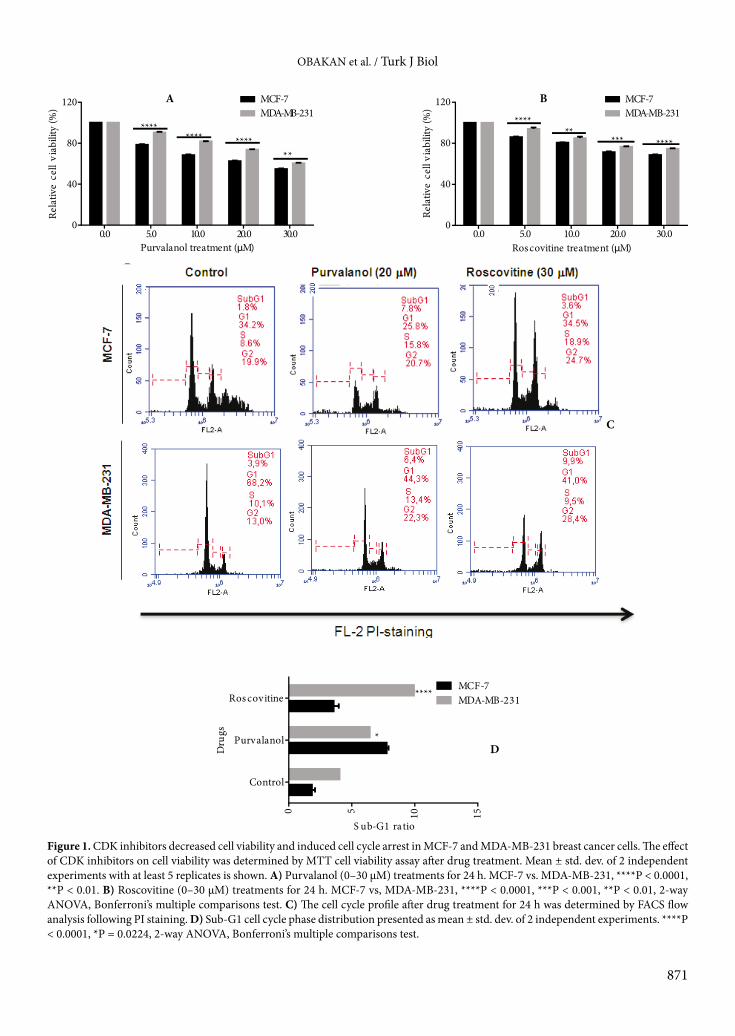
concentrations for each CDK inhibitor. As shown in Figure 1A, 20 µM purvalanol decreased cell viability by 37% and 25% in MCF-7 and MDA-MB-231 breast cancer cells, respectively (MCF-7 vs. MDA-MB-231: P < 0.0001). In a similar way, 30 µM roscovitine decreased cell viability by 32% and 25% in MCF-7 and MDA-MB-231 breast cancer cells, respectively (MCF-7 vs. MDA-MB-231: P < 0.001, Figure 1B).
In order to evaluate the potential role of CDK inhibitors in the cell cycle of MCF-7 and MDA-MB-231 breast cancer cells, we performed cell cycle analysis following PI staining. The sub-G1 population was increased following purvalanol (20 µM) or roscovitine (30 µM) treatment by 7.8% and 3.6% compared to untreated control samples in MCF-7 cells, respectively (Figure 1C). Although roscovitine more efficiently induced apoptosis by increasing sub-G1 ratio by 5% in MDA-MB-231 cells, purvalanol increased the sub-G1 ratio by 3.1% and was found to be less effective compared to use in MCF-7 cells (Figure 1D). These moderate cytotoxic drug concentrations were utilized in further experiments.
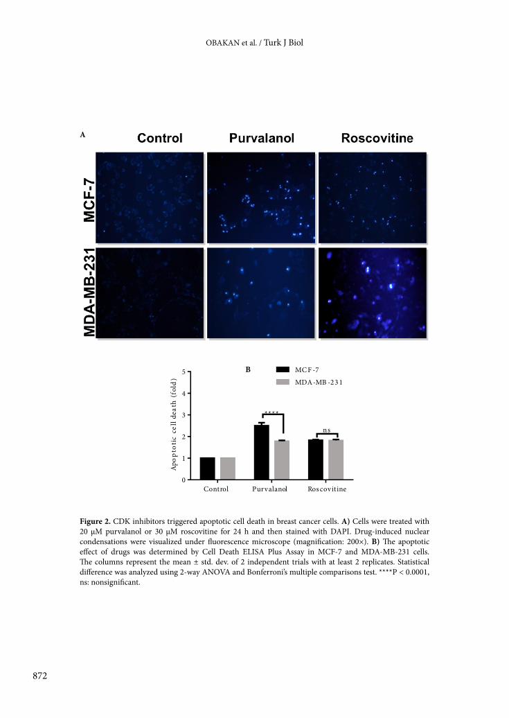
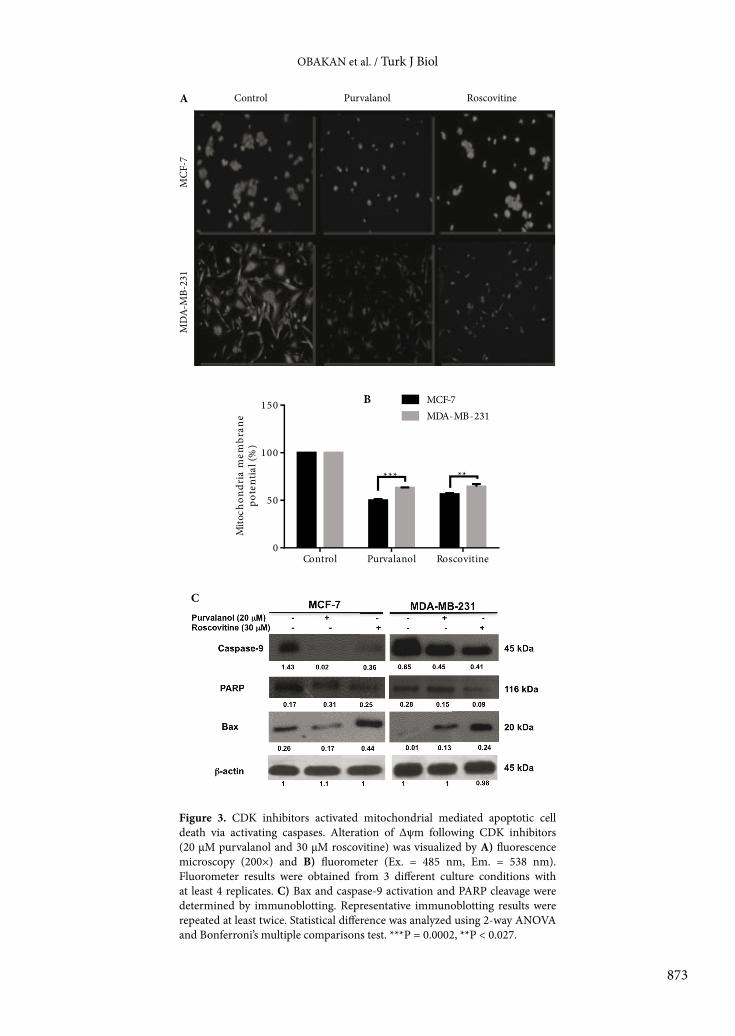
To evaluate the apoptotic potential of each CDK inhibitor in MCF-7 and MDA-MB-231 cells, we performed DAPI staining to detect condensed DNA amount and cell death ELISA assay to confirm DNA fragmentation as a marker of apoptosis. According to Figures 2A and 2B, both CDK inhibitors showed similar responses in MCF-7 breast cancer cells, which were found more sensitive to CDK inhibitors than MDA-MB-231 cells. While roscovitine induced apoptosis by 2.1-fold in MCF-7 and MDA-MB-231 cells, purvalanol induced apoptosis by 2.5- and 1.7-fold in MCF-7 and MDA-MB-231 breast cancer cells, respectively (Figure 2B).3.2. Purvalanol and roscovitine activated the mitochondrial-mediated apoptotic pathway Since Δψm is a crucial step in intrinsic apoptotic pathway induction due to drug treatment, we observed the alteration of Δψm following exposure of cells with moderate cytotoxic concentrations of each CDK inhibitor in breast cancer cells within 24 h (Figures 3A and 3B). According to the DiOC6 staining, 30 µM roscovitine reduced the Δψm in a similar way in both breast cancer cells, but ER– MDA-MB-231 breast cancer cells showed a resistant profile against 20 µM purvalanol treatment as compared to MCF-7 cells. Moreover, each CDK inhibitor induced caspase-9 activation and PARP cleavage in MCF-7 cells (Figure 3C). Although both CDK inhibitors upregulated Bax expression in both MCF-7 and MDA-MB-231 breast cancer cells, the efficiency of purvalanol was lower than that of roscovitine (Figure 3C). 3.3. Purvalanol induced alteration of the MAPK signaling pathway in MCF-7 breast cancer cells To evaluate the effect of CDK inhibitors on the MAPK signaling pathway, we performed immunoblotting analysis. Following drug treatment for 24 h, we determined
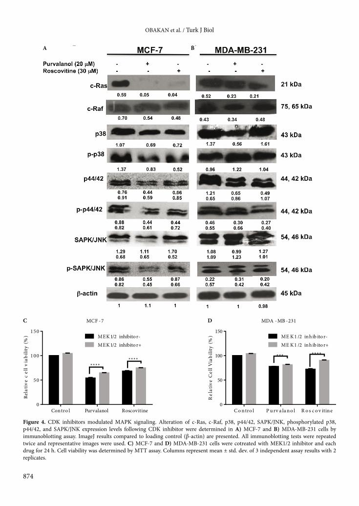
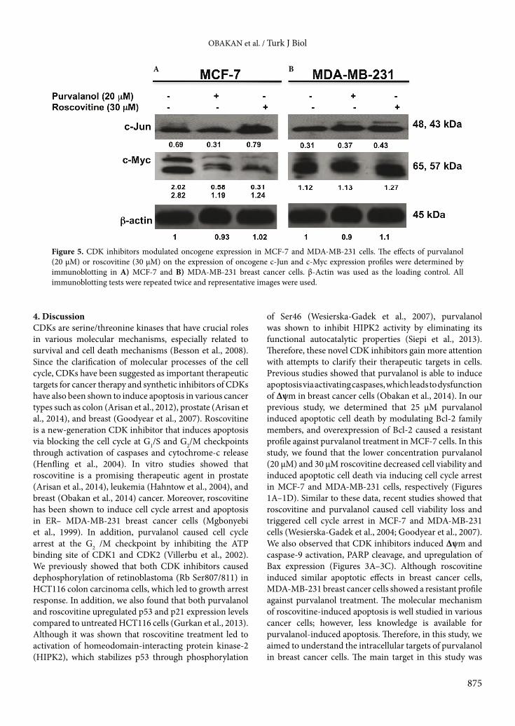
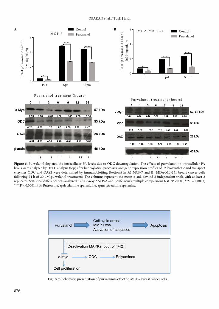
that although both CDK inhibitors abolished c-Ras expression levels in MCF-7 cells, they showed less effect in MDA-MB-231 breast cancer cells. Neither purvalanol nor roscovitine was effective on c-Raf expression levels in either breast cancer cell line. When we investigated p38 expression profile following drug treatment for 24 h in MCF-7 cells, both CDK inhibitors had downregulated p38 and caused dephosphorylation of p38 at the Thr180/Tyr182 site. The p38 expression was slightly downregulated after purvalanol treatment but no significant effect was observed following roscovitine treatment in MDA-MB-231 cells. In addition, phosphorylation of p38 was more effectively increased following purvalanol treatment compared to roscovitine. Both CDK inhibitors downregulated p44/42 in MCF-7 and MDA-MB-231 breast cancer cells. In a similar way, CDK inhibitors caused dephosphorylation of p44/42 at the Thr202/Tyr204 site. Although the drugs did not alter expression levels of SAPK/JNK, purvalanol dephosphorylated SAPK/JNK at the Thr183/Tyr184 site (Figures 4A and 4B). In addition, cells were pretreated with MAPK inhibitor U0126 (10 µM) for 2 h and then cells were exposed to appropriate concentrations of each CDK inhibitor for 24 h. According to cell viability assay, inhibition of MAPKs prevented CDK inhibitor-induced cell viability loss in MCF-7 and MDA-MB-231 cells (P < 0.0001, 2-way ANOVA, Bonferroni’s multiple comparison test). In order to understand the effect of CDK inhibitors on downstream targets of the MAPK signaling cascade, we performed an immunoblotting assay to check the expression levels of c-Jun and c-Myc. While drugs downregulated c-Myc and c-Jun in MCF-7 cells, no significant difference was determined in MDA-MB-231 breast cancer cells (Figures 5A and 5B). 3.4. Purvalanol depleted PAs through downregulation of c-Myc and ODC in MCF-7 breast cancer cellsTo evaluate the resistance phenotype of MDA-MB-231 cells against purvalanol treatment, we examined c-Myc, ODC, and OAZI expression levels in MCF-7 and MDA-MB-231 cells in a time-dependent manner. First we determined the intracellular PA pool after drug treatments. Purvalanol caused depletion of PAs in MCF-7 and MDA-MB-231 breast cancer cells (Figures 6A and 6B). Exposure of MCF-7 cells to purvalanol for 3 h caused downregulation of c-Myc, which was again upregulated within 6–12 h. Long-term exposure of MCF-7 cells to purvalanol for 24 h downregulated c-Myc expression levels. In a similar way, the transcription target of c-Myc and ODC was downregulated starting from 1 h of purvalanol treatment in MCF-7 cells. In accordance with ODC, expression level of OAZI was downregulated in a time-dependent manner after purvalanol treatment in MCF-7 cells (Figure 6A). However, only ODC expression, not c-Myc or OAZI, showed fluctuations in a time-dependent manner in MDA-MB-231 cells (Figure 6B).
OBAKAN et al. / Turk J Biol
871
Purvalanol treatment (µM)
Rela
tive
cell
viab
ility
(%)
0.0 5.0 10.0 20.0 30.00
40
80
120 MCF-7MDA-MB-231
**** ********
**
Ros covitine treatment (µM)
Rela
tive
cell
viab
ility
(%)
0.0 5.0 10.0 20.0 30.00
40
80
120 MCF-7MDA-MB-231
*********
****
S ub-G1 ratio
Dru
gs
0 5 10 15
Control
Purvalanol
Ros covitineMCF-7MDA-MB-231
*
****
Figure 1. CDK inhibitors decreased cell viability and induced cell cycle arrest in MCF-7 and MDA-MB-231 breast cancer cells. The effect of CDK inhibitors on cell viability was determined by MTT cell viability assay after drug treatment. Mean ± std. dev. of 2 independent experiments with at least 5 replicates is shown. A) Purvalanol (0–30 µM) treatments for 24 h. MCF-7 vs. MDA-MB-231, ****P < 0.0001, **P < 0.01. B) Roscovitine (0–30 µM) treatments for 24 h. MCF-7 vs, MDA-MB-231, ****P < 0.0001, ***P < 0.001, **P < 0.01, 2-way ANOVA, Bonferroni’s multiple comparisons test. C) The cell cycle profile after drug treatment for 24 h was determined by FACS flow analysis following PI staining. D) Sub-G1 cell cycle phase distribution presented as mean ± std. dev. of 2 independent experiments. ****P < 0.0001, *P = 0.0224, 2-way ANOVA, Bonferroni’s multiple comparisons test.
A B
C
D
OBAKAN et al. / Turk J Biol
872
Apop
totic
cel
l dea
th (f
old)
Control Purvalanol Ros covitine0
1
2
3
4
5 MC F -7
MDA-MB -23 1
****
ns
Figure 2. CDK inhibitors triggered apoptotic cell death in breast cancer cells. A) Cells were treated with 20 µM purvalanol or 30 µM roscovitine for 24 h and then stained with DAPI. Drug-induced nuclear condensations were visualized under fluorescence microscope (magnification: 200×). B) The apoptotic effect of drugs was determined by Cell Death ELISA Plus Assay in MCF-7 and MDA-MB-231 cells. The columns represent the mean ± std. dev. of 2 independent trials with at least 2 replicates. Statistical difference was analyzed using 2-way ANOVA and Bonferroni’s multiple comparisons test. ****P < 0.0001, ns: nonsignificant.
A
B
OBAKAN et al. / Turk J Biol
873
B
A
MCF
-7M
DA-
MB-
231
A. Purvalanol RoscovitineControlControl
MD
A-M
B-23
1
Purvalanol
MC
F-7
Roscovitine
Mito
chon
dria
mem
bran
epo
tent
ial (
%)
Control Purvalanol Roscovitine0
50
100
150 MCF-7MDA-MB-231
*****
Figure 3. CDK inhibitors activated mitochondrial mediated apoptotic cell death via activating caspases. Alteration of Δψm following CDK inhibitors (20 µM purvalanol and 30 µM roscovitine) was visualized by A) fluorescence microscopy (200×) and B) fluorometer (Ex. = 485 nm, Em. = 538 nm). Fluorometer results were obtained from 3 different culture conditions with at least 4 replicates. C) Bax and caspase-9 activation and PARP cleavage were determined by immunoblotting. Representative immunoblotting results were repeated at least twice. Statistical difference was analyzed using 2-way ANOVA and Bonferroni’s multiple comparisons test. ***P = 0.0002, **P < 0.027.
C
OBAKAN et al. / Turk J Biol
874
C DMCF - 7
Rela
tive
cel
l via
bilit
y (%
)
Contro l Purvalanol Roscovitine0
50
100
150MEK 1/2 inhibito r-MEK1/2 inhibito r+
********
MDA -MB -231
Rel
ativ
e C
ell V
iabi
lity
(%)
C o ntro l P u rv a la n o l R o s c o v itine0
50
100
150ME K 1 /2 inhib ito r-ME K1 /2 inh ib ito r+
*** ****
Figure 4. CDK inhibitors modulated MAPK signaling. Alteration of c-Ras, c-Raf, p38, p44/42, SAPK/JNK, phosphorylated p38, p44/42, and SAPK/JNK expression levels following CDK inhibitor were determined in A) MCF-7 and B) MDA-MB-231 cells by immunoblotting assay. ImageJ results compared to loading control (β-actin) are presented. All immunoblotting tests were repeated twice and representative images were used. C) MCF-7 and D) MDA-MB-231 cells were cotreated with MEK1/2 inhibitor and each drug for 24 h. Cell viability was determined by MTT assay. Columns represent mean ± std. dev. of 3 independent assay results with 2 replicates.
A B
OBAKAN et al. / Turk J Biol
875
4. DiscussionCDKs are serine/threonine kinases that have crucial roles in various molecular mechanisms, especially related to survival and cell death mechanisms (Besson et al., 2008). Since the clarification of molecular processes of the cell cycle, CDKs have been suggested as important therapeutic targets for cancer therapy and synthetic inhibitors of CDKs have also been shown to induce apoptosis in various cancer types such as colon (Arisan et al., 2012), prostate (Arisan et al., 2014), and breast (Goodyear et al., 2007). Roscovitine is a new-generation CDK inhibitor that induces apoptosis via blocking the cell cycle at G1/S and G2/M checkpoints through activation of caspases and cytochrome-c release (Henfling et al., 2004). In vitro studies showed that roscovitine is a promising therapeutic agent in prostate (Arisan et al., 2014), leukemia (Hahntow et al., 2004), and breast (Obakan et al., 2014) cancer. Moreover, roscovitine has been shown to induce cell cycle arrest and apoptosis in ER– MDA-MB-231 breast cancer cells (Mgbonyebi et al., 1999). In addition, purvalanol caused cell cycle arrest at the G2 /M checkpoint by inhibiting the ATP binding site of CDK1 and CDK2 (Villerbu et al., 2002). We previously showed that both CDK inhibitors caused dephosphorylation of retinoblastoma (Rb Ser807/811) in HCT116 colon carcinoma cells, which led to growth arrest response. In addition, we also found that both purvalanol and roscovitine upregulated p53 and p21 expression levels compared to untreated HCT116 cells (Gurkan et al., 2013). Although it was shown that roscovitine treatment led to activation of homeodomain-interacting protein kinase-2 (HIPK2), which stabilizes p53 through phosphorylation
of Ser46 (Wesierska-Gadek et al., 2007), purvalanol was shown to inhibit HIPK2 activity by eliminating its functional autocatalytic properties (Siepi et al., 2013). Therefore, these novel CDK inhibitors gain more attention with attempts to clarify their therapeutic targets in cells. Previous studies showed that purvalanol is able to induce apoptosis via activating caspases, which leads to dysfunction of Δψm in breast cancer cells (Obakan et al., 2014). In our previous study, we determined that 25 µM purvalanol induced apoptotic cell death by modulating Bcl-2 family members, and overexpression of Bcl-2 caused a resistant profile against purvalanol treatment in MCF-7 cells. In this study, we found that the lower concentration purvalanol (20 µM) and 30 µM roscovitine decreased cell viability and induced apoptotic cell death via inducing cell cycle arrest in MCF-7 and MDA-MB-231 cells, respectively (Figures 1A–1D). Similar to these data, recent studies showed that roscovitine and purvalanol caused cell viability loss and triggered cell cycle arrest in MCF-7 and MDA-MB-231 cells (Wesierska-Gadek et al., 2004; Goodyear et al., 2007). We also observed that CDK inhibitors induced Δψm and caspase-9 activation, PARP cleavage, and upregulation of Bax expression (Figures 3A–3C). Although roscovitine induced similar apoptotic effects in breast cancer cells, MDA-MB-231 breast cancer cells showed a resistant profile against purvalanol treatment. The molecular mechanism of roscovitine-induced apoptosis is well studied in various cancer cells; however, less knowledge is available for purvalanol-induced apoptosis. Therefore, in this study, we aimed to understand the intracellular targets of purvalanol in breast cancer cells. The main target in this study was
Figure 5. CDK inhibitors modulated oncogene expression in MCF-7 and MDA-MB-231 cells. The effects of purvalanol (20 µM) or roscovitine (30 µM) on the expression of oncogene c-Jun and c-Myc expression profiles were determined by immunoblotting in A) MCF-7 and B) MDA-MB-231 breast cancer cells. β-Actin was used as the loading control. All immunoblotting tests were repeated twice and representative images were used.
A B
OBAKAN et al. / Turk J Biol
876
Figure 6. Purvalanol depleted the intracellular PA levels due to ODC downregulation. The effects of purvalanol on intracellular PA levels were analyzed by HPLC analysis (top) after benzoylation processes, and gene expression profiles of PA biosynthetic and transport enzymes ODC and OAZI were determined by immunoblotting (bottom) in A) MCF-7 and B) MDA-MB-231 breast cancer cells following 24 h of 20 µM purvalanol treatments. The columns represent the mean ± std. dev. od 2 independent trials with at least 2 replicates. Statistical difference was analyzed using 2-way ANOVA and Bonferroni’s multiple comparisons test. *P < 0.05, ***P = 0.0002, ****P < 0.0001. Put: Putrescine, Spd: triamine spermidine, Spm: tetraamine spermine.
A
Tota
l pol
yam
ine
con
tent
(nM
(mg/
mL–1
))
Pu t S pd S pm0
2
4
6
ControlPurvalanol M C F -7
*
***
********
Pur valanol t reatment (hours)
B
Tota
l pol
yam
ine
con
tent
(nM
(mg
mL–1
))
P u t S p d S p m0
2
4
6 ControlPurvalanol
M D A -M B -2 3 1
***
********
Pur valanol t reatment (hours)
Figure 7. Schematic presentation of purvalanol’s effect on MCF-7 breast cancer cells.
OBAKAN et al. / Turk J Biol
877
to evaluate the MAPK signaling axis in relation with PA biosynthetic machinery.
The Ras/Raf/MEK/ERK signaling axis regulates gene expression of proteins involved in cell survival and apoptosis. Activated c-Raf phosphorylates and activates the MEK/ERK kinase pathway, which leads to activation of downstream targets such as c-Jun and c-Myc (Noguchi et al., 2000). In the present study, we determined the expression levels of c-Ras, which is an upstream molecule involved in the stimulation of survival signals in cancer cells, following purvalanol or roscovitine treatment. We showed that c-Ras expression was downregulated after CDK inhibitor treatment more efficiently in MCF-7 cells (Figure 5A). While JNK and p38 are activated upon cellular stress factors or apoptosis induction, ERK1/2 is generally associated with survival signals such as mitogens (Johnson and Lapadat, 2002). Recent studies have shown that active p38 kinase is necessary to induce G2/M cell cycle arrest after treatment with various chemotherapeutic agents including vanadate and genistein (Frey et al., 2003; Zhang et al., 2003). In addition, asiatic acid activated phosphorylated p38 kinase and correspondingly induced cell cycle arrest and triggered apoptosis in MCF-7 and MDA-MB-231 cells, respectively. Interestingly, in the same study it was shown that ERK1/2 was activated in asiatic acid-mediated apoptosis in breast cancer cells (Hsu et al., 2005). Similarly, another chemotherapeutic agent, rotenone, which is an inhibitor of the mitochondrial electron transport chain complex I, caused the activation of JNK and p38 and the inactivation of ERK1/2 to induce apoptosis in MCF-7 cells (Deng et al., 2010). Conversely to these findings, in this study, phosphorylation of p38 and SAPK/JNK was diminished in MCF-7 cells, but only p38 was upregulated in MDA-MB-231 cells following purvalanol treatment. Moreover, the phosphorylated form of ERK1/2 was decreased after drug treatment in both cell lines (Figures 4A and 4B). In addition, U0126 pretreatment prevented CDK inhibitor-induced cell viability loss in both cell lines (Figures 4C and 4D). Consistent with this result, it was shown that inhibition of MAPKs through U0126 pretreatment prevented cisplatin-induced apoptosis in HeLa cells (Wang et al., 2000). According to these findings, MAPK signaling might activate apoptotic cell death as an early response against purvalanol treatment in breast cancer cells.
In order to evaluate the effect of purvalanol in downstream targets of MAPK signaling pathway, we investigated c-Myc, which is the transcription factor of various molecular targets including PA biosynthetic enzyme ODC. Previous studies showed that the MAPK signaling cascade indirectly affects ODC expression and thereby regulates the intracellular PA pool content (Noguchi et al., 2000). It is well established that intracellular levels of PAs are elevated in malignant cells.
In a similar way, overexpression of c-Myc plays a pivotal role in the regulation of cell cycle progression in cancer cells (Hanson et al., 1994). We determined that purvalanol downregulated c-Myc expression in MCF-7 breast cancer cells but not in MDA-MB-231 breast cancer cells (Figures 5A and 5B). Concomitantly, the target genes of c-Myc were found altered in MCF-7 cells following purvalanol treatment. However, no significant change in ODC expression profile was determined in MDA-MB-231 cells.
c-Myc controls the transcription of ODC, a biosynthetic enzyme of PAs, which increases intracellular PA levels (Bachrach, 2010). Moreover, it was reported that breast cancer cells have higher levels of PAs than normal breast tissue (Wallace et al., 2000). Therefore, the depletion of the PA pool due to ODC downregulation was reported to be an important target in drug-induced apoptotic cell death in colon (Arisan et al., 2012), prostate (Arisan et al., 2014), and breast (Obakan et al., 2014) cancer cells. In addition, overexpression of ODC was reported to attenuate apoptotic cell death in cancer cells (Hsu et al., 2008; Hsieh et al., 2010). Overexpression of ODC induced biosynthesis of PAs and prevented dibenzoylmethane-induced apoptosis in leukemia cells (Wu et al., 2011). Inhibition of ODC by difluoromethylornithine treatment decreased PAs and trigged apoptosis in neuroblastoma cells by increasing cell cycle arrest (Ravanko et al., 2000). Similar to these findings, we determined that purvalanol treatment downregulated c-Myc and ODC expression levels in a time-dependent manner in MCF-7 cells (Figure 6A). However, no significant downregulation was determined in MDA-MDA-231 breast cancer cells (Figure 6B).
The key players of the MAPK signaling cascade, p38, ERK1/2, and JNK1/2 kinases, are well-established molecules in tumorigenesis, metastasis, and angiogenesis. Recent studies showed that increased ODC activity might be suppressed through deactivation of MAPKs in TPA-induced skin tumorigenesis cancer models after guggulsterone treatment (Sarfaraz et al., 2008). Similarly, in this study we found that both CDK inhibitors deactivate p38, ERK1/2, and SAPK/JNK in MCF-7 cells, but do not exert similar effects in MDA-MB-231 breast cancer cells. In addition, c-Myc downregulation, which led to decreased ODC expression levels, was only determined after CDK inhibitor treatment in MCF-7 cells. Although total PA levels were diminished in each breast cancer cell line, ODC expression as well as c-Myc was not altered in MDA-MB-231 cells after purvalanol treatment. To elucidate the molecular action of purvalanol in more detail in estrogen-dependent MCF-7 and estrogen-independent MDA-MB-231 cells, we determined c-Myc, ODC, and OAZI expression profiles in a time-dependent manner. Purvalanol induced c-Myc-mediated ODC downregulation starting from 3 h of treatment in MCF-7 cells. However, purvalanol treatment
OBAKAN et al. / Turk J Biol
878
did not change c-Myc expression levels in MDA-MB-231 cells. Concomitantly ODC and OAZI expression levels were not altered after purvalanol treatment. Therefore, we concluded that purvalanol both activated apoptosis and inhibited cell survival via modulating the upstream factors of c-Myc, p38, ERK1/2, and SAPK/JNK, in MCF-7 cells. Although PA biosynthesis was inhibited after purvalanol treatment in MCF-7 cells, the PA pool was decreased in both breast cancer cell lines. According to our previous studies, purvalanol triggered PA catabolic machinery and depleted PA levels in MCF-7 (Obakan et al., 2014) and MDA-MB-231 (unpublished data) cells.
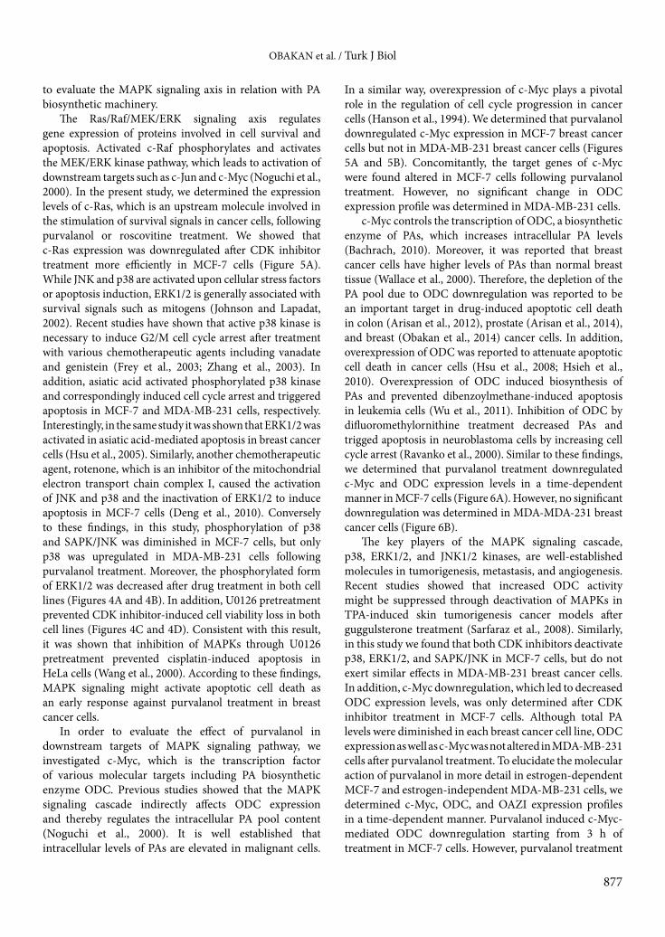
In conclusion, we suggest that purvalanol induced apoptosis through downregulating c-Myc and ODC as a consequence of inhibition of MAPKs in MCF-7 cells. Purvalanol might be a new therapeutic agent for the inhibition of MAPKs, which promoted cell survival mechanisms and proliferation in the treatment of metastatic breast cancer (Figure 7).
Acknowledgements This work was partially supported by a 2209 TÜBİTAK undergraduate research grant and the İstanbul Kültür University Scientific Projects Support Center.
References
Arellano M, Moreno S (1997). Regulation of CDK/cyclin complexes during the cell cycle. Int J Biochem Cell Biol 29: 559–573.
Arisan ED, Obakan P, Coker A, Palavan-Unsal N (2012). Inhibition of ornithine decarboxylase alters the roscovitine-induced mi-tochondrial-mediated apoptosis in MCF-7 breast cancer cells. Mol Med Rep 5: 1323–1329.
Arisan ED, Obakan P, Coker-Gurkan A, Calcabrini A, Agostinelli E, Unsal NP (2014). CDK inhibitors induce mitochondria-medi-ated apoptosis through the activation of polyamine catabolic pathway in LNCaP, DU145 and PC3 prostate cancer cells. Curr Pharm Des 20: 180–188.
Bachrach U (2010). The early history of polyamine research. Plant Physiol Biochem 48: 490–495.
Besson A, Dowdy SF, Roberts JM (2008). CDK inhibitors: cell cycle regulators and beyond. Dev Cell 14: 159–169.
Bodur C, Kutuk O, Karsli-Uzunbas G, Isimjan TT, Harrison P, Ba-saga H (2013). Pramanicin analog induces apoptosis in human colon cancer cells: critical roles for Bcl-2, Bim, and p38 MAPK signaling. PLoS One 8: e56369.
Brown L, Benchimol S (2006). The involvement of MAPK signaling pathways in determining the cellular response to p53 activa-tion: cell cycle arrest or apoptosis. J Biol Chem 281: 3832–3840.
Chen YR, Wang X, Templeton D, Davis RJ, Tan TH (1996). The role of c-Jun N-terminal kinase (JNK) in apoptosis induced by ul-traviolet C and gamma radiation. Duration of JNK activation may determine cell death and proliferation. J Biol Chem 271: 31929–31936.
Coller HA, Grandori C, Tamayo P, Colbert T, Lander ES, Eisenman RN, Golub TR (2000). Expression analysis with oligonucle-otide microarrays reveals that MYC regulates genes involved in growth, cell cycle, signaling, and adhesion. P Natl Acad Sci USA 97: 3260–3265.
Dang CV (1999). c-Myc target genes involved in cell growth, apopto-sis, and metabolism. Mol Cell Biol 19: 1–11.
Deacon K, Mistry P, Chernoff J, Blank JL, Patel R (2003). p38 Mito-gen-activated protein kinase mediates cell death and p21-acti-vated kinase mediates cell survival during chemotherapeutic drug-induced mitotic arrest. Mol Biol Cell 14: 2071–2087.
Deng YT, Huang HC, Lin JK (2010). Rotenone induces apoptosis in MCF-7 human breast cancer cell-mediated ROS through JNK and p38 signaling. Mol Carcinog 49: 141–151.
Evan G, Littlewood T (1998). A matter of life and cell death. Science 281: 1317–1322.
Frey RS, Singletary KW (2003). Genistein activates p38 mitogen-ac-tivated protein kinase, inactivates ERK1/ERK2 and decreases Cdc25C expression in immortalized human mammary epithe-lial cells. J Nutr 133: 226–231.
Gan FY, Gesell MS, Alousi M, Luk GD (1993). Analysis of ODC and c-myc gene expression in hepatocellular carcinoma by in situ hybridization and immunohistochemistry. J Histochem Cyto-chem 41: 1185–1196.
Goodyear S, Sharma MC (2007). Roscovitine regulates invasive breast cancer cell (MDA-MB231) proliferation and survival through cell cycle regulatory protein cdk5. Exp Mol Pathol 82: 25–32.
Gurkan AC, Arisan ED, Obakan P, Palavan-Unsal N (2013). Inhibi-tion of polyamine oxidase prevented cyclin-dependent kinase inhibitor-induced apoptosis in HCT 116 colon carcinoma cells. Apoptosis 18: 1536–1547.
Haan C, Behrmann I (2007). A cost effective non-commercial ECL-solution for Western blot detections yielding strong signals and low background. J Immunol Methods 318: 11–19.
Hahntow IN, Schneller F, Oelsner M, Weick K, Ringshausen I, Fend F, Peschel C, Decker T (2004). Cyclin-dependent kinase inhibi-tor roscovitine induces apoptosis in chronic lymphocytic leu-kemia cells. Leukemia 18: 747–755.
Hanson KD, Shichiri M, Follansbee MR, Sedivy JM (1994). Effects of c-myc expression on cell cycle progression. Mol Cell Biol 14: 5748–5755.
Heby O, Persson L (1990). Molecular genetics of polyamine synthesis in eukaryotic cells. Trends Biochem Sci 15: 153–158.
Henfling ME, Ramaekers FC, Schutte B (2004). Proteasomes act in the pre-mitochondrial signal transduction route towards roscovitine-induced apoptosis. Int J Oncol 25: 1437–1446.
Henriksson M, Luscher B (1996). Proteins of the Myc network: es-sential regulators of cell growth and differentiation. Adv Can-cer Res 68: 109–182.
Hsieh WC, Hsu PC, Liao YF, Young ST, Wang ZW, Lin CL, Tsay GJ, Lee H, Hung HC, Liu GY (2010). Overexpression of ornithine decarboxylase suppresses thapsigargin-induced apoptosis. Mol Cells 30: 311–318.
OBAKAN et al. / Turk J Biol
879
Hsu PC, Hung HC, Liao YF, Liu CC, Tsay GJ, Liu GY (2008). Orni-thine decarboxylase attenuates leukemic chemotherapy drugs-induced cell apoptosis and arrest in human promyelocytic HL-60 cells. Leuk Res 32: 1530–1540.
Hsu YL, Kuo PL, Lin LT, Lin CC (2005). Asiatic acid, a triterpene, induces apoptosis and cell cycle arrest through activation of ex-tracellular signal-regulated kinase and p38 mitogen-activated protein kinase pathways in human breast cancer cells. J Phar-macol Exp Ther 313: 333–344.
Johnson GL, Lapadat R (2002). Mitogen-activated protein kinase pathways mediated by ERK, JNK, and p38 protein kinases. Sci-ence 298: 1911–1912.
Kahana C, Reiss Y (2005). Cell-free assay for ubiquitin-independent proteasomal protein degradation. Methods Mol Biol 301: 83–96.
Komina O, Nosske E, Maurer M, Wesierska-Gadek J (2011). Rosco-vitine, a small molecule CDK inhibitor induces apoptosis in multidrug-resistant human multiple myeloma cells. J Exp Ther Oncol 9: 27–35.
Meijer L (1996). Chemical inhibitors of cyclin-dependent kinases. Trends Cell Biol 6: 393–397.
Mgbonyebi OP, Russo J, Russo IH (1999). Roscovitine induces cell death and morphological changes indicative of apoptosis in MDA-MB-231 breast cancer cells. Cancer Res 59: 1903–1910.
Morgan DO, Fisher RP, Espinoza FH, Farrell A, Nourse J, Chamber-lin H, Jin P (1998). Control of eukaryotic cell cycle progression by phosphorylation of cyclin-dependent kinases. Cancer J Sci Am 4 (Suppl. 1): S77-83.
Noguchi K, Yamana H, Kitanaka C, Mochizuki T, Kokubu A, Kuchi-no Y (2000). Differential role of the JNK and p38 MAPK path-way in c-Myc- and s-Myc-mediated apoptosis. Biochem Bio-phys Res Commun 267: 221–227.
Obakan P, Arisan ED, Ozfiliz P, Coker-Gurkan A, Palavan-Unsal N (2014). Purvalanol A is a strong apoptotic inducer via activat-ing polyamine catabolic pathway in MCF-7 estrogen receptor positive breast cancer cells. Mol Biol Rep 41: 145–154.
Paulovich AG, Toczyski DP, Hartwell LH (1997). When checkpoints fail. Cell 88: 315–321.
Pearson G, Robinson F, Beers Gibson T, Xu BE, Karandikar M, Ber-man K, Cobb MH (2001). Mitogen-activated protein (MAP) kinase pathways: regulation and physiological functions. En-docr Rev 22: 153–183.
Ravanko K, Jarvinen K, Paasinen-Sohns A, Holtta E (2000). Loss of p27Kip1 from cyclin E/cyclin-dependent kinase (CDK) 2 but not from cyclin D1/CDK4 complexes in cells transformed by polyamine biosynthetic enzymes. Cancer Res 60: 5244–5253.
Ringer L, Sirajuddin P, Yenugonda VM, Ghosh A, Divito K, Trabosh V, Patel Y, Brophy A, Grindrod S, Lisanti MP et al. (2010). VMY-1-103, a dansylated analog of purvalanol B, induces cas-pase-3-dependent apoptosis in LNCaP prostate cancer cells. Cancer Biol Ther 10: 320–325.
Sarfaraz S, Siddiqui IA, Syed DN, Afaq F, Mukhtar H (2008). Gug-gulsterone modulates MAPK and NF-kappaB pathways and inhibits skin tumorigenesis in SENCAR mice. Carcinogenesis 29: 2011–2018.
Siepi F, Gatti V, Camerini S, Crescenzi M, Soddu S (2013). HIPK2 catalytic activity and subcellular localization are regulated by activation-loop Y354 autophosphorylation. Biochim Biophys Acta 1833: 1443–1453.
Troppmair J, Bruder JT, Munoz H, Lloyd PA, Kyriakis J, Banerjee P, Avruch J, Rapp UR (1994). Mitogen-activated protein kinase/extracellular signal-regulated protein kinase activation by on-cogenes, serum, and 12-O-tetradecanoylphorbol-13-acetate requires Raf and is necessary for transformation. J Biol Chem 269: 7030–7035.
Villerbu N, Gaben AM, Redeuilh G, Mester J (2002). Cellular effects of purvalanol A: a specific inhibitor of cyclin-dependent kinase activities. Int J Cancer 97: 761–769.
Walhout AJ, Gubbels JM, Bernards R, van der Vliet PC, Timmers HT (1997). c-Myc/Max heterodimers bind cooperatively to the E-box sequences located in the first intron of the rat ornithine decarboxylase (ODC) gene. Nucleic Acids Res 25: 1493–1501.
Wallace HM, Duthie J, Evans DM, Lamond S, Nicoll KM, Heys SD (2000). Alterations in polyamine catabolic enzymes in human breast cancer tissue. Clin Cancer Res 6: 3657–3661.
Wallace HM, Fraser AV (2004). Inhibitors of polyamine metabolism: review article. Amino Acids 26: 353–365.
Wang X, Feith DJ, Welsh P, Coleman CS, Lopez C, Woster PM, O’Brien TG, Pegg AE (2007). Studies of the mechanism by which increased spermidine/spermine N1-acetyltransferase activity increases susceptibility to skin carcinogenesis. Carci-nogenesis 28: 2404–2411.
Wang X, Martindale JL, Holbrook NJ (2000). Requirement for ERK activation in cisplatin-induced apoptosis. J Biol Chem 275: 39435–39443.
Wesierska-Gadek J, Gueorguieva M, Wojciechowski J, Horky M (2004). Cell cycle arrest induced in human breast cancer cells by cyclin-dependent kinase inhibitors: a comparison of the ef-fects exerted by roscovitine and olomoucine. Pol J Pharmacol 56: 635–641.
Wesierska-Gadek J, Schmitz ML, Ranftler C (2007). Roscovitine-activated HIP2 kinase induces phosphorylation of wt p53 at Ser-46 in human MCF-7 breast cancer cells. J Cell Biochem 100: 865–874.
Wu CL, Liao YF, Hung YC, Lu KH, Hung HC, Liu GY (2011). Or-nithine decarboxylase prevents dibenzoylmethane-induced apoptosis through repressing reactive oxygen species genera-tion. J Biochem Mol Toxicol 25: 312–319.
Xia Z, Dickens M, Raingeaud J, Davis RJ, Greenberg ME (1995). Op-posing effects of ERK and JNK-p38 MAP kinases on apoptosis. Science 270: 1326–1331.
Zhang T, Jiang T, Zhang F, Li C, Zhou YA, Zhu YF, Li XF (2010). Involvement of p21Waf1/Cip1 cleavage during roscovitine-induced apoptosis in non-small cell lung cancer cells. Oncol Rep 23: 239–245.
Zhang Z, Leonard SS, Huang C, Vallyathan V, Castranova V, Shi X (2003). Role of reactive oxygen species and MAPKs in vana-date-induced G(2)/M phase arrest. Free Radic Biol Med 34: 1333–1342.