
Venepuncture TrainingVersion 7 (October 2011)

Section 1Objectives and Definition
After completing this section you will:
Know the objectives
Be able to identify what venepuncture is

Define venepuncture
Identify suitable veins for venepuncture
Explore the problems in identifying suitable veins for venepuncture
Identify complications and actions i.e. faint, anxiety, haematoma arterial puncture
Demonstrate health and safety awareness issues related to venepuncture e.g. hand washing, needlestick injury, infection risk, and sharp disposal.
Objectives
What is Venepuncture?
Venepuncture may be defined as puncturing of a vein.
It is performed either:
to withdraw blood for diagnostic purposes
to monitor levels of blood components or drugs

Section 2Policies relating to Venepuncture
After completing this section you will:
Know how to access venepuncture related policies and procedures
Be aware of the policies and procedures pertinent to venepuncture

CONSENT POLICY INCIDENT REPORTING POLICY
Access directly from Shropshire Community Health NHS Trust website
Venepuncture Guidelines
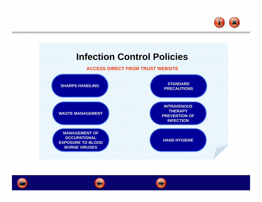
Infection Control Policies
WASTE MANAGEMENT
HAND HYGIENE
INTRAVENOUS THERAPY
PREVENTION OF INFECTION
STANDARD PRECAUTIONS
MANAGEMENT OF OCCUPATIONAL
EXPOSURE TO BLOOD BORNE VIRUSES
ACCESS DIRECT FROM TRUST WEBSITE
SHARPS HANDLING
Section 3Anatomy and Physiology
After completing this section you should be able to understand:
The anatomy and physiology of the arm
The role of veins and arteries
Vein structure
And be able to name common veins

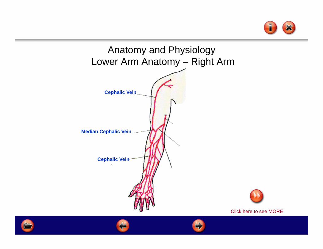
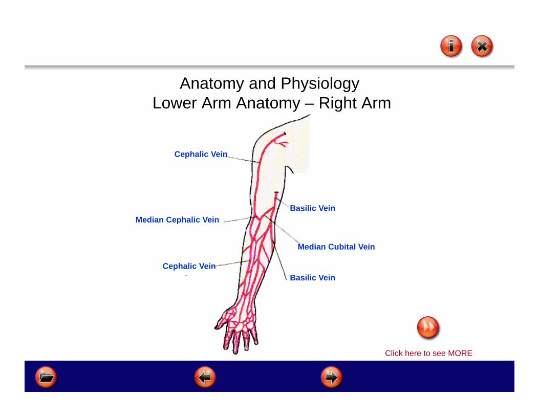
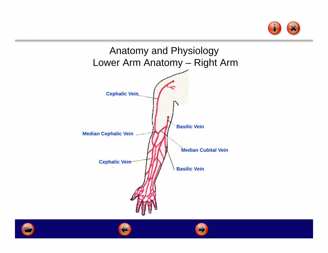
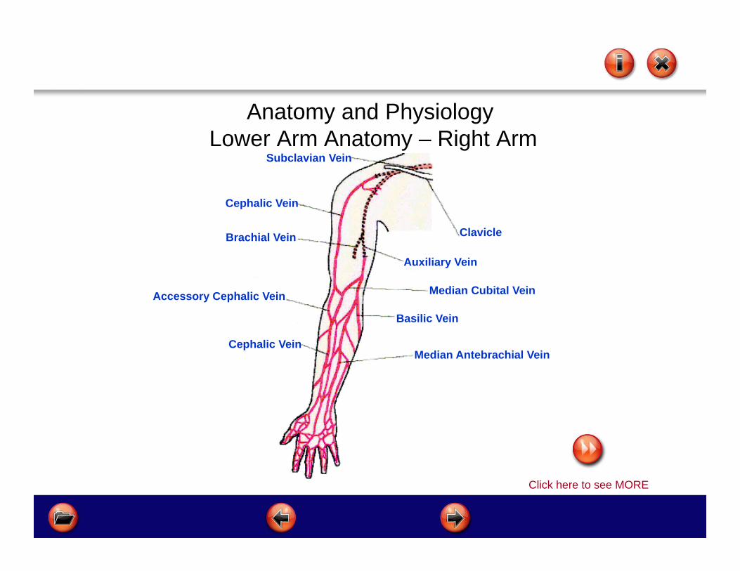
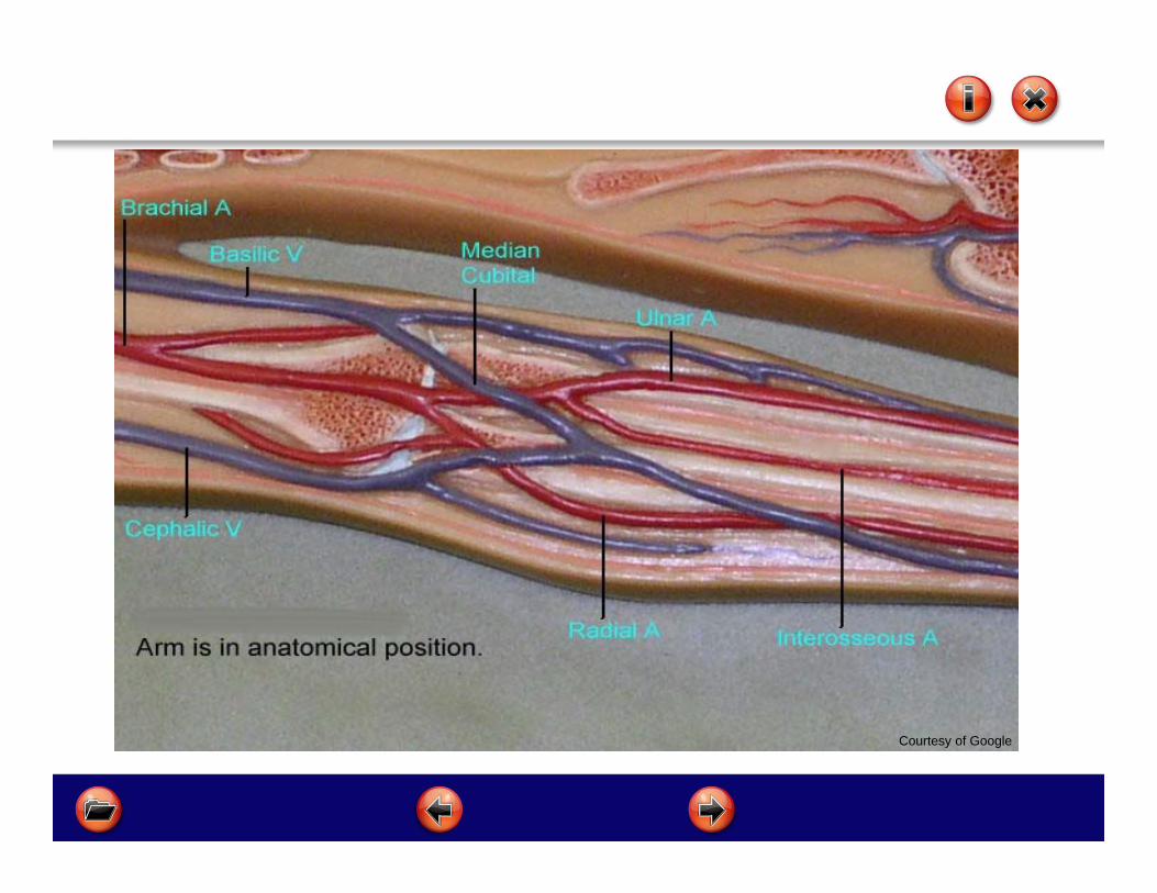
Anatomy and PhysiologyLower Arm Anatomy – Right Arm
Click here to see MORE

Anatomy and PhysiologyLower Arm Anatomy – Right Arm
Cephalic Vein
Click here to see MORE
Cephalic Vein
Anatomy and PhysiologyLower Arm Anatomy – Right Arm
Median Cephalic Vein
Click here to see MORE
Cephalic Vein
Cephalic Vein

Anatomy and PhysiologyLower Arm Anatomy – Right Arm
Basilic Vein
Basilic Vein
Click here to see MORE
Median Cephalic Vein
Cephalic Vein
Cephalic Vein
Anatomy and PhysiologyLower Arm Anatomy – Right Arm
Median Cubital Vein
Click here to see MORE
Median Cephalic Vein
Cephalic Vein
Cephalic Vein
Basilic Vein
Basilic Vein
Anatomy and PhysiologyLower Arm Anatomy – Right Arm
Median Cubital Vein
Median Cephalic Vein
Cephalic Vein
Cephalic Vein
Basilic Vein
Basilic Vein

Cephalic Vein
Basilic Vein
Median Cubital Vein
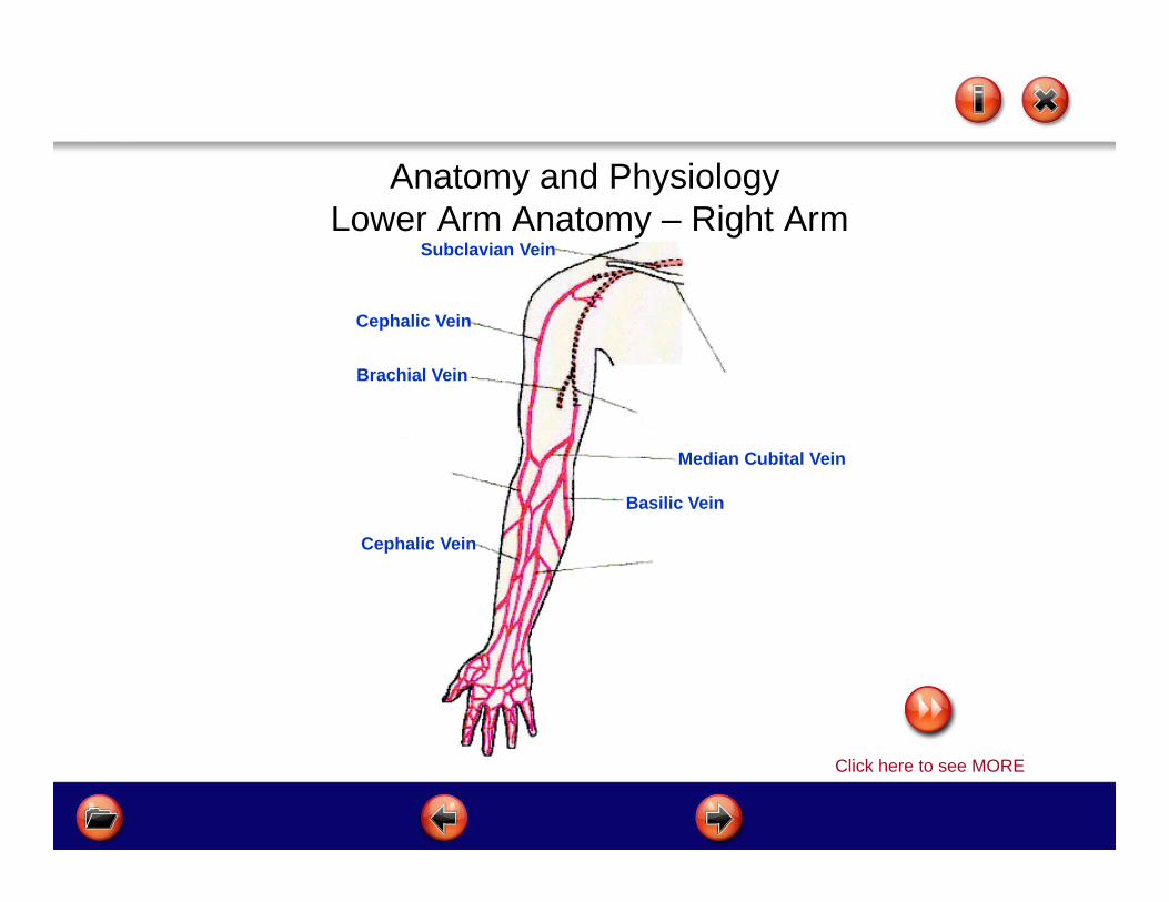
Anatomy and PhysiologyLower Arm Anatomy – Right Arm
Cephalic Vein
Click here to see MORE

Subclavian Vein
Cephalic Vein
Basilic Vein
Median Cubital Vein
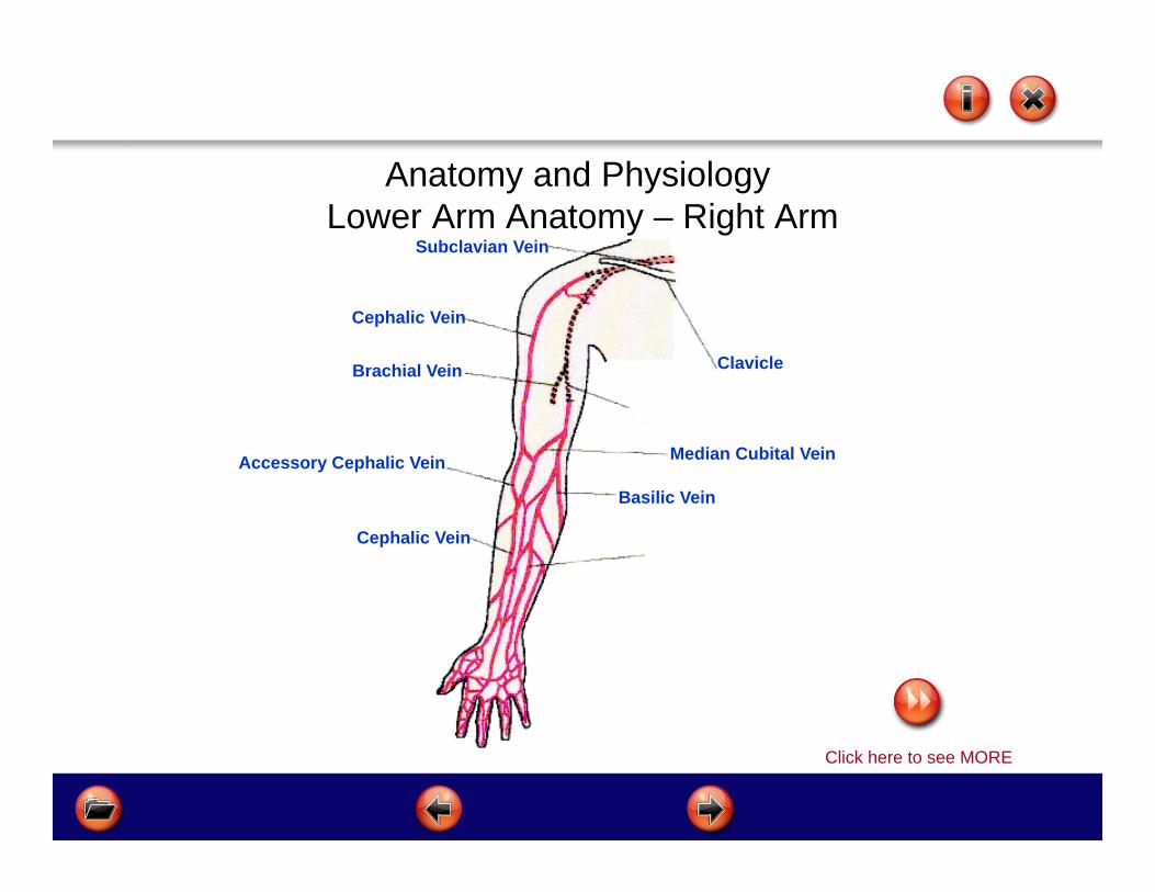
Anatomy and PhysiologyLower Arm Anatomy – Right Arm
Cephalic Vein
Click here to see MORE
Subclavian Vein
Brachial Vein
Cephalic Vein
Basilic Vein
Median Cubital Vein
Anatomy and PhysiologyLower Arm Anatomy – Right Arm
Cephalic Vein
Click here to see MORE

Subclavian Vein
Brachial Vein
Accessory Cephalic Vein
Cephalic Vein
Basilic Vein
Median Cubital Vein
Anatomy and PhysiologyLower Arm Anatomy – Right Arm
Cephalic Vein
Click here to see MORE
Subclavian Vein
ClavicleBrachial Vein
Accessory Cephalic Vein
Cephalic Vein
Basilic Vein
Median Cubital Vein
Anatomy and PhysiologyLower Arm Anatomy – Right Arm
Cephalic Vein
Click here to see MORE

Subclavian Vein
ClavicleBrachial Vein
Accessory Cephalic Vein
Cephalic Vein
Basilic Vein
Median Cubital Vein
Auxiliary Vein
Anatomy and PhysiologyLower Arm Anatomy – Right Arm
Cephalic Vein
Click here to see MORE
Subclavian Vein
ClavicleBrachial Vein
Accessory Cephalic Vein
Cephalic VeinMedian Antebrachial Vein
Basilic Vein
Median Cubital Vein
Auxiliary Vein
Anatomy and PhysiologyLower Arm Anatomy – Right Arm
Cephalic Vein
Click here to see MORE

Subclavian Vein
ClavicleBrachial Vein
Accessory Cephalic Vein
Cephalic VeinMedian Antebrachial Vein
Basilic Vein
Median Cubital Vein
Auxiliary Vein
Anatomy and PhysiologyLower Arm Anatomy – Right Arm
Cephalic Vein

The Role of Veins and Arteries
Veins and Arteries are blood vessels that carry blood to and from the heart
ARTERIES - carry oxygenated blood away from the heart
VEINS - carry de-oxygenated blood to the heart
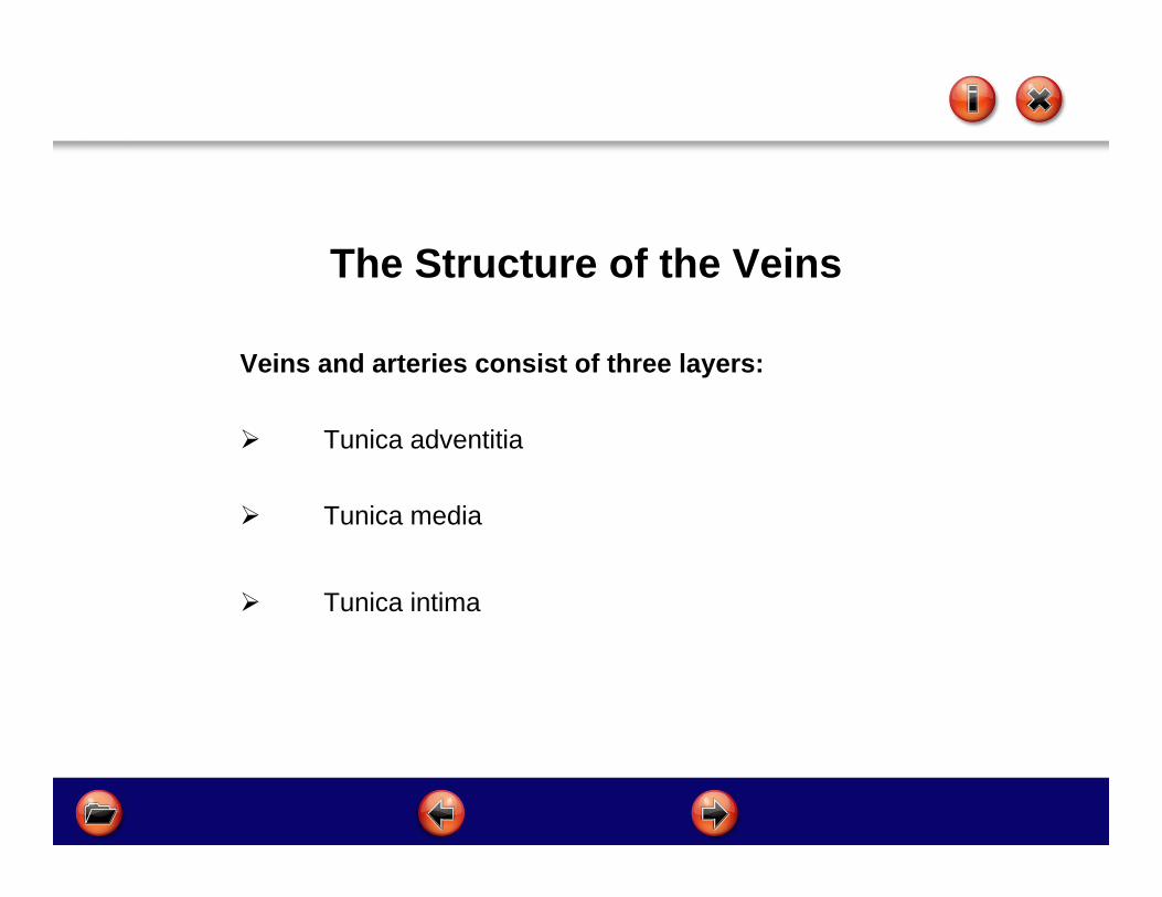
The Structure of the Veins
Veins and arteries consist of three layers:
Tunica adventitia
Tunica media
Tunica intima
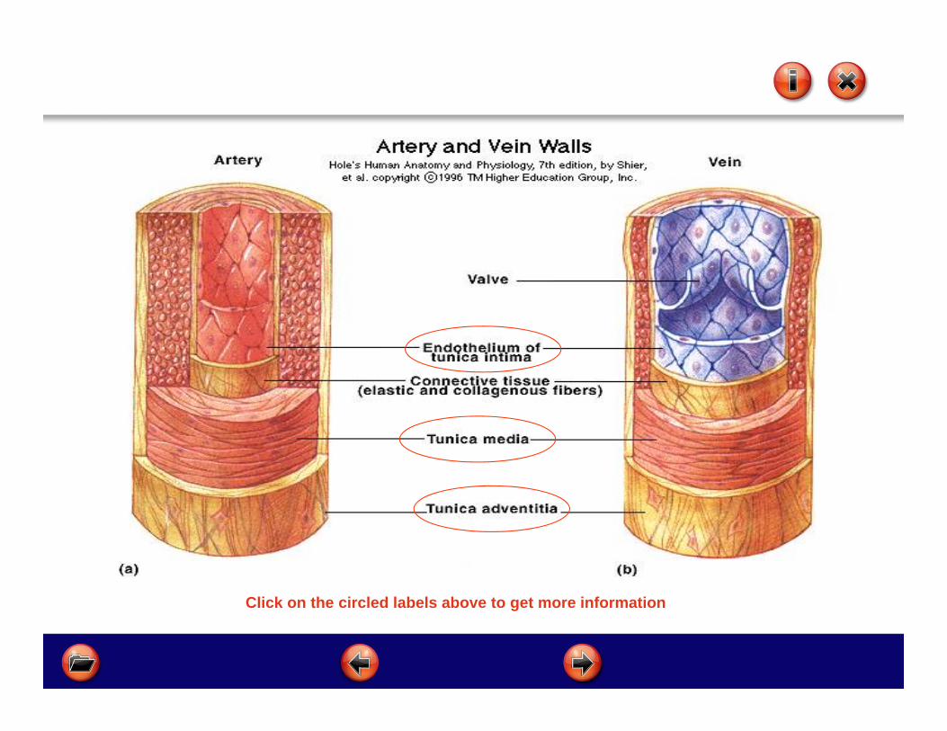
Click on the circled labels above to get more information

TUNICA ADVENTITIA
Strong - composed of connective tissue, collagen and elastic fibres
Allows stretch
It attaches the vein to whatever other tissue it runs through i.e. the skin
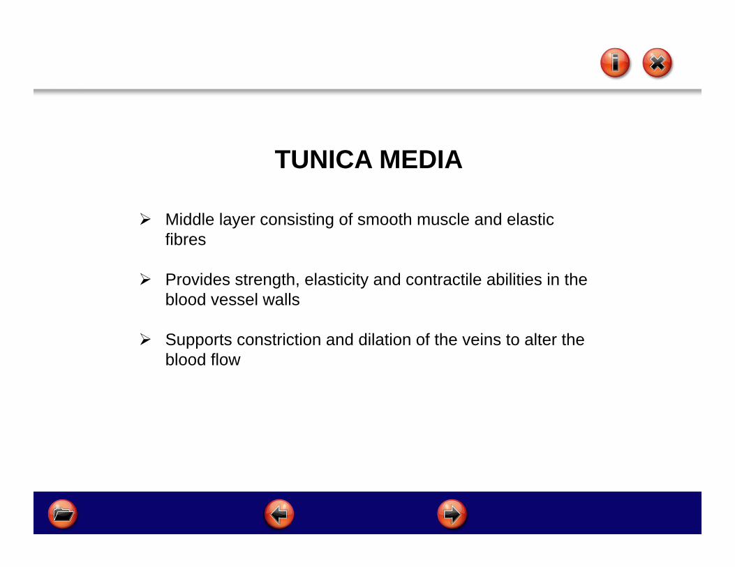
TUNICA MEDIA
Middle layer consisting of smooth muscle and elastic fibres
Provides strength, elasticity and contractile abilities in the blood vessel walls
Supports constriction and dilation of the veins to alter the blood flow
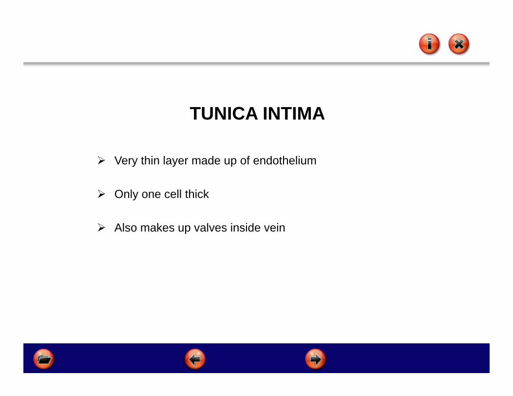
TUNICA INTIMA
Very thin layer made up of endothelium
Only one cell thick
Also makes up valves inside vein
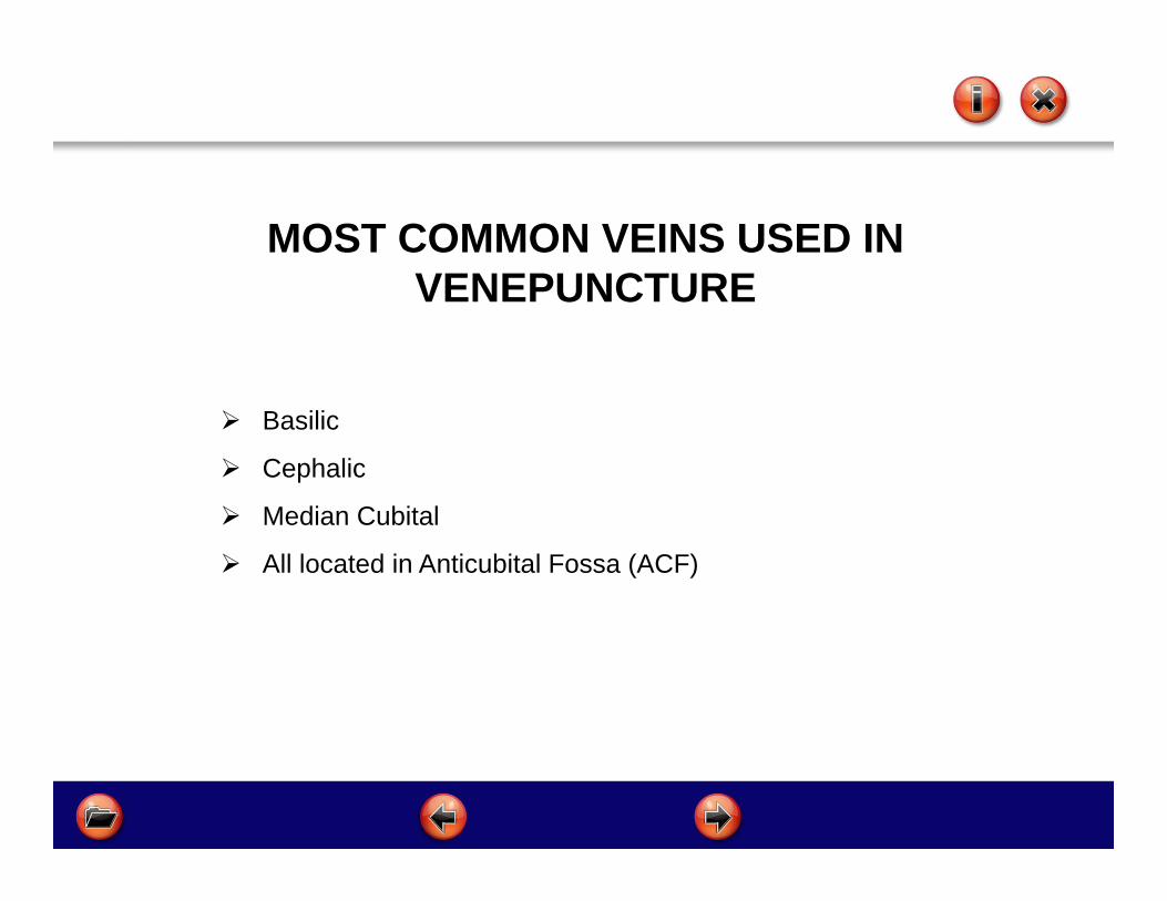
MOST COMMON VEINS USED IN VENEPUNCTURE
Basilic
Cephalic
Median Cubital
All located in Anticubital Fossa (ACF)
Courtesy of Google

Hand Anatomy Metacarpal veins located on the dorsal side of the
hand can be used for venepuncture by experienced practitioners only
Used if venous access to the ACF is poor or difficult
Different equipment is required for this method of sample retrieval
Refer to a more experienced colleague if this method is required until you are more proficient
Section 4Choosing Sites for Venepuncture
After completing this section you will be able to: Identify good veins Avoid bad veins Choose a suitable site Identify influencing factors Improve access to veins
Site Selection
Most frequently used veins are:
median basilic median cubital median cephalic veins in the ante - cubital fossa. Veins on the dorsum of the hand may be used if the
forearm and elbow veins are difficult to identify, but it should be noted that these thin walled easily moveable superficial veins are often more difficult to puncture than the larger, less mobile but palpable veins around the elbow.

Site Selection
You must avoid
Veins in lower extremities
Areas of joint flexion
Veins close to arteries and deeper lying vessels
Veins that may be irritated from previous use
Site Selection
Distal veins should be used first with subsequent venepuncture proximal to previous sites.
Healthy veins are easily detected by palpation. These veins feel soft and bouncy and will refill when depressed.
Always allow adequate time for inspection and palpation of the patients arm and hand to select a site.
In difficult cases, ensure maximum venous dilation before inspection e.g. warming. Use veins in non dominant side if possible.
If in doubt, consult a more experienced colleague
Site Selection
CONDITION AND ACCESSIBILITY
Veins may be tortuous, sclerosed, fibrosed, or thrombosed, inflamed or fragile and unable to accommodate the device used.
If the patient complains of pain or soreness over a particular site, this should be avoided as should areas that are bruised.
Characteristics of Good Veins
Bouncy Soft Refills when depressed Has a large lumen Straight Visible Is well supported

AVOIDveins that are:
Bruised Infected (phlebitis) Oedematous limbs Hard, fibrosed veins Areas of previous venepuncture Veins adjacent to infection Near bone
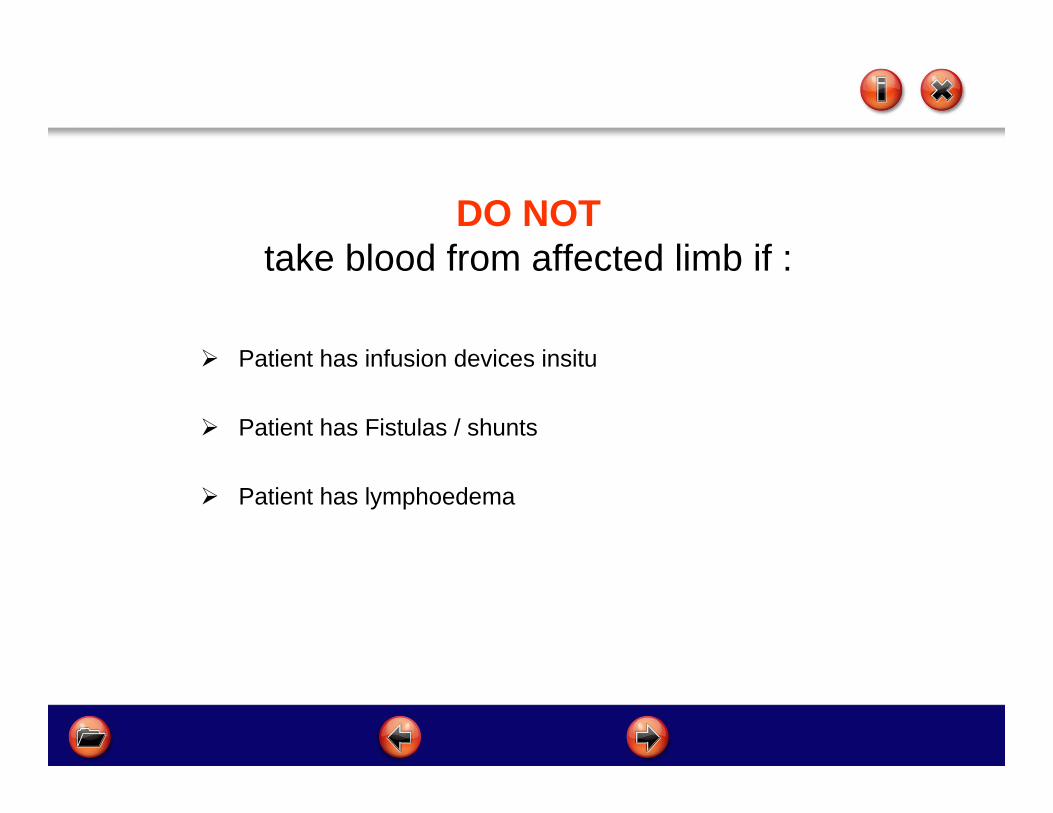
DO NOTtake blood from affected limb if :
Patient has infusion devices insitu
Patient has Fistulas / shunts
Patient has lymphoedema

INFLUENCING FACTORS Temperature of the environment
Patient anxiety
Medication
Age and weight of patient
Position of patient
Injury
Privacy/dignitySOURCE: MALLETT & BAILEY 2001

IMPROVING ACCESS / VEIN PROMINENCE
Tourniquet
Open and close of fist
Lower arm below heart level
Stroke vein in distal direction
Heat

Section 5Blood Collection Devices
After completing this section you will be able to: Choose a device Complete the form Understand the order of the draw Recognise symptoms and prevention of Haemolysis
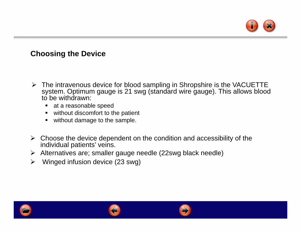
The intravenous device for blood sampling in Shropshire is the VACUETTE system. Optimum gauge is 21 swg (standard wire gauge). This allows blood to be withdrawn: at a reasonable speed without discomfort to the patient without damage to the sample.
Choose the device dependent on the condition and accessibility of the individual patients’ veins.
Alternatives are; smaller gauge needle (22swg black needle) Winged infusion device (23 swg)
Choosing the Device

Specimen Request Form(being replaced by patient specific pre-printed request
forms)Area in green must be completed, coloured squares denote vacutainer bottle to be used

Order of Draw
Red Light Blue Gold Lavender Pink Dark Green Light Green Grey Dark Blue

Vacuette Tube Guide

Haemolysis
Haemolysis results from damage or destruction of red blood cells and liberation of haemoglobin. When the cells are ruptured it causes discolouration of the serum (plasma), staining it pink or slightly red.
Haemolysis elevates potassium, LDH, AST, ALT, phosphorate, magnesium and ammonia levels and decreases levels of red blood cells.
Haemolysis can occur as a result of a medical condition such as a patient with fragile cells, but frequently occurs as a result of how the sample of blood is collected, handled and stored.

Causes of Haemolysis:
The vigorous shaking of bottles Too much time taken to draw and collect the blood Too small needle for the volume of blood taken Frothing as drawing up Drawing from a vein that has a haematoma Storing samples too close to the freezer
compartment in a refrigerator
Haemolysis
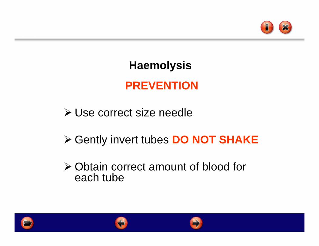
PREVENTION
Use correct size needle
Gently invert tubes DO NOT SHAKE
Obtain correct amount of blood for each tube
Haemolysis

Section 6Collection of Venous Blood Samples
After completing this section you will be able to:
List the equipment needed
Prepare the area/patient
Work through the process

Equipment Required
Clean field
Tourniquet
Vacuette Holder
Needle
Alcohol swab
Non-woven gauze or equivalent
Micropore
Blood specimen bottles
Specimen request form
Gloves
Plastic apron
Sharps bin

Skin Preparation Cleanliness is vital when performing venepuncture as the
skin is breached and an alien device introduced into a sterile circulatory system. The major sources of contamination are: cross infection from practitioner to patient skin flora of the patient
Good hand washing and drying techniques/use of alcohol handrub are essential on the part of the nurse
Firm and prolonged cleansing with an alcohol swab to the site of venepuncture is essential. Allow this area to dry before commencing procedure. Do not fan or blow on area after cleaning.

ProcedureACTION
Approach the patient confidently
Identify patient through confirmation of name and date of birth
Allow patient to ask questions and discuss any problems which have arisen previously
Wash hands and dry carefully
Check hands for visibly broken skin and cover with plaster
Prepare equipment required onto a clean field
RATIONALE A relaxed patient will have
relaxed veins To ensure that correct patient
identified
To obtain the patient consent and co-operation
To minimise the risk of infection

ACTION Check adequate lighting, good
ventilation, privacy and correct patient position
Consult the patient to any preferences and problems previously encountered
Place arm in dependant position, ask patient to clench unclench fist if necessary.
If these measures are unsuccessful, remove tourniquet and apply moist heat.
Select the vein and the device to be used
RATIONALE To ensure patient and nurse
comfort and adequate light source available
To involve the patient in treatment and take patient history which may influence vein choice
To dilate the veins by obstructing the venous return
To promote blood flow and therefore distend vein
To maintain cleanliness
Procedure
ProcedureACTION
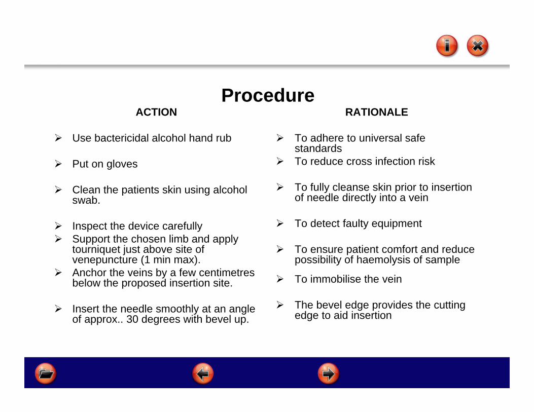
Use bactericidal alcohol hand rub
Put on gloves
Clean the patients skin using alcohol swab.
Inspect the device carefully Support the chosen limb and apply
tourniquet just above site of venepuncture (1 min max).
Anchor the veins by a few centimetres below the proposed insertion site.
Insert the needle smoothly at an angle of approx.. 30 degrees with bevel up.
RATIONALE
To adhere to universal safe standards
To reduce cross infection risk
To fully cleanse skin prior to insertion of needle directly into a vein
To detect faulty equipment
To ensure patient comfort and reduce possibility of haemolysis of sample
To immobilise the vein
The bevel edge provides the cutting edge to aid insertion

ProcedureACTION
Do not exert any pressure on the needle
Slide blood bottle into the vacuette holder and fill blood bottle to the marked fill line
Release the tourniquet as the last blood bottle is almost filled. In some instances this may be requested at the beginning of sampling ( i.e. for blood calcium, as the tourniquet may damage cells)
Place non-woven gauze or equivalent over puncture point.
Remove needle and discard immediately into sharps box, apply digital pressure to puncture site
RATIONALE To prevent a through puncture
occurring or cutting from bevel of needle
To ensure required amount of blood sample is obtained
To decrease pressure within the vein
To reduce the amount of static blood in the vein and the likelihood of leakage
To stop leakage and haematoma formation
To ensure safe disposal and prevent needle stick injury

ProcedureACTION
Apply pressure until bleeding has ceased (about one minute). Longer may be required if clotting mechanisms are influenced by disease or treatment i.e. warfarin
Invert sample 4-6 times
Label the bottles and complete required details on the specimen request form
Inspect the site
Ascertain whether the patient is allergic to plaster
Apply plaster or alternative
RATIONALE To prevent leakage or haematoma
formation
To ensure that the blood is mixed with any additive present
To ensure the specimen is from the right patient , the right tests are performed and results reported to their GP
To check the puncture point has sealed
To prevent allergic reaction
To cover the puncture and prevent leakage or bacteria contamination

Procedure
ACTION Ensure patient is
comfortable
Discard waste
Follow procedure for collection and transportation of samples
RATIONALE To ascertain patient
condition
Safe disposal of waste
To make sure specimens reach lab within the specified time
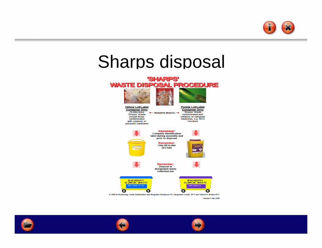
Sharps disposal
Section 7Trouble Shooting
After completing this section you will be able to: Identify potential problems
Understand the causes
Take action to rectify problems

Trouble ShootingPROBLEM CAUSE SUGGESTED ACTION
Excessive Pain Anxiety, Fear, Low pain threshold
Confident, unhurried approach. Use all methods including heat to dilate veins. Use of local anaesthetic cream. Avoid hesitancy and skin tickling.
Frequently used vein Avoid this site, if possible
Nerve touched Remove needle immediately and proceed to different site
Infection Poor cleaning technique Practice good hand washing and skin cleansing
Limited Venous Access
Repeated use Phlebitis
Confident unhurried approachUse a needle of 22 or 23 swg. Only proceed if sure of a successful first attemptConsider referral to more experienced colleague
Bruising due: fragile veins in the elderlyAnticoagulation therapy or low platelet levels
As above plus apply tourniquet gently or do not use. Ensure adequate pressure to puncture site to prevent further damage
Peripheral Shutdown Use all methods to dilate veins. A sphygmomanometer cuff (no higher than 120 mmtlg) may be more effective in restricting venous return. Work quickly if patient in collapsed state. Pull blood back into veins by massaging above the venepuncture site.

Trouble ShootingPROBLEM CAUSE SUGGESTED ACTION
Missed Vein Inadequate AnchoringWrong positioningPoor LightingLess than 100% concentration
Withdraw needle almost to the bevel and manoeuvre gently to realign needle and vein. Re-advance but stop if becomes painful.
Spurt of blood on entry Bevel of needle entering before entire bevel is under skin, due to vein being superficial (ensure tourniquet is not overly tight
Ignore. Reassure patient
Slacken tourniquet if over tight
Blood flow stops Overshooting vein or advancing needle while withdrawing blood. Vein collapse due to contact with valve or vein wall collapsePoor blood flow
Gently ease needle back and continue.Manoeuvre gently. Release and retighten tourniquet and continue
As above and massage above the needle tip to pull blood into the vein.
Haematoma Perforation of opposite wall of veinForgetting to remove tourniquet before removing needleInadequate pressure on puncture site
Insert needle at correct angle. Do not advance needle during procedureRemember next time to slacken off the tourniquet prior to removing final blood sample bottle
Apply adequate pressure on needle removal . Supervise the patient doing the same
PROBLEM CAUSE SUGGESTED ACTION
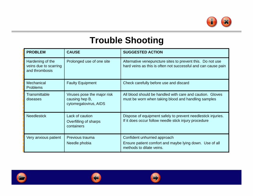
Hardening of the veins due to scarring and thrombosis
Prolonged use of one site Alternative venepuncture sites to prevent this. Do not use hard veins as this is often not successful and can cause pain
Mechanical Problems
Faulty Equipment Check carefully before use and discard
Transmittable diseases
Viruses pose the major risk causing hep B, cytomegalovirus, AIDS
All blood should be handled with care and caution. Gloves must be worn when taking blood and handling samples
Needlestick Lack of cautionOverfilling of sharps containers
Dispose of equipment safely to prevent needlestick injuries. If it does occur follow needle stick injury procedure
Very anxious patient Previous traumaNeedle phobia
Confident unhurried approachEnsure patient comfort and maybe lying down. Use of all methods to dilate veins.
Trouble Shooting

Section 8First Aid and Needlestick Injury
After completing this section you will be able to: Deal with Needlestick Injury
Identify causes, symptoms and treatment for fainting and haemorrhage
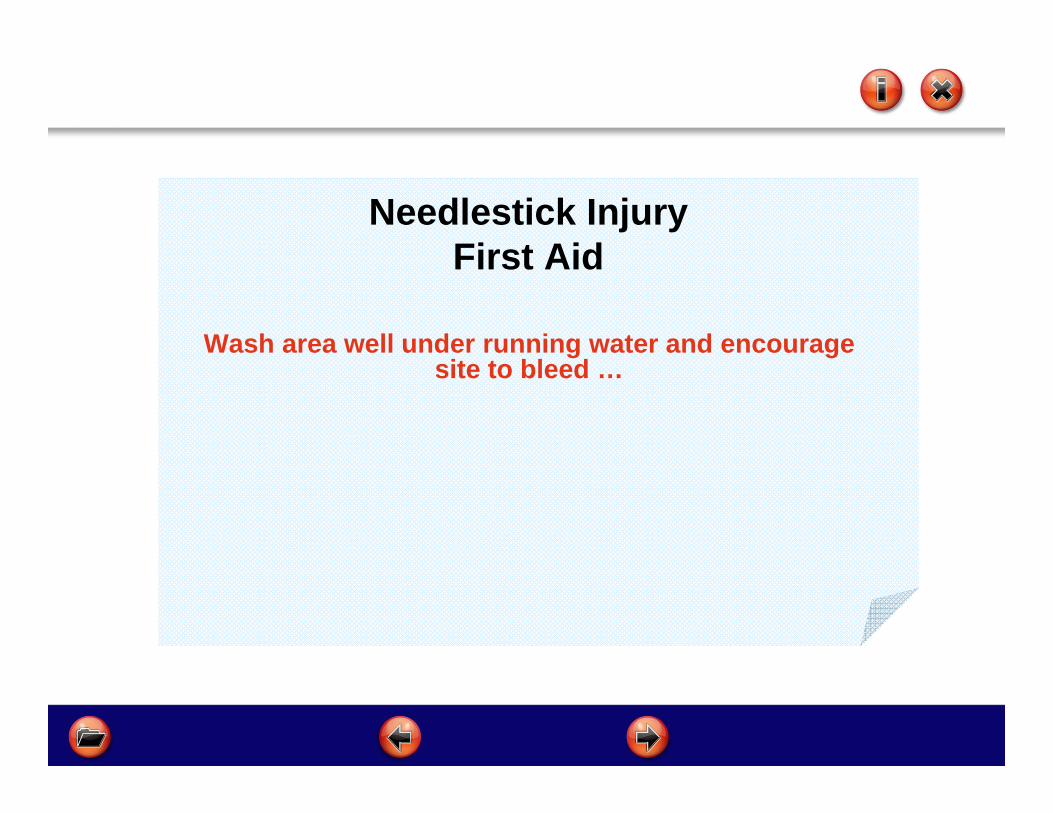
Needlestick InjuryFirst Aid
Wash area well under running water and encourage site to bleed …
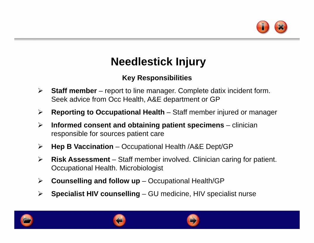
Needlestick InjuryKey Responsibilities
Staff member – report to line manager. Complete datix incident form. Seek advice from Occ Health, A&E department or GP
Reporting to Occupational Health – Staff member injured or manager
Informed consent and obtaining patient specimens – clinician responsible for sources patient care
Hep B Vaccination – Occupational Health /A&E Dept/GP
Risk Assessment – Staff member involved. Clinician caring for patient. Occupational Health. Microbiologist
Counselling and follow up – Occupational Health/GP
Specialist HIV counselling – GU medicine, HIV specialist nurse

First Aid
Fainting
Haemorrhage
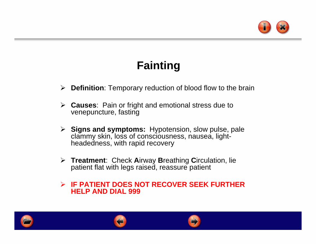
Fainting
Definition: Temporary reduction of blood flow to the brain
Causes: Pain or fright and emotional stress due to venepuncture, fasting
Signs and symptoms: Hypotension, slow pulse, pale clammy skin, loss of consciousness, nausea, light-headedness, with rapid recovery
Treatment: Check Airway Breathing Circulation, lie patient flat with legs raised, reassure patient
IF PATIENT DOES NOT RECOVER SEEK FURTHER HELP AND DIAL 999

Haemorrhage Definition: Prolonged loss of blood from blood vessel
Causes: Arterial puncture, blood clotting disorders, thrombocytopenia, patients on wafarin/aspirin/drugs affecting clotting
Signs and symptoms: Bleeding from venepuncture site that appears prolonged patient may feel faint
Treatment: Wear gloves and apply direct pressure to venepuncture site with clean dressing or pad, elevate and support limb above level of heart, if feeling faint lie patient flat and raise legs, add further dressings over the top of first dressing.
If necessary, seek further help

Section 9Knowledge Quiz & Learner Contract
FormsPrint off the Knowledge Quiz and Learning Contract Complete both documents and bring to the Practical
Workshop Session.You will not be able to attend the workshop if you do not
bring the above completed documents
KNOWLEDGE QUIZ LEARNING CONTRACT

Section 10
Competency Framework
Competency Framework for Assessing Venepuncture
At the taught session you will receive a competency booklet that must be completed and signed off by yourself and your assigned
mentor/assessor
This completed document should be kept in your personal development folder and used as evidence of development at your KSF review
We hope you enjoyed this training experience.
Feedback is important as it helps us to improve our Service delivery.
If you have any comments about the programme please contact
theOD/HR Training Dept on 01743 277590
email us [email protected]


