TRI HARTINI Y
ANATOMY AND HISTOLOGY DEPARTMENT
SCHOOL OF MEDICINE
AIRLANGGA UNIVERSITY
FEMALE REPRODUCTIVE SYSTEM CONSISTS OF :
1 OVARY
2 THE GENITAL TUBES :
- UTERINE TUBES (TUBA FALLOPII)
- UTERUS AND PLACENTA
- VAGINA
3 EXTERNAL GENITALIA
4 MAMMARY GLANDS


OVARY
Lies in cavum abdomen Oval-shaped / almond-shaped Composed of cortex and medulla The surface of ovaries is covered by a layer of
cuboidal epithelium called GERMINAL EPITHELIUM
CORTEX
Consists of three layers, are germinal epithelium, tunica albuginea and the ovarian stroma
The tunica albuginea is a dense connective tissue layer
The ovarian stroma consists of ovarian follicles, corpus luteum, and corpus albicans
MEDULLA Located in central region of ovaries Contains fibroelastic connective tissue There are many blood vessels, lymphatic vessels and
smooth muscles in the medulla
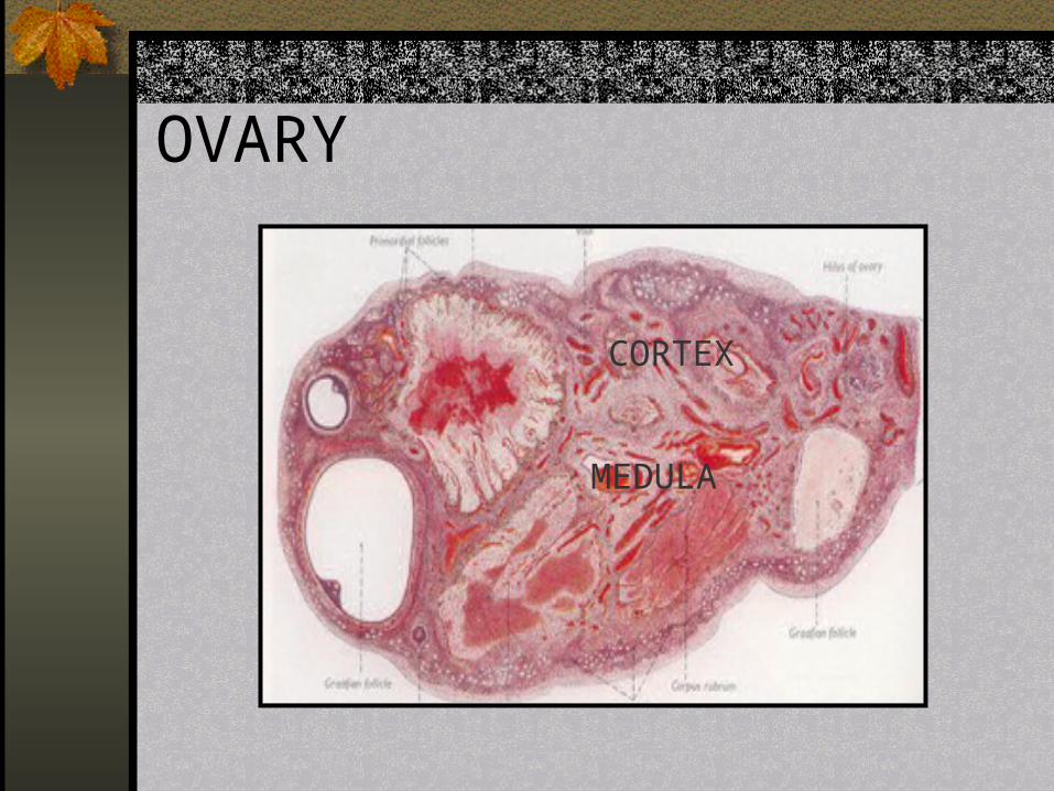
OVARY
MEDULA
CORTEX

OVARIAN CORTEXGERMINAL EPITELIUM
T. ALBUGINEA
STROMA
OVARIAN FOLLICLES
1. PRIMORDIAL FOLLICLE
2. PRIMARY FOLLICLE
3. GROWING FOLLICLE
4. MATURE GRAAFIAN FOLLICLE
5. CORPUS LUTEUM
6. CORPUS ALBICANS
7. ATRETIC FOLLICLE

PRIMORDIAL FOLLICLE Primordial follicle is the earliest stage of
follicular development Appears in the prenatal Consists of oocyte surrounded by single layer
of squamous follicle cellsPRIMARY FOLLICLE Appears in baby after he was born Consists of oocyte surrounded by single layer
of squamous follicle cells Histological appearance is like primordial
follicle

PRIMARY FOLLICLE

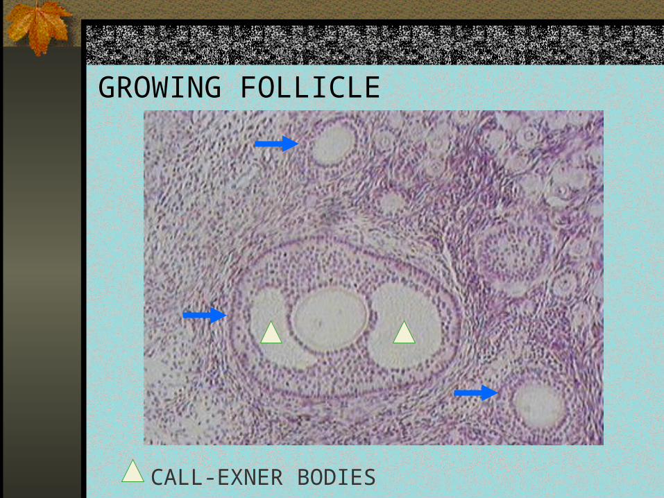
GROWING FOLLICLE
Formed in puberty, stimulated by Follicle Stimulating Hormone (FSH)
The oocyte become larger, together with squamous follicle cells form acidophilic refractil membrane called ZONA PELLUCIDA
Squamous follicle cells become cuboidal, proliferate, form multilayers of cuboidal cells called granulosa cells
Formed cavities that was filled liquor folliculi, called CALL-EXNER BODIES
In this stage, there are THECA FOOLICULI LAYER consists of THECA INTERNA LAYER and THECA EXTERNA LAYER
Theca interna contains cells produced esterogen Theca interna is surrounded by an outer stromal layer called
theca externa The GLASSY MEMBRAN lies between granulosa cells and
theca folliculi layer
GROWING FOLLICLE
CALL-EXNER BODIES

ZONA PELLUCIDA

MATURE GRAAFIAN FOLLICLE Secondary oocyte of mature Graafian follicle will be
expelled from ovary in ovulation Call-exner bodies begin to coalesce, forming a single
cavity called ANTHRUM FOLLICULI The follicle cells form :
CORONA RADIATA : a single layer of follicle cells surrounding oocyte
CUMULUS OOPHORUS : follicle cells located outer of corona radiata, that project into the anthrum fooliculi
MEMBRANA GRANULOSA : multilayer of follicle cells form wall of anthrum folliculi
There is primary oocyte ( oocyte I ) in the primary follicle until in Graafian follicle

The mature Graafian follicle was developed from primary follicle in 10 – 14 days
In this stage, Graafian follicle has the largest anthrum and has a projecting part into the surface of ovary that was called STIGMAIn this region, tunica albuginea and theca folliculi became more slight than in the other sides
Before ovulation occurred, the Graafian follicle had enlarged and had been filled by liquor folliculi. This condition is called PREOVULATORY SWELLING
The anthrum that was filled liquor pressured and ruptured the stigma. Then the oocyte (secondary oocyte), along with zona pellucida and corona radiata moved out from the ovary.
The meiotic I (reduction division) reduced number of chromosom to haploid. So the oocyte I becomes secondary oocyte ( oocyte II )
This process occurred just before ovulation

GRAAFIAN FOLLICLE
1
2
3
1. ANTHRUM FOLLICULI
2. CUMULUS OOPHORUS
3. MEMBRANA GRANULOSA

GRAAFIAN FOLLICLE
CORONA RADIATA
ZONA PELLUCIDA
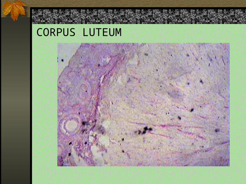
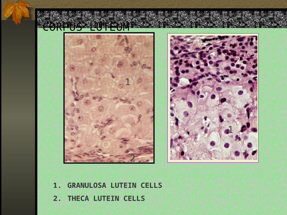
CORPUS LUTEUM Formed from wall of anthrum after ovulation Consists of 2 types of cells :
1. GRANULOSA LUTEIN CELLS Derived from granulosa cells The big and pale cells located in the central of corpus luteum Produce progesteron
2. THECA LUTEIN CELLS Derived from the cells of theca interna layer The small cells and deeply staining nucleus Located in periphery of corpus luteum
If fertilization occurs, corpus luteum will increase in size to form corpus luteum of pregnancy (CORPUS LUTEUM GRAVIDITAS)
Corpus luteum of pregnancy still exist until five month of pregnancy when placenta was formed completely
In this time, progesteron was produced by placenta
Then corpus luteum will become CORPUS ALBICANS
CORPUS LUTEUM

CORPUS LUTEUM
1
2
1.GRANULOSA LUTEIN CELLS
2. THECA LUTEIN CELLS
CORPUS LUTEUM
1
2
1
2
1. GRANULOSA LUTEIN CELLS
2. THECA LUTEIN CELLS

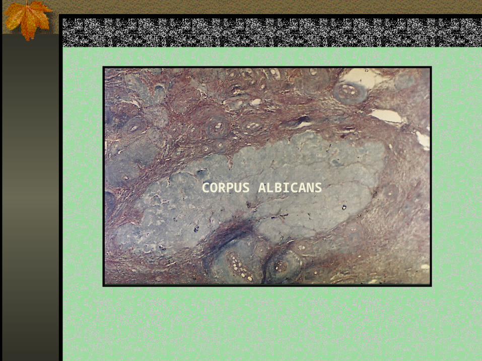
CORPUS ALBICANS
Derived from degeneration of corpus luteum when fertilization doesn’t occurred
Lutein cells undergo apoptosis, remain a pale fibrous scar called CORPUS ALBICANS
It can be formed from follicular atretics and from corpus luteum graviditas when placenta completely formed
Formed just before menstruation

CORPUS ALBICANS
CORPUS ALBICANS
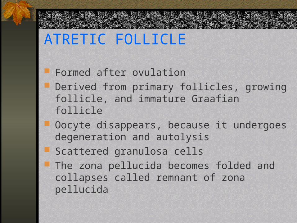
ATRETIC FOLLICLE
Formed after ovulation Derived from primary follicles, growing follicle, and
immature Graafian follicle Oocyte disappears, because it undergoes
degeneration and autolysis Scattered granulosa cells The zona pellucida becomes folded and collapses
called remnant of zona pellucida

ATRETIC FOLLICLE
REMNANT OF ZONA PELLUCIDA

ATRETIC FOLLICLE
UTERINE TUBES ( FALLOPIAN TUBES ) The uterine tubes transport oocyte from the ovary
to the uterus Divided into 4 segments :
1. Infundibulum
2. Ampulla
3. Isthmus
4. Intramural part

INFUNDIBULUM A funnel-shaped segment of the tubes adjacent to the
ovary The segment opens to the peritoneal cavity Folded mucosa is like fingers called FIMBRIAE at
free side of the segment The fimbriae catch the ovum from the ovary
AMPULLA The longest segment of the tube, is approximately
two thirds of the total length The segment has thin wall and large diameter Has branched mucosal folds Site of fertilization

ISTHMUS
The narrow segment The segment has the thickest wall of the other
segments
INTRAMURAL PART Lies within the uterine wall and opens into the cavity
of the uterus
MICROSCOPIC APPERANCE OF UTERINE TUBES The wall of uterine tubes is consists of three
layers :1. The mucosa inner layer of the wall, forms
longitudinal folds that project into lumen throughout its lumen. The folds are most numerous and complex in the ampulla
Consists of two types of cells, are CILIATED CELLS and NON-CILIATED (PEG CELLS)
2. The muscularis is arranged by two layers of smooth muscles
3. The serosa is composed of loose connective tissue with mesothelium in outer surface

UTERINE TUBES
AMPULA OF UTERINE TUBES ISTHMUS

UTERUS
Consists of 5 parts :
1. Fundus
2. Corpus
3. Isthmus
4. Cervix
5. Portio vaginalis
CORPUS Histological appearance of corpus is similar
to fundus They consist of three layers:A. Serosa ( Perimetrium )
The outer layer, arranged by thin layer of connective tissue that covered by mesothelium
B. Myometrium ( muscularis layer )Arranged by smooth muscles that enlarge in pregnancy
C. EndometriumHistological appearance of endometrium depend on menstrual cycle
ENDOMETRIUM Consists of two layers ; epithel and lamina
propria EPITHEL :
A layer of silindrical epithelium ciliated in some definite sites
This epithel continues to lamina propria and forms uterine glands that have branches in the basal site
LAMINA PROPRIA : Just beneath the epithelium, consists of reticular
fibers There are two types of arteries in lamina propria. Straight arteries in the basal site, and spiral
arteries ( coiled arteries) in the upper site
CYCLE CHANGES OF ENDOMETRIUM Depend on menstrual cycle, endometrium divided
into 4 phases :
1. PROLIFERATIVE PHASE : The other name of this phase is estrogenic or
reparative phase In this phase, the epithelium and the lamina propria
are reconstituted The uterine glands proliferate rapidly Coiled arteries are reformed

PROLIFERATIVE PHASE
1
2
1. ENDOMETRIUM
2. MYOMETRIUM

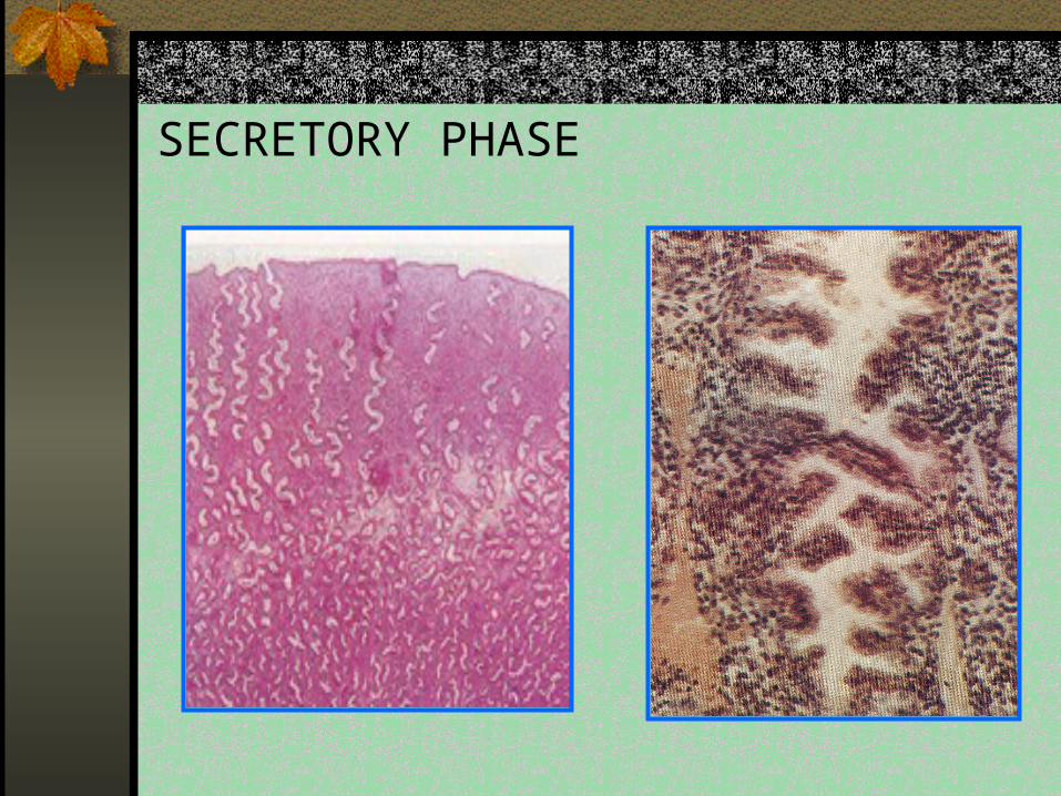
2. SECRETORY PHASE The other name is LUTEAL OR PROGESTATIONAL
PHASE The uterine glands become hypertrophi Endometrium becomes edematous and may
eventually reach a thickness of 5 to 6 mm The glands make saw-shaped appearance The coiled arteries are numerous and extend to the
surface of the endometrium The stromal cells enlarge and transform into decidual
cells In this phase endometrium becomes the thickest
stage so it can divided into two layers
Theese layers are :1. Functional layer the thick part, will
be sloughed off in menstruation and in partus. Divided into :
Compact layer : nearly to the surface, a thin layer with straight glands
Spongious layer : beneath the compact layer, a thick layer with spiral glands
2. Basal layer : the deeper layer attached to myometrium. This layer is retainedduring menstruation
SECRETORY PHASE

PROLIFERATION PHASE SECRETORY PHASE

3. ISCHEMIC PHASE ( premenstrual phase ) Contraction of coiled arteri wall cause the
stratum functionale become ischemic. Infiltration of leucocytes in stroma .
4. MENSTRUAL PHASE :Endometrium is sloughed off and constitute the vaginal discharge.
Vaginal discharge contains :a. blood
b. epithelial cells and sloughing stroma
c. secret of the glands

MENSTRUAL PHASE
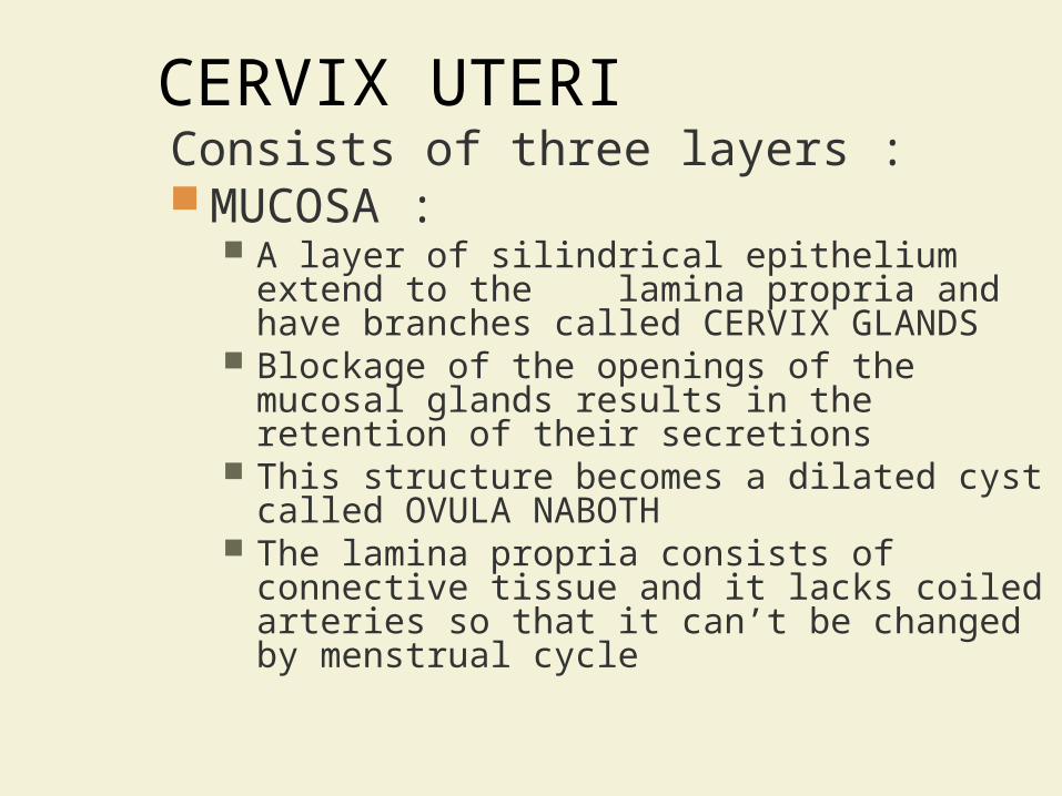
CERVIX UTERIConsists of three layers : MUCOSA :
A layer of silindrical epithelium extend to the lamina propria and have branches called CERVIX GLANDS
Blockage of the openings of the mucosal glands results in the retention of their secretions
This structure becomes a dilated cyst called OVULA NABOTH
The lamina propria consists of connective tissue and it lacks coiled arteries so that it can’t be changed by menstrual cycle

MUSCULARIS : Consists of smooth muscle with
irreguler arrangement The outer is arranged longitudinaly
extending to vagina
ADVENTITIA :Consists of fibroelastic connective tissue

CERVIX UTERI
CERVIX GLANDS 1. OVULA NABOTH

PORTIO VAGINALIS
The portion of the cervix that projects to the vagina
Covered by stratified squamous epithelium
This part is not changed during menstrual cycle


A fibromuscular tube covered by mucosa Consists of theree layers :
1. MUCOSA
Has numerous mocosal folds called RUGAE
Consists of : Non-keratinized stratified squamous epithelium The lamina propria that has numerous elastic
fibers, bood vessels and nerves
2. MUSCULARIS LAYER Consists of two layers of smooth muscles
3. ADVENTITIAL LAYER Consists of thin connective tissue with
numerous blood vessels and autonomic ganglion

VAGINA

GENITALIA EXTERNA ( VULVA ) Consists of theese structures :
1. Clitoris
2. Labia minora
3. Labia majora
4. Vestibular glands

CLITORIS
The erectile structure that is homolougous to the penis
Covered by non-keratinized stratified squamous epithelium
The lamina propria forms propria papil that has numerous blood vessels and sensory nerve endings (MEISSNER’S CORPUSCLES and PACINIAN CORPUSCLES)

A. EPITHELIUM
B. LAMINA PROPRIA
C. ERECTILE TISSUE

LABIA MINORA Folds of skin that border the vestibule Covered by non-keratinized stratified
squamous epithelium with melanin pigment in the deep cells of the epithelium
Large sebaceous glands in the stroma (lamina propria)
The glands doesn’t connect to hair follicles

SEBACEOUS GLANDS

LABIA MINORA
LABIA MAJORA Folds of skin, covers the labia minora The inner surface is covered by non-
keratinized stratified squamous epithelium
The outer surface is covered by keratinized stratified squamous epithelium with pubic hair

VESTIBULAR GLANDS
Consists of :
1. Minor vestibular glands The small mocous glands are present primarily
near the clitoris and around external urethral orifice
2. Major vestibular glands (Bartholin’s glands) The large mucous glands in the lateral wall of
the vestibule Secrete lubricating mucous


Modification of sweat glands in subcutan
They are rudimentary in male Composed of 15 – 20 lobes separated
by interlobular tissue (connective tissue) Each lobes has excretory ducts that
assemble in the apex of the nipple

Histological Appearance1. INACTIVE MAMMARY GLANDS
Composed of numerous intralobular ducts Consists of dense connective tissue and thick
adipose tissue
2. ACTIVE MAMMARY GLANDS The alveoli begin to develop Amount of the adipose and the connective tissue
is decrease The alveoli enlarge forming SACCULI that secret
milk in the late pregnancy
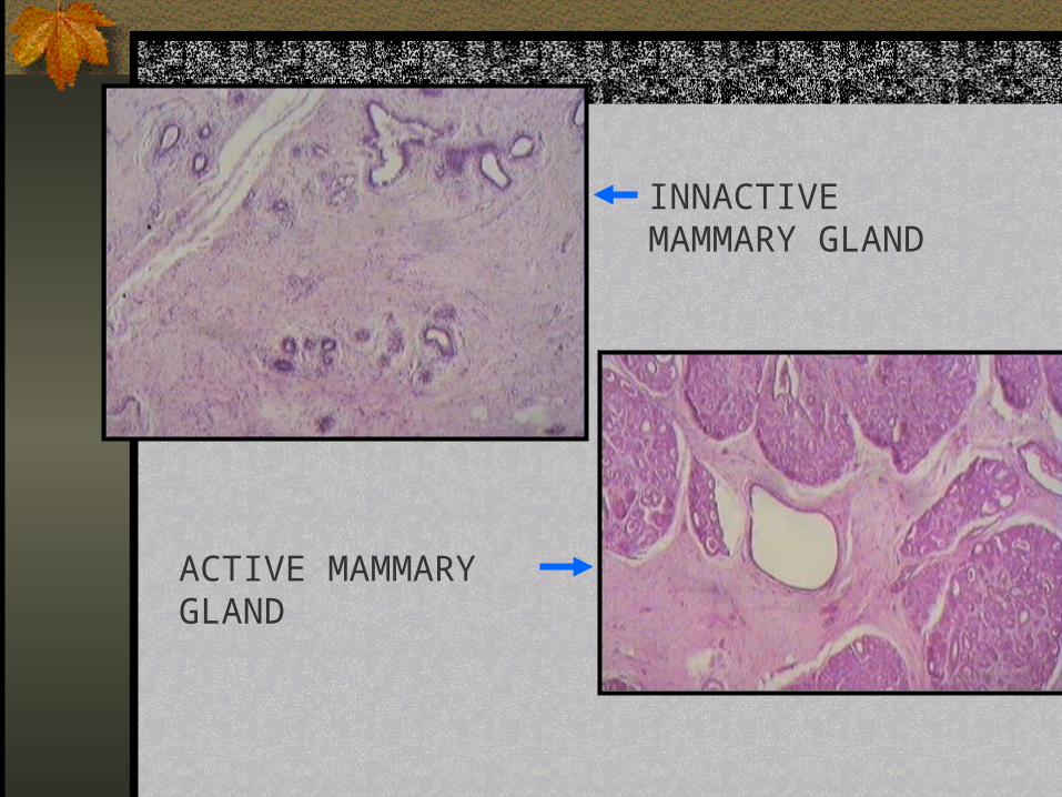
INNACTIVE MAMMARY GLAND
ACTIVE MAMMARY GLAND

THE KEYS OF SUCCESS ARE PERSEVERANCE AND DO THE WORK HARDLY


