
Identification of Extracellular Signal-regulatedKinase 1 (ERK1) Direct Substrates using StableIsotope Labeled Kinase Assay-LinkedPhosphoproteomics*□S
Liang Xue‡, Pengcheng Wang§, Pianpian Cao¶, Jian-kang Zhu‡§,and W. Andy Tao‡�**‡‡§§
Kinase mediated phosphorylation signaling is extensivelyinvolved in cellular functions and human diseases, andunraveling phosphorylation networks requires the identi-fication of substrates targeted by kinases, which has re-mained challenging. We report here a novel proteomicstrategy to identify the specificity and direct substrates ofkinases by coupling phosphoproteomics with a sensitivestable isotope labeled kinase reaction. A whole cell ex-tract was moderately dephosphorylated and subjected toin vitro kinase reaction under the condition in which 18O-ATP is the phosphate donor. The phosphorylated proteinsare then isolated and identified by mass spectrometry, inwhich the heavy phosphate (�85.979 Da) labeled phos-phopeptides reveal the kinase specificity. The in vitrophosphorylated proteins with heavy phosphates are fur-ther overlapped with in vivo kinase-dependent phospho-proteins for the identification of direct substrates withhigh confidence. The strategy allowed us to identify 46phosphorylation sites on 38 direct substrates of extracel-lular signal-regulated kinase 1, including multiple knownsubstrates and novel substrates, highlighting the abilityof this high throughput method for direct kinase substratescreening. Molecular & Cellular Proteomics 13: 10.1074/mcp.O114.038588, 3199–3210, 2014.
Protein phosphorylation regulates almost all aspects of celllife, such as cell cycle, migration, and apoptosis (1), andderegulation of protein phosphorylation is one of the mostfrequent causes or consequences of human diseases includ-ing cancers, diabetes, and immune disorders (2). Up till now,however, known substrates are far from saturation for the
majority of protein kinases (3); thus, mapping comprehensivekinase-substrate relationships is essential to understandingbiological mechanisms and uncovering new drug targets (4).
Accompanied with advances of high-speed and high-reso-lution mass spectrometry, the technique of kinase substratescreening using proteomic strategy is quickly evolving (5–7).Mass spectrometry has been extensively used for kinase-substrate interaction mapping (8) and global phosphorylationprofiling (9). Although thousands of phosphorylation siteshave been detected, complex phosphorylation cascade andcrosstalk between pathways make it difficult for large-scalephosphoproteomics to reveal direct relationships betweenprotein kinases and their substrates (10, 11). Extensive sta-tistics, bioinformatics, and downstream biochemical assaysare mandatory for the substrate verification (12, 13). Anotherstrategy uses purified, active kinases to phosphorylate cellextracts in vitro, followed by mass spectrometric analysis toidentify phosphoproteins. This approach inevitably faces themajor challenge of separating real sites phosphorylated bytarget kinase and the phosphorylation triggered by endoge-nous kinases from cell lysates (14). Analog-sensitive kinaseallele (15) overcomes the issue by utilizing the engineeredkinase that can exclusively take a bulky-ATP analog under thereaction condition. Analog-sensitive kinase allele has beencoupled with �-thiophosphate analog ATP to facilitate themass spectrometric analysis (16–18).
We have introduced kinase assay-linked phosphoproteom-ics (KALIP)1 to link the in vitro substrate identification and
From the ‡Departments of Biochemistry, §Horticulture and Land-scape Architecture, ¶Mathematics, �Medicinal Chemistry and Molec-ular Pharmacology, **Chemistry, and ‡‡Purdue University Center forCancer Research, Purdue University, West Lafayette, Indiana 47907
Received February 12, 2014, and in revised form, July 7, 2014Published, MCP Papers in Press, July 14, 2014, DOI 10.1074/
mcp.O114.038588Author contributions: L.X., J.Z., and W.A.T. designed research; L.X.
and P.W. performed research; L.X., P.C., and W.A.T. analyzed data;L.X. and W.A.T. wrote the paper.
1 The abbreviations used are: BAG3, BAG family molecular chap-erone regulator 3; KALIP, kinase assay-linked phosphoproteomics;regulator 3; BORG4, Cdc42 effector protein 4; EPS15, epidermalgrowth factor receptor pathway substrate 15; ERK1, extracellular sig-nal-regulated kinase 1; FASP, filter aided aided proteome preparation;FDR, false discovery rate; FSBA, adenosine-5�-(4-fluorosulfonylbenzo-ate) hydrochloride; GO, gene ontology; IPA, ingenuity pathway analysis;MAPK, mitogen-activated protein kinase; PolyMAC, polymer-basedmetal-ion affinity capture; proKALIP, protein kinase assay linked withphosphoproteomics; QIKS, quantitative identification of kinasesubstrates; SILAC, stable isotope labeling by amino acids in cellculture; siKALIP, stable isotope labeling kinase assay-linkedphosphoproteomics.
Technological Innovation and Resources© 2014 by The American Society for Biochemistry and Molecular Biology, Inc.This paper is available on line at http://www.mcponline.org
Molecular & Cellular Proteomics 13.11 3199
physiological phosphorylation events together in a highthroughput manner (19, 20). The strategy, however, has onlybeen applied to identify direct substrates of tyrosine kinases.In this study, we expanded the application of KALIP to serine/threonine kinases by introducing a quantitative strategytermed Stable Isotope Labeled Kinase Assay-Linked Phos-phoproteomics (siKALIP). The method was applied to identifydirect substrates of extracellular signal-regulated kinase 1(ERK1), a serine/threonine kinase acting as an essential com-ponent of the Mitogen-activated protein kinase (MAPK) signaltransduction pathway (21). A defect in the MAP/ERK pathwaycauses uncontrolled growth, which likely leads to cancer (22)and other diseases (23–25). ERK1 can be activated by growthfactors such as platelet-derived growth factor (PDGF), epider-mal growth factor (EGF), and nerve growth factor (NGF) (26).Upon stimulation, ERK1 phosphorylates hundreds of sub-strates in various cellular compartments including cytoplasm,nucleus, and membrane (27). Among 38 ERK1 direct sub-strates identified by siKALIP, more than one third are previ-ously discovered by classical molecular biology approaches,highlighting high specificity and sensitivity of the strategy. Theresults also support the hypothesis that ERK1 plays complexroles in multiple pathways that are essential for the cell growthregulation.
EXPERIMENTAL PROCEDURES
Mammalian Cell Culture—HEK293 cells (ATCC) were maintained inDulbecco’s Modified Eagle Medium (DMEM) (Sigma) supplementedwith 10% heat inactivated FBS, 100 �g/ml streptomycin, and 100IU/ml penicillin in 5% CO2 at 37 °C. Human DG-75 B lymphoma cells(ATCC) were grown in RPMI 1640 media (Sigma) supplemented with10% heat inactivated FBS, 1 mM sodium pyruvate, 100 �g/ml strep-tomycin, 100 IU/ml penicillin, and 0.05 mM 2-mercaptoethanol in 5%CO2 at 37 °C. The cells were washed with PBS, trypsinized, collected,and frozen at �80 °C for further use.
Plant Tissue Culture—Seedlings of A. thaliana were grown in 40 mlof half-strength Murashige and Skoog medium at 22 °C in continuouslight on a rotary shaker set at 100 rpm. Twelve-day-old seedlingswere collected and frozen at �80 °C for further use.
In Vitro Kinase Reaction in siKALIP—Mammalian cells were lysedby sonication in lysis buffer containing 50 mM Tris-HCl, pH 7.5, 150mM NaCl, 5 mM EDTA, and 1% Nonidet P-40 on ice. For plant cells,total protein was extracted from 2 g of seedlings by grinding in 2 mlof extraction buffer containing 100 mM Tris-HCl (pH 7.5), 250 mM
NaCl, and 5 mM EDTA. The cell debris was cleared by centrifugationat 16,000 � g for 10 min. The supernatant containing 400 �g solubleproteins was collected. The lysate volume was adjusted to 200 �lusing phosphatase buffer (Roche). 10U of rAPid alkaline phosphatase(Roche) was added and incubated at 37 °C for 3 h. The phosphatasewas deactivated by heating at 75 °C for 5 min. To inhibit endogenouskinases, the sample was incubated with 1 mM 5�-(4-fluorosulfonyl-benzoyl)adenosine (FSBA) with 10% DMSO in Tris-HCl, pH 7.5 at30 °C for 1 h. Excess FSBA is removed by Vivacon filtration units (30kDa cutoff). Samples in the filters were then incubated in buffercontaining 300 ng ERK1 (Sigma), 5 mM MgCl2, and 1 mM �-[18O4]-ATP(Cambridge Isotope Laboratory, Andover, MA) at 30 °C for 1 h. Thereaction was stopped by 8 M urea with 5 mM dithiothreitol. Filter aidedproteome preparation (FASP, San Diego, CA) digestion was per-formed according to the manufacturers’ specifications (Expedeon).
In Vitro Kinase Reaction by Autoradiography—The EPS15 andBAG3 substrates were isolated on beads by specific antibody (Anti-EPS15 from Cell Signaling, Beverly, MA and Anti-BAG3 from Protein-Tech, Chicago, IL). The BORG4 recombinant proteins were pur-chased from Abnova. Substrates were incubated in Tris-HCl, pH 7.5buffer containing 1 �g ERK1 (Sigma), 5 mM MgCl2, and 25 �M coldATP, 2.5 �Ci [�-32P] ATP at 30 °C for 1 h. The reaction was quenchedby boiling the sample with 4X electrophoresis sample buffer (Invitro-gen, Carlsbad, CA). Phosphorylation signal was detected by phos-phor-imager (GE Healthcare, Pittsburgh, PA).
Phosphopeptide Enrichment—Tryptic peptides were first desaltedusing a Sep-pak C18 column (Waters, Milford, MA) and dried. Next,the peptide mixture was resuspended in 100 �l of loading buffer (100mM glycolic acid, 1% trifluroacetic acid, and 50% acetonitrile) towhich 5 nmol of the PolyMAC-Ti (Tymora Analytical, IN) reagent wasadded (28). The mixture was then incubated for 5 min. 200 �l of 300mM HEPES, pH 7.7, was added to the mixture to achieve a final pH of6.3. The solution was incubated with magnetic hydrazide beads tocapture the PolyMAC-Ti dendrimers. The column was gently agitatedfor 10 min and then centrifuged at 2300 � g for 30 s to collect theunbound flow-through. The beads were washed once with 200 �lloading buffer, twice with a mixture of 100 mM acetic acid, 1%trifluoroacetic acid, and 80% acetonitrile, and once with water. Thephosphopeptides were eluted from dendrimers by incubating thebeads twice with 100 �l of 400 mM ammonium hydroxide for 5 min.The eluates were collected and dried under vacuum.
Mass Spectrometric Data Acquisition—Peptide samples were dis-solved in 8 �l of 0.1% formic acid and injected into an EksigentNanoLC Ultra 2D HPLC system. The reverse phase chromatographywas performed using an in-house C18 capillary column packed with5 �m C18 Magic beads resin (Michrom; 75 �m i.d. and 12 cm bedlength). The mobile phase buffer consists of 0.1% formic acid inultra-pure water with an eluting buffer of 0.1% formic acid (Buffer A)in 100% CH3CN (Buffer B) run over a linear gradient (2–35% Buffer B,90 min) with a flow rate of 300 nl/min. The electrospray ionizationemitter tip was generated on the prepacked column with a laser puller(Model P-2000, Sutter Instrument Co.). The Eksigent Ultra2D HPLCsystem was coupled online with a high-resolution hybrid duel-celllinear ion trap Orbitrap mass spectrometer (LTQ-Orbitrap Velos;Thermo Scientific). The mass spectrometer was operated in the data-dependent mode in which a full-scan MS (from m/z 300–1700 withthe resolution of 60,000 at m/z 400) was followed by 20 MS/MS scansof the most abundant ions using collision-induced dissociation (CID).Ions with the charge state of �1 were excluded. The dynamic exclu-sion time was set to 60 s after two fragmentations.
Database Search and Quantitation—The LTQ-Orbitrap raw fileswere searched directly against the Homo sapiens database with noredundant entries (93,289 entries; human International Protein Index(IPI) v.3.83) using the SEQUEST algorithm on Proteome Discoverer(Version 1.3; Thermo Fisher). Peptide precursor mass tolerance wasset to 10 ppm, and MS/MS tolerance was set to 0.8 Da. Searchcriteria included a static modification of �57.0214 Da on cysteineresidues, a dynamic modification of �15.9949 Da on oxidized methi-onine, a dynamic modification of �79.996 Da on normal phosphory-lated serine, threonine, and tyrosine residues, and a dynamic modifi-cation of �85.996 Da on heavy phosphorylated serine, threonine, andtyrosine residues. Searches were performed with full tryptic digestionand allowed a maximum of two missed cleavages on peptides ana-lyzed by the sequence database. False discovery rates (FDR) were setto 1% for each analysis. Proteome Discoverer generated a reverse“decoy” database from the same protein database, and any peptidepassing the initial filtering parameters from this decoy database wasdefined as a false positive. The minimum cross-correlation factor(Xcorr) filter was re-adjusted for each charge state separately in order
Identification of ERK1 Direct Substrates by siKALIP
3200 Molecular & Cellular Proteomics 13.11
to optimally meet the predetermined 1% FDR based on the number ofrandom false-positives matched with the reversed “decoy” database.Thus, each dataset had its own passing parameters. The number ofunique phosphopeptides and nonphosphopeptides were then man-ually counted and compared. Phosphorylation site localization fromCID mass spectra was determined by PhosphoRS scores (29). Forphosphopeptides with ambiguous phosphorylation sites, only onephosphorylation site with the highest score was selected for furtherdata interpretation.
Data Analysis—To find the ERK1 phospho-motif analysis, Motif-X(http://motif-x.med.harvard.edu) was used for predicting the specific-ity of kinases according to identified phosphorylation sites. Parame-ters were set to peptide length � 13, occurrence � 10, and signifi-cance p value less than 0.000001. For the ERK1 binding site analysis,Scansite 2.0 (http://scansite.mit.edu) was used to search Uniprotprotein sequences for high-stringency ERK1/2 binding sequences.The heavy phosphorylated proteins in vitro were submitted to Inge-nuity Pathway Analysis (IPA) (Ingenuity Systems) for the gene ontol-ogy (GO) annotation.
Immunoprecipitation and Western Blotting Experiments—Cellswere collected and lysed by sonication in lysis buffer containingprotease and phosphatase inhibitors (50 mM Tris-HCl, pH 7.5, 150 mM
NaCl, 5 mM EDTA, 1% Nonidet P-40, and 1x Mini Complete proteaseinhibitor mixture (Roche)). Samples were cleared of debris and nor-malized based on the protein concentration. Then 1 mg of lysate waspre-incubated with 20 �l Protein A/G agarose beads (Thermo) for 20min at 4 °C to remove nonspecific bindings, before further incubationwith 10 �g of antibodies for 4 h at 4 °C. Anti-EPS15 rabbit polyclonalantibody from Cell Signaling and anti-BAG3 rabbit polyclonal anti-body from Proteintech were used. The samples were then incubatedwith 20 �l of Protein A/G agarose beads again for capturing for 4 h at4 °C. The beads were washed and bound proteins were eluted byboiling the beads in the SDS loading buffer with 50 mM DTT for 5 min.The eluents were separated on a 12% SDS-polyacrylamide gel and
transferred onto a PVDF membrane. The membranes were probedusing antibodies against proteins of interest.
RESULTS
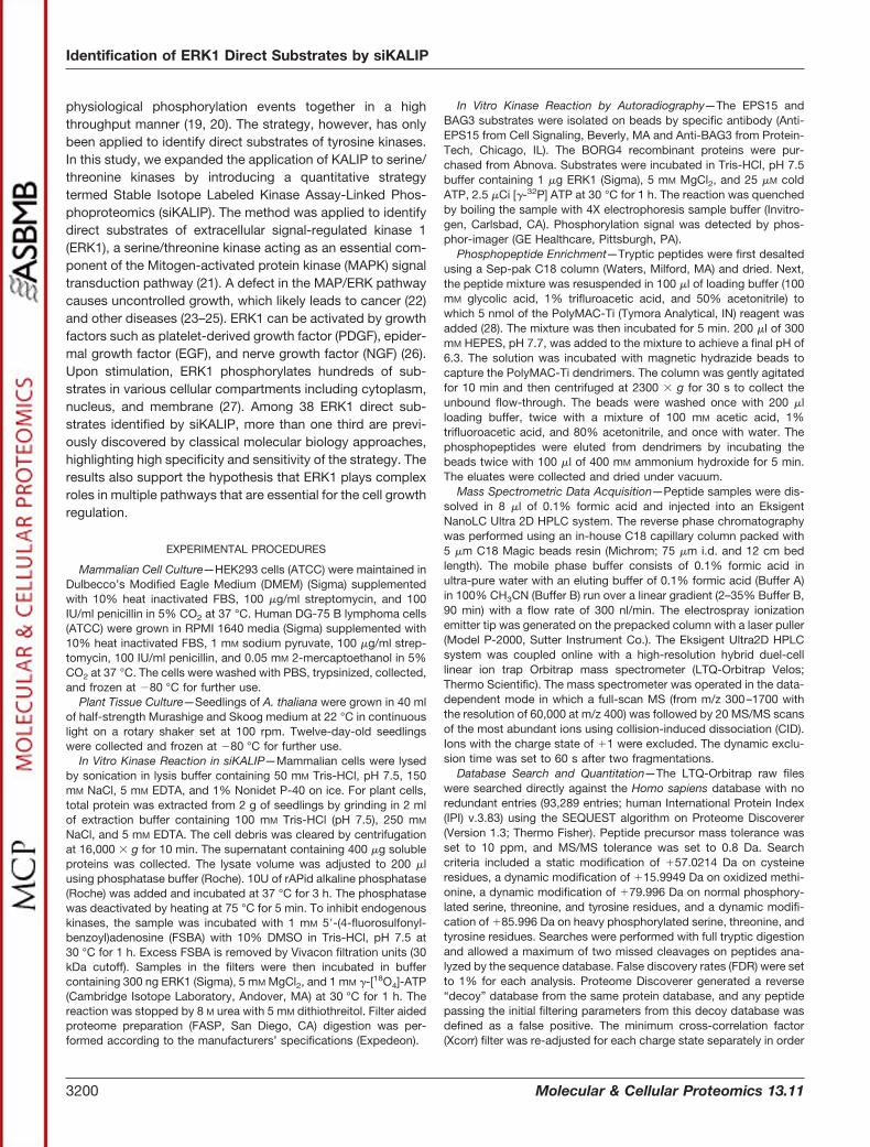
The siKALIP Strategy for Direct Kinase Substrate Identifica-tion—In contrast to tyrosine phosphorylation, it is estimatedthat serine/threonine phosphorylation consists of over 99% ofall phosphorylation (30). In a typical whole cell extract, thou-sands of proteins can be phosphorylated on serine/threonineresidues (31) and present enormously large background inany phosphorylation analysis. It poses great challenges to usewhole cell extracts for in vitro kinase assay. To address theissue, we devised a new strategy, siKALIP, by incorporating�-18O-phosphate ATP and moderate dephosphorylation inthe kinase assay to identify direct kinase substrates in highthroughput (Fig. 1). In general, proteins are extracted from thecells of interest. A phosphatase is added to partially removephosphate groups from endogenous phosphoproteins togenerate a pool of the candidate proteome for the following invitro kinase reaction. Pulsed heating is applied to quench theexogenous phosphatase activity. A generic kinase inhibitorsuch as FSBA is added to deactivate endogenous kinaseactivities and excess FSBA was subsequently removed usingfiltration. The protein solution is then equally divided prior tothe addition of the kinase along with �-[18O4] ATP (sample) or�-[18O4] ATP only (control). After the in vitro kinase reaction,proteins are digested and phosphopeptides are enriched byPolymer-based Metal ion Affinity Capture (PolyMAC) (28), fol-
FIG. 1. Workflow for kinase substrate identification through the integration of in vitro kinase reaction and in vivo phosphoproteo-mics. In the in vitro kinase reaction, cell lysate is dephosphorylated before the kinase assay. Rephosphorylated proteins are digested, andphosphopeptides are enriched and analyzed by mass spectrometry for sequencing and site identification. Theoretical substrate identificationis based on the phosphopeptide having 18O-labeled phosphate groups. In in vivo phosphoproteomics, kinase-dependent phosphorylationevents are identified by comparing wild type and kinase inhibited cells. Bona fide direct substrates are the overlapping phosphoproteinspresent in both in vitro and in vivo datasets.
Identification of ERK1 Direct Substrates by siKALIP
Molecular & Cellular Proteomics 13.11 3201
lowed by mass spectrometric analysis to identify peptidesequences and phosphorylation sites. This procedure gener-ates a list of in vitro direct substrates of the target kinase. Inparallel, quantitative phosphoproteomics is performed usingcells in which the kinase of interest is either active or inhibited.The in vitro experiments by the kinase reaction produce truedirect kinase substrates and false positives because of theloss of physiological environment. On the contrary, endoge-nous phosphoproteomics data include both direct and indi-rect kinase substrates. The overlap of in vitro and in vivocandidates should represent the true direct kinase substratesin high confidence.
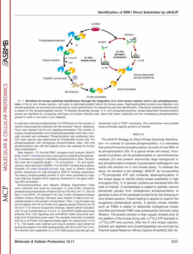
In Vitro Kinase Assay with Stable Isotope Labeled ATP—High background of serine/threonine phosphorylation and po-
tential residual endogenous kinase activities may lead to falsepositives when it is identified in the kinase reaction sample.Previous KALIP strategies used SILAC method to quantify thechange of phosphorylation before and after the kinase reac-tion. To heighten the sensitivity and specificity of the in vitrokinase reaction, 18O labeled-ATP (“heavy” ATP) is introducedin the kinase assay (Fig. 2A). In contrast to the naturallyexisting �-[16O4]-ATP (“light” ATP), when the heavy ATP isutilized by the kinase for phosphorylation, the substrates willhave a phosphate group with three 18O atoms. Therefore, thenewly phosphorylated sites can be qualitatively distinguishedfrom the background in the spectra. The “yes or no” feature ofsiKALIP improves the sensitivity of in vitro kinase assay andmakes the data interpretation more straightforward and re-
FIG. 2. A, Dephosphorylation and 18O-labeled ATP in siKALIP. The cell lysates are dephosphorylated and then incubated with [18O4]-ATPunder the reaction condition. Samples at the basal level, and after the kinase-treated, along with the control are compared. Red and greenphosphates indicate P18O4
3� and P16O43� respectively. The peaks of phosphopeptides are labeled with different colors (yellow, blue, and
purple). Yellow: The endogenous phosphorylation is not completely removed, so the heavy and light isotopic peaks exist. Blue: Theendogenous phosphorylation is removed, and only the heavy phosphorylation exists after kinase assay. Purple: endogenous phosphorylationsites not detected, and phosphorylated by the kinase. B, The histogram depicting the relationship between dephosphorylation and therephosphorylation efficiency. From 0, 1 h, to 3 h phosphatase treatment, endogenous phosphopeptides identification is decreasing, whereasthe newly phosphorylated sites labeled with heavy phosphate are increasing.
Identification of ERK1 Direct Substrates by siKALIP
3202 Molecular & Cellular Proteomics 13.11

producible compared with SILAC-based quantitative analysis.Note that P-O bond on the phosphate is quite stable, and ithas been demonstrated that 18O and 16O exchange did notoccur under the catalysis of kinase or harsh conditions duringthe sample preparation (32).
Dephosphorylation Significantly Improves the Sensitivity ofIn Vitro Kinase Assay—There have been a handful of studiesusing kinase assay to screen the substrates from whole cellextracts (33, 34). Widespread and high dynamic range ofphosphorylation events in whole cell extracts diminishes thesensitivity unless intensive fractionation steps are installed tosimplify the sample. We carefully examined the effect of de-phosphorylation prior to the kinase reaction. Whole cell ex-acts prepared from human embryonic kidney 293 (HEK293)cells were treated with phosphatase and monitored as a func-tion of time. These samples were further split for control andkinase assay with ERK1. Following the in vitro reaction and as-sisted by 18O labeled-ATP, we compared the “heavy” and“light” phosphorylation sites in these samples and a clearcorrelation between dephosphorylation efficiency and kinaseassay sensitivity was observed (Fig. 2B). As expected, a lon-ger dephosphorylation step led to the decreased number oflight phosphorylation sites in the control sample. Interestingly,lower background phosphorylation (number of light phosphor-ylation sites) correlates with higher rephosphorylation level(number of heavy phosphorylation sites) in the kinase assay.Two factors may contribute to the observed correlation: agood percentage of serine/threonine residues were pre-occu-pied by endogenous phosphorylation so that the stoichiom-etry of free residues for new phosphorylation events might betoo low to be detected in the MS; Another factor might be thatlarge amount of endogenous phosphorylation overwhelms thenew phosphorylation events by the kinase (phosphorylated byheavy ATP) during phosphopeptide enrichment and duringthe LC-MS/MS data acquisition. Taken together, dephosphor-ylation of cell lysate by a phosphatase increases the sensitiv-ity of in vitro kinase assay in the strategy.
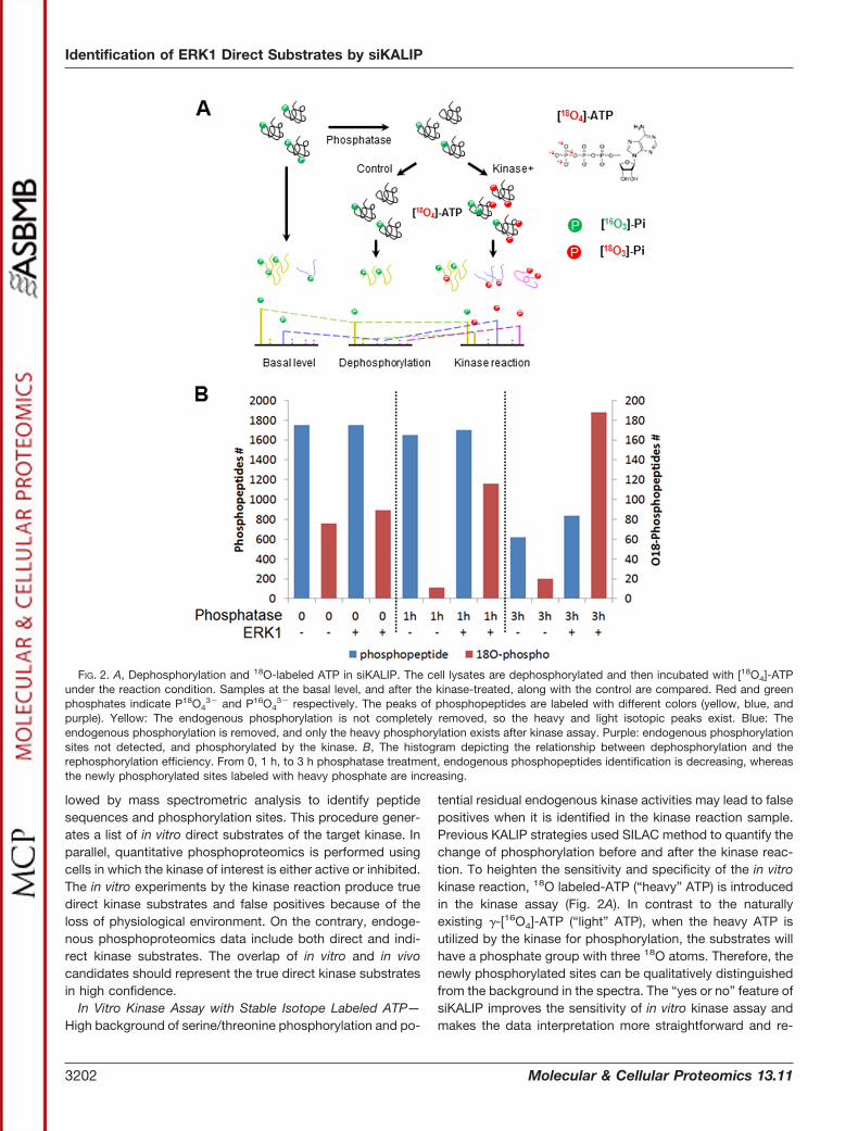
Identification of Direct ERK1 Substrates In Vitro—We ap-plied the siKALIP strategy to identify novel substrates forERK1 in HEK293 cells. Protein lysate from the cells wasdephosphorylated using an alkaline phosphatase, which wassubsequently inactivated by pulse heating. After FSBA treat-ment, the candidate substrate proteins were then split andincubated with or without purified active ERK1 in the kinasereaction buffer at the presence of �-[18O4]-ATP. The reactionwas ceased by 8 M urea followed by trypsin digestion. Phos-phopeptides were enriched and analyzed by mass spectrom-etry. The existence of heavy phosphate was used as anindicator to determine the direct substrates of ERK1 in vitro.supplemental Fig. S1A showed that the progesterone recep-tor membrane component 1 (PGRMC1) could not be phos-phorylated by ERK1 in our experiment because only the lightphosphorylation was detected. In contrast, peptide ASG-QAFELILsPR derived from the known substrate Stathmin had
the heavy phosphorylation form (Fig. 3A), which was easilydistinguished from the background in light phosphory-lation. Many novel substrates were identified. For example,BAG family molecular chaperone regulator 3 (BAG3) had twopossible sites (S377 and S385) that could be phosphorylatedby ERK1 in vitro (supplemental Fig. S1B). A previous reportedsubstrate, cortactin, also had two novel phosphorylation sitesidentified, although neither of these two sites had been de-phosphorylated completely (Fig. 3B). In these cases, the co-existence of light and heavy phosphopeptides allowed us toquantify the kinase activity normalized by residual phosphor-ylation level of substrates.
Validating ERK1 Specificity for the Substrates Recogni-tion—From all identified peptides (supplemental Table S1)using two batches of HEK293 cells (400 �g cell lysate for eachexperiment), a total of 629 unique phosphorylation sites la-beled with heavy phosphate group, representing 400 phos-phoproteins (supplemental Fig. S2, supplemental Table S2)which were identified exclusively in the kinase reaction sam-ples as in vitro ERK1 substrates. The phosphorylation sitesfavored by protein kinases could be dependent on both pri-mary sequence and the protein structures (35). Comparedwith protein array-based screening, siKALIP provides a con-venient and efficient way to determine kinase specificity bytaking advantage of naturally synthesized proteins in an intactcell. We applied the motif analysis (36) to extract the consen-sus sequence of ERK1 among the peptides with heavy la-beled phosphorylation in vitro (Fig. 4A). The enrichment ofproline residue adjacent to the phosphorylated serine wasconsistent with reported ERK1 motif according to Phospho-SitePlus (37) (Fig. 4B). Besides the phosphorylation site motif,ERK1 substrates were reported to have a binding sequencenamed D domain (38) (positively charged residues positionedat three to five residues ahead of a hydrophobic sequence).Using the Scansite (39), we found D domain was also en-riched in the in vitro substrates compared with the UniprotHuman proteome (Fig. 4C). We further compared our phos-phorylation motif and D domain enrichment with other meth-ods previously reported for in vitro MAPK substrate screening,including ASKA for ERK2 substrates (40) and high throughputin vitro kinase assay for p38 (34). Although three studiesshared common features for the consensus motif SP/TP (sup-plemental Fig. S3A, S3B), the similarity of D domain and ERK1motif was higher between Carlson’s and ours (Fig. 4C), im-plying that ERK1 and ERK2 share higher homology comparedwith p38. This result demonstrated high sensitivity and spec-ificity of our approach in extracting the in vitro substrates forserine/threonine kinases.
Identifying Direct Substrate of ERK1 Under the Physiologi-cal Condition by Overlapping In Vitro and In Vivo Phospho-proteins—Under the in vitro reaction condition, kinase spec-ificity is usually compromised because of the high amount ofthe exogenous kinase and the loss of physiological context.To improve our ability to identify true substrates with a low
Identification of ERK1 Direct Substrates by siKALIP
Molecular & Cellular Proteomics 13.11 3203

false positive rate, we coupled our in vitro result with in vivoERK1 regulated phosphoproteins from several previous re-ports (40–44). In these studies, cell permeable inhibitors suchas U0126 were applied in the cell culture. Those inhibitorsinhibited ERK1 by disrupting the ATP binding to the N-termi-nal domain of its specific activator MEK1/2 (45). By comparingthe control cell versus inhibitor treated cells, the ERK1 de-pendent phosphoproteins can be retrieved. Based on thisapproach, Mann’s group identified 98 proteins with 167 phos-phorylation sites using SILAC quantitation (41), whereas Ahn’sgroup identified 35 proteins with 60 phosphorylation sitesusing label-free quantitation (42). In the same year, Hattori’sgroup identified 38 proteins using 2D-DIGE, IMAC, andphosphomotif antibodies (43). Recently Thibault and col-leagues further expanded the ERK1-dependent repertoire,which contains 155 proteins with 232 phosphorylation sitesusing a rat cell line (44). In addition, two previously mentionedstudies identified in vivo substrates of ERK2 (40) and p38 (34),
which also belong to mitogen activation protein kinase(MAPK) family. Overlapping our data with each of those stud-ies was provided in supplemental Fig. S4. Generally speaking,we got 34, 19, 17, and 11 overlapping substrate proteins withCourcelles (44), Pan (41), Old (42), and Kosako’s studies (43),respectively. Considering the high homology between ERK1and ERK2, we also took 16 ERK2 -dependent phosphopro-teins identified by Forest group into the analysis and got threecommon phosphoproteins. Combining those five datasetstogether, we identified 46 direct phosphorylation sites byERK1 (Supplemental Table S3).
Validating Novel ERK1 Direct Substrates by In Vitro KinaseAssay—Among the 38 phosphoproteins containing 46 phos-phorylation sites overlapping in vitro and in vivo, 14 wereknown substrates of ERK1 with 18 phosphorylation sites (sup-plemental Table S3). Note that a novel site T364 was identifiedon the previously characterized substrate cortactin (46). Theremaining 27 phosphorylation sites representing 24 proteins
FIG. 3. MS spectra of different cases of phosphopeptides identified by LC-MS. A, The known phosphorylation site of ERK1. B, The novelphosphorylation sites of ERK1 identified while the endogenous phosphorylation still exist in high level.
Identification of ERK1 Direct Substrates by siKALIP
3204 Molecular & Cellular Proteomics 13.11

were considered to be novel substrates of ERK1. When con-sidering the phosphoprotein regardless of accurate phosphor-ylation sites, we totally identified 72 proteins that were phos-phorylated by ERK1 both in vitro and in vivo (supplementalTable S4).
To gain a global view of ERK1 phosphorylation, we per-formed gene ontology (GO) to analyze the 72 direct sub-
strates. The cellular components analysis indicated that ERK1substrates fell into four major categories (Fig. 5A), cytoplasm,nucleus, membrane, and cytoskeleton, a distribution consis-tent with a previous report (40). In comparison with total humanproteome (28,612 proteins) and phosphoproteome (11,479proteins) having GO annotations from Uniprot and Phospho-SitePlus, respectively, the proportions of cytoplasm, nucleus,and cytoskeleton were significantly increased (Fig. 5B). To-gether with the previous in vivo study that spliceosome phos-phorylation is ERK1 dependent (44), the enrichment of spli-ceosomal proteins in our result further suggested that ERK1probably contributes to the splicing regulation directlythrough phosphorylation. In addition, the direct ERK1 sub-strates could be grouped into several important biologicalprocesses: gene expression, metabolism, cellular assembly,cell cycle, transport, and signal transduction (Fig. 5C). Amongthem, gene expression, metabolism, cell assembly, and cellcycle were significantly enriched compared with human pro-teome (Fig. 5D). ERK1 has been known to regulate geneexpression by phosphorylating transcription factors andtransporters in both the cytosol and nucleus (47). A handful ofsubstrates involved in gene expression regulation fall in thosecategories (supplemental Table S5), including some knownERK1 substrates such as nucleoporin (NUP) (43), translocatedpromoter region (TPR) (48), and heterogeneous nuclear ribo-nucleoprotein (HNRNP) (49). Besides, many substrates playroles in cytoskeleton organization (supplemental Table S6),such as stathmin (50) and cortactin (51).
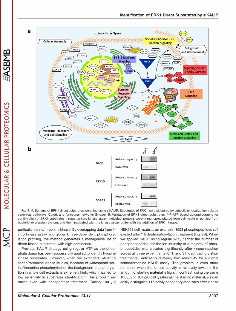
The identified substrates are mapped on commonly anno-tated cellular components and important biological functions(Fig. 6A, supplemental Table S7). Among the newly identifiedsubstrate candidates, we selected three for biochemical val-idation: BAG family molecular chaperone regulator 3 (BAG3),Epidermal growth factor receptor pathway substrate 15(EPS15), and Cdc42 effector protein 4 (BORG4). BAG3 andEPS15 were immunoprecipitated from HEK293 cell lysates,whereas BORG4 was the recombinant protein purified frombacteria. After 32P-ATP in vitro kinase assay, all three proteinswere confirmed to be the direct substrates of ERK1 by auto-radiography (Fig. 6B). EPS15 is involved in cell growth regu-lation, especially the regulation of mitogenic signals and cellproliferation. It was involved in the internalization of ligand-inducible receptors such as EGFR through a variety of pro-tein-protein interactions (52). PHOSIDA (53) showed that theS662 was phosphorylated during mitosis by a phosphopro-teomic study for cell cycle (54). To the best of our knowledge,other than large-scale phosphoproteomic screenings, this isthe first report indicating that EPS15 phosphorylation is di-rectly related to ERK1. The significant increase of the phos-phorylation and the subsequent biological processes such asprotein interaction and membrane translocation suggest thatsuch modification by ERK1 might be essential for EGFR en-docytosis. BAG3 executes the anti-apoptotic function byforming a complex with phospholipase C-gamma and Hsp70/
FIG. 4. A, The ERK1 consensus sequence generated from in vitrosubstrates identified in the kinase reaction; B, Consensus sequenceof ERK1 substrates by PhosphositePlus; C, Histogram comparing thefrequency of high-stringency D domains and ERK1 motif betweensiKALIP-ERK1 substrates, human proteome, reported substrates,substrates identified by Carlson et al. (40), and Knight et al. (34).
Identification of ERK1 Direct Substrates by siKALIP
Molecular & Cellular Proteomics 13.11 3205

Hsc70 under EGF-regulation (55). Several studies pointed outthat S377 and S385 phosphorylation of BAG3 were regulatedin the cell cycle (56) and up-regulated under the EGFR stim-ulation (31). Our research illustrates that direct upstream ki-nase ERK1 might be critical for BAG3 regulation in both cellcycle and EGFR pathway.
Compared with the BAG3 and EPS15, much less is knownabout Cdc42 effector protein 4 (BORG4). It may be involved inthe cell morphology by inducing the actin filament assembly(57). Although the knowledge of BORG4 function is limited, itsS104 phosphorylation appears to be significantly regulatedfor the cell cycle and EGFR pathway, in agreement with otherresearch groups (31, 56). Elucidation of the direct phosphor-ylation relationship between ERK1 and BORG4, as well asother identified substrates in the same network, such as CTT-NBP2 N-terminal-like protein (CTTNBP2NL), cytoplasmic dy-nein 1 light intermediate chain 1 (DYNC1LI1), and stathmin
(STMN1), advances our knowledge those substrates’ func-tions and mechanisms in cell organization regulation.
DISCUSSION
We present here a proteomic strategy that highlights theuse of �-[18O4]-ATP in a kinase reaction with a complexprotein pool prepared from whole cell extracts. The siKALIPapproach offers multiple advantages. First of all, the heavy-ATP has the same biochemical properties as normal ATP andthe labeling 18O does not exchange with 16O; thus, there is aclear relationship between the existence of heavy phosphateand the direct kinase substrate in vitro. Second, the dephos-phorylation step prior to the kinase reaction efficiently re-duces the background phosphorylation and as a result, thesensitivity has been greatly improved while fractionation ofsamples before mass spectrometric analyses is not needed.Third and finally, the strategy can be applied to any kinase, in
FIG. 5. Gene ontology analyses for ERK1 direct substrates. A, Categories of cellular components for identified ERK1 substrates. B,Distribution of cellular components among ERK1 direct substrates, human proteome, and human phosphoproteome. C, Categories ofbiological processes for identified ERK1 substrates. D, Distribution of biological processes among ERK1 direct substrates, human proteome,and human phosphoproteome.
Identification of ERK1 Direct Substrates by siKALIP
3206 Molecular & Cellular Proteomics 13.11
particular serine/threonine kinase. By overlapping data from invitro kinase assay and global kinase-dependent phosphory-lation profiling, the method generates a manageable list ofdirect kinase substrates with high confidence.
Previous KALIP strategy using regular ATP as the phos-phate donor has been successfully applied to identify tyrosinekinase substrates. However, when we extended KALIP toserine/threonine kinase studies, because of widespread ser-ine/threonine phosphorylation, the background phosphoryla-tion in whole cell extracts is extremely high, which has led tolow sensitivity in substrates identification. This problem re-mains even with phosphatase treatment. Taking 100 �g
HEK293 cell lysate as an example, 1653 phosphopeptides stillexisted after 1 h dephosphorylation treatment (Fig. 2B). Whenwe applied KALIP using regular ATP, neither the number ofphosphopeptides nor the ion intensity of a majority of phos-phopeptides was elevated significantly after kinase reactionacross all three experiments (0, 1, and 3 h dephosphorylationtreatments), indicating relatively low sensitivity for a globalserine/threonine KALIP assay. The problem is even moredominant when the kinase activity is relatively low and theamount of starting material is high. In contrast, using the same100 �g of HEK293 cell lysates as the starting material, we caneasily distinguish 116 newly phosphorylated sites after kinase
FIG. 6. A, Scheme of ERK1 direct substrates identified using siKALIP. Substrates of ERK1 were clustered by subcellular localization, relatedcanonical pathways (Color), and functional networks (Shape); B, Validation of ERK1 direct substrates. 32P-ATP based autoradiography forconfirmation of ERK1 substrates through in vitro kinase assay. Individual proteins were immunoprecipitated from cell lysate or purified frombacterial expression system, and then incubated with the kinase assay buffer with the addition of ERK1 kinase.
Identification of ERK1 Direct Substrates by siKALIP
Molecular & Cellular Proteomics 13.11 3207

assay by database search, without any additional complexityon data analysis such as stochastic ion selection for subtrac-tive MS or statistical significance decision when using ratiochange as the criteria. Among the 116 phosphopeptides withheavy phosphate groups, it includes several known sub-strates of ERK1 such as stathmin and cortactin, as well as anew substrate validated in this study, BAG3. These knownand validated substrates were not identified with the originalKALIP protocol.
To further demonstrate the superior specificity and sensi-tivity of siKALIP, we examined two additional kinases, includ-ing serine/threonine kinase SnRK2 in Arabidopsis tissues andspleen tyrosine kinase (SYK) in Human DG75 B cells. Theresults indicated that siKALIP is particularly superior to KALIPfor the serine/threonine kinase based on the identification ofknown and confirmed substrates and higher phosphorylationcontrast between samples with or without the kinase. Asshown in supplemental Fig. S5A, we observed the advantageof using heavy ATP in the SnRK2 kinase assay. Several knownsubstrates were shown phosphorylation in the control groupand thus excluded from the substrate candidate list by theexisting KALIP approach. However, the evidence that thoseproteins could be phosphorylated with SnRK2 in the presenceof heavy ATP revealed that they are likely genuine substratesof SnRK2 while they remain at a high level of basal phosphor-ylation. On the other hand, when comparing existing KALIPand siKALIP, we did not observe a significant improvement onthe identification of phosphorylated tyrosine substrates usingSYK as the exogenous kinase, in which case both regular andheavy ATP generated around one thousand phosphorylationsite in two replicates (supplemental Fig. S5B). We reasonedthat it might be due to the fact that endogenous tyrosinephosphorylation is low and the residual tyrosine phosphory-lation would not affect MS analysis significantly.
Our list of in vitro ERK1 substrates included 32 biochemi-cally validated ERK1 phosphorylation sites present on 22substrate proteins, including TPR, STMN, CTTN, c-Jun,LMNA, PXN, EIF4E, and RhoGEF (supplemental Table S8).When high-throughput data was also taken into account,there were 60 phosphorylation sites representing 46 proteinsshown to be regulated by ERK1 (supplemental Table S9).Furthermore, to the best of our knowledge, 33 additionalproteins can be phosphorylated by ERK1 in vivo (supplemen-tal Table S10). However, the accurate ERK1 phosphorylationsites in those 33 proteins were not clarified since previousstudies did not provide phosphorylation sites information orthe site localization did not exactly match with our data.Overall, the high recovery of known ERK1 substrates vali-dated the sensitivity of our method.
To further evaluate the sensitivity of isotope labeling in vitrokinase assay during siKALIP procedure, we compared ourresult with the three most recent studies that performed highthroughput in vitro mitogen activated protein kinase (MAPK)substrates screening, including ERK1 kinase assay with phos-
phospecific antibody (43), analog-sensitive ERK2 kinase as-say with SILAC (40), and p38 kinase assay with cell lysateusing FSBA and dimethyl labeling quantitation (34). The over-laps of our data with these studies are shown in supplementalFig. S6. We identified about half of the in vitro substrates fromthe report of Kosako (43), whereas the overlaps with Carlson(40) and Knight (34) were smaller, which might be attributed tothe different organisms chosen and less homology betweenp38 and ERK1. The results suggest the capability of siKALIPfor the in vitro kinase substrates identification and the consis-tency with other conventional methods.
Although siKALIP is capable of identifying direct substratesof any kinase in high throughput, there are certain conditionswe need to be aware of. First, prior to the kinase reactionendogenous kinase activity has to be eliminated using a ge-neric kinase inhibitor such as 5�-(4-fluorosulfonylbenzoyl)ad-enosine (FSBA). However, complete inhibition of all endoge-nous kinases is always difficult to achieve so that theremaining kinase activity from the lysate can potentially leadto false positive phosphorylation events. Second, the natureof siKALIP requires sufficient activity of purified kinase in vitro.The reconstitution of purified kinase assay in vitro may requirea prerequisite activation event (58), cofactor (59), or cellularcontext (60). Third, a full spectrum of kinase-dependent phos-phorylation change in vivo is essential. In this study,Courcelles and coworkers have reported the most compre-hensive quantitative phosphoproteomics on ERK1 to ourknowledge (44), which led to the most overlapping substrates(34 proteins) with our in vitro kinase reactions. It also high-lights the importance of highly specific and efficient kinaseinhibition in vivo. Together, only when both in vitro and in vivodatasets are comprehensive and specific can the siKALIPperformance on direct substrate identification be maximized.
* This project has been funded in part by an NSF CAREER awardCHE-0645020 (W.A.T.), and by a National Institutes of Health grantGM088317 (WAT).
□S This article contains supplemental Figs. S1 to S7 and Tables S1to S10.
§§ To whom correspondence should be addressed: Department ofBiochemistry, Purdue University, 175 S University St, West Lafayette,IN 47907. Tel.: 765-494-9605; Fax: 765-494-7897; E-mail: [email protected].
ADDITIONAL INFORMATION: Supplementary data set containing7 figures and 10 tables and annotated sequence spectra supportingidentification of phosphorylated peptides can be downloaded areavailable on the internet through the MCP site. The mass spectrom-etry proteomics data have been deposited to the ProteomeXchangeConsortium (http://proteomecentral.proteomexchange.org) via thePRIDE partner repository (61) with the dataset identifier PXD000357.
REFERENCES
1. Hunter, T. (2000) Signaling–2000 and beyond. Cell 100, 113–1272. Blume-Jensen, P., and Hunter, T. (2001) Oncogenic kinase signalling. Na-
ture 411, 355–3653. Bensimon, A., Heck, A. J., and Aebersold, R. (2012) Mass spectrometry-
based proteomics and network biology. Annu. Rev. Biochem. 81,379–405
Identification of ERK1 Direct Substrates by siKALIP
3208 Molecular & Cellular Proteomics 13.11

4. Cohen, P. (2002) Protein kinases–the major drug targets of the twenty-firstcentury? Nat. Rev. Drug Discov. 1, 309–315
5. Amanchy, R., Zhong, J., Molina, H., Chaerkady, R., Iwahori, A., Kalume,D. E., Gronborg, M., Joore, J., Cope, L., and Pandey, A. (2008) Identifi-cation of c-Src tyrosine kinase substrates using mass spectrometry andpeptide microarrays. J. Proteome Res. 7, 3900–3910
6. Huang, S. Y., Tsai, M. L., Chen, G. Y., Wu, C. J., and Chen, S. H. (2007) Asystematic MS-based approach for identifying in vitro substrates of PKAand PKG in rat uteri. J. Proteome Res. 6, 2674–2684
7. Coba, M. P., Pocklington, A. J., Collins, M. O., Kopanitsa, M. V., Uren, R. T.,Swamy, S., Croning, M. D., Choudhary, J. S., and Grant, S. G. (2009)Neurotransmitters drive combinatorial multistate postsynaptic densitynetworks. Sci. Signal 2, ra19
8. Morandell, S., Grosstessner-Hain, K., Roitinger, E., Hudecz, O., Lindhorst,T., Teis, D., Wrulich, O. A., Mazanek, M., Taus, T., Ueberall, F., Mechtler,K., and Huber, L. A. (2010) QIKS–Quantitative identification of kinasesubstrates. Proteomics 10, 2015–2025
9. Canas, B., Lopez-Ferrer, D., Ramos-Fernandez, A., Camafeita, E., andCalvo, E. (2006) Mass spectrometry technologies for proteomics. BriefFunct. Genomic Proteomic 4, 295–320
10. Bodenmiller, B., and Aebersold, R. (2010) Quantitative analysis of proteinphosphorylation on a system-wide scale by mass spectrometry-basedproteomics. Methods Enzymol. 470, 317–334
11. Kubota, K., Anjum, R., Yu, Y., Kunz, R. C., Andersen, J. N., Kraus, M.,Keilhack, H., Nagashima, K., Krauss, S., Paweletz, C., Hendrickson,R. C., Feldman, A. S., Wu, C. L., Rush, J., Villen, J., and Gygi, S. P. (2009)Sensitive multiplexed analysis of kinase activities and activity-basedkinase identification. Nat. Biotechnol. 27, 933–940
12. Hernandez, M., Lachmann, A., Zhao, S., Xiao, K., and Ma’ayan, A. (2010)Inferring the Sign of Kinase-Substrate Interactions by combining quan-titative phosphoproteomics with a literature-based mammalian kinomenetwork. Proc. IEEE Int. Symp. Bioinformatics Bioeng. 2010, 180–184
13. Bennetzen, M. V., Cox, J., Mann, M., and Andersen, J. S. (2012) Phospho-SiteAnalyzer: A Bioinformatic Platform for Deciphering Phospho Pro-teomes Using Kinase Predictions Retrieved from NetworKIN. J. Pro-teome Res.
14. Kettenbach, A. N., Schweppe, D. K., Faherty, B. K., Pechenick, D., Pletnev,A. A., and Gerber, S. A. (2011) Quantitative phosphoproteomics identifiessubstrates and functional modules of Aurora and Polo-like kinase activ-ities in mitotic cells. Sci. Signal 4, rs5
15. Shah, K., Liu, Y., Deirmengian, C., and Shokat, K. M. (1997) Engineeringunnatural nucleotide specificity for Rous sarcoma virus tyrosine kinase touniquely label its direct substrates. Proc. Natl. Acad. Sci. U. S. A. 94,3565–3570
16. Allen, J. J., Li, M., Brinkworth, C. S., Paulson, J. L., Wang, D., Hubner, A.,Chou, W. H., Davis, R. J., Burlingame, A. L., Messing, R. O., Katayama,C. D., Hedrick, S. M., and Shokat, K. M. (2007) A semisynthetic epitopefor kinase substrates. Nat. Methods 4, 511–516
17. Blethrow, J. D., Glavy, J. S., Morgan, D. O., and Shokat, K. M. (2008)Covalent capture of kinase-specific phosphopeptides reveals Cdk1-cy-clin B substrates. Proc. Natl. Acad. Sci. U. S. A. 105, 1442–1447
18. Chi, Y., Welcker, M., Hizli, A. A., Posakony, J. J., Aebersold, R., andClurman, B. E. (2008) Identification of CDK2 substrates in human celllysates. Genome Biol. 9, R149
19. Xue, L., Wang, W. H., Iliuk, A., Hu, L., Galan, J. A., Yu, S., Hans, M.,Geahlen, R. L., and Tao, W. A. (2012) Sensitive kinase assay linked withphosphoproteomics for identifying direct kinase substrates. Proc. Natl.Acad. Sci. U. S. A. 109, 5615–5620
20. Xue, L., Geahlen, R. L., and Tao, W. A. (2013) Identification of direct tyrosinekinase substrates based on protein kinase assay-linked phosphopro-teomics. Mol. Cell. Proteomics
21. Cargnello, M., and Roux, P. P. (2011) Activation and function of the MAPKsand their substrates, the MAPK-activated protein kinases. Microbiol.Mol. Biol. Rev. 75, 50–83
22. Dhillon, A. S., Hagan, S., Rath, O., and Kolch, W. (2007) MAP kinasesignalling pathways in cancer. Oncogene 26, 3279–3290
23. Zheng, B., Fiumara, P., Li, Y. V., Georgakis, G., Snell, V., Younes, M.,Vauthey, J. N., Carbone, A., and Younes, A. (2003) MEK/ERK pathway isaberrantly active in Hodgkin disease: a signaling pathway shared byCD30, CD40, and RANK that regulates cell proliferation and survival.Blood 102, 1019–1027
24. Hilger, R. A., Scheulen, M. E., and Strumberg, D. (2002) The Ras-Raf-MEK-ERK pathway in the treatment of cancer. Onkologie 25, 511–518
25. McCubrey, J. A., Steelman, L. S., Chappell, W. H., Abrams, S. L., Wong,E. W., Chang, F., Lehmann, B., Terrian, D. M., Milella, M., Tafuri, A.,Stivala, F., Libra, M., Basecke, J., Evangelisti, C., Martelli, A. M., andFranklin, R. A. (2007) Roles of the Raf/MEK/ERK pathway in cell growth,malignant transformation, and drug resistance. Biochim. Biophys. Acta1773, 1263–1284
26. Boulton, T. G., Nye, S. H., Robbins, D. J., Ip, N. Y., Radziejewska, E.,Morgenbesser, S. D., DePinho, R. A., Panayotatos, N., Cobb, M. H., andYancopoulos, G. D. (1991) ERKs: a family of protein-serine/threoninekinases that are activated and tyrosine phosphorylated in response toinsulin and NGF. Cell 65, 663–675
27. Yoon, S., and Seger, R. (2006) The extracellular signal-regulated kinase:multiple substrates regulate diverse cellular functions. Growth Factors24, 21–44
28. Iliuk, A. B., Martin, V. A., Alicie, B. M., Geahlen, R. L., and Tao, W. A. (2010)In-depth analyses of kinase-dependent tyrosine phosphoproteomesbased on metal ion-functionalized soluble nanopolymers. Mol. Cell. Pro-teomics 9, 2162–2172
29. Taus, T., Kocher, T., Pichler, P., Paschke, C., Schmidt, A., Henrich, C., andMechtler, K. (2011) Universal and confident phosphorylation site local-ization using phosphoRS. J. Proteome Res. 10, 5354–5362
30. Hunter, T. (1995) Protein kinases and phosphatases: the yin and yang ofprotein phosphorylation and signaling. Cell 80, 225–236
31. Olsen, J. V., Blagoev, B., Gnad, F., Macek, B., Kumar, C., Mortensen, P.,and Mann, M. (2006) Global, in vivo, and site-specific phosphorylationdynamics in signaling networks. Cell 127, 635–648
32. Fu, C., Zheng, X., Jiang, Y., Liu, Y., Xu, P., Zeng, Z., Liu, R., and Zhao, Y.(2013) A universal and multiplex kinase assay using gamma-[(18)O(4)]-ATP. Chem. Commun. 49, 2795–2797
33. Hennrich, M. L., Marino, F., Groenewold, V., Kops, G. J., Mohammed, S.,and Heck, A. J. (2013) Universal quantitative kinase assay based ondiagonal SCX chromatography and stable isotope dimethyl labeling pro-vides high-definition kinase consensus motifs for PKA and human Mps1.J. Proteome Res. 12, 2214–2224
34. Knight, J. D., Tian, R., Lee, R. E., Wang, F., Beauvais, A., Zou, H., Megeney,L. A., Gingras, A. C., Pawson, T., Figeys, D., and Kothary, R. (2012) Anovel whole-cell lysate kinase assay identifies substrates of the p38MAPK in differentiating myoblasts. Skelet. Muscle 2, 5
35. Sondhi, D., Xu, W., Songyang, Z., Eck, M. J., and Cole, P. A. (1998) Peptideand protein phosphorylation by protein tyrosine kinase Csk: insights intospecificity and mechanism. Biochemistry 37, 165–172
36. Schwartz, D., and Gygi, S. P. (2005) An iterative statistical approach to theidentification of protein phosphorylation motifs from large-scale datasets. Nat. Biotechnol. 23, 1391–1398
37. Hornbeck, P. V., Kornhauser, J. M., Tkachev, S., Zhang, B., Skrzypek, E.,Murray, B., Latham, V., and Sullivan, M. (2012) PhosphoSitePlus: acomprehensive resource for investigating the structure and function ofexperimentally determined post-translational modifications in man andmouse. Nucleic Acids Res. 40, D261–D270
38. Tanoue, T., Adachi, M., Moriguchi, T., and Nishida, E. (2000) A conserveddocking motif in MAP kinases common to substrates, activators, andregulators. Nat. Cell Biol. 2, 110–116
39. Obenauer, J. C., Cantley, L. C., and Yaffe, M. B. (2003) Scansite 2.0:Proteome-wide prediction of cell signaling interactions using short se-quence motifs. Nucleic Acids Res. 31, 3635–3641
40. Carlson, S. M., Chouinard, C. R., Labadorf, A., Lam, C. J., Schmelzle, K.,Fraenkel, E., and White, F. M. (2011) Large-scale discovery of ERK2substrates identifies ERK-mediated transcriptional regulation by ETV3.Sci. Signal. 4, rs11
41. Pan, C., Olsen, J. V., Daub, H., and Mann, M. (2009) Global effects of kinaseinhibitors on signaling networks revealed by quantitative phosphopro-teomics. Mol. Cell. Proteomics 8, 2796–2808
42. Old, W. M., Shabb, J. B., Houel, S., Wang, H., Couts, K. L., Yen, C. Y.,Litman, E. S., Croy, C. H., Meyer-Arendt, K., Miranda, J. G., Brown, R. A.,Witze, E. S., Schweppe, R. E., Resing, K. A., and Ahn, N. G. (2009)Functional proteomics identifies targets of phosphorylation by B-Rafsignaling in melanoma. Mol. Cell 34, 115–131
43. Kosako, H., Yamaguchi, N., Aranami, C., Ushiyama, M., Kose, S., Imamoto,N., Taniguchi, H., Nishida, E., and Hattori, S. (2009) Phosphoproteomics
Identification of ERK1 Direct Substrates by siKALIP
Molecular & Cellular Proteomics 13.11 3209
reveals new ERK MAP kinase targets and links ERK to nucleoporin-mediated nuclear transport. Nat. Struct. Mol. Biol. 16, 1026–1035
44. Courcelles, M., Fremin, C., Voisin, L., Lemieux, S., Meloche, S., and Thi-bault, P. (2013) Phosphoproteome dynamics reveal novel ERK1/2 MAPkinase substrates with broad spectrum of functions. Mol. Syst. Biol. 9,669
45. Favata, M. F., Horiuchi, K. Y., Manos, E. J., Daulerio, A. J., Stradley, D. A.,Feeser, W. S., Van Dyk, D. E., Pitts, W. J., Earl, R. A., Hobbs, F.,Copeland, R. A., Magolda, R. L., Scherle, P. A., and Trzaskos, J. M.(1998) Identification of a novel inhibitor of mitogen-activated proteinkinase kinase. J. Biol. Chem. 273, 18623–18632
46. Kelley, L. C., Hayes, K. E., Ammer, A. G., Martin, K. H., and Weed, S. A.(2010) Cortactin phosphorylated by ERK1/2 localizes to sites of dynamicactin regulation and is required for carcinoma lamellipodia persistence.PLoS One 5, e13847
47. Wada, T., and Penninger, J. M. (2004) Mitogen-activated protein kinases inapoptosis regulation. Oncogene 23, 2838–2849
48. Vomastek, T., Iwanicki, M. P., Burack, W. R., Tiwari, D., Kumar, D., Parsons,J. T., Weber, M. J., and Nandicoori, V. K. (2008) Extracellular signal-regulated kinase 2 (ERK2) phosphorylation sites and docking domain onthe nuclear pore complex protein Tpr cooperatively regulate ERK2-Tprinteraction. Mol. Cell. Biol. 28, 6954–6966
49. Habelhah, H., Shah, K., Huang, L., Ostareck-Lederer, A., Burlingame, A. L.,Shokat, K. M., Hentze, M. W., and Ronai, Z. (2001) ERK phosphorylationdrives cytoplasmic accumulation of hnRNP-K and inhibition of mRNAtranslation. Nat. Cell Biol. 3, 325–330
50. Lovric, J., Dammeier, S., Kieser, A., Mischak, H., and Kolch, W. (1998) Acti-vated raf induces the hyperphosphorylation of stathmin and the reorgani-zation of the microtubule network. J. Biol. Chem. 273, 22848–22855
51. Campbell, D. H., Sutherland, R. L., and Daly, R. J. (1999) Signaling path-ways and structural domains required for phosphorylation of EMS1/cortactin. Cancer Res. 59, 5376–5385
52. Roxrud, I., Raiborg, C., Pedersen, N. M., Stang, E., and Stenmark, H. (2008)
An endosomally localized isoform of Eps15 interacts with Hrs to mediatedegradation of epidermal growth factor receptor. J. Cell Biol. 180,1205–1218
53. Gnad, F., Gunawardena, J., and Mann, M. (2011) PHOSIDA 2011: theposttranslational modification database. Nucleic Acids Res. 39,D253–D260
54. Olsen, J. V., Vermeulen, M., Santamaria, A., Kumar, C., Miller, M. L.,Jensen, L. J., Gnad, F., Cox, J., Jensen, T. S., Nigg, E. A., Brunak, S., andMann, M. (2010) Quantitative phosphoproteomics reveals widespreadfull phosphorylation site occupancy during mitosis. Sci. Signal. 3, ra3
55. Doong, H., Price, J., Kim, Y. S., Gasbarre, C., Probst, J., Liotta, L. A.,Blanchette, J., Rizzo, K., and Kohn, E. (2000) CAIR-1/BAG-3 forms anEGF-regulated ternary complex with phospholipase C-gamma andHsp70/Hsc70. Oncogene 19, 4385–4395
56. Dephoure, N., Zhou, C., Villen, J., Beausoleil, S. A., Bakalarski, C. E.,Elledge, S. J., and Gygi, S. P. (2008) A quantitative atlas of mitoticphosphorylation. Proc. Natl. Acad. Sci. U. S. A. 105, 10762–10767
57. Hirsch, D. S., Pirone, D. M., and Burbelo, P. D. (2001) A new family of Cdc42effector proteins, CEPs, function in fibroblast and epithelial cell shapechanges. J. Biol. Chem. 276, 875–883
58. Rubinfeld, H., and Seger, R. (2005) The ERK cascade: a prototype of MAPKsignaling. Mol. Biotechnol. 31, 151–174
59. Tzivion, G., Luo, Z., and Avruch, J. (1998) A dimeric 14-3-3 protein is anessential cofactor for Raf kinase activity. Nature 394, 88–92
60. Tamas, P., Solti, Z., and Buday, L. (2001) Membrane-targeting is critical forthe phosphorylation of Vav2 by activated EGF receptor. Cell, Signal. 13,475–481
61. Vizcaino, J. A., Cote, R. G., Csordas, A., Dianes, J. A., Fabregat, A., Foster,J. M., Griss, J., Alpi, E., Birim, M., Contell, J., O’Kelly, G., Schoenegger,A., Ovelleiro, D., Perez-Riverol, Y., Reisinger, F., Rios, D., Wang, R., andHermjakob, H. (2013) The PRoteomics IDEntifications (PRIDE) databaseand associated tools: status in 2013. Nucleic Acids Res. 41,D1063–D1069
Identification of ERK1 Direct Substrates by siKALIP
3210 Molecular & Cellular Proteomics 13.11


