
This is the peer-reviewed author’s accepted manuscript of:
Haile-Selassie Y, Melillo SM, Vazzana A, Benazzi S, Ryan TM. A 3.8-million-year-old hominin
cranium from Woranso-Mille, Ethiopia. Nature (2019 Aug 28)
The final published version is available online at:
https://doi.org/10.1038/s41586-019-1513-8
https://www.nature.com/articles/s41586-019-1513-8
This version is subjected to Nature terms for reuse that can be found at: http://www.nature.com/authors/policies/license.html#terms
A 3.8-million-year-old hominin cranium
from Woranso-Mille, Ethiopia Yohannes Haile-Selassie
1,5*, Stephanie M. Melillo
2,5*, Antonino Vazzana
3, Stefano Benazzi
3 & Timothy M. Ryan
4
The cranial morphology of the earliest known hominins in the genus Australopithecus remains unclear. The oldest species
in this genus (Australopithecus anamensis, specimens of which have been dated to 4.2–3.9 million years ago) is known
primarily from jaws and teeth, whereas younger species (dated to 3.5–2.0 million years ago) are typically represented by
multiple skulls. Here we describe a nearly complete hominin cranium from Woranso-Mille (Ethiopia) that we date to 3.8
million years ago. We assign this cranium to A. anamensis on the basis of the taxonomically and phylogenetically
informative morphology of the canine, maxilla and temporal bone. This specimen thus provides the first glimpse of the
entire craniofacial morphology of the earliest known members of the genus Australopithecus. We further demonstrate that
A. anamensis and Australopithecus afarensis differ more than previously recognized and that these two species overlapped
for at least 100,000 years—contradicting the widely accepted hypothesis of anagenesis.
The absence of cranial remains of Australopithecus species that are older
than 3.5 million years has limited our understanding of the evo-lutionary
history of this genus. Here we describe a nearly complete hominin
cranium, dated to approximately 3.8 million years (Myr) ago, that fills a
crucial gap in the hominin fossil record. The specimen shows a
morphology that is more primitive than that of any previ-ously known
Australopithecus cranium, including features that link early
Australopithecus to the Mio-Pliocene genera Sahelanthropus and
Ardipithecus. Derived features are concentrated in the face, appearing in
an unexpected combination that is variably shared with A. afarensis and
paralleling the morphology that is present in later australopiths.
Discoveries of hominin fossils in the past three decades have resulted in
the naming of numerous new taxa, including the earliest species known
thus far1–11. These discoveries have added to our understand-ing of
human evolution by pushing the hominin fossil record into the Miocene
epoch2–4,10, and by showing possible taxonomic diversity5,9, wider
geographical distributions1, the presence of multiple forms of
bipedalism6,12 and a major adaptive shift associated with the origin of
the genus Australopithecus12. At the same time, these discoveries raise
important questions related to hominin taxonomy and system-
atics9,13,14. Although most questions emanate from the fragmentary
(mostly dentognathic) nature of the fossil record and small sample size,
some issues relate to the absence of fossils from critical time periods and
skeletal elements informative in systematics.
The Woranso-Mille study area, located in the Afar region of Ethiopia, has
become one of the most important sites and has yielded homi-nin fossils
from a poorly known period of the mid-Pliocene epoch. Since 2005,
fieldwork at Woranso -Mille has been aimed at answer-ing questions
about mid-Pliocene hominin diversity and testing the hypothesized
ancestor–descendant relationship of A. anamensis and A. afarensis. In
this regard, Woranso-Mille hominin fossils have shown that more than
one hominin species was present during the mid-Pliocene epoch (at least
A. afarensis and an as-yet-unnamed species that is represented by the
Burtele foot)6,15. Another species, Australopithecus deyiremeda, has
also been named on the basis of fossils from 3.5–3.3-Myr-old deposits,
even though this has been contested13. The 3.8–3.6-Myr-old hominin
fossils—which are mostly
dentognathic—from Woranso -Mille have also corroborated the proposed
ancestor–descendant relationship between A. anamensis (4.2–3.9 Myr
ago) and A. afarensis (3.7–3.0 Myr ago)16,17. Fieldwork in 2016 resulted
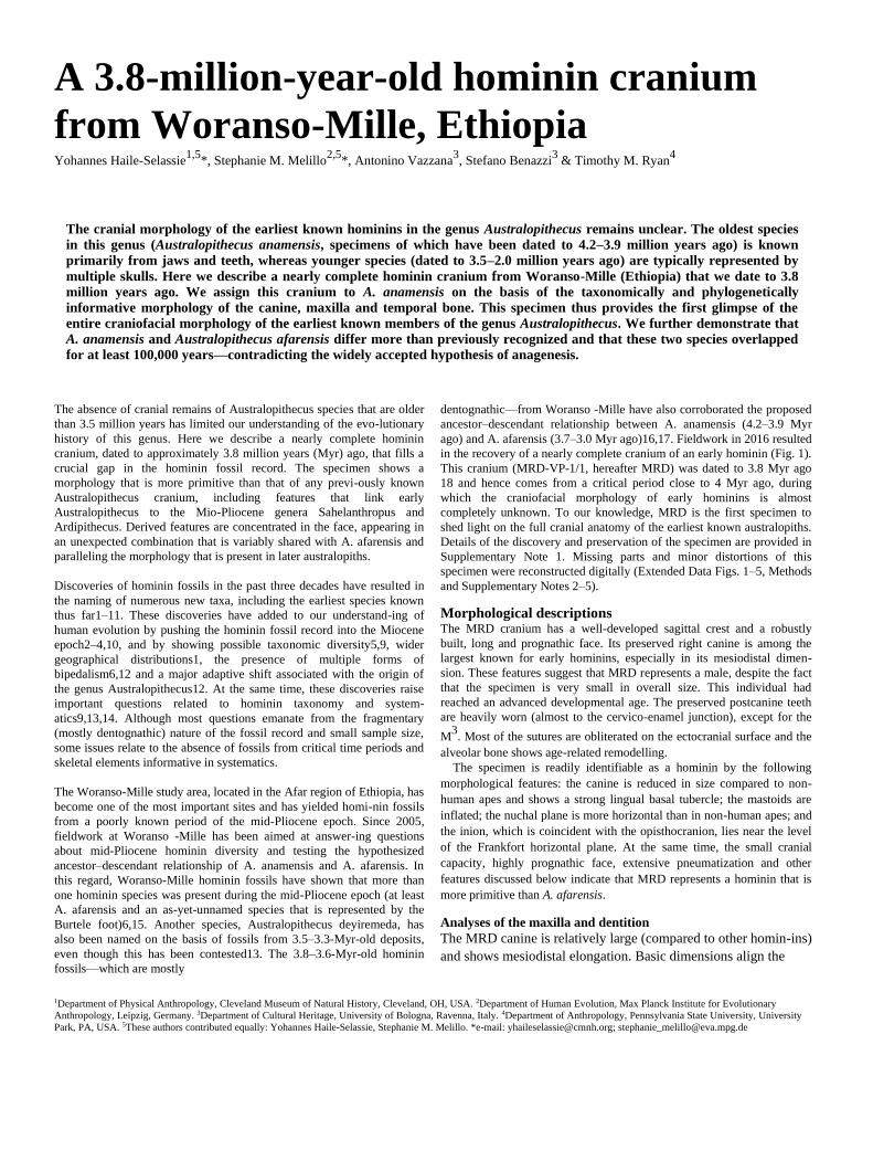
in the recovery of a nearly complete cranium of an early hominin (Fig. 1).
This cranium (MRD-VP-1/1, hereafter MRD) was dated to 3.8 Myr ago
18 and hence comes from a critical period close to 4 Myr ago, during
which the craniofacial morphology of early hominins is almost
completely unknown. To our knowledge, MRD is the first specimen to
shed light on the full cranial anatomy of the earliest known australopiths.
Details of the discovery and preservation of the specimen are provided in
Supplementary Note 1. Missing parts and minor distortions of this
specimen were reconstructed digitally (Extended Data Figs. 1–5, Methods
and Supplementary Notes 2–5). Morphological descriptions The MRD cranium has a well-developed sagittal crest and a robustly
built, long and prognathic face. Its preserved right canine is among the
largest known for early hominins, especially in its mesiodistal dimen-
sion. These features suggest that MRD represents a male, despite the fact
that the specimen is very small in overall size. This individual had
reached an advanced developmental age. The preserved postcanine teeth
are heavily worn (almost to the cervico-enamel junction), except for the
M3. Most of the sutures are obliterated on the ectocranial surface and the
alveolar bone shows age-related remodelling. The specimen is readily identifiable as a hominin by the following
morphological features: the canine is reduced in size compared to non-
human apes and shows a strong lingual basal tubercle; the mastoids are
inflated; the nuchal plane is more horizontal than in non-human apes; and
the inion, which is coincident with the opisthocranion, lies near the level
of the Frankfort horizontal plane. At the same time, the small cranial
capacity, highly prognathic face, extensive pneumatization and other
features discussed below indicate that MRD represents a hominin that is
more primitive than A. afarensis. Analyses of the maxilla and dentition
The MRD canine is relatively large (compared to other homin-ins)
and shows mesiodistal elongation. Basic dimensions align the
1Department of Physical Anthropology, Cleveland Museum of Natural History, Cleveland, OH, USA. 2Department of Human Evolution, Max Planck Institute for Evolutionary
Anthropology, Leipzig, Germany. 3Department of Cultural Heritage, University of Bologna, Ravenna, Italy. 4Department of Anthropology, Pennsylvania State University, University
Park, PA, USA. 5These authors contributed equally: Yohannes Haile-Selassie, Stephanie M. Melillo. *e-mail: [email protected]; [email protected]
a b c
d e f
Fig. 1 | The MRD-VP-1/1 cranium. a, Anterior view. b, Posterior view. c, Superior view. d, Left lateral view. e, Right lateral view. f, Inferior view.
The specimen is oriented in Frankfort horizontal plane. Scale bar, 1 cm.
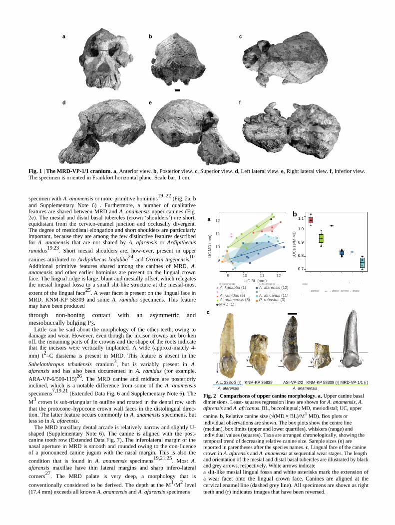
specimen with A. anamensis or more-primitive hominins19–22
(Fig. 2a, b
and Supplementary Note 6) . Furthermore, a number of qualitative features are shared between MRD and A. anamensis upper canines (Fig. 2c). The mesial and distal basal tubercles (crown ‘shoulders’) are short, equidistant from the cervico-enamel junction and occlusally divergent. The degree of mesiodistal elongation and short shoulders are particularly important, because they are among the few distinctive features described for A. anamensis that are not shared by A. afarensis or Ardipithecus
ramidus19,23
. Short mesial shoulders are, how-ever, present in upper
canines attributed to Ardipithecus kadabba24
and Orrorin tugenensis10
.
Additional primitive features shared among the canines of MRD, A. anamensis and other earlier hominins are present on the lingual crown face. The lingual ridge is large, blunt and mesially offset, which relegates the mesial lingual fossa to a small slit-like structure at the mesial-most
extent of the lingual face25
. A wear facet is present on the lingual face in
MRD, KNM-KP 58309 and some A. ramidus specimens. This feature may have been produced
a 12 b
1.1
UC
MD
(m
m)
1U
Csiz
e/M
MD
1.0
11
10
0.9
0.8
9
0.7
9 10 11 12 UC BL (mm)
ramidus O. tugenensis (1) A. deyiremeda (1)
A. kadabba (1) A. afarensis (12)
A. ramidus (5) A. africanus (11) A
.
A
A. anamensis (8) P. robustus (3)
MRD (1)
)
(3)
(1)
(5)
(1)
(4)
(3
a
africanus
anamensis
MRD
afarensis
deyiremed
. A
.
A
.
.
A
through non-honing contact with an asymmetric and
mesiobuccally bulging P3. Little can be said about the morphology of the other teeth, owing to
damage and wear. However, even though the incisor crowns are bro-ken off, the remaining parts of the crowns and the shape of the roots indicate that the incisors were vertically implanted. A wide (approxi-mately 4-
mm) I2–C diastema is present in MRD. This feature is absent in the
Sahelanthropus tchadensis cranium3, but is variably present in A.
afarensis and has also been documented in A. ramidus (for example,
ARA-VP-6/500-115)26
. The MRD canine and midface are posteriorly
inclined, which is a notable difference from some of the A. anamensis
specimens7,19,21
(Extended Data Fig. 6 and Supplementary Note 6). The
M3 crown is sub-triangular in outline and rotated in the dental row such
that the protocone–hypocone crown wall faces in the distolingual direc-tion. The latter feature occurs commonly in A. anamensis specimens, but less so in A. afarensis.
The MRD maxillary dental arcade is relatively narrow and slightly U-shaped (Supplementary Note 6). The canine is aligned with the post-canine tooth row (Extended Data Fig. 7). The inferolateral margin of the nasal aperture in MRD is smooth and rounded owing to the con-fluence of a pronounced canine jugum with the nasal margin. This is also the
condition that is found in A. anamensis specimens19,21,25
. Most A.
afarensis maxillae have thin lateral margins and sharp infero-lateral
corners27
. The MRD palate is very deep, a morphology that is
conventionally considered to be derived. The depth at the M1/M
2 level
(17.4 mm) exceeds all known A. anamensis and A. afarensis specimens
c
* *
ASI-VP-2/2 KNM-KP 58309 (r) MRD-VP-1/1 (r)
A.L. 333x-3 (r) KNM-KP 35839
A. afarensis A. anamensis Fig. 2 | Comparisons of upper canine morphology. a, Upper canine basal
dimensions. Least- squares regression lines are shown for A. anamensis, A.
afarensis and A. africanus. BL, buccolingual; MD, mesiodistal; UC, upper
canine. b, Relative canine size (√(MD × BL)/M1 MD). Box plots or
individual observations are shown. The box plots show the centre line
(median), box limits (upper and lower quartiles), whiskers (range) and
individual values (squares). Taxa are arranged chronologically, showing the temporal trend of decreasing relative canine size. Sample sizes (n) are
reported in parentheses after the species names. c, Lingual face of the canine
crown in A. afarensis and A. anamensis at sequential wear stages. The length
and orientation of the mesial and distal basal tubercles are illustrated by black
and grey arrows, respectively. White arrows indicate
a slit-like mesial lingual fossa and white asterisks mark the extension of
a wear facet onto the lingual crown face. Canines are aligned at the
cervical enamel line (dashed grey line). All specimens are shown as right
teeth and (r) indicates images that have been reversed.

a
b
160
pla
ne
)
150
(orb
ital 140
brea
dth
130
biz
yg
.
120
Re
lative
110
100
(20)
(1)
(1) (1) (2) (2)
(1) (2) (4)
(20)
. tchadensis A A . aethiopicus
. gorilla . ramidus
MRD africanus . robustus P
. boisei
G . afarensis.
. troglodytes
P
S A
P
P
c
bre
adth
(orb
ital pla
ne)
-
alv
eola
r bre
adth
/
biz
yg.
Ma
xill
o
65
60
55
50
45
P. troglodytes (20) G. gorilla (20) MRD (1) S. tchadensis (1) A. ramidus (1) A. afarensis (1) A. africanus (2) P. aethiopicus (1) P. robustus (1) P. boisei (2)
70 80 90 100 110 Sup. facial breadth/bizyg.
breadth (orbital plane)
Fig. 3 | Structure of the face and
neurocranium. a, Anterior view of early
hominin crania. Left to right: S. tchadensis
(TM 266-01-60-1), composite A. ramidus
reconstruction, MRD, A. afarensis (A.L. 444-2), A. africanus (Sts 5). Specimens are oriented in Frankfort horizontal and aligned at orbitale (dashed grey line). b, Bizygomatic (bizyg.) breadth (in the orbital
plane) relative to biorbital breadth. G. gorilla,
Gorilla gorilla; P. aethiopicus, Paranthropus
aethiopicus; P. boisei, Paranthropus boisei; P. troglodytes, Pan troglodytes. c, Bivariate plot
of indices that quantify the ‘facial mask’ shape.
d, Superior view of early hominin crania (in the
same order as in a). Specimens are oriented in
Frankfort horizontal and aligned at glabella
(dashed grey line). e, Index of postorbital
constriction (superior facial breadth/postorbital
constriction). f, Index that quantifies the
braincase shape. Box plot definitions are as in
Fig. 2 and sample sizes are indicated in the
figure panels. Scale bars, 2 cm. Images of A. ramidus and S. tchadensis in a, d were generated, with permission, from CT
scans that have previously been published26,45
. d
e
Po
sto
rbita
l co
nstr
iction
in
de
x
200
180
160
140
120 )
(20) (1) (1)
(1)
)
(3) (1) (1) (4)
trog
(20 (2
A . aethiopicus lody tes
. ramidus . afarensisafricanus. . robustus
. tchadensi MRD
P . boisei
A
P
. gorilla
G
S
A
P
.
P
f
bre
ad
th
200
leng
th/b
ipar
ieta
l
180
Va
ult
160
140
(20) (20)
(1)
(1) (1)
(1)
) )
MRD
(1 (3
. tchadensis
A
.boisei
. gorilla A . aethiopicus
. afarensisafricanus P
. troglodytes S
. P
G
P
and falls among the deepest palates of A. africanus and
Paranthropus robustus (Supplementary Note 7). Analysis of the face Australopiths share a pattern of facial structure that is characterized by a
broad zygomatic region combined with a relatively narrow upper face.
This produces an upward-tapering outline (the ‘hexagonal facial
mask’)27–29
. Although the hexagonal mask is shared among non-robust
and robust australopiths, S. tchadensis, A. ramidus and non-human apes
exhibit relatively broader upper faces and lack pronounced zygo-matic
expansion26,27
. MRD is similar to other australopiths and distinct from
earlier hominins in possessing a broad midface and narrow upper face
(Fig. 3a–c). There are facial similarities that are specifically shared
between MRD and A. afarensis. The external contour of the orbits in
MRD and A. afarensis is squared off superolaterally and the lateral
border of the orbit widens inferiorly. Facial hafting is similar between
MRD and non-robust australopiths: the frontal bone is slightly inclined,
which positions the face below the braincase, and no post-toral sulcus or
frontal trigon is present (Extended Data Fig. 8).
The MRD face is particularly long supero-inferiorly. This morphol-
ogy stands in stark contrast to the short and gracile face reconstructed for A. ramidus . However, comparisons of facial robusticity must consider sexual dimorphism: MRD, A.L. 444-2 and TM 266-01-60-1 probably represent male individuals, whereas the face of the A. rami-
dus composite represents a female26
. Thus some degree of difference
in facial robusticity is expected. The currently available fossil sample does not permit the disentanglement of the morphological differences that are due to sexual dimorphism from those that are taxonomically
diagnostic. Furthermore, it has previously been suggested26
that the
short face of the composite reconstruction of A. ramidus is not repre-sentative of the species. The MRD face is also strongly prognathic, both in the mid-face and subnasally (Extended Data Fig. 8). The projection of the mid-face in MRD is comparable to S. tchadensis, but
MRD lacks upper facial projection3,26
. There are some aspects of the MRD face that are reminiscent of A.
africanus and robust australopiths, and that have traditionally been
considered to be derived. The MRD zygomaticoalveolar crest is nearly
straight and rises steeply from the alveolar margin. As a result, the
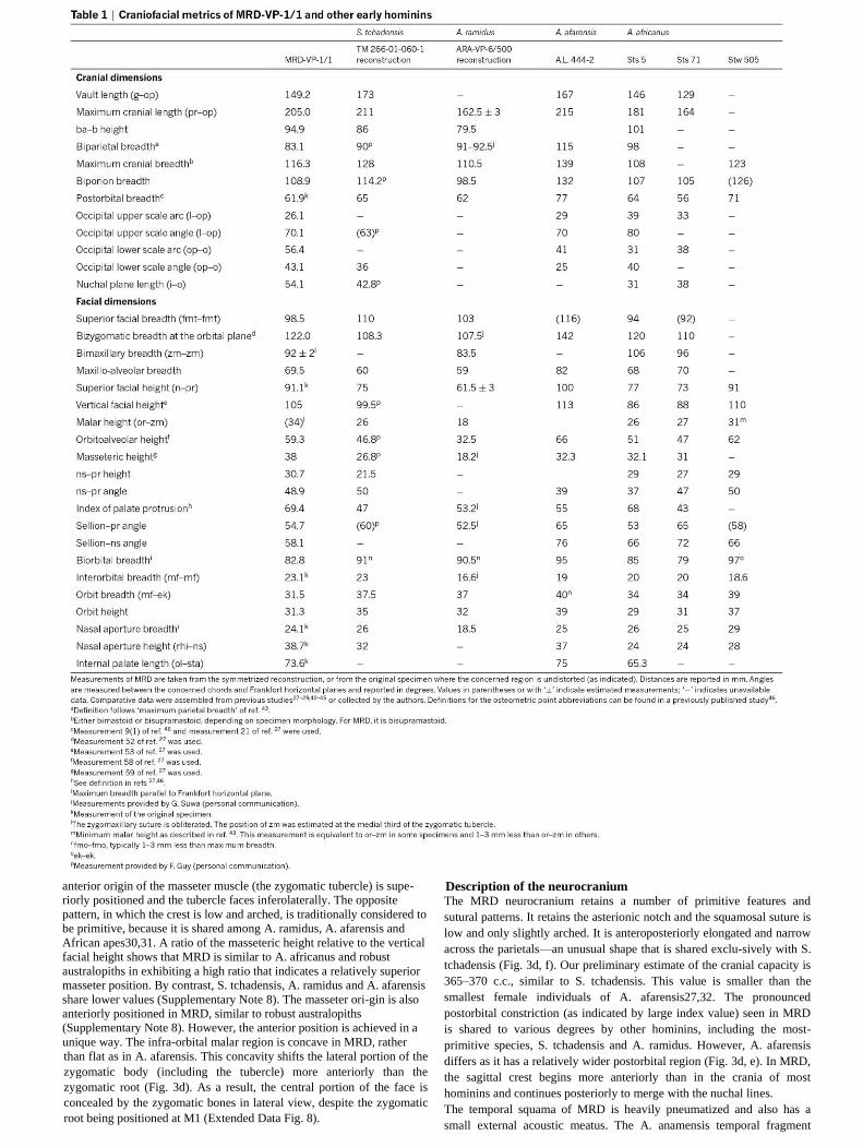
anterior origin of the masseter muscle (the zygomatic tubercle) is supe-
riorly positioned and the tubercle faces inferolaterally. The opposite
pattern, in which the crest is low and arched, is traditionally considered to
be primitive, because it is shared among A. ramidus, A. afarensis and
African apes30,31. A ratio of the masseteric height relative to the vertical
facial height shows that MRD is similar to A. africanus and robust
australopiths in exhibiting a high ratio that indicates a relatively superior
masseter position. By contrast, S. tchadensis, A. ramidus and A. afarensis
share lower values (Supplementary Note 8). The masseter ori-gin is also
anteriorly positioned in MRD, similar to robust australopiths
(Supplementary Note 8). However, the anterior position is achieved in a
unique way. The infra-orbital malar region is concave in MRD, rather
than flat as in A. afarensis. This concavity shifts the lateral portion of the
zygomatic body (including the tubercle) more anteriorly than the
zygomatic root (Fig. 3d). As a result, the central portion of the face is
concealed by the zygomatic bones in lateral view, despite the zygomatic
root being positioned at M1 (Extended Data Fig. 8).
Description of the neurocranium The MRD neurocranium retains a number of primitive features and
sutural patterns. It retains the asterionic notch and the squamosal suture is
low and only slightly arched. It is anteroposteriorly elongated and narrow
across the parietals—an unusual shape that is shared exclu-sively with S.
tchadensis (Fig. 3d, f). Our preliminary estimate of the cranial capacity is
365–370 c.c., similar to S. tchadensis. This value is smaller than the
smallest female individuals of A. afarensis27,32. The pronounced
postorbital constriction (as indicated by large index value) seen in MRD
is shared to various degrees by other hominins, including the most-
primitive species, S. tchadensis and A. ramidus. However, A. afarensis
differs as it has a relatively wider postorbital region (Fig. 3d, e). In MRD,
the sagittal crest begins more anteriorly than in the crania of most
hominins and continues posteriorly to merge with the nuchal lines.
The temporal squama of MRD is heavily pneumatized and also has a
small external acoustic meatus. The A. anamensis temporal fragment
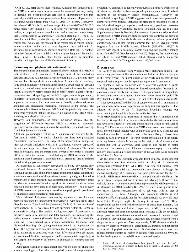
(KNM-KP 29281B) shares these features. Although the dimensions of
the MRD external acoustic meatus cannot be measured precisely owing
to damage, the better-preserved left side is approximately 8.0 mm
vertically and 8.9 mm anteroposteriorly with an estimated ellipse area of
55.9 mm2, which is larger than KNM-KP 29281B7 (40 mm2). However,
the value of MRD falls in the lower range of A. afarensis27 (47.7–109.3
mm2; mean = 79.7 mm2; n = 4). A posterior view reveals a bell-shaped
outline, a compound temporal-nuchal crest and a ‘bare area’ morphology
that is comparable to A. afarensis27 (Extended Data Fig. 9). The MRD
mastoids are inflated, although they are positioned slightly above the
cranial base. As a result, the contour of the cranial base is convex, similar
to the condition in Pan and to some degree to the condition in A.
africanus, but in contrast to A. afarensis (Extended Data Fig. 9). Another
primitive feature of the cranial base is the great length of the nuchal
plane. In MRD, the nuchal plane length—standardized by bimastoid
breadth—is longer than that of TM266-01-60-1 (Table 1).
Taxonomy and phylogenetic relationships The morphological comparisons presented above indicate that MRD is
best attributed to A. anamensis. Although most of the similarities
between MRD and A. anamensis are plesiomorphic, MRD presents many
features that distinguish A. anamensis from A. afarensis: a rel-atively
large and mesiodistally elongated upper canine, a small exter-nal acoustic
meatus, a rounded lateral nasal margin with contribution from the canine
jugum, a relatively narrow palate and an upper canine aligned with the
postcanine row. Morphology of the MRD upper canine is particularly
important in taxonomic attribution, because it displays features that
appear to be apomorphic in A. anamensis (basally posi-tioned crown
shoulders and pronounced mesiodistal elongation of the crown). The
most notable differences between MRD and the previ-ously known A.
anamensis hypodigm include the greater inclination of the MRD canine
and the greater depth of the palate.
However, our comparisons of canine inclination indicate that the
magnitude of dif-ference between MRD and other A. anamensis
specimens is consistent with intraspecific variability (Extended Data Fig.
6 and Supplementary Note 6).
Additional plesiomorphic features of A. anamensis are revealed for the
first time in MRD. The nuchal plane is very long and postorbital
constriction is pronounced. The shape of the neurocranium in superior
view has notable similarities to that of S. tchadensis. However, aspects of
the mid- and upper face show clear affinity to A. afarensis. The facial
mask is hexagonal and the superolateral corner of the orbits is ‘squared
off’ in a distinctive manner. Facial hafting in MRD is similar to the
condition shared between A. afarensis and A. africanus (that is, inclined
frontal lacking a post-toral sulcus).
A. anamensis is consistently recognized as being phylogenetically
positioned between A. ramidus and A. afarensis7,19,20,25,33,34.
Although this idea has both chronological and morphological support, the
ana-tomical composition of the previously known hypodigm is limited to
comparisons of jaws and teeth. Our understanding is thus primarily based
on where A. anamensis fits with regard to documented trends in canine
reduction and the development of masticatory robusticity. The discovery
of MRD presents an opportunity to consider the phylogenetic position of
A. anamensis using craniofacial morphology.
We conducted phylogenetic analyses by augmenting two charac-ter
matrices published by independent observers27,33 with data from MRD
(Supplementary Notes 9 and Supplementary Table 1). In one iteration of
these analyses, MRD was treated as one specimen within the larger A.
anamensis hypodigm. The resulting cladograms depict A. anamensis as
the sister taxon to A. afarensis and later hominins, thus reinforcing the
widely accepted topology (Extended Data Fig. 10a, b). Results are similar
when MRD was treated as a separate operational taxonomic unit
(Extended Data Fig. 10c–g, Supplementary Note 9 and Supplementary
Table 1). Together, these analyses indicate that the phylogenetic position
of A. anamensis is consistent, even when differ-ent anatomical regions
are considered (that is, dentognathic and/or craniofacial morphologies)
and despite inter-observer differences in character list composition and
scoring. Although the addition of craniofacial observations does not change the
accepted topology, it does alter the implied pattern of morphologi-cal
evolution. A. anamensis is generally portrayed as a primitive exten-sion of
A. afarensis; this idea has been supported by the apparent lack of derived
features in the previously known hypodigm19,35. However, the
craniofacial morphology of MRD suggests that A. anamensis possessed a
number of derived features, including the presence of topographic relief in
the infraorbital region, a superiorly and anteriorly positioned masseter
origin, and potentially additional features (Extended Data Fig. 10h, i and
Supplementary Note 9). Notably, the presence of pro-nounced postorbital
constriction in MRD and more primitive homi-nins confirms the previous
suggestion that A. afarensis is derived in showing reduced postorbital
constriction27,35. It also further confirms that the 3.9-Myr-old frontal
fragment from the Middle Awash, Ethiopia (BEL-VP-1/1)36,37, is
derived with regard to postorbital constriction and thus probably belongs
to A. afarensis27,38 (Supplementary Note 10). Together, the secure dating
of BEL-VP-1/1 and MRD indicate that A. afarensis and A. anamensis
overlapped in the Afar Triangle for at least 100,000 years.
Discussion The 3.8-Myr-old cranium from Woranso-Mille elaborates some of the
outstanding questions in Pliocene hominin evolution and fills a major gap
in the fossil record. The morphologies of the MRD canine, maxilla and
temporal region suggest that this specimen represents A. anamensis.
The hypothesis that A. anamensis and A. afarensis constitute a single
evolving chronospecies was based on limited apomorphic features in A.
anamensis, but is mostly due to perceived temporal trends in morphology
in four time-successive samples of the two species from Kanapoi, Allia
Bay, Laetoli and Hadar35. However, the lack of fossil hominins from 3.9–
3.7 Myr ago in general and the lack of complete crania of A. anamensis in
particular have been major impediments to fully test this hypothesis. The
addition of MRD to the A. anamensis hypodigm changes our
understanding of the relationship between the two taxa.
With MRD assigned to A. anamensis, it indicates that A. anamensis can
be clearly distinguished from A. afarensis such that the latter species may
not have been a result of ‘phyletic transformation within an unbranched
lineage’35. Furthermore, the fact that MRD shares some neurocranial and
facial morphological features with younger taxa such as A. africanus and
Paranthropus—albeit considered here to be more likely to have been
caused by parallel evolution—is worth further investigation in the future,
as it may have considerable bearing on the origin of A. africanus and its
relationship with A. afarensis. More work is also needed to better
understand the geology and Pliocene palaeo-geography of the Afar
region18 and establish a refined taxonomy of the Woranso-Mille hominins
from 3.8 to 3.4 Myr ago.
On the basis of the currently available fossil evidence, it appears that
there were at least four time-successive but allopatric A. anamensis
populations (Woranso-Mille, Allia Bay, Asa Issie and Kanapoi) that
showed variable cranial and dentognathic morphology. Although the
cranial morphology of A. anamensis was poorly known thus far, the 3.8-
Myr-old MRD from Woranso-Mille is morphologically similar to the
species from Kanapoi and Asa Issie (4.2–4.1 Myr ago). However, it is
unlikely that an A. anamensis population represented by MRD gave rise to
A. afarensis, as MRD postdates BEL-VP-1/1, which now appears to be
the earliest known representative of A. afarensis with an age of
approximately 3.9 Myr. Although their taxonomic affinity is still
controversial39, it is possible that the few approximately 4.0-Myr-old teeth
from Fejej, Ethiopia, might also belong to A. afarensis40,41. These
observations can be tested with the recovery of crania from Kanapoi, Asa
Issie and Allia Bay and further comparisons to MRD. In sum-mary,
although MRD and other discoveries from Woranso-Mille do not falsify
the proposed ancestor–descendant relationship between A. anamensis and
A. afarensis, they indicate that A. afarensis may not have evolved from a
single ancestral population. Most importantly, MRD shows that despite
the widely accepted hypothesis of anagenesis, A. afarensis did not appear
as a result of phyletic transformation. It also shows that at least two
related hominin species co-existed in eastern Africa around 3.8 Myr ago,
further lending support to mid-Pliocene hominin diversity.
1. Brunet, M. et al. Australopithecus bahrelghazali, une nouvelle espèce
d’Hominidé ancien de la région de Koro Toro (Tchad). C. R. Acad. Sci. IIA
322, 907–913 (1996).
2. Brunet, M. et al. New material of the earliest hominid from the Upper Miocene of Chad. Nature 434, 752–755 (2005).
3. Brunet, M. et al. A new hominid from the Upper Miocene of Chad, Central
Africa. Nature 418, 145–151 (2002).
4. Haile-Selassie, Y. Late Miocene hominids from the Middle Awash, Ethiopia.
Nature 412, 178–181 (2001).
5. Haile-Selassie, Y. et al. New species from Ethiopia further expands Middle
Pliocene hominin diversity. Nature 521, 483–488 (2015).
6. Haile-Selassie, Y. et al. A new hominin foot from Ethiopia shows multiple Pliocene bipedal adaptations. Nature 483, 565–569 (2012).
7. Leakey, M. G., Feibel, C. S., McDougall, I. & Walker, A. New four-million-
year-old hominid species from Kanapoi and Allia Bay, Kenya. Nature 376,
565–571 (1995).
8. Leakey, M. G., Feibel, C. S., McDougall, I., Ward, C. & Walker, A. New
specimens and confirmation of an early age for Australopithecus anamensis.
Nature 393, 62–66 (1998).
9. Leakey, M. G. et al. New hominin genus from eastern Africa shows diverse middle Pliocene lineages. Nature 410, 433–440 (2001).
10. Senut, B. et al. First hominid from the Miocene (Lukeino formation, Kenya).
C. R. Acad. Sci. IIA 332, 137–144 (2001).
11. White, T. D., Suwa, G. & Asfaw, B. Australopithecus ramidus, a new species
of early hominid from Aramis, Ethiopia. Nature 371, 306–312 (1994).
12. White, T. D. et al. Ardipithecus ramidus and the paleobiology of early
hominids. Science 326, 64–86 (2009). 13. Wood, B. & K Boyle, E. Hominin taxic diversity: fact or fantasy? Am. J.
Phys. Anthropol. 159, 37–78 (2016).
14. Haile-Selassie, Y., Melillo, S. M. & Su, D. F. The Pliocene hominin diversity
conundrum: do more fossils mean less clarity? Proc. Natl Acad. Sci. USA
113, 6364–6371 (2016).
15. Haile-Selassie, Y. et al. Dentognathic remains of Australopithecus afarensis
from Nefuraytu (Woranso-Mille, Ethiopia): comparative description,
geology, and paleoecological context. J. Hum. Evol. 100, 35–53 (2016). 16. Haile-Selassie, Y. Phylogeny of early Australopithecus: new fossil evidence
from the Woranso-Mille (central Afar, Ethiopia). Phil. Trans. R. Soc. Lond. B
365, 3323–3331 (2010).
17. Haile-Selassie, Y., Saylor, B. Z., Deino, A., Alene, M. & Latimer, B. M. New
hominid fossils from Woranso-Mille (Central Afar, Ethiopia) and taxonomy
of early Australopithecus. Am. J. Phys. Anthropol. 141, 406–417 (2010).
18. Saylor, B. Z. et al. Age and context of new mid-Pliocene hominin cranium
from Woranso-Mille, Ethiopia. Nature https://doi.org/10.1038/s41586-019-1514-7 (2019).
19. Ward, C. V., Leakey, M. G. & Walker, A. Morphology of Australopithecus
anamensis from Kanapoi and Allia Bay, Kenya. J. Hum. Evol. 41, 255–368
(2001).
20. Ward, C. V., Manthi, F. K. & Plavcan, J. M. New fossils of Australopithecus
anamensis from Kanapoi, West Turkana, Kenya (2003–2008). J. Hum. Evol.
65, 501–524 (2013). 21. Ward, C. V., Plavcan, J. M. & Manthi, F. K. New fossils of Australopithecus
anamensis from Kanapoi, West Turkana, Kenya (2012–2015). J. Hum. Evol.
https://doi.org/10.1016/j.jhevol.2017.07.008 (2017).
22. Manthi, F. K., Plavcan, J. M. & Ward, C. V. New hominin fossils from
Kanapoi, Kenya, and the mosaic evolution of canine teeth in early hominins.
S. Afr. J. Sci. 108, 724 (2012).
23. Suwa, G. et al. Paleobiological implications of the Ardipithecus ramidus
dentition. Science 326, 69–99 (2009). 24. Haile-Selassie, Y., Suwa, G. & White, T. D. Late Miocene teeth from Middle
Awash, Ethiopia, and early hominid dental evolution. Science 303, 1503–
1505 (2004).
25. White, T. D. et al. Asa Issie, Aramis and the origin of Australopithecus.
Nature 440, 883–889 (2006).
26. Suwa, G. et al. The Ardipithecus ramidus skull and its implications for
hominid origins. Science 326, 68–68e7 (2009). 27. Kimbel, W. H., Rak, Y. & Johanson, D. C. The Skull of Australopithecus
afarensis (Oxford Univ. Press, 2004).
28. Guy, F. et al. Morphological affinities of the Sahelanthropus tchadensis (Late
Miocene hominid from Chad) cranium. Proc. Natl Acad. Sci. USA 102,
18836–18841 (2005).
29. Rak, Y. The Australopithecine Face (Academic, 1983).
30. Kimbel, W. H. & Rak, Y. Australopithecus sediba and the emergence of
Homo: Questionable evidence from the cranium of the juvenile holotype MH 1. J. Hum. Evol. 107, 94–106 (2017).
31. Kimbel, W. H., White, T. D. & Johanson, D. C. Cranial morphology of
Australopithecus afarensis: a comparative study based on a composite
reconstruction of the adult skull. Am. J. Phys. Anthropol. 64, 337–388
(1984).
32. Kimbel, W. H. & Rak, Y. The cranial base of Australopithecus afarensis: new
insights from the female skull. Phil. Trans. R. Soc. Lond. B 365, 3365–3376
(2010). 33. Strait, D. S. & Grine, F. E. Inferring hominoid and early hominid phylogeny
using craniodental characters: the role of fossil taxa. J. Hum. Evol. 47, 399–
452 (2004).
34. Dembo, M., Matzke, N. J., Mooers, A. Ø. & Collard, M. Bayesian analysis of
a morphological supermatrix sheds light on controversial fossil hominin
relationships. Proc. R. Soc. Lond. B 282, 20150943 (2015).
35. Kimbel, W. H. et al. Was Australopithecus anamensis ancestral to A. afarensis? A case of anagenesis in the hominin fossil record. J. Hum. Evol.
51, 134–152 (2006).
36. Asfaw, B. The Belohdelie frontal: new evidence of early hominid cranial
morphology from the Afar of Ethiopia. J. Hum. Evol. 16, 611–624 (1987).
37. Renne, P. R. et al. Chronostratigraphy of Mio-Pliocene Sagantole Formation, Middle Awash Valley, Afar rift, Ethiopia. Bull. Geol. Soc. Am. 111, 869–885
(1999).
38. Kimbel, W. H., Johanson, D. C. & Rak, Y. The first skull and other new
discoveries of Australopithecus afarensis at Hadar, Ethiopia. Nature 368, 449–
451 (1994).
39. Ward, C. V. Taxonomic affinity of the Pliocene hominin fossils from Fejej,
Ethiopia. J. Hum. Evol. 73, 98–102 (2014).
40. Fleagle, J. G., Rasmussen, D. T., Yirga, S., Bown, T. M. & Grine, F. E. New hominid fossils from Fejej, Southern Ethiopia. J. Hum. Evol. 21, 145–152
(1991).
41. Kappelman, J. et al. Age of Australopithecus afarensis from Fejej, Ethiopia.J.
Hum. Evol. 30, 139–146 (1996).
42. Kimbel, W. H., Johanson, D. C. & Coppens, Y. Pliocene hominid cranial
remains from the Hadar Formation, Ethiopia. Am. J. Phys. Anthropol. 57,
453–499 (1982).
43. Wood, B. Koobi Fora Research Project: Hominid Cranial Remains Vol. 4
(Clarendon, 1991).
44. Lockwood, C. A. & Tobias, P. V. A large male hominin cranium from
Sterkfontein, South Africa, and the status of Australopithecus africanus. J.
Hum. Evol. 36, 637–685 (1999).
45. Zollikofer, C. P. et al. Virtual cranial reconstruction of Sahelanthropus
tchadensis. Nature 434, 755–759 (2005).
46. Martin, R. & Knussman, R. Anthropologie: Handbuch der vergleichenden Biologie des Menschen Vol. 1 (Gustav Fischer, 1988).
Acknowledgements We thank the Authority for Research and Conservation of
Cultural Heritage (ARCCH) for permission to conduct field and laboratory workthe
Afar people of Woranso-Mille and the Mille District administration for their
hospitality; the project’s fieldwork crew members for their tireless support of field activities; The National Museum of Kenya, National Museum of Tanzania, Ditsong
Museum and the Evolutionary Studies Institute of South Africa for access to
original hominin specimens in their care; Max Planck Institute for Evolutionary
Anthropology for access to comparative hominin computed tomography scan data;
T. White, G. Suwa and B. Asfaw for access to the original A. ramidus material and
for providing images and unpublished measurements; M. Brunet and F. Guy for
images and unpublished measurements of S. tchadensis; W. H. Kimbel for informative discussions and for access to a surface model of the A.L. 444-2
cranium; D. Lieberman (the Peabody Museum (Harvard)), R. Beutel (Phyletisches
Museum Jena), C. Funk (Museum für Naturkunde), M. Tocheri (National Museum
of Natural History (Smithsonian)), U. Olbrich-Schwarz (the Max Planck Institute
for Evolutionary Anthropology) for the use of computed tomography scans of
extant apes; A. Girmaye, M. Endalamaw, Y. Assefa, T. Getachew, S. Melaku and
G. Tekle of ARCCH for access to the fossil collections housed in the
Paleoanthropology Laboratory in Addis Ababa; T. Stecko from the Penn State Center for Quantitative Imaging for assistance with computed tomography
scanning; and N. Meisel and D. N. Kaweesa (Made By Design Lab (Pennsylvania
State University)) for assistance in 3D printing. This research was supported by
grants from the US National Science Foundation (BCS-1124705, BCS-1124713,
BCS-1124716, BCS-1125157 and BCS-1125345) and The Cleveland Museum of
Natural History. Y.H.-S. was also supported by W. J. and L. Hlavin, T. and K.
Leiden, and E. Lincoln. S.B. was supported by the European Research Council
(ERC) under the European Union’s Horizon 2020 research and innovation programme (ERC-724046-SUCCESS; http://www.erc-success.eu). S.M.M. was
supported by the Max Planck Institute for Evolutionary Anthropology, Department
of Human Evolution.
Author contributions Y.H.-S. and S.M.M. conducted fieldwork and collected
data. T.M.R. scanned the specimen using computed tomography. T.M.R., S.B.
and A.V. performed the three-dimensional reconstruction with contributions
from Y.H.-S. and S.M.M. Y.H.-S. and S.M.M. performed the comparative analysis. S.M.M. and Y.H.-S. wrote the paper with contributions from T.M.R., S.B. and A.V
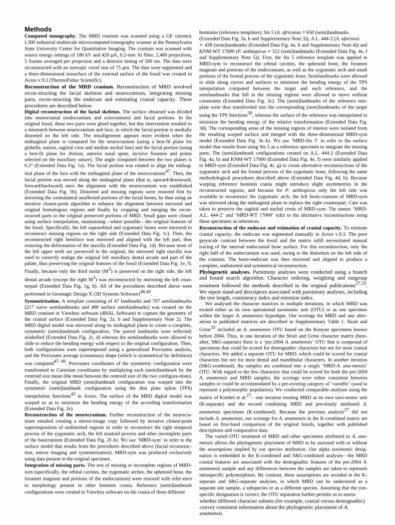
Methods Computed tomography. The MRD cranium was scanned using a GE v|tome|x
L300 industrial multiscale microcomputed tomography scanner at the Pennsylvania
State University Center for Quantitative Imaging. The cranium was scanned with
source energy settings of 180 kV and 420 μA, 0.2-mm Al filter, 2,400 projections,
5 frames averaged per projection and a detector timing of 500 ms. The data were
reconstructed with an isotropic voxel size of 75 μm. The data were segmented and
a three-dimensional isosurface of the external surface of the fossil was created in
Avizo v.9.3 (ThermoFisher Scientific). Reconstruction of the MRD cranium. Reconstruction of MRD involved
recon-structing the facial skeleton and neurocranium, integrating missing
parts, recon-structing the endocast and estimating cranial capacity. These
procedures are described below. Digital reconstruction of the facial skeleton. The surface obtained was divided
into neurocranial (endocranium and ectocranium) and facial portions. In the
original fossil, these two parts were glued together, but this intervention resulted in
a mismatch between neurocranium and face, in which the facial portion is medially
distorted on the left side. The misalignment appears more evident when the
midsagittal plane is computed for the neurocranium (using a best-fit plane for
glabella, nasion, sagittal crest and median nuchal line) and the facial portion (using
a best-fit plane for rhinion, anterior nasal spine, incisive foramen and points
selected on the maxillary suture). The angle computed between the two planes is
6.2° (Extended Data Fig. 1a). The facial portion was rotated to align the midsag-
ittal plane of the face with the midsagittal plane of the neurocranium47
. Then, the
facial portion was moved along the midsagittal plane (that is, upward/downward,
forward/backward) once the alignment with the neurocranium was established
(Extended Data Fig. 1b). Distorted and missing regions were restored first by
mirroring the contralateral unaffected portions of the facial bones, by then using an
iterative closest-point algorithm to enhance the alignment between mirrored and
original homologous regions and finally by cropping and merging the recon-
structed parts to the original preserved portions of MRD. Small gaps were closed
using surface interpolation, maintaining—where possible—the original features of
the fossil. Specifically, the left supraorbital and zygomatic bones were mirrored to
reconstruct missing regions on the right side (Extended Data Fig. 1c). Then, the
reconstructed right hemiface was mirrored and aligned with the left part, thus
restoring the deformation of the maxilla (Extended Data Fig. 1d). Because most of
the left upper teeth are preserved in the original, the mirrored right maxilla was
used to correctly realign the original left maxillary dental arcade and part of the
palate, thus preserving the original features of the fossil (Extended Data Fig. 1e, f).
Finally, because only the third molar (M3) is preserved on the right side, the left
dental arcade (except the right M3) was reconstructed by mirroring the left coun-
terpart (Extended Data Fig. 1g, h). All of the procedures described above were
performed in Geomagic Design X (3D Systems Software)48,49
. Symmetrization. A template consisting of 47 landmarks and 707 semilandmarks
(217 curve semilandmarks and 490 surface semilandmarks) was created on the
MRD cranium in Viewbox software (dHAL Software) to capture the geometry of
the cranial surface (Extended Data Fig. 2a, b and Supplementary Note 2). The
MRD digital model was mirrored along its midsagittal plane to create a complete,
symmetric (semi)landmark configuration. The paired landmarks were reflected/
relabelled (Extended Data Fig. 2c, d) whereas the semilandmarks were allowed to
slide to reduce the bending energy with respect to the original configuration. Then,
both configurations were superimposed using a generalized Procrustes analysis,
and the Procrustes average (consensus) shape (which is symmetrical by definition)
was computed47–49
. Procrustes coordinates of the symmetric configuration were
transformed to Cartesian coordinates by multiplying each (semi)landmark by the
centroid size mean (the mean between the centroid size of the two configura-tions).
Finally, the original MRD (semi)landmark configuration was warped into the
symmetric (semi)landmark configuration using the thin plate spline (TPS)
interpolation functions42
in Avizo. The surface of the MRD digital model was
warped so as to minimize the bending energy of the according transformation
(Extended Data Fig. 2e). Reconstruction of the neurocranium. Further reconstruction of the neurocra-
nium entailed creating a mirror-image copy followed by iterative closest-point
superimposition of undistorted regions in order to reconstruct the right temporal
process of the zygomatic arch, the left mastoid process and other incomplete parts
of the basicranium (Extended Data Fig. 2f–h). We use ‘MRD-sym’ to refer to the
surface model that results from the procedures described above (facial reconstruc-
tion, mirror imaging and symmetrization). MRD-sym was produced exclusively
using data present in the original specimen. Integration of missing parts. The rest of missing or incomplete regions of MRD-
sym (specifically, the orbital cavities, the zygomatic arches, the sphenoid bone, the
foramen magnum and portions of the endocranium) were restored with refer-ence
to morphology present in other hominin crania. Reference (semi)landmark
configurations were created in Viewbox software on the crania of three different
hominins (reference templates): Sts 5 (A. africanus = 650 (semi)landmarks
(Extended Data Fig. 3a, b and Supplementary Note 3)), A.L. 444-2 (A. afarensis
= 438 (semi)landmarks (Extended Data Fig. 4a, b and Supplementary Note 4)) and
KNM-WT 17000 (P. aethiopicus = 312 (semi)landmarks (Extended Data Fig. 4e, f
and Supplementary Note 5)). First, the Sts 5 reference template was applied to
MRD-sym to reconstruct the orbital cavities, the sphenoid bone, the foramen
magnum and portions of the endocranium, as well as the zygomatic arch and small
portions of the frontal process of the zygomatic bone. Semilandmarks were allowed
to slide along curves and surfaces to minimize the bending energy of the TPS
interpolation computed between the target and each reference, and the
semilandmarks that fell in the missing regions were allowed to move without
constraints (Extended Data Fig. 3c). The (semi)landmarks of the reference tem-
plate were thus transformed into the corresponding (semi)landmarks of the target
using the TPS functions50
, whereas the surface of the reference was interpolated to
minimize the bending energy of the relative transformation (Extended Data Fig.
3d). The corresponding areas of the missing regions of interest were isolated from
the resulting warped surface and merged with the three-dimensional MRD-sym
model (Extended Data Fig. 3e–h). We use ‘MRD-Sts 5’ to refer to the surface
model that results from using Sts 5 as a reference specimen to integrate the missing
parts. The (semi)landmark configurations created on A.L. 444-2 (Extended Data
Fig. 4a, b) and KNM-WT 17000 (Extended Data Fig. 4e, f) were similarly applied
to MRD-sym (Extended Data Fig. 4c, g) to create alternative reconstructions of the
zygomatic arch and the frontal process of the zygomatic bone, following the same
methodological procedures described above (Extended Data Fig. 4d, h). Because
warping reference hominin crania might introduce slight asymmetries in the
reconstructed regions, and because for P. aethiopicus only the left side was
available to reconstruct the zygomatic arch, the left hemi-cranium of MRD-sym
was mirrored along the midsagittal plane to replace the right counterpart. Care was
taken to preserve the sagittal and nuchal crests of MRD-sym. The names ‘MRD-
A.L. 444-2’ and ‘MRD-WT 17000’ refer to the alternative reconstructions using
these specimens as references. Reconstruction of the endocast and estimation of cranial capacity. To estimate
cranial capacity, the endocast was segmented manually in Avizo v.9.3. The poor
greyscale contrast between the fossil and the matrix infill necessitated manual
tracing of the internal endocranial bone surface. For this reconstruction, only the
right half of the endocranium was used, owing to the distortion on the left side of
the cranium. The hemi-endocast was then mirrored and aligned to produce a
complete, undistorted and symmetrical reconstruction. Phylogenetic analyses. Parsimony analyses were conducted using a branch and bound search algorithm. Character ordering, weighting and outgroup
treatment followed the methods described in the original publications27,33
.
We report stand-ard descriptors associated with parsimony analyses, including the tree length, consistency index and retention index.
We analysed the character matrices in multiple iterations, in which MRD was
treated either as its own operational taxonomic unit (OTU) or as one specimen
within the larger A. anamensis hypodigm. Our scorings for MRD and any alter-
ations to published matrices are described in Supplementary Table 1. Strait and
Grine33
included an A. anamensis OTU based on the Kenyan specimens known
before 2004. Thus, in one iteration of the Strait and Grine character matrix (here-
after, S&G-separate) there is a ‘pre-2004 A. anamensis’ OTU that is composed of
specimens that could be scored for dentognathic characters but not for most cranial
characters. We added a separate OTU for MRD, which could be scored for cranial
characters but not for most dental and mandibular characters. In another iteration
(S&G-combined), the samples are combined into a single ‘MRD-A. ana-mensis’
OTU. With regard to the few characters that could be scored for both the pre-2004
A. anamensis and MRD samples, the scorings were either consistent between
samples or could be accommodated by a pre-existing category of ‘variable’ (used to
represent a polymorphic population). We conducted comparable analyses using the
matrix of Kimbel et al.27
—one iteration treating MRD as its own taxo-nomic unit
(K-separate) and the second combining MRD and previously attributed A.
anamensis specimens (K-combined). Because the previous analysis27
did not
include A. anamensis, our scorings for A. anamensis in the K-combined matrix are
based on first-hand comparison of the original fossils, together with published
descriptions and comparative data. The varied OTU treatment of MRD and other specimens attributed to A. ana-
mensis allows the phylogenetic placement of MRD to be assessed with or without
the assumptions implied by our species attribution. Our alpha taxonomic desig-
nation is embedded in the K-combined and S&G-combined analyses—the MRD
cranial features are associated with the dentognathic features of the pre-2004 A.
anamensis sample and any differences between the samples are taken to represent
intraspecific polymorphism. By contrast, these assumptions are avoided in the K-
separate and S&G-separate analyses, in which MRD can be understood as a
separate site sample, a subspecies or as a different species. Assuming that the con-
specific designation is correct, the OTU separation further permits us to assess
whether different character subsets (for example, cranial versus dentognathic) convey consistent information about the phylogenetic placement of A.
anamensis.
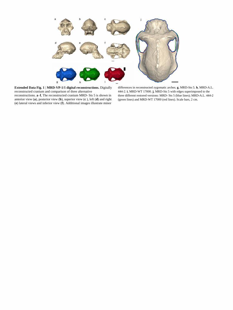
Extended Data Fig. 1 | MRD-VP-1/1 digital reconstructions. Digitally
reconstructed cranium and comparison of three alternative
reconstructions. a–f, The reconstructed cranium MRD- Sts 5 is shown in
anterior view (a), posterior view (b), superior view (c ), left (d) and right
(e) lateral views and inferior view (f). Additional images illustrate minor
differences in reconstructed zygomatic arches. g, MRD-Sts 5. h, MRD-A.L.
444-2. i, MRD-WT 17000. j, MRD-Sts 5 with edges superimposed to the
three different restored versions: MRD- Sts 5 (blue lines); MRD-A.L. 444-2
(green lines) and MRD-WT 17000 (red lines). Scale bars, 2 cm.
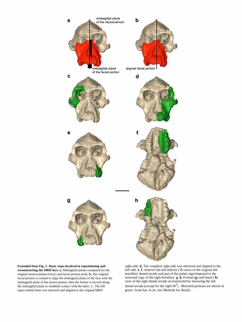
Extended Data Fig. 2 | Basic steps involved in repositioning and
reconstructing the MRD face. a, Midsagittal planes computed for the
original neurocranium (ivory) and facial portion (red). b, The original
facial portion is rotated to align the midsagittal plane of the face with the
midsagittal plane of the neurocranium, then the former is moved along
the midsagittal plane to establish contact with the latter. c , The left
supra-orbital bone was mirrored and aligned to the original MRD
right side. d, The complete right side was mirrored and aligned to the left side. e, f, Anterior (e) and inferior ( f) views of the original left maxillary dental arcade and part of the palate superimposed to the mirrored copy of the right hemiface. g, h, Frontal (g) and basal ( h) view of the right dental arcade reconstructed by mirroring the left
dental arcade (except for the right M3) . Mirrored portions are shown in
green. Scale bar, 4 cm. See Methods for details.
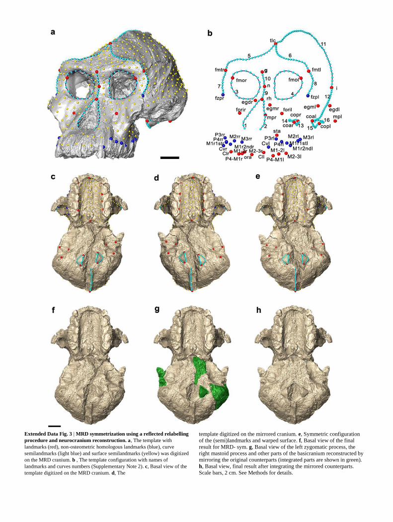
Extended Data Fig. 3 | MRD symmetrization using a reflected relabelling
procedure and neurocranium reconstruction. a, The template with
landmarks (red), non-osteometric homologous landmarks (blue), curve
semilandmarks (light blue) and surface semilandmarks (yellow) was digitized
on the MRD cranium. b , The template configuration with names of
landmarks and curves numbers (Supplementary Note 2). c, Basal view of the
template digitized on the MRD cranium. d, The
template digitized on the mirrored cranium. e, Symmetric configuration
of the (semi)landmarks and warped surface. f, Basal view of the final
result for MRD- sym. g, Basal view of the left zygomatic process, the
right mastoid process and other parts of the basicranium reconstructed by
mirroring the original counterparts (integrated parts are shown in green).
h, Basal view, final result after integrating the mirrored counterparts.
Scale bars, 2 cm. See Methods for details.
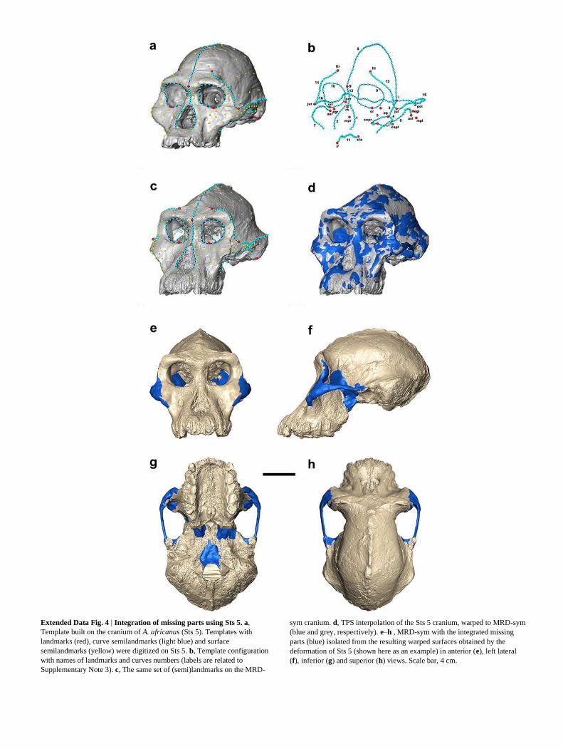
Extended Data Fig. 4 | Integration of missing parts using Sts 5. a,
Template built on the cranium of A. africanus (Sts 5). Templates with
landmarks (red), curve semilandmarks (light blue) and surface
semilandmarks (yellow) were digitized on Sts 5. b, Template configuration
with names of landmarks and curves numbers (labels are related to
Supplementary Note 3). c, The same set of (semi)landmarks on the MRD-
sym cranium. d, TPS interpolation of the Sts 5 cranium, warped to MRD-sym
(blue and grey, respectively). e–h , MRD-sym with the integrated missing
parts (blue) isolated from the resulting warped surfaces obtained by the
deformation of Sts 5 (shown here as an example) in anterior (e), left lateral
(f), inferior (g) and superior (h) views. Scale bar, 4 cm.

Extended Data Fig. 5 | Integration of missing parts using A.L. 444-2 and
KNM-WT 17000. a, e, Templates with landmarks (red), curve
semilandmarks (light blue) and surface semilandmarks (yellow) digitized on
A.L. 444-2 (a) and KNM-WT 17000 (e) crania. b, f, The configurations of
(semi)landmarks with names of landmarks and curves numbers
digitized on A.L. 444-2 (b ) and KNM -WT 17000 (f) crania (labels are
related to Supplementary Notes 4, 5). c, g, Sets of (semi)landmarks on the
MRD-sym cranium. d, h, TPS interpolation of the A.L. 444 -2 (green) and
KMN-WT 17000 (red) crania warped to MRD-sym (grey). Scale bar, 4 cm.
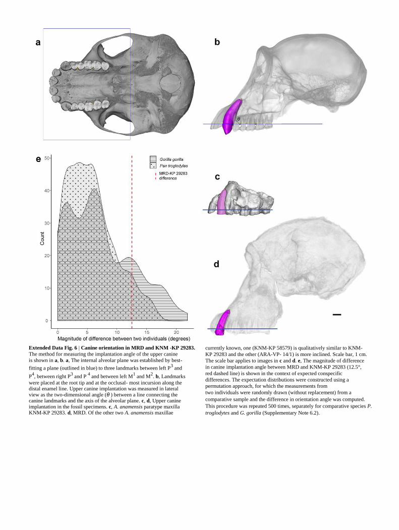
Extended Data Fig. 6 | Canine orientation in MRD and KNM -KP 29283.
The method for measuring the implantation angle of the upper canine is shown in a, b. a, The internal alveolar plane was established by best-
fitting a plane (outlined in blue) to three landmarks between left P3 and
P4, between right P
3 and P
4 and between left M
1 and M
2. b, Landmarks
were placed at the root tip and at the occlusal- most incursion along the distal enamel line. Upper canine implantation was measured in lateral view as the two-dimensional angle (θ ) between a line connecting the canine landmarks and the axis of the alveolar plane. c, d, Upper canine implantation in the fossil specimens. c, A. anamensis paratype maxilla KNM-KP 29283. d, MRD. Of the other two A. anamensis maxillae
currently known, one (KNM-KP 58579) is qualitatively similar to KNM-
KP 29283 and the other (ARA-VP- 14/1) is more inclined. Scale bar, 1 cm.
The scale bar applies to images in c and d. e, The magnitude of difference
in canine implantation angle between MRD and KNM-KP 29283 (12.5°,
red dashed line) is shown in the context of expected conspecific
differences. The expectation distributions were constructed using a
permutation approach, for which the measurements from two individuals were randomly drawn (without replacement) from a
comparative sample and the difference in orientation angle was computed.
This procedure was repeated 500 times, separately for comparative species P.
troglodytes and G. gorilla (Supplementary Note 6.2).
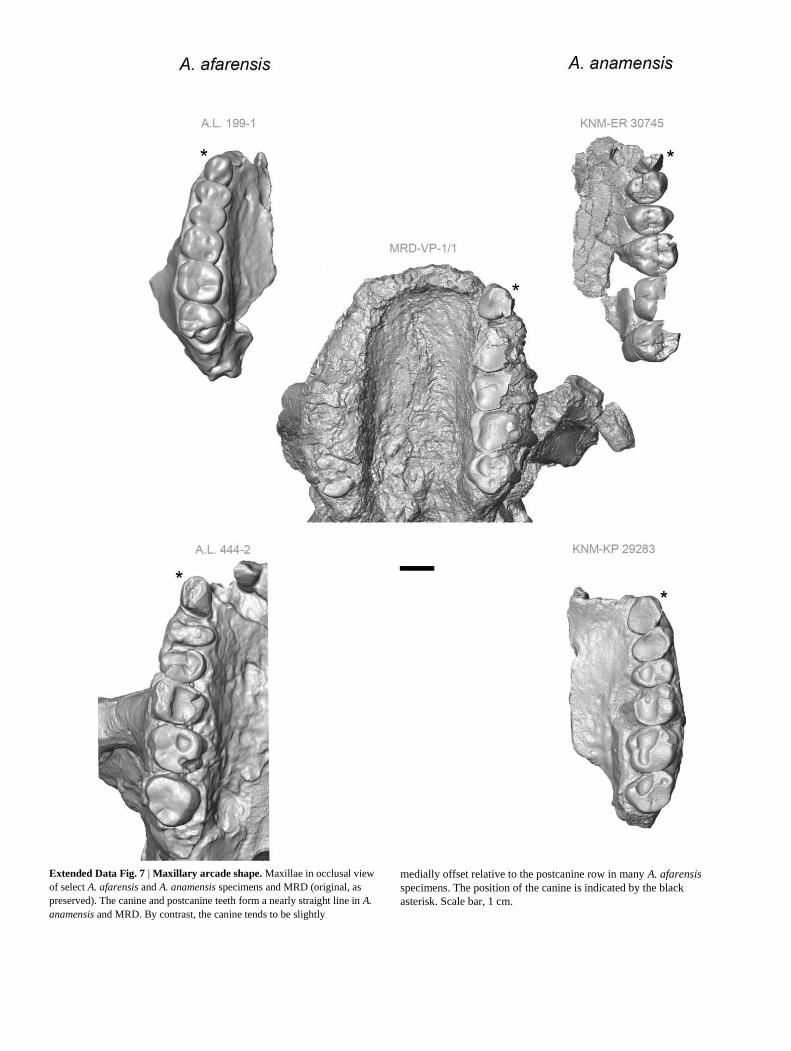
Extended Data Fig. 7 | Maxillary arcade shape. Maxillae in occlusal view
of select A. afarensis and A. anamensis specimens and MRD (original, as
preserved). The canine and postcanine teeth form a nearly straight line in A.
anamensis and MRD. By contrast, the canine tends to be slightly
medially offset relative to the postcanine row in many A. afarensis
specimens. The position of the canine is indicated by the black
asterisk. Scale bar, 1 cm.
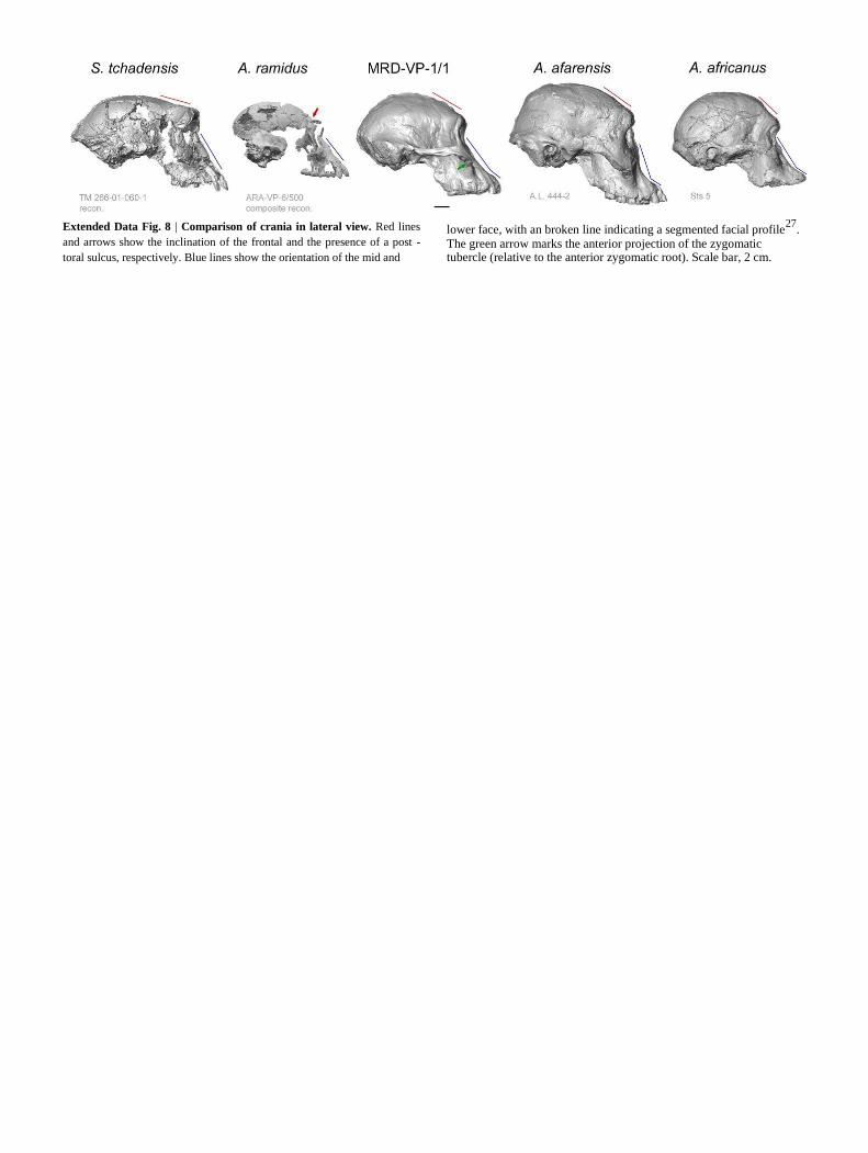
Extended Data Fig. 8 | Comparison of crania in lateral view. Red lines
and arrows show the inclination of the frontal and the presence of a post -
toral sulcus, respectively. Blue lines show the orientation of the mid and
lower face, with an broken line indicating a segmented facial profile27
.
The green arrow marks the anterior projection of the zygomatic tubercle (relative to the anterior zygomatic root). Scale bar, 2 cm.

Extended Data Fig. 9 | Comparison of crania in posterior view. The
transverse contour of the cranial base is convex in African apes, whereas A.
afarensis shows an angular transition between the nuchal region and the
greatly expanded mastoids (red dashed lines). In this regard, A. afarensis
anticipates the morphology of robust australopiths, but A. africanus is less
derived. MRD shows the primitive convex contour of the base, even though
the mastoids are expanded. MRD is also primitive with regard to
the great length of the nuchal plane (black arrows). However, it is similar
to A. afarensis in the configuration of the compound temporal–nuchal
crest (white dashed lines), the bare area (blue hatched triangle), and the
overall ‘bell -shaped’ posterior outline (that is, the parietal walls are
slightly convergent superiorly and the greatest width occurs basally
across the enlarged mastoids).
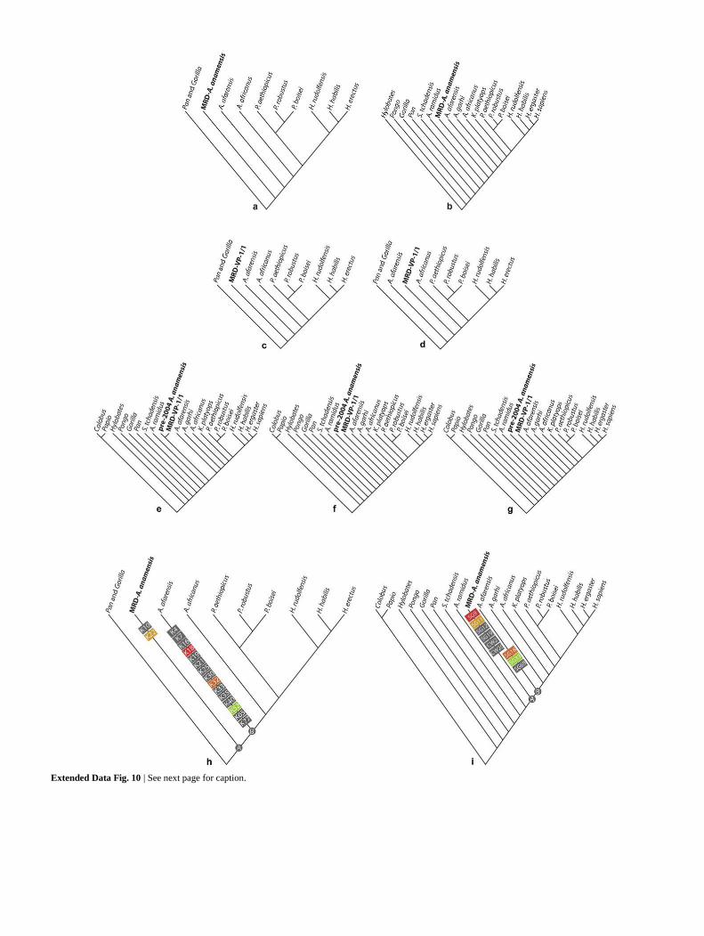
Extended Data Fig. 10 | See next page for caption.

Extended Data Fig. 10 | Results of phylogenetic analyses. a, Cladogram
resulting from the character matrix of ref. 27
, with the addition of MRD and
previously described A. anamensis specimens (combined as a single OTU, K
-combined). Parsimony analysis returned a single most-parsimonious tree (l =
196, C = 0.71, R = 0.70). b, Cladogram resulting from the character matrix of
ref. 33
(and references therein) with the addition of the combined MRD–A.
anamensis OTU (S&G-combined). This analysis returned a single most-
parsimonious tree (l = 429, C = 0.47, R = 0.66) with identical topology. The position of the combined MRD–A. anamensis OTU reinforces accepted relationships and is consistent with geochronology. c–g, Cladograms resulting from analyses in which MRD is treated as a separate OTU (that is,
an OTU bearing observations primarily for cranial characters, but very few dental characters and no mandibular characters.) c, d, Equally parsimonious
cladograms from the K-separate analysis (l = 196, C = 0.71, R = 0.68). e–g, Equally parsimonious
cladograms from the S&G-separate analysis (l = 430, C = 0.47, R = 0.66).
The ‘pre-2004 A. anamensis’ OTU in e–g bears observations primarily on
dentognathic characters. Character scores for MRD are provided in
Supplementary Table 1, sheets 1 and 2. Regardless of whether cranial or dentognathic characters are considered, the phylogenetic placement of MRD and the previously known A. anamensis sample remains stable relative to other hominins. h , i, Cladograms from the K-combined and S&G-combined analyses (as in a and b), with apomorphies added to the cladograms to illustrate the implied pattern of evolutionary change. The character states reconstructed at nodes A and B provide the reference for identifying A. anamensis and A. afarensis apomorphies, which are shown here as rectangles containing their abbreviated character labels. Characters in red, orange, gold and green describe similar morphology
and appear in both previously published studies27,33
. See Supplementary
Note 9 and Supplementary Table 1.
3


