Sensors 2012, 12, 15907-15946; doi:10.3390/s121115907
sensors ISSN 1424-8220
www.mdpi.com/journal/sensors
Review
Thiol Reactive Probes and Chemosensors
Hanjing Peng 1, Weixuan Chen
1, Yunfeng Cheng
1, Lovemore Hakuna
2, Robert Strongin
2 and
Binghe Wang 1,*
1 Department of Chemistry and the Center for Diagnostics and Therapeutics, Center for
Biotechnology and Drug Design, Georgia State University, Atlanta, GA 30302, USA;
E-Mails: [email protected] (H.P.); [email protected] (W.C.); [email protected] (Y.C.) 2
Department of Chemistry, Portland State University, Portland, OR 97207, USA;
E-Mails: [email protected] (L.H.); [email protected] (R.S.)
* Author to whom correspondence should be addressed; E-Mail: [email protected];
Tel.: +1-404-413-5545; Fax: +1-404-413-5543.
Received: 24 September 2012; in revised form: 12 November 2012 / Accepted: 13 November 2012 /
Published: 19 November 2012
Abstract: Thiols are important molecules in the environment and in biological processes.
Cysteine (Cys), homocysteine (Hcy), glutathione (GSH) and hydrogen sulfide (H2S) play
critical roles in a variety of physiological and pathological processes. The selective
detection of thiols using reaction-based probes and sensors is very important in basic
research and in disease diagnosis. This review focuses on the design of fluorescent and
colorimetric probes and sensors for thiol detection. Thiol detection methods include probes
and labeling agents based on nucleophilic addition and substitution, Michael addition,
disulfide bond or Se-N bond cleavage, metal-sulfur interactions and more. Probes for H2S
are based on nucleophilic cyclization, reduction and metal sulfide formation. Thiol probe
and chemosensor design strategies and mechanism of action are discussed in this review.
Keywords: thiols; cysteine; homocysteine; glutathione; hydrogen sulfide; sensors; probes;
detection
Abbreviations
ABD-F: 4-Fluoro-7-sulfamoylbenzofurazan
ADNB: 5-(2-aminoethyl)dithio-2-nitrobenzoate
OPEN ACCESS
Sensors 2012, 12 15908
AIE: aggregation-induced emission
ATP: adenosine triphosphate
AuNPs: gold nanoparticles
BHMT: betaine-homocysteine S-methyltransferase
BIPM: N-[4-(2-benzimidazolyl)phenyl]maleimide
BODIPY: 4,4-difluoro-4-bora-3a,4a-diaza-s-indacene
CAT: cysteine aminotransferase
CBS: cystathionine β-synthase
CE: capillary electrophoresis
ChE: cholinesterase
CMPI, 2-chloro-1-methylpyridinium iodide
CMQT, 2-chloro-1-methylquinolinium tetrafluoroborate
CNS: central nervous system
CSE: cystathionine γ-lyase or CGL
CV: cardiovascular
Cys: cysteine
DAD: diode array detector
DNBS: 2,4-dinitrophenyl sulfonyl group
D-PEN: D-penicillamine or PenA
DSP: dithiobis(succinimidylpropinate)
DTT: dithiothreitol
DTNB: 5,5’-dithiobis(2-nitrobenzoic acid or Ellman’s reagent
EPA: US Environmental Protection Agency
FRET: Förster resonance energy transfer
GCL: γ-glutamate-cysteine ligase or γ-glutamylcysteine synthetase (γ-GCS)
Gly: glycine
Glu: glutamate
GPx: glutathione peroxidase
GSH: glutathione or γ-L-glutamyl-L-cysteinylglycine
GSR: glutathione reductase
GSSG: glutathione disulfide
Hcy: homocysteine
HEPES: 4-(2-hydroxyethyl)-1-piperazineethanesulfonic acid
HPLC: high performance liquid chromatography
5-IAF: 5-iodoacetamidofluorescein
ICT: intramolecular charge transfer
LSPR: localized surface plasmon resonance
LuxS: S-ribosylhomocysteinase
MAT: methionine adenosyltransferase
MLCT: metal-to-ligand charge transfer
mBrB: monobromobimane
MES: 2-(N-morpholino)ethanesulfonic acid
Sensors 2012, 12 15909
3MST: 3-mercaptopyruvate sulfurtransferase
MTs: AdoMet-dependent methyltransfersess
N-DBPM: N-[4-(dimethylamino-2-benzofuranyl)phenyl]maleimide
NIR: near infrared
ODNB: 5-octyldithio-2-nitrobenzoate
PBS: phosphate buffered saline
2-PDS: 2,2’-dipyridyl disulfide
PET: photoinduced electron transfer
PIPES: piperazine-N,N’-bis(2-ethanesulfonic acid)
RNS: reactive nitrogen species
ROS: reactive oxygen species
RSS: reactive sulfur species
SAHH: S-adenosylhomocysteine hydrolase
SBD-F: 7-Fluorobenzo-2-oxa-1,3-diazole-4-sulfonic acid ammonium salt
Ser: serine
TCDI: 1,1’-thiocarbonyldiimidazole
TCEP, tris(2-carboxyethyl)phosphine
tHcy: total Hcy concentration
TNB: 5-thio-2-nitrobenzoate
TP: tiopronin or Thiola
2-TP: 2-thiopyridone
4-TP: 4-thiopyridone
TPM: two-photon microscopy
TPP: triphenylphosphonium
Trx: thioredoxin
UV: ultraviolet
1. Introduction
Sulfhydryl-containing compounds are often referred to as mercaptans due to their ability to react
with mercury. They have unique chemical reactivity and thus special utility in chemical reactions [1]
and in biological processes [2–4]. Thiols and thiophenols are widely used intermediates in synthetic
chemistry; dithiothreitol (DTT) and 2-mercaptoethanol are common antioxidants used in biology
labs [5]; aminothiols, such as cysteine (Cys, 1), homocysteine (Hcy, 2), and γ-L-glutamyl-L-
cysteinylglycine (glutathione or GSH, 3) play essential metabolic roles in biological systems. For
example, Cys plays versatile roles in protein structure and function [6]. The sulfhydryl group of Cys
serves as an ideal nucleophile in nucleophilic enzyme catalysis. Its ability to undergo reversible redox
reactions under physiological conditions is essential for maintaining tertiary and quaternary protein
structures through disulfide formation [7]. Hcy is a key intermediate generated during the biosynthesis
of Cys from the essential amino acid methionine (Met, 4) [3] (Figure 1). It is implicated in the health
of the cardiovascular (CV) system [3]. The tripeptide GSH is present at very high levels (0.1–10 mM)
in the cell (comprising about 90% of non-protein sulfur) and protects the cells against oxidative
Sensors 2012, 12 15910
stress [2,8] among many other functions. GSH levels in cancer cells can impact the effectiveness of
chemotherapy [9]. In addition to the above-mentioned thiols, thiol drugs such as D-penicillamine
(D-PEN or PenA) [10] and tiopronin (TP or Thiola) [11], are also widely used in clinical practice. The
quantitative detection of these drugs and their metabolites are very important in related clinical research.
Hydrogen sulfide (H2S), the simplest mercaptan, has been known as an environmental hazard and toxic
gas for many years. Recently, hydrogen sulfide has been recognized as one of the three gasotransmitters
[12–15], together with nitric oxide (NO) and carbon monoxide (CO), that are endogenously produced
and essential for maintaining the health of cardiovascular system among other roles. Even the unpleasant
smell of mercaptans has found important applications. For example, some small molecule thiols such as
ethanethiol and butanethiol are added to natural gas to help warn of gas leaks.
The metabolism and transportation of these sulfur-containing compounds in biological systems are
closely related to a series of important enzymes and proteins, the deficiency of which could lead to
various physiological/pathological conditions [16–18]. Furthermore, fluctuations in the endogenous
concentration of these thiols indicate the functional state of the corresponding enzymes/proteins and
are correlated with disease [18–20]. Thus, the detection of concentrations of mercaptans has
implications and significance in clinical diagnosis. Among all the biologically important mercaptans,
Hcy has been extensively studied as a biomarker for various reasons [21]. Deficiency in the expression
of enzymes such as cystathionine β-synthase (CBS) and cystathionine γ-lyase (CSE or CGL) or their
cofactors may lead to high levels or abnormal accumulation of Hcy, which characterizes inherited
diseases such as homocystinuria [22], Down syndrome [23,24] and other clinical conditions such as
vitamin (folate [25], cobalamin (vitamin B12) [26] or vitamin B6 [27]) deficiency, cardiovascular
disease [28] and renal failure [29]. The tripeptide GSH is found to be present at low micromolar range
in plasma [30]. However, the cytosol contains 0.1-10 mM GSH, depending on the cell type, while in
most cells the concentration is 1-2 mM [4]. Because GSH provides antioxidant protection for the cell,
the GSH concentration is also significant. The ratio of free GSH and its oxidized state glutathione
disulfide (GSSG, 5), which is normally >100:1 [31], is an indicator for both the corresponding enzyme
(GSSG reductase or enzymes related to de novo GSH synthesis) activity and the redox state of the
cell [32]. Low GSH concentration or [GSH]/[GSSG] ratio is related to inflammation and lung diseases,
such as cystic fibrosis [19,33].
H2S is synthesized in the cell both enzymatically and non-enzymatically. The enzymatic synthesis
of H2S is catalyzed by CBS, CSE [34,35] and cysteine aminotransferase (CAT)/3-mercaptopyruvate
sulfurtransferase (3MST) [36,37]. H2S undergoes fast metabolism and is involved in the regulation of
various systems, such as the cardiovascular [38–41] and the central nervous system (CNS) [42,43].
Concrete evidence has revealed the physiological and therapeutic significance of H2S, leading to a
rapid growth in research activity involving H2S [13,14,44]. Endogenous and exogenous hydrogen
sulfide has been demonstrated to exert either beneficial or detrimental effects in many pathological
conditions. H2S was found to have therapeutic benefit in ischemia-induced heart failure [39,45] and
hyperhomocysteinemia-induced hypertension [46]. The endogenous hydrogen sulfide level is related to
Down syndrome [47] and lung diseases [48]. Exogenous hydrogen sulfide may confer myocardial
protection against ischemia/reperfusion injury and exerts a protective effect against anti-inflammatory
drug-induced gastric mucosal injury [38].
Sensors 2012, 12 15911
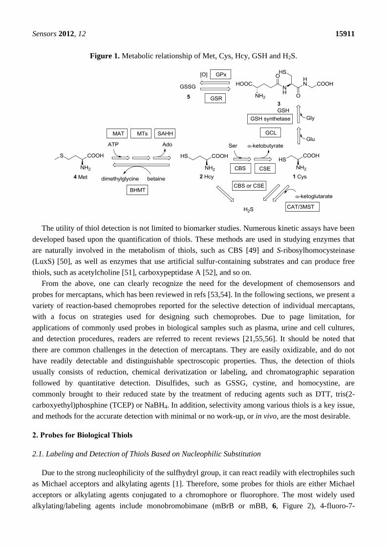
Figure 1. Metabolic relationship of Met, Cys, Hcy, GSH and H2S.
The utility of thiol detection is not limited to biomarker studies. Numerous kinetic assays have been
developed based upon the quantification of thiols. These methods are used in studying enzymes that
are naturally involved in the metabolism of thiols, such as CBS [49] and S-ribosylhomocysteinase
(LuxS) [50], as well as enzymes that use artificial sulfur-containing substrates and can produce free
thiols, such as acetylcholine [51], carboxypeptidase A [52], and so on.
From the above, one can clearly recognize the need for the development of chemosensors and
probes for mercaptans, which has been reviewed in refs [53,54]. In the following sections, we present a
variety of reaction-based chemoprobes reported for the selective detection of individual mercaptans,
with a focus on strategies used for designing such chemoprobes. Due to page limitation, for
applications of commonly used probes in biological samples such as plasma, urine and cell cultures,
and detection procedures, readers are referred to recent reviews [21,55,56]. It should be noted that
there are common challenges in the detection of mercaptans. They are easily oxidizable, and do not
have readily detectable and distinguishable spectroscopic properties. Thus, the detection of thiols
usually consists of reduction, chemical derivatization or labeling, and chromatographic separation
followed by quantitative detection. Disulfides, such as GSSG, cystine, and homocystine, are
commonly brought to their reduced state by the treatment of reducing agents such as DTT, tris(2-
carboxyethyl)phosphine (TCEP) or NaBH4. In addition, selectivity among various thiols is a key issue,
and methods for the accurate detection with minimal or no work-up, or in vivo, are the most desirable.
2. Probes for Biological Thiols
2.1. Labeling and Detection of Thiols Based on Nucleophilic Substitution
Due to the strong nucleophilicity of the sulfhydryl group, it can react readily with electrophiles such
as Michael acceptors and alkylating agents [1]. Therefore, some probes for thiols are either Michael
acceptors or alkylating agents conjugated to a chromophore or fluorophore. The most widely used
alkylating/labeling agents include monobromobimane (mBrB or mBB, 6, Figure 2), 4-fluoro-7-

Sensors 2012, 12 15912
sulfamoylbenzofurazan (ABD-F, 9), 7-fluorobenzo-2-oxa-1,3-diazole-4-sulfonic acid ammonium salt
(SBD-F, 10), 5-iodoacetamidofluorescein (5-IAF, 11), 2-chloro-1-methylpyridinium iodide (CMPI,
12) and 2-chloro-1-methylquinolinium tetrafluoroborate (CMQT, 13) (Figure 2). One common feature
of these reagents is that they all bear a halogen that can undergo nucleophilic substitution with thiols.
mBrB, ABD-F, SBD-F and 5-IAF form fluorescent conjugates with thiols, while CMPI and CMQT
yield UV-absorbing conjugates. The samples could then be analyzed using high performance liquid
chromatography (HPLC) or capillary electrophoresis (CE) coupled with a UV-vis detector (such as a
diode array detector or DAD) or a fluorescence detector. Several commonly used fluorogenic and
chromogenic labeling agents are compared in Table 1 in terms of their reactivity and limit of detection
(LOD). Compared with absorption detection (such as CMPI and CMQT), fluorescent probes (such as
mBrB) show higher sensitivity with detection limits in the picomolar range. This is due to the low
background and less interference from the matrix. Compared to fluorescence detection, absorption
methods, especially the absorptions in the UV range (<400 nm), are more prone to interference from
other biological substrates. It should be noted that the detection limits listed in Table 1 were obtained
from chromatographic methods, which includes an additional separation step. Such data are not
directly comparable to the detection limits obtained from direct detections. In addition, detection limits
reported for most probes by various labs were measured in different solvent systems (buffers or a
mixed solvent of a buffer and an organic solvent). They are not directly comparable either.
Table 1. A comparison of fluorogenic and chromogenic labeling agents.
Compound
λex/λem
(nm) or
Abs. (nm)
Reaction
Time
(min)
Reaction
Temperature/pH LOD
Biological
Sample Refs.
mBrB 380/480 2–20 25 °C/7.4–8.9 0.5 pM plasma [57–60]
ABD-F 380/510 5–60 37–60 °C /7.5–9.3 0.3 pM plasma, cell [61–63]
SBD-F 380/510 60 60 °C/9.5–10.5 0.1–1.5 µM plasma [64,65]
5-IAF 494/521 15–120 25–40 °C/12.5 2–30 nM plasma [66–68]
CMPI 310 15–30 25 °C/8.2 0.3–20 nM urine [69,70]
CMQT 355 1–4 25 °C/7.5 50 nM urine, plasma [71–73]
Among these agents, mBrB (6) can easily undergo SN2 substitution with a sulfhydryl group at
ambient temperature. The thiol-bimane conjugate 7 emits at 480 nm when excited at 380 nm. It has
been extensively used in the quantification of thiols [19,57–60]. Automated separation-quantification
of biological thiols in plasma and urine samples has been developed using mBrB [74]. Other bimane
derivatives, such as monochlorobimane (mCB, 8) [59], have also been used for thiol detection. ABD-F
and SBD-F [75] consist another group of useful reagents for the derivatization of thiols, yielding
fluorescent conjugates (λex 380 nm, λem 510 nm) and have been used for detection of thiols in both
plasma and tissues [62,65,76,77], although these reagents require fairly high temperature and pH for
the substitution reaction. 5-IAF reacts rapidly with thiols at room temperature at pH 12.5 and is used in
CE analysis of thiols in plasma and bacteria [66–68]. CMPI (12) forms thiol conjugates with an
absorption maxima at 310 nm, providing nanomolar detection limits. It has been used for the analysis
of thiols in urine and plasma [69,70]. CMQT (13) is user-friendly because it undergoes a fast
substitution reaction with thiols. The conjugates show a maximum absorption at 355 nm. It has also

Sensors 2012, 12 15913
been used for the quantitative analysis of thiols in urine and plasma [71–73,78,79]. Other UV labeling
agents, such as p-bromophenacyl bromide, have also been reported [80]. However, the short absorption
wavelength (263 nm) could limit their use because of spectral interference from the matrix.
These methods are widely used in both research and clinical analysis. However, one drawback of
these agents is the lack of any selectivity among various thiols because they rely on simple
nucleophilic addition or substitution for chromogenic or fluorescent labeling. As a result, detection and
quantification of individual thiols using these techniques relies on separations such as HPLC or CE to
differentiate various thiol-derivatives. Another disadvantage is interference from excess amount of
labeling agent, especially in the case of fluorescence. This issue can be overcome if the labeling agent
show significantly increased signal after conjugation.
Figure 2. Thiol labeling agents bearing halogen groups.
Due to the high level of electron-deficiency on the phenyl ring, the 2,4-dinitrophenyl sulfonyl
(DNBS) moiety can act as an electron sink when attached to a fluorophore and may incur
photoinduced electron transfer (PET) resulting in the quenching of the fluorescence. Furthermore,
DNBS can easily undergo de-sulfonylation in the presence of thiols, releasing SO2 gas and the attached
fluorophore, thus resulting in a fluorescence increase (Figure 3). This strategy has been used in the
development of many thiol probes. Examples include fluorescein derivatives 14 and 15 (Figure 3),
which are almost non-fluorescent (ΦFL 0.0007 and 0.0003, respectively in HEPES buffer at pH 7.4). In
these two compounds, the hydroxyl group was ―capped‖ as a sulfonate. After reaction with thiols in
HEPES buffer, 16 and 17 are produced, leading to strong fluorescence (ΦFL 0.75 and 0.58, λex 460 nm,
λem 560 nm). Compound 14 shows a low detection limit of 2 pM for thiols such as GSH and Cys. It
was investigated for its application in cholinesterase (ChE) assays [81]. Although 0.7% of compound
14 was observed to be decomposed after 1 h incubation in buffer at 37 °C, high reaction rates with
thiols still allowed for high throughput screening of ChE inhibitors using this probe.

Sensors 2012, 12 15914
Figure 3. Fluorescent probes of thiols bearing a DNBS moiety.
In another example (compound 19, Figure 4) the DNBS moiety was conjugated to a red-emissive
fluorophore, which can be released through reaction with thiols [82]. A donor-π-acceptor architecture
was built into the molecule, which was masked by the DNBS group. After addition of thiol, the
sulfonyl ester moiety collapsed and resulted in the release of aniline as an electron donor. This process
turned the fluorescence on with on/off ratios of 60 for GSH and 110-120 for Cys and Hcy (λex 560 nm,
λem 623 nm), providing a detection limit of about 3 µM. Almost exclusive selectivity was obtained for
Cys over other amino acids. Furthermore, no hydrolysis was detected over 12 h at 37 °C. Its
compatibility over a wide pH range (5.6–9.5) also makes this an excellent probe. The probe was utilized
in the bioimaging of thiols in albino Swiss mouse embryonic fibroblast cells (3T3 lines). The probe
showed good cell permeability and reacted with intracellular thiols. The control experiment using
N-methylmaleimide to consume all free thiols did not show any fluorescence after incubation with the
probe. In another example, a merocyanine fluorophore was conjugated to a DNBS moiety (Compound
20, Figure 4) [83]. Cleavage of the DNBS moiety tunes the intramolecular charge transfer (ICT) state of
the molecule and provides an absorption shift from 380 nm to 530 nm (ε~78,000 M−1
·cm−1
) and a strong
fluorescence emission at 553 nm. Both absorption and emission intensity are linearly related to thiol
concentrations in MeOH/H2O 3:7. A nanomolar detection limit was achieved.
Figure 4. Fluorescent probes of thiols bearing a DNBS moiety.
High photostability, high quantum yield and low pH sensitivity makes 4,4-difluoro-4-bora-3a,4a-
diaza-s-indacene (BODIPY) one of the best fluorescent scaffolds available. Recently a BODIPY-based
fluorescent probe (compound 21, Figure 4) for thiols was reported [84]. The DNBS moiety was

Sensors 2012, 12 15915
conjugated to this probe, providing an efficient fluorescence quenching effect. After exposure to Cys
or GSH in PBS buffer for 10 min, a 20–25-fold increase in fluorescence (λex 527 nm, λem 570 nm) was
observed, presumably as a result of the displacement reaction described. Micromolar detection limit
could be achieved with a moderate selectivity (>3-fold) over other amino acids. The probe was stable
at pH 7.3 for at least 12 hr at room temperature. Imaging using this probe was demonstrated in monkey
renal fibroblast COS-7 cell lines. In 2011, another BODIPY-based red-emitting off-on fluorescent
probe (compound 22, Figure 4) was reported [85]. This probe showed a 46-fold fluorescent
enhancement after exposure to thiols in MeOH/H2O 4:1 (λex 520 nm, λem 590 nm) with a detection
limit of 7 µM. Probe 22 showed moderate specificity toward Cys over Hcy with about 3-fold
difference, while no obvious response was observed for GSH. The fluorescence of the reaction
product remained pH-independent over a wide pH range (pH 2–8). Cellular thiol imaging was
performed in SGC-H446 cells. The specificity for thiols was also confirmed by N-methylmaleimide
control experiments.
Compared to fluorescence, phosphorescence has the advantages of large Stokes shift, long
luminescence lifetimes and ease of measurement. Phosphorescent probes utilizing a ruthenium
complex have been developed for the detection of thiols based on the nucleophilic addition-
fragmentation of DNBS (Figure 5, compound 23) [86]. The probe 23 consists of a DNBS moiety
attached to a Ru(II) poly(1,10-phenanthroline) complex as the luminophore, taking advantage of the
metal-to-ligand charge transfer (MLCT) red emission. The addition of 20 μM of Cys led to the
formation of 24 and a 90-fold increase in phosphorescence emission at 600 nm in a mixed solvent of
acetonitrile/water 4:1 v/v, providing a detection limit in the high nanomolar range. The selectivity for
Cys over other amino acids was over 40 fold. A fluorescent imaging study of this probe was performed
using NCI-H446 cells. Control experiments were carried out with cells pre-treated with
N-methylmaleimide, which covalently conjugate to thiols (this is also discussed in the next section).
Figure 5. A phosphorescent probe of thiols bearing a DNBS moiety.
2.2. Labeling of Thiols Based on Michael Addition
Because of their excellent nucleophilicity, thiols react readily with Michael acceptors [1]. Recently,
the Michael addition reaction has been widely used in the development of chemoprobes for thiols.
Maleimide as one excellent Michael acceptor has found numerous applications in various studies.
Sensors 2012, 12 15916
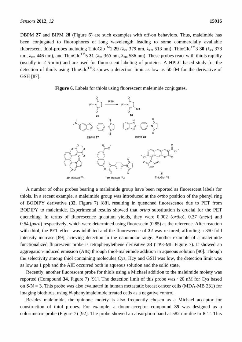
DBPM 27 and BIPM 28 (Figure 6) are such examples with off-on behaviors. Thus, maleimide has
been conjugated to fluorophores of long wavelength leading to some commercially available
fluorescent thiol-probes including ThioGloTM
1 29 (λex 379 nm, λem 513 nm), ThioGloTM
3 30 (λex 378
nm, λem 446 nm), and ThioGloTM
5 31 (λex 365 nm, λem 536 nm). These probes react with thiols rapidly
(usually in 2-5 min) and are used for fluorescent labeling of proteins. A HPLC-based study for the
detection of thiols using ThioGloTM
3 shows a detection limit as low as 50 fM for the derivative of
GSH [87].
Figure 6. Labels for thiols using fluorescent maleimide conjugates.
A number of other probes bearing a maleimide group have been reported as fluorescent labels for
thiols. In a recent example, a maleimide group was introduced at the ortho position of the phenyl ring
of BODIPY derivative (32, Figure 7) [88], resulting in quenched fluorescence due to PET from
BODIPY to maleimide. Experimental results showed that ortho substitution is crucial for the PET
quenching. In terms of fluorescence quantum yields, they were 0.002 (ortho), 0.37 (meta) and
0.54 (para) respectively, which were determined using fluorescein (0.85) as the reference. After reaction
with thiol, the PET effect was inhibited and the fluorescence of 32 was restored, affording a 350-fold
intensity increase [89], acieving detection in the nanomolar range. Another example of a maleimide
functionalized fluorescent probe is tetraphenylethene derivative 33 (TPE-MI, Figure 7). It showed an
aggregation-induced emission (AIE) through thiol-maleimide addition in aqueous solution [90]. Though
the selectivity among thiol containing molecules Cys, Hcy and GSH was low, the detection limit was
as low as 1 ppb and the AIE occurred both in aqueous solution and the solid state.
Recently, another fluorescent probe for thiols using a Michael addition to the maleimide moiety was
reported (Compound 34, Figure 7) [91]. The detection limit of this probe was ~20 nM for Cys based
on S/N = 3. This probe was also evaluated in human metastatic breast cancer cells (MDA-MB 231) for
imaging biothiols, using N-phenylmaleimide treated cells as a negative control.
Besides maleimide, the quinone moiety is also frequently chosen as a Michael acceptor for
construction of thiol probes. For example, a donor-acceptor compound 35 was designed as a
colorimetric probe (Figure 7) [92]. The probe showed an absorption band at 582 nm due to ICT. This

Sensors 2012, 12 15917
band decreased upon addition of thiols such as Cys and GSH, mostly likely due to the nucleophilic
addition of thiols on the quinone ring. A 17-fold fluorescence increase was observed for Cys in a 1:1
mixed solvent of H2O and THF The linear relationship between the absorption intensity change and the
thiol concentration could be used for quantitation.
Figure 7. Michael addition-based probes.
Open chain Michael acceptors are another option for the design of fluorescent sensor of thiols. A
real-time thiol quantitation method reported was based on the modulation of intramolecular PET
quenching upon addition of mercapto species [93]. Water soluble sensor 36 (Figure 8) reacts rapidly
with thiols to form conjugate 37 with a rate constant of 7.0 × 10⁴
M−1
·s−1
in Tris buffer at 25 °C. The
reaction of 50 µM probe 36 and 50 mM β-mercaptoethiol has a t1/2 of 3 ms. The conjugate 37 (λex
400 nm, λem 470 nm) has a fluorescence quantum yield more than 470-fold higher than that of 36. The
detection limit was as low as 0.5 nM. This has enabled the development of a high-throughput
fluorescence assay for glutathione reductase, since the assay requires probes with very short response
time. A similar idea was used in the design of α, β-unsaturated ketone derivative 38 (Figure 8) [94].
Figure 8. Fluorescent labeling of thiols using open chain Michael acceptors.
Upon thiol addition, the conjugation is disrupted and the fluorescence of the coumarin fluorophore
is restored. The probe 38 is a highly sensitive thiol reagent showing over 200-fold increases in
fluorescence (λex 444 nm, λem 496 nm) by forming 39 through Michael addition. The detection limit
was found to be 1 µM for Cys in 25 mM phosphate buffer (pH 7.4). The malononitrile group is an
effective fluorescent quencher and has been utilized in another coumarin-based biothiol probe 40

Sensors 2012, 12 15918
(Figure 8) [95], which forms fluorescent product 41 through reaction with thiols. However compound
40 only showed a relatively low fluorescent enhancement upon reaction with thiols (5.6–12 fold, λex
394 nm, λem 475 nm) in DMSO/HEPES buffer 1:2 (v/v).
The nucleophilic addition of a sulfhydryl group to electron-deficient squaraines has also been used
in the detection of thiols [96]. Compounds 42 and 43 are such derivatives (Figure 9). They showed
strong absorption at 640 nm in acetonitrile/H2O. The addition of thiols to the solution of the probe in
acetonitrile/MES buffer (pH 6.5) leads to the formation of adducts 44 and 45, where the absorption
band at 640 nm is significantly decreased, which is associated with a color change from blue to
colorless. Near infrared (NIR) spectroscopy is emerging as a very powerful tool in tissue imaging
because light in the 650–900 nm range is known to penetrate much deeper than visible light [97]. In
2009, a π-extended NIR squaraine dye 46 (Figure 9) formed by linking two bispyrrole molecules was
reported [98]. Upon addition of thiols such as Cys and GSH, the π-conjugation of probe 46 is
interrupted leading to the formation of 47, which shows significantly increased fluorescence (λex 410 nm,
λem 595 nm and λex 730 nm, λem 802 nm) in both visible and NIR region. The results also confirmed
that the level of the aminothiols in blood doubles after smoking. Probes of similar structures have also
been reported for cyanide detection [99]. However, since one would not normally expect cyanide in
blood, the lack of selectivity over cyanide does not pose a significant interference problem.
Figure 9. Fluorescent labeling based on the Michael addition using squaraines.
Michael addition triggered ring-opening reaction is another strategy in developing thiol reactive
probes (Figure 10). In 2009, a chromene-based colorimetric probe 48 (λmax 292 nm) was reported [100].
The 4-nitrophenolate moiety was generated after Michael addition, leading to the formation of 49 (λmax
Sensors 2012, 12 15919
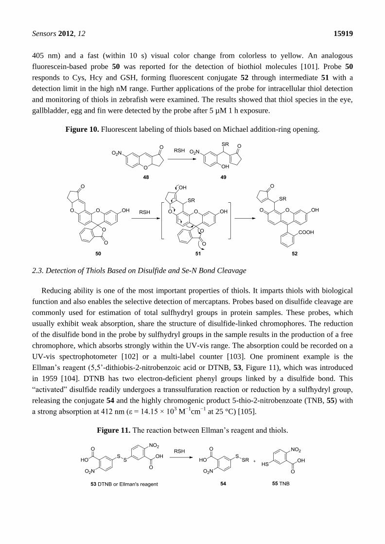
405 nm) and a fast (within 10 s) visual color change from colorless to yellow. An analogous
fluorescein-based probe 50 was reported for the detection of biothiol molecules [101]. Probe 50
responds to Cys, Hcy and GSH, forming fluorescent conjugate 52 through intermediate 51 with a
detection limit in the high nM range. Further applications of the probe for intracellular thiol detection
and monitoring of thiols in zebrafish were examined. The results showed that thiol species in the eye,
gallbladder, egg and fin were detected by the probe after 5 µM 1 h exposure.
Figure 10. Fluorescent labeling of thiols based on Michael addition-ring opening.
2.3. Detection of Thiols Based on Disulfide and Se-N Bond Cleavage
Reducing ability is one of the most important properties of thiols. It imparts thiols with biological
function and also enables the selective detection of mercaptans. Probes based on disulfide cleavage are
commonly used for estimation of total sulfhydryl groups in protein samples. These probes, which
usually exhibit weak absorption, share the structure of disulfide-linked chromophores. The reduction
of the disulfide bond in the probe by sulfhydryl groups in the sample results in the production of a free
chromophore, which absorbs strongly within the UV-vis range. The absorption could be recorded on a
UV-vis spectrophotometer [102] or a multi-label counter [103]. One prominent example is the
Ellman’s reagent (5,5’-dithiobis-2-nitrobenzoic acid or DTNB, 53, Figure 11), which was introduced
in 1959 [104]. DTNB has two electron-deficient phenyl groups linked by a disulfide bond. This
―activated‖ disulfide readily undergoes a transsulfuration reaction or reduction by a sulfhydryl group,
releasing the conjugate 54 and the highly chromogenic product 5-thio-2-nitrobenzoate (TNB, 55) with
a strong absorption at 412 nm (ε = 14.15 × 10³ M
−1cm
−1 at 25 °C) [105].
Figure 11. The reaction between Ellman’s reagent and thiols.

Sensors 2012, 12 15920
One problem with Ellman’s reagent is its low stability. DTNB is relatively stable at pH below 8.
Under more basic conditions DTNB undergoes obvious degradation, leading to undesired background
absorption increase. However, slightly basic conditions (pH values greater than 8) are necessary for
optimal reaction rates. Due to this reason, other reagents with relatively higher stability have been
reported as alternatives. Such examples include n-octyldithionitrobenzoate (ODNB, 56, Figure 12) [106]
and 5-(2-aminoethyl)dithio-2-nitrobenzoate (ADNB, 57) [107,108]. Both are mixed disulfides
bearing one electron donating aliphatic chain. Because of the less activated disulfide bond and one
less TNB group, they both show lower background than Ellman’s reagent for thiol detection. ADNB
was reported to show similar reactivity with thiols as Ellman’s reagent with much slower
hydrolysis [107,108]. On the other hand, the octyl group renders ODNB much more reactive to the
sulfhydryl groups in proteins that reside in a hydrophobic environment. ADNB is more reactive to
sulfhydryl groups in an anionic environment. These sulfhydryl groups usually react much slower with
doubly negatively charged DTNB [106].
2,2’-Dipyridyl disulfide (2-PDS, 58) and 4,4’-dipyridyl disulfide (4-PDS, 59) [109,110] were
reported for the determination of thiols such as GSH and protein thiols. When reduced by thiols,
2-PDS and 4-PDS forms 2-thiopyridone (2-TP) and 4-thiopyridone (4-TP), which absorb strongly at
343 and 324 nm, respectively. These two probes are reported to be more reactive to thiols at lower pH
(3-6) compared with DTNB [111]. The protonation of the nitrogen on pyridinyl ring is believed to
further activate the disulfide bond. Besides, 4-PDS reacts with GSH about 3 (pH 7)-30 (pH 4) times
faster than 2-PDS [109].
Figure 12. Colorimetric probes based on disulfide cleavage.

Sensors 2012, 12 15921
Another interesting probe, Ratio-HPSSC (60) was developed by the Lin group [112]. In this probe
(60, Figure 12), tetrakis(4-hydroxyphenyl)porphyrin is linked to coumarin by a disulfide bond. Due to
the overlap of the coumarin emission (λex 350 nm, λem 459 nm) with the porphyrin absorption (421 nm,
Soret band) and subsequent Förster resonance energy transfer (FRET), the fluorescence of coumarin in
Ratio-HPSSC is almost completely quenched. When exposed to thiols, the disulfide bond is cleaved,
switching off FRET and thus restoring the fluorescence of coumarin. This probe shows good
selectivity and sensitivity to thiols such as Cys with a detection limit lower than 1 μM in PBS
buffer/ethanol. Thiol imaging in live Hela cells was also studied.
Cyclization-release is one of the most popular strategies used in directed drug release. This was also
found to be useful in the development of thiol probes. In a typical example, a disulfide bond is linked
to a fluorophore by a carbamate linkage (61, Figure 13). After the disulfide is reduced by thiols to form
62, the newly formed sulfhydryl group could undergo an intramolecular cyclization with the carbonyl,
releasing the linked fluorophore as a free amine. In the first example published in this class [113], 63
(RhoSS, Figure 13) released rhodamine 110 after incubation with thiols for 1–2 h at 37 °C resulting in
a fluorescence increase at 535 nm. This probe was tested in cellular imaging studies in live Hela cells.
Strong fluorescence response was observed after incubating with cells. When incubated with
N-ethylmaleimide pre-treated cells, where sulfhydryl groups on thiols are capped, no such fluorescence
response was observed. The cellular distribution of the probe was studied by co-staining and the probe
seems to be localized within the cytosol.
Two-photon microscopy (TPM) [114,115] is a relatively new technology that has received great
interest in the past several years owing to its applications in deep tissue imaging (>500 μm).
Specifically, a two-photon microscope generates pulsed laser beams, focusing within less than
femtoliter volumes in the objective, and employs two photons of lower energy to excite the
fluorophore. TPM allows for increased penetration depth, localized excitation, reduced photo-damage
and prolonged observation time [116,117], and thus is superior to conventional one-photon excitation
and confocal microscopy. Recently, two-photon fluorescent probes that could selectively image
biothiols have been developed. For example, ASS (64, Figure 13) is a two-photon fluorescent probe
derived from 2-methylamino-6-acetylnaphthalene. In the presence of thiols the disulfide bond is
cleaved, leading to intramolecular cyclization and release of the fluorophore, which could be detected
by TPM. Fluorescence imaging using this probe in live cells and rat tissue in a depth of 120 μm has
been demonstrated [118]. Based on an analogous concept, SSH-Mito (65, Figure 13) bearing a
triphenylphosphonium (TPP) moiety for mitochondrial targeting was developed recently [119]. TPP
has been demonstrated to specifically transport cargo molecules to mitochondria due to electrostatic
interactions of the positively charged phosphine and the negative potential across the inner
mitochondrial membrane [120].
Recently, a thiol probe aimed at targeting liver cells was described [121]. This probe (66, Figure 13)
contains a galactose subunit and a disulfide-linked naphthalimide. The terminal galactose residue is
recognized by the asialoglycoprotein receptor (ASGP-R), which is expressed on the plasma membrane
of mammalian hepatocytes. This directs the probe selectively to hepatocytes. In the presence of
biothiols, such as GSH, thioredoxin (Trx), Hcy and Cys in PBS buffer, the disulfide bond is cleaved,
followed by an intramolecular cyclization. This uncaps the amino group on the naphthalimide moiety
and leads to a substantial increase in fluorescence intensity at 540 nm. A 10-fold fluorescence increase

Sensors 2012, 12 15922
was observed for thiols at 5.0 mM when 1.0 µM of probe 66 was used in PBS buffer. The target
specificity was confirmed by cellular imaging experiments, in which fluorescence was observed only
in HepG2 cells, but not in other non-hepatocytes such as C2C12, HaCaT, and N2a cells. The results of
tissue imaging experiments using male Sprague-Dawley rats have also confirmed the specificity.
Figure 13. Fluorescent probes based on disulfide cleavage followed by cyclization release.
Probes for the detection of thiols based on gold nanoparticles (AuNPs) have also been reported [122].
Specifically, disulfide linked AuNP clusters (68, Figure 14) are formed by treating AuNPs coated with
dithiobis(succinimidylpropinate) (DSP, 67).
Figure 14. Detection of thiols using disulfide linked AuNPs.
The formation of AuNP clusters was confirmed by localized surface plasmon resonance (LSPR)
spectroscopy and transmission electron microscopy (TEM). When the AuNP clusters are exposed to
small molecule thiols, the GSSG disulfide linkage can be readily reduced, resulting in the reversal of
the cluster formation to form 69 and 70. This is characterized by a significant blue shift (610 nm to
520 nm) of the LSPR spectra. This probe is especially useful for the detection of low molecular weight
Sensors 2012, 12 15923
thiols because by controlling the size of the linker and the NPs, a steric environment is created for the
easy access of low molecular weight thiols but not larger molecules. Different responses were obtained
for various thiols with the best detection limit (to NaSH) being in the low micromolar range.
Glutathione peroxidase (GPx) is an antioxidant enzyme that catalyzes the oxidation of GSH to
GSSG by H2O2. It has a selenocysteine in its active site, forming a catalytic triad with tryptophan (Trp)
and glutamine (Gln). The catalytic mechanism involves the formation of selenyl sulfide as an
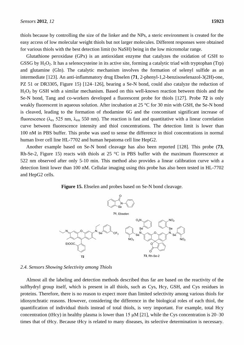
intermediate [123]. An anti-inflammatory drug Ebselen (71, 2-phenyl-1,2-benzisoselenazol-3(2H)-one,
PZ 51 or DR3305, Figure 15) [124–126], bearing a Se-N bond, could also catalyze the reduction of
H2O2 by GSH with a similar mechanism. Based on this well-known reaction between thiols and the
Se-N bond, Tang and co-workers developed a fluorescent probe for thiols [127]. Probe 72 is only
weakly fluorescent in aqueous solution. After incubation at 25 °C for 30 min with GSH, the Se-N bond
is cleaved, leading to the formation of rhodamine 6G and the concomitant significant increase of
fluorescence (λex 525 nm, λem 550 nm). The reaction is fast and quantitative with a linear correlation
curve between fluorescence intensity and thiol concentrations. The detection limit is lower than
100 nM in PBS buffer. This probe was used to sense the difference in thiol concentrations in normal
human liver cell line HL-7702 and human hepatoma cell line HepG2.
Another example based on Se-N bond cleavage has also been reported [128]. This probe (73,
Rh-Se-2, Figure 15) reacts with thiols at 25 °C in PBS buffer with the maximum fluorescence at
522 nm observed after only 5-10 min. This method also provides a linear calibration curve with a
detection limit lower than 100 nM. Cellular imaging using this probe has also been tested in HL-7702
and HepG2 cells.
Figure 15. Ebselen and probes based on Se-N bond cleavage.
2.4. Sensors Showing Selectivity among Thiols
Almost all the labeling and detection methods described thus far are based on the reactivity of the
sulfhydryl group itself, which is present in all thiols, such as Cys, Hcy, GSH, and Cys residues in
proteins. Therefore, there is no reason to expect more than limited selectivity among various thiols for
idiosynchratic reasons. However, considering the difference in the biological roles of each thiol, the
quantification of individual thiols instead of total thiols, is very important. For example, total Hcy
concentration (tHcy) in healthy plasma is lower than 15 μM [21], while the Cys concentration is 20–30
times that of tHcy. Because tHcy is related to many diseases, its selective determination is necessary.
Sensors 2012, 12 15924
Differentiation among various thiols in current methods is mainly based on chromatographic
separation [56,78,129]. Specifically, thiols are derivatized with labeling agents such as mBrB or CMQT
and analyzed by HPLC or CE equipped with a DAD or fluorescence detector. Data could then be
compared with a calibration curve. This process is instrument-dependent and time-consuming. Therefore,
the need for the development of chemoprobes, which allow for selectivity among thiols, is urgent.
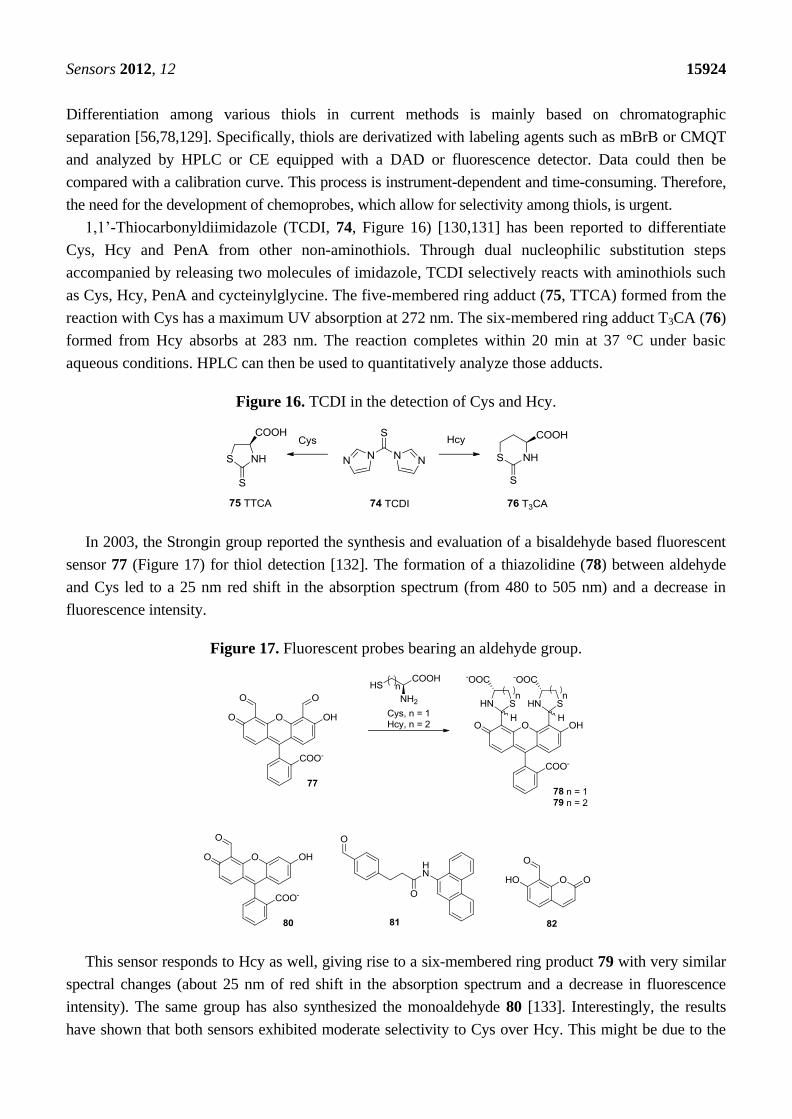
1,1’-Thiocarbonyldiimidazole (TCDI, 74, Figure 16) [130,131] has been reported to differentiate
Cys, Hcy and PenA from other non-aminothiols. Through dual nucleophilic substitution steps
accompanied by releasing two molecules of imidazole, TCDI selectively reacts with aminothiols such
as Cys, Hcy, PenA and cycteinylglycine. The five-membered ring adduct (75, TTCA) formed from the
reaction with Cys has a maximum UV absorption at 272 nm. The six-membered ring adduct T3CA (76)
formed from Hcy absorbs at 283 nm. The reaction completes within 20 min at 37 °C under basic
aqueous conditions. HPLC can then be used to quantitatively analyze those adducts.
Figure 16. TCDI in the detection of Cys and Hcy.
In 2003, the Strongin group reported the synthesis and evaluation of a bisaldehyde based fluorescent
sensor 77 (Figure 17) for thiol detection [132]. The formation of a thiazolidine (78) between aldehyde
and Cys led to a 25 nm red shift in the absorption spectrum (from 480 to 505 nm) and a decrease in
fluorescence intensity.
Figure 17. Fluorescent probes bearing an aldehyde group.
This sensor responds to Hcy as well, giving rise to a six-membered ring product 79 with very similar
spectral changes (about 25 nm of red shift in the absorption spectrum and a decrease in fluorescence
intensity). The same group has also synthesized the monoaldehyde 80 [133]. Interestingly, the results
have shown that both sensors exhibited moderate selectivity to Cys over Hcy. This might be due to the
Sensors 2012, 12 15925
favored formation of a five-membered ring structure. In 2004, the Barbas group reported another probe
81 bearing an aldehyde group [134]. This probe showed a fluorescence increase after condensation with
Cys. Although the authors did not mention the study of homocysteine, this sensor showed a significantly
higher reaction rate with Cys compared to GSH. Based on the same design strategy, the Hong group
described in 2008 a fluorescent probe 82 bearing an aldehyde moiety on the coumarin dye. This probe
was reported to show about 3-fold selectivity toward Cys over Hcy with no response to GSH [135]. In
addition, two photon fluorescent probes have been reported recently bearing an aldehyde moiety,
showing selectivity for cysteine [136].
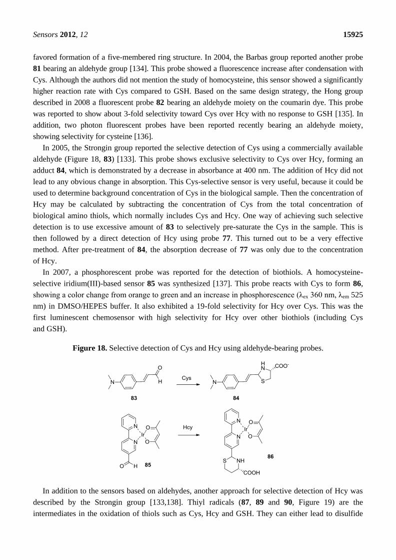
In 2005, the Strongin group reported the selective detection of Cys using a commercially available
aldehyde (Figure 18, 83) [133]. This probe shows exclusive selectivity to Cys over Hcy, forming an
adduct 84, which is demonstrated by a decrease in absorbance at 400 nm. The addition of Hcy did not
lead to any obvious change in absorption. This Cys-selective sensor is very useful, because it could be
used to determine background concentration of Cys in the biological sample. Then the concentration of
Hcy may be calculated by subtracting the concentration of Cys from the total concentration of
biological amino thiols, which normally includes Cys and Hcy. One way of achieving such selective
detection is to use excessive amount of 83 to selectively pre-saturate the Cys in the sample. This is
then followed by a direct detection of Hcy using probe 77. This turned out to be a very effective
method. After pre-treatment of 84, the absorption decrease of 77 was only due to the concentration
of Hcy.
In 2007, a phosphorescent probe was reported for the detection of biothiols. A homocysteine-
selective iridium(III)-based sensor 85 was synthesized [137]. This probe reacts with Cys to form 86,
showing a color change from orange to green and an increase in phosphorescence (λex 360 nm, λem 525
nm) in DMSO/HEPES buffer. It also exhibited a 19-fold selectivity for Hcy over Cys. This was the
first luminescent chemosensor with high selectivity for Hcy over other biothiols (including Cys
and GSH).
Figure 18. Selective detection of Cys and Hcy using aldehyde-bearing probes.
In addition to the sensors based on aldehydes, another approach for selective detection of Hcy was
described by the Strongin group [133,138]. Thiyl radicals (87, 89 and 90, Figure 19) are the
intermediates in the oxidation of thiols such as Cys, Hcy and GSH. They can either lead to disulfide
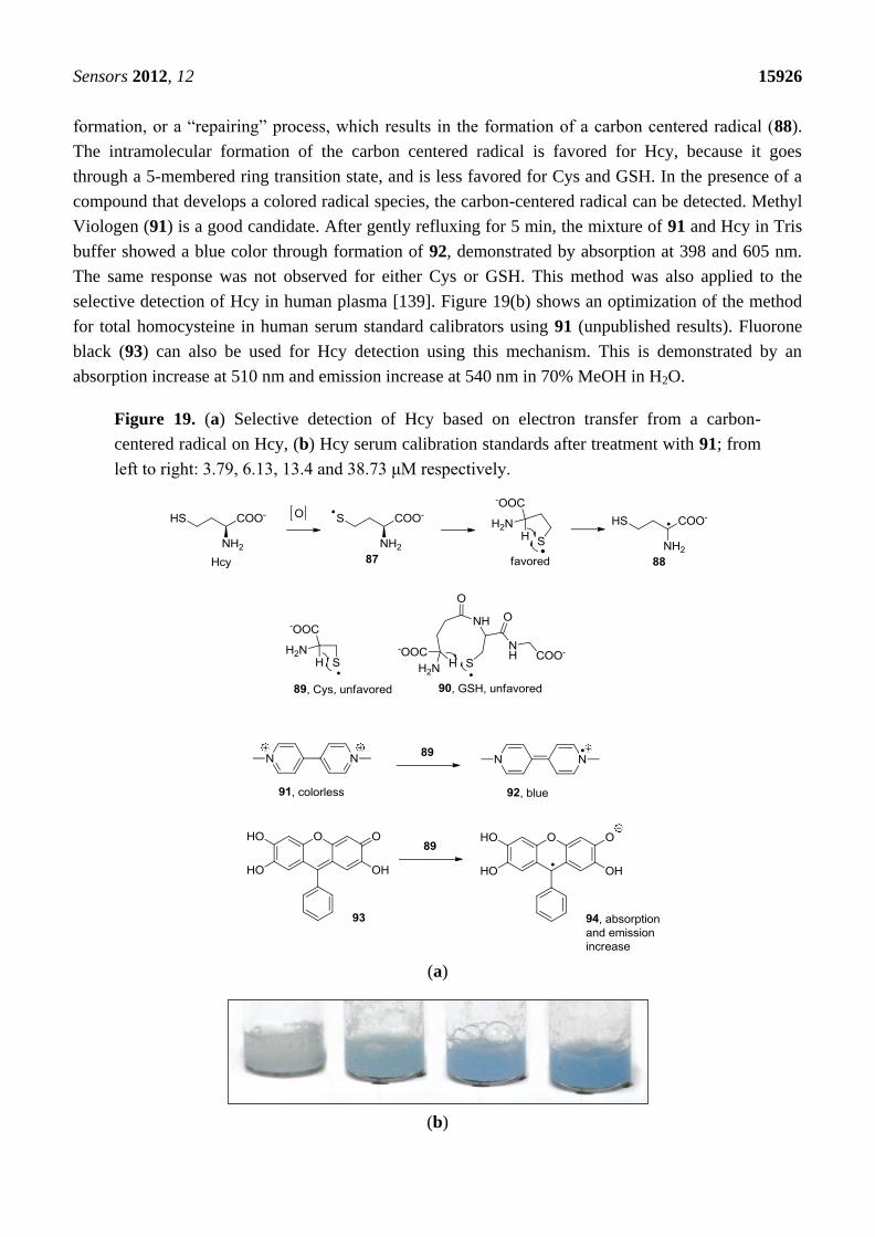
Sensors 2012, 12 15926
formation, or a ―repairing‖ process, which results in the formation of a carbon centered radical (88).
The intramolecular formation of the carbon centered radical is favored for Hcy, because it goes
through a 5-membered ring transition state, and is less favored for Cys and GSH. In the presence of a
compound that develops a colored radical species, the carbon-centered radical can be detected. Methyl
Viologen (91) is a good candidate. After gently refluxing for 5 min, the mixture of 91 and Hcy in Tris
buffer showed a blue color through formation of 92, demonstrated by absorption at 398 and 605 nm.
The same response was not observed for either Cys or GSH. This method was also applied to the
selective detection of Hcy in human plasma [139]. Figure 19(b) shows an optimization of the method
for total homocysteine in human serum standard calibrators using 91 (unpublished results). Fluorone
black (93) can also be used for Hcy detection using this mechanism. This is demonstrated by an
absorption increase at 510 nm and emission increase at 540 nm in 70% MeOH in H2O.
Figure 19. (a) Selective detection of Hcy based on electron transfer from a carbon-
centered radical on Hcy, (b) Hcy serum calibration standards after treatment with 91; from
left to right: 3.79, 6.13, 13.4 and 38.73 μM respectively.
(a)
(b)

Sensors 2012, 12 15927
The most recent work on the selective detection of Hcy and Cys reported by the Strongin group
takes advantage of a Michael addition-lactam formation cascade [140]. The probe (95, Figure 20)
undergoes a Michael addition with the sulfhydryl group to generate conjugates 96. Then an
intramolecular nucleophilic attack from the amino group leads to the formation of lactams 97, release
of a HMBT fluorophore 98, and a subsequent fluorescence change. The difference in reaction rates
with Cys and Hcy provides an option for discrimination of Cys and Hcy. Very recently, Yoon lab has
also reported a cyanine-based NIR emitting fluorescent probe showing selectivity for cysteine using
the same strategy [141].
Figure 20. Discrimination of Cys and Hcy based on Michael addition-lactam formation cascade.
3. Probes for Aromatic Thiols
Aromatic thiols, such as thiophenols, are widely used in the production of pharmaceutical
intermediates, polymers and pesticides [142]. Exposure to thiophenol may lead to damage to the
central nervous system, kidney and liver [143]. Due to conjugation effect with the phenyl ring, the
sulfhydryl group of thiophenol has a pKa much lower than that of alkyl thiols. Thus it is conceivable
that fluorescent labeling agents for thiols would also label thiophenols, and probably more efficiently.
The significant difference in the nucleophilicity under physiological pH conditions has been
successfully used in the selective detection of thiophenols. The Wang group has reported two
fluorescent chemoprobes for thiophenols [144,145]. Compound 99 is based on a nitrobenzofuranzan
fluorophore bearing a DNBS moiety (Figure 21), which shows no fluorescence before the addition of
thiophenol. When thiophenol is added, the sulfhydryl group attacks the dinitrophenyl and releases the
free fluorophore NBD (100), which leads to a dramatic fluorescence increase at an emission
wavelength of 550 nm. A similar design also imparted the same selectivity to compound 101, which
releases a benzoxazole fluorophone 102 after the addition of thiophenol. Both probes have a detection
limit in the low micromolar range.

Sensors 2012, 12 15928
Besides the strategy of using DNBS moiety, another probe (103, Figure 21, ΦFL 0.006) based on the
nucleophilic attack on the dinitrophenyl group was developed by Lin and co-workers [146]. The probe
releases a fluorescent coumarin derivative 104 (λex 461 nm, λem 494 nm, ΦFL 0.50) upon reaction with
thiophenol, showing good selectivity for thiolphenol over 2-mercaptoethanol. The detection limit was
reported to be 1.8 nM for benzenethiols. This method has been tested in water, soil and cell cultures.
Figure 21. Fluorescent probes of thiophenols.
4. Probes for H2S
H2S has been recognized as an endogenously produced gasotransmitter. It plays regulatory roles in
multiple systems, such as the cardiovascular [38–41] and the central nervous system (CNS) [42,43]. It
also shows therapeutic effects in heart diseases [39,45]. Due to the newly recognized biological
significance of H2S, more and more research interest has been focused on the molecular mechanisms
of its biological functions and related therapeutic applications. However, H2S is very unstable because
of its high volatility and high propensity to be oxidized under physiological conditions. Such
properties have made the accurate measurement of hydrogen sulfide a difficult task. A number of
methods have been reported for the detection of H2S [147,148]. These methods include gas
chromatography (GC) [149–152], HPLC [153], and electrochemical methods, which mostly rely on
sulfide ion selective electrodes [154,155] or polarographic methods [156]. Despite the availability of
numerous detection methods, literature reported hydrogen sulfide concentrations vary significantly
among publications, ranging from high micromolar [48,157,158] to low nanomolar [159]. Part of the
reason could be the difference in sample preparation and intrinsic fluctuation of hydrogen sulfide
concentrations. Other reasons might be due to the lack of methods for fast and selective detection. In
any case, it is generally agreed that the development of new approaches is necessary not only for the
selective and instantaneous detection, but also for intracellular imaging.

Sensors 2012, 12 15929
4.1. Probes for H2S based on Nucleophilic Cyclization Reactions
Hydrogen sulfide (H2S) dissociates in aqueous solutions in two sequential steps to HS− and S
2− with
a pKa1 of 6.9 and pKa2 of 12, which means over 75% of H2S exists in the anionic state at physiological
pH. Sulfide is a stronger nucleophile than commonly encountered anions such as chloride and
hydroxide under physiological conditions. This provides the possibility of selective detection of sulfide
amongst various anions. One of the classical methods for the quantification of sulfide is the methylene
blue method [160]. Sulfide reacts with N,N-dimethyl-p-phenylenediamine (105, Figure 22) in the
presence of Zn(OAc)2 and FeCl3 under acidic conditions, yielding methylene blue (106), which
absorbs strongly at 670 nm.
Figure 22. The reaction involved in H2S detection using the methylene blue method.
Samples containing H2S are often preserved by addition of Zn2+
to trap H2S in the form of ZnS.
During analysis, solutions of compound 105 and FeCl3 in HCl are added into the sample. A blue color,
which could be measured by a UV-vis spectrophotometer, usually develops in minutes [161]. This
method shows both good selectivity and sensitivity with a nanomolar detection limit. It has been
approved by the US Environmental Protection Agency (EPA) as a standard method for sulfide
quantitation and has been utilized for hydrogen sulfide determination in many studies [153,157,162].
However, because methylene blue tends to form dimers and trimers [163] at concentrations over 10
μM, a linear calibration curve could not be obtained for sulfide at 10 μM or higher concentrations. In
addition, the use of corrosive reagents and the non-instantaneous nature of this method have also
limited the application of this method.
Figure 23. Selective detection of H2S based on a disulfide cleavage-cyclization strategy.

Sensors 2012, 12 15930
Being the simplest molecule in the thiol family, hydrogen sulfide can undergo two deprotonation
steps. In other words, it can undergo two nucleophilic reactions. This distinguishes it from other thiols
and provides a strategy for sulfide detection with high selectivity. The Xian group reported a
fluorescent probe for H2S, which was designed based on this principle [164]. As shown in Figure 23,
compound 107 reacts with sulfide, releasing mercaptopyridine 108 to form an intermediate 109, which
undergoes a cyclization in situ and releases fluorophore 110 and benzodithiolone 111. This probe is
very selective for H2S in aqueous solution (PBS/acetonitrile 9:1) among thiols such as Cys and GSH, and
gives a linear correlation to sulfide concentrations with a detection limit of low micromolar
concentrations. Fluorescent imaging using this probe and exogenous H2S has been studied in COS7 cells.
Another strategy reported by the He group uses a Michael addition reaction followed by
cyclization [165]. In this study, two fluorescent probes, SFP-1 (112) and SFP-2 (114, Figure 24) were
synthesized. These probes bear an α, β-unsaturated ester group at the ortho position of a benzaldehyde,
which is linked to a fluorophore. The nucleophilic attack by sulfide on the formyl group yields
hemithioacetals, which positions the sulfhydryl group for the following Michael addition to form the
trapped thioacetal 113 and 115, in which the PET effect is interrupted and the fluorescence is recovered.
Figure 24. Detection of H2S based on Michael addition-cyclization.
Both SFP-1 and SFP-2 show 50–100 fold selectivity for sulfide over other thiols including
β-mercaptoethanol, Cys and GSH. The detection limit is about 5–10 μM with a S/N ratio of 3:1. SFP-2

Sensors 2012, 12 15931
was used in the in vivo imaging of endogenously generated H2S triggered by the addition of GSH and
Cys in Hela cells. Along a similar line, probes 116 and 117 have been reported recently by the Xian
group. These probes are based on a Michael addition-cyclization reaction [166]. In probes 116 and
117, the Michael acceptor is activated by two electron withdrawing groups. After incubating the
probes (5 µM) with 100 µM sulfide for 30 min in phosphate buffer, Michael addition-cyclization takes
place to release the fluorophore 110 to form thiolactones 118 and 119, leading to 11 (116) or 160
(117)-fold fluorescence increase (λex = 465 nm, λem = 510 nm), respectively. Imaging of exogenous
H2S was performed in COS7 cells.
Furthermore, chemoprobes developed for other thiols could also be used for H2S detection. For
example, compound 14 (Figure 3), reported by Maeda for the fluorescent detection of thiols, has also
been used for the fluorescent detection of H2S [167]. Of course, in such a case, selectivity is an issue.
4.2. Probes for H2S Based on Reduction Reactions
Sulfide is a fairly strong reducing agent. This is another chemical property of H2S that can be used
in probe design. Azides are known to be reduced by sulfide anion [168]. However, it is not until very
recently that this reaction was utilized in the selective detection of sulfide independently by the labs of
Chang and Wang. Specifically, the Chang group reported the synthesis and evaluation of two
fluorescent probes (SF1, 120 and SF2, 121, Figure 25) based on this strategy [169]. Both probes bear a
fluorescein moiety attached directly to an azido group, which is easily reduced to an amino group by
hydrogen sulfide, resulting in a significant increase in fluorescence. The selectivity of these two probes
was demonstrated among various RSS (reactive sulfur species), RNS (reactive nitrogen species) and
ROS (reactive oxygen species) including GSH, Cys, Na2SO3, NO, H2O2 and O2−
. The probes were also
evaluated for in vivo cell imaging in HEK293T cells using exogenous H2S [169].
At the same time, another fluorescent probe for H2S was reported by the Wang group [170]. It is
based on the reduction of the azido group attached to a sulfonylnaphthalene fluorophore, the dansyl
dye (DNS-Az, 122, Figure 25). This probe showed a dramatic fluorescence increase (λex 340 nm, λem
517 nm, over 40 fold) upon addition of 25 μM of sulfide into phosphate buffer with Tween-20. The
probe was evaluated in different aqueous media, including phosphate buffer and commercial bovine
serum. Good selectivity of DNS-Az for H2S was observed among various anions (>50 fold) and other
reducing agents, including Cys (60-fold) and thiophenol (4-7.5 fold). Another important advantage of
this probe over other reported sulfide probes is the fast response [171]. The reduction reaction is
complete in seconds in bovine serum at room temperature. This is among the fastest H2S fluorescent
probes reported so far. Considering the volatility and low stability of H2S, an ―instant‖ probe is very
important because it allows for accurate detection of sulfide concentration without sample
pre-treatment such as precipitation. The fluorescence intensity could be recorded on a fluorometer or a
multi-label counter. Excellent linear calibration curves were obtained for H2S in different solvents,
including buffer and bovine serum. The probe was used for the measurement of H2S concentration in
blood using the C57BL6/J mouse model. Specifically, a stock solution of DNS-Az was added to the
blood, followed by mixing and reaction at room temperature for 5 min. Blood plasma was then
obtained by centrifugation and fluorescence intensity was analyzed in a 96-well plate and multi-label
counter. The zero point was obtained by trapping sulfide with ZnCl2 and the calibration curve was

Sensors 2012, 12 15932
obtained by using an internal standard method. The blood H2S concentration obtained (31.9 ± 9.4 μM)
is very similar to most previously reported numbers.
Besides DNS-Az, compound 123 has also been synthesized by the Wang group (unpublished
results). This probe is composed of an azido group and the naphthalimide fluorophore moiety. The
synthesis of this compound consists of two steps from 4-bromo-1,8-naphthalic anhydride through amide
formation with n-butylamine and substitution of the bromo group by azido using NaN3. This compound
is sensitive toward sulfide with a 100-fold fluorescence increase (λex 420 nm, λem 530 nm) in response to
the addition of 100 μM of H2S in a mixed solvent of 1:1 phosphate buffer/acetonitrile. The reaction is
complete in 30 s in acetonitrile and 20 min in phosphate buffer/acetonitrile at room temperature.
However, the lower solubility and stability of compound 123 compared with 122 may limit its application.
Further structure optimization is ongoing to obtain improved fluorescent probes for H2S. Two other
naphthalimide probes (compounds 124 and 125, Figure 25) have also been reported recently [172]. Probe
124 is based on the reduction of an azido group and probe 125 is based on the reduction of a nitro
group by sulfide. Both probes can be converted to the corresponding 4-aminonaphthalimide (λex 432
nm, λem 542 nm) by reduction. They were found to show more than 10-fold selectivity toward H2S
over other reactive oxygen, nitrogen, and sulfur species. In PIPES buffer, the reduction reaction is
complete in 90 min (124) and 45 min (125), respectively, upon treatment with 0.5 mM of sulfide.
Fluorescent imaging of exogenous H2S using these two probes were demonstrated in Hela cells.
Figure 25. Fluorescent probes for H2S based on reduction reactions.
Recently, a NIR fluorescent probe (126, Figure 25) for H2S was reported by Han and
co-workers [173]. When the azido group is reduced by H2S, a 50-nm red shift from 610 nm to 660 nm
in absorption and a 40 nm red shift from 710 nm to 750 nm (λex 625 nm) in emission were observed in
Sensors 2012, 12 15933
HEPES buffer. The reaction took 20 min to complete with a micromolar detection limit. This probe
was tested in RAW 264.7 macrophage cells for imaging exogenously added H2S.
Due to its high selectivity and reactivity, redox-based strategy is becoming very useful in the
detection of H2S. The level of activities is very high in this area. Recently, several other probes have
been reported based on this strategy [174–177]. These probes include two-photon fluorescent probes
and protein-based probes.
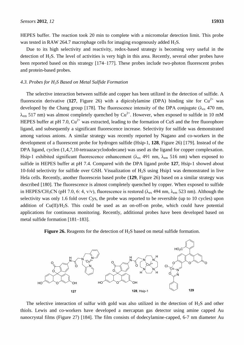
4.3. Probes for H2S Based on Metal Sulfide Formation
The selective interaction between sulfide and copper has been utilized in the detection of sulfide. A
fluorescein derivative (127, Figure 26) with a dipicolylamine (DPA) binding site for Cu2+
was
developed by the Chang group [178]. The fluorescence intensity of the DPA conjugate (λex 470 nm,
λem 517 nm) was almost completely quenched by Cu2+
. However, when exposed to sulfide in 10 mM
HEPES buffer at pH 7.0, Cu2+
was extracted, leading to the formation of CuS and the free fluorophore
ligand, and subsequently a significant fluorescence increase. Selectivity for sulfide was demonstrated
among various anions. A similar strategy was recently reported by Nagano and co-workers in the
development of a fluorescent probe for hydrogen sulfide (Hsip-1, 128, Figure 26) [179]. Instead of the
DPA ligand, cyclen (1,4,7,10-tetraazacyclododecane) was used as the ligand for copper complexation.
Hsip-1 exhibited significant fluorescence enhancement (λex 491 nm, λem 516 nm) when exposed to
sulfide in HEPES buffer at pH 7.4. Compared with the DPA ligand probe 127, Hsip-1 showed about
10-fold selectivity for sulfide over GSH. Visualization of H2S using Hsip1 was demonstrated in live
Hela cells. Recently, another fluorescein based probe (129, Figure 26) based on a similar strategy was
described [180]. The fluorescence is almost completely quenched by copper. When exposed to sulfide
in HEPES/CH3CN (pH 7.0, 6: 4, v/v), fluorescence is restored (λex 494 nm, λem 523 nm). Although the
selectivity was only 1.6 fold over Cys, the probe was reported to be reversible (up to 10 cycles) upon
addition of Cu(II)/H2S. This could be used as an on-off-on probe, which could have potential
applications for continuous monitoring. Recently, additional probes have been developed based on
metal sulfide formation [181–183].
Figure 26. Reagents for the detection of H2S based on metal sulfide formation.
The selective interaction of sulfur with gold was also utilized in the detection of H2S and other
thiols. Lewis and co-workers have developed a mercaptan gas detector using amine capped Au
nanocrystal films (Figure 27) [184]. The film consists of dodecylamine-capped, 6-7 nm diameter Au

Sensors 2012, 12 15934
nanocrystals. When exposed to gaseous thiols such as CH3SH, the amine cap is displaced by thiols,
resulting in an electrical resistance drop due to the reduced distance amongst Au cores. The slope of
the resistance curve shows a linear response to CH3SH between 0.153 and 1.53 ppm in air with the
detection limit of 1.7 ppb. Although the film detector does not show a comparable response to larger
molecules such as octanethiol, it still provides a very sensitive detection of small molecular gaseous
mercaptans such as CH3SH and H2S.
Figure 27. Gaseous mercaptan detection by amine capped Au nanocrystal films.
Because of the biological significance of H2S, the value for the quantitative detection of this small
molecule has been recognized both for diagnostic applications and in basic research. Fluorescent
probes for H2S based on nucleophilic cyclization, reduction and metal sulfide formation have been
reported. Quantitation of H2S was achieved in mouse blood samples with DNS-Az (122). Fluorescence
imaging of endogenous and exogenous H2S was demonstrated by various groups using different cell
lines. AuNPs was also used in the detection of mercaptan gas including H2S and CH3SH. These probes
provide new chemical tools for biomedical researchers in their investigation of the functions and
properties of H2S in biological systems.
5. Conclusions
With the widely recognized importance of biological thiols, there has been increasing interest in
developing new detection methods. Numerous fluorescent probes have been reported so far for the
selective detection of thiols. The probes for biological amino-thiols include those based on
nucleophilic substitution, Michael addition, and disulfide/Se-N bond cleavage. Among these probes,
nucleophilic substitution-based probes (such as mBrB) are commonly used in the fluorometric/
colorimetric labeling of thiols for HPLC quantitation. Disulfide cleavage-based probes (such as
Ellman’s reagent) are frequently used for estimation of total sulfhydryl groups in a protein sample.
Methods based on other probes are still in development. Fluorescent probes for H2S include those
based on nucleophilic cyclization, reduction, and metal sulfide formation. These probes are emerging
as important detection tools of H2S. Generally speaking, excellent methods are available for the
quantitative and selective concentration determinations of various thiols in solution or in sera. Still
needed are methods that are fast enough to address the issues of rapid fluctuations of intracellular
concentrations or in vivo, especially for hydrogen sulfide. Recently, there is growing interest in the
intracellular imaging of various thiols. Fluorescent probes are needed that are: (1) selective, (2) fast
response, (3) cell permeable, and (4) of long wavelength. Among these, especially important is the

Sensors 2012, 12 15935
availability of probes that are fast in response because reaction times on the scale of minutes run the
risk of perturbing the thiol homeostasis mechanisms in such a way that the amount of probe consumed
(and thus fluorescent intensity) may not be correlated with the true concentration at a given time.
However, based on the level of research interest in this area, there are good reasons to be optimistic
that excellent solutions will be found in the not too distant future.
Acknowledgements
We gratefully acknowledge the financial support of this work in the authors’ lab by the Molecular
Basis of Disease Program at GSU through fellowships to HJP, WXC and YFC, the GSU University
fellowship program through a fellowship to HJP, and the National Institutes of Health (GM084933
to BW).
References
1. Koval, I.V. Reactions of thiols. Russ. J. Org. Chem. 2007, 43, 319–346.
2. Meister, A.; Anderson, M.E. Glutathione. Ann. Rev. Biochem. 1983, 52, 711–760.
3. Selhub, J. Homocysteine metabolism. Annu. Rev. Nutr. 1999, 19, 217–246.
4. Forman, H.J.; Zhang, H.Q.; Rinna, A. Glutathione: Overview of its protective roles,
measurement, and biosynthesis. Mol. Aspects Med. 2009, 30, 1–12.
5. Cleland, W.W. Dithiothreitol, a new protective reagent for SH groups. Biochemistry 1964, 3,
480–482.
6. Lipton, S.A.; Choi, Y.B.; Takahashi, H.; Zhang, D.X.; Li, W.Z.; Godzik, A.; Bankston, L.A.
Cysteine regulation of protein function—As exemplified by NMDA-receptor modulation.
Trends Neurosci. 2002, 25, 474–480.
7. Petsko, G.A.; Ringe, D. Protein Structure and Function: From Sequence to Consequence;
New Science Press Ltd.: London, UK, 2004.
8. Meister, A. Glutathione metabolism and its selective modification. J. Biol. Chem. 1988, 263,
17205–17208.
9. Conklin, K.A. Chemotherapy-associated oxidative stress: impact on chemotherapeutic
effectiveness. Integr. Cancer. Ther. 2004, 3, 294–300.
10. Peisach, J.; Blumberg, W.E. A mechanism for the action of penicillamine in the treatment of
Wilson’s disease. Mol. Pharmacol. 1969, 5, 200–209.
11. Zhang, J.G.; Lindup, W.E. Tiopronin protects against the nephrotoxicity of cisplatin in rat renal
cortical slices in vitro. Toxicol. Appl. Pharm. 1996, 141, 425–433.
12. Boehning, D.; Snyder, S.H. Novel neural modulators. Annu. Rev. Neurosci. 2003, 26, 105–131.
13. Martelli, A.; Testai, L.; Breschi, M.C.; Blandizzi, C.; Virdis, A.; Teddei, S.; Calderone, V.
Hydrogen sulfide: novel oppotunity for drug discovery. Med. Res. Rev. 2011,
doi:10.1002/med.20234.
14. Szabo, C. Hydrogen sulphide and its therapeutic potential. Nat. Rev. Drug Discov. 2007, 6,
917–935.
15. Wang, R. Two’s company, three’s a crowd: Can H2S be the third endogenous gaseous
transmitter? FASEB J. 2002, 16, 1792–1798.
Sensors 2012, 12 15936
16. Kluijtmans, L.A.J.; van den Heuvel, L.P.W.J.; Boers, G.H.J.; Frosst, P.; Stevens, E.M.B.;
vanOost, B.A.; den Heijer, M.; Trijbels, F.J.M.; Rozen, R.; Blom, H.J. Molecular genetic
analysis in mild hyperhomocysteinemia: A common mutation in the methylenetetrahydrofolate
reductase gene is a genetic risk factor for cardiovascular disease. Am. J. Hum. Genet. 1996, 58,
35–41.
17. Calonge, M.T.; Gasparini, P.; Chillaron, J.; Chillon, M.; Gallucci, M.; Rousaud, F.; Zelante, L.;
Testar, X.; Dallapiccola, B.; Disilverio, F.; et al. Cystinuria caused by mutations in rbat, a gene
involved in the transport of cystine. Nat. Genet. 1994, 6, 420–425.
18. Lowicka, E.; Beltowski, J. Hydrogen sulfide (H2S)—The third gas of interest for
pharmacologists. Pharmacol. Rep. 2007, 59, 4–24.
19. Wu, G.Y.; Fang, Y.Z.; Yang, S.; Lupton, J.R.; Turner, N.D. Glutathione metabolism and its
implications for health. J. Nutr. 2004, 134, 489–492.
20. Droge, W.; Eck, H.P.; Mihm, S. Hiv-Induced Cysteine Deficiency and T-Cell Dysfunction—A
rationale for treatment with N-Acetylcysteine. Immunol. Today 1992, 13, 211–214.
21. Refsum, H.; Smith, A.D.; Ueland, P.M.; Nexo, E.; Clarke, R.; McPartlin, J.; Johnston, C.;
Engbaek, F.; Schneede, J.; McPartlin, C.; et al. Facts and recommendations about total
homocysteine determinations: An expert opinion. Clin. Chem. 2004, 50, 3–32.
22. Levy, H.L.; Kraus, J.P.; Mudd, S.H. Disorders of Transsulfuration. In The Metabolic and
Molecular Bases of Inherited Disease; Valle, D., Beaudet, A.L., Vogelstein, B., Kinzler, K.W.,
Antonarakis, S.E., Ballabio, A., Scriver, C.R., William, S.S., Childs, B., Eds.; McGraw-Hill:
New York, NY, USA, 1995.
23. Chadefaux, B.; Ceballos, I.; Hamet, M.; Coude, M.; Poissonnier, M.; Kamoun, P.; Allard, D. Is
absence of atheroma in Down syndrome due to decreased homocysteine levels? Lancet 1988,
2, 741.
24. James, S.J.; Pogribna, M.; Melnyk, S.; Pogribny, I.; Chango, A.; Yi, P. Homocysteine
metabolism in children with Down syndrome: In vitro modulation. Am. J. Hum. Genet. 2001, 69,
88–95.
25. Kang, S.S.; Wong, P.W.K.; Norusis, M. Homocysteinemia due to folate deficiency. Metabolism
1987, 36, 458–462.
26. Klee, G.G. Cobalamin and folate evaluation: Measurement of methylmalonic acid and
homocysteine vs vitamin B-12 and folate. Clin. Chem. 2000, 46, 1277–1283.
27. Ubbink, J.B.; van der Merwe, A.; Delport, R.; Allen, R.H.; Stabler, S.P.; Riezler, R.;
Vermaak, W.J.H. The effect of a subnormal vitamin B-6 status on homocysteine metabolism.
J. Clin. Invest. 1996, 98, 177–184.
28. Medina, M.A.; Amores-Sanchez, M.I. Homocysteine: an emergent cardiovascular risk factor?
Eur. J. Clin. Invest. 2000, 30, 754–762.
29. Bostom, A.G.; Culleton, B.F. Hyperhomocysteinemia in chronic renal disease. J. Am. Soc.
Nephrol. 1999, 10, 891–900.
30. Jones, D.P.; Carlson, J.L.; Samiec, P.S.; Sternberg, P.; Mody, V.C.; Reed, R.L.; Brown, L.A.S.
Glutathione measurement in human plasma Evaluation of sample collection, storage and
derivatization conditions for analysis of dansyl derivatives by HPLC. Clin. Chim. Acta 1998,
275, 175–184.
Sensors 2012, 12 15937
31. Akerboom, T.P.M.; Bilzer, M.; Sies, H. Relationship of biliary glutathione disulfide efflux and
intra cellular glutathione disulfide content in perfused rat liver. J. Biol. Chem. 1982, 257,
4248–4252.
32. Griffith, O.W. Biologic and pharmacologic regulation of mammalian glutathione synthesis.
Free Radic. Bio. Med. 1999, 27, 922–935.
33. Rahman, I.; Biswas, S.K. Environmental toxicity, redox signaling and lung inflammation: The
role of glutathione. Mol. Aspects Med. 2009, 30, 60–76.
34. Kruger, W.D.; Chen, X.L.; Jhee, K.H. Production of the neuromodulator H2S by cystathionine
beta-synthase via the condensation of cysteine and homocysteine. J. Biol. Chem. 2004, 279,
52082–52086.
35. Ishii, I.; Akahoshi, N.; Yu, X.N.; Kobayashi, Y.; Namekata, K.; Komaki, G.; Kimura, H. Murine
cystathionine gamma-lyase: Complete cDNA and genomic sequences, promoter activity, tissue
distribution and developmental expression. Biochem. J. 2004, 381, 113–123.
36. Shibuya, N.; Mikami, Y.; Kimura, Y.; Nagahara, N.; Kimura, H. Vascular endothelium expresses
3-mercaptopyruvate sulfurtransferase and produces hydrogen sulfide. J. Biochem. 2009, 146,
623–626.
37. Tanizawa, K. Production of H2S by 3-mercaptopyruvate sulphurtransferase. J. Biochem. 2011,
149, 357–359.
38. Calvert, J.W.; Jha, S.; Gundewar, S.; Elrod, J.W.; Ramachandran, A.; Pattillo, C.B.; Kevil, C.G.;
Lefer, D.J. Hydrogen sulfide mediates cardioprotection through Nrf2 signaling. Circ. Res. 2009,
105, 365-U105.
39. Calvert, J.W.; Elston, M.; Nicholson, C.K.; Gundewar, S.; Jha, S.; Elrod, J.W.; Ramachandran, A.;
Lefer, D.J. Genetic and pharmacologic hydrogen sulfide therapy attenuates ischemia-induced
heart failure in mice. Circulation 2010, 122, 11–19.
40. Zhao, W.M.; Zhang, J.; Lu, Y.J.; Wang, R. The vasorelaxant effect of H2S as a novel endogenous
gaseous K-ATP channel opener. EMBO J. 2001, 20, 6008–6016.
41. Lefer, D.J. A new gaseous signaling molecule emerges: Cardioprotective role of hydrogen
sulfide. P. Natl. Acad. Sci. USA 2007, 104, 17907–17908.
42. Abe, K.; Kimura, H. The possible role of hydrogen sulfide as an endogenous neuromodulator.
J. Neurosci. 1996, 16, 1066–1071.
43. Kimura, H. Hydrogen sulfide as a neuromodulator. Mol. Neurobiol. 2002, 26, 13–19.
44. Moore, P.K.; Bhatia, M.; Moochhala, S. Hydrogen sulfide: from the smell of the past to the
mediator of the future? Trends Pharmacol. Sci. 2003, 24, 609–611.
45. Elrod, J.W.; Calvert, J.W.; Morrison, J.; Doeller, J.E.; Kraus, D.W.; Tao, L.; Jiao, X.Y.;
Scalia, R.; Kiss, L.; Szabo, C.; et al. Hydrogen sulfide attenuates myocardial ischemia-
reperfusion injury by preservation of mitochondrial function. Proc. Natl. Acad. Sci. USA 2007,
104, 15560–15565.
46. Sen, U.; Mishra, P.K.; Tyagi, N.; Tyagi, S.C. Homocysteine to hydrogen sulfide or hypertension.
Cell Biochem. Biophys. 2010, 57, 49–58.
47. Kamoun, P.; Belardinelli, M.-C.; Chabli, A.; Lallouchi, K.; Chadefaux-Vekemans, B.
Endogenous hydrogen sulfide overproduction in down syndrom. Am. J. Med. Genet. 2003, 116A,
310–311.

Sensors 2012, 12 15938
48. Chen, Y.-H.; Yao, W.-Z.; Geng, B.; Ding, Y.-L.; Lu, M.; Zhao, M.-W.; Tang, C.-S. Endogenous
hydrogen sulfide in patients with COPD. Chest 2005, 128, 3205–3211.
49. Aitken, S.M.; Kirsch, J.F. Kinetics of the yeast cystathionine beta-synthase forward and reverse
reactions: Continuous assays and the equilibrium constant for the reaction. Biochemistry 2003,
42, 571–578.
50. Zhu, J.G.; Dizin, E.; Hu, X.B.; Wavreille, A.S.; Park, J.; Pei, D.H. S-ribosylhomocysteinase
(LuxS) is a mononuclear iron protein. Biochemistry 2003, 42, 4717–4726.
51. Ellman, G.L.; Courtney, K.D.; Andres, V., Jr.; Feather-Stone, R.M. A new and rapid colorimetric
determination of acetylcholinesterase activity. Biochem. Pharmacol. 1961, 7, 88–95.
52. Brown, K.S.; Kingsbury, W.D.; Hall, N.M.; Dunn, G.L.; Gilvarg, C. Determination of
carboxypeptidase A using N-acetyl-phenylalanyl-3-thiaphenylalanine as substrate application to
a direct serum assay. Anal. Biochem. 1987, 161, 219–225.
53. Chen, X.; Zhou, Y.; Peng, X.J.; Yoon, J. Fluorescent and colorimetric probes for detection of
thiols. Chem. Soc. Rev. 2010, 39, 2120–2135.
54. Yin, L.L.; Chen, Z.Z.; Tong, L.L.; Xu, K.H.; Tang, B. Progress on fluorescent probes for thiols.
Chinese J. Anal. Chem. 2009, 37, 1073–1081.
55. Toyo’oka, T. Review Recent advances in separation and detection methods for thiol compounds
in biological samples. J. Chromatogr. B. 2009, 877, 3318–3330.
56. Nekrassova, O.; Lawrence, N.S.; Compton, R.G. Analytical determination of homocysteine:
A review. Talanta 2003, 60, 1085–1095.
57. Sakhi, A.K.; Blomhoff, R.; Gundersen, T.E. Simultaneous and trace determination of reduced
and oxidized glutathione in minute plasma samples using dual mode fluorescence detection and
column switching high performance liquid chromatography. J. Chromatogr. A 2007, 1142,
178–184.
58. Ivanov, A.R.; Nazimov, I.V.; Baratova, L.A. Qualitative and quantitative determination of
biologically active low-molecular-mass thiols in human blood by reversed-phase
high-performance liquid chromatography with photometry and fluorescence detection.
J. Chromatogr. A 2000, 870, 433–442.
59. Kamencic, H.; Lyon, A.; Paterson, P.G.; Juurlink, B.H.J. Monochlorobimane fluorometric
method to measure tissue glutathione. Anal. Biochem. 2000, 286, 35–37.
60. Chou, S.T.; Ko, L.E.; Yang, C.S. High performance liquid chromatography with fluorimetric
detection for the determination of total homocysteine in human plasma: Method and clinical
applications. Anal. Chim. Acta 2001, 429, 331–336.
61. Toyo’oka, T.; Tanabe, J.; Kashihara, Y. Determination of intracellular glutathione in rat
hepatocytes after treatment of environmental pollutants by capillary electrophoresis with
laser-induced fluorescence detection. Anal. Chim. Acta 2001, 433, 1–12.
62. Ogasawara, Y.; Mukai, Y.; Togawa, T.; Suzuki, T.; Tanabe, S.; Ishii, K. Determination of plasma
thiol bound to albumin using affinity chromatography and high-performance liquid
chromatography with fluorescence detection: Ratio of cysteinyl albumin as a possible biomarker
of oxidative stress. J. Chromatogr. B. 2007, 845, 157–163.

Sensors 2012, 12 15939
63. Satoh, S.; Shindoh, M.; Min, J.Z.; Toyo’oka, T.; Fukushima, T.; Inagaki, S. Selective and
sensitive determination of lipoyllysine (protein-bound alpha-lipoic acid) in biological specimens
by high-performance liquid chromatography with fluorescence detection. Anal. Chim. Acta 2008,
618, 210–217.
64. Feussner, A.; Rolinski, B.; Weiss, N.; Deufel, T.; Wolfram, G.; Roscher, A.A. Determination of
total homocysteine in human plasma by isocratic high-performance liquid chromatography.
Eur. J. Clin. Chem. Clin. 1997, 35, 687–691.
65. Nolin, T.D.; McMenamin, M.E.; Himmelfarb, J. Simultaneous determination of total
homocysteine, cysteine, cysteinylglycine, and glutathione in human plasma by high-performance
liquid chromatography: Application to studies of oxidative stress. J. Chromatogr. B. 2007, 852,
554–561.
66. Carru, C.; Deiana, L.; Sotgia, S.; Pes, G.M.; Zinellu, A. Plasma thiols redox status by
laser-induced fluorescence capillary electrophoresis. Electrophoresis 2004, 25, 882–889.
67. Zinellu, A.; Carru, C.; Sotgia, S.; Deiana, L. Plasma D-penicillamine redox state evaluation by
capillary electrophoresis with laser-induced fluorescence. J. Chromatogr. B. 2004, 803, 299–304.
68. Musenga, A.; Mandrioli, R.; Bonifazi, P.; Kenndler, E.; Pompei, A.; Raggi, M.A. Sensitive and
selective determination of glutathione in probiotic bacteria by capillary electrophoresis-laser
induced fluorescence. Anal. Bioanal. Chem. 2007, 387, 917–924.
69. Kaniowska, E.; Chwatko, G.; Glowacki, R.; Kubalczyk, P.; Bald, E. Urinary excretion
measurement of cysteine and homocysteine in the form of their S-pyridinium derivatives by
high-performance liquid chromatography with ultraviolet detection. J. Chromatogr. A 1998, 798,
27–35.
70. Bald, E.; Kaniowska, E.; Chwatko, G.; Glowacki, R. Liquid chromatographic assessment of total
and protein-bound homocysteine in human plasma. Talanta 2000, 50, 1233–1243.
71. Kuimierek, K.; Bald, E. Measurement of reduced and total mercaptamine in urine using liquid
chromatography with ultraviolet detection. Biomed. Chromatogr. 2008, 22, 441–445.
72. Kubalczyk, P.; Bald, E. Transient pseudo-isotachophoretic stacking in analysis of plasma for
homocysteine by capillary zone electrophoresis. Anal. Bioanal. Chem. 2006, 384, 1181–1185.
73. Bald, E.; Chwatko, G.; Glowacki, R.; Kusmierek, K. Analysis of plasma thiols by
high-performance liquid chromatography with ultraviolet detection. J. Chromatogr. A 2004,
1032, 109–115.
74. Federici, G.; Pastore, A.; Massoud, R.; Motti, C.; Lo Russo, A.; Fucci, G.; Cortese, C. Fully
automated assay for total homocysteine, cysteine, cysteinylglycine, glutathione, cysteamine, and
2-mercaptopropionylglycine in plasma and urine. Clin. Chem. 1998, 44, 825–832.
75. Imai, K.; Toyo’oka, T. Fluorometric assay of thiols with fluorobenzoxadiazoles. Methods
Enzymol. 1987, 143, 67–75.
76. Toyo’oka, T.; Tanabe, J.; Jinno, H. Determination of rat hepatocellular glutathione by
reversed-phase liquid chromatography with fluorescence detection and cytotoxicity evaluation of
environmental pollutants based on the concentration change. Biomed. Chromatogr. 2001, 15,
240–247.
Sensors 2012, 12 15940
77. Satoh, S.; Toyo’oka, T.; Fukushima, T.; Inagaki, S. Simultaneous determination of alpha-lipoic
acid and its reduced form by high-performance liquid chromatography with fluorescence
detection. J. Chromatogr. B. 2007, 854, 109–115.
78. Kusmierek, K.; Glowacki, R.; Bald, E. Analysis of urine for cysteine, cysteinylglycine, and
homocysteine by high-performance liquid chromatography. Anal. Bioanal. Chem. 2006, 385,
855–860.
79. Kusmierek, K.; Bald, E. Simultaneous determination of tiopronin and D-penicillamine in human
urine by liquid chromatography with ultraviolet detection. Anal. Chim. Acta 2007, 590, 132–137.
80. Huang, T.M.; Yang, B.; Yu, Y.J.; Zheng, X.W.; Duan, G.L. Reverse-phase high performance
liquid chromatography for the determination of tiopronin in human plasma after derivatization
with p-bromophenacyl bromide. Anal. Chim. Acta 2006, 565, 178–182.
81. Maeda, H.; Matsuno, H.; Ushida, M.; Katayama, K.; Saeki, K.; Itoh, N.
2,4-Dinitrobenzenesulfonyl fluoresceins as fluorescent alternatives to Ellman's reagent in
thiol-quantification enzyme assays. Angew. Chem. Int. Ed. 2005, 44, 2922–2925.
82. Bouffard, J.; Kim, Y.; Swager, T.M.; Weissleder, R.; Hilderbrand, S.A. A highly selective
fluorescent probe for thiol bioimaging. Org. Lett. 2008, 10, 37–40.
83. Wang, S.-P.; Deng, W.-J.; Sun, D.; Yan, M.; Zheng, H.; Xu, J.-G. A colorimetric and fluorescent
merocyanine-based probe for biological thiols. Org. Biomol. Chem. 2009, 7, 4017–4020.
84. Li, X.; Qian, S.; He, Q.; Yang, B.; Li, J.; Hu, Y. Design and synthesis of a highly selective
fluorescent turn-on probe for thiol bioimaging in living cells. Org. Biomol. Chem. 2010, 8,
3627–3630.
85. Shao, J.; Guo, H.; Ji, S.; Zhao, J. Styryl-BODIPY based red-emitting fluorescent OFF–ON
molecular probe for specific detection of cysteine. Biosens. Bioelectron. 2011, 26, 3012–3017.
86. Ji, S.; Guo, H.; Yuan, X.; Li, X.; Ding, H.; Gao, P.; Zhao, C.; Wu, W.; Wu, W.; Zhao, J. A
highly selective off-on red-emitting phosphorescent thiol probe with large stokes shift and long
luminescent lifetime. Org. Lett. 2010, 12, 2876–2879.
87. Ercal, N.; Yang, P.; Aykin, N. Determination of biological thiols by high-performance liquid
chromatography following derivatization by ThioGlo maleimide reagents. J. Chromatogr. B
2001, 753, 287–292.
88. Matsumoto, T.; Urano, Y.; Shoda, T.; Kojima, H.; Nagano, T. A thiol-reactive fluorescence
probe based on donor-excited photoinduced electron transfer: Key role of ortho substitution.
Org. Lett. 2007, 9, 3375–3377.
89. Guo, X.-F.; Wang, H.; Guo, Y.-H.; Zhang, H.-S. Selective spectrofluorimetric determination of
glutathione in clinical and biological samples using 1,3,5,7-tetramethyl-8-phenyl-(2-maleimide)-
difluoroboradiaza-s-indacene. Anal. Chim. Acta 2009, 633, 71–75.
90. Liu, Y.; Yu, Y.; Lam, J.W. Y.; Hong, Y.; Faisal, M.; Yuan, W.Z.; Tang, B.Z. Simple biosensor
with high selectivity and sensitivity: Thiol-specific biomolecular probing and intracellular
imaging by AIE fluorogen on a TLC plate through a thiol–ene click mechanism. Chem. Eur. J.
2010, 16, 8433–8438.
91. Kand, D.; Kalle, A.M.; Varma, S.J.; Talukdar, P. A chromenoquinoline-based fluorescent off-on
thiol probe for bioimaging. Chem. Commun. 2012, 48, 2722–2724.
Sensors 2012, 12 15941
92. Zeng, Y.; Zhang, G.; Zhang, D. A selective colorimetric chemosensor for thiols based on
intramolecular charge transfer mechanism. Anal. Chim. Acta 2008, 627, 254–257.
93. Yi, L.; Li, H.Y.; Sun, L.; Liu, L.L.; Zhang, C.H.; Xi, Z. A Highly Sensitive Fluorescence Probe
for Fast Thiol-Quantification Assay of Glutathione Reductase. Angew. Chem. Int. Ed. 2009, 48,
4034–4037.
94. Lin, W.; Yuan, L.; Cao, Z.; Feng, Y.; Long, L. A sensitive and selective fluorescent thiol probe
in water based on the conjugate 1,4-addition of thiols to α,β-unsaturated ketones. Chem. Eur. J.
2009, 15, 5096–5103.
95. Kwon, H.; Lee, K.; Kim, H.-J. Coumarin-malonitrile conjugate as a fluorescence turn-on probe
for biothiols and its cellular expression. Chem. Commun. 2011, 47, 1773–1775.
96. Ros-Lis, J.V.; García, B.; Jiménez, D.; Martínez-Máñez, R.; Sancenón, F.; Soto, J.; Gonzalvo, F.;
Valldecabres, M.C. Squaraines as fluoro−chromogenic probes for thiol-containing compounds
and their application to the detection of biorelevant thiols. J. Am. Chem. Soc. 2004, 126,
4064–4065.
97. Stolik, S.; Delgado, J.A.; Perez, A.; Anasagasti, L. Measurement of the penetration depths of red
and near infrared light in human ―ex vivo‖ tissues. J. Photochem. Photobiol. B 2000, 57, 90–93.
98. Sreejith, S.; Divya, K.P.; Ajayaghosh, A. A near-infrared squaraine dye as a latent ratiometric
fluorophore for the detection of aminothiol content in blood plasma. Angew. Chem. Int. Ed. 2008,
47, 7883–7887.
99. Ros-Lis, J.V.; Martinez-Manez, R.; Soto, J. A selective chromogenic reagent for cyanide
determination. Chem. Commun. 2002, 2248–2249.
100. Huo, F.-J.; Sun, Y.-Q.; Su, J.; Chao, J.-B.; Zhi, H.-J.; Yin, C.-X. Colorimetric detection of thiols
using a chromene molecule. Org. Lett. 2009, 11, 4918–4921.
101. Chen, X.; Ko, S.-K.; Kim, M.J.; Shin, I.; Yoon, J. A thiol-specific fluorescent probe and its
application for bioimaging. Chem. Commun. 2010, 46, 2751–2753.
102. Riddles, P.W.; Blakeley, R.L.; Zerner, B. Reassessment of Ellman's reagent. Methods Enzymol.
1983, 91, 49–60.
103. Taylan, E.; Resmi, H. The Analytical Performance of a Microplate Method for Total Sulfhydryl
Measurement in Biological Samples. Turk. J. Biochem. 2010, 35, 275–278.
104. Ellman, G.L. Tissue sulfhydryl groups. Arch. Biochem. Biophys. 1959, 82, 70–77.
105. Eyer, P.; Worek, F.; Kiderlen, D.; Sinko, G.; Stuglin, A.; Simeon-Rudolf, V.; Reiner, E. Molar
absorption coefficients for the reduced Ellman reagent: reassessment. Anal. Biochem. 2003, 312,
224–227.
106. Faulstich, H.; Tews, P.; Heintz, D. Determination and derivatization of protein thiols by
N-octyldithionitrobenzoic acid. Anal. Biochem. 1993, 208, 357–362.
107. Zhang, H.; Le, M.; Means, G.E. A kinetic approach to characterize the electrostatic environments
of thiol groups in proteins. Bioorg. Chem. 1998, 26, 356–364.
108. Zhu, J.G.; Dhimitruka, I.; Pei, D. 5-(2-aminoethyl)dithio-2-nitrobenzoate as a more base-stable
alternative to Ellman’s reagent. Org. Lett. 2004, 6, 3809–3812.
109. Le, M.; Means, G.E. A procedure for the determination of monothiols in the presence of
dithiothreitol -an improved assay for the reduction of disulfides. Anal. Biochem. 1995, 229,
264–271.

Sensors 2012, 12 15942
110. Grassetti, D.R.; Murray, J.F. Determination of sulfhydryl groups with 2,2'- or
4,4'-dithiodipyridine. Arch. Biochem. Biophys. 1967, 119, 41–49.
111. Riener, C.K.; Kada, G.; Gruber, H.J. Quick measurement of protein sulfhydryls with Ellman’s
reagent and with 4,4'-dithiodipyridine. Anal. Bioanal. Chem. 2002, 373, 266–276.
112. Cao, X.W.; Lin, W.Y.; Yu, Q.X. A ratiometric fluorescent probe for thiols based on a tetrakis
(4-hydroxyphenyl)porphyrin-coumarin scaffold. J. Org. Chem. 2011, 76, 7423–7430.
113. Pires, M.M.; Chmielewski, J. Fluorescence imaging of cellular glutathione using a latent
rhodamine. Org. Lett. 2008, 10, 837–840.
114. Denk, W.; Strickler, J.H.; Webb, W.W. Two-photon laser scanning fluorescence microscopy.
Science 1990, 248, 73–6.
115. Rubart, M. Two-photon microscopy of cells and tissue. Circ. Res. 2004, 95, 1154–1166.
116. Helmchen, F.; Denk, W. Deep tissue two-photon microscopy. Nat. Methods 2005, 2, 932–940.
117. Zipfel, W.R.; Williams, R.M.; Webb, W.W. Nonlinear magic: multiphoton microscopy in the
biosciences. Nat. Biotechnol. 2003, 21, 1368–1376.
118. Lee, J.H.; Lim, C.S.; Tian, Y.S.; Han, J.H.; Cho, B.R. A two-photon fluorescent probe for thiols
in live cells and tissues. J. Am. Chem. Soc. 2010, 132, 1216–1217.
119. Lim, C.S.; Masanta, G.; Kim, H.J.; Han, J.H.; Kim, H.M.; Cho, B.R. Ratiometric detection
of mitochondrial thiols with a two-photon fluorescent probe. J. Am. Chem. Soc. 2011, 133,
11132–11135.
120. Yousif, L.F.; Stewart, K.M.; Kelley, S.O. Targeting mitochondria with organelle-specific
compounds: Strategies and applications. Chembiochem 2009, 10, 1939–1950.
121. Lee, M.H.; Han, J.H.; Kwon, P.-S.; Bhuniya, S.; Kim, J.Y.; Sessler, J.L.; Kang, C.; Kim, J.S.
Hepatocyte-targeting single galactose-appended naphthalimide: A tool for intracellular thiol
imaging in vivo. J. Am. Chem. Soc. 2012, 134, 1316–1322.
122. Mutus, B.; Durocher, S.; Rezaee, A.; Hamm, C.; Rangan, C.; Mittler, S. Disulfide-linked, gold
nanoparticle based reagent for detecting small molecular weight thiols. J. Am. Chem. Soc. 2009,
131, 2475–2477.
123. Rotruck, J.T.; Pope, A.L.; Ganther, H.E.; Swanson, A.B.; Hafeman, D.G.; Hoekstra, W.G.
Selenium: Biochemical role as a component of glutathione peroxidase. Science 1973, 179,
588–590.
124. Bhabak, K.P.; Mugesh, G. Functional mimics of glutathione peroxidase: Bioinspired synthetic
antioxidants. Acc. Chem. Res. 2010, 43, 1408–1419.
125. Mugesh, G.; du Mont, W.W.; Sies, H. Chemistry of biologically important synthetic
organoselenium compounds. Chem. Rev. 2001, 101, 2125–2179.
126. Müller, A.; Cadenas, E.; Graf, P.; Sies, H. A novel biologically active seleno-organic compound-I.
Glutathione peroxidase-like activity in vitro and antioxidant capacity of PZ 51 (Ebselen).
Biochem. Pharmacol. 1984, 33, 3235–3239.
127. Tang, B.; Xing, Y.L.; Li, P.; Zhang, N.; Yu, F.B.; Yang, G.W. A rhodamine-based fluorescent
probe containing a Se-N bond for detecting thiols and its application in living cells. J. Am. Chem.
Soc. 2007, 129, 11666–11667.

Sensors 2012, 12 15943
128. Tang, B.; Yin, L.; Wang, X.; Chen, Z.; Tong, L.; Xu, K. A fast-response, highly sensitive and
specific organoselenium fluorescent probe for thiols and its application in bioimaging.
Chem. Commun. 2009, 5293–5295.
129. Goodman, S.I.; Elsas, L.J.; Rosenblatt, D.S. ASHG/ACMG STATEMENT—Measurement and
use of total plasma homocysteine. Am. J. Hum. Genet. 1998, 63, 1541–1543.
130. Amarnath, V.; Amarnath, K. Specific determination of cysteine and penicillamine through
cyclization to 2-thioxothiazolidine-4-carboxylic acids. Talanta 2002, 56, 745–751.
131. Amarnath, K.; Amarnath, V.; Valentine, H.L.; Valentine, W.M. A specific HPLC-UV method for
the determination of cysteine and related aminothiols in biological samples. Talanta 2003, 60,
1229–1238.
132. Strongin, R.M.; Rusin, O.; St Luce, N.N.; Agbaria, R.A.; Escobedo, J.O.; Jiang, S.; Warner, I.M.;
Dawan, F.B.; Lian, K. Visual detection of cysteine and homocysteine. J. Am. Chem. Soc. 2004,
126, 438–439.
133. Rusin, O.; Wang, W.H.; Xu, X.Y.; Kim, K.K.; Escobedo, J.O.; Fakayode, S.O.; Fletcher, K.A.;
Lowry, M.; Schowalter, C.M.; Lawrence, C.M.; et al. Detection of homocysteine and cysteine.
J. Am. Chem. Soc. 2005, 127, 15949–15958.
134. Tanaka, F.; Mase, N.; Barbas, C.F. Determination of cysteine concentration by fluorescence
increase: Reaction of cysteine with a fluorogenic aldehyde. Chem. Commun. 2004, 15,
1762–1763.
135. Lee, K.S.; Kim, T.K.; Lee, J.H.; Kim, H.J.; Hong, J.I. Fluorescence turn-on probe for
homocysteine and cysteine in water. Chem. Commun. 2008, 46, 6173–6175.
136. Yang, Z.; Zhao, N.; Sun, Y.; Miao, F.; Liu, Y.; Liu, X.; Zhang, Y.; Ai, W.; Song, G.; Shen, X.;
Yu, X.; Sun, J.; Wong, W.-Y. Highly selective red- and green-emitting two-photon fluorescent
probes for cysteine detection and their bio-imaging in living cells. Chem. Commun. 2012, 48,
3442–3444.
137. Chen, H.; Zhao, Q.; Wu, Y.; Li, F.; Yang, H.; Yi, T.; Huang, C. Selective phosphorescence
chemosensor for homocysteine based on an iridium(III) complex. Inorg. Chem. 2007, 46,
11075–11081.
138. Strongin, R.M.; Wang, W.H.; Escobedo, J.O.; Lawrence, C.M. Direct detection of homocysteine.
J. Am. Chem. Soc. 2004, 126, 3400–3401.
139. Escobedo, J.O.; Wang, W.H.; Strongin, R.M. Use of a commercially available reagent for the
selective detection of homocysteine in plasma. Nat. Protoc. 2006, 1, 2759–2762.
140. Yang, X.; Guo, Y.; Strongin, R.M. Conjugate addition/cyclization sequence enables selective and
simultaneous fluorescence detection of cysteine and homocysteine. Angew. Chem. Int. Ed. 2011,
50, 10690–10693.
141. Guo, Z.; Nam, S.; Park, S.; Yoon, J. A highly selective ratiometric near-infrared fluorescent
cyanine sensor for cysteine with remarkable shift and its application in bioimaging. Chem. Sci.
2012, 3, 2760–2765.
142. Harris, R.L.; Bingham, E.; Cohrssen, B. Patty’s Industrial Hygiene and Toxicology, 5th ed.;
John Wiley & Sons, Inc.: New York, NY, USA, 2001; Volume 7.
143. Hathaway, G.J.; Proctor, N.H. Proctor and Hughes' Chemical Hazards of the Workplase;
John Wiley & Sons, Inc.: Hoboken, NJ, USA, 2004.
Sensors 2012, 12 15944
144. Wang, W.; Jiang, W.; Fu, Q.Q.; Fan, H.Y.; Ho, J. A highly selective fluorescent probe for
thiophenols. Angew. Chem. Int. Ed. 2007, 46, 8445–8448.
145. Wang, W.; Jiang, W.; Cao, Y.T.; Liu, Y.A. Rational design of a highly selective and sensitive
fluorescent PET probe for discrimination of thiophenols and aliphatic thiols. Chem. Commun.
2010, 46, 1944–1946.
146. Lin, W.Y.; Long, L.L.; Tan, W. A highly sensitive fluorescent probe for detection of
benzenethiols in environmental samples and living cells. Chem. Commun. 2010, 46, 1503–1505.
147. Lawrence, N.S.; Davis, J.; Compton, R.G. Analytical strategies for the detection of sulfide: A
review. Talanta 2000, 52, 771–784.
148. Hughes, M.N.; Centelles, M.N.; Moore, K.P. Making and working with hydrogen sulfide the
chemistry and generation of hydrogen sulfide in vitro and its measurement in vivo: A review.
Free Radic. Biol. Med. 2009, 47, 1346–1353.
149. Radfordknoery, J.; Cutter, G.A. Determination of carbonyl sulfide and hydrogen sulfide species
in natural waters using specialized collection procedures and gas chromatography with flame
photometric detection. Anal. Chem. 1993, 65, 976–982.
150. Berube, P.R.; Parkinson, P.D.; Hall, E.R. Measurement of reduced sulphur compounds contained
in aqueous matrices by direct injection into a gas chromatograph with a flame photometric
detector. J. Chromatogr. A 1999, 830, 485–489.
151. Hill, P.G.; Smith, R.M. Determination of sulphur compounds in beer using headspace solid-phase
microextraction and gas chromatographic analysis with pulsed flame photometric detection.
J. Chromatogr. A 2000, 872, 203–213.
152. Hyspler, R.; Tichá, A.; Indrová, M.; Zadák, Z.; Hysplerová, L.; Gasparic, J.; Churácek, J. A
simple, optimized method for the determination of sulphide in whole blood by GC–MS as a
marker of bowel fermentation processes. J. Chromatogr. B. 2002, 770, 255–259.
153. Savage, J.C.; Gould, D.H. Determination of sulfide in brain tissue and rumen fluid by
ion-interaction reversed-phase high-performance liquid chromatography. J. Chromatogr.
Biomed. 1990, 526, 540–545.
154. Schiavon, G.; Zotti, G.; Toniolo, R.; Bontempelli, G. Electrochemical detection of trace
hydrogen-sulfide in gaseous samples by porous silver electrodes supported on ion-exchange
membranes (solid polymer electrolytes). Anal. Chem. 1995, 67, 318–323.
155. Spilker, B.; Randhahn, J.; Grabow, H.; Beikirch, H.; Jeroschewski, P. New electrochemical
sensor for the detection of hydrogen sulfide and other redox active species. J. Electroanal. Chem.
2008, 612, 121–130.
156. Doeller, J.E.; Isbell, T.S.; Benavides, G.; Koenitzer, J.; Patel, H.; Patel, R.P.; Lancaster, J.R.;
Darley-Usmar, V.M.; Kraus, D.W. Polarographic measurement of hydrogen sulfide production
and consumption by mammalian tissues. Anal. Biochem. 2005, 341, 40–51.
157. Li, L.; Bhatia, M.; Zhu, Y.Z.; Ramnath, R.D.; Wang, Z.J.; Anuar, F.B. M.; Whiteman, M.;
Salto-Tellez, M.; Moore, P.K. Hydrogen sulfide is a novel mediator of lipopolysaccharide-
induced inflammation in the mouse FASEB J. 2005, 19, 1196–1198.
158. Kamoun, P. Endogenous production of hydrogen sulfide in mammals. Amino Acids 2004, 26,
243–254.
Sensors 2012, 12 15945
159. Whitfield, N.L.; Kreimier, E.L.; Verdial, F.C.; Skovgaard, N.; Olson, K.R. Reappraisal of
H2S/sulfide concentration in vertebrate blood and its potential significance in ischemic
preconditioning and vascular signaling. Am. J. Physiol. Regul. Integr. Comp. Physiol. 2008, 294,
R1930–R1937.
160. Lei, W.; Dasgupta, P.K. Determination of sulfide and mercaptans in caustic scrubbing liquor.
Anal. Chim. Acta 1989, 226, 165–170.
161. Fonselius, S.; Dyrssen, D.; Yhlen, B. Determination of Hydrogen Sulphide. In Methods of
Seawater Analysis; Grasshoff, K., Kremling, K., Ehrhardt, M., Eds.; Wiley-VCH Verlag GmbH:
Weinheim, Germany, 2007.
162. Sowmya, S.; Swathi, Y.; Yeo, A.L.; Shoon, M.L.; Moore, P.K.; Bhatia, M. Hydrogen sulfide:
regulatory role on blood pressure in hyperhomocysteinemia. Vasc. Pharmacol. 2010, 53,
138–143.
163. Lewis, G.N.; Godschmid, O.; Magel, T.T.; Bigeleisen, J. Dimetric and other forms of methylene
blue: absorption and fluorescence of the pure monomer. J. Am. Chem. Soc. 1943, 65, 1150–1154.
164. Liu, C.; Pan, J.; Li, S.; Zhao, Y.; Wu, L.Y.; Berkman, C.E.; Whorton, A.R.; Xian, M. Capture
and visualization of hydrogen sulfide by a fluorescent probe. Angew. Chem. Int. Ed. 2011, 50,
10327–10329.
165. Qian, Y.; Karpus, J.; Kabil, O.; Zhu, H.-L.; Banerjee, R.; Zhao, J.; He, C. Selective fluorescent
probes for live-cell monitoring of sulfide. Nat. Commun. 2011, 2, 495.
166. Liu, C.; Peng, B.; Li, S.; Park, C.-M.; Whorton, A.R.; Xian, M. Reaction based fluorescent
probes for hydrogen sulfide. Org. Lett. 2012, 14, 2184–2187.
167. Yang, X.F.; Wang, L.P.; Xu, H.M.; Zhao, M.L. A fluorescein-based fluorogenic and
chromogenic chemodosimeter for the sensitive detection of sulfide anion in aqueous solution.
Anal. Chim. Acta 2009, 631, 91–95.
168. Kazemi, F.; Kiasat, A.R.; Sayyahi, S. Chemoselective reduction of azides with sodium sulfide
hydrate under solvent free conditions. Phosphorus Sulfur 2004, 179, 1813–1817.
169. Lippert, A.R.; New, E.J.; Chang, C.J. Reaction-based fluorescent probes for selective imaging of
hydrogen sulfide in living cells. J. Am. Chem. Soc. 2011, 133, 10078–10080.
170. Peng, H.; Cheng, Y.; Dai, C.; Wang, B. A fluorescent probe for fast and quantitative detection of
hydrogen sulfide in blood. Angew. Chem. Int. Ed. 2011, 50, 9672–9675.
171. Xuan, W.M.; Sheng, C.Q.; Cao, Y.T.; He, W.H.; Wang, W. Fluorescent probes for the detection
of hydrogen sulfide in biological systems. Angew. Chem. Int. Ed. 2012, 51, 2282–2284.
172. Montoya, L.A.; Pluth, M.D. Selective turn-on fluorescent probes for imaging hydrogen sulfide in
living cells. Chem. Commun. 2012, 48, 4767–4769.
173. Yu, F.; Li, P.; Song, P.; Wang, B.; Zhao, J.; Han, K. An ICT-based strategy to a colorimetric and
ratiometric fluorescence probe for hydrogen sulfide in living cells. Chem. Commun. 2012, 48,
2852–2854.
174. Chen, S.; Chen, Z.-j.; Ren, W.; Ai, H.-w., Reaction-based genetically encoded fluorescent
hydrogen sulfide sensors. J. Am. Chem. Soc. 2012, 134, 9589–9592.
175. Das, S.K.; Lim, C.S.; Yang, S.Y.; Han, J.H.; Cho, B.R. A small molecule two-photon probe for
hydrogen sulfide in live tissues. Chem. Commun. 2012, 48, 8395–8397.

Sensors 2012, 12 15946
176. Wu, Z.S.; Li, Z.; Yang, L.; Han, J.H.; Han, S.F. Fluorogenic detection of hydrogen sulfide via
reductive unmasking of o-azidomethylbenzoyl-coumarin conjugate. Chem. Commun. 2012, 48,
10120–10122.
177. Li, W.; Sun, W.; Yu, X.; Du, L.; Li, M. Coumarin-based Fluorescent Probes for H2S Detection.
J. Fluoresc. 2012, doi:10.1007/s10895-012-1131-4.
178. Chang, S.K.; Choi, M.G.; Cha, S.; Lee, H.; Jeon, H.L. Sulfide-selective chemosignaling by a
Cu(2+) complex of dipicolylamine appended fluorescein. Chem. Commun. 2009, 47, 7390–7392.
179. Sasakura, K.; Hanaoka, K.; Shibuya, N.; Mikami, Y.; Kimura, Y.; Komatsu, T.; Ueno, T.;
Terai, T.; Kimura, H.; Nagano, T. Development of a highly selective fluorescence probe for
hydrogen sulfide. J. Am. Chem. Soc. 2011, 133, 18003–18005.
180. Hou, F.; Cheng, J.; Xi, P.; Chen, F.; Huang, L.; Xie, G.; Shi, Y.; Liu, H.; Bai, D.; Zeng, Z.
Recognition of copper and hydrogen sulfide in vitro using a fluorescein derivative indicator.
Dalton Trans. 2012, 41, 5799–5804.
181. Strianese, M.; Palm, G.J.; Milione, S.; Kuhl, O.; Hinrichs, W.; Pellecchia, C. A FRET enzyme-
based probe for monitoring hydrogen sulfide. Inorg. Chem. 2012, 51, 11220–11222.
182. Hou, F.P.; Huang, L.; Xi, P.X.; Cheng, J.; Zhao, X.F.; Xie, G.Q.; Shi, Y.J.; Cheng, F.J.;
Yao, X.J.; Bai, D.C.; Zeng, Z.Z. A Retrievable and highly selective fluorescent probe for
monitoring sulfide and imaging in living cells. Inorg. Chem. 2012, 51, 2454–2460.
183. Wang, M.Q.; Li, K.; Hou, J.T.; Wu, M.Y.; Huang, Z.; Yu, X.Q. BINOL-based fluorescent sensor
for recognition of Cu(II) and sulfide anion in water. J. Org. Chem. 2012, 77, 8350–8354.
184. Briglin, S.M.; Gao, T.; Lewis, N.S. Detection of organic mercaptan vapors using thin films of
alkylamine-passivated gold nanocrystals. Langmuir 2004, 20, 299–305.
© 2012 by the authors; licensee MDPI, Basel, Switzerland. This article is an open access article
distributed under the terms and conditions of the Creative Commons Attribution license
(http://creativecommons.org/licenses/by/3.0/).


