
THE ROLE OF ORTHOKERATOLOGY IN REFRACTIVE CORRECTION AND AN
ANALYSIS OF CHANGES IN CORNEAL TOPOGRAPHY FOLLOWING OVERNIGHT
ORTHOKERATOLOGY
Dissertation submitted to
THE TAMIL NADU Dr. M.G.R. MEDICAL UNIVERSITY
CHENNAI
with fulfillment of regulations for the award of the degree of
M.S. (OPHTHALMOLOGY)
BRANCH III
MADRAS MEDICAL COLLEGE
CHENNAI – 600003
MARCH 2009

ACKNOWLEDGEMENTS
I wish to express my sincere thanks to Prof. Dr. T P Kalanithi,
MD, Dean, Madras Medical College, Chennai, for permitting me to
conduct this study at the Regional Institute of Ophthalmology and
Government Ophthalmic Hospital, Chennai.
With profound gratitude, I thank Prof. Dr. M. Radhakrishnan,
MS, DO, Director and Superintendent, Regional Institute of
Ophthalmology and Government Ophthalmic Hospital, Chennai, for his
valuable guidance throughout my post graduate course in ophthalmology
and for his constant encouragement to pursue knowledge and learn its
application in practical life.
I am extremely grateful to my mentor, my Chief, Prof. Dr. K.
Vasantha, MS, FRCS (Edin), for giving me every possible opportunity
to learn and grow. I sincerely thank my Chief for the pearls of wisdom
imparted during everyday work, for inculcating in me a desire to achieve
perfection and for her unwavering encouragement and guidance during
the conduct of this study and throughout my post graduate training.
With utmost respect and gratitude, I would like to thank
Prof. Dr. V. Velayutham, MS, DO, former Director and Superintendent,
RIO-GOH, Chennai, for instilling a sense of passion for ophthalmology
and for his encouragement and support to this relatively new endeavour.

I convey my heartfelt thanks to the assistant professors in my unit.
To Dr. M Anand Babu, MS, DO, for being a source of constant
help, guidance and support in all my endeavours.
To Dr. K Mohan, MS, DO, for always being there and for his
help, encouragement and valuable suggestions that made this study
possible.
To Dr. MR Chitra, MS, DO, for her support and guidance
throughout my course.
To Dr. Rajini, MS, DO, who with her cheerful demeanour, has
been a constant source of support and valuable guidance.
To Dr. Kalaiselvi, MS, for her encouragement and support during
my tenure in the unit.
I wish to thank all my Professors, Assistant Professors and my
colleagues for their timely help, advice and support in all my endeavours
throughout my course in ophthalmology.
Finally, I am greatly indebted to all my patients for their sincere
cooperation which made this study possible.

CONTENTS
S.No. TITLE PART I
Page No.
1. INTRODUCTION 1
2. REVIEW OF LITERATURE a. Orthokeratology – history b. Anatomy of the cornea c. Corneal metabolism d. Myopia e. Orthokeratology lenses f. Mechanism of action of ortho-k lenses g. Fitting of ortho-k lenses h. Ortho-k lens care and handling i. Problems with fitting j. The role of corneal topography in orthokeratology
1 11 15 18 20 24 32 36 40 45
PART II
1. AIMS OF THE STUDY 51
2. INCLUSION AND EXCLUSION CRITERIA 52
3. MATERIALS AND METHODS 53
4. OBSERVATION AND ANALYSIS 54
5. ORTHOKERATOLOGY FOR KERATOCONUS PATIENTS
66
6. RESULTS 69
7. DISCUSSION 71
8. CONCLUSION 73
PART III
1. BIBLIOGRAPHY
2. PROFORMA
3. KEY TO MASTER CHART
4. MASTER CHART
5. LIST OF SURGERIES PERFORMED

1
INTRODUCTION
Orthokeratology
The word ‘orthokeratology’ is derived from Greek roots and comes
from ‘ortho’ meaning correct or proper and ‘keratology’ meaning having
to do with the cornea. It thus means forming the cornea to a correct
shape.1 Orthokeratology may also be considered to literally mean “the
study of straight corneas”.2
Orthokeratology is defined as the reduction, modification or
elimination of refractive error by the programmed application of specially
designed rigid gas permeable contact lenses.3
It flattens the central anterior portion of the cornea, thus
compensating for the abnormally long eye, in order to reduce or correct
myopia.
Historical background
Various methods of correcting myopia through manipulation of
ocular structures or exercises have been reported during the last 300
years.2
2
The early Chinese applied small bags of sand to their eyelids
overnight in an attempt to alter their refractive error.2
Various devices, such as the “cornea restorer,” were developed and
used for applying pressure to the front of the globe in the 1860s.4
With the introduction of polymethyl methacrylate (PMMA) corneal
contact lenses, eye care practitioners began to notice that many young
myopic wearers of contact lenses seemed to have some improvement in
their vision, a reduction in their myopic progression or both.5,6,7,8 This
phenomenon was first reported at the 1957 International Congress on
Contact Lenses held in Chicago and numerous papers followed
describing the corneal and refractive changes associated with wearing
PMMA corneal contact lenses.9
In 1962, George Jessen became the first clinician to report on
deliberate attempts to alter corneal curvature with PMMA contact lenses
by means of his “orthofocus” techniques, which he later termed
orthokeratology.2
The orthofocus technique utilized a plano power contact lens that
was fitted flat enough to allow correction of the refractive error by the
tear lens formed behind the contact lens. The overall diameter of the lens

3
was adjusted to the minimum size that would maintain centration. These
flat fitting lenses were extremely uncomfortable for high myopes.2
In 1976, Kerns proposed a definition that considered both the
purpose and methods of orthokeratology as a “purposeful attempt to
modify the corneal curvature to result in a reduction or elimination of a
refractive anomaly by a programmed application of contact lenses”.
Orthokeratology has been practised in the United States for over 30
years10 but with controversial and mainly anecdotal results. Early
techniques such as the May-Grant and Tabb methods10 used a series of
progressively flatter lenses with a total diameter of about 10mm and a
back optic zone diameter (BOZD) of about 8.50mm. The results proved
unpredictable and lens decentration frequently induced significant corneal
distortion. The myopia reduction achieved (approximately 1.25D) was
not as large as claimed.
Orthokeratology has evolved from the “standard” technique to the
“accelerated” technique through the years.
The practice of “standard orthokeratology” involved fitting
conventional rigid contact lenses as flat as possible while still maintaining
acceptable lens position on the cornea. With this technique, the time

4
taken to achieve refractive correction ranged anywhere between three to
ten months with varied myopia reduction rates reported among individual
patients during the treatment time. The method of fitting progressively
flatter lenses also led to an increase in with-the-rule astigmatism due to
high riding of the flat lenses causing inferior corneal steepening. This
phenomenon gives credence to the theory that “corneal power can neither
be created nor destroyed, it is simply redistributed.”
Predicting the success of ortho-k using the early fitting methods
was dependent on the initial shape of the cornea which was determined
based on an inherently inaccurate corneal measurement system of
keratometry. It was thought that the more spherical the cornea and the
lower the eccentricity, the smaller the ortho-k effect. As a result, corneas
that had steeper curves and higher eccentricities were believed to have a
better chance of experiencing a reduced myopia.
Research studies on Standard Orthokeratology
The first controlled study of orthokeratology was conducted by
Kerns in 1973.11-17 It involved an experimental group consisting of 18
subjects (36 eyes) and a control group of 3 spectacle wearers (6 eyes) and
13 PMMA contact lens wearers (26 eyes) with a conventional alignment
fitting. The experimental group wore lenses designed according to the

5
method of Grant and May.2 The study was conducted during a 1000 day
period, after which the lenses were not worn for 60 days. The lens base
curve-cornea relationship ranged from 1.50D steep to 2.75D flat. Among
all subjects, 70% were fitted “flatter than K,” 13% were “on K” and 8%
were “steeper than K.” changes in refractive error ranged from an
increase in myopia of 0.75D to a decrease of 3.00D with an average
reduction of myopia of 0.77D after 10months as measured by a spherical
equivalent refraction. An additional reduction of 0.40D was achieved
very slowly over the next 20months. Of the 36 eyes in the study group,
27% had no change, whereas an increase in with-the-rule astigmatism
developed in 56%. When the lenses were discontinued, both corneal
curvature and visual acuity regressed towards pre-fitting values.
The second controlled study was reported by Binder et al. in
1980.18 20 patients who were fitted with 0.75D to 2.50D “flatter than K”
were compared with 16 patients fitted “on K” during a 40 month period.
The study group was further divided into three response groups. The “no
response” group consisted of five subjects with the highest initial mean
refractive error (-3.95D). The “variable response” group of six subjects
had the lowest initial amount of myopia (-1.98D). The “good response”
group of nine subjects had a moderate amount of myopia initially (-
2.03D). The mean changes in spherical equivalent refractive errors were

6
none, 0.39D and 1.52D for each group, respectively. Most of the myopia
reduction was achieved within the first three months, with the maximum
change occurring at 9 months and only minimal additional changes at 18
months. These investigators concluded that there were no factors that
could predict the outcome of the procedure.
Polse and colleagues published the third controlled (first
randomized and masked) study in 1983.19-22 Forty test subjects were fitted
0.30D “flatter than K” and 40 control subjects were fitted in alignment
with conventional PMMA contact lenses. The subjects were then
followed for 36months. 21 subjects were lost to follow up, leaving 31
treatment and 28 control subjects who completed the study. Although it
was variable, the treatment group after 444 days showed an average
reduction in myopia of 1.01D in comparison with 0.54D in the control
group. The major changes occurred within the first 132 days of lens wear
with little additional change occurring during the remaining 241 days of
the study. Once the lenses were removed, the refractive error returned to
within 25% of baseline after 95 days.
The last of these controlled studies was published by Coon in 1982
and 1984.23,24 Coon selected the Tabb method to fit 24 subjects “steeper
thean K” (0.25 to 0.75D) and followed them for 15 months. Spherical

7
equivalent refractive error was reduced by nearly 1.00D in 5months.
However, by the end of the study, the mean reduction in myopia was only
0.56D. Coon also reported that there was no significant increase in with-
the-rule astigmatism.
From the aforementioned studies, the following conclusions are evident:
Orthokeratology with PMMA contact lenses presents no greater
risks to ocular health than the wearing of standard PMMA lenses.
Although individual cases may vary, on average, only modest
reductions in myopia of about 1.00D can be expected.
Improvement in unaided visual acuity does not directly correlate
with changes in corneal curvature or reduction in myopia
Because the different fitting methods produced about the same
amount of myopia reduction, factors other than the cornea-base
curve relationship must be in play.
Most of any myopic reduction will take place within the first 6
months of treatment, but additional changes may occur at or
beyond 12 months.

8
Induced with-the-rule astigmatism and corneal distortion may
occur, especially when flatter lenses are fitted that may not center
well.
There are no patient characteristics or findings that will predict the
outcome.
Any improvement in refractive error is not permanent and requires
the use of retainer lenses.
Based on the results of these studies, conventional orthokeratology
fell into disfavor among most eye care practitioners and was declared a
non viable option for myopia correction in ophthalmologic literature.25-32
But continued refinements in the techniques of orthokeratology have
enabled clinicians to deliver more controlled and predictable results.
Orthokeratology did not gain widespread acceptance in the initial
years partly due to resistance from the scientific community because they
maintained that altering the central cornea would not be safe. The fact
that only keratometry was available to evaluate, demonstrate and monitor
corneal topographic changes limited the use of ortho-k to a body of fitters
who had ample anecdotal evidence, yet little scientific data.

9
A more scientific approach was conferred to orthokeratology10 with
the advent of:
• Reverse geometry lenses
• Corneal topography measurement so that changes in corneal
curvature can be carefully monitored.
• Highly oxygen permeable materials to improve corneal physiology
Nick Stoyan developed and patented the first of the “reverse
geometry” lenses.
In 1989, Wlodyga and Bryla33 introduced “accelerated”
orthokeratology in a report on 15 patients fitted with a new series of
reverse geometry lens designs manufactured by Contex. These lenses
were designed with secondary curves steeper than their base curves. The
steeper secondary curve served three purposes. The first was to provide
space for the mid-peripheral cornea to move as the central cornea was
flattened. The second purpose was to reduce lens decentration and the
third was to create a tear reservoir to promote good tear exchange.
Because of the reverse geometry, lenses could be fitted with base curves
significantly flatter than those used in standard orthokeratology, resulting
in larger and more rapid changes in corneal curvature and subsequent
improvements in visual acuity.

10
Sami El Hage recommended the use of the term ‘controlled
keratoreformation’ (CKR) to describe a method of orthokeratology that
relied on measurements of the peripheral cornea by photokeratoscopy
instead of keratometry.34,35
John Mountford developed the first effective computerized and
predictable Ortho-k fitting regime in 1994.2

11
Anatomy of the Cornea
The cornea is a transparent, avascular watch glass like structure
forming the anterior one sixth of the eyeball. It forms the principal
refractive surface, contains the intraocular pressure and provides a
protective interface with the environment. These functions can be
subserved by virtue of a specialized substructural organization.36
Histologically, the cornea is composed of five layers:
Epithelium
Bowman’s membrane
Stroma or substantia propria
Descemet’s membrane
Endothelium
1. Corneal epithelium
It is stratified, squamous and non-keratinized. It is 50 to 90 micron
thick and consists of 5 or 6 layers of nucleated cells. The epithelial cells
are arranged in three zones:
• Deep zone: It consists of a single layer of basal columnar cells and
forms the germinative zone.

12
• Fig 1. Photomicrograph of normal human cornea showing the
stratified squamous epithelium (EP), Bowman’s membrane
(BWM), stroma (STR), Descemet’s membrane (DM) and
endothelium (EN) 37

13
• Middle zone: It comprises of 2 to 3 layers of polyhedral cells called
wing cells which are convex anteriorly and cap the basal cells.
• Superficial zone: It has 2 to 3 layers of flattened nucleated cells
called squamous cells.
2. Bowman’s layer
It is a narrow, acellular, homogeneous zone, 8 to 14 micron thick,
simmediately subjacent to the basal lamina of the epithelium.
It is relatively resistant to trauma due to the compact arrangement
of collagen but once destroyed, it can’t be regenerated.
3. Stroma or substantia propria
It is around 560 micron thick (90% of the thickness of cornea) and
comprises of regularly arranged lamellae of collagen bundles in a
proteoglycan ground substance with cells called keratocytes.
The lamellar arrangement is less precise in the anterior stroma.
4. Descemet’s membrane
It is a 10 to 12 micron thick basal lamina produced by the
endothelium. It’s peripheral termination is marked by the Schwalbe’s
line.

14
The major protein is type IV collagen. Descemet’s membrane
readily regenerates following injury.
5. Endothelium
It consists of a single layer of hexagonal cells lying on the
Descemet’s membrane. The normal endothelial cell density is 2500 to
3000 cells/mm2
Turnover of corneal epithelial cells
The corneal epithelium is maintained by a constant cycle of
shedding of superficial cells and proliferation of cells in the basal layer.
The corneal epithelium is replaced approximately weekly by the division
of the basal epithelial cells. The mitotic rate is 10 to 15% per day.38
The XYZ hypothesis of corneal epithelial maintenance
Thoft and Friend (1983) proposed, on the basis of experimental
evidence, that there was both a limbal basal and a corneal basal epithelial
source for corneal epithelial cells. The sequence of events from
proliferation of stem cells to desquamation of superficial corneal
epithelial cells is thought to involve cell division by the slow-cycling
stem cells, whose daughter cells, the transient amplifying cells, migrate

15
centripetally. They undergo limited series of cell divisions prior to
terminal differentiation and ultimate shedding.38,39
X = proliferation of basal cells
Y = centripetal movement of cells
Z = cell loss from the surface
X + Y = Z
Fig 2. The X-Y-Z hypothesis of corneal epithelial maintenance

16
Corneal nutrition and metabolism38
Corneal metabolism depends on oxygen derived predominantly
from the atmosphere with minor amounts supplied by the aqueous
humour and limbal vasculature.40
The aqueous humour has lower oxygen content (approximately
40mm of Hg) as compared to that of tears (155mm of Hg). During sleep
or under closed-eye conditions, oxygen is delivered to the cornea by the
highly vascularised superior palpebral conjunctiva, albeit at reduced
levels (i.e. PO2 21% with eyelids open and 8% with eyelids closed).41,42
The corneal epithelium consumes oxygen at a rate approximately
10 times faster than the stroma does. Most of the metabolic requirements
for glucose, amino acids, vitamins and other nutrients are supplied to the
cornea by the aqueous humour, with lesser amounts available in the tears
or via limbal vasculature. In addition the corneal epithelium has glycogen
stores which can be a source of glucose.
Under both hypoxic and normoxic conditions, glucose derived
from the aqueous humour or the epithelial glycogen stores is converted to
pyruvate by the Embden-Meyerhof pathway (anaerobic glycolysis),

17
yielding two molecules of adenosine triphosphate (ATP) per glucose
molecule.
Under aerobic conditions, pyruvate is then oxidized in the
tricarboxylic acid cycle (Krebs or citric acid cycle) to yield water, carbon
dioxide and 36 molecules of ATP per cycle.
Under hypoxic conditions, such as during contact lens use,
increasing amounts of pyruvate are converted by the enzyme lactate
dehydrogenase to lactate, which diffuses from the epithelium into the
stroma, osmotically inducing epithelial and stromal oedema.43 Epithelial
oedema leads to clinical symptoms such as halo and rainbow formation,
increased glare sensitivity and decreased contrast sensitivity.
Because of the barrier provided by the superficial epithelial cells,
the dispersion of lactate into the tear film is precluded and the elimination
of lactate is dependent on slow diffusion across the stroma and
endothelium into the aqueous humour.
It has been shown that the cornea can maintain a deturgescent state
with sustained oxygen levels as low as 25mm of Hg before oedema is
induced.44

18
For small diameter hard polymethyl methacrylate lenses which are
impermeable to oxygen, good lens movement is essential to allow an
exchange of tears under the lens with oxygenated tears from the periphery
which is accomplished by the action of the lid blink. For larger diameter
soft lenses, especially those used on an extended –wear basis, oxygen
permeability must be sufficient for an adequate supply of oxygen to reach
the cornea by diffusion through the lens itself, in addition to the lesser
effect of the tear pump exchange with these lenses.38
Corneal Cap
The anterior curvature of the cornea is spherical over a small zone
2 to 4 mm in diameter which is decentred upwards and outwards relative
to the visual axis, but correctly centred for the pupillary aperture (which
lies 0.4 mm temporally). This is termed the “corneal cap” or “corneal
apex”. The corneal curvature varies from apex to limbus. There is greater
flattening nasally than temporally and above than below, although
variations occur. Near the limbus, the corneal curvature increases before
entering the trough-like contour of the limbal zone. These features
influence the fitting of contact lenses.45

19
Fig 3. Corneal dimensions37
Fig4. Corneal cap on topography

20
MYOPIA46
Myopia, or short sight, is that form of refractive error wherein
parallel rays of light come to a focus in front of the sentient layer of the
retina when the eye is at rest; the eye is thus relatively too large.
Types of myopia:
1. Axial myopia : due to an increase in the antero-posterior diameter
of the eye
2. Curvature myopia : due to the increase in the curvature of the
cornea or one or both surfaces of the lens
3. Index myopia : due to a change in the refractive index of the lens
In a great majority of cases, myopia is of the axial variety which
occurs due to the antero-posterior lengthening of the eyeball.
Complications of myopia
• Muscae volitantes
• Tears and hemorrhages in the retina
• Retinal detachment
• Posterior cortical cataract
• Posterior staphyloma

21
Myopia can be treated with spectacles, contact lenses and
refractive surgeries. Orthokeratology uses specific types of RGP contact
lenses which attempt to correct myopia by flattening the anterior corneal
surface as a compensation for the enlarged eyeball.

22
Orthokeratology
Orthokeratology makes use of specially designed RGP lenses for
the correction of myopia. These lenses can be used for low degrees of
astigmatism and hypermetropia too.
Lens material47
The lenses are made from a fluorosilicone acrylate polymer with a
water content of less than 1%. This material has a high oxygen
permeability or a high Dk value. The other materials used are Paragon
HDS, Paragon HDS 100, Boston XO and Boston equalens II.
Lens design47
Orthokeratology lenses have evolved significantly over the years.
First generation ortho-k lenses
These were conventional rigid contact lenses fit as flat as possible.
The disadvantages of these lenses were as follows:
• The extremely flat fit resulted in the decentration of the lenses
either up or down causing corneal distortion and increased
astigmatism.

23
• The material used for these lenses was polymethyl methacrylate
(PMMA) which lacked good oxygen permeability and did not
allow for overnight wear. Prolonged wear of such lenses also led to
corneal oedema and further corneal distortion.
• The fitting procedure involved making very small incremental lens
design changes. This process was slow, expensive and tedious for
both the fitter and the patient.
• Myopia reduction did not last very long when lenses were worn
occasionally on a daily wear basis.
Second generation ortho-k lenses
These are special types of contact lenses called “reverse geometry
lenses” (RGLs). With the advent of computer assisted lathes, it has
become feasible to manufacture RGLs with the secondary curve steeper
than the back optic zone radius (BOZR).
The advantages of these lenses are as follows
• They make myopia reduction more predictable and achievable.
• They attain good centration and optimal pressure distribution under
the lens.

24
• Since the materials used have high oxygen permeability, it allows
for overnight wear of the lenses.
• The reverse geometry designs are also found to be useful in fitting
post-refractive surgery cases and post-penetrating keratoplasty
cases as opposed to the conventional gas permeable or soft lenses.
The disadvantages are as follows
• Improperly fitted lenses induce ‘with-the-rule’ astigmatism and the
patients may have diplopia and glare when the pupils become
larger.
Types of Reverse Geometry Lenses47
Three curve RGL
Four curve RGL
Five curve or double RGL
Six curve RGL
Various curves of the RGL47
Base curve / Treatment zone
It flattens the central portion of the cornea and should be at least
4mm in diameter.

25
Reverse zone
It is the steepest part of the lens and marks the transition between
the base curve and the alignment zone.
Alignment zone
It centres the contact lens on the cornea and is also referred to as
the ‘fitting curve’.
Peripheral zone
An aspheric ski edge lift allows good tear exchange and ensures
patient comfort.
Three curve RGLs have a base curve, reverse zone and peripheral
zone.
Four curve RGLs have a base curve, reverse zone, alignment zone
and peripheral zone.
Five curve RGLs have a base curve, reverse zone, two curves in
the alignment zone and peripheral zone.
Six curve RGLs have a base curve, reverse zone, two curves in the
alignment zone and two in the peripheral zone.
Most patients treated with modern ortho-k lenses achieve their
desired myopia reduction with only one pair of lenses as compared to the
old process that often took many pairs to achieve the same.

26
Mechanism of action47
It is presumed that the ortho-k lenses reshape the cornea by moving
the epithelial cells from the centre to the periphery, thus flattening the
anterior surface.
The amount of flattening is roughly 6 microns per dioptre of
myopia.
Ortho-k works by means of a squeeze film force beneath the lens
acting tangentially across the corneal epithelium which gets thinned
centrally and redistributed towards the mid-periphery. In other words, the
thin layer of tear film that exists between the back of the ortho-k lens and
the central cornea has “shear forces” that act hydraulically to force a
redistribution of epithelial cells under the lens from the centre toward the
periphery.
Studies conducted by Helen Swarbrick48 et al, of the University of
New South Wales, Sydney, Australia in 1998 evaluated topography and
pachymetry changes in accelerated orthokeratology lens wearers over a
30 day period. Their study found:

27
o Corneal epithelial cells were redistributed to significant levels
across the corneal surface leading to a thinning of the central
cornea.
o There was concurrent thickening of the midperipheral cornea,
particularly in the stromal layer.
o These changes occurred with no apparent change in the posterior
corneal curvature.
The tear film forces cause a compression that results in
redistribution of the epithelial cells and probably some stromal fibrils
towards the corneal periphery. This produces a reduction in the corneal
sagittal depth which results in a shortening of the axial length of the eye.
Because there is thinning of the central cornea, there are some
similarities with laser refractive surgery, which uses the Munnerlyn
formula to predict how much tissue needs to be removed. This is now
also used in orthokeratology as the most reliable way of predicting the
refractive change10:

28
A = (RD2) / 3
where A = ablation depth (or corneal thinning)
R = refractive error
D = diameter of the treatment zone
The maximum change in epithelial thickness has been suggested as
20 micron compared with the total thickness of the corneal epithelium of
approximately 50micron. Using this figure of 20micron for treatment
zones of 6mm and 4mm, the anticipated refractive changes would be
respectively -1.75D and -3.75D.
Myopic orthokeratology: The posterior shape of the ortho-k lens
creates a positive push force which flattens the centre of the cornea. The
mid peripheral cornea, on the other hand, is subjected to a negative
pressure or pull.
Hypermetropic orthokeratology works on the exact opposite
mechanism wherein the central portion of the cornea is steepened.
Two types of Ortho K lenses are approved by the FDA:
1. Corneal Reshaping Therapy (CRT) from Paragon Vision Sciences
2. Vision Shaping Treatment (VST) from Bausch and Lomb

29
These lenses can be used either as day wear lenses or for overnight
wear. Patients readily accept overnight ortho-k because adaptation is
rapid with minimal lens awareness.
Indications and Patient selection47
Young, emerging myopes
[FDA approval:
For CRT: myopia of -6.00D and astigmatism of upto -1.75 D
For VST: myopia of -5.00D and astigmatism of upto -1.50 D ]
Adolescents and teens who are not eligible for LASIK
Sports men, Swimmers
People who work in dusty, dirty environments
Alternative to spectacles and daily wear contact lenses
Vocational requirement of unaided visual acuity for certain periods
such as police, firemen, military etc
High corneal e-values, over 0.50

30
There is no age restriction but generally patients who are seven
years or older are preferred.
Contraindications47
Acute and subacute inflammations or infections of the anterior
segment
Any disease, injury or abnormality that affects the cornea,
conjunctiva or eyelids
Severe dry eyes
Corneal hypoaesthesia
Systemic disease that may affect the eye or be exacerbated by
wearing contact lenses
Allergy to ingredients of contact lens solutions
Any active corneal infection – bacterial, fungal or viral
Loose eyelids where there is minimal force to mould the corneal
shape

31
The amount of myopia reduction depends upon:
• Original K readings
• Amount of myopia
• Elastic characteristics of the cornea
• Rigidity factor of the cornea
• Fitting of the contact lens
• Duration of lens wear
• Corneal rheology of the individual patient
Ideal candidates
Myopia 0.50 D to 3.00 D
-1.50 D astigmatism or less with the rule
Young emerging myopes
Sportsmen
Children who are not candidates for surgery

32
Adults with an active lifestyle who do not want the option of
surgery
Poor candidates
Against the rule astigmatism -0.75 D or more
Topography showing extreme steep portion of a quadrant
Astigmatism more than half the spherical correction
With the rule astigmatism more than -1.50 D
Internal or lenticular astigmatism
Large pupils where flare may be a problem
Low corneal e-values
Unrealistic patient expectations
Pre fitting evaluation
Spectacle wearers can be fitted with ortho-k lenses immediately.
RGP lens wearers should discontinue the lenses for three weeks and soft
lens users should discontinue it for one week before the ortho-k lens
fitting.

33
The pre fitting examination should include:
• The uncorrected and best corrected visual acuity
• A detailed slit lamp examination including an assessment of the
pupil diameter
• Ophthalmoscopy
• Baseline corneal topography
• Tear film analysis
a) Schirmer’s test (quantitative)
b) Tear film Break-Up time or TBUT (qualitative)

34
Fitting procedure
The fitting of ortho-k lenses is based on the patient’s refractive
error and corneal topographic flat K value.
The ortho-k lens to be fitted is chosen with the help of a
nomogram.
Example
The Base Curve calculation is as follows:
Flat ‘K’ 44.00
Refractive error -3.00
○ Flat ‘K’ 44.00
○ Sphere minus one diopter -3.00 -1.00 = - 4.00
40.00
○ Base curve to order is 40.00 D
Original flat ‘K’ 44.00 D 1st Choice Inventory lens K13
Target power -3.00 D Base Curve 40.00 D (8.43mm)
Eccentricity value 0.4 Target power -3.00 D

35
The ideal fit47
The treatment zone has the target power and the BOZR is flatter
than the flat K value by one dioptre.
The reverse curve is usually 0.60mm to 1.00mm wide.
The alignment curve is around 0.80mm to 1.50mm wide and may
be spherical, aspheric or a tangential straight line.
The peripheral curve is approximately 0.30mm wide.
The fit is evaluated after staining with 2% Fluorescein. An ideal fit
shows a central zone of applanation and 360degree mid-peripheral
pooling.
Characteristics of an ideal fit:
Well centered lens
Broad applanation of atleast 4 mm
Narrow tear channel under the reverse zone
Wide alignment zone
Moderate edge lift with good tear exchange
Slight movement after blinking (1mm)

36
Patients should be instructed in lens removal both by the manual
blink method and by the use of the silicone rubber lens remover.
The patient is then allowed to wear the lenses overnight and
reviewed the next morning.
The centration and movement of the lens are assessed.
The unaided and best corrected visual acuity is noted.
Corneal topography is done to determine centration of the lens and
the amount of flattening achieved. The “bull’s eye” pattern indicates that
the lens is well centered both horizontally and vertically.
A slit lamp examination is performed and side effects, if any, are
enquired into.
A significant degree of myopia reduction often occurs after the first
night. If the initial lens appears to have tightened, a flatter lens should be
fitted. It may also be necessary to repeat the trial with a different lens
because of an unsatisfactory result. This may consist of:
Borderline or minimal improvement in unaided vision
An insignificant reduction in myopia
Unacceptable lens decentration

37
Corneal distortion
Corneal staining even with a well-fitting lens
Excessive dimpling
The initial goal in ortho-k lens fitting is to achieve the desired
amount of myopia reduction. Having reached that point, the goal is then
to try to reduce overnight lens wear to a frequency that will still maintain
the desired visual acuity level and a stable corneal shape.
The follow up examination schedule is as follows:
Next day
1 week
1 month
3 months
6 months
1 year
The patient should wear or bring his/her contact lenses into the
office for each exam visit.

38
Ortho-k lens care and handling47
The patients must be thoroughly trained to wear and care for their
ortho-k lenses using proper hygienic methods whenever the lenses are
handled.
Preparing the lens for wearing:
• Always wash, rinse and dry hands thoroughly before handling
contact lenses.
• Avoid oily soaps, lotions or oily cosmetics prior to handling lenses.
These substances can adhere to the surface of the lenses and be
difficult to remove.
• Handle lenses with the fingertips, avoiding the use of fingernails
because they can scratch or chip the lenses.
• Always start with the same lens first to avoid mix-ups.
• Remove the lens from its storage case and examine it to see if it is
clean, moist and free of any cracks or nicks.

39
Placing the lens on the eye:
After thoroughly washing and drying hands, these steps are
followed to place the lens on the eye –
• Remove the lens from the case.
• Rinse the lens with fresh conditioning solution.
• Inspect the lens for cleanliness, uniform wetness and unwanted
debris.
• Rub several drops of fresh conditioning solution over the surface of
the lens.
• Place the lens on the top of the index finger of the dominant hand.
• Hold down the lower lid with the middle finger of the dominant
hand and lift the upper lid with the other hand.
• Gently place the lens on the centre of the eye, release the lids and
blink. With this, the lens should centre automatically.
• Use the same technique to insert the other lens.
• The wearer should be instructed to place two or three drops of the
recommended wetting solution in each eye before sleeping.

40
Removing the lenses:
• The lens may be removed manually by using the “blink” or
“scissor” method as with standard RGP lenses.
• The removal of large diameter ortho-k lenses may require the
use of a soft silicon rubber removal device.
Before attempting to remove a lens, one should verify whether the
lens is moving. Because of the overnight wear, the lens may be bound in
place on the eye in the morning.
If the lens is stuck, few drops of the recommended lubricating
solution are applied to the eyes in the morning. After a few minutes,
when the lens begins to move freely with the blink, its removal should be
attempted.
If the adhered lens does not move freely after applying the
lubricating drops, it should be loosened manually. While looking
upwards, a finger is placed at the lower eyelid at the lens edge to gently
but firmly apply pressure. Looking downward, the process is repeated
using the fingertip placed on the upper eyelid at the lens edge. The patient
should then look straight ahead and blink several times. Once the lens
begins to move, it can be removed.

41
Cleaning and storing the lenses:
• The lenses should be rubbed gently for 20 seconds on each side
with the recommended multipurpose solution, followed by a
thorough rinse in the same.
• Care must be taken not to press or squeeze the lenses excessively
during and handling since they are susceptible to distortion and
breakage.
• The cleaned lenses should be placed in the proper well of the lens
case and covered completely with the storage solution, allowing
them to soak for a minimum of four hours.

42
Problems with ortho-k lens fitting:47
1. High riding lens:
This occurs if the alignment curve is too flat or with an excessively
tight upper lid.
It can be corrected by
• Steepening the alignment curve with a comparable adjustment to
the reverse curve
• Increasing the overall sag of the lens
• Increasing the lens mass with a greater centre thickness,
incorporating a prism or using material of higher specific gravity
• Steepening the radius of the front surface lenticulation to reduce
the effect of the upper lid or adjusting edge shape and thickness
2. Low riding lens:
This occurs if the alignment curve is too steep or with lid pressure
or gravity.

43
It can be corrected by
• Flattening the alignment curve with a comparable adjustment to the
reverse curve
• Decreasing the overall sag of the lens
• Decreasing the lens mass with a reduced centre thickness or using
material of lower specific gravity
• Adjusting the edge shape, thickness or lenticulation to increase the
upward effect of the lid
3. Lateral decentration:
It may be caused by a flat alignment curve, against the rule
astigmatism or a decentred corneal apex.
It can be corrected by
• A larger total diameter of the lens
• A steeper alignment curve
• Increasing the overall sag of the lens

44
4. Vaulting and air bubbles:
This may be addressed by choosing a flatter lens.
5. Over-responders:
The target power or the wearing time of the lens maybe reduced to
tackle this problem.
6. Under-responders:
The target power or the wearing time of the lens should be
increased to deal with this problem.
7. Central islands:
These are areas of incomplete treatment caused by a steep fitting or
resistant areas of the cornea. They give rise to reduced acuity or distorted
vision.
This can be corrected by
• A flatter fitting to increase the area of central touch
• Improving centration
• Reducing the overall sag of the lens

45
8. Smiley face and Frowny face:
The ‘smiley face’ pattern is usually found inferior to the central
treatment zone. It represents areas of localized corneal steepening caused
by a flat fitting which in turn allows a superior lens decentration. The
‘frowny face’, on the other hand is seen superior to the central treatment
zone and occus due to superior corneal steepening.
This can be corrected by
• A steeper fitting
• A larger total diameter of the lens
• Increasing the overall sag of the lens
9. Corneal stippling:
It occurs when the central or peripheral part of the alignment zone
is too tight preventing proper tear exchange.
A flatter alignment zone should be chosen to address this problem.
10. Lens adhesion:
This is experienced by a fairly high percentage of patients.

46
A lens that adheres may be improved by:
• Using a smaller total diameter
• Altering the alignment curve in relation to any lens decentration
• Flattening the peripheral curve to increase the edge lift
11. Induced astigmatism:
Is the result of a decentred lens. Alterations should be made in the
alignment curve according to the type of decentration to correct the
induced astigmatism.

47
The role of corneal topography in ortho-k lens fitting:
Corneal topography is an integral part of the ortho-k lens fitting
and aftercare procedures. The axial, subtractive and numeric plots are the
most useful in orthokeratology.10
There are two basic methods of measurement of corneal
curvatures:49
1. Reflection (Placido disc) based systems:
They measure the slope of the corneal surface and can use this
information to calculate the radius of curvature and power. They do not
measure elevation.
Examples - Keratometer and Videokeratoscope
2. Projection based systems:
They measure the true corneal shape in terms of elevation from
which slope, curvature and power can be calculated.
Examples - Slit scanning photography, Rasterstereography, Moires
interference, Laser interferometry

48
Bogan and colleagues classified the computerized videokeratography
patterns of normal human corneal topography as follows:50
• Round – 22.6%
• Oval – 20.8%
• Symmetric bowtie – 17.5%
• Asymmetric bowtie – 32.1%
• Irregular – 7.1%
Topographic indices51,52
SIM K1 (Ks) and SIM K2 (Kf) are powers on strong and weak
principal meridians at a radial position of about 3 mm on the cornea.
These values are calculated by averaging the measured values of the 8 to
10th rings on the meridian. Numerical values higher than the normal value
predict keratoconus, cornea-transplanted eyes, and natively steep corneas.
Numerical values lower than the normal values predict myopia-corrected
post LASIK corneas and natively flat corneas.
MIN K (Minimum keratometry value) is the lowest power of the
powers at a radial position of 3 mm on the cornea. In some corneas with
irregular astigmatism, strong and weak principal meridians are not

49
present in 90 degree directions. This will become an important factor
during surgery for correction of astigmatism.
CYL (Simulated keratometric cylinder) is the corneal cylinder
power obtained as the difference between SimK1 and SimK2 (Ks and Kf
respectively)
SAI (Surface Asymmetry Index) is obtained by measuring the
difference in refractive values at a position of 180 degrees symmetry to
each ring over the entire corneal surface. An SAI value of greater than 0.5
is considered abnormal.53 Higher than normal SAI values are induced by
keratoconus, penetrating keratoplasty, myopic refractive surgery, trauma
or warped contact lens.
SRI (Surface Regularity Index) indicates a local variation in the
centre of the cornea and is a value associated with PVA (potential visual
acuity). An increase in SRI means that the corneal surface within the
pupil radius is irregular. A value greater than 1.0 is considered
abnormal.53 Dry eyes, corneal deformation due to wearing of contact
lenses and cornea-transplanted eyes indicate high values. SRC (Area
compensated SRI) is an index obtained by correcting SRI by the area of
the subject to be analyzed.
50
ACP is the average corneal power within the entrance pupil. This
can be regarded as the spherical equivalent value of the cornea. If the
keratometry value is abnormal, ACP also takes an abnormal value.
CEI (Corneal Eccentricity Index) indicates asymmetry of the
cornea. A positive value indicates a prolate cornea. 0 (a nil value)
indicates a spherical cornea. A negative value indicates an oblate cornea.
A keratoconus shows a positive value much higher than those of normal
corneas. Corneal deformation due to wearing of contact lenses and
myopia corrected post LASIK eyes show negative values.
CVP (Coefficient of variation of corneal power) is a numerical
value indicative of an index created from the power distribution over the
whole region on the cornea and calculated from the following formula.
CVP= 1000×(standard deviation SD of powers over the whole region on
cornea/average power over the whole region on cornea).
Where the value of the CVP is high, the cornea shows a multifocal
nature as seen in moderate to advanced keratoconus.
SDP (Standard Deviation of corneal power) is calculated from a
distribution of all corneal powers in the videokeratograph. It is high in
keratoconus, trauma and transplants.

51
IAI (Irregular Astigmatism Index) is an area compensated
average summation of inter ring power variations along every meridian
for the entire corneal surface analyzed. It has high values after penetrating
keratoplasty.
I-SV (Inferior – Superior value) is calculated from the refractive
power difference between five inferior and five superior points 3mm from
the centre at 30 degree intervals.
Analyzed Area gives the fraction of the corneal area covered by
the mires that could be processed by the auto topographer.
The measurement of corneal topography is essential because:
• It provides measurements for the apical radius and eccentricity.
• It can help predict potential failures prior to fitting from these
measurements.
• It can provide a permanent record of corneal shape and power at all
stages of the procedure.
• The subtractive plot gives a record of the progress.

52
• It demonstrates graphically any unacceptable degree of corneal
distortion either before or during fitting.
• The type of distortion gives indications as to whether the fit is flat,
steep or decentred.

53
AIMS OF THE STUDY
a) To determine the amount of refractive correction after single
overnight application of OrthoK lenses in myopes with and without
low degrees of astigmatism
b) To assess corneal topographic changes after overnight
orthokeratology
c) To determine the duration of maintenance of clear vision during
daytime after a single application of OrthoK lenses
d) To evaluate the side effects of OrthoK lens wear
e) To assess the effectiveness of orthokeratology in the refractive
correction of keratoconus patients

54
INCLUSION CRITERIA
• Myopia (-1.0D to -10.0D) with or without astigmatism of -0.25D to
-1.0D
• No prior history of use of RGP contact lenses
• In case of soft lens users, lens wear was discontinued for 1 week
before OrthoK fitting
• No prior documented history of anterior segment inflammation
EXCLUSION CRITERIA
• Myopes with presbyopia
• Astigmatism > 1.0D
• History of corneal refractive surgery
• History of allergic reactions following soft contact lens wear

55
MATERIALS AND METHODS
This study was conducted at the Regional Institute of
Ophthalmology and Government Ophtahalmic Hospital, Chennai
between February 2008 and October 2008.
40 eyes of 20 patients were included in the study.
OrthoK lens fitting was done according to the topographic flat K
value and the spherical equivalent of the refractive error.
Corneal topographic parameters were assessed with TMS Tomey
version 4.
The patients were reviewed the morning after a single OrthoK lens
application. The uncorrected visual acuity, best corrected visual acuity
and topography were recorded. A detailed ocular and slit lamp
examination was performed.
They were asked about side effects, if any.
Five patients were followed up every week for a period of 1 month
and then every month for 3 months. At each visit, the unaided and best
corrected visual acuity was assessed. Corneal topography was performed
to look for centration of the treatment area and consistency in the various
parameters. A slit lamp examination was also performed to look for any
adverse reactions.

56
OBSERVATION AND ANALYSIS
The results were analyzed as follows:
1. The amount of refractive correction achieved after single
overnight application of OrthoK lenses
Table 1
Spherical equivalent (dioptres)
No. of eyes Percentage
0.0 to 1.0 3 7.5%
1.0 to 2.0 20 50%
2.0 to 3.0 14 35%
3.0 to 4.0 3 7.5%
The data shows that 50% of the eyes achieved between 1.0 to 2.0D
of myopic correction after overnight orthokeratology, 35% achieved
between 2.0 to 3.0D and 7.5% each between 0 to 1.0D and 3.0 to 4.0D of
myopic correction.
The mean spherical equivalent refractive correction achieved was
-2.05D.

57
Refractive correction achieved after a single session of overnight
orthokeratology
Fig 1. shows that 50% of the eyes achieved 1.0 to 2.0D of myopic
correction and another 35% achieved 2.0 to 3.0D of myopic correction
after overnight ortho-k lens wear.

58
2. The amount of reduction in the topographic average K reading
after a single overnight OrthoK lens application
Table 2
Reduction in average K value (dioptres)
No. of eyes Percentage
0.0 to 0.5 3 7.5%
0.5 to 1.0 5 12.5%
1.0 to 1.5 8 20%
1.5 to 2.0 13 32.5%
2.0 to 2.5 9 22.5%
2.5 to 3.0 2 5%
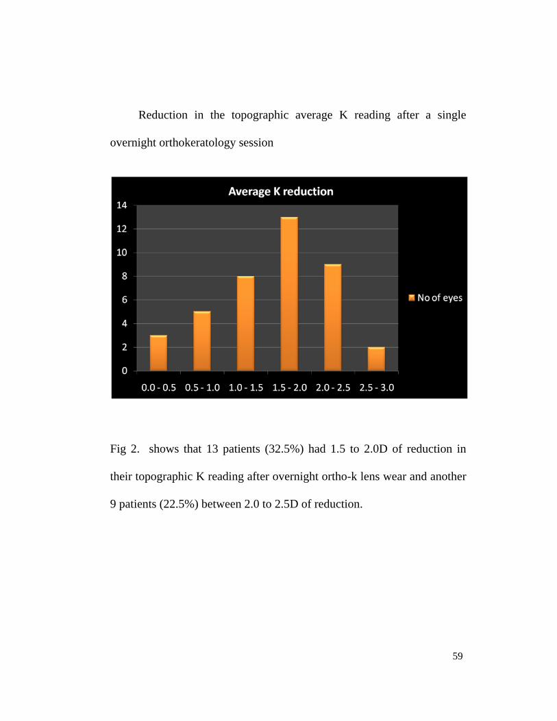
The majority of patients (32.5%) experienced 1.5 to 2.0D of
reduction in the topographic average K value after overnight
orthokeratology. 22.5% had a reduction of 2.0 to 2.5D, 20% had a
reduction of 1.0 to 1.5D, 12.5% had a reduction of 0.5 to 1.0D, 7.5% upto
0.5D and 5% between 2.5 to 3.0D in their average K readings.
59
Reduction in the topographic average K reading after a single
overnight orthokeratology session
Fig 2. shows that 13 patients (32.5%) had 1.5 to 2.0D of reduction in
their topographic K reading after overnight ortho-k lens wear and another
9 patients (22.5%) between 2.0 to 2.5D of reduction.

60
3. The change in corneal eccentricity values after single overnight
ortho-k lens application
Our patient data demonstrated that the corneal eccentricity values
tended to become less positive or more negative after a single overnight
orthokeratology session. This is coherent with the desired change in
shape of the cornea from the normal prolate to the centrally flattened
oblate shape.
Fig 3. shows the trend towards more negative values of corneal
eccentricity after single overnight ortho-k lens application indicating a
change in shape from prolate to oblate

61
4. The changes in Surface Regularity Index (SRI) following a
single overnight application of ortho-k lenses
The SRI values increased after overnight orthokeratology but did
not exceed the values considered as normal (i.e. all the values were below
1.0). This caused no significant visual impairment.
SRI changes following overnight orthokeratology
Fig 4. shows that the SRI values increased after overnight
orthokeratology but were still within the normal limits (less than 1.0).

62
5. The changes in Surface Asymmetry Index (SAI) following a
single overnight application of ortho-k lenses
The SAI values increased after overnight orthokeratology and
exceeded the normal cut-off limit of 0.5 but this asphericity caused no
significant visual impairment.
SAI changes following overnight orthokeratology
Fig 5. shows that the SAI values increased beyond the normal limit after
overnight orthokeratology indicating a trend towards more asphericity.

63
6. The duration of unaided clear vision during daytime after one
overnight orthokeratology session
Table 3
Duration of clear vision No. of patients Percentage
4 to 6 hours 3 15%
6 to 8 hours 12 60%
8 to 10 hours 3 15%
10 to 12 hours 2 10%
60% of the patients had 6 to 8 hours of unaided clear vision during
the day after one night of ortho-k lens wear. 15% patients experienced 4
to 6 hours and 8 to 10 hours of unaided clear vision each and 10% had 10
to 12 hours of clear vision during the day.

64
Duration of unaided clear vision during the day after one night of
orthokeratology
Fig 6. shows that the majority of our patients (60%) had unaided clear
vision that lasted for 6 to 8 hours following a single overnight ortho-k
lens application.

65
7. The occurrence of side effects after overnight orthokeratology
Table 4
Side effects No. of patients Percentage
None 13 65%
Irritation 3 15%
Heaviness of eyelids 2 10%
Epithelial erosion 1 5%
Haloes 1 5%
Overnight orthokeratology was not associated with any side effects
in 65% of our patients. 15% patients experienced ocular irritation, 10%
had heaviness of eyelids and 5% each (1 patient each) developed
epithelial erosion and haloes.

66
The occurrence of side effects after overnight orthokeratology
Fig 7. shows that 65% of the patients experienced no side effects. Ocular
irritation, heaviness of eyelids, epithelial erosion and haloes were seen in
15%, 10%, 5% and 5% of the patients respectively.

67
FOLLOW UP
5 patients (SEQ -2.0D to -5.0D) were followed up weekly for 1
month.
All 5 patients achieved the complete refractive correction at the
end of 1 week.
All of them reported an increase in the duration of daytime clear
vision as the weeks progressed.
Side effects like irritation and haloes resolved over one week.
Topography showed consistent flattening of the central cornea.

68
ORTHOKERATOLOGY FOR KERATOCONUS PATIENTS
Aim
a) To assess the effectiveness of orthokeratology in the refractive
correction of keratoconus patients
b) To evaluate the change in corneal topographic parameters in
keratoconus patients fitted with day-wear orthokeratology contact
lenses
Inclusion criteria
• Mild to moderate keratoconus
• No significant corneal thinning
• Patients who were not candidates for keratoplasty
• RGP lens wearers were asked to discontinue lens wear for 3 weeks before the ortho-k trial
Exclusion criteria
• Advanced keratoconus
• Significant corneal thinning
• Corneal hydrops and scarring
• Intolerance to RGP lenses

69
Materials and Methods
6 eyes of 4 patients with mild to moderate keratoconus were fitted
with ortho-k lenses and the results were evaluated.
A slit lamp examination was performed to confirm the presence of
keratoconus and to rule out significant corneal thinning/scarring.
Corneal topography was done to establish a diagnosis of
keratoconus and also for the purpose of lens fitting.
The uncorrected and best corrected visual acuity were recorded.
The fitting was done according to flat K value and the target
power.
The patients were asked to wear the lenses for 10 hours during the
day and not at night.
They were reviewed the next day and thereafter every week for a
period of 1 month.
At each visit, the visual acuity with the lenses on was measured. A
slit lamp examination was performed and corneal topography was
recorded after removing the contact lenses.

70
Observation and Analysis
Of the six keratoconic eyes fitted with orthokeratology contact
lenses, two eyes showed an improvement of 2 lines of visual acuity on the
Snellen’s chart, one eye showed an improvement of 3 lines, one eye
showed an improvement of 5 lines and two eyes had an improvement of 1
line of visual acuity on the Snellen’s chart.
The mean improvement in visual acuity was 2.33 lines on the
Snellen’s chart.
The corneal topography measured before and after the ortho-k lens
fitting did not show a significant change in the parameters.

71
RESULTS
The mean refractive correction achieved after overnight
orthokeratology was -2D.
50% of the eyes attained 1D to 2D correction and 35% eyes
achieved 2D to 3D of myopia correction.
32.5% eyes showed a flattening of 1.5D to 2.0D in the central
cornea and 22.5% showed flattening of 2D to 2.5D after a single
OrthoK application.
The corneal eccentricity values were indicative of a change in
shape from prolate to oblate.
SRI values increased after overnight orthokeratology but did not
exceed the normal range and caused no significant impairment of
vision
SAI demonstrated a trend towards asphericity without significant
visual impairment.
60% patients were able to maintain clear vision for 6 to 8 hours
during the day.

72
There were no significant side effects reported.
In case of the keratoconus patients, a mean improvement of 2.33
lines of visual acuity on Snellen’s chart was noted as compared to
the best spectacle corrected visual acuity.
Corneal topographic parameters did not show a significant change
in keratoconus patients on a day wear ortho-k schedule.
73
DISCUSSION
Overnight orthokeratology has an important application in the
treatment of myopia especially in the young patients.
It may have a role in retarding the progression of myopia and
reducing the incidence of complications of pathological myopia.54,55
RGP lenses help retard axial elongation during the growth years.56
Orthokeratology gives better visual acuity as compared to
spectacles in patients with keratoconus who can tolerate RGP contact
lenses. It does not induce any topographic changes in keratoconic corneas
when prescribed as day-wear lenses.
Advantages of orthokeratology47
It provides good unaided vision for most of the day.
It is a temporary and completely reversible procedure.
It is a good nonsurgical alternative for children and patients who
cannot undergo laser refractive surgery.
It is not painful.

74
The technique uses established contact lens procedures with
minimal risk of problems.
It is less expensive as compared to refractive surgery.
Disadvantages of orthokeratology47
It can treat only low to medium degrees of myopia.
Several visits are required over the first few months.
For the best results, careful patient compliance is required.

75
CONCLUSION
Our study demonstrates that overnight orthokeratology is a safe
and effective method for the correction of myopia. This is in accordance
with certain studies in literature.21,57,58,59 The central corneal flattening is
consistent as evidenced by topography. The asphericity induced in the
cornea during the treatment process does not significantly impair vision.
Orthokeratology provides unaided clear vision for most part of the day in
a majority of the patients. It is a viable nonsurgical alternative to
refractive surgery.
Orthokeratology improves the visual acuity over and above the best
spectacle corrected visual acuity in patients with keratoconus who can
tolerate RGP contact lenses. These lenses, when given for day-wear, do
not induce any topographic changes in patients with keratoconus.
Initiating orthokeratology at a younger age may have the beneficial
effect of retarding the progression of myopia and significantly reducing
the incidence of complications associated with pathological myopia.

BIBLIOGRAPHY
1. Rehm DS. The Myopia Myth: the truth about nearsightedness and how to prevent it. IMPA 1981; 113
2. Leach N. Orthokeratology. Clinical manual of contact lenses. Lippincott, Williams and Wilkins: Philadelphia 1999; 20 (2): 559-81
3. Mountford J. An analysis of the changes in corneal shape and refractive error induced by accelerated orthokeratology. Int Contact Lens Clinic 1997; 24: 128-43
4. Freeberg DD. Letter to the editor. J Am Optom Assoc 1976; 47: 545
5. Dickinson F. The value of microlenses in progressive myopia. Optician 1957; 133: 263-4
6. Morrison RJ. Observations on contact lenses and the progression of myopia. Contacto 1958; 2: 20-2
7. Bier N. Myopia controlled by contact lenses. Optician 1958;135-427
8. Stone J. The possible influence of contact lenses on myopia. Br J of Physiol Opt 1976; 33: 144
9. Hodd FAB. Changes in corneal shape induced by the use of alignment fitted corneal lenses. Contacto 1965; 9: 18-24
10. Gasson A, Morris J. Orthokeratology and reverse geometry lenses. The Contact lens manual – a practical guide to fitting. Elsevier 2003; 167-86
11. Kerns R. Research in orthokeratology. Part II: experimental design, protocol and method. J Am Optom Assoc 1976; 47: 1275-85

12. Kerns R. Research in orthokeratology. Part III: results and observations. J Am Optom Assoc 1976; 47: 1505-15
13. Kerns R. Research in orthokeratology. Part IV: results and observations. J Am Optom Assoc 1977; 48: 227-38
14. Kerns R. Research in orthokeratology. Part V: results and observations – recovery aspects. J Am Optom Assoc 1977; 48: 345-59
15. Kerns R. Research in orthokeratology. Part VI: statistical and clinical analysis. J Am Optom Assoc 1977; 48: 1534-47
16. Kerns R. Research in orthokeratology. Part VII: examination of techniques, procedures and control. J Am Optom Assoc 1977; 48: 1541-53
17. Kerns R. Research in orthokeratology. Part VIII: results, conclusion and discussion of techniques. J Am Optom Assoc 1978; 49: 308-14
18. Binder PS, May CH, Grant SC. An evaluation of orthokeratology. Ophthalmology 1980; 87: 729-44
19. Brand RJ, Polse KA, Schwalbe JS. The Berkeley Orthokeratology study, part I: general conduct of the study. Am J Optom Physiol Opt 1983; 60: 175-86
20. Polse KA, Brand RJ, Keener RJ et al. The Berkeley Orthokeratology study, part II: efficacy and duration. Am J Optom Physiol Opt 1983; 60: 187-98
21. Polse KA, Brand RJ, Keener RJ et al. The Berkeley Orthokeratology study, part III: safety. Am J Optom Physiol Opt 1983; 60: 321-8

22. Polse KA, Brand RJ, Vastine DW, Schwalbe JS. Corneal change accompanying orthokeratology: plastic or elastic? Results of a randomized controlled clinical trial. Arch Ophthalmol 1983; 101: 1873-8.
23. Coon LJ. Orthokeratology, part I: historical perspective. J Am Optom Assoc 1982; 53: 187-95
24. Coon LJ. Orthokeratology, part II: evaluating the Tabb method. J Am Optom Assoc 1984; 55: 409-18
25. Polse KA. Orthokeratology as a clinical procedure (editorial). Am J Optom Physiol Opt 1977; 54: 345-6
26. Tredici TJ, Shacklett DE. Orthokeratology – help or hindrance? Trans Am Acad Ophthalmol Otolaryngol 1974; 78: OP425-32
27. Eger MJ. Orthokeratolgy – fact or fiction? (editorial) J Am Optom Assoc 1975; 46: 682-3
28. Safir A. Orthokeratology, II. A risky and unpredictable treatment for a benign condition. Surv Ophthalmol 1980; 24: 291-302
29. Tredici TJ. Symposium: clinical management of physiological myopia. Role of orthokeratology: a perspective. Ophthalmology 1979; 86: 698-705
30. Binder PS. Orthokeratology In: Symposium on medical and surgical diseases of the cornea. Transactions of the New Orleans Academy of Ophthalmology. St Louis:Mosby 1998: 149-66
31. Wilson DR, Keeney AH. Corrective measures for myopia. Surv Ophthalmol 1990; 34: 294-304

32. Rubin ML, Milder B. Myopia – a treatable “disease”? Surv Ophthalmol 1976; 21: 65-9
33. Wlodyga RJ, Bryla C. Corneal molding: the easy way. Contact lens spectrum 1989; 4: 58-65
34. El Hage SG. Photokeratoscopy and controlled keratoreformation. Presented at International Symposium of Ophthalmology and Optics. Tokyo, Japan, May 1978
35. El Hage SG, Baker RN. Controlled keratoreformation for post operative radial keratotomy patients. Int Eyecare 1983; 2: 49-53
36. Bron AJ, Tripathi RC, Tripathi BJ. The cornea and sclera. Wolff’s Anatomy of the Eye and the Orbit. Chapman And Hall:London 1997; (8): 7: 233
37. Arffa RC. Anatomy. Grayson’s diseases of the cornea. Mosby:St Louis 1997; (4) 6-7
38. Edelhauser HF, Ubels JL, Hejny C. Cornea and sclera. Adler’s Physiology of the eye. Clinical application. Mosby: St Louis 1997; 10 (4): 47-95
39. Bron AJ, Tripathi RC, Tripathi BJ. The cornea and sclera. Wolff’s Anatomy of the Eye and the Orbit. Chapman And Hall:London 1997; (8): 7: 245
40. Hill RM, Fatt I. How dependent is the cornea on the atmosphere? J Am Optom Assoc 1964; 35: 873
41. Efron N, Carney LG. Oxygen levels beneath the closed eyelid. Invest Ophthalmol Vis Sci 1979; 18: 93

42. Holden BA, Sweeney DF. The oxygen tension and temperature of the superior palpebral conjunctiva. Acta Ophthalmol 1985; 63:100
43. Klyce SD. Stromal lactate accumulation can account for corneal oedema osmotically following epithelial hypoxia in the rabbit. J Physiol 1981; 321: 49
44. Mandell RB, Farrell R. Corneal swelling at low atmospheric oxygen pressures. Invest Ophthalmol Vis Sci 1980; 19: 697
45. Bron AJ, Tripathi RC, Tripathi BJ. The cornea and sclera. Wolff’s Anatomy of the Eye and the Orbit. Chapman And Hall:London 1997; (8): 7: 234
46. Abrams D. Myopia. Duke-Elder’s practice of refraction. 1989; 10: 53-64
47. A guide to overnight orthokeratology. Polymer Technology Corporation, USA 2002; 9-32.
48. Swarbrick HA, Wong G, O’Leary DJ. Corneal response to orthokeratology. Optom Vis Sci 1998; 75: 791-9
49. Corbett, Rosen, O’Bratt. Corneal topography – principles and applications
50. Koch DD, Haft EA. Introduction to corneal topography. Corneal topography: The state of the art. Jaypee:New Delhi 1996; 1: 10
51. Map display methods and applications. Operator manual. Topographic Modeling System TMS 4; Japan 2003: 64-7
52. Klyce, Stephen D. (New Orleans, LA, US), Smolek, Michael K. (New Orleans, LA, US), Fujieda, Masanao (Toyohashi, JP). Corneal topography analysis system, United States. Nidek Co., Ltd. (Aichi, JP). Patent no: 737096 May 2008. http://www.freepatentsonline. com/7370969.html

53. Gemoules G. Therapeutic effects of contact lenses on complicated eyes after refractive surgery. Eye and Contact lens. Lippincott, Williams and Wilkins. January 2005.
54. Myopia Control Study by using Orthokeratology (Reverse Geometry) Lenses by Tommy Yee O.D. FIOS, Houston, Texas , USA (5 years Study) - showed a success rate of 97% in control the progression of myopia (519 patients).
55. Perrigin J, Perrigin D, Quintero S, Grosvenor T. Silicone-acrylate contact lenses for myopia control : 3 years results. Optom and Vis Sci 1990; 67 (10): 764-9.
56. Michael Feldmann. BRACE refractive therapy. Contact lens spectrum. May 2001
57. Mika R, Morgan B, Cron M et al. Safety and efficacy of overnight orthokeratology in myopic children. Optometry 2007 May; 78(5): 225-31.
58. Rah MJ, Jackson JM, Jones LA et al. Overnight orthokeratology: preliminary results of the Lenses and Overnight Orthokeratology (LOOK) study. Optom Vis Sci 2002 Sep; 79(9): 598-605
59. Walline JJ, Rah MJ, Jones LA. The Children's Overnight Orthokeratology Investigation (COOKI) pilot study. Optom Vis Sci 2004 Jun; 81(6): 407-13

PROFORMA
Date: OP No:
Name: Age/Sex:
Right eye Left eye Head posture Facial symmetry Extraocular movements Anterior segment
Eyelids Conjunctiva Cornea Iris AC Pupil diameter Lens
Schirmer’s test Tear film break up time Fundus examination
RIGHT EYE LEFT EYE UCVA: BCVA:
UCVA: BCVA:
Topography: Flat K: E value: Steep K:
Topography: Flat K: E value: Steep K:
Base Curve:
Base Curve:
OK lens: Status 1st fit:
OK lens: Status 1st fit:
Vn with OK lens:
Vn with OK lens:
POK Day 1: UCVA: BCVA: Flat K: E value: Steep K:
POK Day 1: UCVA: BCVA: Flat K: E value: Steep K:

Right eye Left eye
Lens centration Lens movement
FOLLOW UP
RIGHT EYE LEFT EYE POK Week 1: UCVA: BCVA: Flat K: Steep K: E value:
POK Week 1: UCVA: BCVA: Flat K: Steep K: E value:
POK Month 1: UCVA: BCVA: Flat K: Steep K: E value:
POK Month 1: UCVA: BCVA: Flat K: Steep K: E value:
POK Month 3: UCVA: BCVA: Flat K: Steep K: E value:
POK Month 3: UCVA: BCVA: Flat K: Steep K: E value:
POK Month 6: UCVA: BCVA: Flat K: Steep K: E value:
POK Month 6: UCVA: BCVA: Flat K: Steep K: E value:
POK Year 1: UCVA: BCVA: Flat K: Steep K: E value:
POK Year 1: UCVA: BCVA: Flat K: Steep K: E value:
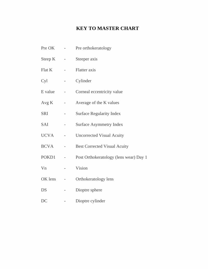
KEY TO MASTER CHART
Pre OK - Pre orthokeratology
Steep K - Steeper axis
Flat K - Flatter axis
Cyl - Cylinder
E value - Corneal eccentricity value
Avg K - Average of the K values
SRI - Surface Regularity Index
SAI - Surface Asymmetry Index
UCVA - Uncorrected Visual Acuity
BCVA - Best Corrected Visual Acuity
POKD1 - Post Orthokeratology (lens wear) Day 1
Vn - Vision
OK lens - Orthokeratology lens
DS - Dioptre sphere
DC - Dioptre cylinder

LIST OF SURGERIES PERFORMED
S. No.
Name Age / Sex
OP / IP No.
Diagnosis Surgery performed
1. Thiruvaimozhi 75/F 407842 BE – MC RE – ECCE with PCIOL
2. Pushpa 60/F 411326 BE – MC LE – ECCE with PCIOL
3. Devi 65/F 411564 BE – PCC LE – ECCE with PCIOL
4. Govindasamy 62/M 412232 RE - MC LE – IMC
RE – ECCE with PCIOL
5. Durairaj 80/M 413582 BE – MC RE – ECCE with PCIOL
6. Palanivel 24/M 1573 RE – Upper lid tear
RE – Upper lid tear suturing
7. Sujatha 18/F 36891 RE – Lower lid chalazion
RE – Chalazion I and C
8. Perumal 57/M 42675 Left sided Chronic dacryocystitis
Left sided DCT
9. Vadivel 47/M 42445 RE – Pterygium
RE – Pterygium excision with conjunctival autograft
10. Lakshmi 55/F 420098 BE – IMC RE – SICS with PCIOL
11. Venkatesh 64/M 420580 RE – PCC LE – Pseudophakia
RE – SICS with PCIOL

12. Patabi 68/M 420720 BE – Nuclear cataract
RE – SICS with PCIOL
13. Ponnusamy 61/M 421086 RE – Pseudophakia LE – PCC
LE – SICS with PCIOL
14. Rani 68/F 421198 RE – IMC LE - Pseudophakia
RE – SICS with PCIOL
15. Rangasamy 54/M 45653 Right sided Chronic dacryocystitis
Left sided DCT
16. Rajesh 35/M 422671 RE – Fungal corneal ulcer
RE- TKP
17. Subhadra 37/F 37658 RE – Pterygium
RE – Pterygium excision with conjunctival autograft
18. Peter 53/M 1786 LE – Scleral tear with uveal tissue prolapse
LE - Scleral tear suturing
19. Anandan 46/M 422702 LE – Non healing fungal corneal ulcer
LE – TKP
20. Selvi 20/F 422783 LE – Perforated fungal corneal ulcer
LE – TKP
21. Ganeshan 30/M 422852 RE – Non healing fungal corneal ulcer
RE – TKP
22. Siva 32/M 2208 RE – Ruptured globe
RE – Corneo-scleral tear

suturing
23. Lakshmi M 63/F 423102 RE – PCC LE – Pseudophakia
RE – Phaco with PCIOL
24. Ravannammal 73/F 423126 BE – PCC RE – Phaco with PCIOL
25. Parthiban 65/M 423227 RE – HMC LE – IMC
RE – SICS with PCIOL
26. Parvathi 58/F 423265 BE – IMC LE – Phaco with PCIOL
27. Sushila 60/F 423311 RE – Pseudophakia LE – PCC
LE – Phaco with PCIOL
28. Manikam 55/M 423388 BE- PCC RE – Phaco with PCIOL
29. Janaki 23/F 21486 LE – Upper lid mass [suspected lipoma]
LE – Excision biopsy of the upper lid mass
30. Palayam 74/M 32561 Right sided lacrimal abscess
I and D of the lacrimal abscess
31. Pavithra 24/F 43265 RE – Pterygium
RE – Pterygium excision with conjunctival autograft
32. Shekhar 47/M 2436 LE – Ruptured globe
LE – Corneo-scleral tear suturing done

BE – Both Eyes
RE – Right Eye
LE – Left Eye
MC – Mature Cataract
IMC – Immature Cataract
HMC – Hyper Mature Cataract
PCC – Posterior Cortical Cataract
ECCE – Extra Capsular Cataract Extraction
SICS – Small Incision Cataract Surgery
PCIOL – Posterior Chamber Intraocular Lens
Phaco – Phacoemulsification
DCT - Dacryocystectomy
TKP – Therapeutic Keratoplasty
I and C – Incision and Curettage
I and D – Incision and Drainage

Fig 1. Photomicrograph of normal human cornea showing the stratified
squamous epithelium (EP), Bowman’s membrane (BWM), stroma (STR),
Descemet’s membrane (DM) and endothelium (EN) 37

Fig 3. Corneal dimensions37
Fig 4. Corneal cap on topography
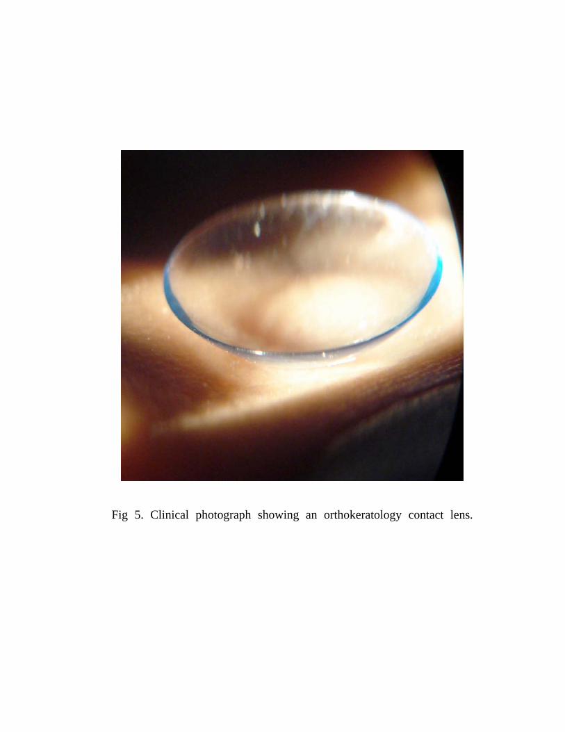
Fig 5. Clinical photograph showing an orthokeratology contact lens.

Fig 6. Four curve reverse geometry lens showing the base curve, reverse
curve, alignment curve and peripheral curve.
Fig 7. Diagram showing the BOZD (back optic zone diameter) and
BOZR (back optic zone radius) of a reverse geometry lens.
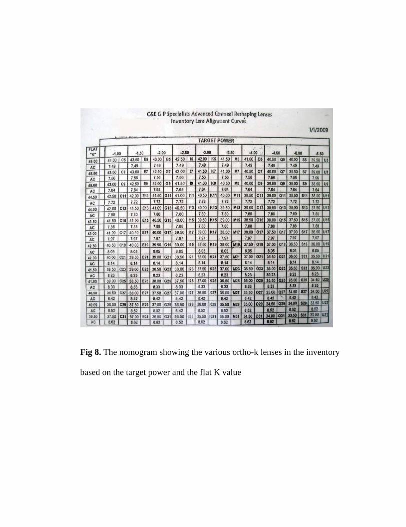
Fig 8. The nomogram showing the various ortho-k lenses in the inventory
based on the target power and the flat K value

Fig 9. Slit lamp photograph showing the characteristics of an ideal fit of
orthokeratology contact lens after staining with 2% Fluorescein dye

Fig 10a. Corneal topography picture before fitting orthokeratology lens.
Fig 10b. Corneal topography picture taken after overnight ortho-k lens
wear showing the flattening of the central cornea and a bull’s eye pattern
of centration.

Fig 11a. Corneal topography picture before fitting orthokeratology lens.
Fig 11b. Corneal topography picture taken after overnight ortho-k lens
wear showing central corneal flattening.

Fig. 12 Slit lamp photograph showing air bubble inferiorly under the contact lens
Fig. 13 Slit lamp photograph showing steep fit of the contact lens

Fig. 14 Corneal topography picture showing eccentric flattening
Fig. 15 Corneal topography picture showing smiley face pattern

No Name Steep K Flat K Cyl E value Avg K SRI SAI Steep K Flat K Cyl E value Avg K SRI SAI1 Amudha PreOK 44.34 @ 89 43.44 @ 179 0.9 0.81 43.89 0.16 0.21 44.72 @ 100 43.54 @ 10 1.18 0.77 44.13 0.08 0.18
POKD1 43.47 @ 94 42.54 @ 4 0.93 0.37 43.01 0.24 0.33 42.96 @ 101 41.77 @ 11 1.19 -0.18 42.36 0.32 0.362 Bharatkumar 44.25 @ 96 43.88 @ 6 0.38 0.66 44.06 0.16 0.22 44.59 @ 96 43.85 @ 6 0.74 0.57 44.22 0.23 0.78
POKD1 42.90 @ 11 41.50 @ 101 1.39 -0.22 42.2 0.66 0.55 42.88 @ 69 42.62 @ 159 0.27 -0.03 42.75 0.39 0.933 Janani 43.17 @ 82 42.56 @ 172 0.61 0.42 42.87 0.12 0.23 43.61 @ 101 42.91 @ 11 0.7 0.31 43.26 0.42 0.2
POKD1 40.41 @ 107 39.74 @ 17 0.67 -0.55 40.07 0.47 0.63 42.01 @ 94 41.24 @ 4 0.77 -0.33 41.63 0.5 0.974 Gayathri 45.69 @103 44.61 @ 13 1.08 0.54 45.15 0.03 0.38 45.68 @ 89 45.02 @ 179 0.65 0.56 45.35 0.07 0.19
POKD1 44.22 @101 42.82 @ 11 1.4 -0.46 43.52 0.02 0.27 43.81 @ 68 43.09 @ 158 0.72 -0.47 43.45 0.18 0.385 Sathyendran 44.23 @ 155 43.70 @ 65 0.53 0.54 43.96 0.07 0.1 43.8 43.8 0 0.55 43.8 0.07 0.12
POKD1 42.52 @ 135 42.12 @ 45 0.4 -0.31 42.32 0.06 0.17 41.89 @ 55 41.68 @ 145 0.21 -0.53 41.79 0.06 0.216 Thirumalaiselv 45.16 @ 37 44.81 @ 127 0.36 0.58 44.99 0.16 0.22 45.64 @ 4 45.05 @ 94 0.59 0.59 45.35 0 0.24
POKD1 42.98 @ 167 42.17 @ 77 0.81 -0.49 42.57 0.56 0.47 44.58 @ 4 43.50 @ 94 1.09 0.48 44.04 0.84 1.297 Adhiba 42.61 @ 73 41.91 @ 163 0.7 0.6 42.26 0.11 0.28 42.59 @ 101 41.60 @ 11 0.99 0.57 42.1 0.07 0.1
POKD1 40.92 @ 80 39.64 @ 170 1.28 -0.44 40.28 0.25 0.26 40.75 @ 101 39.19 @ 11 1.56 -0.05 39.97 0.02 0.288 Prabuvenkatesh 42.50 @138 41.69 @ 48 0.81 0.53 42.09 0.1 0.38 42.56 @ 53 42.06 @ 143 0.5 0.59 42.31 0.04 0.28
POKD1 40.97 @ 142 40.38 @ 52 0.59 -0.17 40.67 0.48 0.48 40.93 40.93 0 -0.28 40.93 0.06 0.269 Shreeyeh 42.58 @ 87 41.63 @ 177 0.95 0.9 42.11 0.13 0.22 43.16 @ 100 42.02 @ 10 1.14 0.68 42.59 0.03 0.29
POKD1 42.29 @ 90 41.67 @ 180 0.62 0.32 41.98 0.61 0.31 42.11 @ 108 41.09 @ 18 1.02 -0.36 41.6 0.56 0.510 Meenadevi 44.45 @ 101 43.85 @ 11 0.61 0.46 44.15 0.27 0.3 44.81 @ 83 44.01 @ 173 0.79 0.46 44.41 0.11 0.25
POKD1 42.03 @ 98 41.24 @ 8 0.79 -0.62 41.64 0.55 1.67 42.67 @ 97 41.37 @ 7 1.3 -0.45 42.02 0.61 1.7511 Nithin 44.69 @ 89 43.96 @ 179 0.73 0.55 44.33 0.2 0.34 45.13 @ 101 44.33 @ 11 0.79 0.58 44.73 0.01 0.21
POKD1 44.00 @ 94 42.60 @ 4 1.41 -0.07 43.3 0.04 0.39 43.78 @ 82 42.75 @ 172 1.04 -0.04 43.26 0.07 0.1712 Bama 44.62 @ 96 44.25 @ 6 0.36 0.45 44.43 0.06 0.21 45.46 @ 83 45.24 @ 173 0.22 0.48 45.35 0.67 0.33
POKD1 44.30 @ 80 43.98 @ 170 0.32 0.3 44.14 0.32 0.43 44.90 @ 96 44.17 @ 6 0.73 -0.23 44.53 0.69 0.81
Right Eye Left Eye

No Name Steep K Flat K Cyl E value Avg K SRI SAI Steep K Flat K Cyl E value Avg K SRI SAIRight Eye Left Eye
13 Anusha 45.29 @ 3 42.56 @ 93 2.73 0.84 43.92 0.88 0.35 43.27 @ 80 42.40 @ 170 0.87 0.74 42.83 0.06 0.12POKD1 42.49 @ 101 41.88 @ 11 0.61 0.45 42.19 0.47 0.56 42.79 @ 73 42.07 @ 163 0.73 0.5 42.43 0.28 0.57
14 Shakthi 44.08 @ 14 43.18 @ 104 0.9 0.64 43.63 0.24 0.4 43.18 @ 93 42.13 @ 3 1.05 0.72 42.65 0.18 0.3442.16 @ 20 42.02 @ 110 0.14 -0.29 42.09 0.51 0.56 40.97 @ 106 40.23 @ 16 0.74 -0.3 40.6 0.21 0.52
15 Sujatha 44.10 @ 97 43.60 @ 7 0.5 0.57 43.85 0.08 0.24 43.00 @ 1 42.40 @ 91 0.6 0.44 42.7 0.04 0.3843.35 @ 100 42.75 @ 10 0.6 -0.12 43.05 0.12 0.42 42.4 @ 8 41.54 @ 98 0.86 -0.26 41.97 0.11 0.49
16 Manohar 42.02 @ 165 41.22 @ 75 0.8 0.73 41.62 0.3 0.32 42.35 @ 70 41.65 @ 160 0.7 0.64 42 0.13 0.2140.02 @ 70 38.82 @ 160 1.2 -0.24 39.42 0.41 0.46 40.04 @ 85 39.00 @ 175 1.04 -0.38 39.52 0.2 0.38
17 Pavithra 42.00 @ 105 41.50 @ 15 0.5 0.48 41.75 0.14 0.19 43.10 @ 10 42.33 @ 100 0.77 0.55 42.71 0.28 0.440.12 @ 96 39.22 @ 6 0.9 -0.3 39.67 0.48 0.24 40.67 @ 80 39.79 @ 170 0.88 -0.22 40.23 0.41 0.54
18 Natarajan 44.70 @ 112 44.30 @ 22 0.4 0.59 44.5 0.22 0.2 43.88 @ 15 43.48 @ 105 0.4 0.48 43.68 0.07 0.1343.07 @ 95 42.27 @ 5 0.8 -0.21 42.67 0.6 0.36 42.49 @ 10 41.59 @ 100 0.9 -0.38 42.04 0.13 0.28
19 Suchithra 41.90 @ 96 41.40 @ 6 0.5 0.44 41.65 0.4 0.14 41.88 @ 4 41.20 @ 94 0.68 0.52 41.54 0.04 0.2440.18 @ 25 39.58 @ 115 0.6 -0.28 39.88 0.2 0.26 40.42 @ 15 39.34 @ 105 1.08 -0.22 39.88 0.09 0.38
20 Nandini 42.65 @ 30 42.15 @ 120 0.5 0.5 42.4 0.05 0.24 42.77 @ 120 42.37 @ 30 0.4 0.64 42.57 0.3 0.141.31 @ 45 40.31 @ 135 1 -0.36 40.81 0.15 0.5 40.59 @ 104 39.79 @ 14 0.8 -0.18 40.19 0.36 0.18
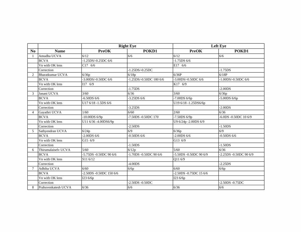
No Name PreOK POKD1 PreOK POKD11 Amudha UCVA 6/12 6/6 6/12 6/6
BCVA -1.25DS/-0.25DC 6/6 -1.75DS 6/6Vn with OK lens C17 6/6 E17 6/6Correction -1.25DS/-0.25DC -1.75DS
2 Bharatkumar UCVA 6/36p 6/18p 6/36P 6/18PBCVA -3.00DS/-0.50DC 6/6 -1.25DS/-0.50DC 180 6/6 -3.00DS/-0.50DC 6/6 -1.00DS/-0.50DC 6/6Vn with OK lens I17 6/9 K17 6/9Correction -1.75DS -2.00DS
3 Janani UCVA 3/60 6/36 3/60 6/36pBCVA -6.50DS 6/6 -3.25DS 6/6 -7.00DS 6/6p -5.00DS 6/6pVn with OK lens U17 6/18 -1.5DS 6/6 U19 6/18 -1.25DS6/6pCorrection -3.25DS -2.00DS
4 Gayathri UCVA 1/60 6/60 2/60 6/36BCVA -10.00DS 6/9p -7.50DS -0.50DC 170 -7.50DS 6/9p -6.0DS -0.50DC 10 6/9Vn with OK lens U11 6/36 -4.00DS6/9p U9 6/24p -2.00DS 6/9Correction -2.50DS -1.50DS
5 Sathyendran UCVA 6/24p 6/9 6/36p 6/9BCVA -2.00DS 6/6 -0.50DS 6/6 -2.00DS 6/6 -0.50DS 6/6Vn with OK lens G15 6/9 G13 6/9Correction -1.50DS -1.50DS
6 Thirumalaiselv UCVA 5/60 6/12p 5/60 6/36BCVA -5.75DS -0.50DC 90 6/6 -1.70DS -0.50DC 90 6/6 -5.50DS -0.50DC 90 6/9 -2.25DS -0.50DC 90 6/9Vn with OK lens S11 6/12 Q11 6/9Correction -4.00DS -2.25DS
7 Adhiba UCVA 6/60 6/6p 6/60 6/6pBCVA -2.50DS -0.50DC 150 6/6 -2.50DS -0.75DC 15 6/6Vn with OK lens I23 6/6p I23 6/6pCorrection -2.50DS -0.50DC -2.50DS -0.75DC
8 Prabuvenkatesh UCVA 6/36 6/6 6/36 6/6
Right Eye Left Eye

No Name PreOK POKD1 PreOK POKD1Right Eye Left Eye
BCVA -1.75DS 6/6 -1.75DS 6/6Vn with OK lens G21 6/6 E21 6/6Correction -1.75DS -1.75DS
9 Shreeyeh UCVA 5/60 6/36 5/60 6/12BCVA -1.75DS -0.50DC 180 6/6 -1.00DS -0.50DC 180 6/6 -1.75DS -0.75DC 180 6/6 -1.00DS -0.25DC 180 6/6Vn with OK lens E25 6/6 With 2nd fit G21 6/6 C25 6/6 With 2nd fit E25 6/6Correction -0.75DS -0.75DS -0.50DC
10 Meenadevi UCVA 3/60 6/9 3/60 6/9BCVA -4.25DS 6/6 -1.50DS 6/6 -4.50DS 6/6 -1.00DC 180 6/6Vn with OK lens O13 6/9 Q13 6/9Correction -2.75DS -3.50DDS
11 Nithin UCVA 6/60 6/12 6/60 6/12BCVA -3.25DS -0.50DC 145 6/6 -1.00DS 6/6 -3.25DS 6/6 -1.00DS 6/6Vn with OK lens M13 6/9 K13 6/9Correction -2.25DS -0.50DC -2.25DS
12 Bama UCVA 5/60 6/36 5/60 6/18BCVA -4.00DS 6/6 -2.50DS 6/6 -3.50DS 6/6 -2.00DS 6/6Vn with OK lens O13 6/6 O9 6/6Correction -1.50DS -1.50DS
13 Anusha UCVA 6/60 6/36p 6/60 6/9BCVA -2.50DS -0.25DC 25 6/6 -2.00DS 6/6 -3.00DS -0.50DC 157 6/6 -1.00DS 6/6Vn with OK lens I17 6/18 K17 6/6pCorrection -0.50DS -0.25DC -2.00DS -0.50DC
14 Shakthi UCVA 6/60 6/12 5/60 6/9pBCVA -2.75DS 6/6 -0.50DS 6/6 -3.00DS 6/6 -0.50DS 6/6Vn with OK lens K17 6/9 K21 6/9Correction -2.25DS -2.50DS
15 Sujatha UCVA 4/60 6/12 4/60 6/12BCVA -4.00DS 6/6 -2.25DS 6/6 -4.00DS 6/6 -2.00DS 6/6

No Name PreOK POKD1 PreOK POKD1Right Eye Left Eye
Vn with OK lens O15 6/9p 019 6/9pCorrection -1.75DS -2.00DS
16 Manohar UCVA 6/60 6/6p 6/60 6/6pBCVA -3.50DS -0.50DC 70 6/9 -1.00DS -0.50DC 70 -3.25DS -0.50DC 160 6/6 -1.00DS -0.50DC 160 6/6Vn with OK lens M25 6/6p M23 6/6pCorrection -2.50DS -2.25DS
17 Pavithra UCVA 6/24 6/6 6/24 6/6BCVA -1.25DS 6/6 6/6 -1.50DS 6/6 6/6Vn with OK lens C23 6/6 E21 6/6Correction -1.25DS -1.50DS
18 Natarajan UCVA 5/60 6/9 5/60 6/9BCVA -3.75DS 6/6 -0.75DS 6/6 -3.75DS 6/6 -0.75DS 6/6Vn with OK lens O13 6/6 O15 6/6pCorrection -3.00DS -3.00DS
19 Suchithra UCVA 6/18 6/6 6/12 6/6BCVA -1.50DS 6/6 6/6 -1.25DS 6/6 6/6Vn with OK lens E25 6/6 C25 6/6Correction -1.50DS -1.25DS
20 Nandini UCVA 6/24p 6/9 6/24 6/9BCVA -2.50DS 6/6 -0.50DS 6/6 -2.00DS 6/6 -0.25DS 6/6pVn with OK lens I21 6/6 G21 6/6pCorrection -2.00DS -1.75DS


