Top Med Chem 7: 47–124DOI: 10.1007/7355_2011_13# Springer-Verlag Berlin Heidelberg 2011Published online: 29 April 2011
The Medicinal Chemistry of Tuberculosis
Chemotherapy
Gwendolyn A. Marriner, Amit Nayyar, Eugene Uh, Sharon Y. Wong,
Tathagata Mukherjee, Laura E. Via, Matthew Carroll, Rachel L. Edwards,
Todd D. Gruber, Inhee Choi, Jinwoo Lee, Kriti Arora, Kathleen D. England,
Helena I.M. Boshoff, and Clifton E. Barry III
Abstract The development of effective chemotherapy for the treatment of tuber-
culosis (TB) began in the 1940s and has been reinvigorated recently due to concern
regarding the emergence of highly drug-resistant TB strains. This chapter explores
the medicinal chemistry efforts that gave rise to current frontline and second-line
drugs in global use today and attempts to comprehensively summarize ongoing
discovery and lead optimization programs being conducted in both the private and
the public sector. TB has a large number of disease-specific considerations and
constraints that introduce significant complexity in drug discovery efforts. Concep-
tually, the disease encompasses all the drug discovery challenges of both infectious
diseases and oncology, and integrating these considerations into programs that
often demand collaboration between industry and academia is both challenging
and rewarding.
Contents
1 Introduction . . . . . . . . . . . . . . . . . . . . . . . . . . . . . . . . . . . . . . . . . . . . . . . . . . . . . . . . . . . . . . . . . . . . . . . . . . . . . . . . 48
1.1 TB: A Global Epidemic . . . . . . . . . . . . . . . . . . . . . . . . . . . . . . . . . . . . . . . . . . . . . . . . . . . . . . . . . . . . . . 48
1.2 The Medical History of Current TB Chemotherapy . . . . . . . . . . . . . . . . . . . . . . . . . . . . . . . . . 49
1.3 The Emergence of Drug-Resistant TB . . . . . . . . . . . . . . . . . . . . . . . . . . . . . . . . . . . . . . . . . . . . . . . 52
1.4 Special Challenges in TB Drug Development . . . . . . . . . . . . . . . . . . . . . . . . . . . . . . . . . . . . . . . 54
2 The Development of Commonly Used First-Line
and Second-Line Agents for TB Therapy . . . . . . . . . . . . . . . . . . . . . . . . . . . . . . . . . . . . . . . . . . . . . . . . . . 55
2.1 Rifamycins . . . . . . . . . . . . . . . . . . . . . . . . . . . . . . . . . . . . . . . . . . . . . . . . . . . . . . . . . . . . . . . . . . . . . . . . . . . 55
2.2 Isoniazid . . . . . . . . . . . . . . . . . . . . . . . . . . . . . . . . . . . . . . . . . . . . . . . . . . . . . . . . . . . . . . . . . . . . . . . . . . . . . 57
2.3 Thioisonicotinamides and Thiosemicarbazones . . . . . . . . . . . . . . . . . . . . . . . . . . . . . . . . . . . . . . 58
2.4 Pyrazinamide . . . . . . . . . . . . . . . . . . . . . . . . . . . . . . . . . . . . . . . . . . . . . . . . . . . . . . . . . . . . . . . . . . . . . . . . 58
2.5 Cycloserine . . . . . . . . . . . . . . . . . . . . . . . . . . . . . . . . . . . . . . . . . . . . . . . . . . . . . . . . . . . . . . . . . . . . . . . . . . 59
This work was funded in part by the Division for Intramural Research, NIAID.
G.A. Marriner, A. Nayyar, E. Uh, S.Y. Wong,
T. Mukherjee, L.E. Via, M. Carroll, R.L. Edwards, T.D. Gruber, I. Choi, J. Lee, K. Arora, K.D.
England, H.I.M. Boshoff, and C.E. Barry III (*)
Tuberculosis Research Section, National Institute of Allergy and Infectious Disease, NIH,
Bethesda, MD, USA
47

2.6 Para-Aminosalicylic Acid . . . . . . . . . . . . . . . . . . . . . . . . . . . . . . . . . . . . . . . . . . . . . . . . . . . . . . . . . . . 60
2.7 Capreomycin . . . . . . . . . . . . . . . . . . . . . . . . . . . . . . . . . . . . . . . . . . . . . . . . . . . . . . . . . . . . . . . . . . . . . . . . . 60
2.8 Aminoglycosides . . . . . . . . . . . . . . . . . . . . . . . . . . . . . . . . . . . . . . . . . . . . . . . . . . . . . . . . . . . . . . . . . . . . . 61
3 Classes of Compounds in Clinical Development . . . . . . . . . . . . . . . . . . . . . . . . . . . . . . . . . . . . . . . . . . 62
3.1 Nitroimidazoles . . . . . . . . . . . . . . . . . . . . . . . . . . . . . . . . . . . . . . . . . . . . . . . . . . . . . . . . . . . . . . . . . . . . . . 62
3.2 Diarylquinolines . . . . . . . . . . . . . . . . . . . . . . . . . . . . . . . . . . . . . . . . . . . . . . . . . . . . . . . . . . . . . . . . . . . . . 67
3.3 Oxazolidinones . . . . . . . . . . . . . . . . . . . . . . . . . . . . . . . . . . . . . . . . . . . . . . . . . . . . . . . . . . . . . . . . . . . . . . 69
3.4 Fluoroquinolones . . . . . . . . . . . . . . . . . . . . . . . . . . . . . . . . . . . . . . . . . . . . . . . . . . . . . . . . . . . . . . . . . . . . 72
3.5 Ethylenediamines . . . . . . . . . . . . . . . . . . . . . . . . . . . . . . . . . . . . . . . . . . . . . . . . . . . . . . . . . . . . . . . . . . . . 77
4 Series in Preclinical Development . . . . . . . . . . . . . . . . . . . . . . . . . . . . . . . . . . . . . . . . . . . . . . . . . . . . . . . . . 80
4.1 Benzothiazinones . . . . . . . . . . . . . . . . . . . . . . . . . . . . . . . . . . . . . . . . . . . . . . . . . . . . . . . . . . . . . . . . . . . . 80
4.2 Nucleosides . . . . . . . . . . . . . . . . . . . . . . . . . . . . . . . . . . . . . . . . . . . . . . . . . . . . . . . . . . . . . . . . . . . . . . . . . . 81
4.3 Macrolides . . . . . . . . . . . . . . . . . . . . . . . . . . . . . . . . . . . . . . . . . . . . . . . . . . . . . . . . . . . . . . . . . . . . . . . . . . . 83
4.4 b-Lactams . . . . . . . . . . . . . . . . . . . . . . . . . . . . . . . . . . . . . . . . . . . . . . . . . . . . . . . . . . . . . . . . . . . . . . . . . . . . 85
4.5 Rhiminophenazines . . . . . . . . . . . . . . . . . . . . . . . . . . . . . . . . . . . . . . . . . . . . . . . . . . . . . . . . . . . . . . . . . . 88
4.6 Pyrroles . . . . . . . . . . . . . . . . . . . . . . . . . . . . . . . . . . . . . . . . . . . . . . . . . . . . . . . . . . . . . . . . . . . . . . . . . . . . . . 89
4.7 Deazapteridines . . . . . . . . . . . . . . . . . . . . . . . . . . . . . . . . . . . . . . . . . . . . . . . . . . . . . . . . . . . . . . . . . . . . . . 90
5 Critical Issues in TB Drug Development . . . . . . . . . . . . . . . . . . . . . . . . . . . . . . . . . . . . . . . . . . . . . . . . . . 91
5.1 Cell Penetration . . . . . . . . . . . . . . . . . . . . . . . . . . . . . . . . . . . . . . . . . . . . . . . . . . . . . . . . . . . . . . . . . . . . . . 91
5.2 Animal Models for Evaluation . . . . . . . . . . . . . . . . . . . . . . . . . . . . . . . . . . . . . . . . . . . . . . . . . . . . . . . 93
5.3 Pharmacological Models for Antitubercular Drugs . . . . . . . . . . . . . . . . . . . . . . . . . . . . . . . . . . 97
5.4 Clinical Development Methodologies . . . . . . . . . . . . . . . . . . . . . . . . . . . . . . . . . . . . . . . . . . . . . . . 99
6 Concluding Remarks . . . . . . . . . . . . . . . . . . . . . . . . . . . . . . . . . . . . . . . . . . . . . . . . . . . . . . . . . . . . . . . . . . . . . . 101
References . . . . . . . . . . . . . . . . . . . . . . . . . . . . . . . . . . . . . . . . . . . . . . . . . . . . . . . . . . . . . . . . . . . . . . . . . . . . . . . . . . . . 102
1 Introduction
1.1 TB: A Global Epidemic
Tuberculosis (TB) is the second major cause of death due to an infectious disease in
adults worldwide with nine million new cases and close to 1.8 million deaths
annually [1]. TB is caused by Mycobacterium tuberculosis (MTb), an airborne
pathogen transmitted among humans which infects macrophages in the lungs.
Two possible outcomes follow macrophage infection: (1) the infected macrophage
can be recognized by effectors of the innate immune system and eradicated; or (2)
the bacilli may further multiply in the cell, ultimately leading to its destruction and
the infection of newmacrophages drawn to the site of infection. The second scenario
may initiate T cell-mediated adaptive immunity enabling the host to eradicate the
bacilli at the initial site of infection. Failure of adaptive immunity to eradicate the
bacilli leads to uncontrolled growth of the organism and subsequent spread through
the lymphatic system to secondary sites. These sites may be in the lung or in some
cases in extra-pulmonary sites, which is manifested as clinical disease with various
degrees of severity which, if not treated, kills more than 50% of patients. There is
also an intermediate situation wherein adaptive immunity may be able to contain the
growth of the organism by controlling its metabolism for years, even decades, until
waning of immunity allows reactivation of this latent form of disease [2]. More
recently, researchers have begun to suspect that latent disease may not be a single
48 G.A. Marriner et al.

metastable state but rather a subtle guerilla war with waxing and waning local
battles on a small scale resulting in a “spectrum” of subclinical active disease [2].
Based on the global incidence of a positive response in the tuberculin skin test,
which is associated with adaptive immunity, it is estimated that about two billion
people are latently infected with MTb [1]. Of these two billion people, about 10%
will develop active tuberculosis in their lifetime, although HIV infection dramati-
cally increases this risk to a 10% annual risk of conversion to active TB [3]. TB is
the major cause of death in HIV-infected individuals. A person with contagious
pulmonary TB infects 10–15 more people on average, which created tuberculosis
epidemics in the developed world until the advent of chemotherapy. Isolation of
patients resulted in limited success in TB control through the establishment of
sanatoria in the mid-nineteenth century. Sanatoria patients would occasionally
achieve spontaneous resolution of the disease, although subsequent relapse rates
were high, highlighting the chronic and dynamic nature of this infection.
Albert Calmette and Camille Guerin produced a strain of Mycobacterium bovis(bacille Calmette-Guerin, BCG) by serial passaging of an isolate of the related
bacillus that causes TB in cattle on potato–bile–glycerin media until it was no longer
virulent in laboratory animals. Vaccination started in the 1920s, but the efficacy of
the vaccine varied greatly depending on factors such as geographic location and
strain of BCG used for vaccination [3, 4]. Large-scale clinical trials throughout the
world have shown that the vaccine protects against severe forms of TB in children
but does not protect against the development of adult pulmonary TB [3]. Thus, BCG
vaccination has not reduced the global incidence of TB. Disturbingly, recent data
suggest that this vaccine applied now for decades on a global scale may also have
accelerated the development of even more virulent forms of TB [5].
In accord with the Millennium Development Goals established by the United
Nations, the World Health Organization’s “Stop TB” Strategy aims to halve the
prevalence of, and mortality due to, TB compared to that seen in 1990 by the year
2015 and to have reduced the incidence of new TB cases to one per million by 2050.
Very few think that these goals remain realistic, given our current progress with
available tools. Our only chance of achieving such progress lies in the development
of better diagnostic methods and new drugs to combat both drug-sensitive and drug-
resistant disease [6].
1.2 The Medical History of Current TB Chemotherapy
Effective chemotherapy for tuberculosis began in 1940s with the discovery and use
of streptomycin (STR, Fig. 1; 1a) and para-aminosalicylic acid (PAS, Fig. 1; 2a)
[7–9]. The first randomized controlled study of STR treatment for TB by the British
Medical Research Council (BMRC) showed that streptomycin was effective in the
short term but that ultimately so many patients developed STR-resistant TB and
hearing loss that at 5 years, no net clinical benefit was seen [10]. Contempora-
neously, PAS was found to be bacteriostatic against MTb (including STR-resistant
strains) in experimental models and able to prevent the development of STR
The Medicinal Chemistry of Tuberculosis Chemotherapy 49

resistance [11, 12]. PAS was also shown to be useful in pulmonary TB patients as
monotherapy, but development of resistance occurred and patients tolerated the
drug poorly, mainly due to gastrointestinal side effects and occasional hepatitis [13].
A BMRC trial in subjects with pulmonary TB found that STR with or without PAS
was as effective, or even slightly more effective, than PAS alone, but that the
combination with PAS greatly reduced the development of drug resistance [10]. By
the end of the 1940s, the standard of care was combined therapy with STR and PAS,
typically given for 12–24 months.
The 1950s were significant because of the discovery and initial use of isonizaid
(INH, Fig. 1; 3a). There were several trials to optimize treatment combinations of INH
with STR and PAS. Although INH was generally well tolerated in patients, some
experienced rash or hepatitis with this drug. INH treatment led to rapid improvement
over the first month of therapy, but recrudescence of disease was common due to
acquired resistance [9, 14, 15]. Drug combination studies showed that INH with STR
was superior to INH with PAS as measured by radiographic, microbiologic, and
clinical improvement. In addition, such studies showed that STR was more effective
than PAS in preventing the emergence of INH resistance [9, 16]. The triple drug
combination of INH, STR, and PASwas found to be better than therapy with INH and
PAS combined and achieved 98% sputum culture conversion at 6months compared to
84% with INH and PAS alone [17]. These early studies also underscored the impor-
tance of extended treatment durations with chemotherapy of less than 1 year, 1 year, or
15 months associated with an 8%, 1.4%, or 0% relapse rate, respectively [17].
Other drugs discovered during the 1950s include cycloserine (Fig. 1; 4a) [18],
ethionamide (Fig. 1; 5a), and the closely related prothionamide (Fig. 1; 5b) [19],
viomycin (Fig. 1; 6a) [20], kanamycin (Fig. 1; 1b-d) [21], and pyrazinamide (PZA,
Fig. 1; 7a) [22]. At the time of their discovery, these drugs were thought to be inferior
to INH, PAS, and STR and were used only in patients with disease refractory to
standard therapy [23].
OH
O
OHH2N
p -Aminosalicylic Acid (PAS)2a
N
O NHNH2
Isoniazid (INH)3a
N
NNH2
O
Pyrazinamide (PZA)7a
ONH
H2N
O
D-cycloserine4a
N
NH2S
R
5a R = Et; Ethionamide 5b R = Pr; Protionamide
Viomycin 6a
Kanamycin1b R1 = NH2, R2 = OH 1c R1 = NH2, R2 = NH2 1d R1 = OH, R2 = NH2
NH
NH
HN
HN
HN
HN
ONH2
H2NO O
OHOH
HN
NH
NH
HO NH
O
NH2
NHH2N
NH
O
HO
HO
OH
NH
NH2
NH
OH3C
OHO
OHO
HOOH
NHCH3O
H
Streptomycin (STR)1a
OHO
OHNH2
OH
O
NH2H2N
O
O
R2
R1
HO
HOOH
Fig. 1 TB drugs introduced in the 1940s and 1950s
50 G.A. Marriner et al.

In the 1960s, care shifted from sanatoria or hospitals to the home after a
landmark study in Madras, India, which showed that care in the home was equally
efficacious to treatment in a sanatorium or hospital [24]. Other TB drugs introduced
during the 1960s include thiacetazone (Fig. 2; 3b) [25], capreomycin (Fig. 2; 6b–e)
[26], and clofazimine (CFM, Fig. 2; 8a)[27]. These early studies highlight one of
the key problems with second-line agents that persists today: tolerability. In the
words of one set of authors of these trials “. . .the patients considered the cure worsethan the disease” [23]. This aspect complicated systematic clinical trials to devise
an optimal regimen or to establish the relative efficacy of many of these new agents.
Notably, these same agents are used in second-line therapy today, where clinicians
confront the same issue. Ethambutol (EMB, Fig. 2; 9a) supplanted PAS in the
standard drug regimen since this drug was better tolerated than PAS and also
allowed the treatment regimen to be shortened to 18 months [28, 29].
Rifampicin (RIF, Fig. 2; 10a), one of the last drugs to be introduced into clinical
practice, revolutionized TB therapy [30]. Landmark clinical trials in the 1970s in
East Africa and Hong Kong showed that addition of RIF to the standard INH/EMB/
STR or INH/STR drug regimens allowed the duration of treatment to be decreased
from 18 to 9 months without increasing the relapse rate [31, 32]. Renewed interest
in PZA was sparked by reports that PZA was more effective than STR in reducing
organ burdens in MTb-infected mice when combined with INH [33, 34]. Clinical
trials at the end of the 1970s and in the 1980s investigated the use of PZA in various
combinations and treatment durations with STR, INH, RIF, EMB, and thiacetazone.
PZA was instrumental in allowing shortening of TB chemotherapy to 6 months
[35–37]. Although thiacetazone was initially used in chemotherapy instead of
RIF due to the high cost of RIF, it was later omitted because of life-threatening
O
OH3C
O
H3C OHOH
HO
NH
NN
H3CO
AcO
CH3 CH3 CH3
OHOHCH3
CH3
1
34
8
NCH3
23 21
O
N NH
SNH2
AcHN
Thiacetazone3b
H2NNH
OR1
O
HN
HNHN
O
NH2
O
O
HNO
HN
NH
HN
NHR2
Capreomycin6b R1= OH, R2 = β−lysyl6c R1= H, R2 = β−lysyl6d R1=OH, R2 =H6e R1=H, R2 =H
β-lysyl =O NH2
NH2
1 2
34
5
HH
N
N N
Cl
H3C CH3
NH
Cl
Clofazimine (CFM)8a
HN
NH
H3CCH3
HO
OH
Ethambutol (EMB)9a
Rifampicin (RIF)10a
Fig. 2 TB drugs introduced in the 1960s and 1970s
The Medicinal Chemistry of Tuberculosis Chemotherapy 51

Stevens–Johnson syndrome in those with HIV coinfection. STR was also largely
replaced by EMB to avoid the requirement of intravenous administration of STR.
The culmination of these studies was the introduction of modern “short-course”
chemotherapy for drug susceptible TB where PZA forms part of the drug regimen
for the first 2 months (the “intensive phase”) in combination with INH, EMB, and
RIF, but then is not included in a subsequent continuation phase of 4 months of
additional treatment with INH and RIF (sometimes in combination with EMB) to
obtain a cure rate greater than 95% [31]. With a new, highly effective treatment
regimen, the world celebrated the end of the “White Plague” and quickly turned its
attention elsewhere.
One could argue, with some justification, that the resulting collapse of research
efforts into developing new antitubercular agents in the 1970s and 1980s happened
too soon. We still have little idea why the substitution of EMB for PAS allowed the
regimen to be shortened from 24 to 18 months, a poor understanding of why adding
RIF allowed the regimen to be shortened to 9 months, and no information at all as to
why adding PZA allowed treatment to be further truncated to 6 months. Perhaps
more importantly, the consequences of widespread global programs of drug treat-
ment in less-controlled environments than the clinical trials supervised by the
BMRC were poorly understood. In retrospect, the trial conclusions in the developed
world have been borne out; widespread TB epidemics are a thing of the past, and
small outbreaks in the USA or Europe are the subjects of alarmed news headlines.
Meanwhile, in the developing world, the rise of drug resistance through improper
drug usage, poor compliance, and lack of government commitment to eradication
programs began in earnest.
1.3 The Emergence of Drug-Resistant TB
As the history of clinical development of TB drugs shows, to limit the risk of
developing resistance developing to every new agent, extensive combination thera-
pies were prescribed. The World Health Organization developed strategies to try to
avoid the acquisition of resistance, primarily in the form of the “directly observed
therapy, short course” (DOTS) which involved implementing a system to monitor
patients’ ingestion of pills and recording compliance and treatment completion
[38, 39]. Central to the DOTS strategy is government commitment to TB control
programs, diagnosis of smear-positive TB cases, observed treatment, ensured drug
supply, and standardized reporting. While DOTS can be effective and is recom-
mended by the World Health Organization, it is programmatically difficult and
expensive. The natural sequence of events then, despite the introduction of both
short-course chemotherapy and DOTS, was that treatment became marked by high
relapse rates, and the 1990s marked a period of increasingly resistant TB ranging
from mono- to multidrug-resistant tuberculosis (MDR-TB). The phrase “MDR-TB”
was coined in the 1990s to refer specifically to isolates that had developed resis-
tance to INH and RIF (according to conventional wisdom the two most important
52 G.A. Marriner et al.

drugs in determining outcome). MDR-TB developed initially by acquisition of
resistance during standard treatment as a result of poor compliance or improper
chemotherapy with subsequent amplification of resistant populations in treated
patients. The development of MDR-TB-infected patients ultimately led to trans-
mission of drug-resistant MTb, first within institutions and hospitals and later in the
community [40–43].
Treatment of MDR-TB and higher degrees of resistance has required reintroduc-
tion of second-line drugs with unproven efficacy in untested combinations as well as
the use of broad-spectrum agents developed for other indications such as fluoroqui-
nolones that have incidental activity against MTb. The treatment of MDR-TB relies
upon a backbone of an injectable agent (kanamycin, capreomycin, or amikacin; see
Sect. 2.8) [21, 26] and a fluoroquinolone (see Sect. 3.4) [44–48]. The choice of
injectable agent and fluoroquinolone for patient treatment is based on drug-sensitivity
results from the sputum-borne strain of the patient in question and prior treatment
history. Drugs from the first-line agents (EMB, PZA, INH, RIF, and STR) are
administered if the strain is sensitive to any of these and combined with second-line
drugs (amikacin, kanamycin, capreomycin, viomycin, enviomycin, fluoroquinolones,
ethionamide, prothionamide, cycloserine, PAS) with a goal of having five active
drugs based on drug-sensitivity results. In cases where extensive resistance does not
allow five drugs to be selected from the first- and second-line agents, agents can be
selected from non-WHO approved lists of third-line agents (rifabutin, macrolides
such as clarithromycin, augmentin, imipenem, clofazimine, linezolid, thioacetazone,
and thioridazine have all been reported for such cases). There is minimal data to
support the use of the third-line agents [49, 50] with the exception of linezolid (see
Sect. 3.3 for further discussion of oxazolidinones) where its use is limited by toxicity
and expense [51, 52]. Even in the case of linezolid, the available data are anecdotal
and not prospectively collected, but ongoing clinical studies are likely to provide data
supporting its use [53, 54]. The duration of treatment for MDR-TB is based on data
from the 1950s where 1–2 years of treatment was best at preventing relapse [55].
Treatment is divided into a 6–9-month intensive phase that includes the injectable
agent followed by a continuation phase of up to 18months for a total of 24–30months
of treatment. The injectable agent is stopped to reduce the potential for nephro- and
ototoxicity associated with these agents. In the case of uncomplicated MDR-TB, cure
rates of greater than 80% have been reported [56].
Following the turn of the century, treatment of MDR-TB with poorly active
second- and third-line agents inevitably gave rise to the emergence of extensively
drug-resistant tuberculosis (XDR-TB) which has been defined based on the loss of the
two components of the MDR treatment backbone perceived to be most important, the
fluoroquinolones and the injectable agents [57]. For patients with XDR-TB lucky
enough to have access to drug-susceptibility testing and the full suite of second- and
third line agents, cure rates now range from 30 to 75% [58–62]. Evenmore disturbing,
there are now reports of totally drug-resistant TB (TDR-TB) for which no chemo-
therapeutic options remain [63, 64]. The end result of the widespread use of drugs to
treat ever-increasingly resistant strains of the organism has been the looming threat of
a return to the pre-chemotherapy era. As these strains have evolved, natural selection
The Medicinal Chemistry of Tuberculosis Chemotherapy 53

will restore whatever fitness costs they incur by acquiring drug resistance, and,
ultimately, these strains will emerge in the developed world again. The pace of our
discovery efforts has been too slow; we are now approaching a situation where we
will have lost all of the achievements of the past. We will need to develop entirely
new regimens, and we urgently need to consider the mistakes that were made 40 years
ago and devise a strategy to avoid repeating them.
1.4 Special Challenges in TB Drug Development
There are four widely accepted primary objectives for improving TB therapy: (1)
shortening and simplifying the treatment for active, drug-sensitive TB, (2) improv-
ing treatment efficacy, safety, and duration for drug-resistant disease, (3) improving
the safety of co-therapy for TB patients coinfected with HIV, and (4) establishing
an effective therapy for latent, persistent TB. The solutions achieving each of these
goals will have different features, but there are some overarching issues that
complicate each area of concern.
One of the most pressing issues in improving TB chemotherapy involves the use
of RIF (see Sect. 2.1 for further discussion of development of RIF as a chemother-
apeutic agent), the most effective drug in reducing patient bacillary burdens in the
first-line regimen for drug-susceptible TB. Recent studies indicate that treatment
outcomes worsen with reduced durations and intermittent use of RIF [65]. Reduc-
ing RIF treatment to 1–2 months results in increased rates of relapse and acquired
resistance compared to established regimens using RIF for a 6-month period.
Additionally, intermittent weekly or twice weekly administrations may promote
relapse and acquired resistance. Although RIF is an essential drug in first-line
therapy for establishing a positive treatment outcome, use of RIF in combination
with various drugs is problematic because of drug–drug interactions as a conse-
quence of RIF’s powerful induction of many cytochrome P450 (Cyp) enzymes.
These enzymes metabolically inactivate other drugs thereby reducing effective
serum concentrations and exposure. RIF particularly induces cytochrome P450
(CYP) 3A4, the most abundant enzyme found in the liver and the gut, which
metabolizes drugs and toxins [66]. RIF is also associated with upregulation of
membrane transporters (P-glycoproteins) that regulate transport of substances
across membranes, which often function as cellular efflux pumps thereby limiting
bioavailability of drugs [67]. HIV-coinfected patients receiving antiretrovirals
whose serum levels are known to be affected by RIF induction of CYP3A4 are
sometimes provided rifabutin as a substitute for RIF. Rifabutin has reduced
CYP3A4 induction and thus simplifies co-therapy; however, current clinical evalu-
ation does not fully support substitution of RIF with rifabutin [68]. Therefore, any
new agent introduced for drug-susceptible disease will likely have to be introduced
in combination with RIF and against a background of strong induction of CYP 3A4.
A further complication in TB drug metabolism is malabsorption [69]. Patients
presenting with TB are often malnourished; weight loss is a hallmark of the disease.
54 G.A. Marriner et al.

Sometimes this is linked to advanced HIV disease where patients are malnourished
or have diarrhea, but it is also a common complication for patients with diabetes
mellitus (DM), another frequent comorbidity of TB patients [69–71].
A particular challenge in the development of new TB drugs is the heterogeneity
of TB pathology [2]. TB shows not only the differences in clinical manifestation but
also the underlying host and pathogen physiology, which poses particular chal-
lenges to antitubercular drug development. Human TB patients harbor a variety of
granulomas resulting in different microenvironments to which the resident MTb is
exposed. Thus, the metabolism of MTb in each lesion is likely to be different. The
presence of discrete populations of MTb in the human host possessing different
susceptibilities to antitubercular drugs might explain the combined activity of
frontline chemotherapy [72]. One theory (supported by virtually no hard evidence)
proposes that rapidly growing bacilli are cleared by drugs such as INH, while
sporadically replicating intracellular organisms are killed more efficiently by
drugs such as RIF and the more slowly dividing bacteria within acidic environments
are selectively sensitive to PZA [72]. Furthermore, it is the eradication of the slowly
growing and non-replicating bacilli which requires such an extended duration of
chemotherapy. PZA is often put forward as a paradigm for a drug that has the
highest capacity to reduce treatment duration. Importantly though, PZA has little or
no in vitro activity (except under conditions where the bacteria is acid-stressed),
and the precise mechanism of action of this drug remains unclear [72]. The
antitubercular activity of PZA was discovered only because it was applied directly
to infected mice, an impractical strategy for evaluating large numbers of com-
pounds. Strictly in vitro, a variety of different growth conditions have been shown
to result in alterations in the susceptibility of MTb to different drugs; for example,
stationary phase [73, 74], anoxia [75], and nutrient deprivation [76, 77] all provide
models of treatment-refractory disease, yet none of these has been validated as
meaningful in predicting clinical efficacy.
Finally, although serum pharmacokinetic data are widely available for many TB
drugs in use and development, TB is not a systemic bacteremia and tissue concentra-
tion studies are scarce. Drug penetration is likely to be limited due to tissue damage
from disease and the loss of vasculature making primary sites of infection difficult to
saturate [78]. A truly effective compound must not only be able to penetrate the
bacterial cell wall, but also be able to reach the bacteria within a fibrous, necrotic, or
cavitary lesion that may harbor the persistent organisms [78, 79].
2 The Development of Commonly Used First-Line
and Second-Line Agents for TB Therapy
2.1 Rifamycins
The rifamycin antibacterials represent one of the most effective and widely used
classes of therapeutic compounds used in modern TB treatment. The first of the
rifamycins, rifampicin (RIF, Fig. 3; 10a), was introduced into TB chemotherapy in
The Medicinal Chemistry of Tuberculosis Chemotherapy 55

the 1960s after extensive structure–activity relationship (SAR) studies performed
on rifamycin B, the natural product produced by Amycolatopsis mediterranei, fromwhich the rifamycins were derived [80]. This isolated natural product was only
active when delivered intravenously, and attaining oral bioavailability of rifampicin
required a considerable effort because of the complex chemistry of this scaffold.
Shortly thereafter, other rifamycin derivatives, rifabutin (Fig. 3; 10b) and rifapen-
tine (Fig. 3; 10c), were developed and currently serve as alternatives to RIF. The
rifamycin-derived antituberculous agents all share a general structure characterized
by a naphthalene core that is spanned by a 19-atom polyketide bridge. SAR studies
have established the role of the aliphatic bridge in stabilizing the overall conforma-
tion of the molecule and positioning the C(1) and C(8) phenols and the C(21) and
C(23) hydroxyl groups for interaction with their bacterial target, RNA polymerase
[81]. As such, modification of the phenol or hydroxyl groups in these positions
abolishes antibacterial activity of the molecule. Conversely, modifications made at
the C(3) and C(4) positions have been the focus of many efforts to improve the oral
bioavailability of the rifamycins, since the C(3) appendages do not appear to
interfere with rifamycin-RNA polymerase binding [82].
The primary mode of action of the rifamycins involves disruption of RNA
transcription through binding of the drug to bacterial DNA-dependent RNA poly-
merase [83]. Accordingly, resistance to the rifamycins occurs primarily through
point mutations acquired in the RNA polymerase b-subunit gene, rpoB [83].
Resistance may also occur through ADP-ribosylation of the alcohol at position
C(21) [84].
The most common adverse effects associated with rifamycin therapy are mild
influenza-like symptoms, hepatotoxicity, and altered liver function. Additionally,
due to the furanonapthoquinone chromophore within the rifamycin structure, bodily
fluids (e.g., sweat, tears, or urine) may take on an orange-red color. As discussed in
Sect. 1.4, rifamycins may also have adverse interactions with other coadministered
drugs, in particular antiretroviral drugs (ARDs). Of the three aforementioned
rifamycins, RIF is the most potent inducer of CYP3A and rifabutin is the least
[85], making it the preferred rifamycin derivate for treating HIV-TB coinfected
patients [86].
O
OH3C
O
H3C OHOH
HO
NH
NN
H3CO
AcO
CH3 CH3 CH3
OHOHCH3
CH3
1
34
8
NR
23 21
O
10a R = CH3; Rifamycin (RIF) 10b R = cyclopentane; Rifabutin
O
OH3C
O
H3C OHO
NH
H3CO
AcO
CH3 CH3 CH3
OHOHCH3
CH3O
N NH
CH3
CH3Rifapentine 10c
1
34
8
23 21Fig. 3 Clinically used
rifamycins
56 G.A. Marriner et al.

2.2 Isoniazid
INH (Fig. 4; 3a) is an analog developed from the antitubercular drug thiacetazone
(Fig. 4; 3b) which had been used effectively in TB patients in the 1940s but was
associated with toxic side effects [87]. In an attempt to improve thiacetazone (3b),
the phenyl ring was replaced with a pyridine ring based on the observation that
nicotinamide (Fig. 4; 3c) had a growth inhibitory effect on MTb. The isonicotinal-
dehyde thiosemicarbazone (Fig. 4; 3d) proved to be more active than thiacetazone,
which inspired evaluation of other intermediates in the synthesis, leading to the
discovery of isonicotinic acid hydrazide (INH, 3a) the best antitubercular agent
developed to date.
Hundreds of derivatives of INH have been synthesized since its original discov-
ery, but none improved on the activity of INH. N-acetyl-INH, an INH metabolite
produced in humans, is inactive, although N-alkyl derivatives such as iproniazid
(Fig. 4; 3e) and hydrazones such as verazide (Fig. 4; 3f) show in vivo efficacy
although the active metabolite in MTb is INH which is released by in vivo hydro-
lysis [88–91].
The minimum inhibitory concentration (MIC) of INH is 0.2 mM against rapidly
growing MTb, with lower activity against slowly growing MTb and practically no
in vitro activity against anaerobically adapted bacteria [92]. INH is a prodrug that
is activated by the KatG catalase to an isonicotinoyl radical that reacts with
nicotinamide-containing molecules such as NAD(P) to yield acyclic isonicoti-
noyl-NAD(P) adducts and their cyclic hemiamidals. The INH-NAD adduct is a
potent inhibitor of the NADH-dependent enoyl-ACP reductase, InhA, involved in
mycolic acid biosynthesis [93–95]. Mutations in katG or inhA confer the majority
of resistance, but other resistant isolates show mutations at targets that use
pyrimidine nucleotides (which are structurally similar to adducts formed during
INH activation) [96]. Isoniazid is well tolerated although side effects as a result of
hepatic enzyme abnormalities resulting in hepatitis occur (especially in older
patients). Also, peripheral neuritis can occur but is easily prevented by pyridoxine
administration.
N NH
SNH2
AcHN
Thiacetazone3b
N
O NHNH2
Isoniazid (INH)3a
N
O NH2
Nicotinamide3c
NN NH
SNH2
Isonicotinaldehyde thiosemicarbazone3d
Iproniazid3e
Verazide3f
N
O
NH
N
OCH3
OCH3
N
O
NH
N(CH3)2
Fig. 4 Thiacetazone
derivatives leading to
isoniazid (INH)
The Medicinal Chemistry of Tuberculosis Chemotherapy 57

2.3 Thioisonicotinamides and Thiosemicarbazones
The thioisonicotinamides, ethionamide (Fig. 5; 5a) and prothionamide (5b) were
discovered during efforts to improve on the MTb inhibitory activity of nicotin-
amide. Thioisonicotinamide (Fig. 5; 5c) showed better in vivo efficacy than in vitro
efficacy [97] which prompted further SAR studies on this series resulting in the
observation that 2-alkyl derivatives were more active than the parent nicotinamide
with 2-ethyl and 2-propyl derivatives showing the best activity. Ethionamide (5a)
is a prodrug that is activated by S-oxidation by a monoxygenase (EtaA) to a
4-pyridylmethane radical intermediate that, similar to the active radical produced
from INH by KatG (discussed in Sect. 2.2), reacts with NAD(P) to form a tight-
binding inhibitor of InhA [98, 99]. The sulfoxide, the major metabolite produced in
humans, is also active against MTb. The thioisonicotinamides have unpleasant
gastrointestinal side effects.
Thiacetazone (Fig. 5; 3b) was discovered to have antitubercular activity in the
1940s and was used as an antitubercular agent despite its toxic side effects [100,
101]. Thiacetazone, similar to the thioisonicotinamides, is activated by EthA
resulting in a reactive intermediate that inhibits mycolic acid oxygenation as well
as cyclopropanation [102, 103]. Thiacetazone causes gastrointestinal disturbances
and, particularly in HIV-infected patients, can cause severe life-threatening skin
reactions known as Stevens–Johnson syndrome [104].
2.4 Pyrazinamide
PZA (Fig. 6; 7a) was developed based on reports describing the antitubercular
activity of vitamin B3 (niacin) [105]. It is unlikely that PZA would be discovered
in modern drug discovery programs since it has no activity against MTb under
normal in vitro growth conditions although it has good activity in infected animals
[106, 107].
Initial SAR studies [106–109] were performed by in vivo assays of derivatives of
nicotinamide (3c) and PZA in infected mice. The presence of a pyrazine heterocy-
cle with a carboxamide at the C(2) position was essential for activity. Modification
of the carboxamide to tetrazole, nitrile, hydrazide, or carboxylic acid (Fig. 6; 7b–e)
leads to completely inactive compounds in vivo. Substitutions on the amide nitro-
gen with either a methyl (Fig. 6; 7f) or an acetyl group (Fig. 6; 7g) were detrimental
N
NH2S
R1
5a R1 = Et; Ethionamide 5b R1 = Pr; Protionamide5c R1 = H; Thioisonicotinamide
N NH
SNH2
AcHN
Thiacetazone3b
Fig. 5 Thioisonicotinamide
Derivatives
58 G.A. Marriner et al.

to activity. Pyrazinoic acid (7e) is considered to be the active metabolite from PZA;
hence, various ester derivatives (e.g., 7h) were synthesized and found to be active
in vitro but inactive in vivo probably due to premature hydrolysis or poor solubility.
However, more stable aminomethylene prodrugs (7i and 7j) did not show improve-
ment in activity presumably because they were not substrates for the amidase.
The thioamide (7k), pyrimidine nucleus (7l), and the pyridazine nucleus (7m,n)
were inactive or weakly active. Thus, PZA is the minimum pharmacophore; further
substitutions on the amide or changes to the pyrazine ring are detrimental to
activity.
Pyrazinamide likely kills MTb by intracellular acidification following hydroly-
sis by MTb nicotinamidase/pyrazinamidase [110], although inhibition of fatty acid
synthase has also been proposed as a mechanism [111–113]. PZA increases serum
uric acid concentrations thereby causing nongouty arthralgia and, when used in
combination with INH and/or RIF, often causes some hepatotoxicity.
2.5 Cycloserine
D-cycloserine (Fig. 7; 4a) is an antibiotic produced by Streptomyces sp. that is
currently used in second-line TB therapy [114]. The isoxazolidinone is the phar-
macophore of D-cycloserine, and attempts to modify it with additional substituents
have been unsuccessful since N-substitution prevents the tautomerization which
is necessary for its activity, and replacement of the heteroatoms on the isoxazoli-
dinone ring leads to dramatic loss of activity [115, 116]. In addition, the stereo-
chemistry is essential since the L-isomer is inactive [116].
D-cycloserine prevents D-alanine incorporation into the bacterial cell wall pepti-
doglycan by forming an irreversible isoxazole-pyridoxal adduct in the enzyme
N
N R
7a R = CONH2; Pyrazinamide (PZA) 7b R = tetrazole7c R = CN7d R = CONHNH27e R = CO2H7f R = CONHCH37g R = CONHAc7h R = CO2Pr7i R = CONHCH2NEt27j R = CONH-N-(morpholinomethyl)7k R = CSNH2
N
N
O
NH2
NN
O
NH2
NN
O
NH2
7l 7m 7n
Fig. 6 Derivatives of PZA
screened in the murine
model of TB
O
NHH2N
O
D-cycloserine4a
Fig. 7 Cycloserine
The Medicinal Chemistry of Tuberculosis Chemotherapy 59

alanine racemase, which converts L-alanine to D-alanine [117]. Although the major
target in MTb is alanine racemase, D-cycloserine also inhibits the D-alanine–D-
alanine ligase involved in synthesis of the terminal D-alanine–D-alanine of the
peptidoglycan UDP-N-acetylmuramyl-pentapeptide [118]. The MIC of this antibi-
otic against MTb is about 100 mM. Because of the side effects observed with D-
cycloserine (CNS effects and hypersensitivity), it is often given at more frequent
but lower doses in TB patients as second-line therapy.
2.6 Para-Aminosalicylic Acid
The success of early clinical trials of PAS (Fig. 8; 2a) in TB patients [119]
prompted synthesis of analogs to enhance the activity of the parent compound.
These analogs showed that PAS exhibits very specific SAR [120].
The mechanism of action of PAS is not fully understood, although folate biosyn-
thesis has been proposed as the target, since inactivation of thymidylate synthase
confers resistance [121]. PAS is generally poorly tolerated in patients due to gastro-
intestinal disturbances often leading to discontinuation of PAS administration.
2.7 Capreomycin
Capreomycin is synthesized by Saccharothrix mutabilis subsp. capreolusa as a
mixture of four related cyclic pentapeptides, capreomycins IA (Fig. 9; 6b), IB
(Fig. 9; 6c), IIA (6d), and IIB (6e). The peptide backbone is made up of 15 unnatural
OHCO2H
Minimum pharmacophorePrimary amine required
Free phenol required
Free acid essential
p-Aminosalicylic Acid (PAS)2a
H2N
Fig. 8 SAR of
p-aminosalicylic acid (PAS)
H2NNH
OR1
O
HN
HNHN
O
NH2
O
O
HNO
HN
NH
HN
NHR2
Capreomycin6b R1= OH, R2 = β−lysyl6c R1= H, R2 = β−lysyl6d R1=OH, R2 =H6e R1=H, R2 =H
β-lysyl =O NH2
NH2
1 2
34
5
HH
Fig. 9 Capreomycin
60 G.A. Marriner et al.

amino acids and either L-serine or L-alanine, known as Capreomycin A and B,
respectively.
A few limited SAR studies [122, 123] have shown that both alanine and serine
at position 1 have antitubercular activity, that small ureido modifications such as
N-methyl groups (but not N,N-dimethyl) are acceptable, whereas N-aryl ureido sub-
stitution increases general antibacterial activity. Capreomycin inhibits protein synthe-
sis by binding at the interface between helix 44 of the 30S subunit and helix 69 of the
50S subunit of the bacterial ribosome [124]. Like the aminoglycosides with which it is
often confused, capreomycin has both nephro- and ototoxic side effects.
2.8 Aminoglycosides
Streptomycin (STR, Fig. 10; 1a), kanamycin (KM, Fig. 10; 1b–d), and amikacin (AK,
Fig. 10; 1e) (Fig. 8) comprise the main aminoglycosides used in TB chemotherapy.
As discussed in Sect. 1.2, the aminoglycosides are still widely used in modern TB
drug regimens although mainly as second-line agents. The general structure of the
aminoglycosides is characterized by an aminocyclitol ring connected to one or more
amino sugars by a glycosidic linkage. The second generation aminoglycosides KM
and AK were largely developed to circumvent resistance mechanisms in other
bacteria, not specifically for MTb; hence, their SAR will not be discussed.
This class of antitubercular compounds primarily acts by binding to the 16S
rRNA of the bacterial 30S ribosomal subunit, which interferes with protein synthe-
sis and ultimately leads to cell death [125]. As such, resistance mechanisms
observed in clinical isolates have principally been the acquisitions of mutations in
the 16S rRNA gene (rrs) and in genes that encode for proteins that interact with the16S rRNA in the region where the drug binds [125–129]. Alternative resistance
mechanisms that have been reported include drug efflux and inactivation by
aminoglycoside-modifying enzymes, but there is little evidence to suggest these
are clinically relevant [130–132]. Common adverse effects associated with amino-
glycoside therapy include nephro- and ototoxicity [133, 134].
NHH2N
NH
O
HO
HO
OH
NH
NH2
NH
OH3C
OHO
OHO
HOOH
NHCH3O
H
Streptomycin1a
OHO
OHNH2
OHO
NH
H2N
OOH
O
OH
H2N
HO
HO
NH2
O OH
Amikacin1e
Kanamycin1b R1 = NH2, R2 = OH1c R1 = NH2, R2 = NH21d R1 = OH, R2 = NH2
OHO
OHNH2
OH
O
NH2H2N
O
O
R2
R1
HO
HOOH
Fig. 10 Aminoglycoside antibiotics
The Medicinal Chemistry of Tuberculosis Chemotherapy 61

3 Classes of Compounds in Clinical Development
3.1 Nitroimidazoles
3.1.1 History
Nitroimidazoles are a class of compounds with growing importance in the field of
tuberculosis chemotherapy. Nitroimidazoles show better activity against obligate
anaerobes than aerobic organisms because their bactericidal activity requires a
bioreduction of the aromatic nitro group whose reduction potential lies beyond
the reach of eukaryotic aerobic redox systems [135, 136]. 2-Nitroimidazole deri-
vatives modified at the 1- and 5-positions were among the first series (Fig. 11) of
this class reported to display antimycobacterial activity [137].
Further improvement in antimicrobial activity was gained by lowering the
reduction potential by changing from 2-nitro- to 5-nitroimidazole derivatives.
A notable example in this class is metronidazole (Fig. 12; 11a), which was the
lead compound from a screen of over 200 derivatives of azomycin (2-nitroimida-
zole) for antitrichomonal activity at the French pharmaceutical company Rhone-
Poulenc in the mid-1950s [136, 138]. Metronidazole (11a), which is bactericidal
against anaerobic non-replicating Mtb in vitro and in hypoxic granulomas in vivo
(as well as other anaerobic bacteria and protozoa) [79, 136], has been in clinical use
for four decades and is listed in the essential drug list by the WHO [139]. In 1989,
Ciba Geigy India was the first to report antitubercular activity from a series of
bicyclic 4- and 5-nitroimidazole [2, 1-b]oxazoles. Their lead compound CGI-17341
N
N
O2N
CH3
OH
N
N
O
O2N
CH3
CGI-1734111b
Metronidazole11a
N
N
R2
NO2
R1
2-nitroimidazole
N
NO2N
O
O
OCF3PA-82411c
N
NO2N
O
CH3
O
N
O
OCF3OPC-67683
12a
24 3
15
NCl
HN
N
N NO2
13a n = 1, NLCQ-113b n = 2, NLCQ-2
·HCl
n
Fig. 12 Nitroimizadoles with antitubercular activity
N
N
O2N
R1
R3
R2
One R group NO2 and one R group bulky and lipophilic preferred
Bulky R1 increases aerobic activity
Fig. 11 Early SAR for
nitroimidazoles
62 G.A. Marriner et al.

(Fig. 12; 11b) was active against drug susceptible and MDR-TB (MIC of 3.3 mM)
[140] and showed dose dependency in a mouse model but was not further developed
due to its mutagenicity in the bacterial Ames assay.
The bicyclic 5-nitroimidazole [2,1-b]oxazole series showed much lower activity
than its 4-nitro counterpart [141]. A decade later, PA-824 (Fig. 12; 11c), the lead
compound from a series of more than 300 nitroimidazooxazine derivatives [142],
and OPC-67683 (Fig. 12; 12a), the lead compound from a series of nitroimidazoox-
azole derivatives [143], were discovered by PathoGenesis (now Novartis) and
Otsuka Pharmaceutical Co. Ltd, respectively. Both compounds showed increased
activity against MTb with potential to decrease the current treatment duration.
Most recently, two nitroimidazo-chloroquine derivatives NLCQ-1 (Fig. 12; 13a)
and NLCQ-2 (Fig. 12; 13b), which are also prodrugs requiring bioreductive activa-
tion, have been reported to show a twofold increase in bactericidal activity against
non-replicating MTb compared to PA-824 [144].
3.1.2 SAR of Nitroimidazooxazines
PA-824 shows bactericidal activity against drug susceptible (MIC range
0.04-0.8 mM) and resistant (MIC range 0.08–1.5 mM) MTb strains [142]. SAR
studies show that the key features responsible for aerobic activity are the nitro
group at the 4-position of the imidazole ring (Table 1, Entry 1), the conformationally
rigid bicyclic system (Table 1, Entry 3) and the lipophilic tail at the 6-position of the
oxazine ring (Table 1, Entries 4 and 5) [145–147]. Antitubercular activity was seen
with a biaryl linker (para > meta > ortho), but these compounds exhibited poor
solubility in most cases [146]. Heterobiaryl analogs improved solubility over biaryl
linkers, and varying lengths of hydrophobic regions at the 6-position of the oxazine
Table 1 SAR of PA-824 [145–147]
Entry Compound name Structure MIC against H37Rv (mM)
1 11d N
ON
O
OCF3
>160
2 11e N
ON
OCH3
O2N >125
3 11f N
OCH3N
O
OCF3
O2N6.25
4 11gN
ON
OO2N
OCF3
0.04
5 11h N
ON
O N
O2NOCF3
N 0.05
The Medicinal Chemistry of Tuberculosis Chemotherapy 63

ring were well tolerated [148] indicating the presence of a large hydrophobic pocket
in the active site of Rv3547 (see below for further discussion of mode of action).
The oxygen at 2-position of the nitroimidazole ring could be substituted with
nitrogen or sulfur with equipotent aerobic activity, but acylation of the nitrogen,
oxidation of sulfur, or replacement of oxygen by a methylene lead to decreased
activity (Table 2) [148]. The S-enantiomer is more than 100-fold more active than
the R-enantiomer. Replacement of the benzylic ether at the 6-position with an
amine marginally increased activity and improved water solubility [148]. Overall
SARs for PA-824 are summarized in Fig. 13.
3.1.3 Biology of Nitroimidazooxazines
Deazaflavin (F420 cofactor)-dependent nitroreductase (Ddn) Rv3547 is responsible
for the reductive activation of the pro-drug PA-824 (11c), generating a reactive
nitrogen species (likely ·NO), production of which correlates with the cidal activity
toward anaerobic non-replicating MTb [149, 150]. PA-824 has been shown
to inhibit cell wall lipid and protein biosynthesis in a dose-dependent manner
Table 2 SAR on heteroatoms of oxazine ring [148, 149]
N
XN
Y
OCF3
O2N
Entry Compound name X Y MIC against H37Rv (mM)
1 PA-824 (11c) O O 0.80
2 11i CH2 O 25
3 11j NH O 0.8
4 11k NAc O 6.25
5 11l S O 0.8
6 11m SO2 O >100
7 11n O NH 0.31
8 11o O NHCO2 0.05
9 11p O NH(CH2)2 0.08
10 11q O NH(CH2)4 0.08
N
NO2N
X
Y
OCF3
S isomer preferred
Polar group required; ether and amine preferred
Heteroatom with available lone pairs preferred
Rigid conformation
required
meta and ortho substitution decreases activity
Large lipophilic groups favored; biaryls, small slightly electronegative
groups retain activity
Linker can be lengthened with either aliphatic or rigid groups
PA-82411c
N
NO2N (R)
O CH3Y
N
O
OCF3
OPC-6768312a
Methyl preferred
R configuration more active
Heteroatom eliminates
mutagenicity
Hydrophilic group improves
bioavailability
Phenyl derivative
most active Phenoxy improves potency
OCF3 preferred; electron- withdrawing required
Para substitutent most potent
Fig. 13 SAR for PA-824 and OPC-67683 series
64 G.A. Marriner et al.

[142, 151]. There is poor correlation between aerobic and anaerobic activity, and
transcriptional profiling analysis suggests that both respiratory inhibition and inhi-
bition of cell wall biosynthesis are related to the activity of PA-824 [152]. Studies in
mice found the daily minimal effective dose and minimal bactericidal dose of PA-
824 to be 12.5 mg/kg and 100 mg/kg, respectively, and it exhibited potent bacteri-
cidal activity in both the initial and continuation phases of treatment [153]. A
combination of PA-824, moxifloxacin, and PZA was shown to cure mice more
rapidly than standard regimen of RIF/INH/PZA [154].
3.1.4 Clinical Use of Nitroimidazooxazines
PA-824 has oral bioavailability (subdose proportional) and a relatively long half-
life (16–20 h in humans) consistent with once a day regimen [155]. Clinical studies
showed that even though PA-824 inhibits excretion of creatinine at high dosage, it
did not affect the glomerular filtration rate, thereby limiting concerns about neph-
rotoxicity [156]. It is non-mutagenic, shows no cross-resistance with current drugs,
and can be coadministered with antiretroviral agents. Phase IIa clinical studies on
patients with newly diagnosed, uncomplicated, smear-positive, pulmonary tuber-
culosis[157] ascertained PA-824 to be safe and well tolerated for 2 weeks with daily
dosing varying from 200 to 1,200 mg/day in which time-frame decrease of bacillary
burdens in sputum was observed [158].
3.1.5 SAR of Nitroimidazooxazoles
OPC-67683 (Fig. 12; 12a) shows potent antitubercular activity against both repli-
cating and non-replicating bacteria and is equipotent against drug-resistant MTb
[143, 159]. Derivatives of these 6-nitro-2,3-dihydroimidazo [2,1-b]oxazoles werenot mutagenic in contrast to the structurally similar CGI-17341 (Fig. 12; 11b), with
heteroatoms at the side chain of 2-position contributing to the absence of mutage-
nicity. Addition of a methyl group at the 2-position was found to improve activity,
and the absolute stereochemistry was found to be critical with the R-enantiomer
(MIC of 180 nM) being 60-fold more active than the S-enantiomer (Table 3).
Subsequent development of R-enantiomers of 6-nitro-2,3-dihydroimidazo [2,1-b]oxazole culminated in identification of lead compound OPC-67683 [159]. Figure 13
compares the SAR for both the oxazine and the oxazole series of antitubercular
nitroimidazoles.
3.1.6 Biology of Nitroimidazooxazoles
OPC-67683 has an MIC of 20 nM, which is more potent than any other nitroimi-
dazole and does not show cross-resistance with currently used antitubercular drugs.
It is active against intracellular MTb in a dose-dependent fashion with OPC-67683
being superior to INH and as effective as RIF [160]. In MTb-infected mice,
The Medicinal Chemistry of Tuberculosis Chemotherapy 65

a combination of OPC-67683 (2.5 mg/kg) with RIF and PZA showed faster rate of
MTb clearance from organs than a standard regimen of RIF, PZA, ETB, and INH
[143]. OPC-67683 is a prodrug that is likely activated by the same nitroreductase as
PA-824 (Ddn/F420 reductase) [141, 143]. Similar to PA-824, it is an inhibitor of
methoxy- and keto-mycolic acid synthesis, which is essential for biosynthesis of the
cell wall [161].
3.1.7 Clinical Use of Nitroimidazooxazoles
OPC-67683 shows a half-life in mouse plasma of 7.6 h with a Cmax of 0.55 mM (6 h,
2.5 mg/kg) [143]. OPC-67683 is not metabolized by liver microsome enzymes,
Table 3 SAR of OPC-67683 [159]
N
N
O
R1O2N
R2
R1 R2 Configuration MIC (mM)
H OPh Racemic 2.98
CH3 OPh (R) 0.18
CH3 OPh (S) 11.37
N
N
O
CH3O2N
O
R3
R3 MIC against MTb strains (mM)
H37Rv H37Rv INH resistant H37Rv RIF resistant
H 0.18 0.18 0.18
Cl 0.08 0.04 0.02
CF3 0.58 0.58 0.29
OCF3 0.56 1.09 0.56
N 2.18 1.09 1.09
N O 2.16 1.08 1.08
N S 2.07 1.04 0.53
N
NO
CH3
O2N
O N O
R4
R4 MIC against MTb strains (mM)
H37Rv H37Rv INH resistant H37Rv RIF resistant
H 0.87 0.87 0.44
p-Cl 0.10 0.10 0.05
p-F 0.83 0.83 0.43
p-OCF3 0.01 0.01 0.01
o-OCF3 0.73 0.73 0.37
m-OCF3 0.04 0.04 0.04
66 G.A. Marriner et al.

making it suitable for coadministration with drugs that induce cytochrome P450
enzymes. It is absorbed better with a high fat diet and up to 400 mg/day can be
tolerated safely without adverse side effects by healthy volunteers [162]. OPC-67683
is less effective than PA-824 in reducing sputum-borne acid-fast bacilli in the first
4 days of treatment in patients with pulmonary TB. This drug is under development
and is currently in the phase II clinical trials for use against MDR-TB [163].
3.2 Diarylquinolines
3.2.1 History
The diarylquinoline TMC207 (Fig. 14; 14a), first reported in 2005, is the first
known antitubercular compound in the diarylquinoline class [164] (for additional
details, see [165, 166]). Tibotec, a subsidiary of Johnson and Johnson, has reported
the vast majority of research and development on TMC207, although recent efforts
by Chattopadhyaya and coworkers have contributed new related compounds [167,
168]. TMC207 was one of the lead compounds discovered in a high-throughput
screen for compounds with activity againstMycobacterium smegmatis (a nonpatho-genic rapid-growing mycobacterium), which were subsequently evaluated against
MTb [164].
3.2.2 SAR of TMC207
Correct absolute and relative configuration of the two stereocenters of TMC207
(Fig. 14; 14a), which have been assigned by NMR and X-ray crystallographic
analysis [169, 170], are required for activity [171, 172]. Sterically undemanding
functional groups can be substituted for the bromine on the quinoline ring without
significant loss of activity, although a bromine atom appears to be preferred. The
naphthyl substituent can be replaced with other electron-poor aryl groups and still
maintain good activity against MTb. Based on initial reports, the dimethyl-
substituted tertiary amine appears to be required for activity, with the replacement
of one methyl substituent with a proton or ethyl substituent resulting in a decrease in
activity [171]. However, more recent reports suggest that the N-monodesmethyl
NH3CO
BrOH
N(CH3)2
(S)
(R)
Electron-deficient aromatic preferred
Hydrophobe preferred
Relative and absolute stereochemistry essential at
both stereocenters
Amine required
Improves physico-chemical properties
dBr preferreFig. 14 SAR of TMC207
The Medicinal Chemistry of Tuberculosis Chemotherapy 67

metabolite of TMC207 produced by oxidation by CYP3A4, a cytochrome P450 that
is potently induced by RIF, maintains significant antitubercular activity [173].
3.2.3 Biology
TMC207 is highly specific for mycobacteria [172]. Both H37Rv and clinical
isolates show MICs in the range of 54–217 nM. TMC207 targets the c subunit of
ATP synthase (atpE gene), a mechanism of action distinct from fluoroquinolones
and other quinoline derivatives [164, 174, 175]. Docking studies have suggested the
tertiary amine of TMC207 serves as an arginine mimic, allowing the compound to
disrupt the proton transport chain of ATP synthase [168, 176]. Point mutations in
atpE confer resistance; these mutations occur at a rate of one in 107 to 108, similar
to the bacterial mutant frequency of rifampicin resistance [164].
Initial in vivo studies showed encouraging results. Treatment of MTb-infected
mice exclusively with TMC207 at 25 mg/kg was as effective as triple combination
therapy of RIF/INH/PZA [164]. Also in mice, the addition of TMC207 to standard
MDR-TB regimens showed an improved cure rate over the standard regimen alone
[177]. TMC207 acts synergistically with PZA in mice [178]. In guinea pigs,
treatment with TMC207 for 6 weeks resulted in almost complete eradication of
MTb bacilli from lesions [179]. Furthermore, TMC207 has also been shown to be
bactericidal in vitro against non-replicating MTb, suggesting that TMC207 might
prove therapeutically effective against latent tuberculosis [180].
Also, a once-weekly schedule of administration of TMC207/rifapentine/PZA
tested in mice was more active than the standard regimen of RIF/INH/PZA given
daily [181]. Because TMC207 has a long half-life in humans (44–64 h in plasma)
[164], once-weekly tuberculosis treatments might one day be possible. However,
metabolism of TMC207 is enhanced by the presence of RIF, suggesting that
coadministration of these drugs might not be straightforward [182].
3.2.4 Clinical Use
In humans, Cmax is reached in 4–5 h [164, 173, 183], and a daily dose of 400 mg
administered daily for 7 days results in a Cmax of 10 mM [183]. A steady-state
concentration of 1 mM, which appears to be required for bactericidal activity [183],
can be maintained with a dosing schedule of 400 mg daily for 2 weeks followed by
reduced doses of 200 mg three times weekly [173]. Adverse events occurred at a
low rate and side effects were considered mild to moderate [164, 173, 183].
In preliminary clinical trials, TMC207 showed significant bactericidal activity
after 4 days of a 7-day trial treating previously untreated TB patients, although
onset of bactericidal activity was delayed in comparison to RIF and INH [173]. In
2009, the first stage of a phase II trial testing TMC207 in combination with a
standard, five-drug, second-line antituberculosis regimen in MDR patients showed
that after 8 weeks of treatment, 48% of study participants receiving the TMC207
regimen converted to negative sputum culture, compared with 9% of those on the
68 G.A. Marriner et al.

standard regimen [183–185]. Additional trials are ongoing in MDR patients [185],
and TMC207 is undergoing further development for drug-susceptible TB [186].
3.3 Oxazolidinones
3.3.1 History
Oxazolidinones are a new structural class of synthetic antibacterial drugs. Reports
of structurally novel anti-infectives by DuPont (Fig. 15; 15a,b) in the mid-1980s
[187] drew the interest of researchers at the former Pharmacia and Upjohn Inc.
(now Pfizer) [188–190]. Two lead compounds, eperezolid (Fig. 15; 15c) and line-
zolid (LZD, Fig. 15; 15d) [191], proved to be exceedingly effective wide-spectrum
drugs, although LZD was better tolerated in clinical trials. Further development of
the oxazolidinone scaffold has yielded PNU-100480 (15e) [191], a linezolid analog
currently in phase II clinical trials [192, 193], as well as DA-7218 (Fig. 15; 15g) and
its metabolite DA-7157 (Fig. 15; 15h), which are in preclinical development
(Dong-A Pharmaceuticals, Ltd.) [194]. Ranbaxy Laboratories Limited (acquired
in 2008 by Daiichi Sankyo Company) has also made contributions in the form of
RBx-7644 (15i) and its more potent analog RBx-8700 (15j) [195], which are in
preclinical development. Additionally, AstraZeneca has developed two oxazolidi-
nones, AZD2563 (15k) [196] (discontinued at preclinical stage) and AZD5847
(structure not yet available) which is starting phase II clinical trials [197, 198].
3.3.2 Structure–Activity Relationship
Because the oxazolidinones were not developed specifically to treat TB, their SARs
have been developed mostly against a number of Gram-positive and Gram-negative
bacteria, and little is known about TB-specific SAR. DuPont was the first to publish
N O
O
NHAc
N
FX
15c X = NCOCH2OH; U-100592 (Eperezolid)15d X = O; U-100755 (Linezolid, LZD)15e X = S; PNU-100480
N O
O
NHAc
X
FO
H3C
15a X = S; DUP-10515b X = C; DUP-721
N O
O
NHAc
N
FN
X
O2N
15i X = O; RBx-7644 (Ranbezolid)15j X = S; RBx-8700
N O
O
NHAc
F
AZD-256315k
F
N
O
HOOH
N O
O
Y
FNNN
NNCH3
15f Y = NHAc; DA-786715g Y = OPO3Na2; DA-721815h Y = OH; DA-7157
Fig. 15 Oxazolidinones
The Medicinal Chemistry of Tuberculosis Chemotherapy 69

their conclusions about the structural motifs required for antibacterial activity
(Fig. 16) [199–201]. These relationships were further refined during the develop-
ment of eperezolid and LZD [202]. Finally, the development of the RBx and DA
compounds has expanded the limits of the functional groups that display antituber-
cular activity [203].
Activities of the oxazolidinones against TB are shown in Table 4. LZD (Fig 15;
15d) has an MIC against first-line susceptible TB strains of 1.55 mM [204]. For the
DA class of compounds, which contain a triazole as the basic side chain, DA-7867
(Fig 15; 15f) proved to be poorly water soluble; hence, a water-soluble prodrug
DA-7128 (Fig 15; 15g, which is metabolized to DA-7157, Fig 15; 15h) was
developed. Interestingly, against MTb, prodrug DA-7128 performed similar to its
(usually more active) metabolite against MTb, giving an MIC of 0.25 mM [207].
3.3.3 Biology
The oxazolidinones inhibit bacterial protein synthesis by binding to the bacterial 23S
rRNA of the 50S subunit, [208, 209] which blocks the interaction between charged
tRNAs at the P site and the A site (Fig. 16) [210]. Specifically, LZD disrupts initiation
of protein synthesis by inhibiting peptide bond formation between the carboxyl
terminus of the N-formylmethionine–tRNA complex residue bound at the P site
N O
O
NHAc
F
N
N O
O
NHAc
X
X = Electron-withdrawing substituent best for activity
N-Aryl group required
S-configuration required
N-Acetylaminomethyl group required
1st Generation SAR
O
N-Aryl group required
N-Acetylaminomethyl group required
S-configuration required
Electron-donating N well tolerated and can improve safety profile
2nd Generation SAR
meta-F dramatically improves activity
N O
O
X
F
R
X = Polar group (i.e. NHAc, OH)
R should contain basic nitrogen - morpholine, piperazine, pyridine, etc. meta-F dramatically
improves activity
3rd Generation SAR
S-configuration required
Fig. 16 Evolution of SAR in oxazolidinone class of antibacterials
Table 4 MIC of various oxazolidinone candidates against TB
Drug candidate MIC against
H37Rv (mM)
[Ref.]
MIC against RIF or INH-
resistant clinical isolates (mM)
[Ref.]
MIC against RIF and
INH-resistant clinical
isolates (mM) [Ref.]
Linezolid (15d) 0.74 [204];
17 [205]
1.40 [204]; 13 [205] 1.24 [204]; 2 [205]
DA-7867 (15f) – 0.15 [206] 0.15 [206]
DA-7157 (15h) – – 0.25 [207]
DA-7218 (15g) – – 0.25–1 [207]
RBx-8700 (15j) – 0.09 [205] 0.34
70 G.A. Marriner et al.

and the amino terminus of the amino acid-tRNA bound at the A site [210]. Crystal
structures show LZD bound near the A site of the 50S ribosomal subunit at the 23S
rRNA in such a way that peptide bond formation should be inhibited [211].
LZD-resistant tuberculosis has been observed in both following in vitro selection
[212] and clinical strains (occurring only rarely) [213] and may arise through an
active efflux system [214]. Many reports have shown that LZD is effective against
MDR-TB both in vitro [215, 216] (MIC < 24 mM) and in vivo [51, 217, 218].
3.3.4 Clinical Use
For DA-7867 (15f), oral bioavailability in rats is 70.8%, with 8.3% not absorbed
and 21.8% eliminated by intestinal first-pass metabolism [194]. LZD (15d) has
nearly 100% bioavailability (regardless of whether or not it is taken with food
[219]), and its half-life of 5.40 � 2.06 h [220] allows for a 12-h dosing schedule
[221]. In healthy human subjects, steady-state plasma concentrations of 63 � 17
mM are obtained at Tmax of 1.03 � 0.62 h [220]. LZD’s major metabolites (Fig. 17;
15l, 15m) are formed via oxidation alpha to the morpholine ring heteroatoms
followed by ring opening [220]. It is metabolized through hepatic oxidation (and
thus should not affect drugs metabolized by cytochrome P450 enzymes); hence,
doses do not have to be altered for patients with renal or hepatic impairment [221].
LZD does not show suppressed antibiotic activity when coadministered with other
antibiotics [221] and even shows synergistic activity with fluoroquinolones and RIF
[222]. PNU-100480 was studied in healthy volunteers (phase I clinical trials)[192,
193] and appeared to be well tolerated at doses of 1,000 mg/day [216]. Addition-
ally, a whole blood assay against MTb showed PNU-100480 to be more effective
N O
O
NH
N
FO
CH3
O
N O
O
NH
N
FS
CH3
O
Presumedenzymatic
Presumed non-enzymatic
N O
O
NH
N
FHO
CH3
O
N O
O
NH
HN
FO
CH3
O
O
OH
HO
O
N O
O
NH
N
FS
CH3
O
metabolism
O
N O
O
NH
N
FS
CH3
O
O
O
+
Linezolid major metabolic pathways
PNU-100480 major metabolic pathway
9.9 ± 1.3%
33.6 ± 14.6%
Excreted unmodified 42.4%±16.4% in urine0.2±0.3% in feces
Fig. 17 Metabolism of LZD (upper) [220] and PNU-100480 (lower)
The Medicinal Chemistry of Tuberculosis Chemotherapy 71

than LZD, although doses of PNU-100480 used were higher (300 mg linezolid vs
1,000 mg PNU-100480 dosed daily until steady-state plasma concentrations were
achieved) [216]. PNU-100480 forms metabolites by the oxidation of the sulfur
atom (Fig. 17; 15n,o).
Side effects reported during phase III clinical trials of linezolid were generally
not severe (however, the duration of exposure in such trials has been notably shorter
than those used in MTb chemotherapy) [221]. More than half of the patients ex-
perienced digestive side effects (including constipation, diarrhea, vomiting, nausea),
rash, headache, insomnia, or dizziness [221]. Hematological side effects including
thrombocytopenia, anemia, leucopenia, or pancytopenia [221], although rare, war-
rant monitoring for longer treatment durations [223]. LZD can cause peripheral and
optic neuropathy [221], and lactic acidosis has been reported in patients on longer
treatment courses [221].
The largest (184 patients) retrospective analysis of patients empirically treated
using LZD in a multidrug regimen for MDR- and XDR-TB patients in a multidrug
regimen showed an overall 59% cure rate for the entire cohort, with an 87% cure
rate in cases with definitive outcomes [224]. The use of LZD was also associated
with a favorable outcome in a retrospective analysis of 176 XDR-TB-infected
patients [62]. No prospective controlled data are available at this point although
two trials are currently underway [53, 54].
3.4 Fluoroquinolones
3.4.1 History
The fluoroquinolones are a synthetic class of antibacterial drugs discovered by the
Sterling-Winthrop Institute in 1962 as an impurity during synthesis of the antima-
larial compound chloroquine [225]. This byproduct, nalidixic acid (Fig. 18; 16a),
was approved by the FDA in 1963 to treat Gram-negative urinary tract infections.
N N
CO2H
CH3
H3C
O
Naldixic Acid16a
N
CO2H
N
O
CH3HN
F
Norfloxacin16b
N
CO2H
N
O
H3CN
F
OCH3
Ofloxacin16d
N
CO2H
N
O
H3CN
F
OCH3
Levfloxacin16e
N
CO2H
N
O
Ciprofloxacin16c
HN
F
Fig. 18 First- and second-generation fluoroquinolones
72 G.A. Marriner et al.

However, despite its good bioavailability and straightforward synthesis, nalidixic
acid has had limited clinical use due to a poor pharmacokinetic profile and narrow
antibacterial spectrum [226]. Interest in the quinolones was renewed in 1980 with
the discovery of the first reported antibacterial fluoroquinolone, norfloxacin
(Fig. 18; 16b), by the Dainippon Pharmaceutical Company [227]. Norfloxacin
showed broad spectrum antibacterial activity 1,000-fold greater than nalidixic
acid [228, 229] as well as improved pharmacokinetic properties, with a longer
half-life and improved solubility [228–230]. Norfloxacin and several other second-
generation fluoroquinolones such as ciprofloxacin (Fig. 18; 16c) (first reported in
1982 by Bayer [231]), ofloxacin (Fig. 18; 16d) (first reported in 1983 by Daiichi
Pharmaceutical Co., Ltd., now Daiichi Sankyo Co., Ltd. [232]), and levofloxacin
(Fig. 18; 16e), which is the isolated S-isomer of racemic mixture ofloxacin (also
developed by Daiichi Pharmaceutical Co., Ltd. [232]), have proven relatively safe
and remain among the most frequently prescribed drugs [226].
Following the discovery of norfloxacin (16b), SARs for the fluoroquinolone core
were studied in detail. This led to the development of a number of analogs with
broader antibacterial activity, better solubility, and longer serum half-lives [226,
229]. Among the third and fourth generations of fluoroquinolones, moxifloxacin
(Fig. 18; 16f) (developed in 1991 by Bayer [233]), which has a bulky hydrophobic
modification at C(7), has been the most successful. Unfortunately, several third and
fourth generation agents have been restricted or withdrawn due to severe adverse
effects (Fig. 19) including temafloxacin (16g), grepafloxacin (16h), trovafloxacin
(16i), and clinafloxacin (16j) [226, 234, 235].
Many new fluoroquinolones are in development such as gemifloxacin (Fig. 20;
16n), patented in 1998 by LG Life Sciences Ltd. [236], and sitafloxacin (Fig. 20; 16o)
(first reported in 1994 by Daiichi Seiyaku Co. [237]) which show activity against a
panel of respiratory pathogens [229]. Sitafloxacin is currently in clinical development;
N
CO2H
N
OF
OCH3
NH
Moxifloxacin 16f
(black box warning)
N
CO2H
N
OF
F
F
HN
CH3
Temafloxacin16g
(withdrawn)
N
CO2H
N
O
HN
FCH3
H3C
Grepafloxacin16h
(withdrawn)
N N
CO2H
N
OF
F
F
H2N
H
H
Trovafloxacin16i
(withdrawn)
N
CO2H
N
OF
H2N
Clinafloxacin16j
(withdrawn)
ClN
CO2H
N
O
HN
F
NH2
F
H3C
CH3Sparfloxacin
16m(no longer
available in US)
N
CO2H
N
O
HN
F
H3C
F
Lomefloxacin16l
(black box warning)
CH3
N
CO2H
N
O
HN
F
H3C
OCH3
Gatifloxacin16k
(oral and injectable not available in US)
Fig. 19 Third- and fourth-generation fluoroquinolones
The Medicinal Chemistry of Tuberculosis Chemotherapy 73

gemifloxacin is a clinically prescribed drug. Recently, novel bacterial topoisomerase
inhibitors (NBTIs) with modes of action similar to the fluoroquinolones have been
reported, including GSK 299423 (Fig. 20; 16p) [238], NXL101 (Fig. 20; 16q) [239],
and a series of tetrahydroindazole compounds [240, 241]. While these new com-
pounds have shown good in vitro activity against a spectrum of both Gram-positive
and Gram-negative microbes including strains resistant to fluoroquinolones, it
remains to be seen whether they will also exhibit activity toward MTb.
3.4.2 Structure–Activity Relationships
While the SAR of the fluoroquinolones has not been analyzed specifically for
mycobacteria, it is reasonable to assume that many of the relationships found in
other types of bacteria will be applicable to MTb (Fig. 21). Modifications at N(1)
control potency, with electron-poor and sterically strained cyclopropyl being opti-
mal, followed by 2,4-difluorophenyl and t-butyl [242]. This substituent also con-
trols Gram-negative and Gram-positive activities, and a 2,4-difluorophenyl group
increases activity against anaerobes. The C(2) position is near the DNA gyrase-
binding site, and thus a sterically undemanding hydrogen atom at R2 is optimal
[244]. The dicarbonyl moiety is required for binding to DNA gyrase and thus is
critical for activity. Modifications at C(5) control in vitro potency with the most
N N
CO2H
N
OF
NH3CO
H2N
Gemifloxacin16n
(black box warning)
N
CO2HO
F
F
ClN
H2N
Sitafloxacin16o
N NHN
OCH3
CN
N
S
O
GSK29942316p
N NHN
OCH3
F
SOH
CO2H
S
Viquidacin (NXL101)16q
(development discontinued)
Fig. 20 New DNA gyrase
inhibitors
Fig. 21 Fluoroquinolone
SAR (adapted from [229, 242,
243])
74 G.A. Marriner et al.

active groups being small electron-rich groups such as -NH2, -OH, and -CH3 [242].
Additionally, C(5) modifications affect activity against both Gram-negative and
Gram-positive organisms. The fluorine atom at C(6) (for which the class is named)
enhances DNA gyrase inhibition [226, 244] and can increase the MIC of the com-
pound 100-fold over that of other substitutions [242]. The most active substituents at
C(7) have been five- and six-membered nitrogen heterocycles, with pyrrolidines
increasing activity against Gram-negative bacteria and piperazines affecting potency
against Gram-positive organisms. The C(8) position controls absorption and half-life,
and optimal modifications for in vivo efficacy include groups that create an electron-
deficient pi system, i.e., N, CF, and CCl [245]. Several modifications that create a N(1)
to C(8) bridge have also been successful, i.e., ofloxacin (Fig. 18; 16d) and levofloxacin
(Fig. 18; 16e), which both display significant gyrase inhibition [244].
3.4.3 Biology
The fluoroquinolones alter DNA topology and block replication by inhibiting two
essential bacterial enzymes, DNA gyrase (topoisomerase II) and topoisomerase IV.
DNA gyrase, encoded by gyrA and gyrB, maintains the levels of supercoiled DNA
required for efficient replication and is the primary target for the fluoroquinolones in
most Gram-negative bacteria [246]. Topoisomerase IV, encoded by parC and parE, isresponsible for decatenation ofDNA following replication and is themajor target of the
fluoroquinolones in many Gram-positive bacteria [229, 247]. Mycobacteria are unique
in that genome sequence analyses have failed to identify DNA topoisomerase IV [229].
Thus, gyrA and gyrB are likely the only targets of the fluoroquinolones in MTb.The MIC for numerous fluoroquinolones has been determined for both wild
type (H37Rv) and clinical isolates of MTb. The MIC values against H37Rv
for the clinically relevant fluoroquinolones are displayed in Table 5 and range
from 0.1 to 5 mM.
3.4.4 Clinical Use
The fluoroquinolones have several pharmacokinetic features that have proven
valuable in treating tuberculosis. For example, the oral bioavailability for many
of the fluoroquinolones is good, ranging anywhere from 70 to 100%, with levels in
Table 5 MIC data for fluoroquinolones commonly used in treatment of MTb
Fluoroquinolone MIC (mM) References
Ciprofloxacin (16c) 1.51 [248]
Gatifloxacin (16k) 1.25 [249]
Levofloxacin (16e) 1.25 [249]
Lomefloxacin (16l) 5 [249]
Moxifloxacin (16f) 0.16 [249]
Ofloxacin (16d) 2.5 [249]
Sparfloxacin (16m) 0.08 [249]
The Medicinal Chemistry of Tuberculosis Chemotherapy 75

the blood peaking soon after administration [248, 250–253]. Moreover, the fluor-
oquinolones are cell permeable and widely distributed throughout the body, which
is important for killing intracellular bacteria and treating disseminated disease
[250]. For the most part, the later generation fluoroquinolones have longer serum
half-lives, but these vary extensively, from 5.37 h for ciprofloxacin to 18.3 h for
sparfloxacin [245]. Finally, most fluoroquinolones are cleared via the kidneys, but
liver metabolism and elimination by a combination of routes do occur for several of
the compounds [250].
Generally, the fluoroquinolones are well tolerated, causing mild side effects that
tend to be self-limiting and rarely require discontinuation or regimen changes [235,
250] (Fig. 22, Table 6). The most frequent adverse events reported include gastro-
intestinal upset, disturbances of the CNS, and skin reactions [226, 234]. A number
of more serious side effects have been documented with fluoroquinolones use as
well. In particular, the fluoroquinolones have been associated with tendonitis and
tendon rupture due to collagen damage, which in 2008 prompted a black box
warning for all currently available drugs within this class [234]. Phototoxicity
due to the generation of reactive oxygen species and inflammatory responses to
sunlight is also commonly reported [226].
While all fluoroquinolones may cause photosensitivity, there is considerable varia-
tion within the class due to structural differences [234]. For example, the presence of
halogen atoms at C(5) or C(8) and a bulky side chain or methyl group at C(5) show the
highest potential for this effect [226, 242]. Moreover, fluoroquinolones can cause QTc
interval prolongation by blocking voltage-gated potassium channels, which has been
Table 6 Notable side effects of selected fluoroquinolones
Fluoroquinolone Adverse effects Implications References
Ciprofloxacin (16c) Tendonitis/tendon rupture Black box warning; 2008 [234]
Clinafloxacin Phototoxicity,
hypoglycemia
Development stopped [243]
Gatifloxacin (16k) Dysglycemia Oral and injectable formulations
no longer available in USA; 2006
[234]
Grepafloxacin (16h) Cardiotoxicity Withdrawn; 1999 [226, 243]
Levofloxacin (16e) Tendonitis/tendon rupture Black box warning; 2008 [234]
Lomefloxacin (16l) Phototoxicity, CNS effects Black box warning; 2008 [234, 243]
Moxifloxacin (16f) QTc interval prolongation,
tendonitis/tendon rupture
Black box warning; 2008 [234]
Ofloxacin (16d) Tendonitis and tendon
rupture
Black box warning; 2008 [234]
Sparfloxacin (16m) Phototoxicity, QTc
interval
prolongation
No longer available in USA [243]
Temafloxacin (16g) Severe hemolytic
reactions,
clotting abnormalities,
renal failure
Withdrawn; 1992 [226, 243]
Trovafloxacin (16i) Hepatotoxicity Withdrawn/limited use; 1999 [226, 234]
76 G.A. Marriner et al.

associated with torsades de pointes syndrome, severe arrhythmia, cardiotoxicity, and
death. However, the severity varies according to structural differences and the dose
administered [226, 234]. Other adverse effects attributed to fluoroquinolone use
include: hepatotoxicity, kidney and liver dysfunction, and dysglycemia [226, 235].
As discussed in Sect. 1.3, patients with MDR-TB receive one of several fluor-
oquinolones used as second-line agents in the treatment of TB, namely gatifloxacin,
levofloxacin, moxifloxacin, or ofloxacin [254, 255]. Based on murine model studies
[256–258], the most active fluoroquinolones are: moxifloxacin ¼ gatifloxacin >levofloxacin > ofloxacin [259]. In addition to the aforementioned fluoroquinolones,
several clinical studies have investigated the efficacy of sparfloxacin and lomeflox-
acin [260]. While sparfloxacin appears efficacious for treating MDR-TB, a role for
lomefloxacin in tuberculosis therapy is unclear [260]. Based on data from the mouse
studies, moxifloxacin and gatifloxacin are currently in phase III clinical trials to
determine whether they can shorten duration of therapy [259, 261, 262]. Thus far,
clinical trials of fluoroquinolones based on extrapolation of murine results of reduced
therapy duration have failed to show similar effects in humans as in mice.
3.5 Ethylenediamines
3.5.1 History
N,N0-diisopropylethylenediamine (Fig. 23; 9b)was the first compound in this series
developed in early 1950s against MTb [263]. Structural modification of the lead
compound led to the discovery of ethambutol (EMB, Fig. 23; 9a) [263–266].
Despite its modest potency, EMB is a first-line drug for the treatment of TB.
3.5.2 Structure–Activity Relationship
Initial studies of structural modifications of EMB concluded that the size and nature of
the alkyl group on the ethylenediamine nitrogens were critical for activity. These
studies confirmed that small a-branched alkyl groups were more effective than alkyl
chains branched at positions other than a and that a longer alkyl chain was detrimental
to activity [264]. Alterations in the linker region of the molecule were deleterious
since any lengthening, incorporation of heteroatoms, or branching of the ethylene
X N
O O
OH
R5
F
R7
R1
R2
Chelates divalent cations
Influences photo-, geno- and cardiotoxicity
May cause drug interactions; influences genotoxicity
Controls genotoxicity; influences drug interactions, photoxicity
Fig. 22 Structure–toxicity
relationships of the
fluoroquinolones (adapted
from [234, 235, 243])
The Medicinal Chemistry of Tuberculosis Chemotherapy 77

linker led to reduced activity. In addition, aryldiamines and cycloalkylamineswere far
less effective than the parent compound. These studies confirmed that the ethylene-
diamine unit is the minimum pharmacophore required for antitubercular activity. Any
change in the basicity of either amino group led to decreased antimycobacterial
activity, with the exception of substitution of the amine with an amide that retained
partial activity in some analogs [267]. Due to the lack of crystallographic information
about the membrane-bound arabinosyltransferase enzyme, which is the presumed
target of EMB [268, 269], a thorough study was undertaken to use combinatorial
chemistry to develop a comprehensive SAR.A library of 63,238 asymmetric diamines
was screened against MTb [270] of which 25 were either more effective or had
comparable activity to the parent compound. The most effective compound, SQ-
109 (Fig. 23; 9c), was chosen for development based on its activity and pharmacoki-
netic properties. A summary of the SAR of the ethylenediamines is shown in Fig. 24.
3.5.3 Biology
Although it was initially assumed that SQ-109 (9c) and EMB (9a) would share the
same arabinosyltransferase target, which catalyzes the transfer of arabinosyl resi-
dues to the cell wall arabinogalactan polymer, SQ-109 retained potency against
EMB resistant strains. In addition, transcriptional profiling studies and analyses of
HN
NH
CH3
H3CCH3
CH3
HN
NH
CH3
H3C
CH3
SQ-1099c
N,N'-diisopropylethylenediamine9b
HN
NH
H3CCH3
HO
OH
Ethambutol (EMB)9a
HN
N
CH3
H3C
CH3
CO2H
CO2H
.
O O
O
OCH3
H3Cpro-SQ-109
9d
HN
NH
SQ-739e
OMe
N
SQ-6099f
N
HO
Fig. 23 Ethylenediamines
HN
NH
H3CCH3
HO
OH
Minimum pharmacophore;branched linker detrimental
Basic nitrogenspreferred
Branching and stereochemistryat α carbon critical for activity in cases where substitution is symmetrical
Fig. 24 SAR of ethylenediamines
78 G.A. Marriner et al.

cell wall-linked sugar residues indicated that MTb responds differently to these
compounds, suggesting that SQ-109 acts on a different target than EMB [271, 272].
3.5.4 Clinical Use
Pharmacokinetic profile of SQ-109 after a single dose administration shows Cmax
after intravenous and oral administration as 1,038 and 135 ng/mL, respectively. The
t1/2 for the drug after i.v. and oral administration were 3.5 and 5.2 h, respectively.
SQ-109 displayed a large volume of distribution into various tissues. SQ-109 levels
in most tissues after a single administration were significantly higher than that in
blood. The highest concentration of SQ-109 was present in lung (>MIC), which
was at least 120-fold (p.o.) and 180-fold (i.v.) higher than that in plasma with the
next ranked tissues being spleen and kidney [273, 274]. SQ-109 is highly unstable
to human microsomes as evidenced by its oxidation, epoxidation, and N-demethyl-
ation and has been shown to have poor oral bioavailability, presumably due to its
poor solubility and first pass metabolism [275]. In a continued effort to enhance
the efficacy of SQ-109, carbamate analogs (Fig. 23; 9d), which act as prodrugs of
the parent compound, have recently been synthesized. Carbamate-based esterase-
sensitive drug conjugates have been used to create prodrugs of both amines and
amidates [276–278]. These carbamates are stable in microsomal assays, but are
substrates for plasma esterases. When administered orally, these prodrugs can
bypass first pass metabolism in the liver. The bioavailability studies of the new
analog 9d, when compared with SQ-109 in a rat model, showed significant
improvement [279]. After oral dosing of 13 mg/kg of SQ-109 or 9d, bioavailability
of free SQ-109 from pro-SQ-109 9d was 91.4% compared to 21.9% from SQ-109
[280]. This study also showed that the concentration of SQ-109 after oral adminis-
tration is higher in lungs than in liver, spleen, and plasma [279], which may be
beneficial for a pathogen predominantly associated with lung disease. SQ-109 has
currently completed phase Ia clinical trials [280].
3.5.5 Other Diamine Derivatives
Another compound, SQ-73 (Fig. 23; 9e), having a moderate MIC at 12.5 mM but a
better therapeutic index (for macrophage toxicity) of 6.4, was studied further as this
compound exhibited better activity in macrophages. In vivo studies with SQ-73
exhibited moderate tissue distribution [271]. A structurally related dipiperidine
class of compounds was also recently reported, with the most effective compound
from this series exhibiting an MIC of 6.25 mM against MTb [281]. After further
optimization and analysis of the dipiperidine library, the compound SQ-609
(Fig. 23; 9f) was selected as the most promising in the class. This compound has
moderate in vitro cytotoxicity in cultured mammalian cells and a suitable therapeu-
tic window. SQ-609 has shown efficacy against intracellular MTb, good aqueous
solubility, and oral bioavailability. In murine studies, SQ-73 (5 mg/kg), SQ-109
The Medicinal Chemistry of Tuberculosis Chemotherapy 79

(10 mg/kg), and SQ-609 (10 mg/kg) all exhibited activity similar to INH (25 mg/kg)
after 3 weeks of treatment [281].
4 Series in Preclinical Development
4.1 Benzothiazinones
4.1.1 History
The nitro-benzothiazinone (BTZ) class was originally derived from a series of
sulfur-containing heterocycles to develop antibacterial and antifungal agent [282].
BTZ-043 (Fig. 25; 17a), the most promising compound among the benzothiazi-
nones, shows high antitubercular activity in vitro, in macrophages, and in the
murine model of chronic TB [283].
4.1.2 Structure–Activity Relationship
From SAR studies, the sulfur atom and the nitro group at positions 1 and 8,
respectively, play a critical role in bactericidal activity. When the nitro is replaced
with either an amine or a hydroxylamine at position 8, the resulting analogs show
a 500 to 5,000-fold decreased activity [283]. More than 30 different BTZ deriva-
tives showed MICs of less than 116 nM against MTb. Electron-withdrawing
group such as CN, CF3, and Cl at the R1 position and 1,4-dioxa-8-azaspiro[4.5]
decane groups with methyl substituents at R2 show promising activity against
MTb.
4.1.3 Biology
The BTZ class of compounds is thought to inhibit decaprenylphosphoryl-b-D-ribose20-epimerase, hereby preventing the conversion of decaprenylphosphoryl ribose
(DPR) into decaprenylphosphoryl arabinose (DPA), which is a substrate for
N
SNO2
F3CO
NO
O
CH3
BTZ-04317a
N
SNO2
R1
O
R2
Minimum pharmacophoreFig. 25 Structure of BTZ-
043 and its pharmacophore
80 G.A. Marriner et al.
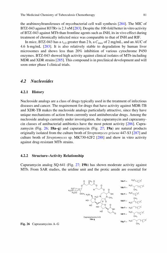
the arabinosyltransferases of mycobacterial cell wall synthesis [284]. The MIC of
BTZ-043 against H37Rv is 2.3 nM [283]. Despite the 100-fold better in vitro activity
of BTZ-043 against MTb than frontline agents such as INH, its in vivo effect during
treatment of chronically infected mice was comparable to that of INH and RIF.
In mice, BTZ-043 has a t1/2 greater than 2 h, a Cmax of 2 mg/mL, and an AUC of
4.6 h�mg/mL [283]. It is also relatively stable to degradation by human liver
microsomes and shows less than 20% inhibition of various cytochrome P450
enzymes. BTZ-043 showed high activity against clinical isolates of MTb including
MDR and XDR strains [285]. This compound is in preclinical development and will
soon enter phase I clinical trials.
4.2 Nucleosides
4.2.1 History
Nucleoside analogs are a class of drugs typically used in the treatment of infectious
diseases and cancer. The requirement for drugs that have activity against MDR-TB
and XDR-TB makes the nucleoside analogs particularly attractive, since they have
unique mechanisms of action from currently used antitubercular drugs. Among the
nucleoside analogs currently under investigation, the capuramycin and caprazamy-
cin classes of antibacterial antibiotics have the most potent activity [286]. Capra-
zamycin (Fig. 26; 18a–g) and capuramycin (Fig. 27; 19a) are natural products
originally isolated from the culture broth of Streptomyces griseus 447-S3 [287] andculture broth of Streptomyces sp. MK730-62F2 [288] and show in vitro activity
against drug-resistant MTb strains.
4.2.2 Structure–Activity Relationship
Capuramycin analog SQ-641 (Fig. 27; 19b) has shown moderate activity against
MTb. From SAR studies, the uridine unit and the protic amide are essential for
N NH
O
OOO
O
H2NNN
O
CH3
O
OH
HO
HOHO
HO
H3C
O
O
R
O
OO
O
O
OCH3H3CO
H3COH3C
CH3H3C
H3C
CH3
H3C
H3C
CH3
H3C
CH3
H3C
H3C
CH3
Caprazamycins A-G18a-g
H
18a R =
18b R =
18c R =
18d R =
18e R =
18f R =
18g R =
11
5
7
9
7
7
5
Fig. 26 Caprazamycins A–G
The Medicinal Chemistry of Tuberculosis Chemotherapy 81

bactericidal activity (Fig. 28) [289]. At the R2 position in Fig. 28, lipophilic groups,
including medium size alkyl chains, phenethyl, and phenyl-type substitutions,
retained moderate activity but benzyl-type substitution showed decreased activity.
When different lipophilic groups were placed at the R1 position, installation of a
decanoate substituent showed the largest increase in whole cell activity compared
to shorter alkanoate chains, likely due to the increased lipophilicity, which (see
Sect. 5.1) increases intracellular uptake into MTb.
CPZEN-45 (Fig. 27; 19f), a caprazamycin analog, is shown in Fig. 27. SAR
studies revealed that the uridine and the aminoribose are crucial for antibacterial
activity [290]. Initially, installation of ester substituents at R1 with R2 alkyl chains
showed that tridecane (C13H27, Fig. 27; 19c) and octadecane (C18H37, Fig. 27; 19d)
esters showed equipotent activity, whereas a 21-carbon chain with unsaturation at
C(18) showed decreased potency. Next, the effect of the amide substituent R2 (19e)
was investigated, which showed that the potency generally increased up to a 21
carbon alkyl chain and exhibited decreased potency with even longer alkyl sub-
stituents. Finally, anilinoamide substituents with n-butyl (CPZEN-45, Fig. 27; 19f),n-hexyl (Fig. 27; 19g), and hexyloxy (Fig. 27; 19h) showed the most potent activity
against MTb. Highly lipophilic molecules are, of course, not good candidates for
lead optimization programs; thus a considerable amount of work is still required to
discover better candidates from CPZEN-45 (19f) as leads for drug development.
N NH
O
O
O
OH3CO
O
C9H19
CONH2
OO
HN
O
HNH3C
OH
OHO
SQ-64119b
N
HN OO
OO
O
H2N
N
N O
CH3
OR1
OHHOHO
HO
H3C
HHN NH
O
O
O
OHHO
CONH2
OO
HN
O
HNOH
OHO
H
Capuramycin19a
19c R1 = COR2, R2 = OC13H2719d R1 = COR2, R2 = OC18H3719e R1 = COR2, R2 = NHalkyl
R1 =
19f R2 = n-Bu; CPZEN-4519g R2 = n-Hex19h R2 = OC6H13
O
NH
R2
Fig. 27 Capuramycin and analogs SQ-641 and CPZEN-45
N NH
O
O
O
OR1H3CO
CONH2
OO
HN
O
OHOH
R2
Uridine unit essential
R1 = lipophilic for better uptake
H
Protic amide requiredFig. 28 SAR for
capuramycins
82 G.A. Marriner et al.

4.2.3 Biology
Translocase I (encoded by mraY) is an essential enzyme involved in the biosynthe-
sis of peptidoglycans, which makes it an attractive target due to its unique presence
in bacteria. Caprazamycin (Fig. 26; 18a–g) and capuramycin (Fig. 27; 19a) inhibit
Translocase I with an IC50 of 18 nM and 90 nM, respectively [289]. The lead
compounds of the series are SQ-641 (Fig. 27; 19b), which has an MIC of
0.67–1.35 mM against drug-susceptible MTb and 0.081–2.71 mM against MDR-
TB [291], and CPZEN-45 (Fig. 27; 19e), which has an MIC of 2.26 and 9.07 mMagainst drug-susceptible and MDR-TB, respectively.
SQ-641 shows promising efficacy in themurinemodel of TB infection and exhibits
strong synergistic effects with EMB, STR, and SQ-109 (see Sect. 2.5) [286]. CPZEN-
45 also exhibits no significant toxicity and a novel mechanism of action making
these nucleoside compounds attractive candidates for TB drug development.
4.3 Macrolides
4.3.1 History
In the early 1950s, the first-generation prototypical macrolide, erythromycin (EM,
Fig. 29; 20a), was discovered. It is a natural antibiotic isolated from Saccharopo-lyspora erythrea [292, 293]. Erythromycin consists of a 14-membered lactone ring
with two attached sugar groups: L-cladinose at the C(3) position and desosamine at
the C(5) position [292, 293]. EM shows antibacterial activity against Gram-positive
bacteria, but no activity has been observed against MTb [292, 293].
4.3.2 Structure–Activity Relationship
In an effort to increase potency against MTb, a series of EM analogs was synthe-
sized with modifications at the 2, 3, 6, 9, 11, and 12 positions of the 14-membered
lactone ring, as well as at the 40 position of cladinose and the 200 position of
O
OCH3H3C
OHHOH3C
H3C
OCH3
O
OCH3
CH3
OHO
H3COH
NCH3
CH3
O
CH3
OH
H3CH3CO
O
OCH3
H3C
OCH3O
H3C
H3CO
CH3
O
O
CH3CH3
OHO
H3C
N
NCH 3
CH3
O
N
N
N
Erythromycin (EM)20a
Telithromycin20b
Fig. 29 Erythromycin (EM), 20a, and telithromycin, 20b
The Medicinal Chemistry of Tuberculosis Chemotherapy 83

desosamine [292–294] (Fig. 30). Specific modifications on the lactone ring such as
6-substitution, 11, 12-carbamate, 11, 12-carbazate, and 9-oxime substitutions
enhance potency [294]. Substitution of fluorine at position 2 in ketolides appears
to improve both potency and selectivity (i.e., cytotoxicity vs activity against MTb)
[293, 294]. The C(6) substituent is critical for activity of the ketolides [293], as it
affords acid stability by preventing internal hemiketalization with the 3-keto group
[292]. In general, ketolides are less potent than the corresponding cladinose-con-
taining compounds for all substituents on the 6-position [293]. Among 9-oxime-
substituted ketolides and macrolides, there is a correlation between the lipophilicity
of the substituent on the 9-position (defined as calculated logP) and the potency
[293], with some C(9) oximes showing submicromolar MIC against MTb [294].
The substituent at 11, 12 position appears to significantly affect potency.
A variety of aryl-substituted 11, 12-carbamate and carbazate macrolides and keto-
lides demonstrated low or submicromolar MICs [294]. Also, the aryl substituent
may be involved in determining cytotoxicity [294]. The substituted 11, 12-carba-
zate compounds demonstrated significant dose-dependent inhibition of MTb
growth in mice, with a 10–20-fold reduction of colony forming units (CFUs) in
lung tissue [294].
To further enhance lipophilicity, the 20 and 400-positions on desosamine and
cladinose rings, respectively, have been modified via esterification, which generally
improved potency and sometimes decreased CYP3A4 inhibition (more commonly
in the cladinose-containing macrolides; see Sect. 4.3.3 for discussion of CYP3A4
inhibition) [293], although the substituent on the 9-position is generally more
important than modifications on 20 and 400 positions [293].
4.3.3 Biology
Macrolides bind reversibly to the 50S subunit of 70S bacterial ribosomes, which
inhibits protein synthesis [293, 295]. Although macrolides are effective for other
bacterial infections, including some mycobacteria, they have not demonstrated
significant efficacy against MTb [293, 294]. Ribosome methylation is the most
O
R9
CH3H3C
OR6R12
H3CH3C
OCH3
O
OCH3
CH3
OR2'
R2
O
H3CR11
NCH3
CH3
R9 = oxime essential for activity; Increased lipophilicity increases both activity and toxicity
Activity strongly depends upon R6
Cladinose at C(3) gives good activity
R2 = F more active and less toxic
R11, R12 = carbamate or carbazate increases potency but strongly inhibits CYP3A
Result of acylation varies
O
CH3
OR4"
H3CH3CO
12
3
4
5
67
89
10
11
12
13
Fig. 30 General SAR for
macrolides (adapted from
[293])
84 G.A. Marriner et al.
widespread mechanism involved with macrolide resistance in MTb, and the gene
ermMT plays an essential role [294–296]. Therefore, the current development goal
for macrolides has been primarily to overcome bacterial resistances resulting from
methylation of the rRNA and drug efflux [294, 295]. Furthermore, since macrolides
are well-known inhibitors of CYP3A4, a cytochrome P450 enzyme, developing
compounds with decreased inhibition against CYP3A4 is critical.
In addition to low efficacy against MTb, the pharmacokinetics of EM are
somewhat unsatisfactory, as it is unstable to gastric acid and displays a short
serum half-life (~1.4 h) [292, 293]. The second-generation macrolides such as
clarithromycin and roxithromycin improved both properties [293], but clarithro-
mycin showed only weak activity against MTb either in vitro or in vivo (200 mg/kg
dose for low-dose aerosol infection mouse models), suggesting that second genera-
tion macrolides cannot be expected to offer significant antimicrobial clinical ben-
efits for TB [293, 294]. Further improvements focused on replacement of the
L-cladinose substituent, as it is associated with both drug efflux (one mechanism
for development of macrolide resistance) and metabolic instability of the macro-
lides [293]. This third generation of macrolides replaced the cladinose ring with a
ketone moiety (ketolides), leading to more metabolically stable drugs [292–294].
However, it appears that C(3) cladinose is important for antitubercular potency of
macrolides, which remain more potent than either ketolides or other substituents
such as 3-OH and 3-carbamoyloxy groups [293, 294]. Telithromycin (Fig. 29; 20b),
the first clinically approved ketolide, has been developed for use against respiratory
pathogens but is not active against MTb [292–294].
Currently, there have been some improvements in in vitro activity with macro-
lides against MTb, but to date no promising drug candidate has emerged. Preclinical
work in this area is ongoing.
4.4 b-Lactams
4.4.1 History
Since the discovery of 6-amino penicillanic acid (penicillin) in 1929 [297], b-lactams
have been one of the most successfully used classes of antibiotics. They are
irreversible inhibitors of peptidoglycan-cross-linking enzymes, D, D-transpeptidases
and D, D-carboxypeptidases [298]. b-lactams are rarely used in chemotherapy of
TB, however, because of the limited permeability of the mycobacterial cell enve-
lope, expression of inactivating enzymes (b-lactamases), and involvement of
b-lactam-insensitive targets in peptidoglycan transpeptidation.
4.4.2 Structure–Activity Relationship
The pharmacophore of the b-lactams is a highly reactive four-membered azetidi-
none ring which is generally fused to a five- or six-membered ring (Table 7). There
The Medicinal Chemistry of Tuberculosis Chemotherapy 85

is an absolute requirement for the b-lactam ring and a carboxylic acid on the fused
ring (or an electron withdrawing moiety such as the sulfonyl as in monobactams).
An amide a to the b-lactam ring is preferred. Conformationally, this core resembles
the acyl-D-alanyl-D-alanine moiety of the natural substrate. The serine nucleophile
in the enzyme active site attacks the electrophilic carbonyl of the b-lactam amide
leading to ring opening and irreversible acylation of the enzyme.
4.4.3 Biology
b-lactams are commonly used in combination with b-lactamase inhibitors such as
clavulanic acid (CA), sulbactam, and tazobactam which themselves are b-lactams.
A variety of b-lactams have been tested for in vitro efficacy against MTb (see
Table 8 for examples). However, despite activity of some b-lactams against MTb
in vitro, especially in the presence of b-lactamase inhibitors, none have to date
shown good efficacy in vivo. Amoxicillin (Table 8; 21a)/CA was found to be
ineffective in mice [302], and imipenem (Table 8; 21f) showed only a 16-fold
reduction in bacterial burden in lungs of infected mice over 4 weeks of treatment
[50]. Furthermore, only a modest decrease in viable numbers was seen in sputum
of patients receiving amoxicillin(Table 8; 21a)/CA or ampicillin(Table 8; 21b)/
sulbactam monotherapy [303]. The poor in vivo efficacy may be due to the
intracellular environment of MTb, making it difficult for drugs to penetrate the
phagosomal compartment. Additionally, the bacterial physiology in vivo may
Table 7 Basic structures of clinically relevant b-lactams and their pharmacologic properties
b-lactamclass
Pharmacophore Spectrum of
activity
Inactivation Adverse effects
Penams
N
S
CH3
CH3
HN
R
O
O
OOH
Gram-positives Classical
b-lactamases,
carbapenemases
Diarrhea,
hypersensitivity,
anaphylaxis,
pseudomembranous
colitis, yeast
infections
Cephems
N
SHN
R1
O
O
O OH
R2
Gram-negatives Extended-
spectrum
b-lactamases,
carbapenemases,
cephalosporinases
Diarrhea,
hypersensitivity,
pseudomembranous
colitis, yeast
infections
Carbapenems
NH3C
O
OOH
HO H
SR
Gram-positives,
Gram-negatives,
anaerobes
Renal
dehydropeptidase,
carbapenemases
Diarrhea, anaphylaxis,
pseudomembranous
colitis,
nephrotoxicity,
neurotoxicity
Mono-
bactams
NO
CH3HN
OR
SO
O
OH
Select Gram-
negatives
Extended-
spectrum
b-lactamases,
carbapenemases
Diarrhea,
pseudomembranous
colitis, yeast infections
86 G.A. Marriner et al.

be different making it less responsive to b-lactam therapy, although the recent
demonstration of activity of meropenem/CA against non-replicating persistent
MTb has raised the possibility of use of this carbapenem against TB [299, 304].
CP-5484 (a carbapenem with activity against MRSA) [305] is currently in
preclinical development for use against tuberculosis. Additionally, the merope-
nem/CA combination has shown potent activity against strains of MTb [299] and
is currently being investigated for possible clinical use.
Table 8 Biological activity of select b-lactams against MTb H37Rv in the presence or absence of
clavulanic acid [299–301]
Compound Structure MIC (mM)
�CA þCA
Amoxicillin (21a)
N
S
CH3
CH3
HN
O
O
OOH
NH2
HO
>250 0.5–3
Ampicillin (21b)
N
S
CH3
CH3
HN
O
O
OOH
NH2 >25 13
Ceftriaxone (21c)
NO
HN
O
N
H3CO
N
S NH2
S
OHO
S
N
N
CH3
O
OH
>230 7–28
Cephalothin (21d)
N
SHN
O
O
O OH
O CH3
O
S>40 25
Meropenem (21e)
NH3C
O
OOH
HO H
S
H CH3
NH N
O
CH3H3C
6.5 0.8
Imipenem (21f)
NH3C
O
OOH
HO H
S
H HNH
NH4 0.5
Aztreonam (21g)
NO
CH3HN
ONO
N
S
H3CH3C
OOH
NH2
SO
O
OH
>147 >147
The Medicinal Chemistry of Tuberculosis Chemotherapy 87

4.5 Rhiminophenazines
4.5.1 History
Clofazimine (CFM, Fig. 31; 8a), a member of the riminophenazine class of
compounds, was originally developed as an antitubercular drug in 1950s, but
inconsistent outcomes in animal studies (effective in mouse models but less activity
against in monkeys and guinea pigs) halted development [306]. Recently, CFM was
reinvestigated, and several analogs of CFM that are active against MDR-TB were
reported [307].
4.5.2 Structure–Activity Relationship
It has been shown that the imino group is essential for activity. Substitution with
electron-withdrawing groups such as -Cl or -CF3 at the R1 and R2 positions results
in higher antituberculosis activity, but increased lipophilicity particularly of R3
substituents can exacerbate the accumulation already observed with CFM in fat
tissues and cells of the reticuloendothelial system [27]. Installation of a tetramethyl-
piperidine in the R3 position showed higher activity than that of ethyl or isopropyl.
4.5.3 Biology
B-4157 (Fig. 32; 8b) exhibited promising in vitro activity against H37Rv and MDR
strains of MTb with MIC range of �114 nM to 228 nM. Tetramethylpiperidine-
substituted riminophenazines (such as B-4169, Fig. 32; 8c) showed MICs of
42.4–169 nM [308]. A generalized membrane disrupting effect, interference with
potassium transport, and generation of reactive oxygen intermediates have been
suggested as the mechanism of action for riminophenazines [27, 309, 310], but
detailed information is still not clear. The very low mutation frequency suggests
that rhiminophenazines may affect multiple aspects of metabolism [27].
The 2,2,6,6-tetramethylpiperidine-substituted riminophenazines such as B4169
have superior activity to CFM against MTb growing in macrophages and are also
N
N NR3
NH
8a R1 = 4-Cl, R2 = 4-Cl, R3 = iPr; Clofazimine (CFM)8b R1 = 4-CF3, R2 = 4-CF3, R3 = Et; B41578c R1 = 3,4,5-Cl R2 = 3,4,5-Cl, R3 = 4-(2,2,6,6-tetramethylpiperidine); B4169
R1
R2
Fig. 31 Antitubercular
rhiminophenazines
88 G.A. Marriner et al.

less toxic in animal models [308]. The B-4157 analog has similar activity to CFM in
infected mice, and at a dose of 20 mg/kg was as effective as similar doses of RIF or
INH in long-term monotherapy in infected animals [307]. The in vivo potency may
be due to the long half-life of CFM in tissues (>70 days after repeated dosing of
human patients) [27]. The absoption, distribution, metabolism, and excretion
(ADME) of CFM analogs have not yet been reported. CFM is 45–62% orally
bioavailable in humans, reaches serum concentrations of 0.7–1 mg/mL, and is
metabolized by the liver through dehalogenation or deamination followed by
glucuronidation or by hydroxylation along with glucuronidation [27]. Riminophe-
nazines are currently in the preclinical development state, but their high potency
makes them attractive drug candidates.
4.6 Pyrroles
4.6.1 History
Naturally occurring pyrrolnitrin (Fig. 32; 22a) and its analogs were tested against
MTb, and the most effective exhibited an MIC of 3.9 mM [311]. However, most of
the compounds from this series were cytotoxic, presumably because of the nitro
group. Structural optimization of pyrrolnitrin and other azole analogs led to the
discovery of the more potent pyrrole, BM-212 (Fig. 33; 22b), exhibiting MIC
values of 1.68 mM against MTb [312]. BM-212 (22b) was also found to be effective
against strains resistant to EMB, INH, amikacin, STR, RIF, and rifabutin, as well as
against MTb growing within a human monocyte cell line.
NH
Cl
Cl
NO2
Pyrrolnitrin lead analog
22a
NHN O
N
N N
CH3
CF3
LL-385822h
N
N R3
CH3
R1
R2
22b R1 = 4-Cl, R2 = 4-Cl, R3 = NCH3; BM-21222c R1 = 4-F, R2 = 4-F, R3 = S22d R1 = 4-F, R2 = 4-F, R3 = S22e R1,R2 = H, R3 = S22f R1 = 4-F, R2 = 2-F, R3 = S22g R1 = 4-CH3, R2 = 4-F, R3 = S
Fig. 32 Pyrrole-based antitubercular compounds
N
N R
CH3R1
R2
R = S or N-CH3 essential; hydrogen bond acceptor
CH3 essential for directing aryl conformation
Substituted Ar ring essential;ortho or para halides more potent, less cytotoxicFig. 33 Pyrrole SAR
The Medicinal Chemistry of Tuberculosis Chemotherapy 89

4.6.2 Structure–Activity Relationship
Using BM-212 (22b) as a lead compound, systematic structural optimization led to
the discovery of improved analogs, with similar or better activity in the range of
0.5–2 mM and an improved therapeutic index (ratio of cytotoxicity to in vitro
activity against MTb) of 16–160 [313–316].
Based on whole cell biological activity, SARs could be deduced, with aromatic
groups at N(1) and C(5) and a methyl group at C(2) as essential features (Fig. 33).
Additionally, methylene-linked thiomorpholine or N-methylpiperazine substituents
at C(3) act as hydrogen bond acceptors to improve activity (Fig. 34; 22b–g) [316,
317]. Thus, a 1,2,3,5-tetrasubstituted pyrrole is the pharmacophore essential for
antitubercular activity (Fig. 32). The 2-methyl group is not involved in any phar-
macophoric interaction but influences the conformation of the substituents at posi-
tions 1 and 3 of the pyrrole ring [316, 317].
4.6.3 Biology
Another pyrrole analog, LL-3858 (Fig. 34; 22h) (first reported by Lupin Limited in
2004), a pyrrole derivative, also complies with this pharmacophore model and is
currently in phase IIa clinical trials in India. This compound has exhibited MIC
values in the range of 0.05–0.1 mM, against MTb. LL-3858 (22h) has been reported
to sterilize the lungs and spleen of infected mice after 12 weeks of treatment, none
of which relapsed after 2 months of therapy termination [318].
4.7 Deazapteridines
4.7.1 History
Tetrahydrofolate (reduced dihydrofolate) is a key cofactor for the synthesis of many
biomolecules, and inhibition of dihydrofolate reductase (DHFR) leads to cell death
[319]. Bacterial DHFR is sufficiently different from human DHFR to serve as a
novel drug target [320]. To this end, the deazapteridines were designed as inhibitors
of mycobacterial DHFR by researchers at the Southern Research Institute [321].
4.7.2 Structure–Activity Relationship
MIC assays against MTb and cytotoxicity assays using Vero cells were used to
compare selectivity for mycobacterial DHFR. Based on the limited number of
structures reported, it appears that a smaller R1 group is better tolerated (com-
pounds SRI-8117 vs SRI-8922 or SRI-8229 vs SRI-8911; Table 9), and either a
secondary or tertiary amine is tolerated (compounds SRI-8710 vs SRI-8117 and
SRI-8687 vs SRI-8686) [321]. 2,5-substituted electron-rich aromatics are preferred
(data not shown). Modeling studies of these small molecules binding to MTb DHFR
90 G.A. Marriner et al.

suggest that the 2-ethoxy or 2-methoxy group acts as a hydrogen bond donor [322,
323]. Additionally, modeling shows that for human DHFR, the cleft to which the
deazapteridines bind is lined with hydrophobic residues, whereas the analogous
MTb DHFR cleft is larger and more accessible to solvent [322, 323].
4.7.3 Biology
Subsequent publications refined the pharmacophore using members of the Myco-bacterium avium complex (MAC), which established that SRI-8686 had the highest
IC50 ratio for MAC DHFR vs human DHFR (0.84 nM vs 2,300 nM, a 2,700-fold
selectivity). SRI-8117 showed similar selectivity (1.1 nM vs 1,000 nM, 900-fold
selectivity) [324]. Other MTb DHFR inhibitors are in early stages of development
[325]. Although the MIC assays suggest that this series may be worth developing,
the on-target effect of these compounds in MTb still needs to be verified.
5 Critical Issues in TB Drug Development
5.1 Cell Penetration
The complex, lipid-rich envelope of MTb acts as a permeation barrier to a broad
range of therapeutic agents and has likely contributed to both the fitness and
the success of the pathogen (Fig. 33). The plasma membrane (PM) forms the
innermost region of the cell envelope and is a typical lipid bilayer, structurally
and functionally similar to the PM of other eubacteria. External to the PM is the
peptidoglycan sacculus. This contains repeating units of N-acetylglucosamine and
Table 9 MIC vs MTb and IC50 vs Vero cells [321]
Compound name R1 R2 R3 MIC vs
H37Rv (mM)
IC50 vs Vero
cells (mM)
SRI-8117 (23a) CH3 2,5-(CH3O)2Ph H 37 2,106
SRI-8922 (23b) CH2CH3 2,5-(CH3O)2Ph H >35 ND
SRI-8710 (23c) CH3 2,5-(CH3O)2Ph CH3 8.8 200
SRI-8686 (23d) CH3 2,5-(CH3CH2O)2Ph H >34 ND
SRI-8687 (23e) CH3 2,5-(CH3CH2O)2Ph CH3 >32 ND
SRI-8202 (23f) CH3 2-CH3-5-CH3OPh H 19 1,421
SRI-8229 (23g) CH3 2-CH3O-5-CH3Ph H 19 231
SRI-8911 (23h) CH2CH3 2-CH3O-5-CH3Ph H >37 ND
SRI-8228 (23i) CH3 2-CH3O-5-CF3OPh H 4.0 2.6
The Medicinal Chemistry of Tuberculosis Chemotherapy 91

N-glycolymuramic acid with stem peptides joined mostly through 3–3 cross-links
rather than the 4–3 linkages commonly found in other bacteria [326, 327].
Peptidoglycan serves as a scaffold for arabinogalactan, a polymer of D-arabinose
and D-galactose, which are covalently attached by b-1,5 linkages [328]. The arabi-
nogalactan chain bridges between peptidoglycan and a thick layer of mycolic acids
where the galactose portion of the polymer is connected to peptidoglycan by a
unique glycosyl–phosphoryl bridge, and the arabinose moieties are ester-linked to
four mycolic acid residues [329, 330]. The mycolic acids that are largely responsi-
ble for the impermeability of the mycobacterial cell wall consist of branched
2-alkyl-3-hydroxy fatty acids 70–90 carbon atoms in length [330]. In contrast to
other bacteria, the MTb membrane also contains a number of lipids with unusual
structures including: phosphatidylinositol mannosides, lipomannans and lipoarabi-
nomannans, trehalose-6,60-dimycolate, sulfolipids, phthicerol dimycocerosates,
and phenolic glycolipids [331]. A loosely attached capsule defines the outermost
layer of MTb and is composed primarily of glucans, arabinomannans, and mannans,
with a small number of lipids and proteins decorating the structure as well.
Historically, the intrinsic resistance of mycobacteria to antibiotics has been
attributed to the low permeability of the cell wall. For example, perturbations in
the cell envelope caused by detergents, mutations, or through inhibition of cell wall
polymer biosynthesis increases the susceptibility of mycobacteria to various classes
of antibiotics, including aminoglycosides, EMB, and RIF [332–335]. Moreover,
data indicate that the outer membrane ofMycobacterium chelonei is 1,000-fold less
Fig. 34 Schematic of the mycobacterial cell envelope
92 G.A. Marriner et al.

permeable to hydrophilic molecules than Escherichia coli and tenfold less perme-
able than the notoriously impermeable Pseudomonas aeruginosa [331, 336, 337].
Instead, hydrophilic molecules such as the cephalosporins likely penetrate the
envelope using water-filled porins located on the outer leaflet of the cell wall
[338]. While typical lipid bilayers are highly permeable to lipophilic molecules,
permeability is inversely correlated with membrane fluidity [331]. When a cell wall
is erected from lipids containing long, hydrocarbon chains with few double bonds
or cyclopropane groups, the result is membrane rigidity [331, 339]. The mycolic
acids within the MTb cell wall are unique in this regard, and accordingly, the inner
leaflet displays extremely low fluidity [340]. In addition, lipids with more than one
fatty acid chain attached to a single head group, similar to the mycolyl-arabinoga-
lactan found in the cell wall of MTb, decrease membrane fluidity further [331].
Data predict that the lipophilic antibiotics such as fluoroquinolones, macrolides,
rifamycins, and tetracyclines do penetrate the bacteria, but this likely occurs via the
lipid bilayer rather than the inefficient porins found on the outer leaflet [331]. In
support of this model, the more hydrophobic agents within an antibacterial class
tend to be more effective against mycobacteria [331].
5.2 Animal Models for Evaluation
As most vertebrates can be infected with a mycobacterial pathogen, it is no surprise
that there are a wide range of TB animal models that to different extents recapitulate
the characteristics of human disease. A summary of some of the current experi-
mental animal models of TB chemotherapy, their typical uses, and the comparative
compound requirement for use in drug efficacy studies are shown in Table 10 and
briefly summarized here.
Zebrafish infected with Mycobacterium marinum are gaining popularity as a
model of TB as the costs and space requirements are quite modest and experimental
work with infected fish or their embryos does not require the biological safely
laboratory level 3 containment of any work with virulent strains of the MTb
complex (M. africanum, M. bovis, M. tuberculosis, and M. caprae). M. marinumis a natural pathogen of fish that causes necrotizing lesions within a nearly trans-
parent host where the progress of infection can be imaged with high resolution
microscopy [341] allowing real-time data collection. This model is contributing to
our understanding of TB pathogenesis and may become useful for drug screening in
the near future [342, 343, 361].
The inbred mouse has been used most extensively in TB studies and can be
infected by a variety of routes including i.v., intranasal inoculation, and by aerosol
exposure. Many strains of mice, each with different genetic backgrounds for
investigating certain immunological parameters, can be reliably maintained for
many months in a state of chronic infection, and used for a variety of different
readouts (Table 10). While the mouse model is often used in studies of MTb strains
The Medicinal Chemistry of Tuberculosis Chemotherapy 93

Table
10
Characteristicsofexperim
entalanim
almodelsoftuberculosischem
otherapy
Model
Pathology
Utility:limitations
Compoundrequirem
ent
(g)[m
assofanim
al]
References
Zebrafish
Necrotizinggranulomain
embryo
Early
granulomaform
ation,high-
throughput:non-pharmacodynam
ic
0.025[100mLdish]
[341–343]
Mice
Pulm
onarypneumonia
andaggregates
of
leukocytes,spleen,andliver
permissive
forgrowth
Standardmodel
fordrugefficacy:very
limited
pathology
0.35[25g]
[79,344–347]
Rats
Sim
ilar
tomice
AcceptedPK/PD
model:little
additional
inform
ationcf.mice
4.6
[0.3
kg]
[348,349]
Guinea
pigs
Pulm
onary,splenic,andliver
lesions;50%
solid,50%
necrotizinggranulomas
Smallsize
withmore
similarityto
human
pathology:non-cavitating
12.6
[0.9
kg]
[79,350–353]
Rabbits
Lim
ited
infectionwithMtb
strains;
pulm
onarysolidandnecrotizinglesions,
cavities;extensivesecondarylesions
(M.bo
visonly)
Pathologyincludingpulm
onary,CNS
system
,ocularsites:highly
sensitiveto
GIdistressfrom
manyagents
40[3.5
kg]
[354–357]
Nonhuman
primates
Diverse
presentationoflesionsin
multiple
organs
Pathologyaccurately
reproduceshuman,
PK
similar:expensive,dosingcanbe
difficult
56[5
kg]
[358–360]
94 G.A. Marriner et al.

to define the contribution of various mycobacterial genes to virulence or in studies
of immunological responses to MTb infection, it does not reproduce the lung
pathology observed in the human [290, 344]. However, the poor sterilizing activity
of INH and the treatment-shortening effect of treating with INH, RIF, and PZA
containing regimens in the mouse model are superficially similar to their efficacy in
human studies; hence, it is argued that the mouse model is predictive of human
relapse rates [345]. Since the mouse does not develop the latent disease that
characterizes the spectrum of human TB but rather develops a chronic disease
marked by very high bacterial burdens and progressive destruction of lung tissue, it
has been argued that this animal model is unsuitable for testing drugs under
development for latent disease [362]. The development of the gamma interferon
gene-disrupted C57BL6 mouse has shortened initial in vivo drug evaluation to
about 2 weeks, but may only reflect drug bioavailability as the host immune system
is crippled and unable to control bacterial replication, resulting in fulminant
disseminated infection [363]. The discovery of the sst1 susceptibility locus in the
C3HeB/FeJ mouse has provided an immune competent mouse model (C3H.B6-sst1mice) with lesions demonstrating central necrosis which more closely resemble
those observed in diseased human lungs [346]. Experiments to benchmark standard
tuberculosis chemotherapy studies to determine relapse rates in this model are
underway [364]. The “Cornell” model, where chemotherapy is used to completely
sterilize mouse tissues of MTb bacilli, or studies where a combination of BCG
vaccination and drug treatment are used to sterilize tissues require extended dura-
tions of chemotherapy (>8 months) and have been argued to reflect the length and
sterilizing activity of TB drug regimens in humans [365–367]. Results of these
studies have been used to determine optimal drug combinations and drug exposures
in human clinical trials to identify treatment-shortening regimens for human clini-
cal trials [368–370].
Actual data substantiating the predictive ability of the mouse model to determine
the length of treatment are very sparse. Anatomically, one major predictor of
relapse in patients is the presence of cavities, a pathologic feature not represented
in any mouse model [371]. Recent trials of shorter therapies based on substituting
moxifloxacin for various components of the standard frontline regimen have failed
to recapitulate the therapy-shortening observed in mice models [372]. Unfortu-
nately, this evidence seems to be widely disregarded in the rush to introduce new
regimens based exclusively on their comparative efficacy in mice.
A larger rodent, the Wistar or Sprague Drawley rat, is often used in medical
research and is widely accepted as a model for toxicology studies. The rat model
allows dual comparison of toxicology and efficacy in the same animal and, as a
result, has been used in several TB drug development studies. In one of these studies,
RIF was reported to give a good dose–response curve for bacillary clearance, but
other TB chemotherapeutics gave less encouraging results, [348] suggesting that this
model may not be a suitable model for evaluation of antitubercular drugs.
The guinea pig, used by Robert Koch in the late 1800s to demonstrate that the
MTb bacillus was the etiological agent of TB, was one of the earliest animal models
in mycobacterial research. Even with very low aerosolized infectious doses, these
The Medicinal Chemistry of Tuberculosis Chemotherapy 95

animals experience rapid progression to granulomatous non-cavitary disease that is
ultimately fatal [350]. Because of its extreme susceptibility to MTb infection, the
guinea pig is used extensively in vaccine studies, but to a lesser extent in drug
efficacy studies because of its relatively large size [352, 353]. Due to its high
susceptibility to MTb infection, it is also gaining popularity for virulence testing
of gene knockout mutants of MTb, since large differences in organ burdens are
generally observed with attenuated strains of MTb in the guinea pig [179, 351].
Recently, the sterilizing activity of a species-specific, human-equivalent dosage of a
INH, RIF, and PZA regimen given in a 2-week intensive phase followed by a 6-
month continuation phase of biweekly dosing (the so-called Denver regimen) was
tested in both the guinea pig and the BALB/c mouse [369]. The guinea pigs were
found to respond to treatment more quickly and have lower relapse rates in the
6 months following treatment as compared to similarly treated mice, although
the guinea pigs experienced gastrointestinal toxicities of unknown origin making
the model more challenging.
The New Zealand White rabbit, a relatively resistant animal to TB infection, has
often been used to study the development of lung pathology including necrotizing
lesions and cavities after either i.v. or aerosol infection with M. bovis, MTb, and
even M. avium strains [354, 357, 373, 374]. It has been used for studies of extra-
pulmonary dissemination and/or growth of MTb especially in studies of pathogen-
esis of the central nervous system as a model of human TB meningitis [355, 375]. It
is also used for testing indwelling venous lines and ports containing drugs which are
technically not feasible on smaller animals such as mice, but less often used for oral
drug efficacy studies due to the large amount of compound needed for efficacy
testing in rabbits (see Table 10) [376]. On the other hand, it is particularly
advantageous that oral dosing and PK/PD blood collection are possible without
anesthesia in this species. It has been reported that latent disease can be achieved
in the rabbit model with certain MTb strains although additional validation is
required [377].
Nonhuman primates (NHP), especially cynomolgus and rhesus macaques, have
a long history in TB research for both vaccine and drug testing, but the advent of
more restrictive laboratory practices and the requirement for BSL-3 housing has
made the model prohibitively expensive and thus less utilized [344, 378]. Like other
species, aerosol infection or direct installation of the bacilli into the lung is the usual
route of infection. These monkeys reproduce the spectrum of disease observed in
humans including pulmonary, extrapulmonary, and latent tuberculosis infection
(LTBI) as well as many of the different types of granulomas observed in human
patients [379]. Low dose infection of the cynomolgus macaque is associated with
the induction of LTBI with roughly 60% of animals showing no signs of disease
after skin test conversion [359]. The genetic similarity between NHP and humans
has allowed use of the same immunological reagents such as TNF-a blockers to
elicit reactivation disease from LTBI and other reagents to query host immune
function for vaccine studies as those that have been used in human clinical trials
[380]. A disadvantage with these relatively large NHPs is the amount of GMP or
minimally GLP compound needed (Table 10) and the requirement for anesthesia
96 G.A. Marriner et al.

for most manipulations including dosing to achieve reliable administration in drug
studies. For these and other regulatory reasons, NHP work is usually reserved for
proof of concept studies to establish the link between findings in prior (lower)
animal studies and anticipated outcome in humans or as the final stage in a
preclinical drug development pipeline before seeking approval for investigational
studies in humans.
5.3 Pharmacological Models for Antitubercular Drugs
An evaluation of the pharmacological properties of new TB drugs is essential for
effective treatment and overall cure of disease. Currently, most TB drugs are
evaluated through preclinical animal models to establish appropriate dosing levels
that promote optimal bacterial killing with limited toxicity. In vivo efficacy for TB
drugs is not solely dependent on plasma concentration but more significantly
dependent on tissue concentrations near and within lesions [78]. These concentra-
tions must remain above MIC levels for an effective period of time to eradicate
bacilli (described as Time > MIC). Lower levels are associated with the develop-
ment of resistance and relapse of disease. However, due to the duration of treat-
ment, maintaining high levels of drugs in combination often poses severe issues of
toxicities and tolerabilities for the patient [381]. Thus, pharmacological evaluations
for TB drugs depend on optimizing treatment to an often narrowly constrained
therapeutic window.
Effective therapy with any drug is dependent on the relationship between
pharmacokinetic and pharmacodynamics (PK/PD) parameters [382, 383]. Pharma-
cokinetics (PK) defines the ADME properties of the compound, while pharmaco-
dynamics (PD) reveals the correlation between the serum concentration and the
biological effect, efficacy or toxicity. For most drugs, the primary measurement
under evaluation is plasma concentration. However, it is more relevant to assess
drug levels within the infected lesion. Many efforts have attempted to define key
parameters for TB drugs such as Cmax/MIC (ratio of peak serum drug levels to
MIC), AUC > MIC (overall drug exposure over the dosing interval must be greater
than MIC), and Time > MIC (time period at which the drug remains in the blood
must be greater than MIC per dosing interval) which are all established from plasma
concentrations [78, 384]. Typically, high Cmax/MIC ratios can offer sterilizing
activity as well as limit adaptive resistance or the selection of resistant subpopula-
tions whereas for TB, AUC > MIC and T > MIC are thought to be most relevant
for both to maintain long-term exposure above MIC with limited dosing. These
parameters are typically measured from blood, yet it is presumed that the primary
driving factor for efficacy in TB therapy involves lesion penetration at effective
concentrations [78, 290]. It is this factor that will eradicate persistent bacilli and
circumvent the development of resistance.
The Medicinal Chemistry of Tuberculosis Chemotherapy 97

There are several confounding factors that play a role in antitubercular drug
efficacy in vivo. As with most drugs, stability and bioavailability with limited
metabolism are important. Delivery to the site of infection at active concentrations
increases the overall efficacy of any drug. However, active pulmonary tuberculosis
is a chronic complex disease with a diverse spectrum of lesions within the lung.
Predominantly caseous lesions are central to the “life cycle” of a pulmonary TB
infection, as they eventually erode into air passages to allow bacilli to reach blood
vessels and permit dissemination. These granulomas are generally poorly vascular-
ized, hypoxic, lipid-rich, and often fortified with fibrous tissue forming an impene-
trable fortress for the TB bacilli [290]. The ability of a drug to penetrate these
lesions and kill bacilli is most critical to eradicate the bacteria and prevent
disseminated or extensive disease.
Currently, there are various models designed to assess the pharmacological
activity of TB drugs. Traditional models focus on determining drug levels in the
blood from preclinical and clinical animal and human evaluation [385]. These
studies are used to optimize dosing and evaluate tolerance. Dose fractionation
models in animals can determine relevant PK indices with a strong correlation
with PD effects. This type of experiment can elucidate important information for
clinical development and optimal dosing strategies to prioritize compounds through
drug development [258, 386, 387]. Recent in vitro models have been designed to
mimic human PK (half-lives and dosing schedules) to assess the development of
resistance [388]. The data obtained from these models help to identify drug-
exposure breakpoints required for maximal bactericidal activity and the suppres-
sion of drug resistance. Current lesion penetration studies involving tissues from
animals and human resections are providing important information in regard to
drug levels found in the various lesion types enabling a better understanding to drug
efficacy and therapeutic response [290].
Finally, the use of therapeutic drug monitoring (TDM) has been a useful tool for
assessing drug levels during treatment in the clinical setting [71]. The use of TDM
in tuberculosis treatment can allow physicians the ability to adjust dosing to provide
an efficacious therapeutic concentration throughout the extensive duration of treat-
ment [71]. Patients who most benefit are those with complications which may alter
drug exposure, such as those on co-therapy for HIV for which there are known
drug–drug interactions, those with diabetes mellitus with typical delayed absorption
or malabsorption concerns, those with renal failure undergoing dialysis, and those
experiencing hepatic dysfunction (see Sect. 1.4) [69]. TDM in combination with
bacteriological and clinical data can be a useful tool to assess treatment and ensure
as successful outcome [71].
Understanding pharmacological activities of TB drugs is essential not only for
addressing drug levels for effective sterilizing activity and optimizing dosing
strategies, but more importantly, they are also useful in limiting the development
of acquired resistance. PK/PD for TB agents is relevant to understanding important
phenomena associated with TB. Efforts are ongoing to develop PK/PD analyses
which will effectively predict success or failure of new antituberculosis drugs and
combination regimens.
98 G.A. Marriner et al.

5.4 Clinical Development Methodologies
The goal of TB chemotherapy is to cure clinical symptoms, prevent the develop-
ment of resistance, and to prevent relapse. Since TB treatment entails 6–24 months
of chemotherapy depending on drug susceptibility patterns, early markers that
predict durable cure would greatly facilitate and speed the evaluation of new
drugs. Currently, it requires 2 years of follow-up after termination of chemotherapy
to capture more than 90% of relapses, thus necessitating trials lasting up to 4 years
during evaluation of new therapies. The FDA recognizes the need to develop new
drugs for TB and the associated need to look for early predictors of durable cure,
making regulatory approval processes for such trials logistically easier.
Biomarkers that predict durable cure could include pathogen-specific measure-
ments, determination of host responses to the pathogen or nonspecific disease-
associated responses, and imaging. Unfortunately, no biomarker of any nature has
been validated as of this time. Microbiologic markers traditionally used to evaluate
TB chemotherapy include: 2-month culture conversion (in which sputum samples
taken at regular intervals during chemotherapy are evaluated for eradication of
culturable MTb at two months posttreatment), days to positivity (the time required
to obtain mycobacterial growth from sputum in liquid culture), and serial counts of
CFUs of MTb from sputum samples on agar combined with scoring acid-fast bacilli
in sputum during treatment. Two-month culture positivity as surrogate marker for
relapse was first recognized and used as a surrogate endpoint to predict treatment
efficacy during the BMRC studies where culture negativity at 2 months was
associated with cure and lack of subsequent relapse and is widely used for current
evaluation of TB chemotherapeutic trials [389–392].
While other culture-dependent methods show promise, they do not always
predict 2-month culture conversion and there is no data to correlate findings with
risk of relapse [390, 393]. In a large ongoing clinical trial (REMoxTB), where
moxifloxacin is evaluated as a replacement for INH or EMB, various culture-
dependent and -independent methods will be assessed against the primary endpoint
of relapse within 2 years of treatment termination, which may give some insight
into the utility of other biomarkers [394].
Culture-dependent methods suffer from their long turnaround time, often several
weeks, due to the slow growth of mycobacteria such as MTb. Potential culture-
independent pathogen-derived biomarkers include detection of MTb DNA [395],
lipoarabinomannan [396, 397] in urine, MTb mRNA in sputum [393], MTb char-
acteristic volatile organic compounds in breath [398], and several other pathogen-
derived biomarkers currently under evaluation.
Host biomarkers of disease include interferon-gamma release assays in
response to MTb antigens [399, 400], measurements of non-MTb-specific host
responses such as C-reactive protein, serum interleukin-2, neopterin and procal-
citonin which still require a definitive TB diagnosis by other means [401–403].
These may especially be useful as clinical trial endpoints in smear-negative,
The Medicinal Chemistry of Tuberculosis Chemotherapy 99

paucibacillary, extra-pulmonary, and pediatric TB where microbiologic culture
is less reliable.
Imaging biomarkers such as high resolution computed tomography (HRCT)
have been shown to be helpful in diagnosing active TB and distinguishing it from
latent disease [404], and may be useful in detecting the early stages of disease in TB
contacts [405, 406] but is also used to evaluate treatment response. Positron
emission tomography (PET) using 2-fluorodeoxyglucose has been used to visualize
inflammatory regions in pulmonary mycobacteriosis caused by MTb and M. aviumbut is even less discriminating than HRCT in distinguishing between TB and
nontuberculous mycobacterial infections [407].
Traditionally, clinical trials for new TB drugs include a 7–14-day assessment of
early bactericidal activity (EBA) of a drug given as monotherapy (often at different
doses). Daily sputum microbiology is performed and bacterial counts quantified.
Unfortunately, reproducibility of such microbiological assays is questionable, and
EBA rarely gives a definitive answer. In most cases, ambiguous EBA results are
ignored and a longer study (2 months or more depending on the available safety and
toxicity data) of the new drug or placebo in combination with current TB drugs
followed by completion of chemotherapy with regular extended phase regimens.
These studies are followed up with expensive phase III trials that give the new drug
as part of combined therapy (6–24 months) with prolonged follow-up after treat-
ment completion [369]. The caveat to using EBA as a predictor of successful
treatment is that drugs such as RIF and PZA, which have a poor EBA but have
good activity against MTb populations in certain lesions, would score as poor drugs
[408]. PZA is the essential drug that allowed the shortening of treatment of drug-
sensitive TB from 9 to 6 months [409], indicating that EBA studies may not be a
good way to cull ineffective drugs.
Phase II trials typically evaluate sputum conversion at 2 months as a predictor of
durable cure (cure without relapse), but the utility of 2-month culture conversion as
a biomarker was derived from studies of drug-sensitive TB and may not apply to
MDR-TB. In addition, evaluation of new drugs is typically compared to the
standard of care, which can achieve a 95% cure rate in clinical settings. Thus, the
sample size of such a trial must be large to detect either non-inferiority or superior-
ity of new agents. With MDR- and XDR-TB, the cure rates are much lower, which
may make it easier to detect the effect of the investigational drug, but such study
populations are much more heterogeneous due to different degrees and spectra of
drug susceptibility results, different background drug regimens, and a wide range
of disease severity and treatment history all of which may confound results. Some
of these problems can be dealt with by randomized stratification of strong covari-
ates, for example by stratifying for fluoroquinolone sensitivity [259].
In summary, EBA studies, while having the advantage of seeing the effect of a
single drug on MTb in lesions that are the source of sputum-borne bacilli, are
clearly not a useful predictor of drug efficacy. Studies of combination therapy using
2-month culture conversion as an end point have the advantage of a partially
validated end point with a weak correlation for drug-sensitive disease but should
be interpreted with caution in drug-resistant disease. Imaging modalities such as
100 G.A. Marriner et al.

HRCT and PET are attractive endpoints that do not require culture and evaluated
disease at the relevant site of infection but, like host-derived biomarkers, require
validation for predicting treatment efficacy.
6 Concluding Remarks
TB drug discovery remains a challenging enterprise at every level. There is an
urgent need to validate criteria for lead compounds with promise to reduce treat-
ment duration, treat drug-resistant disease, and provide utility for prophylaxis of
subclinical disease. Poorly validated in vitro assays, poorly validated in vivo assays,
and a severe lack of predictive animal models all compromise efforts to improve the
quality of the TB drug pipeline. Existing SAR around series explored in the 1950s
and 1960s provides some starting points, but the chemistry of most of these series is
either intractable or linked to enzymatic activation of prodrug and is therefore
difficult to optimize systematically. As in most anti-infectives, target-based strate-
gies have failed to lead to viable candidates, and in most cases the reason for this
failure has been unexplained. In the current climate, it is no surprise that the
momentum is toward improving whole cell screening and attempting to identify
leads with novel targets. Biological studies working toward more predictive assays
are in progress and very promising. For example, the promising use of titratable
promoter elements to systematically knock down genes and mimic the pharmaco-
logical action of a drug can truly validate drug action and directly address “target
vulnerability” by quantitating how much a target needs to be inhibited to affect cell
death [410]. Current efforts to understand the “systems biology” of MTb also have
the potential to reshape the landscape and improve our ability to select targets.
The nitroimidazoles and diarylquinolines, series developed specifically for TB,
provide some reason for optimism and perhaps a sort of loose road map for how to
approach the problems. Both series suffer from critical limitations including
extremely poor water solubility and potential hERG problems. In general, one
serious problem that persists in new drug discovery programs for TB lies in the
lack of broad application of preclinical ADME and toxicology studies in lead
optimization. The academic sector lacks an appreciation of the role of such studies
and lacks resources dedicated to performing them. Both series may well fail
because of these considerations, but at a minimum it has made clinical development
programs for these two new classes considerably more complex than if these issues
had been addressed systematically in lead optimization. Candidate selection in most
cases continues to happen predominantly by MIC and mouse activity, and it is
doubtful that the situation will improve more than incrementally without more
sophistication in lead optimization programs. Nonetheless, there are ongoing phase
II studies with these agents, and it is crucial that clinical development of
these compounds is paired with meaningful attempts to understand the relationship
of the preclinical studies that led to selection of the candidates with the ultimate
clinical properties of these agents. Through such efforts, in vitro and in vivo assays
The Medicinal Chemistry of Tuberculosis Chemotherapy 101

(not to mention phase I and II trials) could perhaps be optimized to be more
predictive, and these could be used to guide both backup programs for the existing
agents and optimization efforts for new series not yet in play.
Funding sources for TB drug discovery have also expanded dramatically over
the last 10 years and now include significant efforts from private foundations (such
as the Bill and Melinda Gates’ Foundation through its TB Drug Accelerator
Program) as well as public institutions such as the US National Institutes of Health
and the European Union’s Framework Programmes. Many of the activities being
funded are taking place at the interface of academic laboratories and commercial
pharmaceutical companies using a wide variety of models for cooperation.
Although this is precisely where such programs appear to be best placed, there
has been a rather steep learning curve as these two cultures are brought together
with competing needs. Nonetheless, there is significant cause for excitement at the
number and quality of programs that are currently operating. If we can successfully
leverage the strengths of both academia and industry, there is hope that we will be
able to raise the victory flag in earnest in the long struggle against TB.
References
1. Dye C, Lonnroth K, Jaramillo E, Williams BG, Raviglione M (2009) Trends in tuberculosis
incidence and their determinants in 134 countries. Bull World Health Organ 87:683–691
2. Barry CE III, Boshoff HI, Dartois V, Dick T, Ehrt S, Flynn J, Schnappinger D, Wilkinson RJ,
Young D (2009) The spectrum of latent tuberculosis: rethinking the biology and intervention
strategies. Nat Rev Microbiol 7:845–855
3. Young D, Dye C (2006) The development and impact of tuberculosis vaccines. Cell
124:683–687
4. Kaufmann SH, Hussey G, Lambert PH (2010) New vaccines for tuberculosis. Lancet
375:2110–2119
5. Parwati I, van Crevel R, van Soolingen D (2010) Possible underlying mechanisms for
successful emergence of the Mycobacterium tuberculosis Beijing genotype strains. Lancet
Infect Dis 10:103–111
6. Dye C, Williams BG (2010) The population dynamics and control of tuberculosis. Science
328:856–861
7. Iseman MD (2002) Tuberculosis therapy: past, present and future. Eur Respir J Suppl
36:87s–94s
8. Murray JF (2004) A century of tuberculosis. Am J Respir Crit Care Med 169:1181–1186
9. Thoren M, Hinshaw HC (1952) Therapy of pulmonary tuberculosis with isoniazid alone and
in combination with streptomycin and with para-amino-salicylic acid. Stanford Med Bull
10:316–318
10. Marshall G (1948) Streptomycin treatment of pulmonary tuberculosis. Br Med J 2:769–782
11. Youmans GP, Raleigh GW, Youmans AS (1947) The tuberculostatic action of para-amino-
salicylic acid. J Bacteriol 54:409–416
12. Graessle OE, Pietrowski JJ (1949) The in vitro effect of para-aminosalicylic acid (PAS) in
preventing acquired resistance to streptomycin by Mycobacterium tuberculosis. J Bacteriol57:459–464
13. Dooneief AS, Buchberg A, Steinbach MM (1950) Para-aminosalicylic acid (PAS) in chronic
pulmonary tuberculosis. N Engl J Med 242:859–862
14. Bowen DA, Collins DM (1952) Development of drug resistance to isoniazid in cases of
pulmonary tuberculosis. Tubercle 33:276–278
102 G.A. Marriner et al.

15. Joiner CL, Maclean KS, Carroll JD, Marsh K, Collard P, Knox R (1954) Isoniazid and P. A.
S. in chronic pulmonary tuberculosis: a warning. Lancet 267:663–666
16. Marshall G (1955) Various combinations of isoniazid with streptomycin or with P.A.S. in the
treatment of pulmonary tuberculosis; seventh report to the Medical Research Council by
their Tuberculosis Chemotherapy Trials Committee. Br Med J 1:435–445
17. Capon AW (1954) Streptomycin and PAS vs. streptomycin, PAS and isoniazid in the
treatment of pulmonary tuberculosis. Can Med Assoc J 70:62–67
18. Hudgins PC, Patnode RA, Cummings MM (1955) The effect of cycloserine on growing and
resting tubercle bacilli. Am Rev Tuberc 72:685–686
19. Hutton PW, Tonkin IM (1960) Ethionamide (1314’) with streptomycin in acute tuberculosis
of recent origin in Uganda Africans: a pilot study. Tubercle 41:253–256
20. Bartz QR, Ehrlich J, Mold JD, Penner MA, Smith RM (1951) Viomycin, a new tuberculo-
static antibiotic. Am Rev Tuberc 63:4–6
21. Patnode RA, Hudgins PC (1958) Effect of kanamycin on Mycobacterium tuberculosisin vitro. Am Rev Tuberc 78:138–139
22. Kaida K, Sugiyama K (1959) Clinical experience with PZA-INH therapy: report on study of
resected specimens following the above therapy in particular. Dis Chest 36:378–388
23. Somner AR, Brace AA (1962) Ethionamide, pyrazinamide and cycloserine used successfully
in the treatment of chronic pulmonary tuberculosis. Tubercle 43:345–360
24. Tuberculosis Chemotherapy Center M (1959) A concurrent comparison of home and sana-
torium treatment of pulmonary tuberculosis in South India. Bull World Health Organ
21:51–144
25. Bienenstock J, Shaldon S (1963) Thiacetazone in tuberculosis. Lancet 2:817–818
26. Cuthbert J, Bruce LG (1964) Treatment of pulmonary tuberculosis by capreomycin and PAS:
a small preliminary trial. Tubercle 45:205–210
27. (2008) Clofazimine. Tuberculosis (Edinb) 88:96–99
28. Doster B, Murray FJ, Newman R, Woolpert SF (1973) Ethambutol in the initial treatment of
pulmonary tuberculosis. U.S. Public Health Service tuberculosis therapy trials. Am Rev
Respir Dis 107:177–190
29. Lees AW, Tyrrell WF, Smith J, Allan GW (1970) Ethambutol in the retreatment of chronic
pulmonary tuberculosis. Br J Dis Chest 64:85–89
30. Fisher L (1971) Rifampin–new and potent drug for TB treatment. Bull Natl Tuberc Respir
Dis Assoc 57:11–12
31. Mitchison DA (2005) The diagnosis and therapy of tuberculosis during the past 100 years.
Am J Respir Crit Care Med 171:699–706
32. (1974) Controlled clinical trial of four short-course (6-month) regimens of chemotherapy for
treatment of pulmonary tuberculosis. Third report. East African-British Medical Research
Councils. Lancet 2:237–240
33. McCune RM Jr, Tompsett R (1956) Fate ofMycobacterium tuberculosis in mouse tissues as
determined by the microbial enumeration technique. I. The persistence of drug-susceptible
tubercle bacilli in the tissues despite prolonged antimicrobial therapy. J Exp Med
104:737–762
34. Grosset J (1978) The sterilizing value of rifampicin and pyrazinamide in experimental short-
course chemotherapy. Bull Int Union Tuberc 53:5–12
35. (1986) Long-term follow-up of a clinical trial of six-month and four-month regimens of
chemotherapy in the treatment of pulmonary tuberculosis. Singapore Tuberculosis Service/
British Medical Research Council. Am Rev Respir Dis 133:779–783
36. (1988) Five-year follow-up of a clinical trial of three 6-month regimens of chemotherapy
given intermittently in the continuation phase in the treatment of pulmonary tuberculosis.
Singapore Tuberculosis Service/British Medical Research Council. Am Rev Respir Dis
137:1147–1150
37. (1983) Controlled clinical trial of 4 short-couse regimens of chemotherapy (three 6-month
and one 8-month) for pulmonary tuberculosis. Tubercle 64:153–166
The Medicinal Chemistry of Tuberculosis Chemotherapy 103

38. (1991) Controlled trial of 2, 4, and 6 months of pyrazinamide in 6-month, three-times-weekly
regimens for smear-positive pulmonary tuberculosis, including an assessment of a combined
preparation of isoniazid, rifampin, and pyrazinamide. Results at 30 months. Hong Kong
Chest Service/British Medical Research Council. Am Rev Respir Dis 143:700–706
39. Bayer R, Wilkinson D (1995) Directly observed therapy for tuberculosis: history of an idea.
Lancet 345:1545–1548
40. Edlin BR, Tokars JI, Grieco MH, Crawford JT, Williams J, Sordillo EM, Ong KR, Kilburn
JO, Dooley SW, Castro KG et al (1992) An outbreak of multidrug-resistant tuberculosis
among hospitalized patients with the acquired immunodeficiency syndrome. N Engl J Med
326:1514–1521
41. Friedman CR, Stoeckle MY, Kreiswirth BN, Johnson WD Jr, Manoach SM, Berger J,
Sathianathan K, Hafner A, Riley LW (1995) Transmission of multidrug-resistant tuberculo-
sis in a large urban setting. Am J Respir Crit Care Med 152:355–359
42. Bifani PJ, Plikaytis BB, Kapur V, Stockbauer K, Pan X, Lutfey ML, Moghazeh SL, Eisner
W, Daniel TM, Kaplan MH, Crawford JT, Musser JM, Kreiswirth BN (1996) Origin and
interstate spread of a New York City multidrug-resistant Mycobacterium tuberculosis clonefamily. JAMA 275:452–457
43. Agerton T, Valway S, Gore B, Pozsik C, Plikaytis B, Woodley C, Onorato I (1997)
Transmission of a highly drug-resistant strain (strain W1) of Mycobacterium tuberculosis.Community outbreak and nosocomial transmission via a contaminated bronchoscope. JAMA
278:1073–1077
44. (1992) A controlled study of rifabutin and an uncontrolled study of ofloxacin in the retreat-
ment of patients with pulmonary tuberculosis resistant to isoniazid, streptomycin and
rifampicin. Hong Kong Chest Service/British Medical Research Council. Tuber Lung Dis
73:59–67
45. Kohno S, Koga H, Kaku M, Maesaki S, Hara K (1992) Prospective comparative study of
ofloxacin or ethambutol for the treatment of pulmonary tuberculosis. Chest 102:1815–1818
46. Sahoo RC (1993) Ofloxacin in the retreatment of patients with pulmonary tuberculosis
resistant to isoniazid, streptomycin and rifampicin – a south Indian experience. Tuber
Lung Dis 74:140–141
47. Kennedy N, Berger L, Curram J, Fox R, Gutmann J, Kisyombe GM, Ngowi FI, Ramsay AR,
Saruni AO, Sam N, Tillotson G, Uiso LO, Yates M, Gillespie SH (1996) Randomized
controlled trial of a drug regimen that includes ciprofloxacin for the treatment of pulmonary
tuberculosis. Clin Infect Dis 22:827–833
48. Moadebi S, Harder CK, Fitzgerald MJ, Elwood KR, Marra F (2007) Fluoroquinolones for the
treatment of pulmonary tuberculosis. Drugs 67:2077–2099
49. Bergmann JS, Woods GL (1998) In vitro activity of antimicrobial combinations against
clinical isolates of susceptible and resistant Mycobacterium tuberculosis. Int J Tuberc LungDis 2:621–626
50. Chambers HF, Turner J, Schecter GF, Kawamura M, Hopewell PC (2005) Imipenem for
treatment of tuberculosis in mice and humans. Antimicrob Agents Chemother 49:2816–2821
51. Anger HA, Dworkin F, Sharma S, Munsiff SS, Nilsen DM, Ahuja SD (2010) Linezolid use
for treatment of multidrug-resistant and extensively drug-resistant tuberculosis, New York
City, 2000-06. J Antimicrob Chemother 65:775–783
52. Schecter GF, Scott C, True L, Raftery A, Flood J, Mase S (2010) Linezolid in the treatment
of multidrug-resistant tuberculosis. Clin Infect Dis 50:49–55
53. Barry CE (2010) Linezolid Pharmacokinetics (PK) in Multi-Drug Resistant (MDR) and
Extensively-Drug Resistant (XDR) Tuberculosis (TB) (S30PK). Clinical Trial NCT00727844.
Sponsored by National Institute for Allergy and Infectious Diseases. Accessed from National
Library of Medicine and National Institutes of Health (US), ClinicalTrials.gov
54. MacKenzie WR (2010) Linezolid to treat extensively drug-resistant tuberculosis. Clinical
Trial NCT00691392. Sponsored by the Centers for Disease Control. Accessed from National
Library of Medicine and National Institutes of Health (US), ClinicalTrials.gov
104 G.A. Marriner et al.

55. Ross JD, Horne NW, Grant IW, Crofton JW (1958) Hospital treatment of pulmonary
tuberculosis; a follow-up study of patients admitted to Edinburgh hospitals in 1953. Br
Med J 1:237–242
56. Caminero JA, Sotgiu G, Zumla A, Migliori GB (2010) Best drug treatment for multidrug-
resistant and extensively drug-resistant tuberculosis. Lancet Infect Dis 10:621–629
57. Centers for Disease Control and Prevention (2006) Emergence of Mycobacterium tubercu-losis with extensive resistance to second-line drugs–worldwide, 2000–2004. MMWR Morb
Mortal Wkly Rep 55:301–305
58. Kwon YS, Kim YH, Suh GY, Chung MP, Kim H, Kwon OJ, Choi YS, Kim K, Kim J, Shim
YM, Koh WJ (2008) Treatment outcomes for HIV-uninfected patients with multidrug-
resistant and extensively drug-resistant tuberculosis. Clin Infect Dis 47:496–502
59. Jacobson KR, Tierney DB, Jeon CY, Mitnick CD, Murray MB (2010) Treatment outcomes
among patients with extensively drug-resistant tuberculosis: systematic review and meta-
analysis. Clin Infect Dis 51:6–14
60. Dheda K, Shean K, Zumla A, Badri M, Streicher EM, Page-Shipp L, Willcox P, John MA,
Reubenson G, Govindasamy D,WongM, Padanilam X, Dziwiecki A, van Helden PD, Siwendu
S, Jarand J, Menezes CN, Burns A, Victor T, Warren R, Grobusch MP, van der Walt M,
Kvasnovsky C (2010) Early treatment outcomes and HIV status of patients with extensively
drug-resistant tuberculosis in South Africa: a retrospective cohort study. Lancet 375:1798–1807
61. Migliori GB, Sotgiu G, D’Arcy Richardson M, Centis R, Facchini A, Guenther G, Spane-
vello A, Lange C (2009) MDR-TB and XDR-TB: drug resistance and treatment outcomes.
Eur Respir J 34:778–779
62. Jeon DS, Kim DH, Kang HS, Hwang SH, Min JH, Kim JH, Sung NM, Carroll MW, Park SK
(2009) Survival and predictors of outcomes in non-HIV-infected patients with extensively
drug-resistant tuberculosis. Int J Tuberc Lung Dis 13:594–600
63. Velayati AA, Farnia P, Masjedi MR, Ibrahim TA, Tabarsi P, Haroun RZ, Kuan HO, Ghanavi
J, Varahram M (2009) Totally drug-resistant tuberculosis strains: evidence of adaptation at
the cellular level. Eur Respir J 34:1202–1203
64. Velayati AA, Masjedi MR, Farnia P, Tabarsi P, Ghanavi J, Ziazarifi AH, Hoffner SE (2009)
Emergence of new forms of totally drug-resistant tuberculosis bacilli: super extensively
drug-resistant tuberculosis or totally drug-resistant strains in Iran. Chest 136:420–425
65. Menzies D, Benedetti A, Paydar A, Martin I, Royce S, Pai M, Vernon A, Lienhardt C,
Burman W (2009) Effect of duration and intermittency of rifampin on tuberculosis treatment
outcomes: a systematic review and meta-analysis. PLoS Med 6:e1000146
66. Zhou SF, Xue CC, Yu XQ, Li C, Wang G (2007) Clinically important drug interactions
potentially involving mechanism-based inhibition of cytochrome P450 3A4 and the role of
therapeutic drug monitoring. Ther Drug Monit 29:687–710
67. Finch CK, Chrisman CR, Baciewicz AM, Self TH (2002) Rifampin and rifabutin drug
interactions: an update. Arch Intern Med 162:985–992
68. Davies G, Cerri S, Richeldi L (2007) Rifabutin for treating pulmonary tuberculosis.
Cochrane Database Syst Rev:CD005159
69. Peloquin CA (2001) Pharmacological issues in the treatment of tuberculosis. Ann NY Acad
Sci 953:157–164
70. Sen T, Joshi SR, Udwadia ZF (2009) Tuberculosis and diabetes mellitus: merging epidemics.
J Assoc Physicians India 57:399–404
71. Peloquin CA (2002) Therapeutic drug monitoring in the treatment of tuberculosis. Drugs
62:2169–2183
72. Mitchison DA (2000) Role of individual drugs in the chemotherapy of tuberculosis. Int J
Tuberc Lung Dis 4:796–806
73. Herbert D, Paramasivan CN, Venkatesan P, Kubendiran G, Prabhakar R, Mitchison DA
(1996) Bactericidal action of ofloxacin, sulbactam-ampicillin, rifampin, and isoniazid on
logarithmic- and stationary-phase cultures of Mycobacterium tuberculosis. Antimicrob
Agents Chemother 40:2296–2299
The Medicinal Chemistry of Tuberculosis Chemotherapy 105

74. Garcia-Tapia A, Rodriguez JC, Ruiz M, Royo G (2004) Action of fluoroquinolones and
linezolid on logarithmic- and stationary-phase culture of Mycobacterium tuberculosis. Che-motherapy 50:211–213
75. Wayne LG, Sohaskey CD (2001) Nonreplicating persistence ofMycobacterium tuberculosis.Annu Rev Microbiol 55:139–163
76. Xie Z, Siddiqi N, Rubin EJ (2005) Differential antibiotic susceptibilities of starved Myco-bacterium tuberculosis isolates. Antimicrob Agents Chemother 49:4778–4780
77. Betts JC, Lukey PT, Robb LC, McAdam RA, Duncan K (2002) Evaluation of a nutrient
starvation model of Mycobacterium tuberculosis persistence by gene and protein expression
profiling. Mol Microbiol 43:717–731
78. Dartois V, Barry CE (2010) Clinical pharmacology and lesion penetrating properties of
second- and third-line antituberculous agents used in the management of multidrug-resistant
(MDR) and extensively-drug resistant (XDR) tuberculosis. Curr Clin Pharmacol 5:96–114
79. Via LE, Lin PL, Ray SM, Carrillo J, Allen SS, Eum SY, Taylor K, Klein E, Manjunatha U,
Gonzales J, Lee EG, Park SK, Raleigh JA, Cho SN, McMurray DN, Flynn JL, Barry CE III
(2008) Tuberculous granulomas are hypoxic in guinea pigs, rabbits, and nonhuman primates.
Infect Immun 76:2333–2340
80. Sensi P (1983) History of the development of rifampin. Rev Infect Dis 5(Suppl 3):S402–406
81. Lancini G, Zanichelli W (1977) Structure-activity relationships in rifamycins. In: Perlman D
(ed) Structure-activity relationships among the semisynthetic antibiotics. Academic, New
York, pp 531–600
82. Campbell EA, Korzheva N, Mustaev A, Murakami K, Nair S, Goldfarb A, Darst SA (2001)
Structuralmechanism for rifampicin inhibition of bacterial RNApolymerase. Cell 104:901–912
83. Floss HG, Yu TW (2005) Rifamycin-mode of action, resistance, and biosynthesis. Chem Rev
105:621–632
84. Baysarowich J, Koteva K, Hughes DW, Ejim L, Griffiths E, Zhang K, Junop M, Wright GD
(2008) Rifamycin antibiotic resistance by ADP-ribosylation: structure and diversity of Arr.
Proc Natl Acad Sci USA 105:4886–4891
85. Li AP, Reith MK, Rasmussen A, Gorski JC, Hall SD, Xu L, Kaminski DL, Cheng LK (1997)
Primary human hepatocytes as a tool for the evaluation of structure-activity relationship in
cytochrome P450 induction potential of xenobiotics: evaluation of rifampin, rifapentine and
rifabutin. Chem Biol Interact 107:17–30
86. Narita M, Stambaugh JJ, Hollender ES, Jones D, Pitchenik AE, Ashkin D (2000) Use of
rifabutin with protease inhibitors for human immunodeficiency virus-infected patients with
tuberculosis. Clin Infect Dis 30:779–783
87. Fox HH (1952) The chemical approach to the control of tuberculosis. Science 116:129–134
88. Kakimoto S, Tone I (1965) Antituberculous compounds. 23. Alkyl- and acylisonicotinic acid
hydrazides. J Med Chem 8:868
89. Fox HH (1953) The chemical attack on tuberculosis. Trans NY Acad Sci 15:234–242
90. Rubbo SD, Edgar J, Vaughan G (1957) Chemotherapy of tuberculosis. I. Antituberculous
activity of verazide and related hydrazones. Am Rev Tuberc 76:331–345
91. Rubbo SD, Rouch LC, Egan JB, Waddington AL, Tellesson WG (1958) Chemotherapy of
tuberculosis. III. Verazide in the treatment of pulmonary tuberculosis. Am Rev Tuberc
78:251–258
92. Wayne LG, Sramek HA (1994) Metronidazole is bactericidal to dormant cells of Mycobac-terium tuberculosis. Antimicrob Agents Chemother 38:2054–2058
93. Vilcheze C, Jacobs WR Jr (2007) The mechanism of isoniazid killing: clarity through the
scope of genetics. Annu Rev Microbiol 61:35–50
94. Rozwarski DA, Grant GA, Barton DH, JacobsWR Jr, Sacchettini JC (1998)Modification of the
NADH of the isoniazid target (InhA) from Mycobacterium tuberculosis. Science 279:98–10295. Rawat R, Whitty A, Tonge PJ (2003) The isoniazid-NAD adduct is a slow, tight-binding
inhibitor of InhA, the Mycobacterium tuberculosis enoyl reductase: adduct affinity and drug
resistance. Proc Natl Acad Sci USA 100:13881–13886
106 G.A. Marriner et al.
96. Argyrou A, Jin L, Siconilfi-Baez L, Angeletti RH, Blanchard JS (2006) Proteome-wide
profiling of isoniazid targets inMycobacterium tuberculosis. Biochemistry 45:13947–13953
97. Rist N, Grumbach F, Libermann D (1959) Experiments on the antituberculous activity of
alpha-ethylthioisonicotinamide. Am Rev Tuberc 79:1–5
98. Wang F, Langley R, Gulten G, Dover LG, Besra GS, Jacobs WR Jr, Sacchettini JC (2007)
Mechanism of thioamide drug action against tuberculosis and leprosy. J Exp Med 204:73–78
99. DeBarber AE, Mdluli K, Bosman M, Bekker LG, Barry CE III (2000) Ethionamide activa-
tion and sensitivity in multidrug-resistant Mycobacterium tuberculosis. Proc Natl Acad Sci
USA 97:9677–9682
100. Domagk G (1950) Investigations on the antituberculous activity of the thiosemicarbazones
in vitro and in vivo. Am Rev Tuberc 61:8–19
101. Behnisch R, Mietzsch F, Schmidt H (1950) Chemical studies on thiosemicarbazones with
particular reference to antituberculous activity. Am Rev Tuberc 61:1–7
102. Alahari A, Trivelli X, Guerardel Y, Dover LG, Besra GS, Sacchettini JC, Reynolds RC,
Coxon GD, Kremer L (2007) Thiacetazone, an antitubercular drug that inhibits cyclopropa-
nation of cell wall mycolic acids in mycobacteria. PLoS ONE 2:e1343
103. Alahari A, Alibaud L, Trivelli X, Gupta R, Lamichhane G, Reynolds RC, Bishai WR,
Guerardel Y, Kremer L (2009) Mycolic acid methyltransferase, MmaA4, is necessary for
thiacetazone susceptibility in Mycobacterium tuberculosis. Mol Microbiol 71:1263–1277
104. Lawn SD, Frimpong EH, Acheampong JW (1999) Life-threatening cutaneous reactions to
thiacetazone-containing antituberculosis treatment in Kumasi, Ghana. West Afr J Med
18:249–253
105. Mc Kenzie D, Malone L (1948) The effect of nicotinic acid amide on experimental tubercu-
losis of white mice. J Lab Clin Med 33:1249–1253
106. Rogers EF, LeanzaWJ, BeckerHJ,MatzukAR,O’Neill RC, BassoAJ, SteinGA, Solotorovsky
M,Gregory FJ, Pfister K III (1952) Antitubercular diazine carboxamides. Science 116:253–254
107. Kushner S, Dalalian H, Sanjurjo JL, Bach FL, Safir SR, Smith VK, Williams JH (1952)
Experimental chemotherapy of tuberculosis. 2. The synthesis of pyrazinamides and related
compounds. J Am Chem Soc 74:3617–3621
108. Felder E, Pitre D, Tiepolo U (1962) N-Morpholinomethyl-pyrazinamide: chemico-physical
characteristics and determination in biological fluids. Minerva Med 53:1699–1703
109. Chung WJ, Kornilov A, Brodsky BH, Higgins M, Sanchez T, Heifets LB, Cynamon MH,
Welch J (2008) Inhibition of M. tuberculosis in vitro in monocytes and in mice by amino-
methylene pyrazinamide analogs. Tuberculosis (Edinb) 88:410–419
110. Scorpio A, Zhang Y (1996) Mutations in pncA, a gene encoding pyrazinamidase/nicotina-
midase, cause resistance to the antituberculous drug pyrazinamide in tubercle bacillus. Nat
Med 2:662–667
111. Boshoff HI, Mizrahi V (2000) Expression of Mycobacterium smegmatis pyrazinamidase in
Mycobacterium tuberculosis confers hypersensitivity to pyrazinamide and related amides.
J Bacteriol 182:5479–5485
112. Boshoff HI, Mizrahi V, Barry CE III (2002) Effects of pyrazinamide on fatty acid
synthesis by whole mycobacterial cells and purified fatty acid synthase I. J Bacteriol 184:
2167–2172
113. Zimhony O, Cox JS, Welch JT, Vilcheze C, Jacobs WR Jr (2000) Pyrazinamide inhibits the
eukaryotic-like fatty acid synthetase I (FASI) of Mycobacterium tuberculosis. Nat Med
6:1043–1047
114. Epstein IG, Nair KG, Boyd LJ (1956) The treatment of human pulmonary tuberculosis with
cycloserine: progress report. Dis Chest 29:241–257
115. Kim MG, Strych U, Krause K, Benedik M, Kohn H (2003) N(2)-substituted D, L-cycloserine
derivatives: synthesis and evaluation as alanine racemase inhibitors. J Antibiot (Tokyo)
56:160–168
116. Neuhaus F (1967) D-cycloserine and O-carbamoyl-D-serine. In: Gottlieb D, Shaw P (eds)
Antibiotics I (mode of action) pp 40-83. Springer-Verlag, New York
The Medicinal Chemistry of Tuberculosis Chemotherapy 107
117. Peisach D, Chipman DM, Van Ophem PW, Manning JM, Ringe D (1998) D-cycloserine
inactivation of D-amino acid aminotransferase leads to a stable noncovalent protein complex
with an aromatic cycloserine-PLP derivative. J Am Chem Soc 120:2268–2274
118. Feng Z, Barletta RG (2003) Roles of Mycobacterium smegmatis D-alanine:D-alanine ligase
and D-alanine racemase in the mechanisms of action of and resistance to the peptidoglycan
inhibitor D-cycloserine. Antimicrob Agents Chemother 47:283–291
119. Lehmann J (1949) The treatment of tuberculosis in Sweden with para-aminosalicylic acid:
a review. Dis Chest 16:684–703, illust
120. Doub L, Schaefer JJ, Bambas LL, Walker CT (1951) Some derivatives of 4-amino-2-
hydroxybenzoic acid (para-aminosalicylic acid). J Am Chem Soc 73:903–906
121. Rengarajan J, Sassetti CM, Naroditskaya V, Sloutsky A, Bloom BR, Rubin EJ (2004) The
folate pathway is a target for resistance to the drug para-aminosalicylic acid (PAS) in
mycobacteria. Mol Microbiol 53:275–282
122. Nomoto S, Shiba T (1977) Chemical studies on tuberactinomycin. XIII. Modification
of beta-ureidodehydroalanine residue in tuberactinomycin N. J Antibiot (Tokyo) 30:
1008–1011
123. Linde RG, Birsner NC, Chandrasekaran RY, Clancy J, Howe RJ, Lyssikatos JP, MacLelland
CP, Magee TV, Petitpas JW, Rainville JP, Su WG, Vu CB, Whipple DA (1997) Cyclic
homopentapeptides.3. Synthetic modifications to the capreomycins and tuberactinomycins:
compounds with activity against methicillin-resistant Staphylococcus aureus and vancomycin-
resistant Enterococci. Bioorg Med Chem Lett 7:1149–1152
124. Stanley RE, Blaha G, Grodzicki RL, Strickler MD, Steitz TA (2010) The structures of the
anti-tuberculosis antibiotics viomycin and capreomycin bound to the 70S ribosome. Nat
Struct Mol Biol 17:289–293
125. Kotra LP, Haddad J, Mobashery S (2000) Aminoglycosides: perspectives on mechanisms of
action and resistance and strategies to counter resistance. Antimicrob Agents Chemother
44:3249–3256
126. Ramaswamy S, Musser JM (1998) Molecular genetic basis of antimicrobial agent resistance
in Mycobacterium tuberculosis: 1998 update. Tuber Lung Dis 79:3–29
127. Bottger EC, Springer B (2008) Tuberculosis: drug resistance, fitness, and strategies for
global control. Eur J Pediatr 167:141–148
128. Suzuki Y, Katsukawa C, Tamaru A, Abe C, Makino M, Mizuguchi Y, Taniguchi H (1998)
Detection of kanamycin-resistant Mycobacterium tuberculosis by identifying mutations in
the 16S rRNA gene. J Clin Microbiol 36:1220–1225
129. Okamoto S, Tamaru A, Nakajima C, Nishimura K, Tanaka Y, Tokuyama S, Suzuki Y,
Ochi K (2007) Loss of a conserved 7-methylguanosine modification in 16S rRNA confers
low-level streptomycin resistance in bacteria. Mol Microbiol 63:1096–1106
130. Magnet S, Blanchard JS (2005) Molecular insights into aminoglycoside action and resis-
tance. Chem Rev 105:477–498
131. Zaunbrecher MA, Sikes RD Jr, Metchock B, Shinnick TM, Posey JE (2009) Overexpression
of the chromosomally encoded aminoglycoside acetyltransferase eis confers kanamycin
resistance in Mycobacterium tuberculosis. Proc Natl Acad Sci USA 106:20004–20009
132. Ramon-Garcia S, Martin C, De Rossi E, Ainsa JA (2007) Contribution of the Rv2333c efflux
pump (the Stp protein) from Mycobacterium tuberculosis to intrinsic antibiotic resistance in
Mycobacterium bovis BCG. J Antimicrob Chemother 59:544–547
133. Peloquin CA, Berning SE, Nitta AT, Simone PM, Goble M, Huitt GA, Iseman MD, Cook JL,
Curran-Everett D (2004) Aminoglycoside toxicity: daily versus thrice-weekly dosing for
treatment of mycobacterial diseases. Clin Infect Dis 38:1538–1544
134. (2008) Handbook of anti-tuberculosis agents. Introduction. Tuberculosis (Edinb) 88:85–86
135. Lamp KC, Freeman CD, Klutman NE, Lacy MK (1999) Pharmacokinetics and pharmaco-
dynamics of the nitroimidazole antimicrobials. Clin Pharmacokinet 36:353–373
136. Barry CE III, Boshoff HI, Dowd CS (2004) Prospects for clinical introduction of nitroimi-
dazole antibiotics for the treatment of tuberculosis. Curr Pharm Des 10:3239–3262
108 G.A. Marriner et al.

137. Cavalleri B, Ballotta R, Arioli V, Lancini G (1973) New 5-substituted 1-alkyl-2-nitroimi-
dazoles. J Med Chem 16:557–560
138. Roe FJ (1977)Metronidazole: review of uses and toxicity. J Antimicrob Chemother 3:205–212
139. Bendesky A, Menendez D, Ostrosky-Wegman P (2002) Is metronidazole carcinogenic?
Mutat Res 511:133–144
140. Ashtekar DR, Costa-Perira R, Nagrajan K, Vishvanathan N, Bhatt AD, Rittel W (1993)
In vitro and in vivo activities of the nitroimidazole CGI 17341 against Mycobacteriumtuberculosis. Antimicrob Agents Chemother 37:183–186
141. Barry CE III, Blanchard JS (2010) The chemical biology of new drugs in the development for
tuberculosis. Curr Opin Chem Biol 14:456–466
142. Stover CK, Warrener P, VanDevanter DR, Sherman DR, Arain TM, Langhorne MH,
Anderson SW, Towell JA, Yuan Y, McMurray DN, Kreiswirth BN, Barry CE, Baker WR
(2000) A small-molecule nitroimidazopyran drug candidate for the treatment of tuberculosis.
Nature 405:962–966
143. Matsumoto M, Hashizume H, Tomishige T, Kawasaki M, Tsubouchi H, Sasaki H, Shimo-
kawa Y, Komatsu M (2006) OPC-67683, a nitro-dihydro-imidazooxazole derivative with
promising action against tuberculosis in vitro and in mice. PLoS Med 3:e466
144. Papadopoulou MV, Bloomer WD, McNeil MR (2007) NLCQ-1 and NLCQ-2, two new
agents with activity against dormant Mycobacterium tuberculosis. Int J Antimicrob Agents
29:724–727
145. Kim P, Zhang L, Manjunatha UH, Singh R, Patel S, Jiricek J, Keller TH, Boshoff HI, Barry
CE III, Dowd CS (2009) Structure-activity relationships of antitubercular nitroimidazoles. 1.
Structural features associated with aerobic and anaerobic activities of 4- and 5-nitroimida-
zoles. J Med Chem 52:1317–1328
146. Palmer BD, Thompson AM, Sutherland HS, Blaser A, Kmentova I, Franzblau SG, Wan B,
Wang Y, Ma Z, Denny WA (2010) Synthesis and structure-activity studies of biphenyl
analogues of the tuberculosis drug (6S)-2-nitro-6-{[4-(trifluoromethoxy)benzyl]oxy}-6,7-
dihydro-5H-imidazo[2, 1-b][1,3]oxazine (PA-824). J Med Chem 53:282–294
147. Sutherland HS, Blaser A, Kmentova I, Franzblau SG, Wan B, Wang Y, Ma Z, Palmer BD,
Denny WA, Thompson AM (2010) Synthesis and structure-activity relationships of antituber-
cular 2-nitroimidazooxazines bearing heterocyclic side chains. J Med Chem 53:855–866
148. Kim P, Kang S, Boshoff HI, Jiricek J, Collins M, Singh R, Manjunatha UH, Niyomrattanakit
P, Zhang L, Goodwin M, Dick T, Keller TH, Dowd CS, Barry CE III (2009) Structure-
activity relationships of antitubercular nitroimidazoles. 2. Determinants of aerobic activity
and quantitative structure-activity relationships. J Med Chem 52:1329–1344
149. Singh R, Manjunatha U, Boshoff HI, Ha YH, Niyomrattanakit P, Ledwidge R, Dowd CS,
Lee IY, Kim P, Zhang L, Kang S, Keller TH, Jiricek J, Barry CE III (2008) PA-824 kills
nonreplicating Mycobacterium tuberculosis by intracellular NO release. Science 322:
1392–1395
150. Manjunatha UH, Boshoff H, Dowd CS, Zhang L, Albert TJ, Norton JE, Daniels L, Dick T,
Pang SS, Barry CE III (2006) Identification of a nitroimidazo-oxazine-specific protein
involved in PA-824 resistance in Mycobacterium tuberculosis. Proc Natl Acad Sci USA
103:431–436
151. (2008) Pa-824. Tuberculosis (Edinb) 88:134–136
152. Manjunatha U, Boshoff HI, Barry CE (2009) The mechanism of action of PA-824: novel
insights from transcriptional profiling. Commun Integr Biol 2:215–218
153. Tyagi S, Nuermberger E, Yoshimatsu T, Williams K, Rosenthal I, Lounis N, Bishai W,
Grosset J (2005) Bactericidal activity of the nitroimidazopyran PA-824 in a murine model of
tuberculosis. Antimicrob Agents Chemother 49:2289–2293
154. Nuermberger E, Tyagi S, Tasneen R, Williams KN, Almeida D, Rosenthal I, Grosset JH
(2008) Powerful bactericidal and sterilizing activity of a regimen containing PA-824,
moxifloxacin, and pyrazinamide in a murine model of tuberculosis. Antimicrob Agents
Chemother 52:1522–1524
The Medicinal Chemistry of Tuberculosis Chemotherapy 109

155. Ginsberg AM, Laurenzi MW, Rouse DJ, Whitney KD, Spigelman MK (2009) Safety,
tolerability, and pharmacokinetics of PA-824 in healthy subjects. Antimicrob Agents
Chemother 53:3720–3725
156. Ginsberg AM, Laurenzi MW, Rouse DJ, Whitney KD, Spigelman MK (2009) Assessment of
the effects of the nitroimidazo-oxazine PA-824 on renal function in healthy subjects.
Antimicrob Agents Chemother 53:3726–3733
157. Dawson R, Diacon A (2010) A phase IIa trial to evaluate the safety, tolerability, extended
early bactericidal activity and pharmacokinetics of 14 days’ treatment with four oral doses
of PA-824 in adult participants with newly diagnosed, uncomplicated, smear-positive,
pulmonary tuberculosis. In: PA-824-CL-007: phase IIa evaluation of early bactericidal
activity in pulmonary tuberculosis. Clinical Trial NCT00567840. Sponsored by the Global
Alliance for TB Drug Development. Accessed from National Library of Medicine and
National Institutes of Health (US), ClinicalTrials.gov
158. Diacon AH, Dawson R, Hanekom M, Narunsky K, Maritz SJ, Venter A, Donald PR, van
Niekerk C, Whitney K, Rouse DJ, Laurenzi MW, Ginsberg AM, Spigelman MK (2010)
Early bactericidal activity and pharmacokinetics of PA-824 in smear-positive tuberculosis
patients. Antimicrob Agents Chemother 54:3402–3407
159. Sasaki H, Haraguchi Y, Itotani M, Kuroda H, Hashizume H, Tomishige T, Kawasaki M,
Matsumoto M, Komatsu M, Tsubouchi H (2006) Synthesis and antituberculosis activity of a
novel series of optically active 6-nitro-2,3-dihydroimidazo[2,1-b]oxazoles. J Med Chem
49:7854–7860
160. Saliu OY, Crismale C, Schwander SK, Wallis RS (2007) Bactericidal activity of OPC-67683
against drug-tolerant Mycobacterium tuberculosis. J Antimicrob Chemother 60:994–998
161. van den Boogaard J, Kibiki GS, Kisanga ER, Boeree MJ, Aarnoutse RE (2009) New drugs
against tuberculosis: problems, progress, and evaluation of agents in clinical development.
Antimicrob Agents Chemother 53:849–862
162. Rivers EC, Mancera RL (2008) New anti-tuberculosis drugs with novel mechanisms of
action. Curr Med Chem 15:1956–1967
163. Diacon, AH, Rustomjee, R, Dawson R (2007) A phase 2, multi-center, non-controlled, open-
label dose escalation trial to assess the safety, tolerability, pharmacokinetics, and efficacy of
orally administered OPC-67683 two times daily to patients with pulmonary multidrug-
resistant tuberculosis refractory to conventional treatment. In: Safety and pharmacokinetics
(PK) in multidrug-resistant (MDR) refractive tuberculosis. Clinical Trial NCT01131351.
Sponsored by Otsuka Pharmaceutical Development and Commercialization, Inc. Accessed
from National Library of Medicine and National Institutes of Health (US), ClinicalTrials.gov
164. Andries K, Verhasselt P, Guillemont J, Gohlmann HW, Neefs JM, Winkler H, Van Gestel J,
Timmerman P, Zhu M, Lee E, Williams P, de Chaffoy D, Huitric E, Hoffner S, Cambau E,
Truffot-Pernot C, Lounis N, Jarlier V (2005) A diarylquinoline drug active on the ATP
synthase of Mycobacterium tuberculosis. Science 307:223–227165. Matteelli A, Carvalho AC, Dooley KE, Kritski A (2010) TMC207: the first compound of a
new class of potent anti-tuberculosis drugs. Future Microbiol 5:849–858
166. Arjona A (2008) TMC-207 mycobacterial ATP synthase inhibitor treatment of tuberculosis.
Drugs Future 33:1018–1024
167. Upadhayaya RS, Vandavasi JK, Kardile RA, Lahore SV, Dixit SS, Deokar HS, Shinde PD,
Sarmah MP, Chattopadhyaya J (2010) Novel quinoline and naphthalene derivatives as potent
antimycobacterial agents. Eur J Med Chem 45:1854–1867
168. Upadhayaya RS, Vandavasi JK, Vasireddy NR, Sharma V, Dixit SS, Chattopadhyaya J
(2009) Design, synthesis, biological evaluation and molecular modelling studies of novel
quinoline derivatives againstMycobacterium tuberculosis. Bioorg Med Chem 17:2830–2841
169. Gaurrand S, Desjardins S, Meyer C, Bonnet P, Argoullon JM, Oulyadi H, Guillemont J
(2006) Conformational analysis of r207910, a new drug candidate for the treatment of
tuberculosis, by a combined NMR and molecular modeling approach. Chem Biol Drug
Des 68:77–84
110 G.A. Marriner et al.

170. Petit S, Coquerel G, Meyer C, Guillemont J (2007) Absolute configuration and structural
features of R207910, a novel anti-tuberculosis agent. J Mol Struct 837:252–256
171. Andries, KJLM, Van Gestel, JFE (2005) Use of substituted quinoline derivatives for the
treatment of drug resistant mycobacterial diseases. Johnson & Johnson: United States.
WO2005117875
172. Lounis N, Guillemont J, Veziris N, Koul A, Jarlier V, Andries K (2010) R207910 (TMC207):
a new antibiotic for the treatment of tuberculosis. Med Mal Infect 40:383–390
173. Rustomjee R, Diacon AH, Allen J, Venter A, Reddy C, Patientia RF, Mthiyane TC,
De Marez T, van Heeswijk R, Kerstens R, Koul A, De Beule K, Donald PR, McNeeley DF
(2008) Early bactericidal activity and pharmacokinetics of the diarylquinoline TMC207 in
treatment of pulmonary tuberculosis. Antimicrob Agents Chemother 52:2831–2835
174. Petrella S, Cambau E, Chauffour A, Andries K, Jarlier V, Sougakoff W (2006) Genetic basis
for natural and acquired resistance to the diarylquinoline R207910 in mycobacteria. Anti-
microb Agents Chemother 50:2853–2856
175. Koul A, Dendouga N, Vergauwen K, Molenberghs B, Vranckx L, Willebrords R, Ristic Z,
Lill H, Dorange I, Guillemont J, Bald D, Andries K (2007) Diarylquinolines target subunit c
of mycobacterial ATP synthase. Nat Chem Biol 3:323–324
176. de Jonge MR, Koymans LH, Guillemont JE, Koul A, Andries K (2007) A computational
model of the inhibition of Mycobacterium tuberculosis ATPase by a new drug candidate
R207910. Proteins 67:971–980
177. Lounis N, Veziris N, Chauffour A, Truffot-Pernot C, Andries K, Jarlier V (2006) Combina-
tions of R207910 with drugs used to treat multidrug-resistant tuberculosis have the potential
to shorten treatment duration. Antimicrob Agents Chemother 50:3543–3547
178. Ibrahim M, Truffot-Pernot C, Andries K, Jarlier V, Veziris N (2009) Sterilizing activity of
R207910 (TMC207)-containing regimens in the murine model of tuberculosis. Am J Respir
Crit Care Med 180:553–557
179. Lenaerts AJ, Hoff D, Aly S, Ehlers S, Andries K, Cantarero L, Orme IM, Basaraba RJ (2007)
Location of persisting mycobacteria in a Guinea pig model of tuberculosis revealed by
r207910. Antimicrob Agents Chemother 51:3338–3345
180. Koul A, Vranckx L, Dendouga N, Balemans W, Van den Wyngaert I, Vergauwen K,
Gohlmann HW, Willebrords R, Poncelet A, Guillemont J, Bald D, Andries K (2008)
Diarylquinolines are bactericidal for dormant mycobacteria as a result of disturbed ATP
homeostasis. J Biol Chem 283:25273–25280
181. Veziris N, Ibrahim M, Lounis N, Chauffour A, Truffot-Pernot C, Andries K, Jarlier V (2009)
A once-weekly R207910-containing regimen exceeds activity of the standard daily regimen
in murine tuberculosis. Am J Respir Crit Care Med 179:75–79
182. Lounis N, Gevers T, Van Den Berg J, Andries K (2008) Impact of the interaction of R207910
with rifampin on the treatment of tuberculosis studied in the mouse model. Antimicrob
Agents Chemother 52:3568–3572
183. Diacon AH, Pym A, Grobusch M, Patientia R, Rustomjee R, Page-Shipp L, Pistorius C,
Krause R, Bogoshi M, Churchyard G, Venter A, Allen J, Palomino JC, De Marez T, van
Heeswijk RP, Lounis N, Meyvisch P, Verbeeck J, Parys W, de Beule K, Andries K, Mc
Neeley DF (2009) The diarylquinoline TMC207 for multidrug-resistant tuberculosis. N Engl
J Med 360:2397–2405
184. Barry CE III (2009) Unorthodox approach to the development of a new antituberculosis
therapy. N Engl J Med 360:2466–2467
185. Tibotec-Virco Virology BVBA Clinical Trial (2009) A Phase II, Open-Label Trial With
TMC207 as Part of a Multi-drug Resistant Tuberculosis (MDR-TB) Treatment Regimen in
Subjects with Sputum Smear-positive Pulmonary Infection with MDR-TB. Clinical Trial
NCT00910871. Sponsored by Tibotec BVBA. Accessed from National Library of Medicine
and National Institutes of Health (US), ClinicalTrials.gov
186. Webb S (2009) Public-private partnership tackles TB challenges in parallel. Nat Rev Drug
Discov 8:599–600
The Medicinal Chemistry of Tuberculosis Chemotherapy 111

187. Gregory WA (1984) p-Oxazolidinylbenzene compounds as antibacterial agents. E. I. DuPont
de Nemours and Company: United States US4461772
188. Zurenko GE, Yagi BH, Schaadt RD, Allison JW, Kilburn JO, Glickman SE, Hutchinson DK,
Barbachyn MR, Brickner SJ (1996) In vitro activities of U-100592 and U-100766, novel
oxazolidinone antibacterial agents. Antimicrob Agents Chemother 40:839–845
189. Ford CW, Hamel JC, Wilson DM, Moerman JK, Stapert D, Yancey RJ, Hutchinson DK,
Barbachyn MR, Brickner SJ (1996) In vivo activities of U-100592 and U-100766, novel
oxazolidinone antimicrobial agents, against experimental bacterial infections. Antimicrob
Agents Chemother 40:1508–1513
190. Jones RN, Johnson DM, Erwin ME (1996) In vitro antimicrobial activities and spectra of U-
100592 and U-100766, two novel fluorinated oxazolidinones. Antimicrob Agents Chemother
40:720–726
191. Barbachyn MR, Brickner SI, Hutchinson DK (1997) Substituted oxazine and thiazine
oxazolidinone antimicrobials. Pharmacia & Upjohn Co.: United States US199713
192. Pfizer, Inc. (2009) A Phase 1, Double-Blind, Randomized, Placebo-Controlled Study
To Evaluate The Safety, Tolerability, Pharmacokinetics And Pharmacodynamics Of
PNU-100480 (PF-02341272) After Administration Of Multiple Escalating Oral Doses To
Healthy Adult Subjects. Clinical Trial NCT 0990990 Sponsored by Pfizer. Accessed from
National Library of Medicine and National Institutes of Health (US), ClinicalTrials.gov
193. Pfizer, Inc. (2009) A Phase 1 Study To Evaluate The Safety, Tolerability, And Pharmaco-
kinetics Of PNU-100480 (PF-02341272) After First Time Administration Of Ascending
Oral Doses To Healthy Adult Subjects Under Fed And Fasted Conditions. Clinical Trial
NCT00871949 Sponsored by Pfizer. Accessed from National Library of Medicine and
National Institutes of Health (US), ClinicalTrials.gov
194. Bae SK, Chung WS, Kim EJ, Rhee JK, Kwon JW, Kim WB, Lee MG (2004) Pharmaco-
kinetics of DA-7867, a new oxazolidinone, after intravenous or oral administration to rats:
Intestinal first-pass effect. Antimicrob Agents Chemother 48:659–662
195. Rudra S, Yadav A, Rao A, Srinivas A, Pandya M, Bhateia P, Mathur T, Malhotra S,
Rattan A, Salman M, Mehta A, Cliffe IA, Das B (2007) Synthesis and antibacterial activity
of potent heterocyclic oxazolidinones and the identification of RBx 8700. Bioorg Med Chem
Lett 17:6714–6719
196. Wookey A, Turner PJ, Greenhalgh JM, Eastwood M, Clarke J, Sefton C (2004) AZD2563,
a novel oxazolidinone: definition of antibacterial spectrum, assessment of bactericidal
potential and the impact of miscellaneous factors on activity in vitro. Clin Microbiol Infect
10:247–254
197. Meier PA (2009) A Phase-1, Single Center, Double-blind, Randomized, Placebo-controlled,
Parallel-group Study to Assess the Safety, Tolerability and Pharmacokinetics (Including
Food Effect) of Ascending Oral Doses of AZD5847 in Healthy Male Subjects and Female
Subjects of Non-childbearing Potential. Clinical Trial NCT01037725. Sponsored by Astra-
Zeneca. Accessed from National Library of Medicine and National Institutes of Health (US),
ClinicalTrials.gov
198. Shaw A (2010) A Phase I, Single-center, Double-blind, Randomized, Placebo-controlled,
Parallel-group, Multiple Ascending Dose Study to Assess the Safety, Tolerability, and
Pharmacokinetics of AZD5847 Following Oral Administration to Healthy Male Subjects
and Female Subjects of Non-childbearing Potential. Clinical Trial NCT0116258. Sponsored
by AstraZeneca. Accessed from National Library of Medicine and National Institutes of
Health (US), ClinicalTrials.gov
199. Gregory WA, Brittelli DR, Wang CLJ, Wuonola MA, McRipley RJ, Eustice DC,
Eberly VS, Bartholomew PT, Slee AM, Forbes M (1989) Antibacterials – synthesis and
structure activity studies of 3-aryl-2-oxooxazolidines.1. The B-group. J Med Chem 32:
1673–1681
200. Gregory WA, Brittelli DR, Wang CLJ, Kezar HS, Carlson RK, Park CH, Corless PF, Miller
SJ, Rajagopalan P, Wuonola MA, McRipley RJ, Eberly VS, Slee AM, Forbes M (1990)
112 G.A. Marriner et al.

Antibacterials – synthesis and structure activity studies of 3-aryl-2-oxooxazolidines. 2. The
A group. J Med Chem 33:2569–2578
201. Park CH, Brittelli DR, Wang CLJ, Marsh FD, Gregory WA, Wuonola MA, McRipley RJ,
Eberly VS, Slee AM, Forbes M (1992) Antibacterials – synthesis and structure activity
studies of 3-aryl-2-oxooxazolidines. 4. Multiply-substituted aryl derivatives. J Med Chem
35:1156–1165
202. Barbachyn MR, Ford CW (2003) Oxazolidinone structure-activity relationships leading to
linezolid. Angew Chem Int Ed 42:2010–2023
203. Selvakumar N, Rajulu GG, Reddy KCS, Chary BC, Kumar PK, Madhavi T, Praveena K,
Reddy KHP, Takhi M, Mallick A, Amarnath PVS, Kandepu S, Iqbal J (2008) Synthesis,
SAR, and antibacterial activity of novel oxazolidinone analogues possessing urea function-
ality. Bioorg Med Chem Lett 18:856–860
204. Alcala L, Ruiz-Serrano MJ, Turegano CPF, Garcia de Viedma D, Diaz-Infantes M, Marin-
Arriaza M, Bouza E (2003) In vitro activities of linezolid against clinical isolates of
Mycobacterium tuberculosis that are susceptible or resistant to first-line antituberculous
drugs. Antimicrob Agents Chemother 47:416–417
205. Rao M, Sood R, Malhotra S, Fatma T, Upadhyay DJ, Rattan A (2006) In vitro bactericidal
activity of oxazolidinone, RBx 8700 against Mycobacterium tuberculosis and Mycobacte-rium avium complex. J Chemother 18:144–150
206. Sood R, Rao M, Singhal S, Rattan A (2005) Activity of RBx 7644 and RBx 8700,
new investigational oxazolidinones, against Mycobacterium tuberculosis infected murine
macrophages. Int J Antimicrob Agents 25:464–468
207. Vera-Cabrera L, Gonzalez E, Rendon A, Ocampo-Candiani J, Welsh O, Velazquez-Moreno
VM, Choi SH, Molina-Torres C (2006) In vitro activities of DA-7157 and DA-7218 against
Mycobacterium tuberculosis and Nocardia brasiliensis. Antimicrob Agents Chemother
50:3170–3172
208. Lin AH, Murray RW, Vidmar TJ, Marotti KR (1997) The oxazolidinone eperezolid binds to
the 50S ribosomal subunit and competes with binding of chloramphenicol and lincomycin.
Antimicrob Agents Chemother 41:2127–2131
209. Aoki H, Ke LZ, Poppe SM, Poel TJ, Weaver EA, Gadwood RC, Thomas RC, Shinabarger
DL, Ganoza MC (2002) Oxazolidinone antibiotics target the P site on Escherichia coliribosomes. Antimicrob Agents Chemother 46:1080–1085
210. Shinabarger DL, Marotti KR, Murray RW, Lin AH, Melchior EP, Swaney SM,
Dunyak DS, DemyanWF, Buysse JM (1997) Mechanism of action of oxazolidinones: effects
of linezolid and eperezolid on translation reactions. Antimicrob Agents Chemother 41:
2132–2136
211. Wilson DN, Schluenzen F, Harms JM, Starosta AL, Connell SR, Fucini P (2008) The
oxazolidinone antibiotics perturb the ribosomal peptidyl-transferase center and effect
tRNA positioning. Proc Natl Acad Sci USA 105:13339–13344
212. Hillemann D, Rusch-Gerdes S, Richter E (2008) In vitro-selected linezolid-resistant
Mycobacterium tuberculosis mutants. Antimicrob Agents Chemother 52:800–801
213. Richter E, Rusch-Gerdes S, Hillemann D (2007) First linezolid-resistant clinical isolates of
Mycobacterium tuberculosis. Antimicrob Agents Chemother 51:1534–1536
214. Escribano I, Rodriguez C, Llorca B, Garcia-Pachon E, Ruiz M, Royo G (2007) Importance of
the efflux pump systems in the resistance of Mycobacterium tuberculosis to Fluoroquino-
lones and linezolid. Chemotherapy 53:397–401
215. Erturan Z, Uzun M (2005) In vitro activity of linezolid against multidrug-resistantMycobac-terium tuberculosis isolates. Int J Antimicrob Agents 26:78–80
216. Prammananan T, Chaiprasert A, Leechawengwongs M (2009) In vitro activity of linezolid
against multidrug-resistant tuberculosis (MDR-TB) and extensively drug-resistant (XDR)-
TB isolates. Int J Antimicrob Agents 33:190–191
217. Schaaf HS, Willemse M, Donald PR (2009) Long-term linezolid treatment in a young child
with extensively drug-resistant tuberculosis. Pediatr Infect Dis J 28:748–750
The Medicinal Chemistry of Tuberculosis Chemotherapy 113

218. von der Lippe B, Sandven P, Brubakk O (2006) Efficacy and safety of linezolid in multidrug
resistant tuberculosis (MDR-TB) – a report of ten cases. J Infect 52:92–96
219. Welshman IR, Sisson TA, Jungbluth GL, Stalker DJ, Hopkins NK (2001) Linezolid absolute
bioavailability and the effect of food on oral bioavailability. Biopharm Drug Dispos
22:91–97
220. Slatter JG, Stalker DJ, Feenstra KL, Welshman IR, Bruss JB, Sams JP, Johnson MG, Sanders
PE, Hauer MJ, Fagerness PE, Stryd RP, Peng GW, Shobe EM (2001) Pharmacokinetics,
metabolism, and excretion of linezolid following an oral dose of C-14 linezolid to healthy
human subjects. Drug Metab Dispos 29:1136–1145
221. Metaxas EI, Falagas ME (2009) Update on the safety of linezolid. Expert Opin Drug Saf
8:485–491
222. Diaz JCR, Ruiz M, Lopez M, Royo G (2003) Synergic activity of fluoroquinolones and
linezolid against Mycobacterium tuberculosis. Int J Antimicrob Agents 21:354–356
223. Gerson SL, Kaplan SL, Bruss JB, Le V, Arellano FM, Hafkin B, Kuter DJ (2002) Hemato-
logic effects of linezolid: summary of clinical experience. Antimicrob Agents Chemother
46:2723–2726
224. Eker B, Ortmann J, Migliori GB, Sotgiu G, Muetterlein R, Centis R, Hoffmann H, Kirsten D,
Schaberg T, Ruesch-Gerdes S, Lange C, German TG (2008) Multidrug- and extensively
drug-resistant tuberculosis, Germany. Emerg Infect Dis 14:1700–1706
225. Lesher GY, Froelich EJ, Gruett MD, Bailey JH, Brundage RP (1962) 1,8-Naphthyridine
derivatives. A new class of chemotherapeutic agents. J Med Pharm Chem 91:1063–1065
226. Mitscher LA (2005) Bacterial topoisomerase inhibitors: quinolone and pyridone antibacter-
ial agents. Chem Rev 105:559–592
227. Nakamura S, Nakata K, Katae H, Minami A, Kashimoto S, Yamagishi J, Takase Y, Shimizu
M (1983) Activity of AT-2266 compared with those of norfloxacin, pipemidic acid, nalidixic
acid, and gentamicin against various experimental infections in mice. Antimicrob Agents
Chemother 23:742–749
228. Koga H, Itoh A, Murayama S, Suzue S, Irikura T (1980) Structure-activity relationships of
antibacterial 6,7- and 7,8-disubstituted 1-alkyl-1,4-dihydro-4-oxoquinoline-3-carboxylic
acids. J Med Chem 23:1358–1363
229. De Souza MV (2005) New fluoroquinolones: a class of potent antibiotics. Mini Rev Med
Chem 5:1009–1017
230. Wise R (1984) Norfloxacin – a review of pharmacology and tissue penetration. J Antimicrob
Chemother 13(Suppl B):59–64
231. Grohe DK, Zeiler H-j, Metzger KG (1984) 7-amino-1-cyclopropyl-4-oxo-1,4-dihydro-
naphthyridine-3-carboxylic acids, process for their preparation and pharmaceutical compo-
sitions containing them. Bayer AG: Germany. EP0049355
232. Tanaka Y, Hayakawa I, Hiramitsu T (1982) 1,8-cyclic substituted quinoline derivative. Dai
Ichi Seiyaku Co Ltd: Japan; JP57203085
233. Grohe KO, Zeiler H-j, Metzger KG (1991) 7-amino-1-cyclopropyl-4-oxo-1,4-dihydro-
quinoline- and naphthyridine-3-carboxylic acids, processes for their preparation and anti-
bacterial agents containing these compounds. Bayer AG: United States. US5077429
234. Liu HH (2010) Safety profile of the fluoroquinolones: focus on levofloxacin. Drug Saf
33:353–369
235. Mandell L, Tillotson G (2002) Safety of fluoroquinolones: an update. Can J Infect Dis 13:54–61
236. Hong CY, Kim YK, Chang JH, Kim SH, Choi H, Nam D, Kwak J-H, Jeong YN, Oh JI, Kim
MY (1998) Quinoline carboxylic acid derivatives having 7-(4-amino-methyl-3-oxime) pyr-
rolidine substituent and processes for preparing thereof. LG Chemical Ltd, Seoul
237. Hayakawa IC, Atarashi S, Imamura M, Kimura Y (1994) Spiro compound. Daiichi Seiyaku
Co., Ltd, Tokyo
238. Bax BD, Chan PF, Eggleston DS, Fosberry A, Gentry DR, Gorrec F, Giordano I, Hann MM,
Hennessy A, Hibbs M, Huang J, Jones E, Jones J, Brown KK, Lewis CJ, May EW, Saunders
MR, Singh O, Spitzfaden CE, Shen C, Shillings A, Theobald AJ, Wohlkonig A, Pearson ND,
114 G.A. Marriner et al.

Gwynn MN (2010) Type IIA topoisomerase inhibition by a new class of antibacterial agents.
Nature 466:935–940
239. Black MT, Stachyra T, Platel D, Girard AM, Claudon M, Bruneau JM, Miossec C (2008)
Mechanism of action of the antibiotic NXL101, a novel nonfluoroquinolone inhibitor of
bacterial type II topoisomerases. Antimicrob Agents Chemother 52:3339–3349
240. Gomez L, Hack MD, Wu J, Wiener JJ, Venkatesan H, Santillan A Jr, Pippel DJ, Mani N,
Morrow BJ, Motley ST, Shaw KJ, Wolin R, Grice CA, Jones TK (2007) Novel pyrazole
derivatives as potent inhibitors of type II topoisomerases. Part 1: synthesis and preliminary
SAR analysis. Bioorg Med Chem Lett 17:2723–2727
241. Wiener JJ, Gomez L, Venkatesan H, Santillan A Jr, Allison BD, Schwarz KL, Shinde S, Tang
L, Hack MD, Morrow BJ, Motley ST, Goldschmidt RM, Shaw KJ, Jones TK, Grice CA
(2007) Tetrahydroindazole inhibitors of bacterial type II topoisomerases. Part 2: SAR
development and potency against multidrug-resistant strains. Bioorg Med Chem Lett
17:2718–2722
242. Domagala JM (1994) Structure-activity and structure-side-effect relationships for the quino-
lone antibacterials. J Antimicrob Chemother 33:685–706
243. Rubinstein E (2001) History of quinolones and their side effects. Chemotherapy 47(Suppl
3):3–8, discussion 44–48
244. Gootz TD, Brighty KE (1996) Fluoroquinolone antibacterials: SAR mechanism of action,
resistance, and clinical aspects. Med Res Rev 16:433–486
245. Bolon MK (2009) The newer fluoroquinolones. Infect Dis Clin North Am 23:1027–1051
246. Gellert M, O’Dea MH, Itoh T, Tomizawa J (1976) Novobiocin and coumermycin inhibit
DNA supercoiling catalyzed by DNA gyrase. Proc Natl Acad Sci USA 73:4474–4478
247. Takei M, Fukuda H, Kishii R, Hosaka M (2001) Target preference of 15 quinolones against
Staphylococcus aureus, based on antibacterial activities and target inhibition. Antimicrob
Agents Chemother 45:3544–3547
248. (2008) Moxifloxacin. Tuberculosis (Edinb) 88:127–131
249. Lougheed KE, Taylor DL, Osborne SA, Bryans JS, Buxton RS (2009) New anti-tuberculosis
agents amongst known drugs. Tuberculosis (Edinb) 89:364–370
250. Berning SE (2001) The role of fluoroquinolones in tuberculosis today. Drugs 61:9–18
251. (2008) Gatifloxacin. Tuberculosis (Edinb) 88:109–111
252. Wise R, Honeybourne D (1999) Pharmacokinetics and pharmacodynamics of fluoroquino-
lones in the respiratory tract. Eur Respir J 14:221–229
253. (2008) Levofloxacin. Tuberculosis (Edinb) 88:119–121
254. Lalloo UG, Ambaram A (2010) New antituberculous drugs in development. Curr HIV AIDS
Rep 7:143–151
255. Ma Z, Lienhardt C, McIlleron H, Nunn AJ, Wang X (2010) Global tuberculosis drug
development pipeline: the need and the reality. Lancet 375:2100–2109
256. Alvirez-Freites EJ, Carter JL, Cynamon MH (2002) In vitro and in vivo activities of
gatifloxacin against Mycobacterium tuberculosis. Antimicrob Agents Chemother 46:
1022–1025
257. Poissy J, Aubry A, Fernandez C, Lott MC, Chauffour A, Jarlier V, Farinotti R, Veziris N
(2010) Should moxifloxacin be used for the treatment of XDR-TB? An answer from the
murine model. Antimicrob Agents Chemother 54(11):4765–4771
258. Shandil RK, Jayaram R, Kaur P, Gaonkar S, Suresh BL, Mahesh BN, Jayashree R, Nandi V,
Bharath S, Balasubramanian V (2007) Moxifloxacin, ofloxacin, sparfloxacin, and ciproflox-
acin against Mycobacterium tuberculosis: evaluation of in vitro and pharmacodynamic
indices that best predict in vivo efficacy. Antimicrob Agents Chemother 51:576–582
259. Lienhardt C, Vernon A, Raviglione MC (2010) New drugs and new regimens for the
treatment of tuberculosis: review of the drug development pipeline and implications for
national programmes. Curr Opin Pulm Med 16:186–193
260. Bryskier A, Lowther J (2002) Fluoroquinolones and tuberculosis. Expert Opin Investig
Drugs 11:233–258
The Medicinal Chemistry of Tuberculosis Chemotherapy 115

261. Gillespie SH (2009) A randomized placebo-controlled double blind trial comparing 1) a two
month intensive phase of ethambutol, moxifloxacin, rifampicin, pyrazinamide versus the
standard regimen (two months ethambutol, isoniazid, rifampicin, pyrazinamide followed
by four months isoniazid and rifampicin) for the treatment of adults with pulmonary
tuberculosis. In: Controlled comparison of treatment shortening regiments in pulmonary
tuberculosis (REMoxTB) Clinical Trial NCT 00864383. Sponsored by University College,
London. Accessed from National Library of Medicine and National Institutes of Health (US),
ClinicalTrials.gov.
262. Lienhardt, C (2005) A randomized open-label controlled trial of a 4-month gatifloxacin-
containing regimen versus standard regiment for the treatment of adult patients with
pulmonary tuberculosis. Clinical Trial NCT 00216385. Sponsored by Institut de Recherche
pour le Developpement. Accessed from National Library of Medicine and National Institutes
of Health (US), ClinicalTrials.gov.
263. Thomas JP, Baughn CO, Wilkinson RG, Shepherd RG (1961) A new synthetic compound
with antituberculous activity in mice: ethambutol (dextro-2,2’-(ethylenediimino)-di-l-buta-
nol). Am Rev Respir Dis 83:891–893
264. Shepherd RG, Wilkinson RG (1962) Antituberculous agents. II. N, N’-Diisopropylethylene-diamine and analogs. J Med Pharm Chem 91:823–835
265. Shepherd RG, Baughn C, Cantrall ML, Goodstein B, Thomas JP, Wilkinson RG (1966)
Structure-activity studies leading to ethambutol, a new type of antituberculous compound.
Ann NY Acad Sci 135:686–710
266. Wilkinson RG, Shepherd RG (1969) Compositions and method of treating mycobacterium
tuberculosis with 2,2’-(ethylenediimino)-di-1-butanols. American Cyanamid Co: United
States, US3463861
267. Hausler H, Kawakami RP, Mlaker E, Severn WB, Stutz AE (2001) Ethambutol analogues as
potential antimycobacterial agents. Bioorg Med Chem Lett 11:1679–1681
268. Belanger AE, Besra GS, Ford ME, Mikusova K, Belisle JT, Brennan PJ, Inamine JM (1996)
The embAB genes of Mycobacterium avium encode an arabinosyl transferase involved in
cell wall arabinan biosynthesis that is the target for the antimycobacterial drug ethambutol.
Proc Natl Acad Sci USA 93:11919–11924
269. Telenti A, Philipp WJ, Sreevatsan S, Bernasconi C, Stockbauer KE, Wieles B, Musser JM,
Jacobs WR Jr (1997) The emb operon, a gene cluster of Mycobacterium tuberculosis
involved in resistance to ethambutol. Nat Med 3:567–570
270. Lee RE, Protopopova M, Crooks E, Slayden RA, Terrot M, Barry CE III (2003) Combinato-
rial lead optimization of [1,2]-diamines based on ethambutol as potential antituberculosis
preclinical candidates. J Comb Chem 5:172–187
271. Protopopova M, Hanrahan C, Nikonenko B, Samala R, Chen P, Gearhart J, Einck L, Nacy
CA (2005) Identification of a new antitubercular drug candidate, SQ109, from a combinato-
rial library of 1,2-ethylenediamines. J Antimicrob Chemother 56:968–974
272. Boshoff HI, Myers TG, Copp BR, McNeil MR, Wilson MA, Barry CE III (2004) The
transcriptional responses of Mycobacterium tuberculosis to inhibitors of metabolism: novel
insights into drug mechanisms of action. J Biol Chem 279:40174–40184
273. Jia L, Tomaszewski JE, Hanrahan C, Coward L, Noker P, Gorman G, Nikonenko B,
Protopopova M (2005) Pharmacodynamics and pharmacokinetics of SQ109, a new
diamine-based antitubercular drug. Br J Pharmacol 144:80–87
274. Jia L, Noker PE, Coward L, Gorman GS, Protopopova M, Tomaszewski JE (2006) Interspe-
cies pharmacokinetics and in vitro metabolism of SQ109. Br J Pharmacol 147:476–485
275. Jia L, Tomaszewski JE, Noker PE, Gorman GS, Glaze E, Protopopova M (2005) Simulta-
neous estimation of pharmacokinetic properties in mice of three anti-tubercular ethambutol
analogs obtained from combinatorial lead optimization. J Pharm Biomed Anal 37:793–799
276. Sun X, Zeckner D, Zhang Y, Sachs RK, Current WL, Rodriguez M, Chen SH (2001)
Prodrugs of 3-amido bearing pseudomycin analogues: novel antifungal agents. Bioorg
Med Chem Lett 11:1881–1884
116 G.A. Marriner et al.
277. Maryanoff BE, McComsey DF, Costanzo MJ, Yabut SC, Lu T, Player MR, Giardino EC,
Damiano BP (2006) Exploration of potential prodrugs of RWJ-445167, an oxyguanidine-
based dual inhibitor of thrombin and factor Xa. Chem Biol Drug Des 68:29–36
278. Burkhart DJ, Barthel BL, Post GC, Kalet BT, Nafie JW, Shoemaker RK, Koch TH (2006)
Design, synthesis, and preliminary evaluation of doxazolidine carbamates as prodrugs
activated by carboxylesterases. J Med Chem 49:7002–7012
279. Meng Q, Luo H, Liu Y, Li W, Zhang W, Yao Q (2009) Synthesis and evaluation of carbamate
prodrugs of SQ109 as antituberculosis agents. Bioorg Med Chem Lett 19:2808–2810
280. (2008) Sq109. Tuberculosis (Edinb) 88:159–161
281. Protopopova N, Bogatcheva E (2003) Methods of use and compositions for the diagnosis and
treatment of infectious disease. Sequella, Inc.: United States. WO2003096987
282. Makarov V, Riabova OB, Yuschenko A, Urlyapova N, Daudova A, Zipfel PF, Mollmann U
(2006) Synthesis and antileprosy activity of some dialkyldithiocarbamates. J Antimicrob
Chemother 57:1134–1138
283. Makarov V, Manina G, Mikusova K, Mollmann U, Ryabova O, Saint-Joanis B, Dhar N,
Pasca MR, Buroni S, Lucarelli AP, Milano A, De Rossi E, Belanova M, Bobovska A,
Dianiskova P, Kordulakova J, Sala C, Fullam E, Schneider P, McKinney JD, Brodin P,
Christophe T, Waddell S, Butcher P, Albrethsen J, Rosenkrands I, Brosch R, Nandi V,
Bharath S, Gaonkar S, Shandil RK, Balasubramanian V, Balganesh T, Tyagi S, Grosset J,
Riccardi G, Cole ST (2009) Benzothiazinones kill Mycobacterium tuberculosis by blocking
arabinan synthesis. Science 324:801–804
284. Mikusova K, Huang H, Yagi T, Holsters M, Vereecke D, D’Haeze W, Scherman MS,
Brennan PJ, McNeil MR, Crick DC (2005) Decaprenylphosphoryl arabinofuranose, the
donor of the D-arabinofuranosyl residues of mycobacterial arabinan, is formed via a two-
step epimerization of decaprenylphosphoryl ribose. J Bacteriol 187:8020–8025
285. Pasca MR, Degiacomi G, Ribeiro AL, Zara F, De Mori P, Heym B, Mirrione M, Brerra R,
Pagani L, Pucillo L, Troupioti P, Makarov V, Cole ST, Riccardi G (2010) Clinical isolates of
Mycobacterium tuberculosis in four European hospitals are uniformly susceptible to ben-
zothiazinones. Antimicrob Agents Chemother 54:1616–1618
286. Nikonenko BV, Reddy VM, Protopopova M, Bogatcheva E, Einck L, Nacy CA (2009)
Activity of SQ641, a capuramycin analog, in a murine model of tuberculosis. Antimicrob
Agents Chemother 53:3138–3139
287. Yamaguchi H, Sato S, Yoshida S, Takada K, Itoh M, Seto H, Otake N (1986) Capuramycin, a
new nucleoside antibiotic. Taxonomy, fermentation, isolation and characterization. J Anti-
biot (Tokyo) 39:1047–1053
288. Igarashi M, Takahashi Y, Shitara T, Nakamura H, Naganawa H, Miyake T, Akamatsu Y
(2005) Caprazamycins, novel lipo-nucleoside antibiotics, from Streptomyces sp. II. Structureelucidation of caprazamycins. J Antibiot (Tokyo) 58:327–337
289. Hotoda H, Furukawa M, Daigo M, Murayama K, Kaneko M, Muramatsu Y, Ishii MM,
Miyakoshi S, Takatsu T, Inukai M, Kakuta M, Abe T, Harasaki T, Fukuoka T, Utsui Y, Ohya
S (2003) Synthesis and antimycobacterial activity of capuramycin analogues. Part 1: substi-
tution of the azepan-2-one moiety of capuramycin. Bioorg Med Chem Lett 13:2829–2832
290. Hirano S, Ichikawa S, Matsuda A (2008) Structure-activity relationship of truncated analogs
of caprazamycins as potential anti-tuberculosis agents. Bioorg Med Chem 16:5123–5133
291. Koga T, Fukuoka T, Doi N, Harasaki T, Inoue H, Hotoda H, Kakuta M, Muramatsu Y,
Yamamura N, Hoshi M, Hirota T (2004) Activity of capuramycin analogues against Myco-bacterium tuberculosis, Mycobacterium avium and Mycobacterium intracellulare in vitro
and in vivo. J Antimicrob Chemother 54:755–760
292. Douthwaite S (2001) Structure-activity relationships of ketolides vs. macrolides. Clin Micro-
biol Infect 7(Suppl 3):11–17
293. Zhu ZJ, Krasnykh O, Pan D, Petukhova V, Yu G, Liu Y, Liu H, Hong S, Wang Y, Wan B,
Liang W, Franzblau SG (2008) Structure-activity relationships of macrolides against Myco-bacterium tuberculosis. Tuberculosis (Edinb) 88(Suppl 1):S49–S63
The Medicinal Chemistry of Tuberculosis Chemotherapy 117

294. Falzari K, Zhu Z, Pan D, Liu H, Hongmanee P, Franzblau SG (2005) In vitro and in vivo
activities of macrolide derivatives against Mycobacterium tuberculosis. Antimicrob Agents
Chemother 49:1447–1454
295. Chhabria M, Jani M, Patel S (2009) New frontiers in the therapy of tuberculosis: fighting
with the global menace. Mini Rev Med Chem 9:401–430
296. Buriankova K, Doucet-Populaire F, Dorson O, Gondran A, Ghnassia J-C, Weiser J, Pernodet
J-L (2004) Molecular basis of intrinsic macrolide resistance in the Mycobacterium tubercu-losis complex. Antimicrob Agents Chemother 48:143–150
297. Fleming A (1929) On the antibacterial action of cultures of a penicillium, with special
reference to their use in the isolation of B. influenzae. Br J Exp Pathol 10:226–236
298. Tipper DJ, Strominger JL (1965) Mechanism of action of penicillins: a proposal based
on their structural similarity to acyl-D-alanyl-D-alanine. Proc Natl Acad Sci USA 54:
1133–1141
299. Hugonnet J-E, Tremblay LW, Boshoff HI, Barry CE III, Blanchard JS (2009) Meropenem-
clavulanate is effective against extensively drug-resistant Mycobacterium tuberculosis.Science 323:1215–1218
300. Segura C, Salvado M, Collado I, Chaves J, Coira A (1998) Contribution of beta-lactamases
to beta-lactam susceptibilities of susceptible and multidrug-resistant Mycobacterium tuber-culosis clinical isolates. Antimicrob Agents Chemother 42:1524–1526
301. Chambers HF, Moreau D, Yajko D, Miick C, Wagner C, Hackbarth C, Kocagoz S, Rosen-
berg E, Hadley WK, Nikaido H (1995) Can penicillins and other beta-lactam antibiotics be
used to treat tuberculosis? Antimicrob Agents Chemother 39:2620–2624
302. Gupta R, Lavollay M, Mainardi J-L, Arthur M, Bishai WR, Lamichhane G (2010) The
Mycobacterium tuberculosis protein LdtMt2 is a nonclassical transpeptidase required for
virulence and resistance to amoxicillin. Nat Med 16:466–469
303. Chambers HF, Kocagoz T, Sipit T, Turner J, Hopewell PC (1998) Activity of amoxicillin/
clavulanate in patients with tuberculosis. Clin Infect Dis 26:874–877
304. Holzgrabe U (2009) Meropenem-clavulanate: a new strategy for the treatment of tuberculo-
sis? ChemMedChem 4:1051–1053
305. Maruyama T, Yamamoto Y, Kano Y, KurazonoM, Matsuhisa E, Takata H, Takata T, Atsumi
K, Iwamatsu K, Shitara E (2007) CP5484, a novel quaternary carbapenem with potent anti-
MRSA activity and reduced toxicity. Bioorg Med Chem 15:6379–6387
306. Barry VC, Belton JG, Conalty ML, Denneny JM, Edward DW, O’Sullivan JF, Twomey D,
Winder F (1957) A new series of phenazines (rimino-compounds) with high antituberculosis
activity. Nature 179:1013–1015
307. Reddy VM, Nadadhur G, Daneluzzi D, O’Sullivan JF, Gangadharam PR (1996) Antituber-
culosis activities of clofazimine and its new analogs B4154 and B4157. Antimicrob Agents
Chemother 40:633–636
308. van Rensburg CE, Joone GK, Sirgel FA, Matlola NM, O’Sullivan JF (2000) In vitro
investigation of the antimicrobial activities of novel tetramethylpiperidine-substituted phe-
nazines against Mycobacterium tuberculosis. Chemotherapy 46:43–48
309. Oliva B, O’Neill AJ, Miller K, Stubbings W, Chopra I (2004) Anti-staphylococcal activity
and mode of action of clofazimine. J Antimicrob Chemother 53:435–440
310. Cholo MC, Boshoff HI, Steel HC, Cockeran R, Matlola NM, Downing KJ, Mizrahi V,
Anderson R (2006) Effects of clofazimine on potassium uptake by a Trk-deletion mutant of
Mycobacterium tuberculosis. J Antimicrob Chemother 57:79–84
311. Di Santo R, Costi R, Artico M, Massa S, Lampis G, Deidda D, Pompei R (1998) Pyrrolnitrin
and related pyrroles endowed with antibacterial activities againstMycobacterium tuberculo-sis. Bioorg Med Chem Lett 8:2931–2936
312. Deidda D, Lampis G, Fioravanti R, Biava M, Porretta GC, Zanetti S, Pompei R (1998)
Bactericidal activities of the pyrrole derivative BM212 against multidrug-resistant and
intramacrophagic Mycobacterium tuberculosis strains. Antimicrob Agents Chemother
42:3035–3037
118 G.A. Marriner et al.

313. Biava M, Cesare Porretta G, Deidda D, Pompei R, Tafi A, Manetti F (2003) Importance of
the thiomorpholine introduction in new pyrrole derivatives as antimycobacterial agents
analogues of BM 212. Bioorg Med Chem 11:515–520
314. Biava M, Porretta GC, Deidda D, Pompei R, Tafi A, Manetti F (2004) Antimycobacterial
compounds. New pyrrole derivatives of BM212. Bioorg Med Chem 12:1453–1458
315. Biava M, Porretta GC, Poce G, Deidda D, Pompei R, Tafi A, Manetti F (2005) Antimyco-
bacterial compounds. Optimization of the BM 212 structure, the lead compound for a new
pyrrole derivative class. Bioorg Med Chem 13:1221–1230
316. Biava M, Porretta GC, Poce G, Supino S, Deidda D, Pompei R, Molicotti P, Manetti F,
Botta M (2006) Antimycobacterial agents. Novel diarylpyrrole derivatives of BM212
endowed with high activity toward Mycobacterium tuberculosis and low cytotoxicity.
J Med Chem 49:4946–4952
317. Biava M, Porretta GC, Manetti F (2007) New derivatives of BM212: a class of antimyco-
bacterial compounds based on the pyrrole ring as a scaffold. Mini Rev Med Chem 7:65–78
318. (2008) LL-3858. Tuberculosis (Edinb) 88:126
319. Chan DCM, Anderson AC (2006) Towards species-specific antifolates. Curr Med Chem
13:377–398
320. Lange RP, Locher HH, Wyss PC, Then RL (2007) The targets of currently used antibacterial
agents: lessons for drug discovery. Curr Pharm Des 13:3140–3154
321. Suling WJ, Reynolds RC, Barrow EW, Wilson LN, Piper JR, Barrow WW (1998) Suscept-
ibilities of Mycobacterium tuberculosis and Mycobacterium avium complex to lipophilic
deazapteridine derivatives, inhibitors of dihydrofolate reductase. J Antimicrob Chemother
42:811–815
322. da Cunha EFF, Ramalho TC, de Alencastro RB, Maia ER (2004) Interactions of 5-deazap-
teridine derivatives with Mycobacterium tuberculosis and with human dihydrofolate reduc-
tases. J Biomol Struct Dyn 22:119–130
323. da Cunha EFF, Ramalho TC, Reynolds RC (2008) Binding mode analysis of 2,4-diamino-5-
methyl-5-deaza-6-substituted pteridines with Mycobacterium tuberculosis and human dihy-
drofolate reductases. J Biomol Struct Dyn 25:377–385
324. Suling WJ, Seitz LE, Pathak V, Westbrook L, Barrow EW, Zywno-Van-Ginkel S, Reynolds
RC, Piper JR, Barrow WW (2000) Antimycobacterial activities of 2,4-diamino-5-deazapter-
idine derivatives and effects on mycobacterial dihydrofolate reductase. Antimicrob Agents
Chemother 44:2784–2793
325. El-Hamamsy M, Smith AW, Thompson AS, Threadgill MD (2007) Structure-based design,
synthesis and preliminary evaluation of selective inhibitors of dihydrofolate reductase from
Mycobacterium tuberculosis. Bioorg Med Chem 15:4552–4576
326. Mahapatra S, Yagi T, Belisle JT, Espinosa BJ, Hill PJ, McNeil MR, Brennan PJ, Crick DC
(2005) Mycobacterial lipid II is composed of a complex mixture of modified muramyl and
peptide moieties linked to decaprenyl phosphate. J Bacteriol 187:2747–2757
327. Lavollay M, Arthur M, Fourgeaud M, Dubost L, Marie A, Veziris N, Blanot D,
Gutmann L, Mainardi J-L (2008) The peptidoglycan of stationary-phase Mycobacteriumtuberculosis predominantly contains cross-links generated by L,D-transpeptidation.
J Bacteriol 190:4360–4366
328. Daffe M, Brennan PJ, McNeil M (1990) Predominant structural features of the cell wall
arabinogalactan of Mycobacterium tuberculosis as revealed through characterization of
oligoglycosyl alditol fragments by gas chromatography/mass spectrometry and by 1H and13C NMR analyses. J Biol Chem 265:6734–6743
329. McNeil M, Daffe M, Brennan PJ (1990) Evidence for the nature of the link between the
arabinogalactan and peptidoglycan of mycobacterial cell walls. J Biol Chem 265:
18200–18206
330. McNeil M, Daffe M, Brennan PJ (1991) Location of the mycolyl ester substituents in the cell
walls of mycobacteria. J Biol Chem 266:13217–13223
331. Brennan PJ, Nikaido H (1995) The envelope of mycobacteria. Annu Rev Biochem 64:29–63
The Medicinal Chemistry of Tuberculosis Chemotherapy 119

332. Hui J, Gordon N, Kajioka R (1977) Permeability barrier to rifampin in mycobacteria.
Antimicrob Agents Chemother 11:773–779
333. Mizuguchi Y, Udou T, Yamada T (1983) Mechanism of antibiotic resistance in Mycobacte-rium intracellulare. Microbiol Immunol 27:425–431
334. David HL, Rastogi N, Clavel-Seres S, Clement F, Thorel MF (1987) Structure of the cell
envelope of Mycobacterium avium. Zentralbl Bakteriol Mikrobiol Hyg A 264:49–66
335. Rastogi N, Goh KS, David HL (1990) Enhancement of drug susceptibility of Mycobacte-rium avium by inhibitors of cell envelope synthesis. Antimicrob Agents Chemother
34:759–764
336. Draper P (1998) The outer parts of the mycobacterial envelope as permeability barriers.
Front Biosci 3:D1253–1261
337. Jarlier V, Nikaido H (1990) Permeability barrier to hydrophilic solutes in Mycobacteriumchelonei. J Bacteriol 172:1418–1423
338. Nguyen L, Thompson CJ (2006) Foundations of antibiotic resistance in bacterial physiology:
the mycobacterial paradigm. Trends Microbiol 14:304–312
339. Jarlier V, Nikaido H (1994) Mycobacterial cell wall: structure and role in natural resistance
to antibiotics. FEMS Microbiol Lett 123:11–18
340. Liu J, Rosenberg EY, Nikaido H (1995) Fluidity of the lipid domain of cell wall from
Mycobacterium chelonae. Proc Natl Acad Sci USA 92:11254–11258
341. Davis JM, Clay H, Lewis JL, Ghori N, Herbomel P, Ramakrishnan L (2002) Real-time
visualization of mycobacterium-macrophage interactions leading to initiation of granuloma
formation in zebrafish embryos. Immunity 17:693–702
342. Volkman HE, Pozos TC, Zheng J, Davis JM, Rawls JF, Ramakrishnan L (2010) Tuberculous
granuloma induction via interaction of a bacterial secreted protein with host epithelium.
Science 327:466–469
343. Makky K, Duvnjak P, Pramanik K, Ramchandran R, Mayer AN (2008) A whole-animal
microplate assay for metabolic rate using zebrafish. J Biomol Screen 13:960–967
344. Flynn JL (2006) Lessons from experimental Mycobacterium tuberculosis infections.
Microbes Infect 8:1179–1188
345. Nuermberger E (2008) Using animal models to develop new treatments for tuberculosis.
Semin Respir Crit Care Med 29:542–551
346. Kramnik I (2008) Genetic dissection of host resistance to Mycobacterium tuberculosis: thesst1 locus and the Ipr1 gene. Curr Top Microbiol Immunol 321:123–148
347. Lenaerts AJM, Gruppo V, Brooks JV, Orme IM (2003) Rapid in vivo screening of experi-
mental drugs for tuberculosis using gamma interferon gene-disrupted mice. Antimicrob
Agents Chemother 47:783–785
348. Gaonkar S, Bharath S, Kumar N, Balasubramanian V, Shandil RK (2010) Aerosol infection
model of tuberculosis in wistar rats. Int J Microbiol 2010:426035
349. McFarland CT, Ly L, Jeevan A, Yamamoto T, Weeks B, Izzo A, McMurray D (2010) BCG
vaccination in the cotton rat (Sigmodon hispidus) infected by the pulmonary route with
virulent Mycobacterium tuberculosis. Tuberculosis (Edinb) 90:262–267350. McMurray DN (2001) Disease model: pulmonary tuberculosis. Trends Mol Med 7:
135–137
351. Ordway DJ, Shanley CA, Caraway ML, Orme EA, Bucy DS, Hascall-Dove L, Henao-
Tamayo M, Harton MR, Shang S, Ackart D, Kraft SL, Lenaerts AJ, Basaraba RJ, Orme
IM (2010) Evaluation of standard chemotherapy in the guinea pig model of tuberculosis.
Antimicrob Agents Chemother 54:1820–1833
352. Abdul-Majid K-B, Ly LH, Converse PJ, Geiman DE, McMurray DN, Bishai WR (2008)
Altered cellular infiltration and cytokine levels during early Mycobacterium tuberculosis
sigC mutant infection are associated with late-stage disease attenuation and milder immuno-
pathology in mice. BMC Microbiol 8:151
353. Gupta UD, Katoch VM (2009) Animal models of tuberculosis for vaccine development.
Indian J Med Res 129:11–18
120 G.A. Marriner et al.

354. Converse PJ, Dannenberg AM Jr, Estep JE, Sugisaki K, Abe Y, Schofield BH, Pitt ML
(1996) Cavitary tuberculosis produced in rabbits by aerosolized virulent tubercle bacilli.
Infect Immun 64:4776–4787
355. Tsenova L, Sokol K, Freedman VH, Kaplan G (1998) A combination of thalidomide plus
antibiotics protects rabbits from mycobacterial meningitis-associated death. J Infect Dis
177:1563–1572
356. Kolodny NH, Goode ST, Ryan W, Freddo TF (2002) Evaluation of therapeutic effec-
tiveness using MR imaging in a rabbit model of anterior uveitis. Exp Eye Res 74:
483–491
357. Manabe YC, Dannenberg AM Jr, Tyagi SK, Hatem CL, Yoder M, Woolwine SC, Zook BC,
Pitt MLM, Bishai WR (2003) Different strains ofMycobacterium tuberculosis cause variousspectrums of disease in the rabbit model of tuberculosis. Infect Immun 71:6004–6011
358. Capuano SV III, Croix DA, Pawar S, Zinovik A, Myers A, Lin PL, Bissel S, Fuhrman C,
Klein E, Flynn JL (2003) Experimental Mycobacterium tuberculosis infection of cynomol-
gus macaques closely resembles the various manifestations of human M. tuberculosisinfection. Infect Immun 71:5831–5844
359. Lin PL, Rodgers M, Smith L, Bigbee M, Myers A, Bigbee C, Chiosea I, Capuano SV,
Fuhrman C, Klein E, Flynn JL (2009) Quantitative comparison of active and latent tubercu-
losis in the cynomolgus macaque model. Infect Immun 77:4631–4642
360. Sharpe SA, Eschelbach E, Basaraba RJ, Gleeson F, Hall GA, MyIntyre A, Williams A, Kraft
SL, Clark S, Gooch K, Hatch G, Orme IM, Marsh PD, Dennis MJ (2009) Determination of
lesion volume by MRI and stereology in a macaque model of tuberculosis. Tuberculosis
(Edinb) 89:405–416
361. Tobin DM, Vary JC Jr, Ray JP, Walsh GS, Dunstan SJ, Bang ND, Hagge DA, Khadge S,
King M-C, Hawn TR, Moens CB, Ramakrishnan L (2010) The lta4h locus modulates
susceptibility to mycobacterial infection in zebrafish and humans. Cell 140:717–730
362. Scanga CA, Mohan VP, Joseph H, Yu K, Chan J, Flynn JL (1999) Reactivation of latent
tuberculosis: variations on the Cornell murine model. Infect Immun 67:4531–4538
363. Ilson BE, Jorkasky DK, Curnow RT, Stote RM (1989) Effect of a new synthetic hexapeptide
to selectively stimulate growth hormone release in healthy human subjects. J Clin Endocrinol
Metab 69:212–214
364. Pichugin AV, Yan B-S, Sloutsky A, Kobzik L, Kramnik I (2009) Dominant role of the sst1
locus in pathogenesis of necrotizing lung granulomas during chronic tuberculosis infection
and reactivation in genetically resistant hosts. Am J Pathol 174:2190–2201
365. Miyazaki E, Chaisson RE, Bishai WR (1999) Analysis of rifapentine for preventive therapy
in the Cornell mouse model of latent tuberculosis. Antimicrob Agents Chemother
43:2126–2130
366. Mitchison DA (2004) The search for new sterilizing anti-tuberculosis drugs. Front Biosci
9:1059–1072
367. Nuermberger EL, Yoshimatsu T, Tyagi S, Bishai WR, Grosset JH (2004) Paucibacillary
tuberculosis in mice after prior aerosol immunization withMycobacterium bovis BCG. InfectImmun 72:1065–1071
368. Andries K, Gevers T, Lounis N (2010) Bactericidal potencies of new regimens are not
predictive for their sterilizing potencies in a murine model of tuberculosis. Antimicrob
Agents Chemother 54(11):4540–4544
369. Ahmad Z, Nuermberger EL, Tasneen R, Pinn ML, Williams KN, Peloquin CA, Grosset JH,
Karakousis PC (2010) Comparison of the ’Denver regimen’ against acute tuberculosis in the
mouse and guinea pig. J Antimicrob Chemother 65:729–734
370. Rosenthal IM, Zhang M, Almeida D, Grosset JH, Nuermberger EL (2008) Isoniazid or
moxifloxacin in rifapentine-based regimens for experimental tuberculosis? Am J Respir Crit
Care Med 178:989–993
371. Benator D, Bhattacharya M, Bozeman L, Burman W, Cantazaro A, Chaisson R, Gordin F,
Horsburgh CR, Horton J, Khan A, Lahart C, Metchock B, Pachucki C, Stanton L, Vernon A,
The Medicinal Chemistry of Tuberculosis Chemotherapy 121

Villarino ME, Wang YC, Weiner M, Weis S, Tuberculosis Trials C (2002) Rifapentine and
isoniazid once a week versus rifampicin and isoniazid twice a week for treatment of drug-
susceptible pulmonary tuberculosis in HIV-negative patients: a randomised clinical trial.
Lancet 360:528–534
372. Mitchison DA, Chang KC (2009) Experimental models of tuberculosis: can we trust the
mouse? Am J Respir Crit Care Med 180:201–202
373. Emori M, Saito H, Sato K, Tomioka H, Setogawa T, Hidaka T (1993) Therapeutic efficacy of
the benzoxazinorifamycin KRM-1648 against experimentalMycobacterium avium infection
induced in rabbits. Antimicrob Agents Chemother 37:722–728
374. Dorman SE, Hatem CL, Tyagi S, Aird K, Lopez-Molina J, Pitt MLM, Zook BC, Dannenberg
AM Jr, Bishai WR, Manabe YC (2004) Susceptibility to tuberculosis: clues from studies with
inbred and outbred New Zealand White rabbits. Infect Immun 72:1700–1705
375. Tsenova L, Mangaliso B, Muller G, Chen Y, Freedman VH, Stirling D, Kaplan G (2002) Use
of IMiD3, a thalidomide analog, as an adjunct to therapy for experimental tuberculous
meningitis. Antimicrob Agents Chemother 46:1887–1895
376. Kailasam S, Daneluzzi D, Gangadharam PR (1994) Maintenance of therapeutically active
levels of isoniazid for prolonged periods in rabbits after a single implant of biodegradable
polymer. Tuber Lung Dis 75:361–365
377. Manabe YC, Kesavan AK, Lopez-Molina J, Hatem CL, Brooks M, Fujiwara R, Hochstein K,
Pitt MLM, Tufariello J, Chan J, McMurray DN, Bishai WR, Dannenberg AM Jr, Mendez S
(2008) The aerosol rabbit model of TB latency, reactivation and immune reconstitution
inflammatory syndrome. Tuberculosis (Edinb) 88:187–196
378. Flynn JL, Capuano SV, Croix D, Pawar S, Myers A, Zinovik A, Klein E (2003) Non-human
primates: a model for tuberculosis research. Tuberculosis (Edinb) 83:116–118
379. Pecherstorfer M, Thiebaud D (1992) Treatment of resistant tumor-induced hypercalcemia
with escalating doses of pamidronate (APD). Ann Oncol 3:661–663
380. Langermans JAM, Doherty TM, Vervenne RAW, van der Laan T, Lyashchenko K, Greenwald
R, Agger EM, Aagaard C,Weiler H, van Soolingen D, DalemansW, Thomas AW, Andersen P
(2005) Protection of macaques against Mycobacterium tuberculosis infection by a subunit
vaccine based on a fusion protein of antigen 85B and ESAT-6. Vaccine 23:2740–2750
381. Zaleskis R (2006) Adverse effects of anti-tuberculosis chemotherapy. Eur Resp Dis:47–49
382. Craig WA (2003) Basic pharmacodynamics of antibacterials with clinical applications to the
use of beta-lactams, glycopeptides, and linezolid. Infect Dis Clin North Am 17:479–501
383. Andes D, Craig WA (2002) Animal model pharmacokinetics and pharmacodynamics:
a critical review. Int J Antimicrob Agents 19:261–268
384. Barroso EC, Pinheiro VGF, Facanha MC, Carvalho MRD, Moura ME, Campelo CL,
Peloquin CA, Guerrant RL, Lima AAM (2009) Serum concentrations of rifampin, isoniazid,
and intestinal absorption, permeability in patients with multidrug resistant tuberculosis. Am J
Trop Med Hyg 81:322–329
385. Davies GR, Nuermberger EL (2008) Pharmacokinetics and pharmacodynamics in the devel-
opment of anti-tuberculosis drugs. Tuberculosis (Edinb) 88(Suppl 1):S65–S74
386. Jayaram R, Shandil RK, Gaonkar S, Kaur P, Suresh BL, Mahesh BN, Jayashree R, Nandi V,
Bharath S, Kantharaj E, Balasubramanian V (2004) Isoniazid pharmacokinetics-pharmaco-
dynamics in an aerosol infection model of tuberculosis. Antimicrob Agents Chemother
48:2951–2957
387. Jayaram R, Gaonkar S, Kaur P, Suresh BL, Mahesh BN, Jayashree R, Nandi V, Bharat S,
Shandil RK, Kantharaj E, Balasubramanian V (2003) Pharmacokinetics-pharmacodynamics
of rifampin in an aerosol infection model of tuberculosis. Antimicrob Agents Chemother
47:2118–2124
388. Gumbo T, Louie A, Deziel MR, Parsons LM, Salfinger M, Drusano GL (2004) Selection of a
moxifloxacin dose that suppresses drug resistance in Mycobacterium tuberculosis, by use of
an in vitro pharmacodynamic infection model and mathematical modeling. J Infect Dis
190:1642–1651
122 G.A. Marriner et al.

389. Conde MB, Efron A, Loredo C, De Souza GRM, Graca NP, Cezar MC, Ram M,
Chaudhary MA, Bishai WR, Kritski AL, Chaisson RE (2009) Moxifloxacin versus etham-
butol in the initial treatment of tuberculosis: a double-blind, randomised, controlled phase II
trial. Lancet 373:1183–1189
390. Dorman SE, Johnson JL, Goldberg S, Muzanye G, Padayatchi N, Bozeman L, Heilig CM,
Bernardo J, Choudhri S, Grosset JH, Guy E, Guyadeen P, Leus MC, Maltas G, Menzies D,
Nuermberger EL, Villarino M, Vernon A, Chaisson RE, Tuberculosis Trials C (2009)
Substitution of moxifloxacin for isoniazid during intensive phase treatment of pulmonary
tuberculosis. Am J Respir Crit Care Med 180:273–280
391. Burman WJ, Goldberg S, Johnson JL, Muzanye G, Engle M, Mosher AW, Choudhri S,
Daley CL, Munsiff SS, Zhao Z, Vernon A, Chaisson RE (2006) Moxifloxacin versus
ethambutol in the first 2 months of treatment for pulmonary tuberculosis. Am J Respir Crit
Care Med 174:331–338
392. Johnson JL, Hadad DJ, Dietze R, Maciel ELN, Sewali B, Gitta P, Okwera A, Mugerwa RD,
Alcaneses MR, Quelapio MI, Tupasi TE, Horter L, Debanne SM, Eisenach KD, Boom WH
(2009) Shortening treatment in adults with noncavitary tuberculosis and 2-month culture
conversion. Am J Respir Crit Care Med 180:558–563
393. Li L, Mahan CS, Palaci M, Horter L, Loeffelholz L, Johnson JL, Dietze R, Debanne SM,
Joloba ML, Okwera A, Boom WH, Eisenach KD (2010) SputumMycobacterium tuberculo-sis mRNA as a marker of bacteriologic clearance in response to antituberculosis therapy.
J Clin Microbiol 48:46–51
394. Wallis RS, Pai M, Menzies D, Doherty TM, Walzl G, Perkins MD, Zumla A (2010)
Biomarkers and diagnostics for tuberculosis: progress, needs, and translation into practice.
Lancet 375:1920–1937
395. Cannas A, Goletti D, Girardi E, Chiacchio T, Calvo L, Cuzzi G, Piacentini M, Melkonyan H,
Umansky SR, Lauria FN, Ippolito G, Tomei LD (2008) Mycobacterium tuberculosis DNAdetection in soluble fraction of urine from pulmonary tuberculosis patients. Int J Tuberc
Lung Dis 12:146–151
396. Reither K, Saathoff E, Jung J, Minja LT, Kroidl I, Saad E, Huggett JF, Ntinginya EN,
Maganga L, Maboko L, Hoelscher M (2009) Low sensitivity of a urine LAM-ELISA in the
diagnosis of pulmonary tuberculosis. BMC Infect Dis 9:141
397. Tessema TA, Bjune G, Assefa G, Svenson S, Hamasur B, Bjorvatn B (2002) Clinical and
radiological features in relation to urinary excretion of lipoarabinomannan in Ethiopian
tuberculosis patients. Scand J Infect Dis 34:167–171
398. Phillips M, Cataneo RN, Condos R, Ring Erickson GA, Greenberg J, La Bombardi V,
Munawar MI, Tietje O (2007) Volatile biomarkers of pulmonary tuberculosis in the breath.
Tuberculosis (Edinb) 87:44–52
399. Carrara S, Vincenti D, Petrosillo N, Amicosante M, Girardi E, Goletti D (2004) Use of a T cell-
based assay for monitoring efficacy of antituberculosis therapy. Clin Infect Dis 38:754–756
400. Wassie L, Demissie A, Aseffa A, Abebe M, Yamuah L, Tilahun H, Petros B, Rook G, Zumla
A, Andersen P, Doherty TM, Group VS (2008) Ex vivo cytokine mRNA levels correlate with
changing clinical status of ethiopian TB patients and their contacts over time. PLoS ONE
3:e1522
401. Turgut T, Akbulut H, Deveci F, Kacar C, Muz MH (2006) Serum interleukin-2 and neopterin
levels as useful markers for treatment of active pulmonary tuberculosis. Tohoku J Exp Med
209:321–328
402. Baylan O, Balkan A, Inal A, Kisa O, Albay A, Doganci L, Acikel CH (2006) The predictive
value of serum procalcitonin levels in adult patients with active pulmonary tuberculosis. Jpn
J Infect Dis 59:164–167
403. Hosp M, Elliott AM, Raynes JG, Mwinga AG, Luo N, Zangerle R, Pobee JO, Wachter H,
Dierich MP, McAdam KP, Fuchs D (1997) Neopterin, beta 2-microglobulin, and acute phase
proteins in HIV-1-seropositive and -seronegative Zambian patients with tuberculosis. Lung
175:265–275
The Medicinal Chemistry of Tuberculosis Chemotherapy 123
404. Lee H-M, Shin JW, Kim JY, Park IW, Choi BW, Choi JC, Seo JS, Kim CW (2010)
HRCT and whole-blood interferon-gamma assay for the rapid diagnosis of smear-negative
pulmonary tuberculosis. Respiration 79:454–460
405. Lee SW, Jang YS, Park CM, Kang HY, Koh W-J, Yim J-J, Jeon K (2010) The role of chest
CT scanning in TB outbreak investigation. Chest 137:1057–1064
406. Wang YH, Lin AS, Lai YF, Chao TY, Liu JW, Ko SF (2003) The high value of high-
resolution computed tomography in predicting the activity of pulmonary tuberculosis. Int
J Tuberc Lung Dis 7:563–568
407. Demura Y, Tsuchida T, Uesaka D, Umeda Y, Morikawa M, Ameshima S, Ishizaki T,
Fujibayashi Y, Okazawa H (2009) Usefulness of 18F-fluorodeoxyglucose positron emission
tomography for diagnosing disease activity and monitoring therapeutic response in patients
with pulmonary mycobacteriosis. Eur J Nucl Med Mol Imaging 36:632–639
408. Sirgel FA, Fourie PB, Donald PR, Padayatchi N, Rustomjee R, Levin J, Roscigno G, Norman
J, McIlleron H, Mitchison DA (2005) The early bactericidal activities of rifampin and
rifapentine in pulmonary tuberculosis. Am J Respir Crit Care Med 172:128–135
409. Jindani A, Dore CJ, Mitchison DA (2003) Bactericidal and sterilizing activities of antitu-
berculosis drugs during the first 14 days. Am J Respir Crit Care Med 167:1348–1354
410. Ehrt S, Schnappinger D (2006) Controlling gene expression in mycobacteria. Future Micro-
biol 1:177–184
124 G.A. Marriner et al.

http://www.springer.com/978-3-642-23486-6


