
Teeth and Parodontium structure, development
Ivo Klepáček IK

OROFACIAL SYSTEM Multifunctional complex of structures
CNS Muscles Joints
Teeth Jaws
Periodontium (parodontium)
fonation speech mastication digestion IK

TEETH
tooth Dens, dentis lat.
Odus, odontos gr.
DENTES
(dens incisivus, caninus, premolaris, molaris (Y5 formula) IK

Zuby – teeth části parts
• Korunka crown (corona)
• krček neck (cervix)
• kořen root (radix)
• dřeň pulp (pulpa) IK
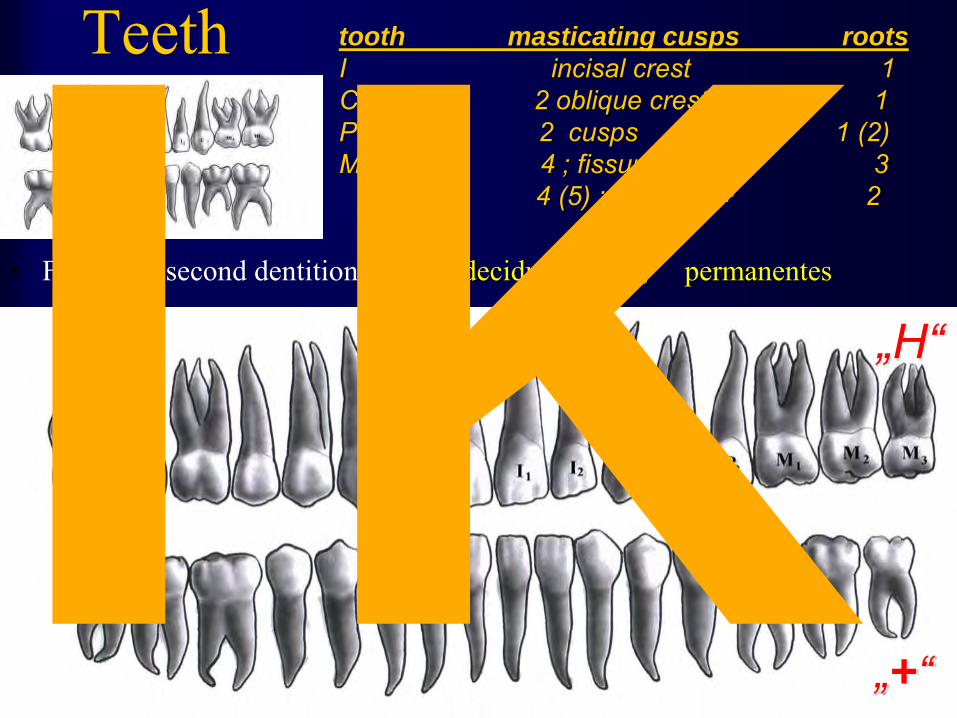
Teeth
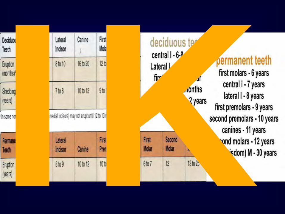
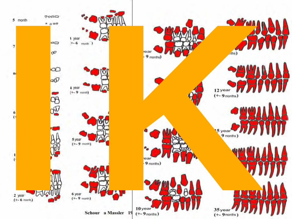
• Firast and second dentition dentes decidui (lactales), permanentes
tooth masticating cusps roots I incisal crest 1 C 2 oblique crests 1 P 2 cusps 1 (2) M - upper 4 ; fissures „H“ 3 - lower 4 (5) ; fissures + 2
„H“
„+“ IK

IK

IK

FDI Fedérale Dentaire Internationale ADA American Dental Association IK
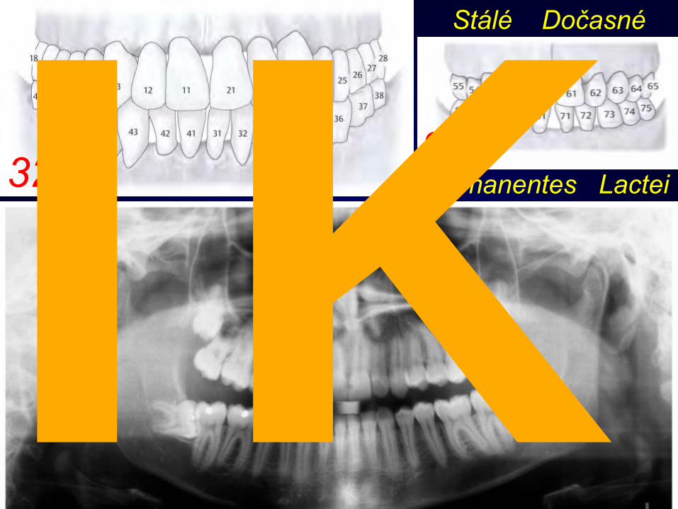
Stálé Dočasné
Permanentes Lactei 32 20 IK
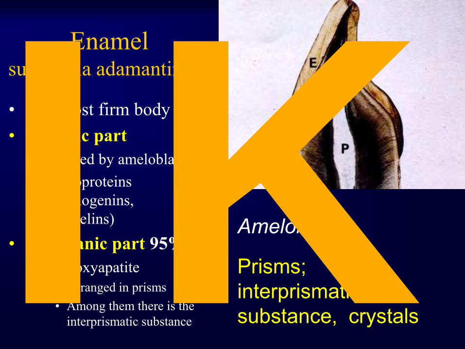
Enamel substantia adamantinosa
• The most firm body part • Organic part
– Formed by ameloblasts – glykoproteins
(amelogenins, enamelins)
• Anorganic part 95% – hydroxyapatite
• Arranged in prisms • Among them there is the
interprismatic substance
Prisms; interprismatic substance, crystals
Ameloblasts IK

Ameloblast: structure Secretion and reabsorbtion during enamelic matrix formation
Tomes fibers
Secretion
Tomes fiber
reabsorbtion
Nexus , desmosomes, tight junctions
Sir John Tomes,
(1815-1895), Engl. surgeon IK

Enamel structure Crystals, prisms and interprismatic substance (short and long molecules)
Prisms proceed to surface in various angles IK

Parazones Diazones IK

Hunter-Schreger strips, striae
Retzius strips, striae
Perikymata ridges, grooves IK
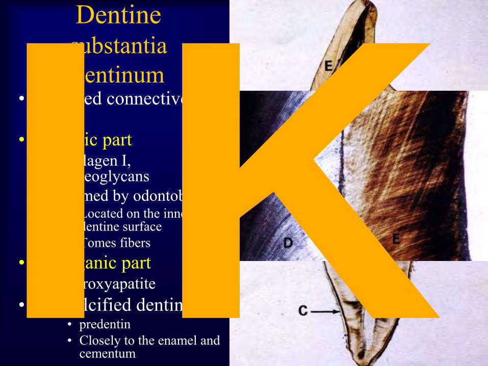
Dentine substantia dentinum
• calcified connective tissue
• Organic part – colllagen I,
proteoglycans – Formed by odontoblasts
• Located on the inner dentine surface
• Tomes fibers • Anorganic part
– hydroxyapatite • No-calcified dentine
• predentin • Closely to the enamel and
cementum
IK

Dentin odontoblasts
dentine tubules
Retzius lines vonEbner
Developmental order: Primary Secondary Terciary Folowing location: Mantle Circumpulpal Interdentin Globular Predentin Odontoblasts, matrix,
fibers
IK
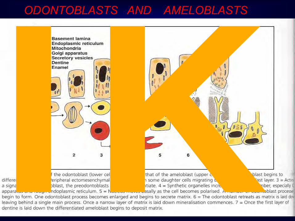
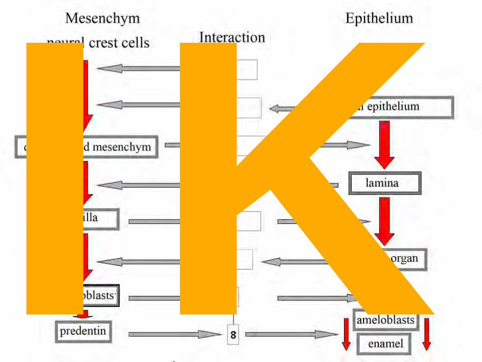
ODONTOBLASTS AND AMELOBLASTS
IK
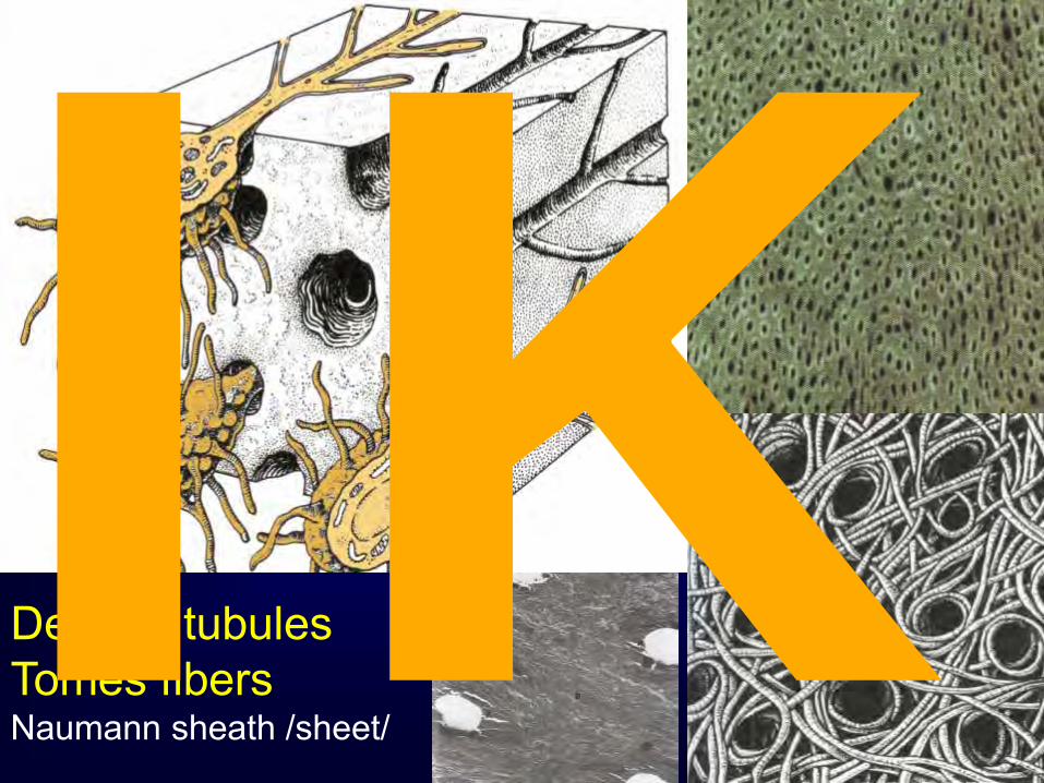
Dentine tubules Tomes fibers Naumann sheath /sheet/ IK

Mantle dentine Intermingling processes granular Circumpulpal dentine matrix rich Interdentine interglobular
Predentine matrix poor odontoblasts
Dentine structure
External coat Mantle dentine
10-30um; contains alfa-fibrills
Inner dentine Circumpulpal dentine
Stripped; exhibits regular secretion and mineralization layers
Predentine Amorphous; area of synthesis; polymorphous, contains proteoglycans, tropocollagen, glycoproteins IK

Neonatal line
Incremental lines (associated with dentine maturation) Von Ebner Adresen Neonatal Relation between primary and secondary dentine
Mineralizing in birth IK

Flow of fluid around Tomes fibers and nerve endings inside canals are responsible for dentine sensitivity IK
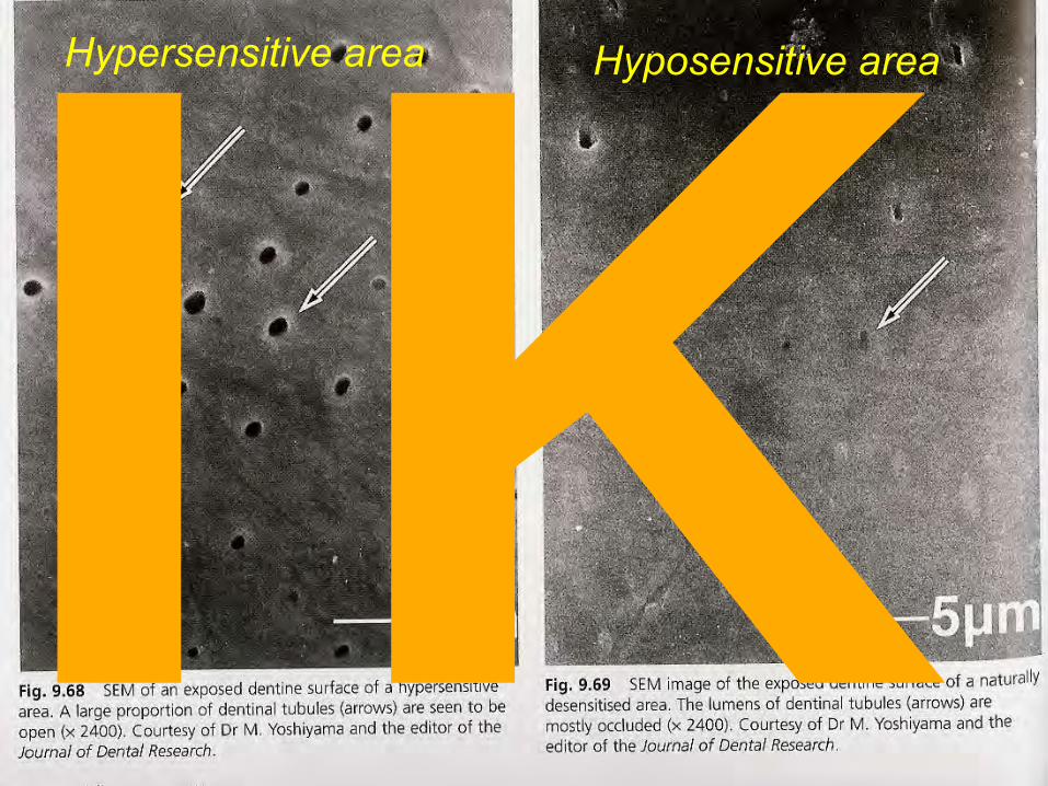
Hypersensitive area Hyposensitive area
IK
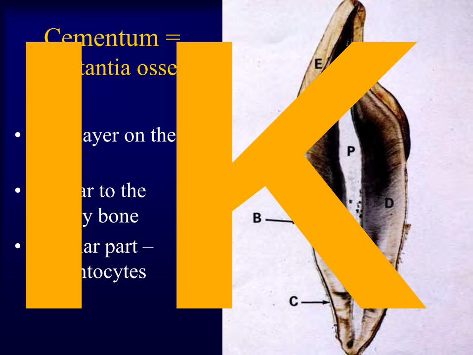
Cementum = substantia ossea
• Thin layer on the root
• Similar to the spongy bone
• Cellular part – cementocytes IK

Cementum Cementoblasts, mucoprotein substance, fibers
Cellulare: Collagen fibers + intercellulare substance + cementocytes Non cellulare: Collagen fibers + intercellular substance
IK

Relation between cementum, dentine and enamel
IK

Hypercementosis IK

Pulp
fibroblasts
ramification
• Odontoblasts, • Weil subodontoblastic layer • Layer rich by nuclei
Pulpocytes (mesenchymal cells, fibrocytes) basic substance (collagen fibers, sugars, elastic fibers) free cells (histiocytes, monocytes, plasmatic cells)
IK

IK

IK
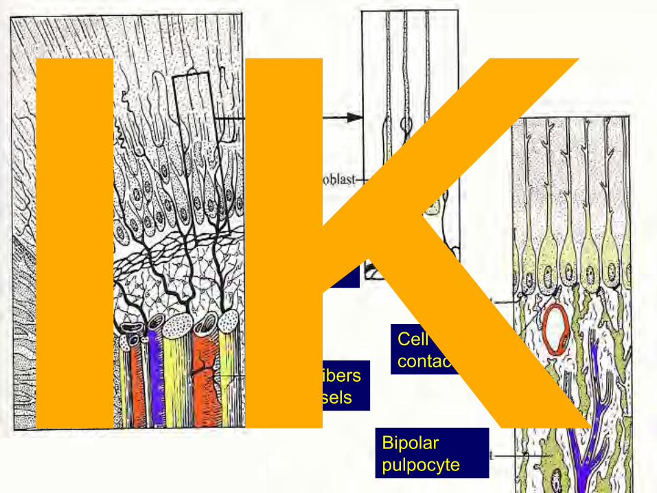
Raschkow plexus
Nerve fibers with vesels
Cell contacts
Bipolar pulpocyte
IK
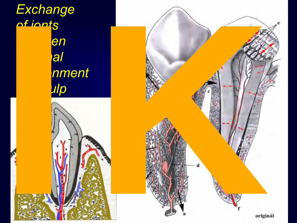
Exchange of ionts between external environment and pulp IK
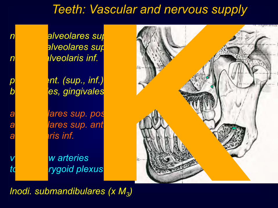
Teeth: Vascular and nervous supply
n.V/2 br.alveolares sup. post. br.alveolares sup. ant. n.V/3 n. alveolaris inf. plexus dent. (sup., inf.) br. dentales, gingivales aa. alveolares sup. post. aa. alveolares sup. ant. a. alveolaris inf. vv. – follow arteries to the pterygoid plexus lnodi. submandibulares (x M3) IK

Periodontium (Parodontium) Cementum, Cortical layer of alveolus Periodontal ligaments Gingiva (Mucoperiosteum) Cells Fibers Matrix Ligaments Plasma Vessels Nerves
“hydroelastic pillow“ IK

PARODONTIUM Periodontium IK

Sulcus gingivae
Paramarginal sulcus
Gingiva = relation to the teeth – “cuff (collar) attachment“ IK

Gingiva proper (attached) =
pink, stippled, keratizing
Alveolar mucosa (“loose gingiva“) =
shiny red, nonkeratizing
Gingiva = fibrous tissue + mucous membrane Free: Interdental; embrasured; circumdental Attached: Adjacent,fixed IK

Gum = Gingiva • Non-keratinized
epithelium - parakeratosis
• Multilayered epithelium
• Gingival groove • Junctional epithelium
– gingivodental junction / closure IK
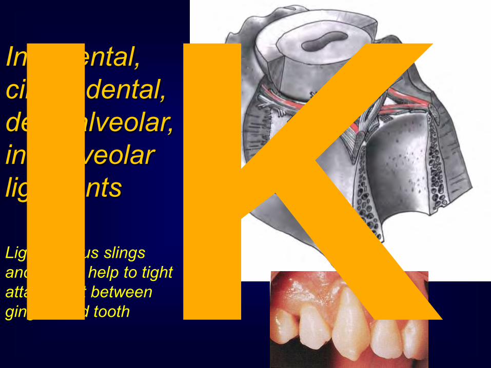
Interdental, circumdental, dentoalveolar, interalveolar ligaments
Ligamentous slings and circles help to tight attachment between gingiva and tooth IK

Upper teeth arch exhibits parabolic form Lower teeth arch exhibits hyperbolic form IK

Arches
parabolic
hyperbolic IK

Function of the Tooth
fixation system
Tooth fixation, elasticity, (hydroelastic cushion) nutrition, asistance during eruption
0.3-0.5
0.1-0.2
About 0.2 mm
IK
IK
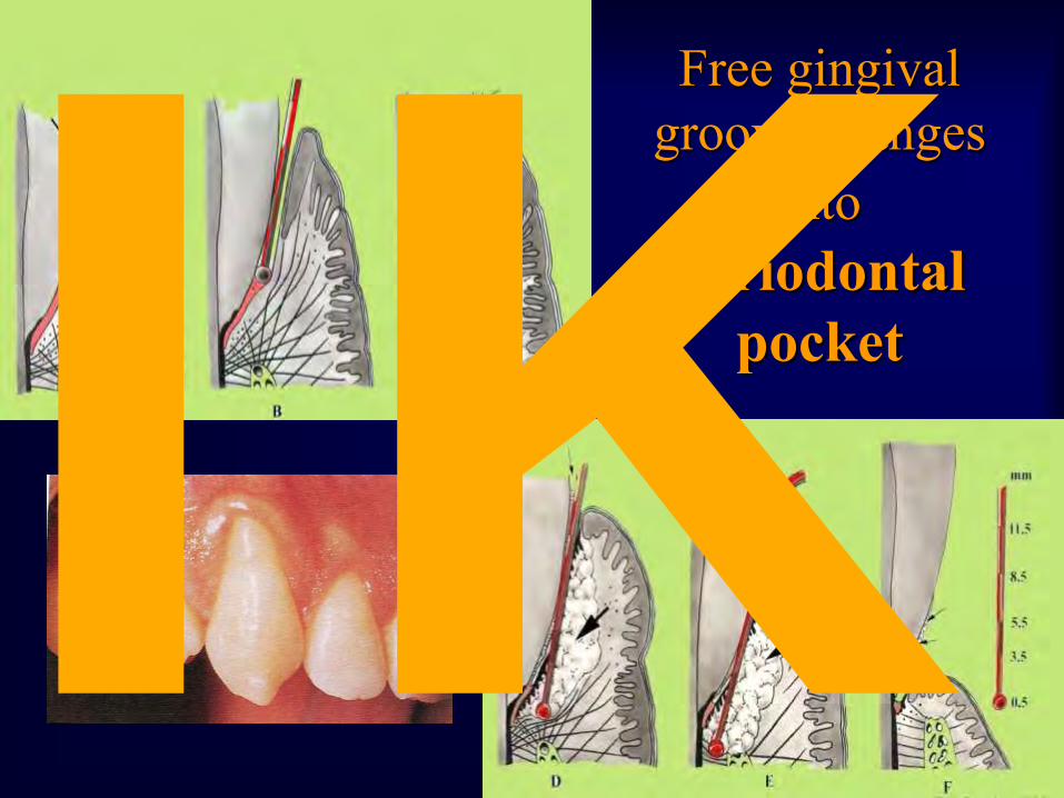
Free gingival groove changes
into periodontal
pocket IK
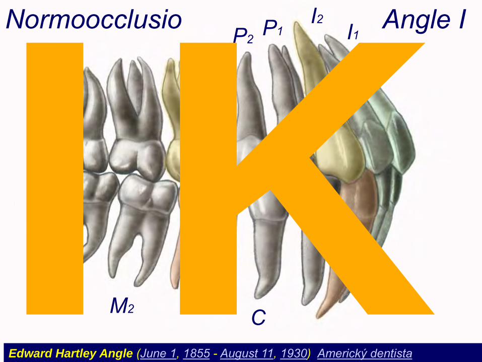
I1 I2
P2 P1
M1
M3
M2 C
Normoocclusio Angle I
Edward Hartley Angle (June 1, 1855 - August 11, 1930) Americký dentista
IK
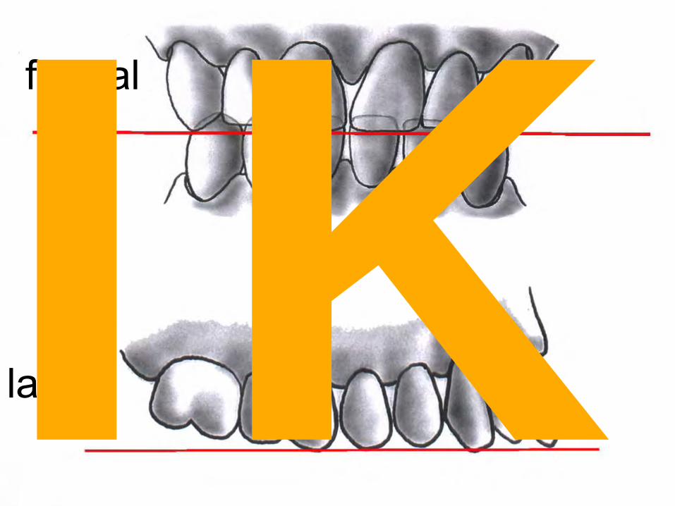
frontal
lateral IK

Wilson curve
Spee curve
Monson
IK

Variations and
anomalies • Mesiodens • Paramolar • Tuberculum Carabelli • Divergention or convergention of roots • Fusion of roots • Intradental location of tooth (dens in dente) • Root hyperplasia IK

IK

Teeth as a whole complex mordex = dentition
• ortodental position (vertical axes of teeth) • articulation = occlusion
– 80% psalidodontia (scissor-like occlusion) = norm – progenia = lower teeth in front of the upper ones – (hiatodontia (= mordex apertus), stegodontia, prognathia,
opisthodontia) IK

IK

IK

Anodontia;
hypodontia;
diastema
Tooth abnormalities in shape and position;
Gingivitis IK

Bodily shift
Mesial shift
Resorbtion -
Apposition + IK
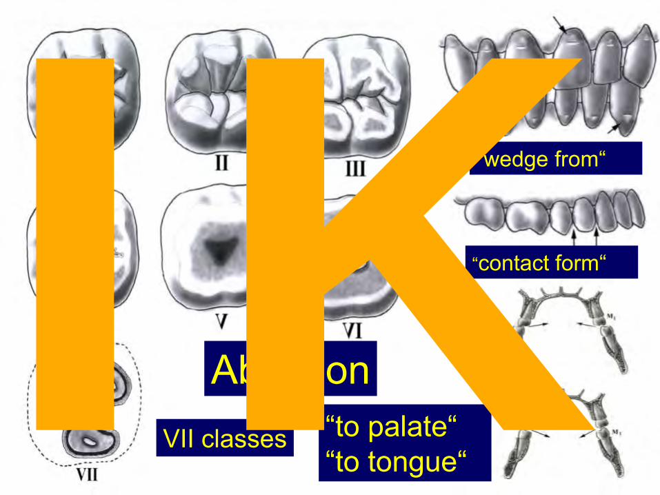
“to palate“ “to tongue“
“wedge from“
“contact form“
Abrasion VII classes IK

IK
IK

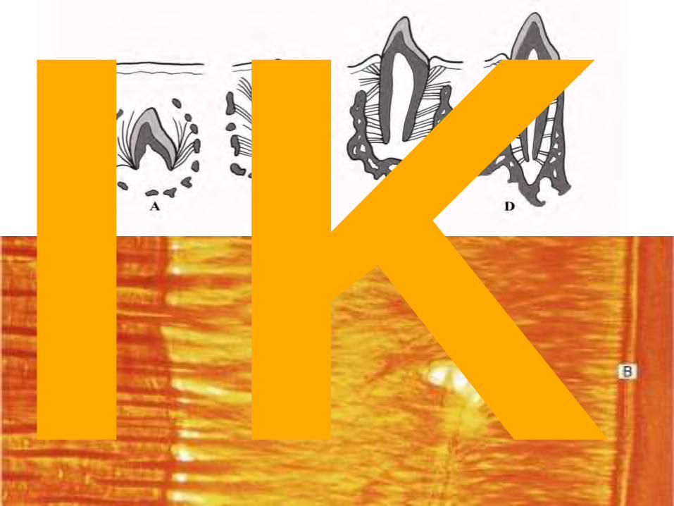
Tooth development
• week 6 development of the dental lamina (dental molding) – Thick epithelium
inside oralmucous membrane
• Each molding has about 10 center of the proliferations – Dental buds IK

Tooth developmental stages • Dental bud
– Local thick epithelium, 10 in each jaw
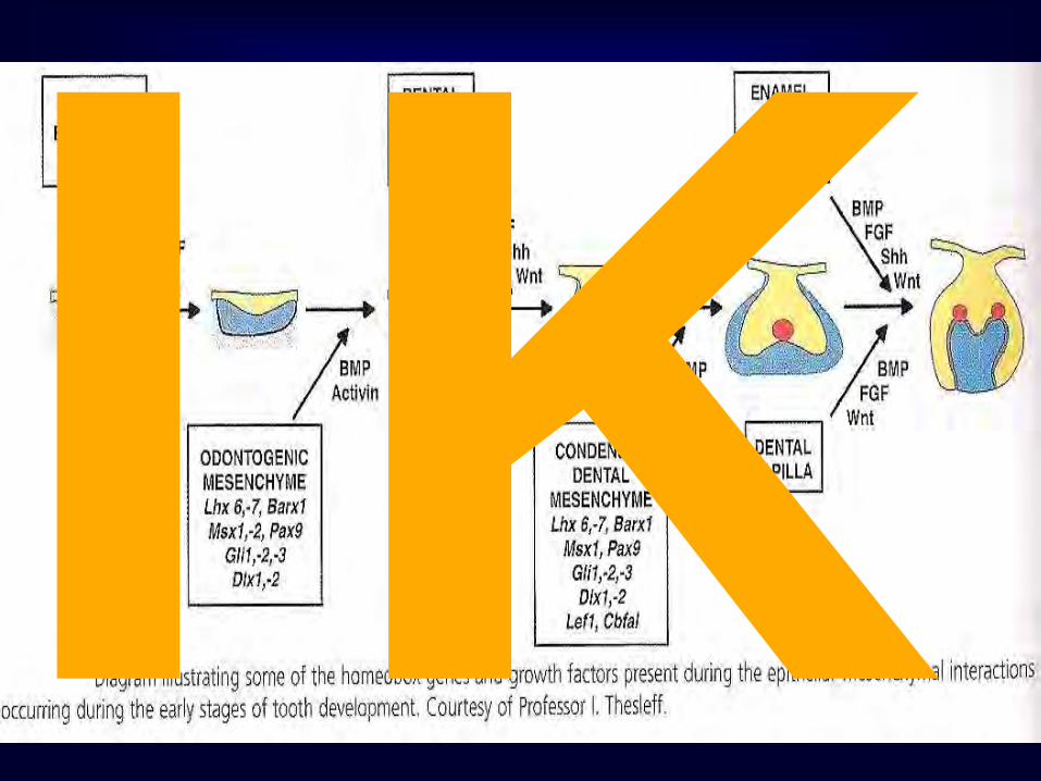
• Dental cap – Ectoderm part → enamel organ – Invagination of the mesenchyme → dental papilla
week 8
IK

• Dental bud (hat) → bell – External dental organ – Dental reticulum – Inner dental organ – Dental papilla → dental pulp – Dentl sac → cementum, periodontal ligaments
Tooth developmental stages
week 8
IK
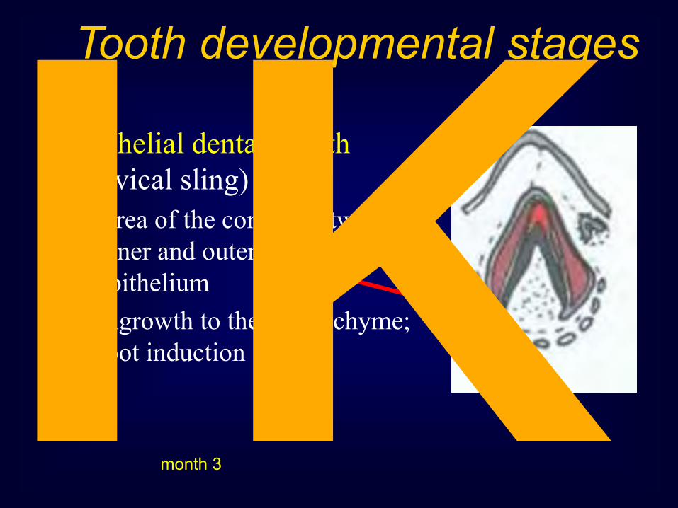
• Epithelial dental sheath (cervical sling) – Area of the contact between
inner and outer enamelic epithelium
– Ingrowth to the mesenchyme; root induction
Tooth developmental stages
month 3 IK

IK

IK
IK
IK
IK

IK

Použitá literatura
R. Čihák: Anatomie 1, 2, 3 Grada Publishing 2003
M. Dykes : Anatomy 2th edition, Mosby 2002
S.Snell: Clinical anatomy for Medical Students 6th edition, Lippincott, Williams & Wilkins
I.Klepáček, J.Mazánek et al.: Klinická anatomie ve stomatologii
Grada Publishing 2001
G.J.Tortora : Principles of Human Anatomy 4th edition, Williams & Wilkins
K.L.Moore, A.F.Dalley: Clinically Oriented Anatomy 4th edition, Williams & Wilkins
F.H.Netter: anatomický atlas člověka Vlastní archív
IK


