REVIEW ARTICLE
Should Return to Sport be Delayed Until 2 Years After AnteriorCruciate Ligament Reconstruction? Biological and FunctionalConsiderations
Christopher V. Nagelli1,2,4,5 • Timothy E. Hewett1,2,3,4,5
� Springer International Publishing Switzerland 2016
Abstract Anterior cruciate ligament (ACL) tears are
common knee injuries sustained by athletes during sports
participation. A devastating complication of returning to
sport following ACL reconstruction (ACLR) is a second
ACL injury. Strong evidence now indicates that younger,
more active athletes are at particularly high risk for a
second ACL injury, and this risk is greatest within the first
2 years following ACLR. Nearly one-third of the younger
cohort that resumes sports participation will sustain a
second ACL injury within the first 2 years after ACLR.
The evidence indicates that the risk of second injury may
abate over this time period. The incidence rate of second
injuries in the first year after ACLR is significantly greater
than the rate in the second year. The lower relative risk in
the second year may be related to athletes achieving
baseline joint health and function well after the current
expected timeline (6–12 months) to be released to unre-
stricted activity. This highlights a considerable debate in
the return to sport decision process as to whether an athlete
should wait until 2 years after ACLR to return to
unrestricted sports activity. In this review, we present
evidence in the literature that athletes achieve baseline
joint health and function approximately 2 years after
ACLR. We postulate that delay in returning to sports for
nearly 2 years will significantly reduce the incidence of
second ACL injuries.
Key Points
Young, active anterior cruciate ligament (ACL)
reconstructed (ACLR) athletes who return to high-
level sports sustain a disproportionately greater
incidence of second ACL injuries within the first
2 years after ACLR.
The evidence in the literature indicates that the
ACLR athletes do not regain baseline, or not
significantly different from baseline, knee joint
biological health and function until approximately
2 years after ACLR.
The incidence of second ACL injuries will
significantly decrease if ACLR athletes delay a
return to high-level activity until 2 years after
ACLR.
1 Introduction
Anterior cruciate ligament (ACL) tears are common mus-
culoskeletal injuries sustained by athletes who participate
in landing and pivoting sports. ACL reconstruction
& Timothy E. Hewett
1 Orthopedic Biomechanics Laboratories, Mayo Clinic,
200 First Street SW, RO_Gu_01_28BIOM, Rochester,
MN 55905, USA
2 Department of Orthopedic Surgery and Sports Medicine
Center, Mayo Clinic, Rochester, MN, USA
3 Department of Physical Medicine and Rehabilitation,
Mayo Clinic, Rochester, MN, USA
4 Department of Biomedical Engineering, The Ohio State
University, Columbus, OH, USA
5 Department of Physiology and Biomedical Engineering and,
Mayo Clinic, Rochester, MN, USA
123
Sports Med
DOI 10.1007/s40279-016-0584-z
(ACLR) is the current clinical standard to provide
mechanical stability to the joint and return to sports (RTS)
in a timely manner [1]. One of the most devastating, and all
too common, complications following a return to activity is
a second ACL injury. A systematic review of prospective
studies with a minimum follow-up time of 5 years found
that the pooled percentage of autograft failure rates and
contralateral ACL tears was 5.8 and 11.8 %, respectively
[2]. Long-term follow-up periods of C10 years have
reported second injury rates between 23 and 27 % [3–5].
Several factors have been implicated in second ACL injury
risk, including graft placement [6–8], graft type [9–13], sex
[14–16], age [5, 14, 17–19], time from surgery [10, 20, 21],
activity level [22, 23], and aberrant neuromuscular and
biomechanical adaptations [24].
The active, young athlete who resumes activity follow-
ing ACLR has a greater propensity for a second ACL
injury [8, 9, 13, 14, 17–19, 25, 26]. The probability of a
second injury increases three- to sixfold when the athlete is
aged \20 years [19]. Injury rates in this younger cohort
have been reported to be almost as high as 30 % in the
literature [5, 17, 20, 24]. In addition, the increased risk in
this group is apparent immediately upon returning to
sports. The evidence strongly indicates that second ACL
injury risk is greatest within the first 2 years after ACLR
for young athletes returning back to high-level sports
[15, 19, 20, 22, 24, 26]. A young athlete who returns to
sport within 1 year is 15 times more likely to suffer a
second ACL injury than a healthy athlete with no medical
history of a knee injury [15]. This elevated risk remains
evident within 2 years of returning to activity, when an
athlete is approximately six times more likely to sustain a
second injury than an uninjured counterpart [20]. While the
risk in this young cohort is high initially, this evidence
indicates that the risk may be abated over time as athletes
are still recovering baseline joint health and function.
These athletes are at a disproportionately higher risk of
second ACL injury within the first 2 years after ACLR.
Therefore, waiting to reintegrate into high-level sports
activity will significantly benefit the ACLR athlete.
The high secondary injury rates within 2 years after
ACLR in the young active cohort highlight the significant
impact of returning to sport too early. The marked increase
in early second ACL injuries also correspond with the shift
from a more conservative postoperative treatment of
ACLR athletes as described by Paulos et al. [27] to the
accelerated rehabilitation program suggested by Shel-
bourne and Nitz [28]. The lower relative risk of second
ACL injuries in athletes who delay a return to activity after
ACLR may be related to graft healing, recovering knee
joint homeostasis, and restoring normal joint motion prior
to being released to unrestricted sports activity. A complete
resolution of symptoms and deficits after ACLR may be
directly related to the biological and functional recovery of
the knee joint. In this review, we present the evidence that
athletes achieve baseline joint health and function
approximately 2 years after ACLR. The postulate outlined
below is that a delay in return to unrestricted high-level
sports participation for at least 2 years will significantly
reduce the incidence of second ACL injuries.
2 Biological Recovery of the Knee
Athletes may require a longer postoperative recovery per-
iod than the typically advocated 6–12 months to facilitate
the biological recovery of the joint [29]. The ACL graft
must undergo repopulation and proliferation of cells, re-
vascularization, and re-innervation to successfully restore
the native properties of the ligament [30]. In addition, ACL
graft maturation, through assimilation and re-ligamentiza-
tion, is critical to the reinstatement of ACL integrity
[31, 32]. These innate properties may be regained, but the
current evidence indicates that this healing occurs at a
slower rate than the timeframe in which athletes commonly
return to activity [33–36]. Furthermore, trauma from the
injury is not restricted to the ligament alone; it also affects
the surrounding joint tissues. A majority of ACL tears
occur as non-contact episodes that involve the knee
absorbing a high external load causing substantial trauma
to the articular cartilage and subchondral bone [37–40].
Partial recovery of the joint is deleterious and may lead to a
knee joint environment that is unable to endure the forces
associated with a return to activity. Therefore, if time from
ACLR is utilized among the RTS criteria in determining
whether an athlete is ready to RTS, it should ensure that the
ACL graft and surrounding knee joint tissue are completely
recovered prior to returning to sport. Ideally, an athlete
returns to sport once ACL and whole joint integrity is re-
established.
2.1 Bone Bruises
The prevalence of bone bruises concomitant with ACL
injuries is high, as approximately 80 % of individuals
present with subchondral lesions and bone marrow edema
[38, 41, 42]. These occult osseous lesions are identifiable
by magnetic resonance imaging (MRI) as alterations in
signal intensity [43, 44]. Despite conflicting evidence of
their clinical relevance and an absence of association with
second ACL injury risk [45–48], the presence of bone
bruises at follow-up may indicate that the knee has not
normalized.
A recent systematic review [49] investigated the char-
acteristics of bone bruises associated with ACL tears, their
clinical relevance, and their progression over time. The
C. V. Nagelli, T. E. Hewett
123
studies that included follow-up imaging observed that
chondral defects were still present 1 year after ACL injury
[49]. One limitation of the studies that were included in the
review was the relative short-term follow-up period of
\2 years [49]. Hence, whether these effects persisted at
2 years post-injury is not clear. Boks et al. [50] performed
a systematic review of follow-up studies to examine the
natural course of post-traumatic occult bone lesions as
detected by MRI imaging. A review of 13 studies
demonstrated that a natural healing response may be
observed for bone bruises after acute trauma, but the time
to recovery may depend on the extent of disturbance [51].
At follow-up imaging, the percentage of complete resolu-
tions of bone bruises in the knee ranged from 88 % after
11–16 months of follow-up to 100 % after 5–12 months of
follow-up [51]. The course of healing was variable and
possibly related to the severity and location of the bone
bruise.
Costa-Paz et al. [47] conducted a follow-up study of
bone bruises associated with ACL ruptures. Preoperative
MRIs verified the presence of bone bruises and were used
to develop a three-level classification system based on
severity and location [47]. The more severe a bone bruise,
the higher the classification it received. Follow-up MRIs
were conducted at an average of 34 months following
ACLR and revealed the resolution of all type I and type II
bone bruises, except one type II [47]. However, all type III
lesions had persistent evidence of abnormality on MRI
scans that was consistent with cartilage thinning or cortical
depression, indicating that recovery was not complete [47].
Bone bruises may be recovering up to 1 year following
ACLR and require a longer recovery period than the
standard timeline according to which athletes are returning
to activity.
Bone scans are important tools to evaluate both osseous
metabolic activity of the joint and potential bone loss
resulting from the trauma of the injury [52]. Bone loss is
not directly implicated in second ACL injury, but a
metabolically active joint and substantial bone loss are
signs of a degenerative joint environment and a knee that is
not prepared for unrestricted sports activity. In addition,
ACLR may slow the process of knee joint homeostasis,
further delaying its full recovery [53]. Nyland et al. [54]
conducted a systematic review of osseous deficits after
ACL injury and ACLR. A review of the literature indicated
that involved limb bone integrity is decreased after ACL
injury and that premorbid bone integrity is not re-estab-
lished after ACLR and accelerated rehabilitation [54].
Zerahn et al. [55] conducted a 2-year prospective cohort
study that evaluated the relationship between subjective
knee function (International Knee Documentation Com-
mittee [IKDC] score), self-reported signs of instability
(Lysholm score), and bone mineral density (BMD). There
was a significant decline in BMD in the proximal tibia of
the ACLR limb at the 4- and 12-month follow-up com-
pared with controls and the uninjured contralateral limb
[55]. The BMD of the medial tibia had returned to normal
levels at 24 months, but the lateral tibia had significantly
lower BMD than the uninjured limb and healthy controls
[55]. Furthermore, there was a significant improvement in
Lysholm score, level of activity, and knee function at
24 months [55]. The improvement in self-reported knee
function at 24 months was also associated with an increase
in BMD in the ACLR limb [55]. In summary, the recovery
of bone to baseline level may requisite a significant delay
in a RTS of nearly 2 years [56–58].
2.2 Mechanoreceptors and Sensory Afferents
Sensory nerve fibers and mechanoreceptors (Golgi tendon
organs, Pacinian corpuscles, and Ruffini nerve endings)
account for nearly 3 % of the ACL’s tissue volume and for
the sensory function of the ligament [59, 60]. In addition, a
reflex loop between these sensory constituents and the
surrounding musculature is involved in dynamic joint sta-
bility and proprioception [61, 62]. However, once the ACL
is disrupted, the native sensory function is lost and re-
innervation is not fully restored, regardless of ACLR
[63–65]. To compensate, ACLR athletes must develop
extra-articular sensation and control of the joint through the
mechanoreceptors and sensory nerve fibers in peri-articular
tissue. This loss in sensory function is strongly indicated by
proprioceptive deficits following ACL injury and ACLR
[66, 67]. Because proprioception of the knee is not objec-
tively defined and can be measured in several ways, we
limit our discussion in this current opinion article to tasks
of joint position matching and threshold to detection of
passive moments, which are commonly measured for joint
kinesthesia; however, we have discussed alternative, more
sensitive and specific methodologies in a separate review
[67].
This proprioceptive compensation strategy is not
immediately developed, but is instead slowly gained long
after ACLR and a release from sports participation. Iwasa
et al. [68] evaluated 38 patients following a hamstrings
tendon (HT) ACLR in joint position tasks from 3 to
24 months at 3-month intervals. A total of 30 patients had
improved joint position sense up until the final follow-up at
24 months, whereas eight patients did not have any
improvement in proprioception at any time in the course of
the study [68]. This study indicated that a longer postop-
erative recovery may be required for a complete resolution
of proprioceptive deficits. MacDonald et al. [69] conducted
‘threshold to detection of passive movement’ testing of
ACL-deficient, ACLR with HT autograft, and ACLR with
patellar tendon (PT) autograft at an average follow-up of
Waiting 2 Years to Return to Sport after ACL Reconstruction
123
31 months. No statistically significant differences were
observed during testing between the three groups at follow-
up [69].
Deficits in proprioception are not exclusive to the ACL-
injured and ACLR limb; they have been widely reported in
the uninjured limb but to a lesser extent [70]. While
symmetry is important, the assessment of proprioception
using the contralateral limb is a fundamental issue that
should be controlled. Roberts et al. [71] compared bilateral
proprioceptive deficits in ACLR and healthy volunteers at a
mean of 2 years from surgery and observed no differences
at 2 years within or between groups during the active and
visual reproduction tests [71]. Risberg et al. [72] investi-
gated proprioception in ACLR and control cohorts with
and without bracing. After controlled rehabilitation, the
cohorts were studied at a mean follow-up of 2 years. No
significant differences were observed during the threshold
to detection of passive moments between the ACLR knees
and the contralateral uninjured knees [72]. Further, there
were no significant differences during the same task
between the ACLR and control groups or the uninjured
contralateral limbs and the control group [72]. These
studies indicate that continued improvement in proprio-
ceptive function is observed up to 2 years after ACLR.
The current evidence regarding proprioception presents
conflicting results. Evidence indicating that proprioceptive
function can be fully restored to baseline levels is limited.
However, the longitudinal evidence indicates an improve-
ment of proprioceptive function over time and a recovery
to not significantly different from baseline well after ath-
letes are commonly released to sports activity. Gokeler
et al. [73] recently reviewed the literature to evaluate the
clinical relevance of proprioceptive deficits. They found
limited evidence to indicate that proprioceptive deficits
detected by the current measurement techniques adversely
affect function in ACL-deficient and ACLR athletes [73].
Further research is needed to develop more sensitive and
relevant measurements for proprioception and sensorimo-
tor function. However, the relative contribution of the loss
of ACL sensory information to chronic joint dysfunction
and second ACL injury risk is unclear.
2.3 Graft Maturation
The restoration of normal knee function may depend on
ACL graft maturation toward a biologic structure similar to
that of the native ACL. The healing and metaplasia of the
ACL graft commonly became known as ligamentization
[31]. Histological analysis of graft tissue from animal
models is commonly used to study the early, remodeling,
and mature phases of the ACL graft ligamentization
sequence [32, 74]. The biomechanical properties of the
ACL graft are significantly influenced by the remodeling
phase, the phase in which the graft is mechanically the
weakest and most susceptible to injury [30, 75]. Claes et al.
[33] conducted a systematic review of the current literature
on the ligamentization process in humans. A key finding
from the review is that humans undergo the same liga-
mentization sequence as animals, but the timeline of
healing is substantially different [33]. The ligamentization
processes occurs in humans over a much longer duration
than that originally observed in animal studies; specifically,
humans have a much slower remodeling phase [33].
A recent systematic review examined the literature on
the ligamentization process in HT autograft used in human
ACLR [34]. Within this review, the results of the liga-
mentization process in HT autografts were compared with
the other commonly used graft in ACLR, the PT autograft
[34]. The results indicated that the HT autograft has a
significantly delayed remodeling phase, which occurs
between 12 and 24 months [34]. Alternatively, PT auto-
grafts undergo the remodeling phase during the 6- to
12-month timeline [34]. These time periods correspond not
only to the time that athletes are returning to sport, but also
to the time when athletes are at a greater risk for a second
ACL injury.
A non-invasive approach to examine the ACL graft and
its stages of healing in humans is achieved through various
imaging modalities that are now clinically available [76]. A
recovery to nearly native ACL properties in imaging
studies is indicated approximately 2 years after ACLR.
Vogl et al. [77] conducted a 2-year prospective study that
evaluated the ACL graft healing process using contrast-
enhanced MRI. Revascularization of the ACL graft closely
resembled that of the native ACL at the 2-year follow-up
[77]. Zaffagnini et al. [78] studied the histological liga-
mentization changes in PT autograft at 6, 12, 24, 48, and
120 months following ACLR using transmission electron
microscopy (TEM). The results of the study indicated that
progressive ultrastructural changes towards the normal
ACL were observed for up to 24 months [78]. Notably, no
further changes were observed in the TEM imaging after
the 24-month time point after ACLR [78]. Variability in
healing was observed in these studies, but longer follow-up
periods indicated a complete structural and morphological
resemblance to the native ACL [79–82]. A significant delay
in RTS following ACLR to nearly 2 years may allow
complete healing of the ACL graft and possibly the pre-
vention of early failure in ACLR knees.
3 Functional Recovery of the Knee
Restoring mechanical stability after ACL injury by electing
to undergo ACLR does not address residual functional
deficits [83–85]. Postoperative rehabilitation protocols treat
C. V. Nagelli, T. E. Hewett
123
the localized dysfunction and related symptoms, including
joint effusion, limited range of motion, and deficits in
quadriceps muscle strength and activation. Functional
impairments implicated in second ACL injury continue to
persist when athletes are returning to sport and are mea-
surable for several months afterwards [84–87]. The evi-
dence in the literature indicates that the recovery of knee
function is the last of the remaining sequelae to normalize
following ACLR [88–91]. Specifically, ACLR athletes
demonstrate characteristic deficits in neuromuscular con-
trol and knee extension strength that do not begin to reach
baseline levels until at least 2 years after ACLR.
3.1 Neuromuscular Control
Similar to primary ACL injury, the majority of second
ACL injuries occur as non-contact episodes [92], and
failure to actively control the knee during multi-planar
movements may lead to an increased risk of second ACL
injury. A recent prospective study [24] screened 56 high-
risk ACLR athletes at the time they were medically cleared
to resume sports participation. At 1 year after the initial
assessment and actively participating in sport, 13 of the
athletes sustained a second ACL injury [24]. A combina-
tion of neuromuscular and biomechanical factors, including
transverse plane hip moments, frontal plane knee angles,
sagittal plane knee moments, and deficits in postural sta-
bility predicted second injury with 92 % sensitivity and
88 % specificity [24]. These findings are important because
they demonstrate that residual deficits in neuromuscular
control are highly predictive of a second ACL injury after
athletes resume sports participation.
Persistent faulty neuromuscular characteristics are evi-
dent in even basic biomechanical tasks such as walking.
Gokeler et al. [88] recently conducted a systematic review
that included a comprehensive overview of kinematic and
kinetic variables present during gait in athletes following
ACLR. A review of over 20 studies with a mean gait
analysis at 29.3 months (range between 3 weeks and
5.7 years) observed that altered biomechanics in all three
planes are common after ACLR and may persist up to
5 years after ACLR [88]. A more recent systematic review
and meta-analysis [89] compared the knee kinematic and
kinetics during walking of ACLR knees to healthy controls
and uninjured contralateral limbs. The analysis of 34
studies identified lower peak flexion moments during
6–12 months post-ACLR and lower peak flexion angles
during 1–3 years and C3 years after ACLR [89]. However,
the pooled data provided evidence of no significant dif-
ference between peak knee adduction moments after
3 years following ACLR [89]. The recovery of normal
neuromuscular control during daily activities such as gait
may be significantly delayed after ACLR.
To date, two studies have reported the progressive
recovery of neuromuscular control during gait C2 years
following ACLR. Roewer et al. [84] recruited a cohort of
high-risk athletes and assessed knee strength and biome-
chanics during gait at 6 months and then again at 2 years
after ACLR. The acute post-injury assessment revealed
asymmetrical knee angles, knee moments, and hip and
knee power that also persisted to 6 months after ACLR
[84]. At 2 years after surgery, quadriceps strength contin-
ued to improve and the kinematic and kinetic asymmetries
that were present at 6 months were resolved, which indi-
cated that athletes have the capability to functionally
improve up to 2 years after ACLR [84]. Webster et al. [93]
also assessed longitudinal changes in knee biomechanics
during gait at an initial assessment at 10 months and again
at 3 years after ACLR. An improvement in knee extension
and internal rotation was documented over time [93]. Pri-
marily, knee biomechanics remained relatively unchanged
from the initial and final assessment [93]. However, the
results indicated that normal gait may recover over the
longer time frames, past the 1-year time point [93].
Altered neuromuscular control is also commonly
reported during sports-related, high-demand activities fol-
lowing ACLR. Sports-specific jumping tasks have revealed
alterations in force generation and attenuation at the ACLR
knee up to 2 years after ACLR and beyond [94–96]. Per-
formance during functional hop testing is a common
assessment for clearance of athletes for unrestricted sports
activities. A recent systematic review [97] reported the
results of functional performance testing at differing time
points following ACLR. A review of 88 studies and nearly
5000 patients observed that the four standard hop tests
(single-leg, cross-over, triple, and timed 6-m hop tests)
were the common functional assessments [97]. The results
indicated that athletes reach 90 % limb symmetry index
(LSI) at 6–9 months postoperatively [97]. However, results
from more demanding functional tasks, such as endurance
hop testing, indicated larger deficits over the same 6- to
9-month time points [97]. These deficits appeared to nor-
malize at 24 months post-ACLR, with reported LSI in the
mid-90s, which are comparable to results from the standard
hop tests [97].
More recently, groups have utilized more invasive
techniques to study in vivo dynamic knee function after
ACLR [98]. Dual fluoroscopy and dynamic stereo-radiog-
raphy is becoming more widely accessible because it pro-
vides better precision and more reliability than the use of
high-speed cameras or marker-based motion capture sys-
tems [98]. Hoshino et al. [99] investigated whether knee
kinematics and joint contact mechanics can be restored
after ACLR with a double-bundle or single-bundle graft
using dynamic stereo X-ray to capture biplane radiographic
images. The athletes were, on average, a little more than
Waiting 2 Years to Return to Sport after ACL Reconstruction
123
1 year out from ACLR and were performing downhill
treadmill running [99]. The study concluded that neither
ACLR procedure restored normal knee kinematics or
medial joint sliding [99]. In addition, developments in
quantitative MRI have allowed researchers to evaluate the
composition and structures within the knee. A recent study
used volumetric dynamic imaging to assess contact pat-
terns during an active knee flexion/extension task in ath-
letes an average of 2 years post-ACLR [100]. There were
no reported significant differences in cartilage thickness
between the ACLR and healthy control groups [100].
However, the ACLR group demonstrated a greater reduc-
tion in the fraction of water bound by proteoglycan and
greater contact in the medial and posterior portion of the
knee than did the control group [100]. In contrast to the
commonly used high-speed cameras, these systems are far
more expensive, expose the patient to more radiation, and
are highly complex. Regardless of technique used to assess
kinematics and kinetics, athletes are still presenting with
large deficits in the 6- to 12-month time frame, but are
normalizing closer to 2 years after ACLR.
Failure to recover normal knee function may be related
to the inability to restore intrinsic properties of the native
ACL. Although functional asymmetries may abate over
time, normal neuromuscular control may never fully
recover following ACLR [88]. A new standard of neuro-
muscular control that involves deficits not significantly
different from baseline function may be inevitable in some
athletes.
3.2 Quadriceps Strength
Lower extremity muscle weakness is a debilitating and
ubiquitous impairment resulting from ACL injury.
Although the relative ratio between quadriceps and ham-
string strengths has been implicated in primary ACL injury
risk [101], the direct relationship of knee extensor and
flexor strength to second ACL has not been assessed. The
majority of sports medicine clinicians and physical thera-
pists advocate a recovery of quadriceps and hamstring
torque production equivalent to the contralateral limb prior
to participating in high-level sports activity. However, the
evidence in the literature strongly indicates that quadriceps
strength deficits are commonly observed for sev-
eral months and years following ACLR in spite of formal
rehabilitation focusing on rebuilding muscle strength [91].
A systematic review and meta-analysis investigated the
influence of graft choice on isokinetic muscle strength
4–24 months following ACLR [90]. The study observed
athletes who elect to receive a PT autograft demonstrated a
greater knee extensor deficit and lower knee flexor deficits
than patients with an HT autograft [90]. Importantly, the
strength deficits were unresolved up to 2 years following
ACLR [90]. The impact of lower extremity muscle weak-
ness has significant functional implications as athletes with
greater strength deficits demonstrate decreased functional
performance and landing strategies that increase risk of
second ACL injury [102, 103].
Recovery of quadriceps and hamstring strength is
commonly observed over time. However, evidence clearly
indicates that athletes consistently demonstrate significant
knee extensor and flexor strength deficits during the
expected time frame to RTS, but these deficits begin to
normalize approximately 2 years after ACLR. A few
studies that demonstrate a recovery of knee strength at
2 years are highlighted below. Aune et al. [104] longitu-
dinally compared isokinetic knee extensor and flexor
strength between athletes who received two different
autografts for ACLR. At 24 months after surgery, nearly
symmetrical knee extensor and flexor strength measure-
ments were reported within the groups, and significant
differences between the two cohorts were not observed
[104]. Further, Inagaki et al. [105] assessed clinical out-
comes 2 years following ACLR and documented that
average peak isokinetic knee extensor and flexor mea-
surements at 2 years demonstrated symmetry comparable
to that of an uninjured population (\15 % deficit) [105].
Longitudinal prospective randomized clinical trials may
provide valuable insight into trends in clinical outcomes. In
one such study, Aglietti et al. [106] assessed 120 athletes in
a prospective randomized study to compare two unique
autografts fixed with modern devices at 4, 12, and
24 months. An isokinetic dynamometer was used to mea-
sure concentric knee extensors and flexors muscle at 60�,120�, and 180�/s [106]. At the 2-year follow-up evaluation,
knee extensor and flexor strength of the involved limb was
comparable to that of the contralateral side; in some
instances, it was even greater [106]. Furthermore, a marked
improvement in extensor strength was observed over time
regardless of the graft type that only became symmetrical
with the contralateral side 2 years postoperatively [106]. In
addition, Maletis et al. [107] also conducted a similar
prospective randomized study using similar autografts and
assessment time points. Isokinetic knee extension/flexion
measurements were also conducted at 60�, 180�, and 300�/s[107]. Similarly, at the 2-year follow-up, clinically mean-
ingful significant differences were not observed in knee
extension and flexion strength [107]. Extensor muscle
strength once again significantly improved over time and
became comparable to that of the contralateral side at
2 years [107]. It is common to measure variable measures
of knee strength following ACLR, but the recovery of
strength seems to be sustained beyond the 2-year time
point.
Longitudinal studies of more than 2 years have also
demonstrated the sustained recovery of knee strength.
C. V. Nagelli, T. E. Hewett
123
Studies that evaluated athletes for over 5 years after ACLR
reported quadriceps strength measurements comparable to
those of a healthy athlete population, with minimal deficits
of between 6 and 10 % [108–110]. Another benefit of delay
of return to pre-injury level of activity for up to 2 years
after ACLR is the recovery of baseline knee strength and a
potential reduction of second ACL injury risk.
3.3 Effect of Surgical Techniques and Postoperative
Rehabilitation
In addition, functional recovery of the knee after ACLR
may be related to surgical techniques utilized in ACLR and
postoperative rehabilitation programs. These topics are
highly researched and discussed in sports medicine but
consensus is minimal. Here, we give a brief overview of
the recent evidence and how these factors may affect early
failures. In particular, femoral tunnel placement during
ACLR has highlighted the impact of surgical techniques on
second ACL injury risk. A recent report has observed that
femoral tunnel malposition was the most commonly cited
reason for a graft failure in a large ACLR cohort [111].
Furthermore, the Danish Knee Ligament Registry observed
an increased risk of revision after anteromedial femoral
tunnel placement compared with a transtibial approach
[112]. Current techniques in ACLR over the past sev-
eral years have focused on reproducing more anatomical
tunnel placement and graft geometry to restore knee
kinematics [98]. Xu et al. [113] demonstrated in a cadav-
eric study that an anatomic anteromedial tunnel placement
can lead to biomechanical advantages when compared with
a non-anatomic placement, and the anatomic placement
better restores knee kinematics to the native intact ACL
state [113]. However, the anatomic placement had signifi-
cantly greater in situ forces than the non-anatomic place-
ment, making it more vulnerable to graft failure [113].
Therefore, delaying a return to activity longer than the
expected timeline may significantly help the recovering
knee withstand the forces of returning to sport. It is likely
that misplaced tunnels are responsible for early traumatic
failures and may predispose a graft to aberrant biome-
chanics, causing it to rupture at a later time [114]. Although
differences in outcomes may be reported, it is generally
thought that the recent trend in surgical practice of a more
anatomic placement to resemble the native ligament is
better for the outcome of the graft as long as it is allowed to
heal completely [115, 116]. Because the significant
majority of athletes undergo ACLR, it is also important to
understand how surgery and rehabilitation together can be
utilized to optimize patient knee outcomes.
Postoperative rehabilitation programs are considered
essential for rebuilding lower extremity muscle strength
and restoring joint mobility and neuromuscular control
with the goal of returning to pre-injury levels of activity.
Currently, there is little consensus regarding an optimal
rehabilitation program to safety and effectively return
athletes to their pre-injury level of sport. A systematic
review of 33 randomized clinical trials observed that sev-
eral of the studies had significant flaws and that little evi-
dence could be derived from them [117]. The authors
concluded there was evidence for high-intensity neuro-
muscular electrical stimulation, volitional exercises, and
neuromuscular training [117]. More recently a randomized
controlled trial assigned 74 ACLR patients to either a
neuromuscular training program or a traditional strength
training program and followed for 2 years after ACLR
[118]. The neuromuscular training group reported greater
global knee function and reduced pain during activity,
while the strength training group demonstrated greater
hamstring strength at 2 years post-ACLR [118]. However,
there were no significant differences between the two
groups in Cincinnati knee score at 1 and 2 years [118].
Grindem et al. [119] compared the preoperative and 2-year
postoperative self-reported outcomes of ACLR patients
who underwent progressive preoperative and postoperative
rehabilitation compared with the institution’s standard of
care. The patients enrolled in the preoperative and post-
operative rehabilitation program demonstrated significantly
greater Knee Injury and Osteoarthritis Outcome Score
(KOOS) in all subscales preoperatively and 2 years post-
operatively than did patients in the standard of care group
[119]. Furthermore, a group investigated whether there
were any long-term differences (at 2–4 years after ACLR)
in athletes who underwent a home-based or physical ther-
apist-supervised rehabilitation program in the first
3 months after ACLR [120]. The home-based group had a
significantly higher ACL quality of life questionnaire score
at a mean of 38 months after ACLR, but there were no
significant differences in knee extension/flexion range of
motion, knee laxity, lower extremity strength, and IKDC
score [120]. The optimal rehabilitation program is still
unknown, and the lack of a standard rehabilitation treat-
ment protocol is a major limitation to returning athletes
back to sport safely. Regardless of the postoperative
rehabilitation protocol, the outcomes following ACLR
begin to normalize at nearly 2 years or later. The combi-
nation of time and comprehensive functional criteria is
critical to return to high-demand activity, but the current
approach of accelerated rehabilitation and timeline to
recover is detrimental to the athlete.
Surgical techniques and postoperative rehabilitation
programs are certainly a topic of ongoing debate and
research studies. Importantly, regardless of surgical tech-
niques and rehabilitation protocols, athletes returning to
activity too early, prior to recovering baseline knee joint
health and function, increase their risk for a subsequent
Waiting 2 Years to Return to Sport after ACL Reconstruction
123
ACL injury. The evidence in the literature indicates that
significantly delaying a return to high-level sports until
nearly 2 years will benefit the athlete.
4 Conclusion
Deficits in knee function and biological health of the knee
joint are all common up to 1 year following ACLR. A
marked improvement in joint health and function and a
resolution of symptoms is strongly indicated at 2 years and
beyond following ACLR. The 2-year time point post-
ACLR is not arbitrary but is evident based on a discerning
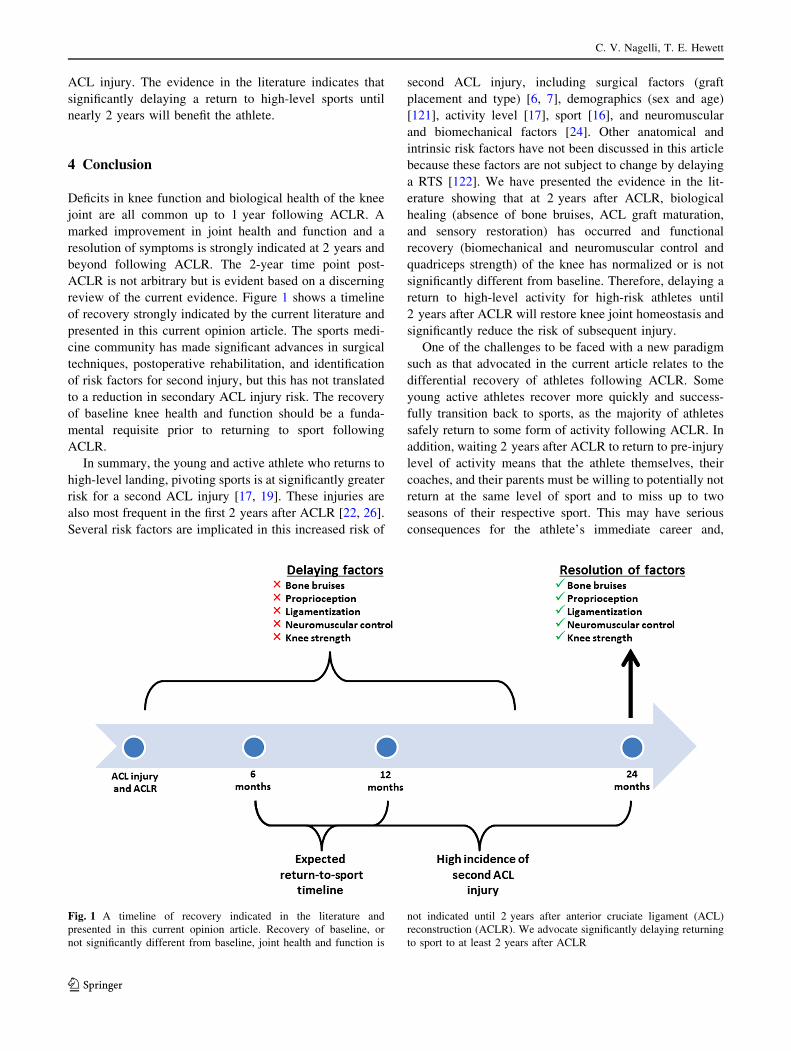
review of the current evidence. Figure 1 shows a timeline
of recovery strongly indicated by the current literature and
presented in this current opinion article. The sports medi-
cine community has made significant advances in surgical
techniques, postoperative rehabilitation, and identification
of risk factors for second injury, but this has not translated
to a reduction in secondary ACL injury risk. The recovery
of baseline knee health and function should be a funda-
mental requisite prior to returning to sport following
ACLR.
In summary, the young and active athlete who returns to
high-level landing, pivoting sports is at significantly greater
risk for a second ACL injury [17, 19]. These injuries are
also most frequent in the first 2 years after ACLR [22, 26].
Several risk factors are implicated in this increased risk of
second ACL injury, including surgical factors (graft
placement and type) [6, 7], demographics (sex and age)
[121], activity level [17], sport [16], and neuromuscular
and biomechanical factors [24]. Other anatomical and
intrinsic risk factors have not been discussed in this article
because these factors are not subject to change by delaying
a RTS [122]. We have presented the evidence in the lit-
erature showing that at 2 years after ACLR, biological
healing (absence of bone bruises, ACL graft maturation,
and sensory restoration) has occurred and functional
recovery (biomechanical and neuromuscular control and
quadriceps strength) of the knee has normalized or is not
significantly different from baseline. Therefore, delaying a
return to high-level activity for high-risk athletes until
2 years after ACLR will restore knee joint homeostasis and
significantly reduce the risk of subsequent injury.
One of the challenges to be faced with a new paradigm
such as that advocated in the current article relates to the
differential recovery of athletes following ACLR. Some
young active athletes recover more quickly and success-
fully transition back to sports, as the majority of athletes
safely return to some form of activity following ACLR. In
addition, waiting 2 years after ACLR to return to pre-injury
level of activity means that the athlete themselves, their
coaches, and their parents must be willing to potentially not
return at the same level of sport and to miss up to two
seasons of their respective sport. This may have serious
consequences for the athlete’s immediate career and,
Fig. 1 A timeline of recovery indicated in the literature and
presented in this current opinion article. Recovery of baseline, or
not significantly different from baseline, joint health and function is
not indicated until 2 years after anterior cruciate ligament (ACL)
reconstruction (ACLR). We advocate significantly delaying returning
to sport to at least 2 years after ACLR
C. V. Nagelli, T. E. Hewett
123
depending on the athlete’s potential, their future long-term
career. However, the current approach of early accelerated
rehabilitation programs and the expected timeline to
recovery of 6–12 months is deleterious because the athlete
is not completely recovered, which predisposes them to an
increased risk for a second ACL injury. The young active
athlete who attempts to resume sports participation at the
same competitive level has a nearly one in three chance of
going on to experience a second ACL injury within the first
or second year post-ACLR. Therefore, the evidence
advocates that these athletes delay a return to sports for
2 years to mitigate the unacceptably high risk for a second
ACL injury, especially in those aged\20 years.
Acknowledgments The authors would like to thank Drs. Stephanie
Di Stasi, Wendy Hurd, and Kate Webster for their input, clinical
expertise, editorial work, and conversations regarding the topic pre-
sented in this article.
Compliance with Ethical Standards
Funding The authors acknowledge funding from the National Insti-
tute of Arthritis and Musculoskeletal and Skin Diseases: R01-
AR049735, R01-AR055563, and R01AR056259 to TEH.
Conflicts of interest Christopher Nagelli and Timothy Hewett have
no conflicts of interest relevant to the content of this review.
References
1. Mall NA, Chalmers PN, Moric M, et al. Incidence and trends of
anterior cruciate ligament reconstruction in the United States.
Am J Sports Med. 2014;42(10):2363–70.
2. Wright RW, Magnussen RA, Dunn WR, et al. Ipsilateral graft
and contralateral ACL rupture at five years or more following
ACL reconstruction a systematic review. J Bone Joint Surg Am.
2011;93A(12):1159–65.
3. Bourke HE, Salmon LJ, Waller A, et al. Survival of the anterior
cruciate ligament graft and the contralateral ACL at a minimum
of 15 years. Am J Sports Med. 2012;40(9):1985–92.
4. Drogset JO, Grontvedt T, Robak OR, et al. A sixteen-year fol-
low-up of three operative techniques for the treatment of acute
ruptures of the anterior cruciate ligament. J Bone Joint Surg Am.
2006;88(5):944–52.
5. Morgan MD, Salmon LJ, Waller A, et al. Fifteen-year survival
of endoscopic anterior cruciate ligament reconstruction in
patients aged 18 years and younger. Am J Sports Med.
2016;44(2):384–92.
6. Brophy RH, Selby RM, Altchek DW. Anterior cruciate ligament
revision: double-bundle augmentation of primary vertical graft.
Arthroscopy. 2006;22(6):683 (e1–5).
7. Marchant BG, Noyes FR, Barber-Westin SD, et al. Prevalence of
nonanatomical graft placement in a series of failed anterior cruciate
ligament reconstructions. Am J SportsMed. 2010;38(10):1987–96.
8. Hui C, Salmon LJ, Kok A, et al. Fifteen-year outcome of
endoscopic anterior cruciate ligament reconstruction with
patellar tendon autograft for ‘‘isolated’’ anterior cruciate liga-
ment tear. Am J Sports Med. 2011;39(1):89–98.
9. Leys T, Salmon L, Waller A, et al. Clinical results and risk
factors for reinjury 15 years after anterior cruciate ligament
reconstruction: a prospective study of hamstring and patellar
tendon grafts. Am J Sports Med. 2012;40(3):595–605.
10. Laboute E, Savalli L, Puig P, et al. Analysis of return to com-
petition and repeat rupture for 298 anterior cruciate ligament
reconstructions with patellar or hamstring tendon autograft in
sportspeople. Ann Phys Rehabil Med. 2010;53(10):598–614.
11. Gifstad T, Foss OA, Engebretsen L, et al. Lower risk of revision
with patellar tendon autografts compared with hamstring auto-
grafts: a registry study based on 45,998 primary ACL reconstruc-
tions in Scandinavia. Am J Sports Med. 2014;42(10):2319–28.
12. Persson A, Fjeldsgaard K, Gjertsen JE, et al. Increased risk of
revision with hamstring tendon grafts compared with patellar
tendon grafts after anterior cruciate ligament reconstruction: a
study of 12,643 patients from the Norwegian Cruciate Ligament
Registry, 2004–2012. Am J Sports Med. 2014;42(2):285–91.
13. Maletis GB, Inacio MC, Desmond JL, et al. Reconstruction of
the anterior cruciate ligament: association of graft choice with
increased risk of early revision. Bone Joint J. 2013;95-
B(5):623–8.
14. Shelbourne KD, Gray T, Haro M. Incidence of subsequent injury
to either knee within 5 years after anterior cruciate ligament
reconstruction with patellar tendon autograft. Am J Sports Med.
2009;37(2):246–51.
15. Paterno MV, Rauh MJ, Schmitt LC, et al. Incidence of con-
tralateral and ipsilateral anterior cruciate ligament (ACL) injury
after primary ACL reconstruction and return to sport. Clin J
Sport Med. 2012;22(2):116–21.
16. Allen MM, Pareek A, Krych AJ, et al. Are female soccer players
at an increased risk of second anterior cruciate ligament injury
compared with their athletic peers? Am J Sports Med. 2016.
doi:10.1177/0363546516648439.
17. Kamien PM, Hydrick JM, Replogle WH, et al. Age, graft size,
and Tegner activity level as predictors of failure in anterior
cruciate ligament reconstruction with hamstring autograft. Am J
Sports Med. 2013;41(8):1808–12.
18. Magnussen RA, Lawrence JT, West RL, et al. Graft size and
patient age are predictors of early revision after anterior cruciate
ligament reconstruction with hamstring autograft. Arthroscopy.
2012;28(4):526–31.
19. Webster KE, Feller JA, Leigh WB, et al. Younger patients are at
increased risk for graft rupture and contralateral injury after
anterior cruciate ligament reconstruction. Am J Sports Med.
2014;42(3):641–7.
20. Paterno MV, Rauh MJ, Schmitt LC, et al. Incidence of second
ACL injuries 2 years after primary ACL reconstruction and
return to sport. Am J Sports Med. 2014;42(7):1567–73.
21. Maletis GB, Inacio MC, Reynolds S, et al. Incidence of post-
operative anterior cruciate ligament reconstruction infections:
graft choice makes a difference. Am J Sports Med.
2013;41(8):1780–5.
22. Salmon L, Russell V, Musgrove T, et al. Incidence and risk factors
for graft rupture and contralateral rupture after anterior cruciate
ligament reconstruction. Arthroscopy. 2005;21(8):948–57.
23. Borchers JR, Pedroza A, Kaeding C. Activity level and graft
type as risk factors for anterior cruciate ligament graft failure: a
case-control study. Am J Sports Med. 2009;37(12):2362–7.
24. Paterno MV, Schmitt LC, Ford KR, et al. Biomechanical mea-
sures during landing and postural stability predict second ante-
rior cruciate ligament injury after anterior cruciate ligament
reconstruction and return to sport. Am J Sports Med.
2010;38(10):1968–78.
25. Fauno P, Rahr-Wagner L, Lind M. Risk for revision after
anterior cruciate ligament reconstruction is higher among ado-
lescents: results from the Danish registry of knee ligament
reconstruction. Orthop J Sports Med. 2014;2(10):232596711
4552405.
Waiting 2 Years to Return to Sport after ACL Reconstruction
123
26. Lind M, Menhert F, Pedersen AB. Incidence and outcome after
revision anterior cruciate ligament reconstruction: results from
the Danish registry for knee ligament reconstructions. Am J
Sports Med. 2012;40(7):1551–7.
27. Paulos L, Noyes FR, Grood E, et al. Knee rehabilitation after
anterior cruciate ligament reconstruction and repair. Am J Sports
Med. 1981;9(3):140–9.
28. Shelbourne KD, Nitz P. Accelerated rehabilitation after anterior
cruciate ligament reconstruction. J Orthop Sports Phys Ther.
1992;15(6):256–64.
29. Grindem H, Snyder-Mackler L, Moksnes H, et al. Simple
decision rules can reduce reinjury risk by 84 % after ACL
reconstruction: the Delaware-Oslo ACL cohort study. Br J
Sports Med. 2016;50(13):804–8.
30. Scheffler SU, Unterhauser FN, Weiler A. Graft remodeling and
ligamentization after cruciate ligament reconstruction. Knee
Surg Sports Traumatol Arthrosc. 2008;16(9):834–42.
31. Amiel D, Kleiner JB, Roux RD, et al. The phenomenon of
‘‘ligamentization’’: anterior cruciate ligament reconstruction with
autogenous patellar tendon. J Orthop Res. 1986;4(2):162–72.
32. Arnoczky SP, Tarvin GB, Marshall JL. Anterior cruciate liga-
ment replacement using patellar tendon. An evaluation of graft
revascularization in the dog. J Bone Joint Surg Am.
1982;64(2):217–24.
33. Claes S, Verdonk P, Forsyth R, et al. The ‘‘ligamentization’’
process in anterior cruciate ligament reconstruction what hap-
pens to the human graft? a systematic review of the literature.
Am J Sports Med. 2011;39(11):2476–83.
34. Pauzenberger L, Syre S, Schurz M. ‘‘Ligamentization’’ in
hamstring tendon grafts after anterior cruciate ligament recon-
struction: a systematic review of the literature and a glimpse into
the future. Arthroscopy. 2013;29(10):1712–21.
35. Abe S, Kurosaka M, Iguchi T, et al. Light and electron micro-
scopic study of remodeling and maturation process in autoge-
nous graft for anterior cruciate ligament reconstruction.
Arthroscopy. 1993;9(4):394–405.
36. Rougraff B, Shelbourne KD, Gerth PK, et al. Arthroscopic and
histologic analysis of human patellar tendon autografts used for
anterior cruciate ligament reconstruction. Am J Sports Med.
1993;21(2):277–84.
37. Borchers JR, Kaeding CC, Pedroza AD, et al. Intra-articular
findings in primary and revision anterior cruciate ligament
reconstruction surgery: a comparison of the MOON and MARS
study groups. Am J Sports Med. 2011;39(9):1889–93.
38. Spindler KP, Schils JP, Bergfeld JA, et al. Prospective study of
osseous, articular, and meniscal lesions in recent anterior cru-
ciate ligament tears by magnetic resonance imaging and
arthroscopy. Am J Sports Med. 1993;21(4):551–7.
39. Boden BP, Dean GS, Feagin JA Jr, et al. Mechanisms of anterior
cruciate ligament injury. Orthopedics. 2000;23(6):573–8.
40. Rosen MA, Jackson DW, Berger PE. Occult osseous lesions doc-
umented by magnetic resonance imaging associated with anterior
cruciate ligament ruptures. Arthroscopy. 1991;7(1):45–51.
41. Dunn WR, Spindler KP, Amendola A, et al. Which preoperative
factors, including bone bruise, are associated with knee pain/
symptoms at index anterior cruciate ligament reconstruction
(ACLR)? A Multicenter Orthopaedic Outcomes Network
(MOON) ACLR Cohort Study. Am J Sports Med. 2010;38(9):
1778–87.
42. Vellet AD, Marks PH, Fowler PJ, et al. Occult posttraumatic
osteochondral lesions of the knee: prevalence, classification, and
short-term sequelae evaluated with MR imaging. Radiology.
1991;178(1):271–6.
43. Mink JH, Deutsch AL. Occult cartilage and bone injuries of the
knee: detection, classification, and assessment with MR imag-
ing. Radiology. 1989;170(3 Pt 1):823–9.
44. RoemerFW,FrobellR,HunterDJ, et al.MRI-detected subchondral
bone marrow signal alterations of the knee joint: terminology,
imaging appearance, relevance and radiological differential diag-
nosis. Osteoarthritis Cartilage. 2009;17(9):1115–31.
45. Johnson DL, Bealle DP, Brand JC Jr, et al. The effect of a
geographic lateral bone bruise on knee inflammation after acute
anterior cruciate ligament rupture. Am J Sports Med.
2000;28(2):152–5.
46. Boks SS, Vroegindeweij D, Koes BW, et al. Clinical conse-
quences of posttraumatic bone bruise in the knee. Am J Sports
Med. 2007;35(6):990–5.
47. Costa-Paz M, Muscolo DL, Ayerza M, et al. Magnetic resonance
imaging follow-up study of bone bruises associated with anterior
cruciate ligament ruptures. Arthroscopy. 2001;17(5):445–9.
48. Hanypsiak BT, Spindler KP, Rothrock CR, et al. Twelve-year
follow-up on anterior cruciate ligament reconstruction: long-
term outcomes of prospectively studied osseous and articular
injuries. Am J Sports Med. 2008;36(4):671–7.
49. Papalia R, Torre G, Vasta S, et al. Bone bruises in anterior
cruciate ligament injured knee and long-term outcomes. A
review of the evidence. Open Access. J Sports Med. 2015;6:
37–48.
50. Boks SS, Vroegindeweij D, Koes BW, et al. Magnetic resonance
imaging abnormalities in symptomatic and contralateral knees:
prevalence and associations with traumatic history in general
practice. Am J Sports Med. 2006;34(12):1984–91.
51. Boks SS, Vroegindeweij D, Koes BW, et al. Follow-up of
posttraumatic ligamentous and meniscal knee lesions detected at
MR imaging: systematic review. Radiology. 2006;238(3):
863–71.
52. Dye SF, Chew MH. The use of scintigraphy to detect increased
osseous metabolic activity about the knee. Instr Course Lect.
1994;43:453–69.
53. Leppala J, Kannus P, Natri A, et al. Effect of anterior cruciate
ligament injury of the knee on bone mineral density of the spine
and affected lower extremity: a prospective one-year follow-up
study. Calcif Tissue Int. 1999;64(4):357–63.
54. Nyland J, Fisher B, Brand E, et al. Osseous deficits after anterior
cruciate ligament injury and reconstruction: a systematic liter-
ature review with suggestions to improve osseous homeostasis.
Arthroscopy. 2010;26(9):1248–57.
55. Zerahn B, Munk AO, Helweg J, et al. Bone mineral density in
the proximal tibia and calcaneus before and after arthroscopic
reconstruction of the anterior cruciate ligament. Arthroscopy.
2006;22(3):265–9.
56. Sievanen H, Kannus P, Heinonen A, et al. Bone mineral density
and muscle strength of lower extremities after long-term
strength training, subsequent knee ligament injury and rehabil-
itation: a unique 2-year follow-up of a 26-year-old female stu-
dent. Bone. 1994;15(1):85–90.
57. Dye SF, Chew MH. Restoration of osseous homeostasis after
anterior cruciate ligament reconstruction. Am J Sports Med.
1993;21(5):748–50.
58. van Meer BL, Waarsing JH, van Eijsden WA, et al. Bone
mineral density changes in the knee following anterior cruciate
ligament rupture. Osteoarthritis Cartilage. 2014;22(1):154–61.
59. Zimny ML, Schutte M, Dabezies E. Mechanoreceptors in the
human anterior cruciate ligament. Anat Rec. 1986;214(2):
204–9.
60. Schultz RA, Miller DC, Kerr CS, et al. Mechanoreceptors inhuman cruciate ligaments. A histological study. J Bone Joint
Surg Am. 1984;66(7):1072–6.
61. Solomonow M, Baratta R, Zhou BH, et al. The synergistic
action of the anterior cruciate ligament and thigh muscles in
maintaining joint stability. Am J Sports Med. 1987;15(3):
207–13.
C. V. Nagelli, T. E. Hewett
123
62. Dyhre-Poulsen P, Krogsgaard MR. Muscular reflexes elicited by
electrical stimulation of the anterior cruciate ligament in
humans. J Appl Physiol. 2000;89(6):2191–5.
63. Krogsgaard MR, Fischer-Rasmussen T, Dyhre-Poulsen P.
Absence of sensory function in the reconstructed anterior cru-
ciate ligament. J Electromyogr Kinesiol. 2011;21(1):82–6.
64. Ochi M, Iwasa J, Uchio Y, et al. The regeneration of sensory
neurones in the reconstruction of the anterior cruciate ligament.
J Bone Joint Surg Br. 1999;81(5):902–6.
65. Ochi M, Iwasa J, Uchio Y, et al. Induction of somatosensory
evoked potentials by mechanical stimulation in reconstructed
anterior cruciate ligaments. J Bone Joint Surg Br.
2002;84(5):761–6.
66. Nyland J, Brosky T, Currier D, et al. Review of the afferent
neural system of the knee and its contribution to motor learning.
J Orthop Sports Phys Ther. 1994;19(1):2–11.
67. Hewett TE, Paterno MV, Myer GA. Strategies for enhancing
proprioception and neuromuscular control of the knee. Clin
Orthop Relat Res. 2002;402:76–94.
68. Iwasa J, Ochi M, Adachi N, et al. Proprioceptive improvement
in knees with anterior cruciate ligament reconstruction. Clin
Orthop Relat Res. 2000;381:168–76.
69. MacDonald PB, Hedden D, Pacin O, et al. Proprioception in
anterior cruciate ligament-deficient and reconstructed knees. Am
J Sports Med. 1996;24(6):774–8.
70. Negahban H, Mazaheri M, Kingma I, et al. A systematic review
of postural control during single-leg stance in patients with
untreated anterior cruciate ligament injury. Knee Surg Sports
Traumatol Arthrosc. 2014;22(7):1491–504.
71. Roberts D, Friden T, Stomberg A, et al. Bilateral proprioceptive
defects in patients with a unilateral anterior cruciate ligament
reconstruction: a comparison between patients and healthy
individuals. J Orthop Res. 2000;18(4):565–71.
72. Risberg MA, Beynnon BD, Peura GD, et al. Proprioception after
anterior cruciate ligament reconstruction with and without brac-
ing. Knee Surg Sports Traumatol Arthrosc. 1999;7(5):303–9.
73. Gokeler A, Benjaminse A, Hewett TE, et al. Proprioceptive
deficits after ACL injury: are they clinically relevant? Br J
Sports Med. 2012;46(3):180–92.
74. Sanchez M, Anitua E, Azofra J, et al. Ligamentization of tendon
grafts treated with an endogenous preparation rich in growth
factors: gross morphology and histology. Arthroscopy.
2010;26(4):470–80.
75. Weiler A, Peters G, Maurer J, et al. Biomechanical properties
and vascularity of an anterior cruciate ligament graft can be
predicted by contrast-enhanced magnetic resonance imaging. A
two-year study in sheep. Am J Sports Med. 2001;29(6):751–61.
76. Rabuck SJ, Baraga MG, Fu FH. Anterior cruciate ligament
healing and advances in imaging. Clin Sports Med.
2013;32(1):13–20.
77. Vogl TJ, Schmitt J, Lubrich J, et al. Reconstructed anterior
cruciate ligaments using patellar tendon ligament grafts: diag-
nostic value of contrast-enhanced MRI in a 2-year follow-up
regimen. Eur Radiol. 2001;11(8):1450–6.
78. Zaffagnini S, De Pasquale V, Marchesini Reggiani L, et al.
Neoligamentization process of BTPB used for ACL graft: his-
tological evaluation from 6 months to 10 years. Knee.
2007;14(2):87–93.
79. Gohil S, Annear PO, Breidahl W. Anterior cruciate ligament
reconstruction using autologous double hamstrings: a compar-
ison of standard versus minimal debridement techniques using
MRI to assess revascularisation. A randomised prospective
study with a one-year follow-up. J Bone Joint Surg Br.
2007;89(9):1165–71.
80. Ge Y, Li H, Tao H, et al. Comparison of tendon-bone healing
between autografts and allografts after anterior cruciate ligament
reconstruction using magnetic resonance imaging. Knee Surg
Sports Traumatol Arthrosc. 2015;23(4):954–60.
81. Li H, Tao H, Cho S, et al. Difference in graft maturity of the
reconstructed anterior cruciate ligament 2 years postoperatively:
a comparison between autografts and allografts in young men
using clinical and 3.0-T magnetic resonance imaging evaluation.
Am J Sports Med. 2012;40(7):1519–26.
82. Suomalainen P, Moisala AS, Paakkala A, et al. Double-bundle
versus single-bundle anterior cruciate ligament reconstruction:
randomized clinical and magnetic resonance imaging study with
2-year follow-up. Am J Sports Med. 2011;39(8):1615–22.
83. Eitzen I, Holm I, Risberg MA. Preoperative quadriceps strength
is a significant predictor of knee function two years after ante-
rior cruciate ligament reconstruction. Br J Sports Med.
2009;43(5):371–6.
84. Roewer BD, Di Stasi SL, Snyder-Mackler L. Quadriceps
strength and weight acceptance strategies continue to improve
two years after anterior cruciate ligament reconstruction.
J Biomech. 2011;44(10):1948–53.
85. Di Stasi S, Hartigan EH, Snyder-Mackler L. Sex-specific gait
adaptations prior to and up to 6 months after anterior cruciate
ligament reconstruction. J Orthop Sports Phys Ther. 2015;45(3):
207–14.
86. Di Stasi SL, Logerstedt D, Gardinier ES, et al. Gait patterns
differ between ACL-reconstructed athletes who pass return-to-
sport criteria and those who fail. Am J Sports Med.
2013;41(6):1310–8.
87. Xergia SA, Pappas E, Zampeli F, et al. Asymmetries in func-
tional hop tests, lower extremity kinematics, and isokinetic
strength persist 6 to 9 months following anterior cruciate liga-
ment reconstruction. J Orthop Sports Phys Ther. 2013;43(3):
154–62.
88. Gokeler A, Benjaminse A, van Eck CF, et al. Return of normal
gait as an outcome measurement in acl reconstructed patients. A
systematic review. Int J Sports. Phys Ther. 2013;8(4):441–51.
89. Hart HF, Culvenor AG, Collins NJ, et al. Knee kinematics and
joint moments during gait following anterior cruciate ligament
reconstruction: a systematic review and meta-analysis. Br J
Sports Med. 2016;50(10):597–612.
90. Xergia SA, McClelland JA, Kvist J, et al. The influence of graft
choice on isokinetic muscle strength 4-24 months after anterior
cruciate ligament reconstruction. Knee Surg Sports Traumatol
Arthrosc. 2011;19(5):768–80.
91. Palmieri-Smith RM, Thomas AC, Wojtys EM. Maximizing
quadriceps strength after ACL reconstruction. Clin Sports Med.
2008;27(3):405–24 (vii–ix).
92. Wright RW, Huston LJ, Spindler KP, et al. Descriptive epi-
demiology of the Multicenter ACL Revision Study (MARS)
cohort. Am J Sports Med. 2010;38(10):1979–86.
93. Webster KE, Feller JA, Wittwer JE. Longitudinal changes in
knee joint biomechanics during level walking following anterior
cruciate ligament reconstruction surgery. Gait Posture.
2012;36(2):167–71.
94. Delahunt E, Sweeney L, Chawke M, et al. Lower limb kinematic
alterations during drop vertical jumps in female athletes who
have undergone anterior cruciate ligament reconstruction.
J Orthop Res. 2012;30(1):72–8.
95. Delahunt E, Prendiville A, Sweeney L, et al. Hip and knee joint
kinematics during a diagonal jump landing in anterior cruciate
ligament reconstructed females. J Electromyogr Kinesiol.
2012;22(4):598–606.
96. Paterno MV, Ford KR, Myer GD, et al. Limb asymmetries in
landing and jumping 2 years following anterior cruciate liga-
ment reconstruction. Clin J Sport Med. 2007;17(4):258–62.
97. Abrams GD, Harris JD, Gupta AK, et al. Functional perfor-
mance testing after anterior cruciate ligament reconstruction: a
Waiting 2 Years to Return to Sport after ACL Reconstruction
123
systematic review. Orthop J Sports Med. 2014;2(1):2325967
113518305.
98. Tashman S, Araki D. Effects of anterior cruciate ligament
reconstruction on in vivo, dynamic knee function. Clin Sports
Med. 2013;32(1):47–59.
99. Hoshino Y, Fu FH, Irrgang JJ, et al. Can joint contact dynamics
be restored by anterior cruciate ligament reconstruction? Clin
Orthop Relat Res. 2013;471(9):2924–31.
100. Kaiser J, Vignos MF, Liu F, et al. American Society of
Biomechanics Clinical Biomechanics Award 2015: MRI
assessments of cartilage mechanics, morphology and composi-
tion following reconstruction of the anterior cruciate ligament.
Clin Biomech (Bristol, Avon). 2016;34:38–44.
101. Myer GD, Ford KR, Barber Foss KD, et al. The relationship of
hamstrings and quadriceps strength to anterior cruciate ligament
injury in female athletes. Clin J Sport Med. 2009;19(1):3–8.
102. Schmitt LC, Paterno MV, Hewett TE. The impact of quadriceps
femoris strength asymmetry on functional performance at return
to sport following anterior cruciate ligament reconstruction.
J Orthop Sports Phys Ther. 2012;42(9):750–9.
103. Schmitt LC, Paterno MV, Ford KR, et al. Strength asymmetry
and landing mechanics at return to sport after anterior cruciate
ligament reconstruction. Med Sci Sports Exerc. 2015;47(7):
1426–34.
104. Aune AK, Holm I, Risberg MA, et al. Four-strand hamstring
tendon autograft compared with patellar tendon-bone autograft
for anterior cruciate ligament reconstruction—a randomized
study with two-year follow-up. Am J Sports Med. 2001;29(6):
722–8.
105. Inagaki Y, Kondo E, Kitamura N, et al. Prospective clinical
comparisons of semitendinosus versus semitendinosus and gra-
cilis tendon autografts for anatomic double-bundle anterior
cruciate ligament reconstruction. J Orthop Sci. 2013;18(5):
754–61.
106. Aglietti P, Giron F, Buzzi R, et al. Anterior cruciate ligament
reconstruction: bone-patellar tendon-bone compared with dou-
ble semitendinosus and gracilis tendon grafts. A prospective,
randomized clinical trial. J Bone Joint Surg Am. 2004;86–
A(10):2143–55.
107. Maletis GB, Cameron SL, Tengan JJ, et al. A prospective ran-
domized study of anterior cruciate ligament reconstruction: a
comparison of patellar tendon and quadruple-strand semitendi-
nosus/gracilis tendons fixed with bioabsorbable interference
screws. Am J Sports Med. 2007;35(3):384–94.
108. Keays SL, Bullock-Saxton JE, Keays AC, et al. A 6-year follow-
up of the effect of graft site on strength, stability, range of
motion, function, and joint degeneration after anterior cruciate
ligament reconstruction: patellar tendon versus semitendinosus
and Gracilis tendon graft. Am J Sports Med. 2007;35(5):729–39.
109. Lautamies R, Harilainen A, Kettunen J, et al. Isokinetic
quadriceps and hamstring muscle strength and knee function
5 years after anterior cruciate ligament reconstruction: com-
parison between bone-patellar tendon-bone and hamstring
tendon autografts. Knee Surg Sports Traumatol Arthrosc.
2008;16(11):1009–16.
110. Moisala AS, Jarvela T, Kannus P, et al. Muscle strength eval-
uations after ACL reconstruction. Int J Sports Med.
2007;28(10):868–72.
111. Morgan JA, Dahm D, Levy B, et al. Femoral tunnel malposition
in ACL revision reconstruction. J Knee Surg. 2012;25(5):361–8.
112. Rahr-Wagner L, Thillemann TM, Pedersen AB, et al. Increased
risk of revision after anteromedial compared with transtibial
drilling of the femoral tunnel during primary anterior cruciate
ligament reconstruction: results from the Danish Knee Ligament
Reconstruction Register. Arthroscopy. 2013;29(1):98–105.
113. Xu Y, Liu J, Kramer S, et al. Comparison of in situ forces and
knee kinematics in anteromedial and high anteromedial bundle
augmentation for partially ruptured anterior cruciate ligament.
Am J Sports Med. 2011;39(2):272–8.
114. Kato Y, Maeyama A, Lertwanich P, et al. Biomechanical
comparison of different graft positions for single-bundle anterior
cruciate ligament reconstruction. Knee Surg Sports Traumatol
Arthrosc. 2013;21(4):816–23.
115. Fu FH, van Eck CF, Tashman S, et al. Anatomic anterior cru-
ciate ligament reconstruction: a changing paradigm. Knee Surg
Sports Traumatol Arthrosc. 2015;23(3):640–8.
116. Middleton KK, Hamilton T, Irrgang JJ, et al. Anatomic anterior
cruciate ligament (ACL) reconstruction: a global perspective.
Part 1. Knee Surg Sports Traumatol Arthrosc. 2014;22(7):
1467–82.
117. Risberg MA, Lewek M, Snyder-Mackler L. A systematic review
of evidence for anterior cruciate ligament rehabilitation: how
much and what type? Phys Ther Sport. 2004;5(3):125–45.
118. Risberg MA, Holm I. The long-term effect of 2 postoperative
rehabilitation programs after anterior cruciate ligament recon-
struction: a randomized controlled clinical trial with 2 years of
follow-up. Am J Sports Med. 2009;37(10):1958–66.
119. Grindem H, Granan LP, Risberg MA, et al. How does a com-
bined preoperative and postoperative rehabilitation programme
influence the outcome of ACL reconstruction 2 years after
surgery? A comparison between patients in the Delaware-Oslo
ACL Cohort and the Norwegian National Knee Ligament
Registry. Br J Sports Med. 2015;49(6):385–9.
120. Grant JA, Mohtadi NG. Two- to 4-year follow-up to a com-
parison of home versus physical therapy-supervised rehabilita-
tion programs after anterior cruciate ligament reconstruction.
Am J Sports Med. 2010;38(7):1389–94.
121. Wiggins AJ, Grandhi RK, Schneider DK, et al. Risk of sec-
ondary injury in younger athletes after anterior cruciate ligament
reconstruction: a systematic review and meta-analysis. Am J
Sports Med. 2016;44(7):1861–76.
122. Christensen JJ, Krych AJ, Engasser WM, et al. Lateral tibial
posterior slope is increased in patients with early graft failure
after anterior cruciate ligament reconstruction. Am J Sports
Med. 2015;43(10):2510–4.
C. V. Nagelli, T. E. Hewett
123


