Portland State University Portland State University
PDXScholar PDXScholar
Dissertations and Theses Dissertations and Theses
1986
Restriction mapping and expression of recombinant Restriction mapping and expression of recombinant
plasmids containing the arsenic resistance genes of plasmids containing the arsenic resistance genes of
the plasmid R45 the plasmid R45
Terry M. Coons Portland State University
Follow this and additional works at: https://pdxscholar.library.pdx.edu/open_access_etds
Part of the Genetics and Genomics Commons, and the Microbiology Commons
Let us know how access to this document benefits you.
Recommended Citation Recommended Citation Coons, Terry M., "Restriction mapping and expression of recombinant plasmids containing the arsenic resistance genes of the plasmid R45" (1986). Dissertations and Theses. Paper 3597. https://doi.org/10.15760/etd.5481
This Thesis is brought to you for free and open access. It has been accepted for inclusion in Dissertations and Theses by an authorized administrator of PDXScholar. Please contact us if we can make this document more accessible: [email protected].
AN ABSTRACT OF THE THESIS OF Terry M. Coons for the Master
of Science in Biology presented July 29, 1986.
Title: Restriction Mapping and Expression of Recombinant
Plasmids Containing the Arsenic Resistance Genes
of the Plasmid R45.
APPROVED BY MEMBERS OF THE THESIS COMMITTEE:
Johf ~· Golbeck
'/ ( , ,/
The trivalent (arsenite) and pentavalent (arsenate)
forms of arsenic are introduced into the environment
through the use of arsenic in herbicides, pesticides,
fertilizers, and the smelting of arsenic-bearing ores.
Bacteria resistant to arsenic are readily isolated from
surface waters, sewage, and clinical infections. Although
some bacterial resistance is provided by inducible
phosphate transport systems that discriminate against
arsenate, marked resistance is carried on bacterial
plasmids.
A 6.9 kilobase fragment previously derived from one
such plasmid, R45, and containing the genes for inducible
resistance to arsenite and arsenate was ligated into the
cloning vectors puce and pUC9 in opposite orientations and
transformed into Escherichia coli JM 105. Insertion into
the multiple cloning site of the pUC vectors places the
inserted fragment under the inducible control of the lac
operon promoter. An attempt was made to determine the
direction of transcription in the fragment by growth in
10-3 M isopropyl-~-D-thiogalactoside prior to challenge
with arsenite.
2
The minimal inhibitory concentrations of arsenite for
strains with and without recombinant plasmids were
determined. Recombinant plasmids conferred up to five-fold
greater resistance to arsenite than was observed in strains
containing intact pUC vectors or no plasmid at all.
A restriction map of the fragment was constructed.
The fragment contained six restriction sites for the
enzymes Pvu I, Bgl II, Sph I and Sal I.

---------------········--
The recombinant plasmids were transformed into the
maxicell strain l· coli CSR 603 to facilitate identi
fication of the polypeptides encoded by the cloned
fragment. Polypeptides of apparent molecular weights 62,
16.S and 13.S kilodaltons were identified.
3
TO THE OFFICE OF GRADUATE STUDIES AND RESEARCH:
The members of the Committee approve the thesis of
Terry H. Coons presented July 29, 1986.
APPROVED:
Mary L. TiWlor :/
Deborah A. Duffield/
Golbeck
/' /;
Richard Peterson, Chairman, Department of Biology
Bernard Ross, Dean of Graduate Studies and Research
1--
RESTRICTION MAPPING AND EXPRESSION
of RECOMBINANT PLASMIDS CONTAINING
THE ARSENIC RESISTANCE GENES
OF THE PLASMID R45
by
TERRY M. COONS
A thesis submitted in partial fulfillment of the
requirements for the degree of
MASTER OF SCIENCE
in
BIOLOGY
Portland State University
1986
ACKNOWLEDGMENTS
This thesis is dedicated to Jack Myers, my advisor and
friend. Special thanks to Kathleen Sampson for sharing her
knowledge and material support, and to Bonnie R. Lee, for
her dedication and production skills.
TABLE OF CONTENTS
ACKNOWLEDGEMENTS
LIST OF TABLES
LIST OF FIGURES
INTRODUCTION
MATERIALS AND METHODS
Bacterial Strains and Sources ......... .
Growth Media .......................... .
Isolation of Plasmid DNA .............. .
Isolation of Bacteriophage A DNA ..... .
Digestion of DNA with Restriction Endo-nucleases ............................. .
Electrophoresis of DNA Fragments ...... .
Isolation of DNA Fragments ..... ; ...... .
Construction of Recombinant Plasmids ...
Transformation ........................ .
Minimal Inhibitory Concentration of Arseni te ........................... .
Induction of Arsenite Resistance by I PTG .................................. .
Restriction Mapping of the Arsenite Resistance Fragment ................... .
Identification of Plasmid-Determined Proteins .............................. .
PAGE
iii
vi
vii
1
9
9
11
12
13
15
16
16
17
18
20
20
21
22
RESULTS
Isolation and Ligation of Arsenic Resistant Fragment DNA ................ .
Transformation of Recombinant Plasmids.
Confirmation of Recombinant Plasmids ...
Restriction Map ....................... .
Minimal Inhibitory Concentrations ..... .
Arsenite Resistance Induction Assays ...
Identification of Plasmid-Encoded Proteins .............................. .
DISCUSSION ................................. .
LIST OF REFERENCES ......................... .
v
PAGE
28
28
28
29
36
41
41
49
55
60
LIST OF TABLES
TABLE PAGE
I. List of Abbreviations ............. . 10
II. Minimal Inhibitory Concentration of Arseni te ....................... . 42

LIST OF FIGURES
FIGURE PAGE
1 . Electrophoretic Analysis of Restriction Digests of Recombinant Plasmids ...... . 30
2. Opposite Orientation of Insertions in Recombinant Plasmids .............. . 32
3. Pvu I Restriction Map of Recombinant Plasmids ............................. . 34
4. Double Digests of the R4S Fragment .... 38
s. Single Digests and Restriction Map of the R45 Fragment .................. . 40
6. Optimal Arsenite Concentration for Induction of Resistance .............. . 44
7. Induction of Arsenite Resistance with IPTG ............................ . 47
8. Plasmid-Determined Proteins .......... . 51
9. P-Lactamase and 36 Kdal Protein ...... . 53
INTRODUCTION
Elemental arsenic is found in the earth's crust, and
oxides of arsenic are produced as by-products from the
smelting of ores and the burning of fossil fuels in power
plants (7). Arsenic is also added to the environment as an
ingredient of many insecticides, herbicides and phosphate
fertilizers (36). Practices causing increased erosion, as
well as the use of arsenic in sheep dips to control ticks
and fleas, also contribute to the incidence of arsenic in
the environment. Major hazards to public health arise from
local contamination of drinking water in areas surrounding
mining or agricultural operations or from job-related
inhalation of particles (7). In one study, 73 of 1500
freshwater samples exceeded the limit for arsenic
recommended by the U.S. Public Health Service (7).
The two most common forms of inorganic arsenic are the
pentavalent arsenate and the trivalent arsenite salts.
Their mechanisms of toxicity differ. Arsenate toxicity
arises from its similarity to phosphate both in geometry
and reactivity (20). Arsenate can substitute for phosphate
in all phosphorolytic reactions; however, the resulting
compounds are much less stable and are hydrolyzed as soon
as they dissociate from the surface of an enzyme. An

2
important example is the oxidation of glyceraldehyde
3-phosphate in the presence of phosphate to form
1,3-diphosphoglycerate. Normally, the 1-phosphoryl group
is then transferred to ADP to generate ATP. If arsenate is
present and substitutes for phosphate, the resulting
compound, 1-arseno-3 phosphoglycerate, is unstable and is
quickly hydrolyzed to 3-phosphoglyceiate. Thus, even
though glyceraldehyde 3-phosphate continues to be oxidized,
synthesis of ATP is blocked, and the cell is gradually
depleted of energy. The energy used to form the arsenate
intermediates is also lost to the cells.
Arsenite is a sulfhydryl agent which reacts rapidly
with the thiol groups of proteins, resulting in enzyme
inactivation (20). It is particularly active against
dithiol groups, such as lipoic acid:
O-As-o- T Hi" + ~COOH SH SH )
Lipoic acid is a component of the a-ketoglutarate and
pyruvate dehydrogenase complexes. Normally, these enzymes
catalyze the oxidative decarboxylation of pyruvate and
a -ketoglutarate to form acetyl CoA and succinyl CoA,
respectively. An acetyl group from pyruvate is transferred
to a thiol group on lipoic acid. By preventing this
reaction from occurring, arsenite causes the accumulation
in the cell of pyruvate and other a-keto acids and blocks
complete oxidation of pyruvate via the tricarboxylic acid
cycle.
3
Possible mutagenic effects of arsenite are suggested
by the observation that low levels of arsenite inhibit
excision repair in Escherichia coli following ultraviolet
radiation by inhibiting formation of single-strand breaks
in the DNA (9). Increased incidences of respiratory
cancers have been reported in metallurgical, chemical and
agricultural workers who are exposed to increased levels of
arsenic (36). Although respiratory cancers are most
common, cancers of the skin and internal organs have also
been reported. Confirmatory carcinogenesis in laboratory
animals is lacking, however (7). Based on the observation
that arsenite reduces the ability of ~· coli to repair
ultraviolet-induced damage, it may act as a co-carcinogen
(34).
Bacteria resistant to arsenate and arsenite are
commonly isolated from sewage, surface water and clinical
infections. In one study of river and sewage-isolated
enteric bacteria, 38.43 were found to be resistant to
arsenite (35).
Different possible mechanisms of resistance include:
1. alterations of target sites
2. enzymatic degradation
3. enzymatic alteration
4. altered transport
In K· coli, chromosomal mutations to arsenate
resistance are known. This resistance reflects the fourth
mechanism -- altered transport -- and involves two systems
responsible for the uptake of phosphate (34, 40, 42). The
Pit system is the primary inorganic phosphate transport
system in K· coli. It is constitutive and does not
discriminate between phosphate and arsenate. Under
limiting phosphate conditions, however, the phosphate
specific Pst system is induced, which more effectively
discriminates between phosphate and arsenate. Chromosomal
mutations that result in the loss of a functional Pit
system (pit-) confer the arsenate-resistant phenotype
(33) since, under this condition, only the phosphate
specific Pst system operates. Chromosomal resistance to
arsenite has been less extensively studied, but alteration
of the membrane-bound ATPase used for ATP synthesis has
been suggested. This was based on the existence of an
arsenite resistant mutant which was also uncoupled and
therefore unable to grow on non-fermentable carbon sources
(34).
4
The resistance to high concentrations of arsenite and
arsenate observed in natural isolates, however, is
associated with bacterial plasmids (3, 22, 26, 35).
Plasmids conferring antibiotic resistances are often called
5
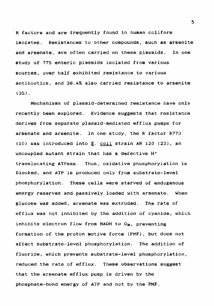
R factors and are frequently found in human coliform
isolates. Resistances to other compounds, such as arsenite
and arsenate, are often carried on these plasmids. In one
study of 775 enteric plasmids isolated from various
sources, over half exhibited resistance to various
antibiotics, and 38.4% also carried resistance to arsenite
(35).
Mechanisms of plasmid-determined resistance have only
recently been explored. Evidence suggests that resistance
derives from separate plasmid-mediated efflux pumps for
arsenate and arsenite. In one study, the R factor R773
(10) was introduced into i· coli strain AN 120 (23), an
uncoupled mutant strain that has a defective H•
translocating ATPase. Thus, oxidative phosphorylation is
blocked, and ATP is produced only from substrate-level
phosphorylation. These cells were starved of endogenous
energy reserves and passively loaded with arsenate. When
glucose was added, arsenate was extruded. The rate of
efflux was not inhibited by the addition of cyanide, which
inhibits electron flow from NADH to 02, preventing
formation of the proton motive force (PMF), but does not
affect substrate-level phosphorylation. The addition of
fluoride, which prevents substrate-level phosphorylation,
reduced the rate of efflux. These observations suggest
that the arsenate efflux pump is driven by the
phosphate-bond energy of ATP and not by the PMF.

6
Using the same strains and methods, a similar system
was suggested for arsenite resistance (30). The two
systems were found to be independent, since inactivation of
one by mutation did not affect the activity of the other
(3). Whether arsenite and arsenate are pumped out by the
same efflux pump or two separate ones, as well as the
components and their location, is unknown.
The fact that two plasmids confer resistance to a
given substance is not proof of identical resistance
mechanisms or of significant DNA homology. For example,
using restriction enzyme analysis and DNA-DNA hybridization
techniques, four genetically distinct tetracycline
resistance determinants have been identified in a broad
range of bacterial plasmids (18). These differences result
in variations in expression of resistance to tetracycline
and its analogs. Recognition sequences for four
restriction enzymes were missing in some of the resistance
markers studied, suggesting genetic dissimilarity. Colony
blot DNA hybridization was performed against the
tetracycline resistance marker in the transposon Tn10. The
resistance determinants were divided into four genetically
distinct classes based on DNA-DNA hybridization. These
classes also show phenotypic differences in resistance to
tetracycline and its analogs. This multiplicity of
genetically distinct determinants specifying a common
resistance suggests the possibility of parallel yet
distinct biochemical pathways leading to the same
phenotypic result.
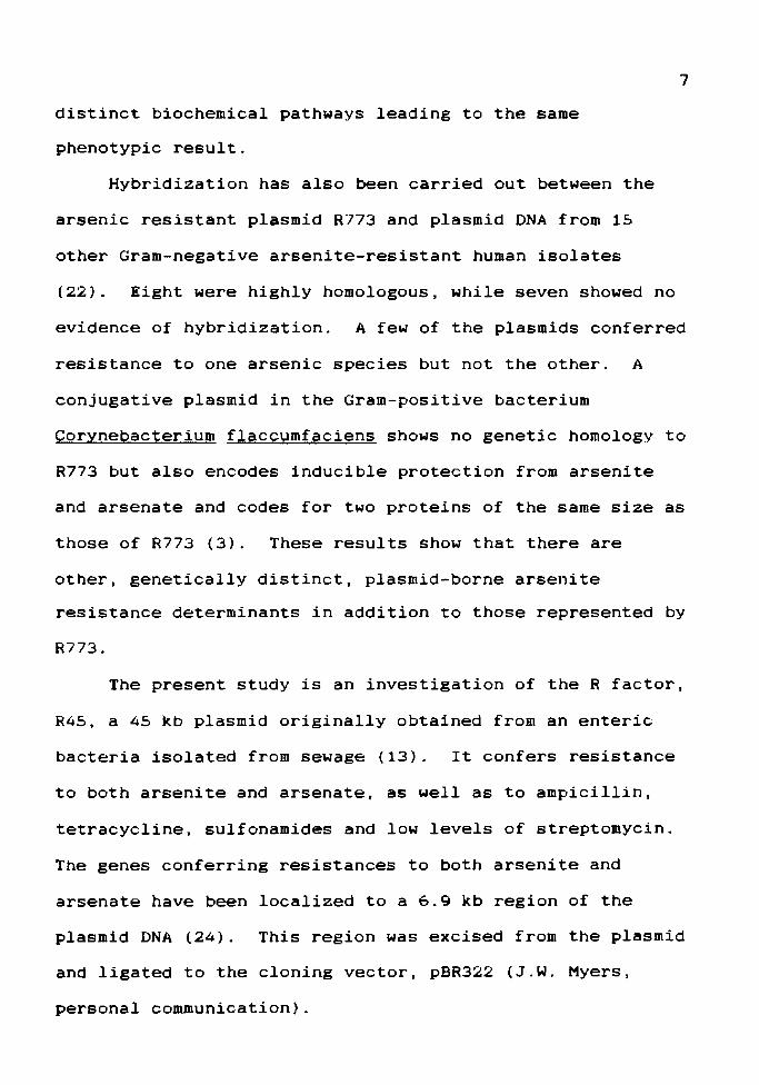
Hybridization has also been carried out between the
arsenic resistant plasmid R773 and plasmid DNA from 15
other Gram-negative arsenite-resistant human isolates
7
(22). Eight were highly homologous, while seven showed no
evidence of hybridization. A few of the plasmids conferred
resistance to one arsenic species but not the other. A
conjugative plasmid in the Gram-positive bacterium
Corynebacterium f laccumf aciens shows no genetic homology to
R773 but also encodes inducible protection from arsenite
and arsenate and codes for two proteins of the same size as
those of R773 (3). These results show that there are
other, genetically distinct, plasmid-borne arsenite
resistance determinants in addition to those represented by
R773.
The present study is an investigation of the R factor,
R45, a 45 kb plasmid originally obtained from an enteric
bacteria isolated from sewage (13). It confers resistance
to both arsenite and arsenate, as well as to ampicillin,
tetracycline, sulfonamides and low levels of streptomycin.
The genes conferring resistances to both arsenite and
arsenate have been localized to a 6.9 kb region of the
plasmid DNA (24). This region was excised from the plasmid
and ligated to the cloning vector, pBR322 (J.W. Myers,
personal communication).
8
The goals of the present study were 1) to construct a
restriction map of the region of the R45 plasmid containing
the arsenite-resistance determinant; 2) to determine the
direction of transcription of the cloned fragment
containing this region; and 3) to identify the proteins
determined by the fragment.
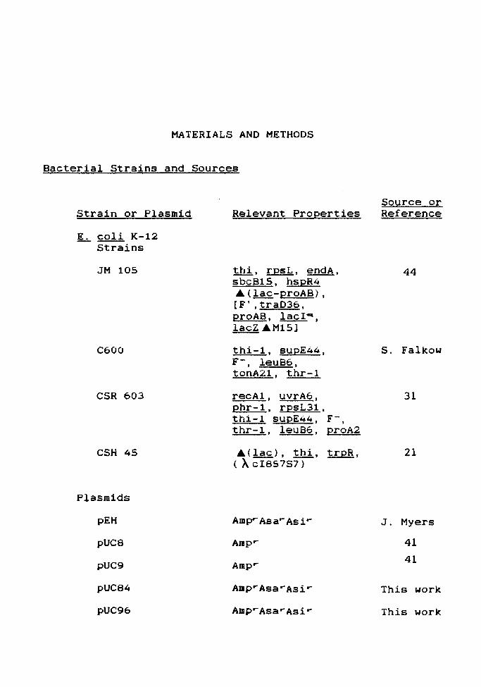
MATERIALS AND METHODS
Bacterial Strains and Sources
Strain or Plasmid
L.. coli K-12 Strains
JM 105
C600
CSR 603
CSH 45
Plasmids
pEH
pUC8
pUC9
pUC84
pUC96
Relevant Properties
thi, rpsL, endA, sbc815, hspR4 A ( lac-proAB), [F', traD36, proAB, lac! ... , lacZ AM15]
thi-1, supE44, F-, leuB6, tonA21, thr-1
recAl, uvrA6, phr-1, rpsL31, thi-1 supE44, F-, thr-1, leuB6, proA2
A(lac), thi, trpR, (AcI8S7S7)
Amp-- Asa ... Asi ...
Amp,..
Amp.-
AmprAsa.-Asi ...
Amp ... Asa.-Asir
Source or Reference
44
S. Falkow
31
21
J. Myers
41
41
This work
This work

amp
asi
asa
LB
LA
lac
IPTG
MOPS
F'
r
s
MCS
SDS
kb
Kdal
MIC
RNAP
PMF
X-gal
TABLE I
TABLE OF ABBREVIATIONS
ampicillin
arsenite
arsenate
Luria broth
Luria agar
lactose operon
isopropyl-~-D-thiogalactoside
morpholinopropane sulf onic acid
10
carrying the ~. coli sex factor F, with a chromosomal insertion
resistant
sensitive
multiple cloning site
sodium dodecyl sulfate
kilobases
kilodaltons
minimal inhibitory concentration
RNA polymerase
proton motive force
5-bromo-4-chloro-3-indolyl-~-D-galactoside
11
Glycerol stocks and strains constructed in this study were
verified on appropriate selective media prior to use.
Short-term working stocks were maintained on these media at
4°C. Long-term stocks were stored in 503 glycerol at
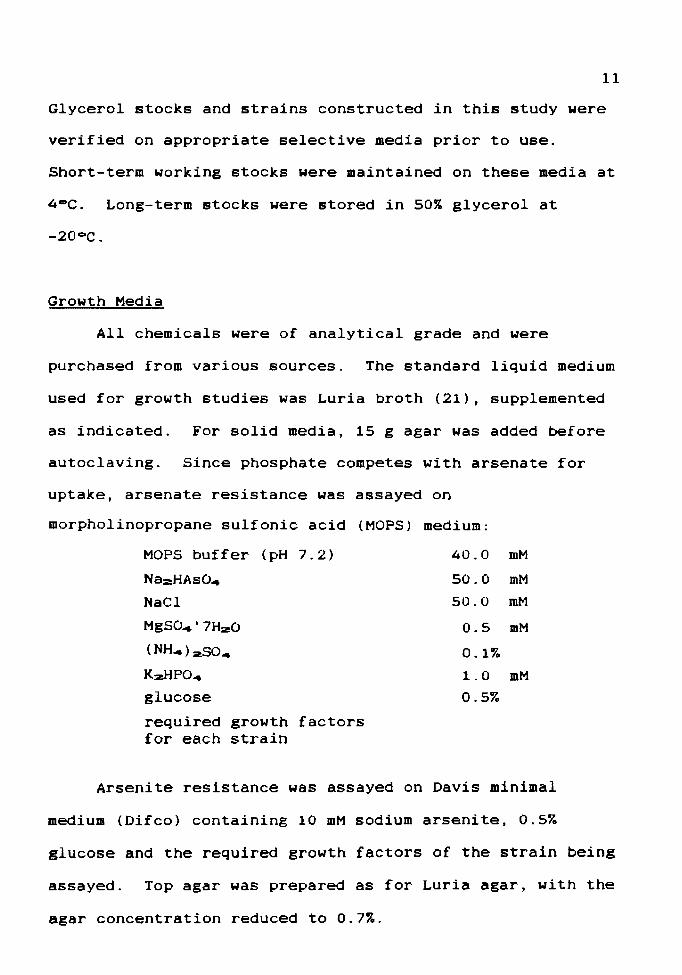
Growth Media
All chemicals were of analytical grade and were
purchased from various sources. The standard liquid medium
used for growth studies was Luria broth (21), supplemented
as indicated. For solid media, 15 g agar was added before
autoclaving. Since phosphate competes with arsenate for
uptake, arsenate resistance was assayed on
morpholinopropane sulfonic acid (MOPS) medium:
MOPS buff er (pH 7.2) 40.0 mM
Na2HAso .. 50.0 mM
NaCl 50.0 mM
MgS0.,.'7HzO 0.5 mM
(NH ... )2504 0.13
KzHP04 1. 0 mM glucose 0.53
required growth factors for each strain
Arsenite resistance was assayed on Davis minimal
medium (Difeo) containing 10 mM sodium arsenite, 0.53
glucose and the required growth factors of the strain being
assayed. Top agar was prepared as for Luria agar, with the
agar concentration reduced to 0.73.
12
Isolation of Plasmid DNA
Plasmid DNA was extracted from verified strains by the
method of Birnboim and Ooly (1). The centrifuged cell
paste was re-suspended in 10 ml 25 mM Tris-Cl (pH 8), 10 mM
Na2EDTA and 13 (w/v) glucose. To lyse the cells, 20 ml
0.2 M NaOH containing 13 SOS was added. At high pH,
plasmid DNA remains covalently closed, while the linear
chromosomal fragments become single-stranded. A S M
acidified potassium acetate solution (15) was added to
lower the pH and precipitate proteins, high molecular
weight RNA and single-stranded ONA. The plasmid ONA
renatured to the supercoiled form and remained in the
supernatant. The precipitate was removed by
centrifugation, and the plasmid DNA was precipitated with
two volumes of 953 ethanol. After centrifugation the
pellet was dissolved in 6 ml TE buffer (15).
To remove contaminating chromosomal ONA, RNA, and
proteins, the solution was centrifuged in a cesium chloride
density gradient containing 0.6 ml ethidium bromide (EtBr)
(10 mg/ml) to purify the plasmid DNA (2). Ethidium bromide
intercalates into the DNA, reducing its density. Since
linear and open circular DNA are not as physically
constrained as the covalently closed circular plasmid ONA,
the first two forms bind more ethidium bromide molecules
than the third, become less dense, and band with a
different bouyant density. The DNA was centrifuged in the
13
Ti 50 rotor of a Beckman preparative ultracentrifuge for 36
hours at 45,000 rpm. DNA bands were visualized with an
ultraviolet lamp, and the lower plasmid ONA band was
extracted. Ethidium bromide was removed from the plasmid
DNA solution by extraction with isopropanol saturated with
5 M NaCl {4). Remaining impurities were removed by ethanol
precipitation of the ONA in two volumes of ethanol in the
presence of 2.5 M ammonium acetate, and the pellet was
suspended in 100 ul TE buffer containing 1 unit/ml RNAse
T1. (ternase) {4).
Isolation of Bacteriophage A DNA
E. coli CSH 45 contains the prophage A cl857S7 ( 4) .
This viral strain has two mutations, cl857 and S7. The cl
repressor gene is responsible for inhibiting the expression
and replication of A and keeping it in the lysogenic
state. The cl857 mutation results in production of a
temperature sensitive repressor. The S7 mutation renders
the phage lysis-deficient, and it continues to multiply
inside the cell. K· coli CSH 45 was grown on agar at
30°C and 42°C (4). Cells picked from colonies showing
no growth at the non-permissive temperature were grown in
LB at 32°C for one hour, and the temperature was
increased to 43°C for 20 minutes to inactivate the
repressor and induce the lytic cycle.
The cultures were allowed to grow for an additional
14
three hours. They were then centrifuged and suspended in
10 ml A diluent (4). One ml chloroform was added to lyse
the bacterial cells. The phage was separated from cellular
debris by passage through a CsCl density block gradient
(4). A lysate was laid over two CsCl solutions having
densities of 1.6 and 1.4, placed in a Beckman SW 65
swinging bucket rotor, and centrifuged in the Beckman
preparative ultracentrifuge for two hours at 30,000 rpm.
The phage, with a density of about 1.5, collected at the
interface between the two CsCl solutions and was removed.
The DNA was isolated by treating the lysate with SOS,
followed by SM potassium acetate. Proteins were removed by
centrifugation for 30 minutes in a microfuge. The A ONA
was precipitated with ethanol and suspended in l
ml ternase. Absorbancy readings were taken in a Beckman
spectrophotomer at 260nm to determine the concentration of
DNA (1 O.D.zao=50 ug/ml). Portions of the DNA were
diluted with ternase to give standards of 10, 5, 2.5, 1.25,
and 0.625 ug/ml. These were used to estimate ONA
concentration based on EtBr fluorescence.
Saran Wrap was placed across a Chromato-vue""
transilluminator in a darkroom {15). The prepared
standards and DNA samples were pipetted onto the Saran Wrap
and mixed with 5 ul of EtBr (2 mg/ml}. Ultraviolet light
was transmitted through the samples, and photographs were
taken with a Polaroid MP-4 Land camera using a yellow
15
filter and Polaroid Type 57 film. The concentration of DNA
was then determined by comparing the fluorescence of the
samples with that of the known standards.
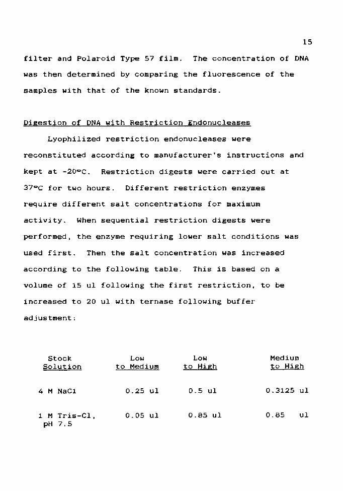
Digestion of DNA with Restriction Endonucleases
Lyophilized restriction endonucleases were
reconstituted according to manufacturer's instructions and
kept at -20-c. Restriction digests were carried out at
37°C for two hours. Different restriction enzymes
require different salt concentrations for maximum
activity. When sequential restriction digests were
performed, the enzyme requiring lower salt conditions was
used first. Then the salt concentration was increased
according to the following table. This is based on a
volume of 15 ul following the first restriction, to be
increased to 20 ul with ternase following buffer
adjustment:
Stock Low Low Medium Solution to Medium to High to High
4 M NaCl 0.25 ul 0.5 ul 0.3125 ul
1 M Tris-Cl, 0.05 ul 0.85 ul 0.85 ul pH 7.5
Electrophoresis of ONA Fragments
Agarose gels ranging from 0.7 to 1.23 (w/v) were
prepared in tris-acetate electrophoresis buffer (15)
containing 0.25 ug/ml of ethiduim bromide (39). Samples
containing 100-200 ng ONA and one-quarter volume of ONA
tracking dye (1) were run in a horizontal slab gel
apparatus (16) at 3 V/cm. The running time varied with
the specific procedure.
Isolation of DNA Fragments
16
Digested DNA was separated by electrophoresis, and
the gel was placed on an ultraviolet transilluminator to
visualize the bands. The gel was sliced, and a strip of
Whatman DE 81 OEAE cellulose paper was inserted in front
of the desired band (5). The DNA was moved onto the paper
by electrophoresis at lOOV for 30 minutes. The paper was
then removed, packed into the barrel of a disposable
plastic 1 ml syringe and placed in a 15 ml Corex
centrifuge tube. The paper was first washed by pipetting
200 ul of 0.1 M NaCl, 0.1 mM EDTA, 10 mM Tris-Cl (pH 8)
into each syringe and centrifuging at 2000 rpm for 30
seconds in a Sorval GLC-1 centrifuge. This procedure was
repeated twice and the eluates discarded. lo remove the
ONA from the paper, a 1.5 ml polypropylene tube was placed
in the Corex tube to catch the eluate, and the paper was
washed by adding 100 ul of a 1.0 M NaCl, 0.1 M EDTA, 10 mM
17
Tris-Cl (pH 8.0) solution, followed by centrifugation for
30 seconds at 2000 rpm. This was repeated three times,
for a total DNA eluate of 400 ul. Two volumes of 953
ethanol were added to the eluate, the tubes were placed in
a dry ice-ethanol bath for five minutes to precipitate the
DNA and then centrifuged in a microfuge for 30 minutes.
The pellet was suspended in 100 ul ternase and mixed
with an equal volume of a 1:1 (v/v) mixture of phenol
equilibrated with TE buffer and chloroform mixed 24:1
(v/v) with isoamyl alcohol (15) and then centrifuged 15
seconds in a microfuge to separate the organic and aqueous
layers. The upper aqueous layer was removed to a new
tube. To maximize DNA recovery, the remaining organic
layer was re-extracted with 100 ul TE buffer. The upper
aqueous layer was again removed and mixed with the first
aqueous portion. Residual phenol was then removed by
mixing with an equal volume of chloroform and centrifuging
as before. The aqueous layer was removed, and the DNA was
precipitated with ammonium acetate and ethanol, as
previously described.
Construction of Recombinant Plasmids
Vector DNA was digested with Hind III, followed by
Eco RI, using buffers and conditions previously
described. The enzymes were then removed by phenol
extraction. Ligations were carried out using 200 ng of

each vector DNA with an equimolar amount of isolated
fragment DNA. A tube containing 400 ng of vector DNA
digested with Hind Ill alone was included as a ligase
control.
Two units of T4 DNA ligase were added to each
reaction mixture, along with lOX ligation buffer (15):
0.5 M Tris-Cl, pH 7.4 0.1 M MgS04
10.0 mM spermidine 0.1 M dithiothreitol
10.0 mM adenosine triphosphate 1.0 mg/ml bovine serum albumin
The reaction mixtures were incubated at 4°C for 12 hours.
Ligated plasmids were then used to transform host cells.
Transformation
K· coli JM 105 and CSR 603 were prepared for
18
transformation (14) by growing cultures to a density of 5 x
107 cells/ml. Cell density was determined using a
Klett-Summerson Photoelectric Colorimeter, Model 800-3,
with a red #66 filter. The culture was chilled on ice for
10 minutes and then centrifuged at 4000 rpm at 0°C for
five minutes. The pellets were suspended in one-half the
original culture volume in a cold (4°C) SO mM CaC12
solution, centrifuged again and resuspended in CaClz at
one-fifteenth the original volume. Aliquots of 0.2 ml were
stored at 4°C for 24 hours. Up to 40 ng of plasmid DNA
was added to each tube. The tubes were kept on ice for 30

minutes and then transferred to a 42°C water bath for two
minutes. Then 1.0 ml LB was added and the cells were
incubated for two hours at 37°C to allow for expression
of plasmid-carried ampicillin resistance. Successful
transformation was detected by plating the cells on Luria
agar containing ampicillin at a final concentration of 50
ug/ml.
When transformation was carried out with pUC vectors,
amp~ transformants containing vectors with inserts could
be distinguished from those without inserts by the
following procedure. Approximately 108 cells from each
transformation tube were plated on LA containing amp.,
IPTG (4X10⁻⁴ M} and X-gal (0.004~} (32}. The pUC
vector codes only for the promoter-proximal region of
19
~-galactosidase (a donor} and the JM105 F' plasmid
specifies a ~-galactosidase functional only in the
promoter-distal region ( w donor} ; the two complementary
polypeptide chains can associate to produce active
~-galactosidase (intracistronic complementation}. Such
activity can be detected by the production of the insoluble
blue dye dibromodichloroindigo (19} when X-gal is
hydrolyzed by the enzyme. If the transformant has an
insert in the MCS, the coding region for the promoter
proximal ~-galactosidase is interrupted and no a donor
polypeptide is made. Transformants containing pUC vector
inserts produce white colonies under these conditions.
20
Three ml of molten top L agar was mixed with 0.2 ml of
transformed cells, and 100 ul of 0.1 M IPTG was added,
along with 50 ul of 23 X-gal (dissolved in N,N-dimethyl
formamide) (37). The top agar was poured over LA
previously spread with ampicillin. The plates were
incubated for 12 hours at 37°C.
Minimal Inhibitory Concentration
The two E. coli host strains used, JM 105 and CSR 603,
and two recombinant-plasmid-bearing strains, JM 105(pUC84)
and CSR 603(pUC84), were grown overnight in 10 ml LB, with
and without the addition of l mM arsenite. These were
subcultured 1:100 into side-arm flasks containing the same
media and shaken at 37°C until they reached a density of
2.5 X 1oe cells/ml. From each flask, 50 ul was spread
over the surf ace of LA plates containing the following
concentrations of arsenite: 2, 4, 6, 8, 10, 12, 15 and 20
mM. The plates were incubated for 48 hours at 37°C, and
the colonies were counted. The lowest concentration of
arsenite on which no growth was observed was taken to be
the minimal inhibitory concentration of arsenite for that
strain (32, 34).
Induction of Arsenite Resistance by IPTG
The~. coli strains JM 105, JM 105(pUC8), JM

21
105(pUC84) and JM 105(pUC96) were grown overnight in 10 ml
LB, with the following additions for each strain:
1. None 2. 1 mM arsenite 3. 10-3 M IPTG (25)
Cultures were diluted 1:100 into the same media in side-arm
flasks and shaken at 37°C until they reached a density of
1.5 X 10⁸ cells/ml. This dilution and subsequent growth
period was repeated. The cultures were challenged by the
addition of arsenite to a final concentration of 15 mM, and
the growth of each was monitored over a period of several
hours.
Restrictioh Mapping of the Arsenite Resistance Fragment
Aliquots of 0.5 ug of the isolated R45 fragment ONA
were digested with the restriction enzymes Pvu I, Bgl II,
Sph I and Sal I. Resulting fragments were separated by
electrophoresis through 1.03 agarose for two hours at 100
V. The distance from the origin of each separated band was
measured in millimeters, and compared with a standard curve
of the log molecular weight against the distance traveled
by the A DNA Hind III-digested fragments. By this means,
the molecular weights of the digestion products were
estimated (16).
A portion of each single digest was then subjected to
a separate, second digestion with each of the remaining
three enzymes under analysis. The digestion products of
22
the second digestion were separated by electrophoresis and
the fragment sizes estimated as described for the first.
Identification of Plasmid-Encoded Proteins
The recA- Y.Y.r.:. genotype of the i..:_ coli strain CSR
603 (31) was confirmed by spreading 109 cells of an
overnight culture grown from a single CSR 603 colony on LA
and irradiating with a total UV dose of O.S J/m2 • The
plates were incubated for 24 hours at 37°C in the dark
(A. Sancar and G. Sancar, personal communication). Plates
having fewer than 100 survivors were used in the following
procedures.
Recombinant plasmids and intact pUC vectors were
transforned into i..:_ coli CSR 603. Cultures were grown
overnight in LB with the following additions. Ampicillin
was included to select against variants that may have lost
the plasmid.
Culture Additions
603 none
603(pUC8) so ug/ml amp
603 ( pUC84) , uninduced so ug/ml amp
603 ( pUC84) , induced so ug/ml amp + 10 mM asi
603(pUC96), uninduced 50 ug/ml amp
603 ( pUC96) , induced 50 ug/ml amp + 10 mM asi
The cultures were diluted 1:100 into 10 ml of the same
media without ampicillin in side-arm flasks and grown to a
----------------------------····---
23
density of 2 X 10• cells/ml. The cultures were diluted
1:100 and grown to the same cell density to insure that
cells were in log phase before proceeding. All cultures
were centrifuged at 4000 rpm for five minutes, and the
pellets were suspended in 10 ml Davis Minimal broth. They
were then irradiated with swirling in a petri dish, for a
total UV dose of 5 J/m2 , to inactivate chromosomal DNA.
The suspensions were centrifuged and the pellets
resuspended in 10 ml LB with the additions previously
described. The cultures were incubated for three hours,
0.1 ml of a freshly made cycloserine solution (200 ug/ml)
was added, and the cultures were incubated for 12 hours.
From each culture, 1.5 ml was centrifuged in a microfuge
for five minutes, and the cells were resuspended in 40 ul
of lysing buffer (8):
0.05 M Tris-Cl, pH 6.8
23 sos 153 glycerol
23 2-mercaptoethanol
0.0053 bromophenol blue
These mixtures were boiled for three minutes to lyse the
cells and then stored at 4°C until needed.
Polyacrylamide gels were used for the separation of
proteins by the method of Laemmli (12). Stock solutions
were as follows:
1. 303 acrylamide
2. 4X running gel buffer {1.S M Tris-Cl, pH 8.8).
3. 4X stacking gel buffer (O.S M Tris-Cl, pH 6.8).
4. 103 {w/v) sodium dodecyl sulfate (SOS)
5. 103 (w/v) ammonium persulfate (APS)
6. Commercial stock of TEMED
(N,N,N',N'-tetramethylethylenediamine)
7. Tank buffer (0.025 M Tris-Cl, pH 8.3, 0.192 M
glycine and 0.13 SDS).
8. 13 Coomassie blue R-250 stain stock. This stock
was used to make a 0.1253 Coomassie blue, 503
methanol, 103 acetic acid stain for gels.
9. 503 methanol, 103 acetic acid (primary destaining
solution).
10. 53 methanol, 7% acetic acid (secondary destaining
solution).
24
Glass plates with 1.0 mm spacer bars were sealed with
1.03 agarose to form a sandwich 16.5 X 19 cm. A 12.53
separating gel solution was prepared:
12.S ml monomer solution
7.5 ml 4 X running gel buffer
0.3 ml 103 SDS
9.7 ml HzO
To this, 10 ul TEMED and 0.75 ml APS were added. This
solution was quickly delivered into the glass sandwich to
within 5 cm of the top, and 1.0 ml isopropanol was layered
25
over the gel to form a level surface. The gel was allowed
to polymerize two hours. A stacking gel solution was
prepared:
1.33 ml monomer solution
2.5 ml 4 X stacking gel buffer
0.1 ml 103 SDS
6 .1 ml H:zO
Five ul TEMED and 0.25 ml APS were added, the sandwich was
filled with this solution, and a 16-well comb was
inserted. The stacking gel was allowed to polymerize at
least one hour. The sandwich was sealed in place in a
Watson vertical gel apparatus with 1.03 agarose. Ten ul of
each sample (1.5 ug protein) and protein standard (1.0 ug
protein} were pipetted into the wells, and the gel was
electrophoresed at 70 V constant voltage until the dye
front began to enter the separating gel. The voltage was
then increased ~o 170 V, and the gel was run 4-5 hours
(39). Protein standards used were:
Mixture
A
Containing
lactalbumin carbonic anhydrase
Molecular weight
14.2 Kdal 29.0 Kdal
B Trypsin inhibitor 20.1 Kdal glyceraldehyde-3-phosphate
c
dehydrogenase 36.0 Kdal bovine serum albumin 66.0 Kdal
trypsinogen egg albumin
24.0 Kdal 45.0 Kdal
26
Gels were stained by placing them in a glass dish and
covering them with 100 ml Coomassie blue stain. The pan
was shaken on a Kraft rotary shaken at 60 rpm for one
hour. The gel was drained, immersed in 100 ml of primary
destaining solution and shaken for one hour. The gel was
again drained, covered with 100 ml secondary destaining
solution and shaken overnight.
Alternatively, gels were stained with silver (43),
either alone or following Coomassie blue (9). The gel was
soaked overnight in 503 methanol to fix the proteins,
rehydrated under running water for one hour and drained.
The silver staining solution was prepared by mixing 21 ml
of 0.363 NaOH with 1.4 ml of 14.8 M NH.OH. The solution
was stirred while 4.0 ml of stock AgN03 solution (4.0 g
AgN03 dissolved in 20 ml HzO) was added dropwise, to
prevent precipitation. The solution was poured over the
gel, and the gel was then agitated on the rotary shaker for
15 minutes. The gel was rinsed for 30 minutes under
running water and then drained. A developing solution
containing 0.0053 (w/v) citric acid and 0.0023 (v/v) 373
formaldehyde was freshly prepared and poured over the
drained gel. The gel was then agitated over a white
background until protein bands began to appear. The
process was stopped by pouring 503 methanol over the
drained gel and agitating for 15 minutes. Protein size was
estimated from a plot of the log of the size of the protein

27
standards in kilodaltons against the distance traveled from
the origin in millimeters.
Stained gels were photographed with a Polaroid 4 X 5
Land camera using Polaroid Type 55 positive/negative film.
The gels were rinsed in running water overnight to remove
all traces of methanol, soaked in an aqueous solution of
13 glycerol and 103 acetic acid for 45 minutes, placed on
either Whatman 3 MM filter paper or cellophane sheets, and
dried at so~c for two hours in a BioRad Model 224 Gel
Slab Dryer.

RESULTS
Isolation and Ligation of Arsenic-kesistant Fragment DNA
The source of the insert DNA used to construct
recombinant plasmids in this study was the previously
constructed plasmid, pEH, containing the arsenic-resistance
region from R4S ligated into the cloning vector, pBR (John
Myers, personal communication). Plasmid DNA was isolated
by methods previously described. Plasmid DNA yield was
routinely 1.0-2.0 mg per liter of starting culture. The
R45-derived fragment was then separated from pBR by double
digestion with Hind III and Eco RI. Each cloning vector
was subjected to a double digest with Hind III and Eco RI
and 200 ng was mixed with 500 ng of the fragment and
ligated with T4 ligase. After ligation, the DNA was used
to transform ~. coli strain JM 105.
Transformation of Recombinant Plasmids
Transformants were selected by mixing 0.3 ml
transformed cultures with IPTG and X-gal in top agar and
pouring this over LA spread with ampicillin. Blue colonies
revealing a functional ~-galactosidase and thus an intact
vector were rejected. White colonies reflect an insert in
the MCS of the vector. These constituted up to 203 of
29
observed colonies. White colonies were picked and spread
on LA containing ampicillin and 10 mM arsenite, to confirm
the asir phenotype. Short-term working stocks were
maintained on this medium at 4°C. -Since the pUC vectors
differ only in their orientation of the MCS, the fragment
insertion in pUC8 should have the opposite orientation of
the insert in pUC9.
Confirmation of Recombinant Plasmids
To confirm that the resistance was due to the presence
of a plasmid with a molecular weight equal to the sum of
those of the inserted fragment (6.9 kb} and the pUC vector
(2.7 kb), plasmid DNA was extracted from several separately
maintained clones of both supposed orientations. This DNA
was digested with Hind III and Eco RI, and samples
containing 100-200 ng DNA were electrophoresed through a
0.73 agarose gel. Double-digested pUC and pEH plasmid DNA
were included in the gel as size markers. Bacteriophage A DNA was digested with Hind III, producing seven fragments
of known size -- 23.67, 9.67, 6.66, 4.26, 2.30, 1.96, and
0.59 kb respectively. This mixture was used as size
markers in this and other agarose gels. Fig. 1 shows that
all recombinant plasmids contained an insert of the
expected size.
To confirm that the recombinant plasmids derived from
pUC8 and pUC9 did indeed contain the R45 fragment in

23.67-
9.46-
6.66-
1 2 3 4 5
30
6 7
Figure 1. Electrophoretic Analysis of Restriction Digests
of Recombinant Plasmids
Plasmid DNA was removed from transformed ~- coli JM 105
strains and sequentially digested with EcoR I and Hind
I I I. Lane 1, pEH; lane 2, A H; lane 3, 105 ( pUC83) ; lane 4,
105(pUC84); lane 5, 105(pUC92); lane 6, 105(pUC96); lane 7,
pUC8. Size standards are shown on the left in kilobases.
31
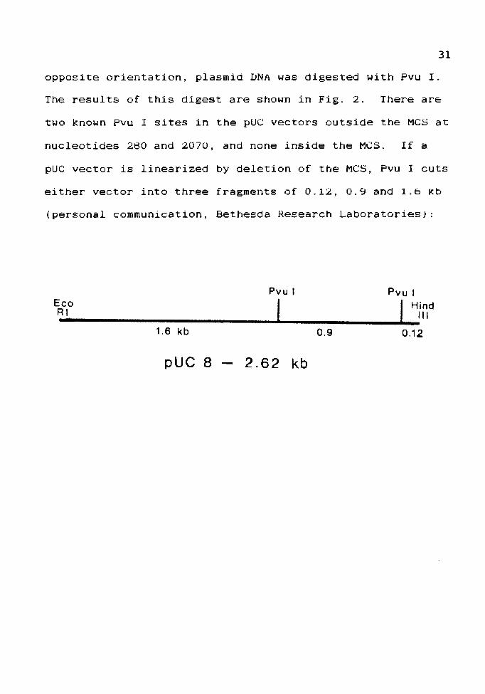
opposite orientation, plasmid DNA was digested with Pvu I.
The results of this digest are shown in Fig. 2. There are
two known Pvu I sites in the pUC vectors outside the MCS at
nucleotides 280 and 2070, and none inside the MCS. If a
pUC vector is linearized by deletion of the MCS, Pvu I cuts
either vector into three fragments of 0.12, 0.9 and 1.b kb
(personal communication, Bethesda Research Laboratories):
Pvu I Pvu I
~-c-,0------------------------------~'----------------...1.f ....:H;nd 1.6 kb 0.9 0.12
pUC 8 2 .62 kb
23.67-9.46-6.66-4.26-
2.30-1.96-
1 2 3 4 5
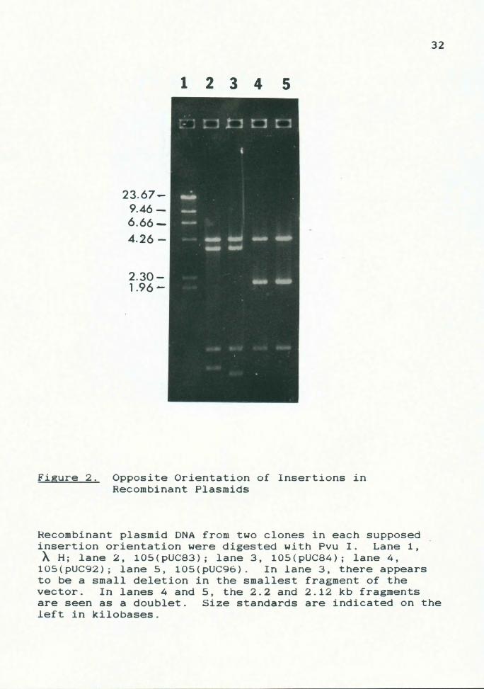
Figure 2. Opposite Orientation of Insertions in Recombinant Plasmids
32
Recombinant plasmid DNA from two clones in each supposed insertion orientation were digested with Pvu I. Lane 1, AH; lane 2, 105(pUC83); lane 3, 105(pUC84); lane 4,
105(pUC92); lane 5, 105(pUC96). In lane 3, there appears to be a small deletion in the smallest fragment of the vector. In lanes 4 and 5, the 2.2 and 2.12 kb fragments are seen as a doublet. Size standards are indicated on the left in kilobases.
33
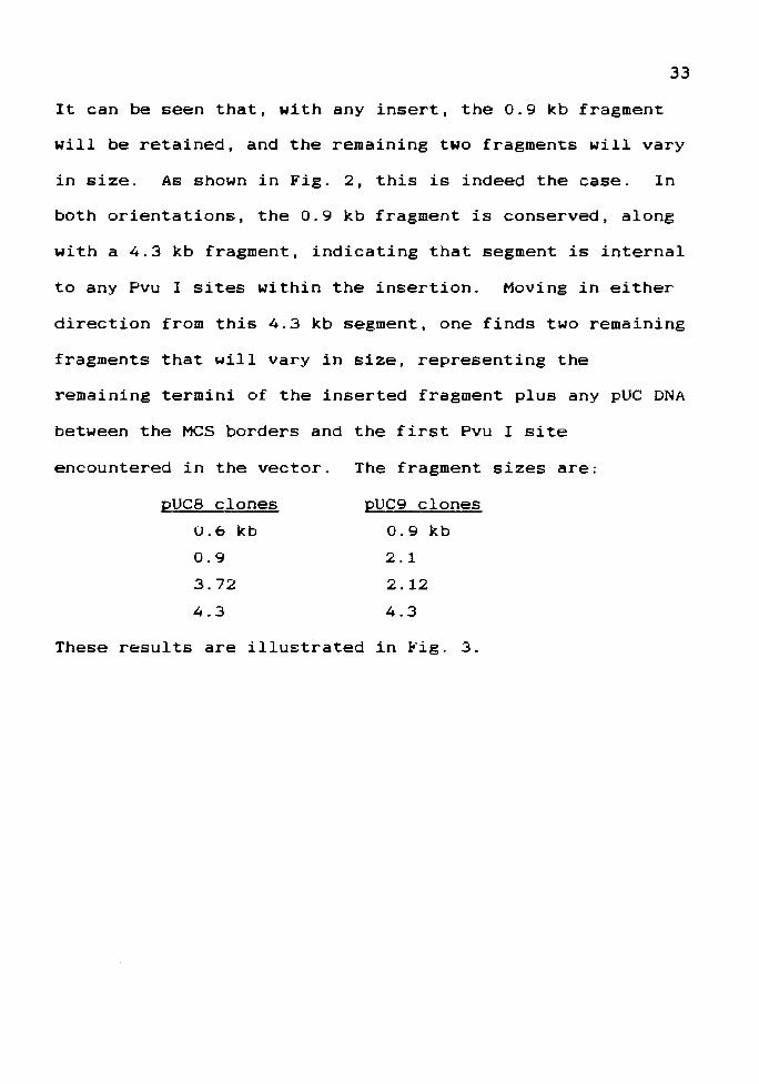
It can be seen that, with any insert, the 0.9 kb fragment
will be retained, and the remaining two fragments will vary
in size. As shown in Fig. 2, this is indeed the case. In
both orientations, the 0.9 kb fragment is conserved, along
with a 4.3 kb fragment, indicating that segment is internal
to any Pvu I sites within the insertion. Moving in either
direction from this 4.3 kb segment, one finds two remaining
fragments that will vary in size, representing the
remaining termini of the inserted fragment plus any pUC DNA
between the MCS borders and the first Pvu I site
encountered in the vector.
pUC8 clones
0.6 kb
0.9
3.72
4.3
The fragment sizes are:
pUC9 clones
0.9 kb
2.1
2.12
4.3
These results are illustrated in Fig. 3.

34
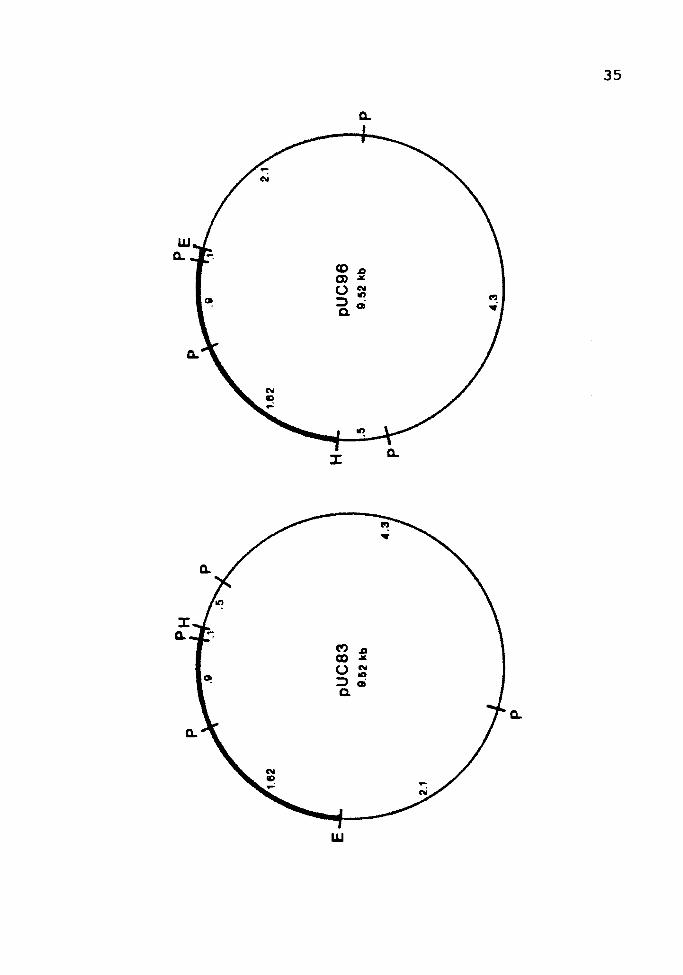
Figure 3. Pvu I Restriction Map of Recombinant Plasmids
The Pvu I restriction sites (P) are shown on a recombinant
plasmid in each orientation. The Eco RI (E) and Hind III
(H) termini of the vector (thick line) are also indicated.
The fragment sizes are shown in kilobases.
.,,
m
:::c
~ !"' c UI (') ~ CQ ~ Ol
.,, m
36
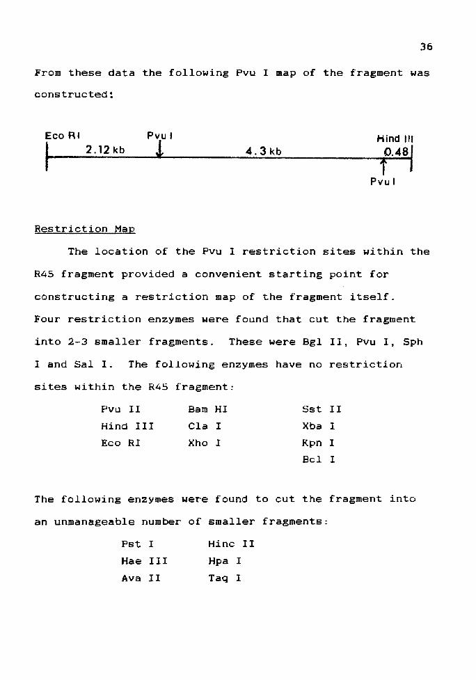
From these data the following Pvu I map of the fragment was
constructed~
Eco R~
I 2 .12 kb
Restriction Map
Pvul
! 4.3 kb Hind Ill
0.481 f
Pvul
The location of the Pvu I restriction sites within the
R45 fragment provided a convenient starting point for
constructing a restriction map of the fragment itself.
Four restriction enzymes were found that cut the fragment
into 2-3 smaller fragments. These were Bgl II, Pvu I' Sph
I and Sal I. The following enzymes have no restriction
sites within the R45 fragment:
Pvu II Barn HI Sst II
Hind III Cla I Xba I
Eco RI Xho I Kpn I
Bel 1
The following enzymes were found to cut the fragment into
an unmanageable number of smaller fragments:
Pst I
Hae III
Ava II
Hine II
Hpa I
Taq I
37
Aliquots of isolated R45 fragment DNA were restricted
with each of the four selected enzymes, and the resulting
fragment sizes estimated by electrophoresis with DNA
standards. A portion of each single digest was then
separately restricted with each of the remaining enzymes,
resulting in a series of double digests. Each of these was
analyzed on a 1.23 agarose gel. The results are given in
Fig. 4.
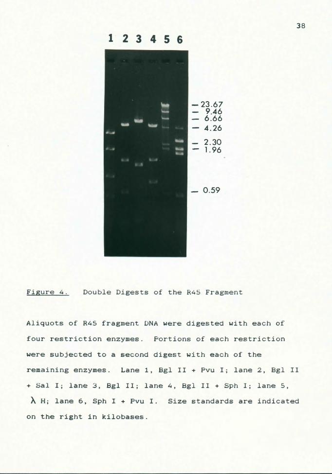
Figure 4.
123456
-23.67 9.46 6.66 4.26
2.30 1.96
- 0.59
Double Digests of the R45 Fragment
Aliquots of R45 fragment DNA were digested with each of
four restriction enzymes. Portions of each restriction
were subjected to a second digest with each of the
38
remaining enzymes. Lane 1, Bgl II + Pvu I; lane 2, Bgl II
+ Sal I; lane 3, Bgl II; lane 4, Bgl II + Sph I; lane 5,
AH; lane 6, Sph I + Pvu I. Size standards are indicated
on the right in kilobases.
39
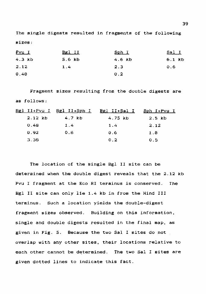
The single digests resulted in fragments of the following
sizes:
Pvu I
4.3 kb
2.12
0.48
Bgl II
5.6 kb
1.4
Sph I
4.6 kb
2.3
0.2
Sal I
6.1 kb
0.6
Fragment sizes resulting from the double digests are
as follows:
Bgl II+Pvu I
2 .12 kb
0.48
0.92
3.38
Bgl II+Sph I
4.7 kb
1.4
0.8
Bgl II+Sal I
4.75 kb
1.4
0.6
0.2
Sph I+Pvu I
2.5 kb
.2.12
1.8
0.5
The location of the single Bgl II site can be
determined when the double digest reveals that the 2.12 kb
Pvu I fragment at the Eco RI terminus is conserved. The
Bgl II site can only lie 1.4 kb in from the Hind III
terminus. Such a location yields the double-digest
fragment sizes observed. Building on this information,
single and double digests resulted in the final map, as
given in Fig. 5. Because the two Sal I sites do not
overlap with any other sites, their locations relative to
each other cannot be determined. The two Sal I sites are
given dotted lines to indicate this fact.
I
.70.6
Eco RI .2 0.6
2 .12
' ' t
f ¥ Sa Sa
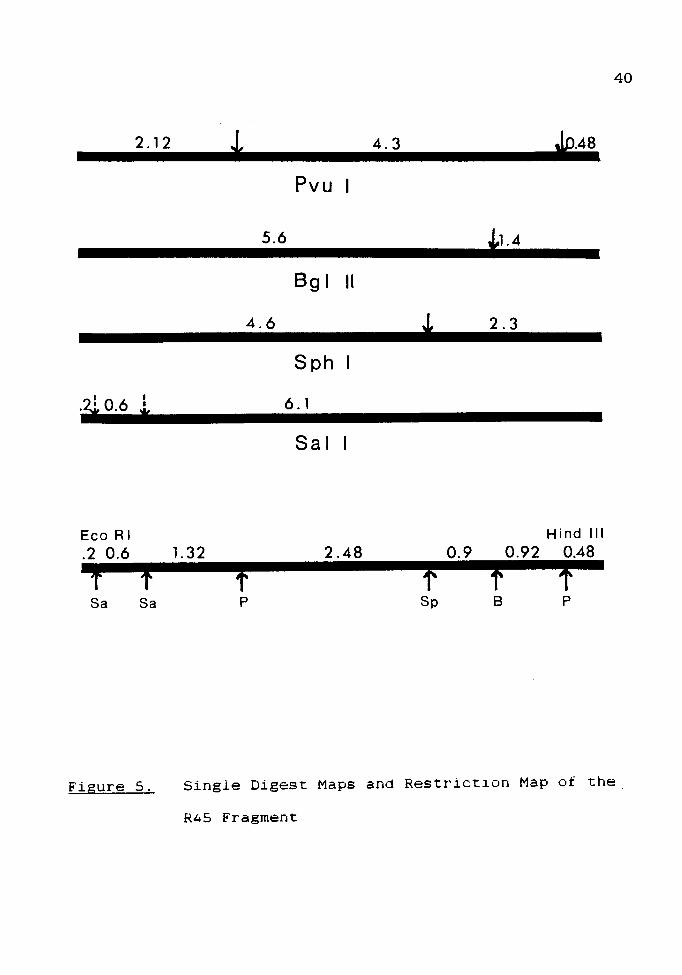
Figure 5.
* 4.3
Pvu
5.6
Bgl II
4.6
Sph
6. l
Sal
l.32 2.48
f p
*1.4
i 2.3
0.9 0.92
f f Sp B
Jp.48
Hind Ill
0.48
f p
40
Single Digest Maps and Restriction Map of the
R45 Fragment

41
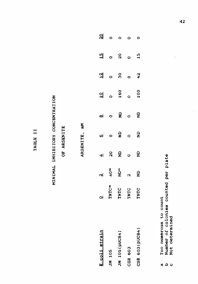
Minimal Inhibitory Concentration of Arsenite
The minimal inhibitory concentration of arsenite for
each strain used, with or without recombinant plasmids, was
determined by plating the strains on LA containing
increasing amounts of arsenite. This allowed the selection
of an arsenite concentration that would clearly distinguish
between plasmid and non-plasmid-bearing strains. The
results are given in Table II.
The minimal inhibitory concentration is taken to be
that concentration of arsenite at which no growth is
observed for a given strain. Minimal inhibitory
concentrations were:
JM 105
6mM
JM 105(pUC84)
20 mM
CSR 603
4 mM
CSR 603(pUC84)
20 mM
Although MIC's determined by this method are
approximate, a sharp distinction can be seen between the
resistances of plasmid and non-plasmid-bearing strains.
Arsenite Resistance Induction Assays
Since the arsenite resistance fragment has been
ligated into the pUC vectors next to the lac promoter,
arsenite resistance may be induced from the lac promoter
using IPTG. Since resistance would be induced only in
clones with the fragment in the proper orientation with
regard to the promoter, it should be possible to determine
in which direction -- from the Eco RI site to the Hind III
TAB
LE
II
MIN
IMA
L IN
HIB
ITO
RY
C
ON
CEN
TRA
TIO
N
OF
AR
SEN
ITE
AR
SEN
ITE
, mM
E.c
oli
str
ain
JM
10
5
TN
TC
• 40
1:.>
2
0
0
JM
10
5(p
UC
84
) TN
TC
ND
N
D
CS
R
60
3
TNTC
2
0 0
CS
R
60
3(p
UC
84
) TN
TC
ND
N
D
ND
a T
oo
nu
mer
ou
s to
co
un
t b
Num
ber
of
co
lon
ies
co
un
ted
p
er
pla
te
c N
ot
dete
rmin
ed
0 0
ND
1
60
0 0
ND
1
00
0 0
0
30
2
0
0
0 0
0
42
1
5
0
43
site or vice versa -- the fragment is transcribed. When
recombinant clones are pre-grown in IPTG, a lag in growth
would be observed with one pUC recombinant plasmid upon
addition of arsenite, because transcription must be induced
from the arsenite promoter. In the other orientation,
resistance will have been previously induced by IPTG from
the lac promoter, and no such lag would be observed.
Experiments were conducted to find the optimum
arsenite concentration that would consistently induce
resistance and yet be subinhibitory to uninduced cultures.
1.0 mM arsenite was selected for this purpose (Fig. 6).
One strain of each orientation -- JM 105(pUC84) and JM
105(pUC96) was chosen for study.
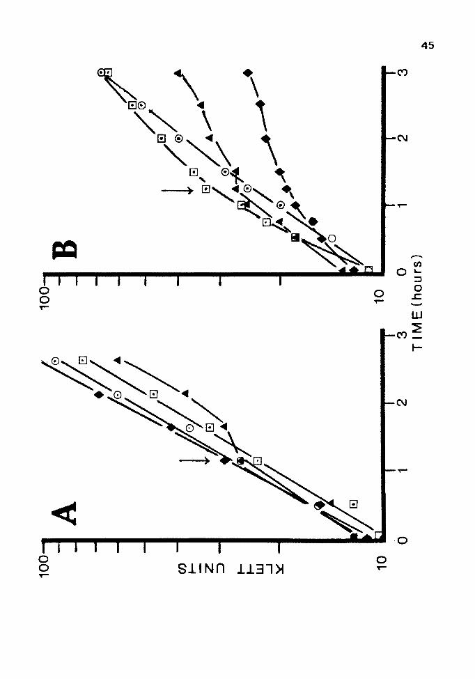
Figure 6. Optimal Arsenite Concentration for Induction
of Resistance
44
A: Cultures of JM 105(pUC84) were grown overnight in LB,
subcultured and grown to log phase. At the time
indicated by the arrow, 0.5 mM, 0; 1.0 mM, D; 5 mM,
A ; or no arsenite, + , were added to the culture.
B: Cultures of JM 105(pUC84) and JM 105(pUC8) were grown
as above and challenged with 10 mM arsenite at the
time indicated by the arrow. JM 105 (pUC 84) was
induced with 0. 5 mM, 0 ; 1. 0 mM, D ; or no arseni te, .& •
JM 105 (pUC 8) was induced with 1 mM arsenite, + .
0
-I -U::> s: m :J 0
~o en -
......
I\)
0 KLETT UNITS
...... 0
>
...... 0 0
...... 0 0
46
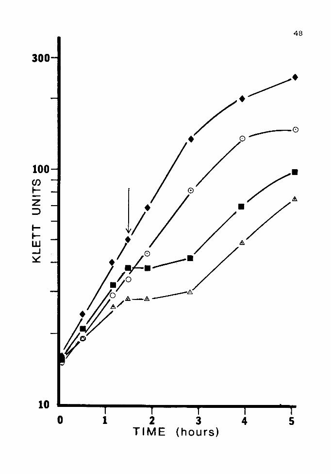
Unfortunately, IPTG did not induce resistance in
either orientation under any of the conditions used.
Results from a clone in the pUCB orientation are shown in
Fig. 7. Results in clones of the opposite orientation were
identical.
47
Figure 7. Induction of Resistance with IPTG
Cultures of JM 105(pUC84) were grown overnight in LB
containing the following inducers: 1mM arsenite,O; 2mM
lPTG,•; none,~. The cultures were subcultured and grown
to log phase twice before being challenged with 15 mM
arsenite at the time indicated by the arrow. A culture
that contained no inducer and was not challenged,+, was
included as a control.
(SJno4) 3~11 s £ z l
sv
0 01
A r m --4 --4
c z --4 CJ)
001
00£
49
Identification of Plasmid-Determined Proteins
The plasmids pUCS, pUC84 and pUC96 were transformed
into the maxicell strain ~- coli CSR 603. Uninduced and
arsenite-induced strains were irradiated as previously
described. This is a modification of the original maxicell
procedure (31), since radioactive amino acids were not
added. Due to the high copy number of the pUC vectors
(approximately 200 per cell) (11), it was expected that
recombinant plasmid proteins would be detected without the
use of radioactivity.
Because the arsenite resistance genes are inducible,
proteins from cultures grown with arsenite can be analyzed
for differences on a gel beside those from uninduced
cultures. In order to maximize these differences, cells
were induced in 10 mM arsenite, rather than the
subinhibitory 1 mM arsenite. The host and pUC-bearing
strains are not viable at this concentration, so arsenite
was not included in the growth media for these strains.
In comparing induced recombinant-bearing strains
against the uninduced strains, the following differences
were apparent:
1. Three proteins were visible in induced lanes only,
with apparent molecular weights of 62, 16.5 and 13.5
Kdal. Whereas the two smaller proteins were seen on
gels stained with either silver or Coomassie blue, the
62 Kdal protein was seen only on Coomassie

50
blue-stained gels (Fig. 8).
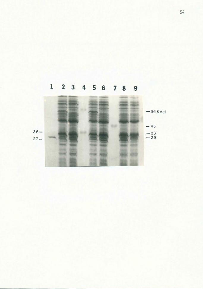
2. When ampicillin was included in the medium, the mature
27 Kdal ~-lactamase enzyme (37) encoded by the pUC
vector was visible in all lanes, except that of the
plasmid-less host strain (Fig. 9).
3. On most gels, a 36 Kdal protein was visible in all
lanes except those of the induced strains {Fig. 9).
51
Figure 8. Plasmid-Determined Proteins
Various CSR 603 strains were irradiated and grown in LB
without ampicillin for three hours. Cycloserine was added,
and the cultures were grown eight hours before lysing.
Cultures in lanes 2 and 5 also contained 10 mM arsenite.
Lanes 1, 4 and 7, protein standards; lanes 2 and 3,
603(pUC96); lanes 5 and 6, 603(pUC84); lane 8, 603(pUC8);
lane 9, CSR603. The sizes of plasmid-determined proteins
are indicated on the left side of the figure. Protein
standard sizes are indicated on the right. The gel was
stained with silver, then with Coomassie blue (6).

52
123456789
-66 Kdal
62-
-45
-36
-29
-24
-20.1
16 .5-
13.5-
53
Figure 9. ~-Lactamase and 36 Kdal Protein
Various CSR 603 strains were irradiated and grown for 6
hours in LB with 50 ug/ml ampicillin before lysing.
Cycloserine was not added. Cultures in lanes 2 and 5 also
contained 10 mM arsenite. Lanes 1, 4 and 7, protein
standards; lanes 2 and 3, 603(pUC96}; lanes 5 and 6,
603(pUC84}; lane 8, 603(pUC8); lane 9, 603. The
~-lactamase protein (27 Kdal) encoded by the pUC vectors
and the 36 Kdal protein absent in induced strains are
indicated by arrows on the left; protein standard sizes are
indicated on the right. The gel was stained with Coomassie
blue.
36-
27-
12345678 9
54
-66 Kdal
-45
-36 -29
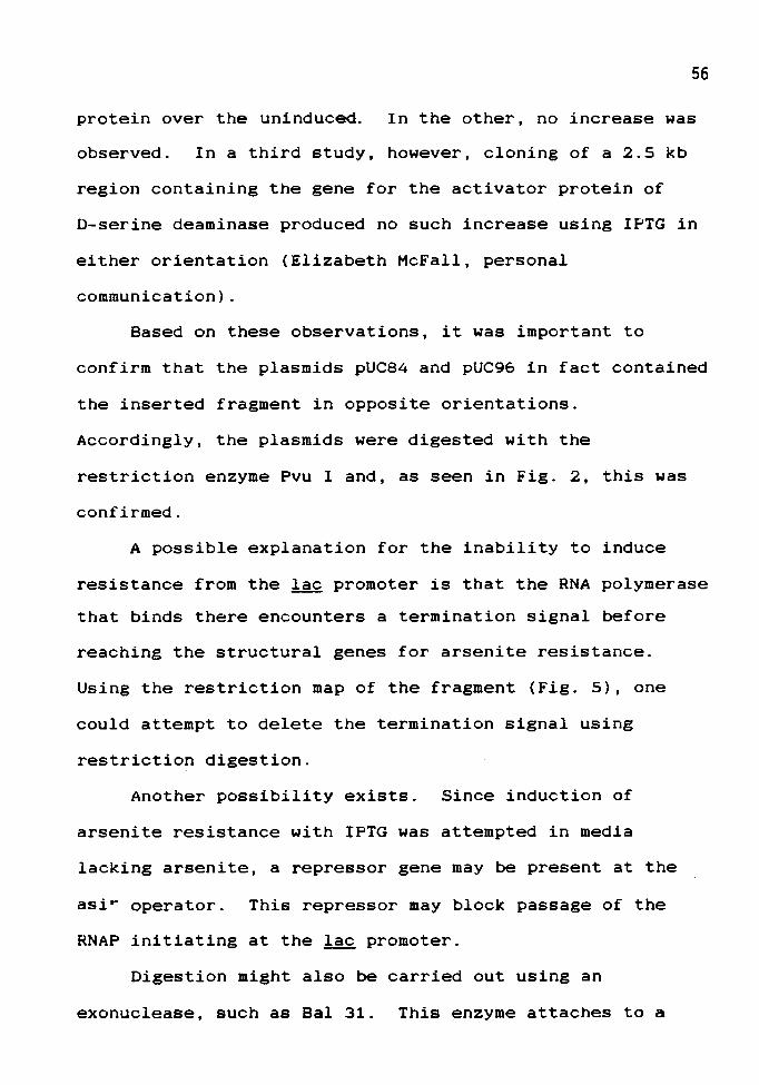
DISCUSSION
Comparison of the minimal inhibitory concentrations of
arsenite for strains growing on LA shows that the MIC's of
the ~- coli strains JM 105 and CSR 603 are similar -- 6 mM
and 4 mM, respectively. Transformation of recombinant
plasmids containing the Eco RI-Hind III fragment of R45
into these strains increases arsenite resistance by up to
five-fold (the MIC was 20 mM in both cases), confirming
successful transfer of the arsenite resistance region from
R45.
Strains bearing recombinant plasmids were grown in
liquid medium with and without arsenite and then challenged
by the addition of arsenite. Pre-induced strains continued
exponential growth while uninduced strains showed a growth
lag of 1-2 hours. These results indicate that the inserted
fragment contains the control elements for the resistance
genes.
Resistance to arsenite could not, however, be induced
in either pUC84 or pUC96 using IPTG. Hence, no conclusion
can be drawn regarding the direction of transcription. In
another study, a 3.5 kb fragment of DNA containing the gene
for diadenosine tetraphosphatase was cloned into the pUC
vectors (17). In one orientation, addition of IPTG
produced an eight-fold increase in production of this
56
protein over the uninduced. In the other, no increase was
observed. In a third study, however, cloning of a 2.5 kb
region containing the gene for the activator protein of
D-serine deaminase produced no such increase using IPTG in
either orientation (Elizabeth McFall, personal
communication).
Based on these observations, it was important to
confirm that the plasmids pUC84 and pUC96 in fact contained
the inserted fragment in opposite orientations.
Accordingly, the plasmids were digested with the
restriction enzyme Pvu I and, as seen in Fig. 2, this was
confirmed.
A possible explanation for the inability to induce
resistance from the lac promoter is that the RNA polymerase
that binds there encounters a termination signal before
reaching the structural genes for arsenite resistance.
Using the restriction map of the fragment (Fig. S), one
could attempt to delete the termination signal using
restriction digestion.
Another possibility exists. Since induction of
arsenite resistance with IPTG was attempted in media
lacking arsenite, a repressor gene may be present at the
asir operator. This repressor may block passage of the
RNAP initiating at the lac promoter.
Digestion might also be carried out using an
exonuclease, such as Bal 31. This enzyme attaches to a
57
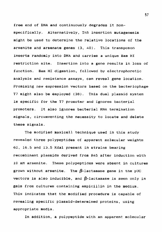
free end of DNA and continuously degrades it non
specifically. Alternatively, Tn5 insertion mutagenesis
might be used to determine the relative locations of the
arsenite and arsenate genes (3, 40). This transposon
inserts randomly into DNA and carries a unique Bam HI
restriction site. Insertion into a gene results in loss of
function. Bam HI digestion, followed by electrophoretic
analysis and resistance assays, can reveal gene location.
Promising new expression vectors based on the bacteriophage
T7 might also be ~mployed (38). This dual plasmid system
is specific for the T7 promoter and ignores bacterial
promoters. lt also ignores bacterial RNA termination
signals, circumventing the necessity to locate and delete
these signals.
The modified maxicell technique used in this study
revealed three polypeptides of apparent molecular weights
62, 16.5 and 13.5 Kdal present in strains bearing
recombinant plasmids derived from R45 after induction with
10 mM arsenite. These polypeptides were absent in cultures
grown without arsenite. The P-lactamase gene in the pUC
vectors is also inducible, and ~-lactamase is seen only in
gels from cultures containing ampicillin in the medium.
This indicates that the modified procedure is capable of
revealing specific plasmid-determined proteins, using
appropriate media.
In addition, a polypeptide with an apparent molecular
-----------------------····---~---
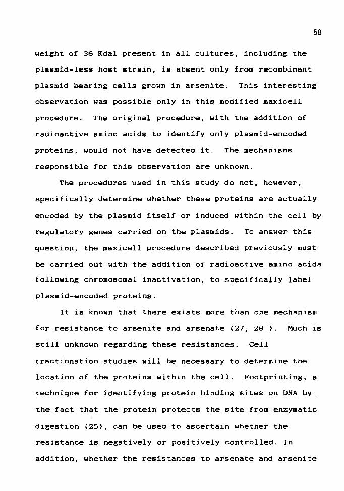
58
weight of 36 Kdal present in all cultures, including the
plasmid-less host strain, is absent only from recombinant
plasmid bearing cells grown in arsenite. This interesting
observation was possible only in this modified maxicell
procedure. The original procedure, with the addition of
radioactive amino acids to identify only plasmid-encoded
proteins, would not have detected it. The mechanisms
responsible for this observation are unknown.
The procedures used in this study do not, however,
specifically determine whether these proteins are actually
encoded by the plasmid itself or induced within the cell by
regulatory genes carried on the plasmids. To answer this
question, the maxicell procedure described previously must
be carried out with the addition of radioactive amino acids
following chromosomal inactivation, to specifically label
plasmid-encoded proteins.
It is known that there exists more than one mechanism
for resistance to arsenite and arsenate (27, 28 ). Much is
still unknown regarding these resistances. Cell
fractionation studies will be necessary to determine the
location of the proteins within the cell. Footprinting, a
technique for identifying protein binding sites on DNA by
the fact that the protein protects the site from enzymatic
digestion (25), can be used to ascertain whether the
resistance is negatively or positively controlled. In
addition, whether the resistances to arsenate and arsenite
59
are due to separate efflux pumps or one pump is modified to
accommodate the separate arsenic compounds remains to be
determined.
In this study, the proteins determined by the arsenic
resistance region of the plasmid R45 have been identified.
Comparison of these proteins with those of other arsenic
resistance plasmids will help illuminate the differences
and similarities between their mechanisms of resistance.
In addition, a restriction map of the region has been
prepared which can be used to further localize the arsenic
resistance genes and study their organization and
regulation.
REFERENCES
1. Birnboim, H.C., and J. Doly. 1979. A Rapid Alkaline Extraction Procedure for Screening Recombinant Plasmid DNA. Nuc; Acids. Res. 7:1513.
2. Casse, Francine, C. Boucher, J.S. Julliot, M. Michel and J. Denarie. 1979. Identification and Characterization of Large Plasmids in Rhizobium meliloti Using Agarose Gel Electrophoresis. J. Gen. Microbiol. 113:229
3. Chen, Chih-Ming, Harry L.T. Mobley and Barry P. Rosen. 1985. Separate Resistances to Arsenate and Arsenite (Antimonate) Encoded by the Arsenical Resistance Operon of R Factor R773. J. Bacteriol. 161:758.
4. Davis, Ronald W., David Botstein and John R. Roth. 1980. Advanced Bacterial Genetics. Cold Spring Harbor Laboratory.
5. Dretzen, G., M. Bellard, P. Sassone-Corsi and P. Chambon. 1981. A Reliable Method for the Recovery of DNA Fragments from Agarose and Acrylamide Gels. Anal. Biochem. 112:295.
6. Dzandu, James K., Mercy E. Deh, Denise L. Barratt and Gary E. Wise. 1984. Detection of Erthrocyte Membrane Proteins, Sialoglyco-proteins, and Lipids in the Same Polyacrylamide Gel Using a Double-Staining Technique. Proc. Nat. Acad. Sci. 81:1733
7. Ferguson, John F. And Jerome Gavis. 1972. A Review of the Arsenic Cycle in Natural Waters. Water Res. 6:1259.
8. Ferrazza, David and Stuart B. Levy. 1980. Biochemical and Immunological Characterization of an R Plasmid-Encoded Protein with Properties Resembling Those of Major Cellular Outer Membrane Proteins. J. Bacteriol. 144:149 ·
61
9. Fong, Kenneth, Frank Lee and Richard Bockrath. 1980.
10.
Effects of Sodium Arsenite on Single-Strand DNA Break Formation and Post-Replication Repair in E. coli Following UV Irradiation. Mutat. Res. 70:151.
Hedges, R.W. and s. Baumberg. Arsenic Compounds Conferred by Between Strains of Escherichia 115:459.
1973. Resistance to a Plasmid Transmissible coli. J. Bacteriol.
11. Heincz, Maria C., Susan M. Bornstein and Elizabeth McFall. 1984. Purification and Characterization of d-Serine Deaminase Activator Protein. J. Bacteriol. 160:42.
12. Laemmli, U.K. 1970. Cleavage of Structural Proteins During the Assembly of the Head of Bacteriophage T4. Nature 227:680.
13. Lawn, A.M., Elinor Meynell, G.G. Meynell, Naomi Datta. 1967. Sex Pili and the Classification of Sex Factors in the Enterobacteriaceae. Nature 216:343.
14. Mandel, M. and A. Higa. 1970. Calcium-Dependent Bacteriophage DNA Infection. J. Mol. Biol. 53:159.
15. Maniatis, T., E.F. Fritsch and J. Sambrook. 1982. Moleculuar Cloning: A Laboratory Manual. Cold Spring Harbor Laboratory.
16. McDonell, Michael W., Martha N. Simon and F. William Studier. 1977. Analysis of Restriction Fragments of T7 DNA and Determination of Molecular Weights by Electrophoresis in Neutral and Alkaline Gels. J. Mol. Biol. 110:119
17. Mechulam, Yves, Michel Fromant, Patrice Mellot, Pierre Plateau, Sylvie Blanchin-Roland, Guy Fayat and Sylvain Blanquet. 1985. Molecular Cloning of the Escherichia coli Gene for Diadenosine 5', 5" '-P 1
, P 4 -Tetraphosphate Pyrophosphohydrolase. J. Bacteriol. 164:63.
18. Mendez, Beatriz, Chikanori Tachibana and Stuart B. Levy. 1980. Heterogeneity of Tetracycline Resistance Determinants. Plasmid 3:99.
19. Messing, Joachim. 1983. New Ml3 Vectors for Cloning. In Methods in Enzymology, Vol. 101. Academic Press.
62 .
20. Metzler, David E. 1977. Biochemistry. Academic Press.
21. Miller, J.M. ed. 1972. Experiments in Molecular Genetics. Cold Spring Harbor Laboratory, New York.
22. Mobley, Harry L.T., Simon Silver, F.D. Porter and Barry P. Rosen. 1984. Homology Among Arsenate Resistance Determinants of R Factors in Escherichia coli. Antimicrob. Agents Chemother. 25:157.
23. Mobley, Harry L.T. and Barry P. Rosen. 1982. Energetics of Plasmid-Mediated Arsenate Resistance in Escherichia coli. Proc. Natl. Acad. Sci. 79:6119.
24. Myers, John w .• W. Herman Taylor and Mary L. Taylor. 1983. Localization of Genes Coding for Arsenate and Arsenite Resistance on the Plasmid, R45. Abstr. Oregon Acad. Sci.
25. Nick, Harry and Walter Gilbert. 1985. Detection in vivo of Protein-DNA Interactions Within the lac Operon of Escherichia coli. Nature 313:795.
26. Novick, Rochard P. and Christine Roth. 1968. Plasmid-Linked Resistance to Inorganic Salts in Staphylococcus aureus. J. Bacteriol. 95:1335.
27. Osborne, F.H. and H.L. Ehrlich. 1976. Oxidation of Arsenite by a Soil Isolate of Alcaligenes. J. Appl. Bact. 41:295.
28. Phillips, Shirley E. and Mary L. Taylor. 1976. Oxidation of Arsenite to Arsenate by Alcaligenes faecalis. Appl. Environ. Microbiol. 32:392.
29. Pugsley, Anthony P. and Carl A. Schnaitmen. 1978. Identification of Three Genes Controling Production of New Outer Membrane Pore Proteins in Escherichia coli K-12. J. Bacteriol. 135:1118.
30. Rosen, Barry P. and Miguel G. Borbolla. 1984. A Plasmid-Encoded Arsenite Pump Produces Arsenite Resistance in Escherichia coli. Biochem. Biophys. Res. Commun. 124:760.
31. Sancar, Aziz, Adelle M. Hack and W. Dean Rupp. 1979. Simple Method for Identification of Plasmid-Coded Proteins. J. Bacteriol. 137:692.

32. Schleif, Robert F. And Pieter c. Wensink. 1981. Practical Methods in Molecular Biology. Springer-Verlag.
33. Silver, S., K. Budd, K.M. Leahy, W.V. Shaw, D.
63
Hammond, R.P. Novick, G.R. Willsky, M.H. Malamy and H. Rosenberg. 1981. Inducible Plasmid-Determined Resistance to Arsenate, Arsenite and Antimony(Ill} in Escherichia coli and Staphycoloccus aureus. J. Bacteriol. 146:983.
34. Smiley, D.G. 1981. A Genetic and Physiological Study of an Arsenite Resistant, Uncoupled Mutant of Escherichia coli. Portland State University. M.S. Thesis.
35. Smith, H. Williams, Z. Parsell and P. Green. 1978. Thermosensitive Antibiotic Resistance Plasmids in Enterobacteria. J. Gen. Microbiol. 109:37.
36. Sunderman, F. William, Jr. 1976. A Review of the Carcinogenicities of Nickel, Chromium and Arsenic Compounds in Man and Animals. Prev. Med. 5:279.
37. Sutcliffe, J. Gregor. 1978. Nucleotide Sequence of the Ampicillin Resistance Gene of Escherichia coli Plasmid pBR322. Proc. Nat. Acad. Sci. 75:3737.
38. Tabor, Stanley and Charles c. Richardson. 1985. A Bacteriophage T7 RNA Polymerase/Promoter System for Controlled Exclusive Expression of Specific Genes. Proc. Natl. Acad. Sci. 82:1074
39. Thomas, Christopher M., Richard Meyer and Donald R. Helinski. 1980. Regions of Broad-Host-Range Plasmid RK2 Which are Essential for Replication and Maintenance. J. Bacteriol. 141:213.
40. Trezona, Thomas. 1981. Plasmid-Mediated Resistance to Arsenite and Arsenate in Escherichia coli. Portland State University. M.S. Thesis.
41. Vieira, Jeffrey and Joachim Messing. 1902. The pUC Plasmids, an M13mp7-Derived System for Insertion Mutagenesis and Sequencing with Synthetic Universal Primers. Gene 19:259.
42. Willsky, Gail R. And Michael H. Malamy. 1980. Effect of Arsenate on Inorganic Phosphate Transport in Escherichia coli. J. Bacteriol. 144:366.

64
43. Wray, Wayne, T. Boulikas, Virginia P. Wray and Ronald Hancock. 1981. Silver Staining of Proteins in Polyacrylamide Gels. Anal. Biochem. 118:197.
44. Yanisch-Perron, Celeste, Jeffrey Vieira and Joachim Messing. 1985. Improved M13 Phage Cloning Vectors and Host Strains: Nucleotide Sequences of the M13mp18 and pUC19 Vectors. Gene 33:103.


