Regio- and stereoselective hybride transfer reactions inmodel systems related to the redox-couple NAD+/NADHCitation for published version (APA):Kok, de, P. M. T. (1988). Regio- and stereoselective hybride transfer reactions in model systems related to theredox-couple NAD+/NADH. Eindhoven: Technische Universiteit Eindhoven. https://doi.org/10.6100/IR285955
DOI:10.6100/IR285955
Document status and date:Published: 01/01/1988
Document Version:Publisher’s PDF, also known as Version of Record (includes final page, issue and volume numbers)
Please check the document version of this publication:
• A submitted manuscript is the version of the article upon submission and before peer-review. There can beimportant differences between the submitted version and the official published version of record. Peopleinterested in the research are advised to contact the author for the final version of the publication, or visit theDOI to the publisher's website.• The final author version and the galley proof are versions of the publication after peer review.• The final published version features the final layout of the paper including the volume, issue and pagenumbers.Link to publication
General rightsCopyright and moral rights for the publications made accessible in the public portal are retained by the authors and/or other copyright ownersand it is a condition of accessing publications that users recognise and abide by the legal requirements associated with these rights.
• Users may download and print one copy of any publication from the public portal for the purpose of private study or research. • You may not further distribute the material or use it for any profit-making activity or commercial gain • You may freely distribute the URL identifying the publication in the public portal.
If the publication is distributed under the terms of Article 25fa of the Dutch Copyright Act, indicated by the “Taverne” license above, pleasefollow below link for the End User Agreement:
www.tue.nl/taverne
Take down policyIf you believe that this document breaches copyright please contact us at:
providing details and we will investigate your claim.
Download date: 24. Feb. 2020
REGIO- AND STEREOSELECTIVE HYDRIDE
TRANSFER REACTIONS
IN MODEL SYSTEMS RELATED TO THE REDOX-COUPLE
NAD+/NADH
P.M.T. DE KOK
REGIO- AND STEREOSELECTIVE HYDRIDE TRANSFER REACTIONS IN MODEL SYSTEMS RELATED TO THE REDOX-COUPLE
NAD+/NADH
REGIO- AND STEREOSELECTIVE HYDRIDE
TRANSFER REACTIONS
IN MODEL SYSTEMS RELATED TO THE REDOX-COUPLE
NAD+/NADH
PROEFSCHRIFT
TER VERKRIJGING VAN DE GRAAD VAN DOCTOR AAN DE TECHNISCHE UNIVERSITEIT EINDHOVEN,
OP GEZAG VAN DE RECTOR MAGNIFICUS, PROF. DR. F.N. HOOGE, VOOR EEN COMMISSIE
AANGEWEZEN DOOR HET COLLEGE VAN DEKANEN IN HET OPENBAAR TE VERDEDIGEN OP
DINSDAG 24 MEl 1988 TE 14.00 UUR
DOOR
PETRUS MARIA THERESIA DE KOK
GEBOREN TE TILBURG
DIT PROEFSCHRIFT IS GOEDGEKEURD DOOR DE PROMOTOREN:
PROF. DR. H.M. BUCK EN
PROF. DR: E.M. MEIJER

aan Dorry, Rob en Joran.
Chapter I
General introduction
I.l Enzymes
1.2 Nicotinamide Adenine Dinucleotide dependent
Dehydrogenases
I.3 Amide out-of-plane orientation
I.4 Outline of this thesis
References and notes
Chapter II
Dithionite reductions of axial chiral 2,4-dimethyl-3-
carballloyl pyridiniUII cations
II.l Introduction
II.2 Stability of the axial chiral carbonyl out-of-
plane orientation
II.3 Dithionite reductions of axial chiral pyridinium
7
7
7
ll
15
16
18
18
19
cations .. 24
II.4 Dithionite reductions of quino1iunium compounds 27
II.5 Experimental section 31
References and notes 41
Chapter III
Stereoselective oxidation of 3-(N,N-dimethylcarbamoyl)-
1,2,4-trimethy1-1,4-dihydropyridine 43
III.l Introduction 43
III.2 Outline of the synthesis 44
III.3 Generation and determination of an enantio-
meric excess
III.4 Absolute configuration at C(4)
III.5 Determination of the CQ out-of-plane orien-
tation in the oxidized form
III.6 Stereochemistry of oxidation
III. 7 Conclusions
III.S Experimental section
References and notes
45
47
49
57
58
61
64
OJapter IV
Synthesis and reduction of an axial chi.ral bicpclic
pyridinita~ cation
IV.l Introduction
IV.2 Synthesis and axial chirality
IV.3 Stereoselectivity of reduction
IV.4 Conclusions
IV.S Experimental section
References and notes
OJapter V
Analysis of the interactions of li1A1)+ with Horse Liver
Alcobol Dehydrogenase using 110lec::ular •c:banics
V.l Introduction
v.a Procedure for calculational studies
V.3 Results and discussion
V.4 Conclusions
Appendix
References and notes
OJapter VI
Molecular Mc:hanics calculation of the ~try of JW)+
derivatives, IIOdified in the nicotioaai.de grodp, in a
69
69
69
71
n 73
76
79
79
80
82
93
95
96
ternary CCIIIIple:z:: with Horse Liver Alcobol Dehydrogenase 99
VI.l Introduction 99
VI.2 Procedure for calculational studies 100
VI.3 Results 103
VI.4 Discussion 108
VI. 5 Conclusions 110
Appendix 111
References and notes 113
s.-ary 116
Sallenvatting 118
CurriculUII vitae 121
DaDJr:.woord 122
aiAP'fER I
General introduction
I.l EnzymeS
Chemists have long been fascinated by the remarkable rate accele
ration and high regio- and stereoselectivities obtained under the
mild conditions of numerous enzyme catalyzed reactions. It has been
suggested that the major source of enzymatic efficacy results from
enzymes achieving their catalytic power by using intrinsic nonco
valent binding energy of substrates, bound in the active complex, to
lower the free-energy barrier of reaction1 • A striking fact is that
the stereochemical requirements for the stereospecificity that the
enzymes .manifest are simultaneously and completely fulfilled by the
chiral environment furnished within the enzyme/substrate complex.
The process of rate enhancement and stereoselectivity requires a
complex macromolecule.
Introducing only stereospecificity in non-enzymatic reactions,
mimicing the "in vivo" process, reduces the catalyst's complexity.
In order to design efficient artificial model systems, it is of
crucial importance to gather insight in the interplay of individual
intrinsic features which the enzyme can deploy to the task.of
introducing stereospecificity.
I.Z Nicotinamide Adenine Dinucleotide dependent dehydrogenases
Among many enzymes playing physiological important roles "in
vivo", the pyridine nucleotide dependent oxidoreductases have attrac
ted organic chemists attempting to simulate enzymatic efficacy in
simplified non-enzymatic MAD(p)+tMAD(P)H model systems2. These
enzymes are extremely important in that they supply energy to cells
of all living organisms through oxidation-reduction reactions 3 ,
which are the most fundamental processes in biological systems. They
effect a reversible and stereoselective tranfer of hydrogen between
-7-
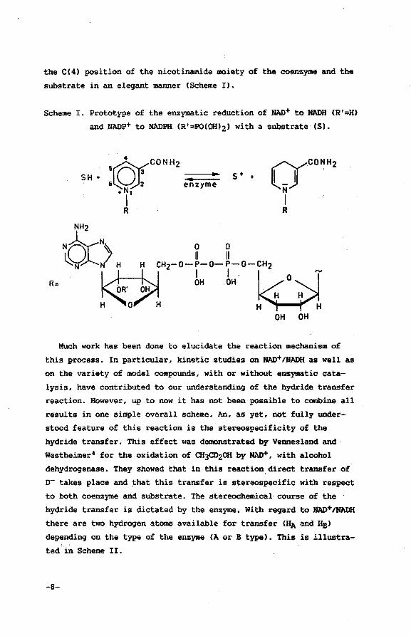
the C(4) position of the nicotinamide moiety of the coenzyme and the
substrate in an eleqant manner (Scheme I).
Scheme I. Prototype of the enzymatic reduction of NAD+ to NADH CR'=H)
and NADP+ to NADPH CR'=POCOIUz) with a substrate CS).
SH + s + +
R:
NH2
tO:> H " cH,-o-Lo-Lo-cH2
II-~ . OH OH O . H H
H 0 H H H OH OH
Much work has been done to elucidate the reaction mechanism of
this process. In particular, kinetic studies on NAD+/NADH as well as
on the variety of model compounds, with or without enzymatic cata~
lysis, have contributed to our understandinq of the hydride transfer
reaction. However, up to now it has not been possible to combine all
results in one simple overall scheme. An, as yet, not fully under
stood feature of this reaction is the stereospecificity of the
hydride transfer. This effect was demonstrated by Vennesland and
Westheimer4 for the oxidation of CH3CD20H by NAD+, with alcohol
dehydroqenase. They showed that in this reaction_direct transfer of
o- takes place and ~hat this transfer is stereospecific with respect
to both coenzyme and substrate. The stereochemical course of the
hydride transfer is dictated by the enzyme. With reqard to NAD+JNADH
there are two hydroqen atoms available for transfer CHA and HB)
dependinq on the type of the enzyme (A orB type). This is .illustrated .. in Scheme II.
-8-
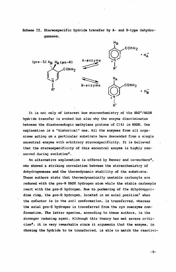
Scheme II. Stereospecific hydride transfer by A- and B-type dehydro
genases.
A-enzyme
~
~ B-enzyme
It is not only of interest how stereochemistry of the NAD+/NADH
hydride transfer is evoked but also why the enzyme discriminates
between the diastereotopic methylene protons of C(4l in NADH. One
explanation is a "historical" one. All the enzymes from all orga
nisms acting on a particular substrate have descended from a single
ancestral enzyme with arbitrary stereospecificity. It is believed
that the stereospecificity of this ancestral enzyme is highly con
served during evolution5 •
An alternative explanation is offered by Benner and co-workers 6 ,
who showed a striking correlation between the stereochemistry of
dehydrogenases and the thermodynamic stability of the substrate.
These authors state that thermodynamically unstable carbonyls are
reduced with the pro-R NADH hydrogen atom while the stable carbonyls
react with the pro-S hydrogen. Due to puckering of the dihydropyri
dine ring, the pro-R hydrogen, located in an axial position7 when
the cofactor is in.the anti conformation, is transferred, whereas
the axial pro-S hydrogen is transferred from the syn coenzyme con
formation. The latter species, according to these authors, is the
stronger reducing agent. Although this theory has met severe criti
cism8, it is very remarkable since it arguments that the enzyme, in
chosing the hydride to be transferred, is able to match the reactivi-
-9-
ty of NADH with the reactivity of the substrate. Of course, evolu
tionary pressure has produced enzymes of which the stereochemistry
is fixed, i.e. reactivity of NADH is adapted to the reactivity of
its natural substrate.
From an enzymology point of view, the generation of stereospecifi
city is quite straightforward. Branden9 showed in his crystallogra
phic work on horse liver alcohol dehydrogenase (LADH; A specific)
the interactions relevant for coenzyme and substrate binding. X-ray
data combined with model building techniques revealed that both the
substrate and the nicotinamide moiety of the coenzyme are fixed deep
inside the binding cleft. Due to steric hindrance as well as to in
teractions with exactly positioned sites of the enzyme, both sub
strate and coenzyme take up well-defined positions, in which the B
side of the nicotinamide group is shielded by the hydrophobic wall
of the cleft whereas the A side (Scheme I) is directed toward the
substrate. This feature is essential for the stereochemistry of the
enzymatic hydride transfer and it is not surprising to find that the
stereospecificity is substantially less when the reaction is carried
out under non-enzymatic conditions10 • Since discrimination between
the A and B side of the pyridinium ring is anly possible when the
amide group is present, it is clear that its (relative) orientation
within the ternary complex is essential for the stereochemistry in
the enzymatic hydride transfer process. The stereospecificity
results from the positioning of the nicotinamide moiety with respect
to the substrate which in turn is largely determined by the hydrogen
bonds between the amide group and the enzyme. Until now, little
effort has been made to investigate the effect of the orientation of
the amide moiety upon the stereochemistry of the process under enzy
matic conditions.
The relevance of the CONH2 group to the dynam~cs in the enzyme
catalyzed stereospe~ific hydride transfer has been suggested by
Dutlerll for LADH. He envisaged the (dihydro)pyridine ring to have
enough freedom of motion to change its position during the hydrogen
transfer, a movement possibly accompanied by rotation of the CONH2
group out of the plane of the (dihydro)pyridine ring.
-10-
!.3 Amide out-of-plane orientation
This thesis elaborates a new concept explaining the stereospecifi
city of the enzymatic~ hydride transfer of NAD+tNADH, that is to say,
how the substrate can distinguish between the two hydrogen atoms
available for transfer. The basic idea is that, once the coenzyme
has formed a reactive complex with a suitable enzyme, the CONH2
group loses the freedom to rotate, for instance, by the formation of
hydrogen bonds. When the CONH2 group is fixed, and most likely this
will be in an out-of-plane orientation with respect to the dihydro
pyridine ring, HA and HB may migrate to the substrate but with very
different rates. This causes the reaction to be effectively stereo
specific.
The primary goal of the investigations presented in this thesis
is to provide experimental evidence for the correlation between the
stereochemical course of the hydride transfer and the CONH2 out-of
plane orientation, using NAD+tNADH model compoundsl2,13.
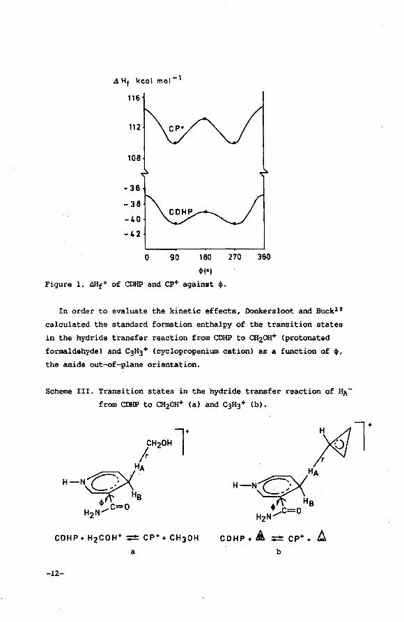
Quantum chemical calculations performed by Donkersloot and Buckl 3
on NAD+/NADH model compounds established that it is unlikely that
under experimental conditions stereospecificity of NAD+tNADH hydride
transfer reactions originates from a permanent axial chirality of
the amide moiety of NAD+tNADH. They calculated the total standard
enthalpy of formation (AHf 0) of 3-carbamoyl-1,4-dihydropyridine
(CDHP) and 3-carbamoyl pyridinium cation (Cp+) with MIND0/3 using
complete structural optimization, but keeping the torsion angle (~)
around the C(3)-cONH2 bond fixed at certain values. Figure 1 shows
the resulting AHf0 as a function of ~ (~=0° corresponds to the oxy
gen of the CONH2 group syn-oriented with respect to C(4); 0°<~<180°
corresponds to the oxygen of the CONH2 group being on the same side
of the (dihydro)pyridine ring as HA, etc.). The enthalpy barriers
are low enough for the rotation of the CONH2 group to be almost
free: for CDHP there are barriers at ~=0° and 180° of 3.9 and 1.7
kcal mol-l, respectively. For cp+, on the other hand, values of 4.6
and 3.3 kcal mol-l, respectively, were calculated. It is therefore
concluded that permanent axial chirality in NAD+tNADH due to an
amide out-of-plane rotation is absent.
-11-
4 H1 kc:al mol- 1
116
112
108
-36
-38
-40
-42
0 90 180 ,,., Figure 1. Mlf0 of CDHP and ep+ against '·
270 360
In order to evaluate the kinetic effects. Donkersloot and Buck13
calculated the standard formation enthalpy of the transition states
in the hydride transfer reaction from CDHP to CHzoa+ (protonated
formaldehyde) and C3H3+ (cyclopropenium cation) as a function of t. the amide out-of-plane orientation.
Scheme III. Transition states in the hydride transfer reaction of HA
from CDHP to CH2oa+ (a) and C3H3+ (b).
a
-12-
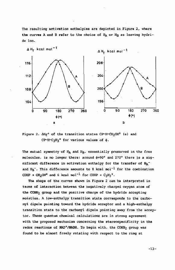
The resulting activation enthalpies are depicted in Figure 2, where
the curves A and B refer to the choice of HA or He as leaving hydri
de ion.
4 Hf. kcal mol-l 4 Hf kcal mot- 1
116 208
112 204
108 200
104 196
0 90 180 270 360 0 90 180 .,., 4»1•)
a b
Figure 2. &lf0 of the transition states CP..·FroCH20H+ (a) and
CP-H-c3H3+ for various values of +•
270 360
The mutual symmetry of HA and Hs, essentially preserved in the free
molecules, is no longer there: around +=90° and 270° there is a sig
nificant difference in activation enthalpy for the transfer of HA
and Hs-· This difference amounts to 8 kcal mol-l for the combination
CDHP + CH20H+ and 6 kcal mol-l for CDHP + C3H3 +.
The shape of the curves shown in Figure 2 can be interpreted in
terms of interaction between the negatively charged oxygen atom of
the CONH2 group and the positive charge of the hydride accepting
moieties. A low-enthalpy transition state corresponds to the carbo
nyl dipole pointing toward the hydride acceptor and a high-enthalpy
transition state to the carbonyl dipole pointing away from the accep
tor. These quantum chemical calculations are in strong agreement
with the proposed mechanism concerning the stereospecificity in the
redox reactions of NAD+/NADH. To begin with, the CONH2 group was
found to be almost freely rotating with respect to the ring at
-13-
normal temperatures. This would account for the observed lack of the
stereospecificity under non-enzymatic conditions. On the other hand,
the calculations demonstrate that the stereospecificity can come to
expression by fixation of the CONH2 group in an out-of-plane orien
tation originating from interactions with the enzyme. Recently an
enzyme-mediated CONH2 out-of-plane orientation was detected in crys
tallized enzyme/coenzyme/substrate complexes. X-ray diffraction data
(2.9 A resolution, crystallographic R factor of 0.22) derived from
the ternary complex of·NAD-bonded LADH (A specific), obtained by
Eklund et al.l 4 , showed the amide group rotated 30° out of the
plane, with the carbonyl oxygen atom situated at the A side. This
orientation of the amide moiety results from interactions with speci
fic sites of the enzyme, i.e. hydrogen bonding of the carbonyl group
of the CONH2 moiety with the main chain nitrogen atom of Phe-319 and
the NH2 group with the carbonyl groups of Val-292 and Gly-317. Addi
tionally, the B side was adequately shielded, thus making this side
inaccessible to the substrate due to Thr-178. High resolution X-ray
data obtained for a binary complex of NAD+ and glyceraldehyde-3-phos
phate dehydrogenase (GAPDH, B specificity) from Bacillus stearother
mophilus (resolution 1.8 A, crystallographic R factor of 0.177)15
revealed that in this enzyme complex, the planes of the carboxamide
group and the pyridine ring are inclined at an angle of about 22°.
Again the carbonyl dipole is directed towards the substrate-binding
region, this time favouring the B specific hydride transfer process.
The difference in stereoselectivity arises from the syn conformation
of the nicotinamide group. The glycosidic torsion angle is approxi
mately 80° in contrast to the value of 260° observed for the anti
conformation of the nicotinamide group in NAD+ bound to LADH. Al
though the results obtained from crystallized complexes are not to
their full extent representative for the dYnamic_behaviour of an
enzyme in solution,_ the observed out-of-plane orientation of the
CONHz group supports the proposed mechanis~. In this context it
should be noted that in both cases the nicotinamide group is situ
ated in the interior of the enzyme, a region in which the influence
of the enzyme-surrounding medium will probably be minimized.
-14-
I.4. Outline of this thesis
The goal of the investigations outlined in this thesis is to esta
blish the CO out-of-plane mediated stereochemistry in the hydride
transfer reactions of NAD+tNADH model compounds and to study co
enzyme (analogue) interactions in a ternary complex with LADH.
Chapter II describes a study in which the axial chiral stability
and the regioselectivity of reduction of 2,4-dimethyl-3-carbamoylpy
ridinium compounds are tested. It will be shown that a high stabi
lity of the carbonyl out-of-plane orientation and 1,4-regioselec
tivity of dithionite reductions are mutually exclusive features in
this type of NAD+ model compounds.
In chapter III the direct synthesis of the 1,4 reduced 2,4-dime
thyl-3-carbamoylpyridinium compounds is outlined and the stereo
selectivity in the oxidation process is tested.
Chapter IV deals with (the stereo- and regioselectivity in) the
reduction of a pyridinophane compound, in which the carbonyl out-of
plane orientation is generated by interconnecting the nicotinamide
C(6) position with the amide nitrogen atom using a relatively short
Cs alkyl bridge.
The results presented in chapter III and chapter IV subscribe the
role of the carbonyl out-of-plane orientation in the stereochemistry
of hydride transfer reactions. In both cases a syn orientation of
the migrating hydrogen atom and the CO dipole is observed. These
results are in complete accordance with the theory developed by
Donkersloot and Buck.
Chapters V and VI present a molecular mechanics study in which
interactions of NAD+ and NAD+ derivatives in a ternary complex of
horse liver alcohol dehydrogenase with DMSO were simulated and their
relative importance to the coenzyme (analogue) geometry is evalu
ated. The results of this calculational study demonstrate that the
"in vivo" reactivity of NAD+ derivatives can be qualitatively rela
ted to the calculated position of the NAD+ pyridinium moiety in the
active site.
-15-
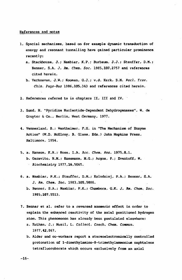
References and notes
1. Special mechanisms, based on for example dynamic transduct~on of
energy and resonant tunnelling have gained particular prominence
recently:
a. Stackhouse, J.; Nambiar, K.P.; Burbaum, J.J.; Stauffer, D.M.;
Benner, S.A. J. Am. Chem. Soc. 1985,107,2757 and references
cited herein.
b. Verhoeven, J.W.; Koomen, G.J.; v.d. Kerk, S.M. Reel. Trav.
Chim. Pays-Bas 1986,105,343 and references cited herein.
2. References refered to in chapters II, III and IV.
3. Sund, H. "Pyridine Nucleotide-Dependent Dehydrogenases", W. de
Gruyter & Co., Berlin, West .Germany, 1977.
4. Vennesland, B.; Westheimer, F.H. in "The Mechanism of Enzyme
Action" (W.D. McElroy, B. Glass, Eds.) John Hopkins Press,
Baltimore, 1954.
5. a. Hanson, K.R.; Rose, I.A. lice. Chem. Res. 1975,8,1.
b. Garavito, R.M.; Rossmann, M.G.; Argos, P.; Eventoff, W.
Biochemistry 1977,16,5065.
6. a. Rambiar. P.K.; Stauffer, D.M.; Kolodziej, P.A.; Benner, S.A.
J. Jim. Chem. Soc. 1983,105,5886.
b. Benner, S .A.; Rambiar, P .K.; Chambers, G.K. J. Jim. Chem. Soc.
1985,107,5513.
7. Benner et al. refer to a reversed anomeric effect in order to
explain the enhanced reactivity of the axial positioned hydrogen
atom. This phenomenon has already been postulated elsewhere:
a. Kuthan, J.; Musil, L. Collect. Czech. Chem. Commun.
1977,42,867.
b. Alder and co-workers report a stereoelectronical1y controlled
protonation of 1-dimethylamino--8-trimethylammonium naphtalene
tetrafluoroborate which occurs exclusively from an axial
-16-
direction. Alder. R.W.; Bryce. M.R •• Goode. N.C. J. Chem.
Soc., Perkin II 1982,477.
c. Brounts. R.H.A.M.; Buck, H.M. J. Am, Chem. Soc. 1983.105,1284.
d. Rob, F.; v. Ramesdonk. H.J.; v. Gerresheim, W.; Bosma, P.;
Scheele. J.J.; Verhoeven, J.W. J. Am. Chem. Soc. 1984.106,3826.
8. Oppenheimer, N.J. J. Am. Chem. Soc. 1984.106,3032.
9. Branden. C.I. reference 2. p 325.
10. Chemical reductions of NAD+ in DzO revealed that the pro-R
hydrogen incorporation predominates. The reported percentages of
enantiomeric excess range from 12% to 44%:
a. Fisher, H.F.; Ofner, P.; Conn, E.E.; Vennesland, B.;
Westheimer, F.H. J. Biol. Chem. 1953,202,687.
b. Pullman, M.E.; San Pietro, A.; Colowick, S.P. J. Biol. Chem.
1954,206,129.
c. San Pietro, A.; Kaplan, N.O.; Colowick, S.P. J. Biol. Chem.
1955.212.941.
d. Kaplan considered this phenomenon to be the result of stac
king of the purine and pyridine .rings of NAD+ in solution.
One of the helix structures (M) is present in excess:
Kaplan, N. 0.; Sarma, R. "Pyridine Nucleotide-Dependent
Dehydrogenases" <H. Sund Ed.) p 39, Springer-Verlag 1970.
11. Outler. H. reference 2. p 347.
12. a. Donkersloot, M.C.A.; Buck, H.M. J. Am. Chem. Soc.
1981,103,6549.
b. Brounts, R.H.A.M.; Buck, H.M. J. Am. Chem. Soc. 1983,105,1284.
13. Donkersloot, M.C.A.; Buck, H.M. J. Am. Chem. Soc. 1981,103,6554.
14. Eklund, H.; Samama, J.P.; Jones. T.A. Biochemistry 1984,23,5982.
15. Skarzynski, T.; Moody. P.C.E.; Wonacott. i\.J. ·J. Mol. Biol.
1987,193,171.
-17-
aiAP'l'ER II
Dithionite reductions of axial ch.iral 2,4-di.methyl-3-carballloy1
pyridinium cations1
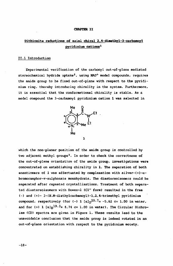
II.l Introduction
Experimental verification of the carbonyl out-of-plane mediated
stereochemical hydride uptake 2 , using NAD+ model compounds, requires
the amide group to be fixed out-of-plane with respect to the pyridi
nium ring, thereby introducing chirality in the system. Furthermore,
it is essential that the conformational chirality is stable. As a
model compound the 3-carbamoyl pyridinium cation 1 was selected in
1
which the non-planar position of the amide group is controlled by
two adjacent methyl groups 3 • In order to check the correctness of
the out-of-plane orientation of the amide group, investigations were
concentrated on establishing chirality in 1. The separation of both
enantiomers of 1 was effectuated by complexation with silver-(+)-«
bromocamphor-~-sulphonate monohydrate. The diastereoisomers could be
separated after repeated crystallizations. Treatment of both separa
ted diastereoisomers with Dowex-2 eel- form) resulted in the free
(-) and (+)- 3-(N,N-diethylcarbamoyl)-1,2,4-trimethyl pyridinium
compound, respectively (for<-> 1 [«]Dl9.7= -5.62 c= 1.00 in water,
and for(+) 1 [«] 019.3= 4.74 c= 1.00 in water). The Circular Dichro
ism (CD) spectra are given in Figure 1. These results lead to the
unavoidable conclusion that the amide group is indeed rotated in an
out-of-plane orientation with respect to the pyridinium moiety.
-18-
1
+2
+t
A.[nm)
-t
-2
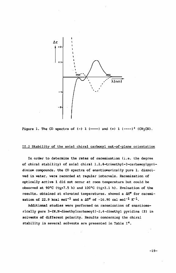
Figure 1. The CD spectra of (-) 1 (----) and (+) 1 (----) 3 (CH3CN).
!!.2 Stability of the axial chiral carbonyl out-of-plane orientation
In order to determine the rates of racemization (i.e. the degree
of chiral stability) of axial chiral 1,2,4-trimethyl-3-carbamoylpyri
dinium compounds, the CD spectra of enantiomerically pure 1, dissol
ved in water, were recorded at regular intervals. Racemization of
optically active 1 did not occur at room temperature but could be
observed at 90°C <t%=7.5 h) and 100°C <t%=3.1 h). Evaluation of the
results, obtained at elevated temperatures, showed a aH~ for racemi
zation of 22.9 kcal mol-l and a ~S~ of -16.90 cal mol-l K-1.
Additional studies were performed on racemization of enantiome
rically pure 3-(N,N-dimethylcarbamoyl)-2,4-dimethyl pyridine (2) in
solvents of different polarity. Results concerning the chiral
stability in several solvents are presented in Table ! 4 •
-19-
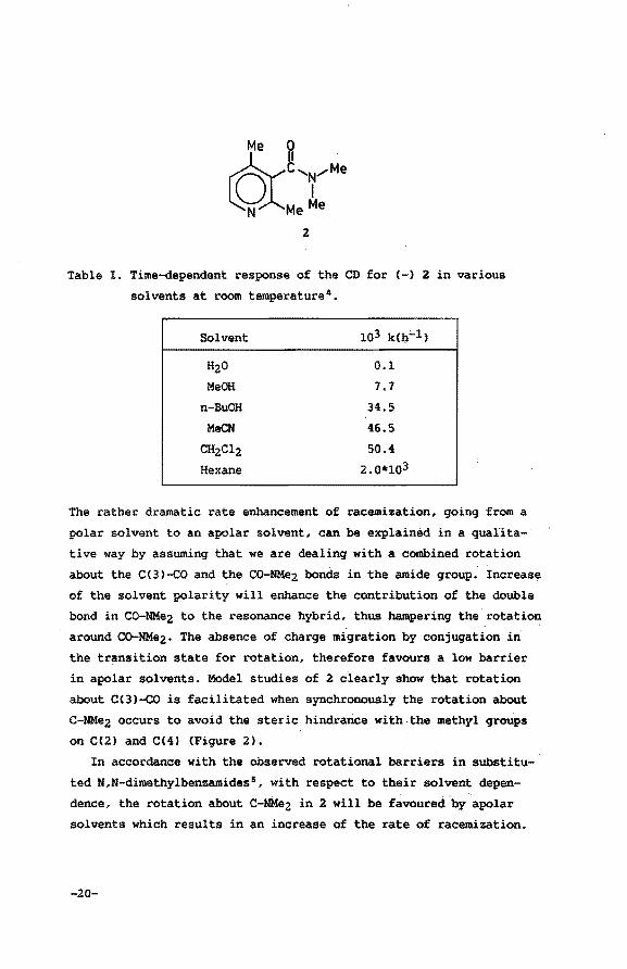
Table I. Time-dependent response of the CD for (-) 2 in various
solvents at room temperature 4 •
Solvent 103 k(b;...l)
H;zO 0.1
MeOH 7.7
n-BuOH 34.5
MeCN 46.5
CH2c12 50.4
Hexane 2.Qiirl03
The rather dramatic rate enhancement of racemization, going from a
polar solvent to an apolar solvent, can be explained in a qualita
tive way by assuming that we are dealing with a combined rotation
about the C(3)-co and the CO-NMe2 bonds in the amide group. Increase
of the solvent polarity will enhance the contribution of the double
bond in co-NMez to the resonance hybrid, thus hampering the rotation
around co-NMe2. The absence of charge migration by conjugation in
the transition state for rotation, therefore favours a low barrier
in apolar solvents. Model studies of 2 clearly show that rotation
about CC3)-co is facilitated when synchronously the rotation about
C-NMez occurs to avoid the steric hindrance with.the methyl groups
on C(2) and C(4) (Figure 2).
In accordance with the observed rotational barriers in substitu- ·
ted N,N-dimethylbenzamides5 , with respect to their solvent depen
dence, the rotation about C-NMe;z in 2 will be favoured by apolar
solvents which results in an increase of the rate of racemization.
-20-
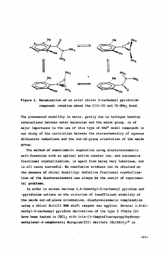
Figure 2. Racemization of an axial chiral 3-carbamoyl pyridinium
compound; rotation about the C(3)-c0 and CO-NMe2 bond.
The pronounced stability in water, partly due to hydrogen bonding
interactions between water molecules and the amide group, is of
major importance to the use of this type of NAD+ model compounds in
our study of the correlation between the stereochemistry of aqueous
dithionite reductions and the out-of-plane orientation of the amide
group.
The method of enantiomeric separation using diastereoisomeric
salt-formation with an optical active counter ion, and successive
fractional crystallization, is apart from being very laborious, not
in all cases succesful. No conclusive evidence can be obtained on
the absence of chiral stability; defective fractional crystalliza
tion of the diastereoisomers can always be the result of experimen
tal problems.
In order to screen various 2,4-dimethyl-3-carbamoyl pyridine and
-pyridinium cations on the criterion of insufficient stability of
the amide out-of-plane orientation, diastereoisomeric complexation
using a chiral Eu(III) NMR shift reagent was applied. Several 2,4-di
methyl-3-carbamoyl pyridine derivatives of the type 3 (Table II)
have been tested in CDCl3 with tris-[3-(heptafluoropropylhydroxy
methylene)-d-camphorato] Europium(III) derivate (Eu(hfc)3) 6 in
-21-
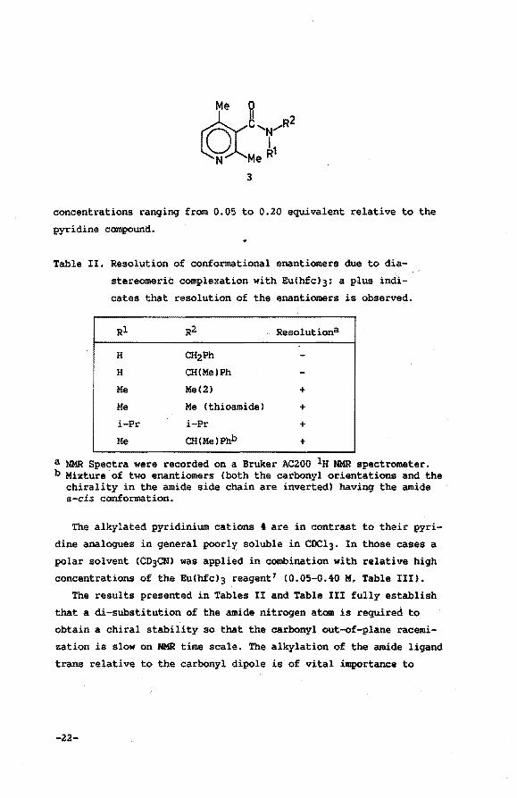
concentrations ranging from 0.05 to 0.20 equivalent relative to the
pyridine compound.
Table II. Resolution of conformational enantiomers due to dia
stereomeric complexation with Eu(hfcl3; a plus indi
cates that resolution of the enantiomers is observed.
Rl
H
H
Me
Me
i-Pr
Me
R2
CH2Ph CH(Me)Ph
Me(2)
Me Cthioamide)
i-Pr
CH(Me)Phb
Resolutiona
+ + + +
a NMR Spectra were recorded on a Bruker AC200 lH NMR spectrometer. b Mixture of two enantiomers (both the carbonyl orientations and the
chirality in the amide side chain are inverted) having the amide s-cis conformation.
The alkylated pyridinium cations 4 are in contrast to their pyri
dine analogues in general poorly soluble in CDCl3. In those cases a
polar solvent (CD3CN) was applied in combination with relative high
concentrations of the Eu(hfc)3 reagent 7 (0.05-0.40 M, Table III).
The results presented in Tables II and Table III fully establish
that a di-substitution of the amide nitrogen atom is required to
obtain a chiral stability so that the carbonyl out-of-plane racemi
zation is slow on NMR time scale. The alkylation of the amide ligand
trans relative to the carbonyl dipole is of vital importance to
-22-
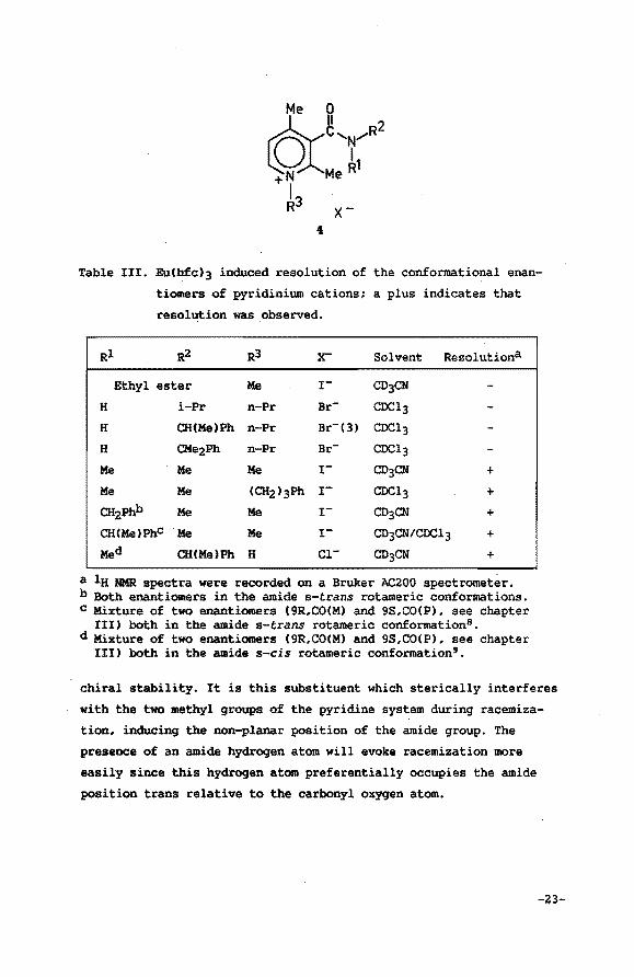
Table III. Eu(hfc)3 induced resolution of the conformational enan
tiomers of pyridinium cations; a plus indicates that
resolution was observed.
Rl R2 R3 x- Solvent Resolutiona
Ethyl ester Me r CD3CN
H i-Pr n-Pr Br- CDCl3
H CH(Me)Ph n-Pr Br-(3) CDCl3
H CMe2Ph n-Pr Br- CDCl3
Me Me Me r CD3CN +
Me Me (CH2)3Ph r CDCl3 +
CHzPhb Me Me r CD3CN + CH(MeJPhc Me Me r- CD3CN/COCl3 + Med CH(MeJPh H Cl- CD3CN +
a la NMR spectra were recorded on a Bruker AC200 spectrometer. b Both enantiomers in the amide a-trans rotameric conformations. c Mixture of two enantiomers (9R,OO(M) and 9S,CO(P), see chapter
III) both in the amide s-trans rotameric conformation8 • d Mixture of two enantiomers (9R,CO(M) and 9S,CO(P), see chapter
III) both in the amide s-cis rotameric conformation'.
chiral stability. It is this substituent which sterically interferes
with the two methyl groups of the pyridine system during racemiza
tion, inducing the non-planar position of the amide group. The
presence of an amide hydrogen atom will evoke racemization more
easily since this hydrogen atom preferentially occupies the amide
position trans relative to the carbonyl oxygen atom.
-23-
II.3 Dithionite reductions of axial chiral pyridinium cations
In the previous section it is outlined that the 3-(N,N-dialkyl
ated-cambamoyl)-1,2,4-trimethyl cations meet the requirement of m high degree of axial chiral stability of the carbonyl out-of-plane
orientation. In some cases enantiomeric separation was accomplis
hed3,8,9, Still these compounds, in order to be useful in testing
the concept of carbonyl out-of-plane mediated stereoselective
hydride incorporation, should also undergo hydride uptake resulting
in the formation of the corresponding 1,4-dihydro pyridine deriva
tive. Unfortunately no reduction of the N,N-diethyl derivative 1 in
an aqueous solution of sodium dithionite could be observed. The lack
of reactivity is probably caused by steric and electronic shielding
resulting from the C(2) and C(4) methyl groups.
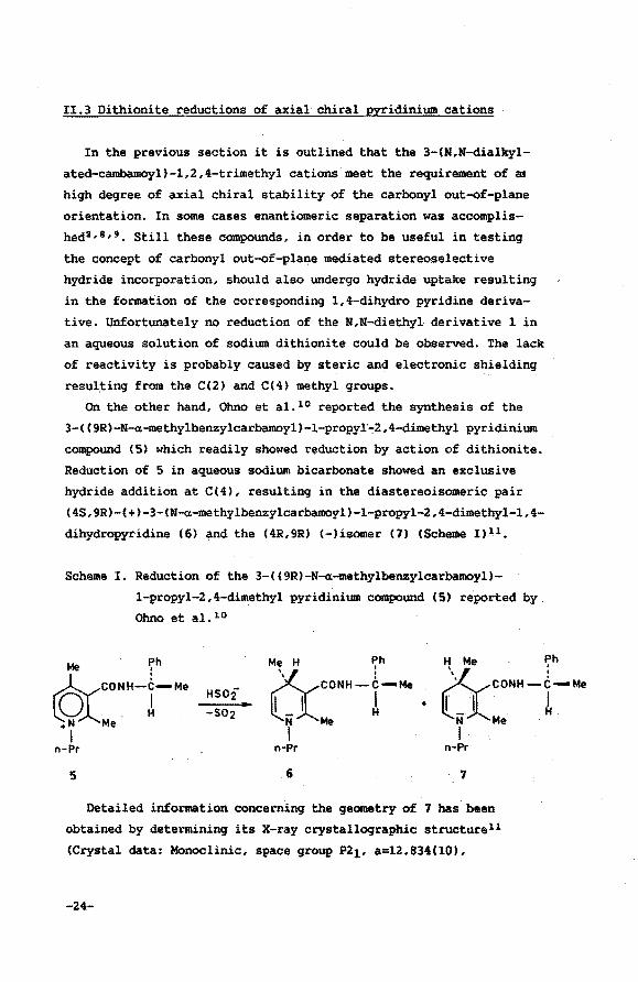
On the other hand, Ohno et al. 10 reported the synthesis of the
3-( (9R)-N-a-methylbenzylcarbamoyl)-l-propyl'-2,4-dimethyl pyridinium
compound (5) which readily showed reduction by action of dithionite.
Reduction of 5 in aqueous sodium bicarbonate showed an exclusive
hydride addition at C(4), resulting in the diastereoisomeric pair
(4S,9R)-(+)-3-(N-a-methylbenzylcarbamoyl)-l-propyl-2,4-dimethyl-1,4-
dihydropyridine (6) and the (4R,9R) (-)isomer (7) <Scheme 1) 1 1,
Scheme I. Reduction of the 3-((9R)-N-a-methylbenzylcarbamoyl)
l-propyl-2,4-dimethyl pyridinium compound (5) reported by
Ohno et al. 10
Me ~h
©rCONH-r-·• +N Me I
n-Pr
5
HSOi
-S02
Me H Ph
~co••--I-Mo N Me I
n-Pr
6
H Me Ph
• )(__co••--t-"' llti~Me
I n-Pr
7
Detailed information concerning the geometry of 7 has been
obtained by determining its X-ray crystallographic structurell
(Crystal data: Monoclinic, space group P2l, a=l2.834(10),
-24-
b=5.108(6), c=l3.166(ll)A, ~=92.98(8) 0 , Dm=l.lO g cm-3, Dc=l.l4 g
cm-3, Z=2). It appeared that the planes of the dihydropyridine ring
and of the carbonyl group, located at the B side of the dihydropyri
dine moiety (p), are inclined at an angle of 65°. This observation,
in combination with the 4R chirality inevitably leads to the conclu
sion that the proton at C(4) is syn-orientated with respect to the
carbonyl group. Since neither compound 5 nor 6 and 7 meet the crite
rion of sufficient axial chiral stability of the carbonyl out-of
plane orientation (verified using the procedure outlined in section
II.2), the sgn orientation observed can apparantly be introduced
during crystallization. ~e out-of-plane orientation results from
intermolecular hydrogen bonds in which the CONH group is likely to
be involved (a ~ bond of 2.96A represents the shortest inter
molecular contact). Since interconversion of the carbonyl out-of
plane orientation in the dihydro compound is virtually free, the
system is able to adopt the sterically most preferable geometry of
both possible diastereoisomeric CO orientations. These considera
tions emphasize the importance of unambiguous axial chiral stability
of the carbonyl out-of-plane orientation.
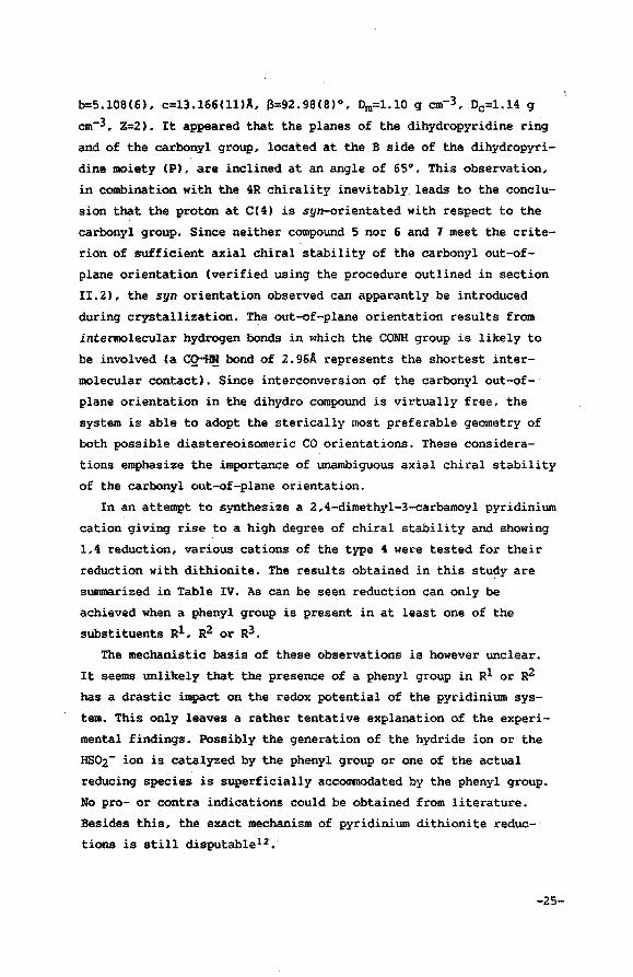
In an attempt to synthesize a 2,4-dimethyl-3-carbamoyl pyridinium
cation giving rise to a high degree of chiral stability and showing
1,4 reduction, various cations of the type 4 were tested for their
reduction with dithioriite. The results obtained in this study are
summarized in Table IV. As can be seen reduction can only be
achieved when a phenyl group is present in at least one of the
substituents Rl, ~ or R3.
The mechanistic basis of these observations is however unclear.
It seems unlikely that the presence of a phenyl group in Rl or ~
has a drastic impact on the redox potential of the pyridinium sys
tem. This only leaves a rather tentative explanation of the experi
mental findings. Possibly the generation of the hydride ion or the
HS02- ion is catalyzed by the phenyl group or one of the actual
reducing species is superficially accommodated by the phenyl group.
No pro- or contra indications could be obtained from literature.
Besides this, the exact mechanism of pyridinium dithionite reduc
tions is still disputablel2,
-25-
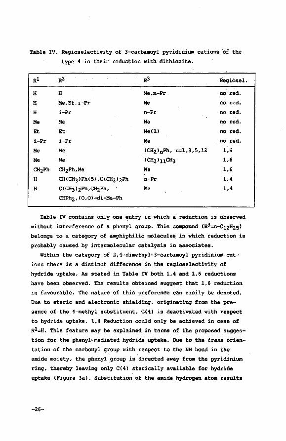
Table IV. Regioselectivity of 3-carbamoyl pyridinium cations of the
type 4 in their reduction with dithionite.
Rl ~ R3 Regiosel.
H H Me,n-Pr no red.
H Me,Et,i-Pr Me no red.
H i-Pr n-Pr no red.
Me Me Me no red.
Et Et Me(l) no red.
i-Pr i-Pr Me no red.
Me Me <CHa>nPh, n=l,3,5,12 1,6
Me Me <CH2>11CH3 1,6
CH2Ph CH2Ph,Me Me 1,6
H CH(CH3)Ph(S),C(CH3)2Ph n-Pr 1,4
H C(CH3)2Ph,CH2Ph, Me 1,4
CHPh2,(0,0)-di~e-Ph
Table IV contains only one entry in which a reduction is observed
without interference of a phenyl group. This compound (Rl=n-c12H2s>
belongs to a category of amphiphilic molecule$ in which reduction is
probably caused by intermolecular catalysis in associates.
Within the category of 2,4-dimethyl-3-carbamoyl pyridinium cat
ions there is a distinct difference in the regiose1ectivity of
hydride uptake. As stated in Table IV both 1,4 and 1,6 reductions
have been observed. The results obtained suggest that 1,6 reduction
is favourable. The nature of this preference can easily be denoted.
Due to steric and electronic shielding, originating from the pre
sence of the 4-methyl substituent, C(4) is deactivated with respect
to hydride uptake. 1,4 Reduction could only be achieved in case of
R1=H. This feature may be explained in terms of the proposed . sugges
tion for the phenyl-mediated hydride uptake. Due to the trans orien-. .
tation of the carbonyl group with respect to the NH bond in the
amide moiety, the phenyl group is directed away from the pyridinium
ring, thereby leaving only CC4) sterically available for hydride
uptake (Figure 3a). Substitution of the amide hydrogen atom results
-26-
in the loss of conformational restrictions of the amide moiety.
Therefore, occasionally, both C(6) and C(4) are located within the
vicinity of the phenyl group (Figure 3b). Since C(6) is less deacti
vated, hydride addition will take place preferably at this position.
0 s Me/ :~c fpH
R~~·r · · \ tu._~ Me H--e
I v '\ H H
a b
Figure 3. The phenyl group is directed away from C(6) due to the
trans orientation of the CONH group (a); when the amide
proton is substituted, C(6) becomes in the vicinity of the
phenyl group (b).
In summary it can be concluded that introduction of a phenyl
group in the amide moiety can only lead to 1,4 reduction when the
amide group is monosubstituted (presence of CONH), although a high
degree of chiral stability requires a dialkylated amide group (Sec
tion II.2). The above mentioned results inevitably lead to the con
clusion that introduction of the phenyl group in one of the substi
tuents Rl, R2 or R3 in 4 will not combine adequate chiral stability
and 1,4 reducibility within one pyridinium compound.
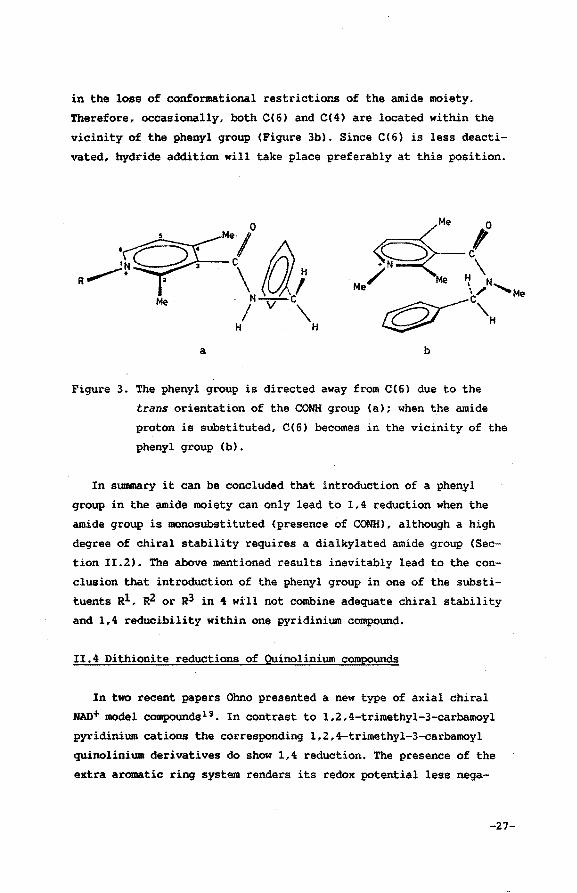
II.4 Dithionite reductions of Quinolinium compounds
In two recent papers Ohno presented a new type of axial chiral
MAD+ model compounds13 • In contrast to 1,2,4-trimethyl-3-carbamoyl
pyridinium cations the corresponding 1,2,4-trimethyl-3-carbamoyl
quinolinium derivatives do show 1,4 reduction. The presence of the
extra aromatic ring system renders its redox potential less nega-
-27-
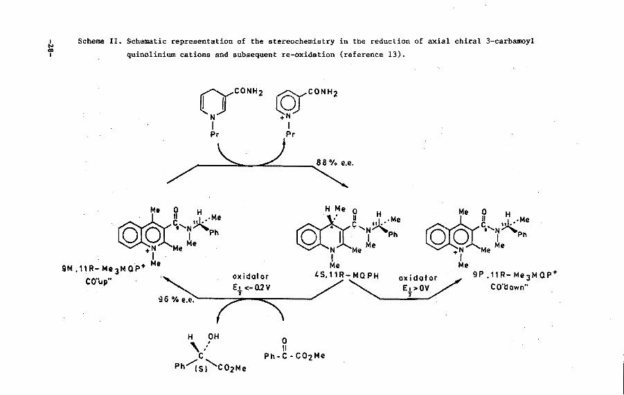
~ Scheme II. Schematic representation of the stereochemistry in the reduction of axial chiral 3-carbamoyl
f quinolinium cations and subsequent re-oxidation (reference 13).
(YCONH 2
N I Pr
Me ~
~~•'N~Me ~y.-( I Ph
,..N Me Me
9M,11R-Me 3MoP•
CO"up"
I Me
H OH , ... c
Ph~si'co2Me
~CONH2
,..N I Pr
H Me o H roc. ' II uJ •• ·Me c~ . '~ Ph
N Me Me
I OO
Me ~ H
c,,N~Me I Ph
,..N Me Me I . Me Me
4S,11R-MOPH oxidotor 9P.11R-Me 3 MOP+
~ CO'l:fown''
0 II
Ph-C-C02Me
I N \0 I
PNAH PNA
"-____) /--
9P,11R- Me 3 MaP+
CO'tlown"
HO H ':' c Ph/(Rj'co2Me
4R, 11 R - M Q PH
0 II
Ph-C-C02Me
oxidator Et >OV 'i
9M,11R -Me 3MaP+
CO"up"
tive14 and at the same time it prohibits incorporation· of the formal
hydride ion at the C(6) position. Ohno in his experiments uses a
1,2,4-trimethyl-3-carbamoyl quinolinium cation which bears a chiral
~-methylbenzyl substituent in the amide group. The presence of the
phenyl group in the ~-methylbenzyl substituent is not decisive for
the reducibility of these compounds, this in contrast to what is
observed for the reduction of the corresponding pyridinium deriva
tives <Section II.3). The 3-(N,N-dimethylcarbamoyl)-1,2,4-trimethyl
quinolinium iodide has been syntbesized·and subsequently reduced
with dithionite to yield its 1,4-dihydro derivative. The stereoche
mistry of this reaction could not be elucidated since enantiomeric
resolution effectuated by fractional crystallization of the diaste
reoisomeric (+)-«-bromoeamphor-~-sulphonate (Sections II.l and II.5)
could not be accomplished. Absence of chiral stability seems unli
kely since this compound is very similar to its pyridinium analogue
which readily could be separated into its enantiomers using this
technique. It should be noted in this context that in la NMR experi
ments Eu!hfcl3-induced enantiotopic resolution was observed. This
indicates that racemization is at least slow on NMR time scale.
Ohno, on the other hand, was able to separate both axial chiral
isomers chromatographically. Combination of axial chirality and
configurational chirality in the amide substituent renders both
isomers to be diastereoisomers. A schematic representation of the
dithionite reduction and subsequent re-oxidation of both quinolinium
diastereoisomers is given in Scheme II.
This study clearly shows that hydride incorporation in axial
chiral 3-carbamoyl quinolinium systems takes place syn relative to
the CO dipole. Loss of axial chiral stability occurs in the final
stage of the reaction. The CO out-of-plane orientation in the resul
ting 1,4-dihydroquinoline derivative is not stable at all. The net
result is a transfe~ of the chiral information from the carbonyl
dipole to configurational chirality of C(4) in the 1,4-dihydro deri
vative.
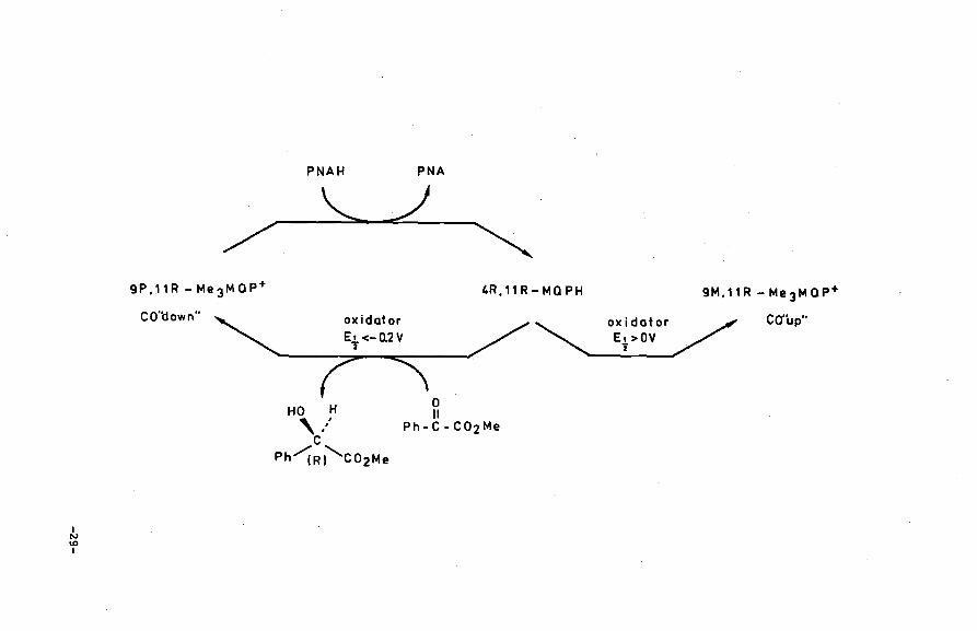
The reversed process, i.e. oxidation of the dihydroquinoline
systems obeys the same rule in case moderate oxidizing agents are
used. During the oxidation process the carbonyl dipole, oriented
-30-
towards the substrate, is being gradually fixed in a stable out-of
plane orientation. The use of very strong oxidizing agents <E%>0 V),
on the contrary, induces a reversed stereoselectivity with regard to
the carbonyl out-of-plane orientation. It should be noted however
that the mechanistic basis of the stereochemistry in this type of
reaction of highly reactive quinones (p-chloranil and DDQ) is no
longer controlled by electrostatic attraction forces between the
carbonyl group of the amide moiety and the slightly positively
charged regions of the substrate. The four partly negatively charged
quinone substituents can, in these cases, easily overrule the
carbonyl-substrate interaction forces. A more detailed mechanistic
interpretation of these results will be offered in Section III.7.
II.S Experimental section
Spectroscopy
60 MHz lH NMR spectra were recorded on a Varian EM360A or a
Hitachi Perkin Elmer R-24B NMR spectrometer, 200 MHz lH NMR spectra
on a Bruker AC200 spectrometer and 300 MHz lH NMR data were obtained
on a Bruker CXP 300 spectrometer. Me4Si was used as internal stan
dard<& 0.00). The CD spectral data were gained from a Jobin Yvon
Dichrograph Mark III-S and specific rotations from a polarimeter
type AA-10 manufactured by Optical Activity Ltd. A Fisher-Johns
apparatus was employed to determine the, (uncorrected) melting points.
Synthesis
Ethyl-2,4-dimethyl-1,4-dihydronicotinate15 , ethyl-2,4-dimethylni
cotinate15 and the 2,4-dimethyl-3-carbamoyl pyridine hydrochlo
ride3•11 were prepared using literature procedures. The successive
reaction steps are: conversion into the acid-chloride using SOC12,
and then to the amide using the appropriate amine, already outlined
in detail in previous literature3 • 10 • An example of the direct con
version of the ethyl-2,4-dimethylnicotinate into the corresponding
amide is also presented in the experimental section.
-31-
Prior to reduction with sodium dithionite, the 3-carbamoyl pyri~
dine derivatives were treated with an alkylhalogenide in order to
obtain the corresponding. cations. General procedures will be
mentioned.
In case synthesis of compounds is in accordance with (slighly ~
difiedl literature procedures, the discussion in the experimental
section confines itselves to listing the relevant spectroscopical
parameters.
All solvents and materials used during synthesis were reagent
grade and were used as received or purified as required.
2,4-dimethyl.,-3-carbamoyl pyridine compounds
Direct amination16 of ethyl 2,4-dimethyl nicotinate, synthesis of
3-(N,N-dimethylcarbamoyl)-2,4-dimethyl pyridine (2)
To an ice cold mixture of dimethylamine· (1. 5 ml, 1. 0 g, 22.2 mmol)
in dry THF (10 ml), a (15%) solution of BuLi in hexane (9.3 ml, 15.8
mmoll was added dropwise. After one hour at 0°C, the ester (1.00 g,
5.58 mmoll in dry THF (15 mll was introduced. During a period of four
hours the mixture was allowed to come to room temperature. It was sub
sequently poured into water and acidified with 4N aqueous hydrochloric
acid. The organic solvents were evaporated and the resulting aqueous
solution was scrubbed with ether .• After basification, the mixture was
extracted with dichloromethane. Drying of the organic layers, filtra
tion and evaporation of the solvent yielded the corresponding amide
(0.9 g, 5.05 mmol, 91%).
Mp 50-51°C. lH NMR CCDCl3): S 2.23(s,3H,CHJ), 2.44(s,3H,CH3), 2.81
(s,3H,NCH3), 3.15(s,3H,NCHJ), 6.9l(d,lH,pyrH), 8.28(d, lH, pyrH). The
diastereoisomers, obtained after complexation of the hydrochloride of
2 with ( + l or (-) -«-bromocamphor-11'-sulphonate monohydrate could be
separately isolate~ after repeated treatment with acetone. Mp
198.0-199.0°C. Anal. calcd.: C,49.08; H,5.97; N,5.72. Found (two
diastereoisomers resp.l: C,48.93, 49.38; H,6.00, 6.02; N,5.88, 5.51.
[«]D21.0:+55.00, -55.10 CH20l. Treatment with NHJIHzO yielded the (+)
and(-) enantiomers: Mp 52.0-54.0°C. Anal. Calcd.: C,67.38; H,7.92;
N,15.72. Found: C,67.14, 67.32; H,7.93, 7.89; N,l5.95, 16.19.
-32-
[a.] 021.0=+1. 80,-1. 72 (H20l. An alternative route. for the enantiomeric
separation showed corresponding analytical data4 •
3-(11,11-diethylcarbamoyU -:a, 4-dilllethyl pyridine
lH NMR (CDCl3l: & 1.05(t,3H,CH3l• 1.27(t,3H,CH3), 2.25(s,3H,CH3),
2.47(s,3H,CH3), 3.10(q,2H,CH2l• 3.62(q,2H,CH2l• 6.93(d, lH,pyrH),
8.30(d,lH,pyrH). Enantiomeric separation can be obtained using the
procedure as is outlined for 2.
Compounds of Table II
3-(ll,ll-dilllethylthiocarbamoyl)-2,4-dimethyl pyridine
To a solution of 2 (3.7 g, 21 mmol) in pyridine (90 ml), pulverized
P2S5 (14 g, 63 mmol) was added. The reaction mixture was kept at ll5°C
during a period of 24 h. After removal of the solvent in vacu, water
was added (50 ml). The resulting mixture was basified (pH 8) with
MaHC03. Subsequent extraction with dichloromethane and drying of the
organic fractions yielded after evaporation of the solv~nt the thio
amide derivative (1.5 g, 37%).
lH NMR (CDCl3l: & 2.23(s,3H,C(4)CH3), 2.44(s,3H,C(2lCH3l• 3.00(s,3H,
MCH3l. 3.60(s,3H,MCH3l• 6.93(d,lH,pyrH), 8.23(d,lH,pyrHl.
3-(11,11-diisopropylcarbamoyl)-2,4-dimethyl pyridine
lH NMR (CDCl3l: & 1.13(d,6H,2CH3l, 1.58(d,6H,2CH3), 2.25(s,3H,CH3),
2.47(s,3H,CH3), 3.52(m,2H,2CH), 6.87(d,lH,pyrHl, 8.18(d,lH,pyrH).
3-(11-methyl~(a.-methylbenzyl)carbamoyl)-2,4-dimethyl pyridine
Synthesis was outlined by Bastiaansen et al. 8 The relevant spectro
scopic data were in good agreement with those described.
3-CJI--benzylcarbamoyl)-2,4-dimethyl pyridine
lH NMR (CDCl3l: & 2.25(s,3H,CH3), 2.38(s,3H,CH3), 4.53(d,2H,CH2),
6.52(br s,lH,NH), 6.78(d,lH,pyrH), 7.23(s,5H,Ph), 8.08(d,lH,pyrH).
3-UIJ-(a;-methylbenzyl)carbamoylh~.t-dimethyl pyridine
Synthesis and spectroscopic data according to those reported by
Ohno et al. 10
-33-
!-SUbstituted 2,4-dimethyl~3-carbamoyl pyridinium compounds
Methylation of the 2,4-dimethyl-3-carbamoyl pyridine compounds.
The pyridine derivatives of 2, dissolved in an excess of CH3I were
stirred until the reaction was complete (TLC). The remainde~ of the
CH3I was evaporated. The residue was repeatedly treated with dry ether
in order to obtain a crystalline deposite.
Alkylation of the 2,4-dimethyl-3-carbamoyl pyridine compounds with
n-propyl bromide.
The alkylation, using n-PrBr, was carried out according to the
literature10 •
Alkylation with w-phenylalkyl bromide.
To a solution of the 3-carbamoyl pyridine derivative (5 mmol) in
nitromethane (5-10 ml), an excess of the bromide was added (4-7 eq:). ·
This mixture was stirred for 20 hrs. at an appropriate temperature
CPhCHzBr and PhCCHzl3Br: room temp.; Ph(CHz>sBr and PhCCH2>1zBr:
lOO"C). Work-up was performed as mentioned in the methylation reac
tion. The PhCCH2JsBr and the Ph(CHz>lzBr were prepared analogous to
the procedure mentioned by Friedman et a1.11
PhCCHzl5Br : Bp 90-105"C/1.5mm. la NMR CCDCl3): & 1.20 to 2.10(m,6H,
3CHz), 2.57(t,2H,CH2>, 3.30(t,2H,CHz), 7.10(s,5H,Ph).
Ph(CHz)lzBr: Bp 165-170"C/O.Olmm. la.NMR CCDCl3): & 1.10 to 2.00(m,
20H,l0CHz), 2.57(t,2H,CH2>, 3.32(t,2H,CHz), 7.08(s,5H,Ph).
Alkylation with dodecyl bromide.
A mixture of the 3-carbamoyl pyridine compound (22 mmol) and dodecyl
bromide (100 mmol, 25 g) was heated for two days at lOO"C in absence
of a solvent. The separated solid material was collected and treated
with dry ether.
3-(N,N-diethylcar~l)-1,2,4-trimethyl pyridinium iodide(l)
Mp 177-178"C. 1H NMR CCDCl3l: & 1.00(t,3H,CH3), 1.45(t,3H,CH3), 2.50
(s,3H,CH3), 2.78(s,3H,CH3), 3.35(q,2H,CHz), 3.60(q,2H,CH2), 4.47(s,3H,
CH3l, 7.78(d,lH,pyrH), 913(d,lH,pyrH). Anal. Calcd. C,44.84; H,6.08;
N,8.04. Found C,44.58; H,5.89; N,S.OO. Separation of the enantiomers
was effectuated by complexation with Ag-.(+)-«-bromocamphor-11'-s\lli>ho--
-34-
nate monohydrate (BKS). The diastereoisomers could be separated after
repeated crystallizations from acetone. Treatment of both diastereo
isomers with Dowex-2 (Cl- form) yielded the pure enantiomers (Cl
form).
(-): Mp 132.0-134.0°C. Anal. Calcd. <monohydrate): C,56.82; H,8.44;
N,l0.19. Found: C,56.64; H,8.65; N,l0.26. [a] 019.7:-5.62 (c=l.OO in
water); lH NMR CCDCl3l: o 0.90(t,3H,CH3), 1.43(t,3H,CH3), 2.48(s,3H,
CH3l, 2.78(s,3H,CH3), 3.23(g,2H,CH2l, 3.57(g,2H,CH2l, 4.52(s,3H, CH3),
7.82(d,lH,pyrH), 9.36(d,lH,pyrH).
(+): Mp 130.0-132.0°C. Anal. Found: C,56.78; H8.60; N,l0.42.
[a]019.3=4.74 Cc=l.OO in water).
3-((9R)-R-m-metbylbenzylcarbamoyl)-l-propyl-2,4-dimethyl pyridinium
braai.de (5)
Synthesis was outlined in the work of Ohno et a1.10 The relevant
spectoscopic data. were in excellent agreement with those described.
Compounds of category 4 (Table III and Table IV)
Ethyl 1,2,f-trimetbyl-3-carboxylate pyridinium iodide
lH NMR CCD3CNl: o 1.39(t,3H,CH3), 2.55(s,3H,CH)l, 2.68(s,3H,CH)l.
4.18(s,3H,CH3), 4.50(g,2H,CH2), 7.26(d,lH,pyrH), 7.78(d,lH,pyrH).
1,2,4-trimetbyl-3-carballloyl pyridinium iodide 1H NMR <DzOl: o 2.50(s,3H,CH3), 2.67~s,3H,CHJl, 4.10Cs,3H,CH3),
7.57(d,lH,pyrH), 8.40(d,lH,pyrH).
1-o-Propyl-2,4-dimethyl-3-carbamoyl pyridinium iodide
Mp 233-234°C. Anal Calcd.: C,48.36; H,6.27; N,l0.26. Found:
C,48.37; H,6.29; N,l0.53. la NMR CD20l: & 1.08(t,3H,CH3l,
2.03(m,2H,CH2), 2.65(s,3H,CH3), 2.87(s,3H,CHJ), 4.53(t,2H,CH2),
7. 72(d, lH,pyrH), 8. 58(d,lH,pyrH).
3-C--.etbylcarbamoyl)-1,2,4-trimethyl pyridinium iodide 1H NMR CD20l: & 2.62(s,3H,CH3), 2.80(s,3H,CH3), 3.07(s,3H,CH3);
4.27(s,3H,CHJ}, 7. 75(d,lH,pyrH), 8. 60(d,lH,pyrH).
-35-
3-(N-etbylcarbaaoyl)-1,2,4-trimethyl pyridiniua iodide
lH NMR (CDCl3l: & 1.25(t,3H,CH3), 2.58(s,3H,CH3), 2.83(s,3H,CH3),
3.40Cq,2H,CH:z), 4.23(s,3H,CH3), 7.52Cd,lH,pyrH), 8.47(d,lH,pyrH)
8.88(br t,lH,NH).
3-(N-isopropylcarbamoyl)-1,2,4-trimethyl pyridinium iodide
Mp 233-235°C. lH NMR CCDCl3l: & 1.36(d,6H,2CH3), 2.53(s,3H,CH3),
2.76(s,3H,CH3), 4.26(m,lH,CHMe:z), 4.26(s,3H,NCH3), 7.63(d,lH,pyrH),
8.00(d,1H,NH), 8.95(d,lH,pyrH).
3-(N-isopropylcarbamoyl)-1-n-propyl-2,4-dimetbyl pyridinium bromide
lH NMR CCDC13l: & 1.07(t,3H,CH3), l.32(d,6H,2CH3), 2.03Cm,2H,CH2l,
2.53Cs,3H,CH3), 2.80Cs,3H,CH3), 4.22(m,1H,CHMez), 4.55(t,2H,CH:zl,
7.67(d,lH,pyrH), 8.30(d,lH,NH), 9.00(d,lH,pyrH).
3-(N ,N-dimethylcarbamoyl)-1 ,2 ,4-trimethyl pyridinium iodide
Mp 255-256°C (ethanol). Enantiomeric separation with BKS. Anal.
Calcd.: C,41.26; H,5.35; N,8.75. Found(-): C,41.34~ 8,5.35~ N,8.75.
(+): C,41.35; 8,5.36; N,8.59. [«]o:Z0=-1.99 and +1.95 resp. CH:zOl. lH
NMR CD:zOl: & 2.60(s,3H,CH3), 2.78(s,3H,CH3)., 3.05(s,3H~CH3), 3.28
(s,38,CH3), 4.30(s,38,CH3l, 7.73(d,l8,pyrH), 8.70Cd,lH,pyrH).
3-(N,N-diisopropylcarbamoyU-1,2,4-trimetby1 pyridinium iodide
18 NMR to20l: S 1.32(d,6H,2CH3L 1.65(d,GH,2CH3), 2.63"(s,3H,CH3),
2. 78(s,3H,CH3), 3.82(m,2H,2CH), 4.27(s,3H,CH3l, 7 .82(d,lH,pyrH),.
8.62(d,18,pyrH).
3-(N,N-dimetbylcarbamoyl)..:.l-benzy1-2 ,4-dimetbyl pyridinium bromide
Mp 189.4-l90.0°C. 1H NMR <DiO>: S 2.47(s,3H,CH3), 2.57(s,3H,CH3),
2. 87 (s,38,CH3), 3 .10(s,3H,CH3), 5. 73 (s,2H,CH:zL 7. 30(m, SH,Ph),
7.80(d,lH,pyr8), 8.~3(d,18,pyr8).
3-(N,N-dimetbylcarbamoy1)-1-(3-phenylpropyll:-2,4-dimetbyl pyridiniua
bromide 1 . . .··· 8 NMR (D20l: & 2.13(t,28,CH:zl, 2.40(s,3H,CH3), 2.50(s,3H,CH3),
2.73(t,28,CH:zPh), 2.83(s,3H,NCH3), 3.13(s,3H,NCH3), 4.43(t,2H,NCH:z),
7.13(m,58,Ph), 7.60(d,l8,pyr8), 8.43(d,lH,pyrH) •
.:;.36-
3-UT,IT-diJiethylcarbamoyl)-1-(5-phenylpentyl)-2,4-dimethyl pyridinium
bromde.
lH NMR (CDCl3): & 1.67(m,6H,3CH2), 2.50(s,3H,CHJ), 2.80(s,3H,CH3),
2.97(s,3H,NCH3), 3.20(s,3H,NCH3), 3.30(t,2H,CH2Ph), 4.73(t,2H,CH2N),
7.76(d,lH,pyrH), 9.17(d,lH,pyrH).
3-(N,R-diaethylcarbamo71)-l-(12-phenyldodecyl)-2.4-dimethyl pyridinium
brc.ide
lH NMR CCDCl3): & 1.27(m,20H,l0CHz), 2.47Cs,3H,CH3), 2.57(t,2H,
CH2Ph), 2.80(s,3H,CH3), 2.97(s,3H,CH3N). 3.13(s,3H,CH3N), 4.74(t,2H,
CH2N), 7.10(m,5H,Ph), 7.74(d,lH,pyrH), 9.13(d,lH,pyrH).
3-(N,IT-diJiethylcarbamo7U-1-dodecyl-2 ,4-dimethyl pyridinium brOIIlide
lH NMR CCDCl3): & 0.90(t,3H,CHJ), 1.25(m,20H,l0CH2), 2.50(s,3H,
CH3), 2.77(s,3H,CHJ), 3.00(s,3H,CHJ), 3.17(s,3H,CH3), 4.73(t,2H,NCH2),
7.80(d,lH,pyrH), 9.06(d,lH,pyrH).
3-(N,~lcarbamoyl)-1,2,4-trimethyl pyridinium iodide
Mp 153.5-155.5°C. lH NMR (020): & 2.3l(s,3H,CHJ), 2.39(s,3H,CHJ),
4.05(s,3H,CHJN), 4.33(m,2H,CH2), 4.87(m,2H,CHz), 6.77 to 7.49(m,l0H,
2Ph), 7.70(d,lH,pyrH), 8.57(d,lH,pyrH).
3-(N-benzyl-N-methylcarbamoyl)-1,2,4-trimethyl pyridinium iodide
Mp 184-185°C. Anal. Calcd.: C,51.52; H,5.34; N,7.07. Found:
C,51.28; H,5.39; N,6.95. For detailed lH NMR spectroscopic and X-ray
diffraction data of the rotamers: See Bastiaansen et al. 8
3-(11-a.,a.-dimethy lbenzylcarbamoyl) -1-n-propyl-2, 4-dimethyl pyridinium
iodide
Mp 220-222°C. Anal. Calcd.: C,54.80; H,6.21;. 11,6.39. Found:
C,54.60; H,6.17; 11,6.24. la NMR CCDCl3): & l.lO(m,SH,Et), 1.84(s,6H,
CCCH3)2), 2. 54(s,3H,CH3), 2. 73 (s,3H,CH3), 4. 43Ct ,2H,NCHzL 7. 40(m,
SH,Ph), 7.63(d,lH,pyrH), 8.73(d,lH,pyrH), 8.93(s,lH,NH).
3-(11-a.,a..-dimethylbenzylcarbamoyl )-1 ,2 ,4-trimethyl pyridinium iodide
Mp 233-235°C. Anal. Calcd.: C,52.69; H,5.65; N,6.83. Found:
-37-
C,52.54; H,5.11; N,7.15. lH NMR (DMSO-d6): & l.70{s,6H,C(CH3)2),
2.47(s,3H,CH3), 2.67(s,3H,CH~), 4.23(s,3H,CH]), 7.37(m,5H,Ph),
7.87(d,lH,pyrH), 8.87(d,lH,pyrH), 8.97(s,lH,NHl.
3-()J-benzy1carbamoy1 )-1,2 .4-trimethy1 pyridi.n:ium iodide
Mp 196-198°C. lu NMR (D20l: & 2.30(s,3H,CH]l, 2.43(s,3H,CH3),
3.87(s,3H,CH3), 4.22(s,2H,CH2), 6.84(m,SH,Ph), 7.13(d,lH,pyrH),
7.9l(d,lH,pyrH).
. .
3-(N-benzhydry1carbamoyU -1,2 ,4-trimethy1 pyridinium iOdide
lH NMR {CDCl]l: & 2.33(s,3H,CH]l, 2.50(s,3H,CH3), 4.03(s,3H,CH]l,
6. 43 (d, lH,CH), 7.08-7.38 (m,10H,2Ph), 7. 45(d, lH,pyrH), 8. 70(d,lH,pyrH),
8.95(d,lH,NH).
3-(N-(0,0)-dimethy1fenylcarbamoy1)-1,2,4-trimethy1 pyridinium iodide
Mp 218-219°C. Anal. calcd.: C,51.53; H,5 .. 34; N,7.07. Found:
C,S0.66; H,5.23; N,7.06. lH NMR (CD3CNl: & 2.39(s,6H,2CH3), 2.7l(s,3H,
CH3l, 2.86(s,3H,CH]l, 4.15(s,3H,CH3), 7.17(s,3H,Ph), 7.74(d,lH,pyrH),
8.53{d,lH,pyrH), 8.62(br s,lH,NH).
3-(~-.ethy1benzyl-N-methy1carbamoyl)-1,2,4-trimethyl pyridinium
iodide
Synthesis according to the procedure reported in literature 8.
3-(~-methylbenzyl-N-.ethy1carbamoyl )-2, 4-dimethyl pyridine-1£1
Synthesis according to the procedure reported in literature9 •
Reductions with sodium dithionite. General procedurelO
To an lN aqueous sodium bicarbonate (120 ml, saturated with Ar)
solution of the pyridinium compound (1.2 mmolL CH2Cl2 (120 ml, satu
rated with Arl was added. Sodium dithionite (10 Eq) was added in por
tions to the stirred mixture. The agitation was continued for 3 to 4
hours in an Ar-flushed flask, excluded from light. The dichloromethane
layer was separated, dried and evaporated, resulting in the crude
dihydro compound.
-38-
Dihydro derivatives of the compounds of category 4 (Table IV)
3-(N,N-dimethy1carbamoy1)-1-benzy1-2,4-dimethy1-1,6-dihydropyridine
lH NMR (CDCl3): & 1.57(m,3H,CH3), 1.80(s,3H,CH3), 2.93(s,3H,CH3N),
2.98(s,3H,CH3), 3.74(dd,2H,C(6)H2), 4.14(s,2H,CH2N), 4.70(m,1H,H(5)),
7.17(m,5H,Ph).
3-(N,N-dimethy1carbamoy1)-1-(3-pheny1propy1)-2,4-dimethy1-1,6-dihydro
pyridine
1H NMR CCDCl3): & 1.28 to 3.25(m,6H,3CH2), 1.48(m,3H,CH3), 1.63(s,3H
,CH3), 2.82(s,3H,CH3), 2.83(s,3H,CH3), 3.64(dd,2H,C(6)H2), 4.57(m,1H,
H(5)), 7.00(m,SH,Ph).
3-(N,N-dimethy1carbamoy1)-1-(5-phenylpenty1)-2,4-dimethyl-1,6-dihydro
pyridine
1H NMR CCDCl3): & 1.20 to 3.12(m,10H,SCH2), 1.50(m,3H,CH3), 1.72(s,
3H,CH3), 2.89(2s,6H,2CH3), 3.71(dd,2H,C(6)H2), 4.66(m,1H,H(5)), 7.04(m,
SH,Ph).
3-(N,N-dimethy1carbamoy1)-1-(12-pheny1dodecy1)-2,4-dimethy1-1,6-dihy
dropyridine
1H NMR CCDCl3): & 1.20 to 3.12(m,24H,12CH2), 1.59(m,3H,CH3), 1.83(s,
3H,CH3), 3.00(2s,6H,2CH3), 3.77(dd,2H,C(6)H2), 4.69(m,1H,H(5)), 7.09(m,
SH,Ph).
3-(N,N-dimethy1carbamoy1)-1-dodecy1-2,4-dimethy1-1,6-dihydropyridine
1H NMR CCDCl3): & 0.8l(t,3H,CH3), 1.20(m,20H,10CH2), 1.53(m,3H,
CH3), 1.76(s,3H,CH3), 2.9l(s,6H,2CH3N), 3.30(t,2H,CH2N), 3.74(dd,2H,
C(6)H2), 4.70(m,1H,H(5)).
3-(N,N-dibenzy1carbamoy1)-1,2,4-trimethy1-1,6-dihydropyridine 1H NMR CCDCl3): & 1.63(s,3H,CH3), 1.81(s,3H,CH3), 2.67(s,3H,CH3N),
3.67(dd,2H,C(6)H2), 4.20 to 4.90(2d,4H,2CH2), 4.63(m,1H,C(5)H), 6.77
to 7.49(m,l0H,2Ph).
-39-
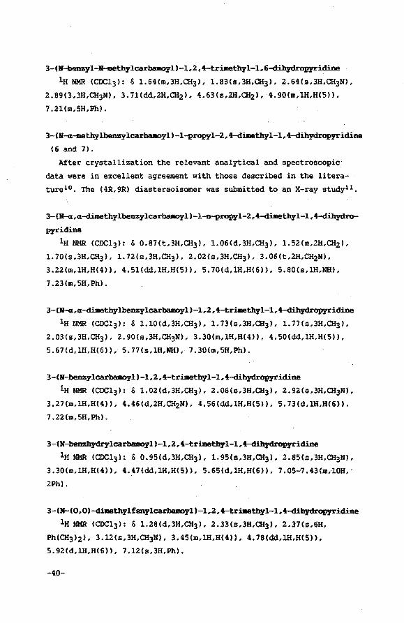
3-(N-beDzyl-11-methylcarbamoyl )-l,l,t-trilllethyl-1,6-dihydrop:yridine
lH NMR (CDCl3l: & 1.64Cm,3H,CH]l, 1.83(s,3H,CH]l, 2.64(s,3H,CH]Ml,
2. 89(3 ,3H,CH3N>, 3. 7l(dd,2H,CH2l, 4.63(s,2H,CHzl, 4. 90(m,1H,H(5)),
7 .ll(m,5H,Ph).
3-(N-«-methylbenzylcarbamoyl)-1-propyl-2,4-dimethyl-1,4-dihydropyridine
(6 and 7).
After crystallization the relevant analytical and spectroscopic
data were in excellent agreement with those described in the litera
turelo'. The C4R,9R) diastereoisomer was submitted to an X-ray studyll,
3-(~,u-dimethylbenzylcarbamoyl)-1-n-propyl-2,4-dilllethyl-1,4-dihydro
pyridine
lH NMR (CDC13>: S 0.87(t,3H,CH3), 1.06(d,3H,CH3L 1.52(m,2H,CHzl,
1.70(s,3H,CH]), 1.72(s,3H.CH]), 2.02(s,3H,CH]), 3.06(t,2H,CHzNI.
3.22(m,lH,H(4)), 4.5l(dd,lH,H(5)), 5.70(d,iH,H(6)), 5.80(s,lH,NH),
7.23(m,5H,Ph).
3-CN-u,u-dimethylbenzylcarbamoyl)-1,2,4-trimethyl-1,4-dihydropyridine
lH NMR CCDCl3l: & 1.10(d,3H,CH]l, l.73(s,3H,CH3), 1.77(s,3H,CH]),
2.03(s,3H,CH]l, 2.90(s,3H,CH]Ml, 3.30(m,lH,H(4)), 4.50Cdd,lH,H(5)),
5.67(d,lH,H(6)), 5. 77(s,lH,NH), 7 .30(m,5H,Ph).
3-(N-benzylcarbamoyl)-1,2,4-trimethyl-1,4-dibydropyridine 1H NMR CCDCl3l: S 1.02(d,3H,CH3), 2.06(s,3H,CH3), 2.92(s,3H,CH]Ml,
3.27(m,lH,H(4)), 4.46(d,2H,CH2N), 4.56(dd,lH,H(5)), 5.73(d,lH,H(6)),
7.22(m,5H,Ph).
3-(N-benzhydrylcarbamoyl)-1,2,4-trimethyl-1,4-dibydrop:yridine
lH NMR CCDCl3l: & 0.95(d,3H,CH]l, 1.95(s,3H,CH3), 2.85(s,3H,CH3N),
3. 30 (m, 1H,H(4l), 4. 47(dd, lH,HC 5)), 5. 65(d, lH,H(6)), 7.05-'7. 43(m,10H,'
2Ph).
3-(N-(0,0)-dimethylfenylcarbamoyl)-1,2,4-trimethyl-1,4-dibydrop:yridine 1H NMR CCDCl3l: & 1.28(d,3H,CH3), 2.33(s,3H,CH3), 2.37(s,6H,
PhCCH3l2l, 3.12(s,3H,CH3N), 3.45(m,lH,H(4)), 4.78(dd,lH,H(5)),
5.92(d,1H,H(6)), 7.12(s,3H,Ph).
-40-
References and notes
1. de Kok, P.M.T.; Donkersloot, M.C.A.; v. Lier, P.M.; Meulendijks,
G.H.W.M.; Bastiaansen, L.A.M.; v. Hooff, H.J.G.; Kanters, J.A.;
Buck, H.M. Tetrahedron 1986,42,941.
2. Donkersloot, M.C.A.; Buck, H.M. J. Am. Chem. Soc. 1981,103,6554.
3. v. Hooff, H.J.G.; v. Lier, P.M.; Bastiaansen, L.A.M.; Buck, H.M.
Reel. Trav. Chim. Pays-Bas 1982,101,191.
4. v. Lier, P.M.; Meulendijks, G.H.W.M.; Buck, H.M. Reel. Trav. Chim.
Pays-Bas 1983,102,337.
5. a. Jackman, L.M.; Kavanagh, T.E.; Haddon, R.C. Org. Magn. Res.
1969,1,109.
b. Sattler, H.J,; Schunack, w. Chem. Ber. 1975,108,730.
6. Schurig, w. Kontakte <Darmstadt> 1985,2,22.
1. Sweeting, L.M.; Crans, D.C.; Whitesides, G.M. J. Org. Chem.
1987,52,2273.
8. Bastiaansen, L.A.M.; Kanters, J.A.; v.d. Steen, F.H.; de Graaf,
J.A.C., Buck, H.M. J. Chem. Soc., Chem. Commun. 1986,536.
9. Bastiaansen, L.A.M.; Vermeulen, T.J.M.; Buck, H.M.; Smeets, W.J.J.;
Kanters, J.A.; Spek, A.L. J. Chem. Soc., Chem. Commun. 1988,230.
10. Ohno, A.; Ikeguchi, M.; Kimura, T.; Oka, S •. J. Am. Chem. Soc.
1979,101,7036.
11. v. Lier, P.M.; Donkersloot, M.C.A.; Koster, A.S.; v. Hooff, H.J.G.;
Buck, H.M. Reel. Trav. Chim. Pays-Bas 1982,101,119.
-41-
12. a. Wallenfels, K.; Schuly, H. Liebigs Ann. 1959,621,178.
b. Blankenhorn, G; Moore, E.G. J. Am. Chem. Soc. 1980,102,1092.
c. Louis-Andre, O; Gelbard, G. Bull. Soc. Chim. Fr. l986,t,565.
13. a. Ohno, A.; Kashiwagi, M.; Ishihara, Y.; Ushida, S.; Oka, S.
Tetrahedron 1986,42,961.
b. Olmo, A.; Ohara, M.; Oka, S. J. Am. Chem. Soc. 1986,108,6438.
14. Reduction potentials of quinolinium cations are less negative than
those reported for the pyridinium analogues. E.g. E0 of 1-benzyl-3-
carbamoyl guino1inium bromide amounts -158 mV whereas the value
obtained for the corresponding pyridinium cation is -361 mV.
Ostovic, D.; Han Lee, I.-S.; Roberts, R.M.G.; Kreevoy, M.M. J. Org.
Chem. 1985,50,4206.
15. a. Hantzsch, A. Ber. 1890,23,1474.
b. Tsuda, K.; Sato, Y.; Itekawa, N.; Mishima, H. J. Org. Chem.
1956,21,800.
16. Procedure adopted from: Yang, K.-W.; Cannon, J.G.; Rose, J.G.
Tetrahedron Lett. 1970,21,1791.
17. Friedman, L.; Shami, A. J. Am. Chem. Soc. 1974,96,7101.
-42-
CHAPTER III
Stereoselective oxidation of 3-(N,N-dimetbylcarbamoyl)-
1,2,4-trimethyl-1,4-dihydropyridine
III.l Introduction
In the preceding chapter the regioselectivity of reduction of axial
chiral 2,4-dimethyl-3-carbamoyl pyridinium cations was examined1 • It
was concluded that both 1,4-reducibility and a high degree of axial
chiral stability could not be combined within one compound of this
kind. For this reason the proposed relation between the stereospecifity
of reduction and the orientation of the carbonyl group2 could not be
tested. Ohno3, on the other hand, was able to establish the principle
of carbonyl out-of-plane mediated stereospecificity of reduction using
quinolinium analogues (Section II.4). Moreover he showed that the
stereochemistry in the oxidation of the corresponding dihydroquinoline
compounds is in full agreement with this concept in case moderate
oxidizing agents are used. This observation justifies the investigation
of the stereochemistry of oxidation of the dihydro analogues of the
compounds presented in chapter II. Especially NADH modelcompounds
devoid of any configurational stereochemical element other than
chirality on C(4) are of interest4 •
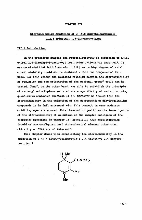
This chapter deals with establishing the stereochemistry in the
oxidation of 3-(M,M-dimethylcarbamoyl)-1,2,4-trimethyl-1,4-dihydro
pyridine 1.
1
-43-
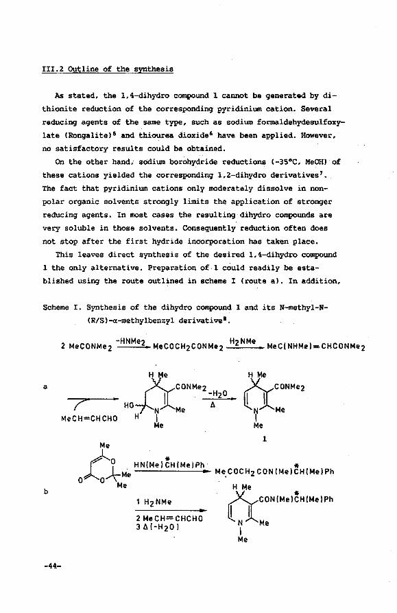
III.2 Outline of the synthesis
As stated, the 1,4-dihydro compound 1 cannot be generated by di
thionite reduction of the corresponding pyridini~ cation. Several
reducing agents of the same type, such as sodium formaldehydesulfoxy
late (Rongalite) 5 and thiourea dioxide6 have been applied. However,
no satisfactory results could be obtained.
On the other hand,; sodium borohydride reductions (-35°C, MeOHJ of
these cations yielded the corresponding 1,2-dihydro derivatives 7 •
The fact that pyridinium cations only moderately dissolve in non
polar organic solvents strongly limits the application of stronger
reducing agents. In most cases the resulting dihydro compounds are
very soluble in those solvents. Consequently reduction often does
not stop after the first hydride incorporation has taken place.
This leaves direct synthesis of the desired 1,4-dihydro compound
1 the only alternative. Preparation of 1 could readily be esta
blished using the route outlined in scheme I <route a). In addition,
Scheme I. Synthesis of the dihydro compound 1 and its N-methyl-N
(R/S)-a.-methylbenzyl derivative8 •
a
b
-44-
T MeCH=CHCHO
Me
.)-.f N. H NINe} ~H INeiP.
o~o'\ e Me
2 MeCH=CHCHO 3ld-H20 I
H Me ~CONMez
llNJl.Me I Me
1
* Me. COCH2 CON IMe}CH(Me}Ph
H Me a CONINe I~H(Ne I P•
N Me I Me
a more general method is developed to generate derivatives of 1
which differ from 1 in their amide group substituents. This method
is briefly mentioned here since it presents an elegant way of intro
ducing chiral substituents in the amide group {Scheme I, route b).
III.3 Generation and determination of an enantiomeric excess
The first problem deals with the separation of the two configura
tional enantiomers of 1 resulting from chirality of C(4). It should
be noted that conformational chirality resulting from a stable CO
out-of-plane orientation is absent 3 • Oreiding models showed that the
sp3 hybridization of C(4) decreases the rotational barrier of the
amide out-of-plane orientation. Moreover disruption of the aromatic
character of the pyridine ring results in an enhanced preference for
the in-plane amide orientation. Consequently, the lH NMR spectrum of
1 shows no sign at all of the existence of diastereoisomers.
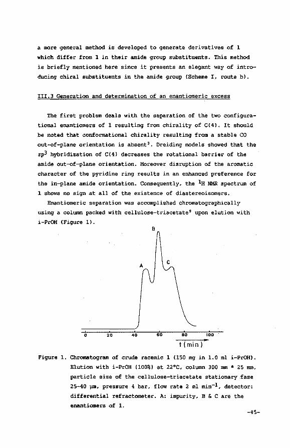
Enantiomeric separation was accomplished chromatographically
using a column packed with cellulose-triacetate9 upon elution with
i-PrOH (Figure 1). B
A
0 20 40 tOO
t (min l
Figure 1. Chromatogram of crude racemic 1 (150 mg in 1.0 ml i-PrOH).
Elution with i-PrOH (100%) at 22°C, column 300 mm * 25 mm,
. particle size of the cellulose-triacetate stationary fase
25-40 pm, pressure 4 bar, flow rate 2 ml min-1, detector:
differential refractometer. A: impurity, B & C are the
enantiomers of 1. -45-
Batches of both enantiomers could be obtained with enantiomeric
excesses ranginq from 33% to 95%.
-1.0
-2.0
250 . . • I . ' I I
' ' I I
• t I I t • I ' . . .. . . ..
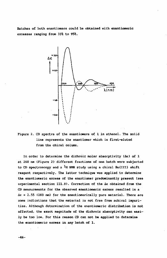
Fiqure 2. CD spectra of the enantiomers of 1 in ethanol. The solid
line represents the enantiomer which is first-eluted
from the chiral column.
In order to determine the dichroic molar absorptivity (A£) of 1
at 260 nm (Fiqure 2) different fractions of one batch were subjected
to CD spectroscopy and a la NMR study using a chiral Eu(III) shift
reaqent respectively. The latter technique was applied to determine
the enantiomeric excess of the enantiomer predominantly present (see
experimental section III.8). Correction of the 4e obtained from the
CD measurements for the observed enantiomeric excess resulted in a
4e = 2.55 (260 nm) for the enantiomerically pure material. There are
some indications that the material is not free from achiral impuri
ties. Although determination of the enantiomeric distribution is not
affected, the exact maqnitude of the dichroic absorptivity can easi
ly be too low. For this reason CD can not be applied to determine
the enantiomeric excess in any batch of 1.
-46-
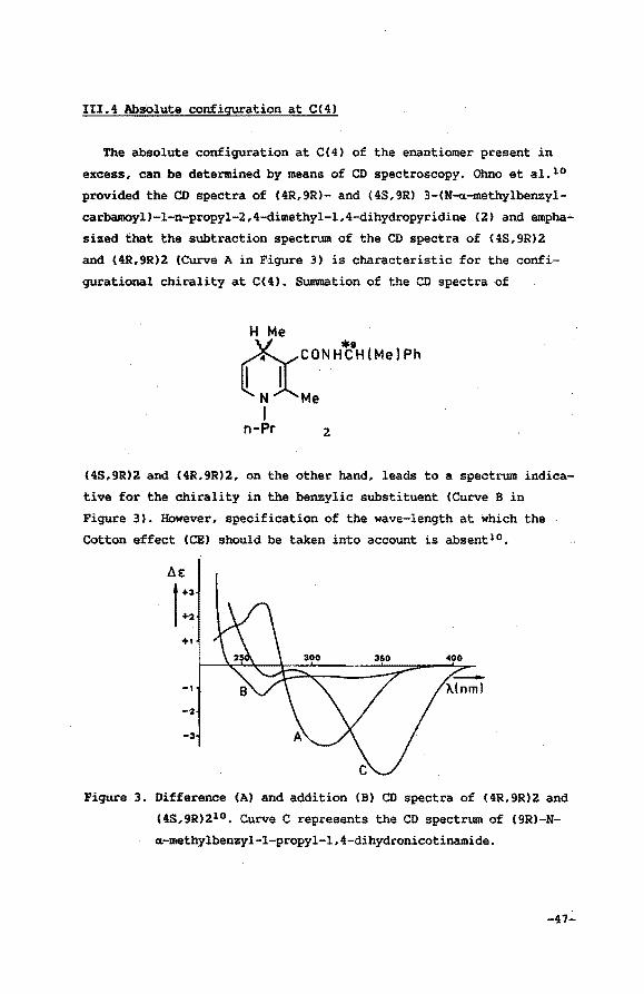
III.4 Absolute configuration at C(4)
The absolute configuration at C(4) of the enantiomer present in
excess, can be determined by means of CD spectroscopy. Ohno et al. 10
provided the CD spectra of (4R,9R)- and (4S,9R) 3-(N-oc-methylbenzyl
carbamoyl)-1-n-propyl-2,4-dimethyl-1,4-dihydropyridine (2) and empha
sized that the subtraction spectrum of the CD spectra of (4S,9R)2
and (4R,9R)2 (Curve A in Figure 3) is characteristic for the confi
gurational chirality at C( 4) . Summation of the CD spectra of
&CONH'f:0H(Me(Ph
N Me I
n-Pr 2
(4S,9R)2 and (4R,9R}2, on the other hand, leads to a spectrum indica~
tive for the chirality in the benzylic substituent (Curve B in
Figure 3). However, specification of the wave-length at which the
Cotton effect (CE) should be taken into account is absentl 0 •
t.e:
I +3
.f-2
+I
-I
Figure 3. Difference (A) and addition (B) CD spectra of (4R,9R)2 and
(4S,9R)210 • Curve C represents the CD spectrum of (9R)-N
«-methy1benzyl-l-propyl-1,4-dihydronicotinamide.
-47-
This assumption of Ohno only holds if the CE's resulting from
both chiral centra can be considered totally independent. If this is
true and no additional dissymmetry would be involved, both the
curves B and C (Figure 3), the latter depicting the CD spectrum of
C 9R)-H-a-methylbenzyl-l-propyl-l, 4-dihydronicotinaJ.Uidell, should be
identical. Moreover, in the region of interest (assignment of confi
guration at C(4)) both curves should be near or equal to zero. This
is surely not the case in the region of 340 nm. The CE of the dihy
dropyridine electronic absorption at 340 nm in curve C is the result
of distortion of the dihydropyridine structure due to the chirality
at C(9) (chirality at C(4) and axial chiral stability of the CO
out-of-plane orientation are absent). Apparently, the CE of curve A
observed at 311 nm originates from the same effect. The hypochromic
shift is due to reduced conjugation bet~een the dihydropyridine ring
and the amide moiety as a result of steric interference of the amide
substituents with the C(2) and C(4) methyl groups. This is illustra
ted by the results of an X-ray crystallographic study12 of C4R,9R)2
(Section II.3) which showed that the amide group is rotated 65° out
of plane. Consequently, information indicative for the isolated.
chirality of C(4) to be obtained from curve A. in the region of 310
nm, is obscured. The CE observed in the difference spectrum A at 264
nm, however, meets all the above mentioned criteria. Curves Band C
which should both be indicative for the isolated chirality at C(9)
are almost identical and deviate only slightly from zero. Curve A,
apart from being more pronounced, bears an opposite sign. It is
concluded that the CE in this region should be taken into account in
order te determine the absolute configuration at C(4) in 2,4-dime
thyl-3-carbamoyl-1,4-dihydropyridine compounds. A positive CE at 265
nm is indicative for 4R chirality whereas a negative one indicates a
4S configuration.
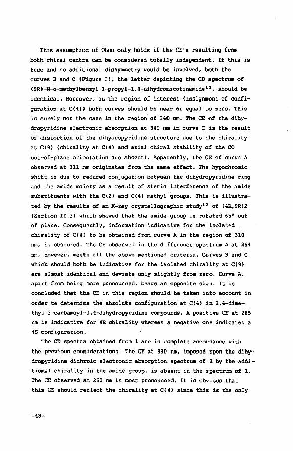
The CD spectra obtained from 1 are in complete accordance with
the previous considerations. The CE at 330 nm, imposed upon the dihy
dropyridine dichroic electronic absorption spectrum of 2 by the addi
tional chirality in the amide group, is absent in the spectrum of 1.
The CE observed at 260 nm is most pronounced. It is obvious that
this CE should reflect the chirality at C(4) since this is the only
-48-
permanent chirality present in this system (interconversion of the
CO out-of-plane orientation is fast on NMR time scale). The solid
line of Figure 2 can be assigned to the 4R enantiomer. of 1 and the
CD spectrum represented by the broken line to the 4S stereoisomer.
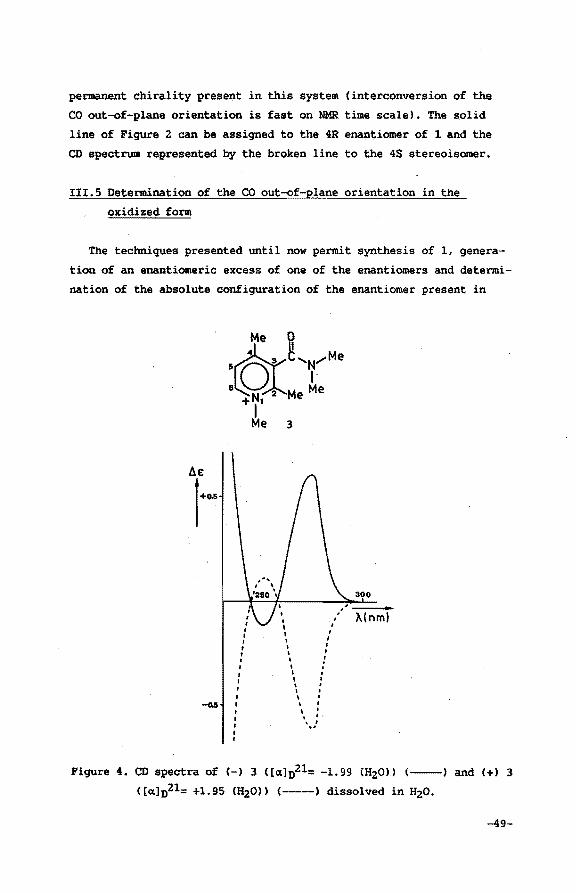
III.S Determination of the CO out-of-plane orientation in the
oxidized form
The techniques presented until now permit synthesis of 1, genera
tion of an enantiomeric excess of one of the enantiomers and determi
nation of the absolute configuration of the enantiomer present in
• I
• ' I I
' I • I
, , , , I . . . ' .
' \ . . . ' ',,!
300
X(nml
Figure 4. CD spectra of (-) 3 ( [a.] 021= -1.99 m2ou (--) and (+) 3
C[a.]n21= +1.95 CH20ll (-----) dissolved in H20.
-49-
excess. In order to be able to evaluate the stereospecificity of the
subsequent oxidation process, a method should be developed to deter
mine the absolute conformation of the carbonyl out-of-plane orienta
tion in the resulting axial chiral pyridinium cation 3.
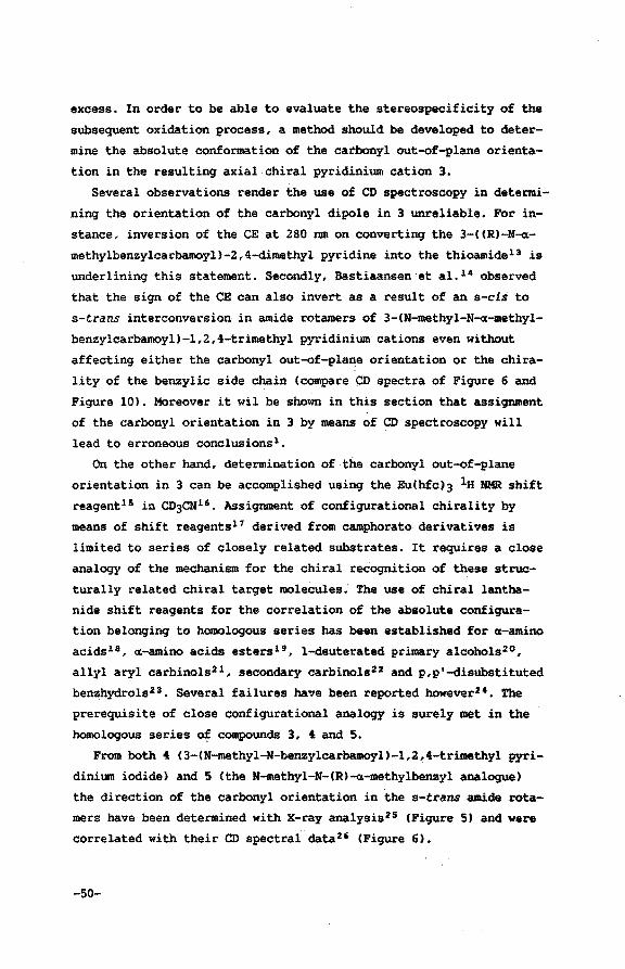
Several observations render the use of CD spectroscopy in determi
ning the orientation of the carbonyl dipole in 3 unreliable. For in
stance, inversion of the CE at 280 nm on converting the 3-((R)-H~
methylbenzylcarbamoyl)-2,4-dimethyl pyridine into the thioamide13 is
underlining this statement. Secondly, Bastiaansen et al. 14 observed
that the sign of the CE can also invert as a result of an s-cis to
s-trans interconversion in amide rotamers of 3-(N-methyl-N-a-methyl
benzylcarbamoyl)-1,2,4-trimethyl pyridinium cations even without
affecting either the carbonyl out-of-plane orientation or the chira
lity of the benzylic side chain (compare CD spectra of Figure 6 and
Figure 10). Moreover it wil be shown in this section that assignment
of the carbonyl orientation in 3 by means of CD spectroscopy will
lead to erroneous conclusions1•
On the other hand, determination of the carbonyl out-of-plane
orientation in 3 can be accomplished using the Eu(hfc)3 lH NMR shift
reagent15 in CD3CN16 • Assignment of configurational chirality by
means of shift reagents17 derived from camphorato derivatives is
limited to series of closely related substrates. It requires a close
analogy of the mechanism for the chiral recognition of these struc
turally related chiral target molecules. The use of chiral lantha
nide shift reagents for the correlation of the absolute configura
tion belonging to homologous series has been established for «-amino
acids18 , «-amino acids estersl 9 , 1-deuterated primary alcoholszo,
allyl aryl carbinols21 , secondary carbinols22 and p,p'-disubstituted
benzhydrols2 3 • Several failures have been reported however24. The
prerequisite of close configurational analogy is surely met in the
homologous series o~ compounds 3, 4 and 5.
From both 4 (3-(N-methyl-N-benzylcarbamoyl)-1,2,4-trimethyl pyri
dinium iodide) and 5 (the N-methyl-N-(R)-a-methylbenzyl analogue)
the direction of the carbonyl orientation in the s-trans amide rota
mars have been determined with X-ray analysis2 5 (Figure 5) and were
correlated with their CD spectral data26 (Figure 6).
-so-
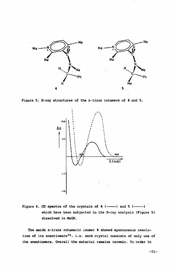
Me-N~Qo Me
~~ Me \
N H,, ,,Me
'·c ~---Ph Me
4 5
Figure S. X-ray structures of the s-trans rotamers of 4 and 5.
+2
_,
-2
,"" .. , I \
' . ' . I 1
I ' I
I . . I
I I I
I
' ' ' ' ' \ ' \
" .... 300
X(nml
Figure 6. CD spectra of the crystals of 4 (--) and 5 (---)
which have been subjected to the X-ray analysis (Figure 5)
dissolved in MeOH.
The amide s-trans rotameric isomer 4 showed spontaneous resolu
tion of its enantiomers26 , Le. each crystal consists of only one of
the enantiomers. Overall the material remains racemic. In order to
-51-
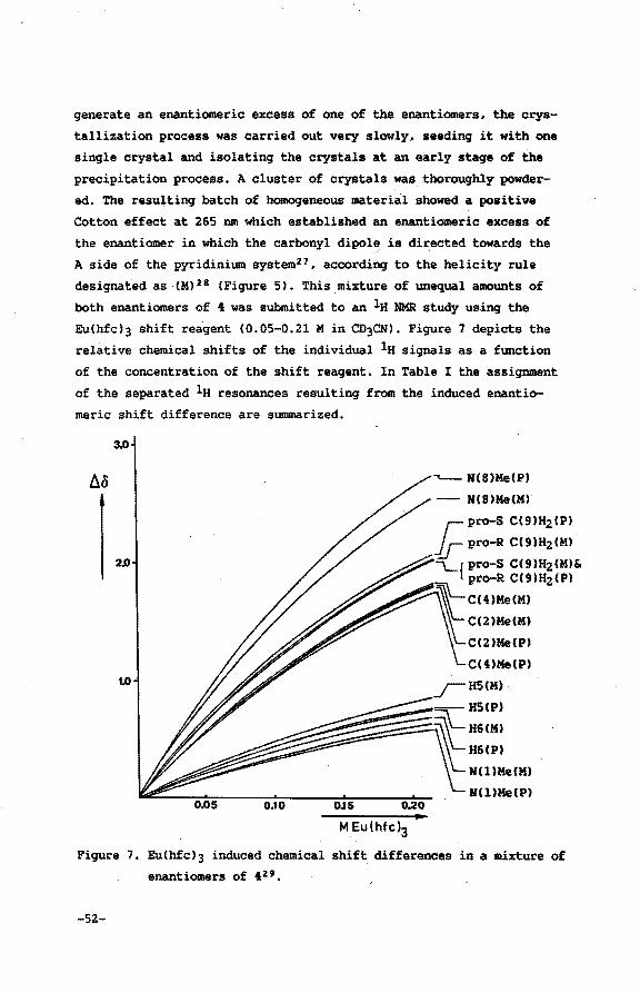
generate an enantiomeric excess of one of the enantiomers, the crys
tallization process was carried out very slowly, seeding it with one
single crystal and isolating the crystals at an early stage of the
precipitation process. A cluster of crystals was thoroughly powder
ed. The resulting batch of homogeneous material showed a positive
Cotton effect at 265 nm which established an enantiomeric excess of
.the enantiomer in which the carbonyl dipole, is di~ected towards the
A side of the pyridinium system27 , according to the helicity rule
designated as UO 28 (Figure 51. This mixture of unequal amounts of
both enantiomers of 4 was submitted to an la NMR study using the
Eu(hfcl3 shift reagent (0.05-0.21 Min CD3CNl. Figure 7 depicts the
relative chemical shifts of the individual la signals as a function
of the concentration of the shift reagent. In Table I the assignment
of the separated la resonances resulting from the induced enantio
meric shift difference are summarized.
"'-- N(8)Me(P)
- N(8)Me(M)
r--pro-S C(91H&(P)
~pro-R CC91H2(M)
\......{pro-S C(91H2(MI& ~ pro-R CC91H&CPI
\
C(4)Me(M)
CUJMe(M)
C(l)Me(PJ
C(4)Me(P)
_;-HS(MI
--HS(P)
~~tlH6(M) H6(P)
N(l)Me(MI
N(l)Me(P) 0,10 OJS Q.20
MEu(hfc)3
Figure 7. EuChfcl3 induced chemical shift differences in a mixture of
enantiomers of 42'.
-52-
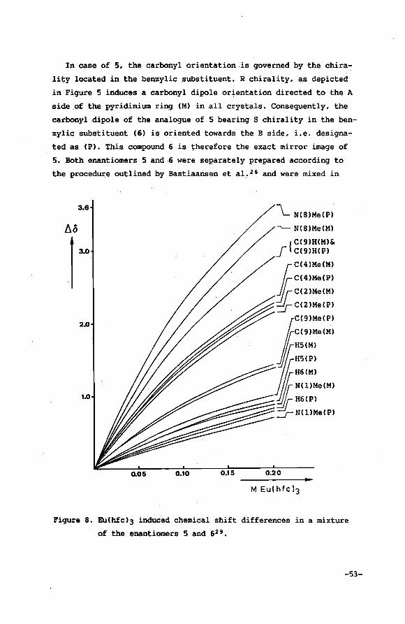
In case of 5, the carbonyl orientation is governed by the chira
lity located in the benzylic .substituent. R chirality, as depicted
in Figure 5 induces a carbonyl dipole orientation directed to the A
side of the pyridinium ring (M) in all crystals. Consequently, the
carbonyl dipole of the analogue of 5 bearing S chirality in the ben
zylic subst.ituent (6) is oriented towards the B side, i.e. designa
ted as (P). This compound 6 is therefore the exact mirror image of
5. Both enantiomers 5 and·6 were separately prepared according to
the procedure outlined by Bastiaansen et a1. 26 and were mixed in
3.6
2.0
1.0
0.05 0.10
\__ N(S)Me(P)
-.____ N(S)Me(M)
{ C(9)H(M)&
_f C(9)H(P)
1C(4)Me(M)
/{C(4)Me(P)
1_[_C(2)Me(M)
:::J C(2 )Me (P)
JC(9)Me(P)
~C(9)MaiMI
fi~~~L .. IJj;; H61PI
-::;:...-:::::~:::::::::: _r- N (1 ) Me ( P)
0.15 0.20
M Eu(hfcl3
Figure 8. Eu(hfc)3 induced chemical shift differences in a mixture
of the enantiomers 5 and 62 9 •
-53-
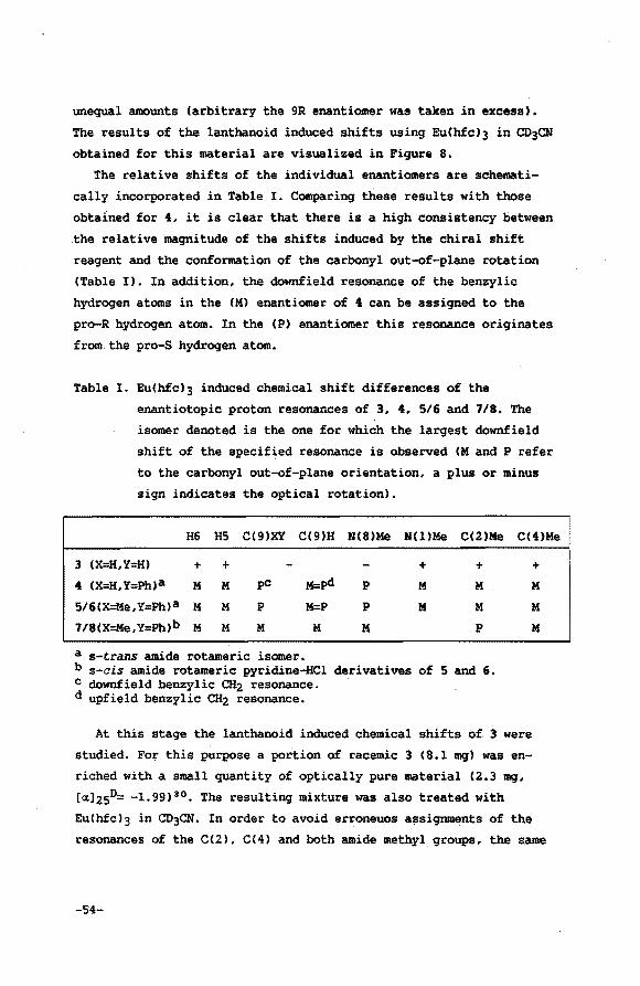
unequal amounts (arbitrary the 9R enantiomer was taken in excess).
The results of the lanthanoid induced shifts using Eu(hfc)3 in CD3CN
obtained for this material are visualized in Figure 8.
The relative shifts of the individual enantiomers are schemati
cally incorporated in Table I. Comparing these results with those
obtained for 4, it is clear that there is a high consistency between
.the relative magnitude of the shifts induced by the chiral shift
reagent and the conformation of the carbonyl out-of-plane· rotation
(Table I). In addition, the downfield resonance of the benzylic
hydrogen atoms in the (M) enantiomer of 4 can be assigned to the
pro-R hydrogen atom. In the (P) enantiomer this resonance originates
from.the pro-S hydrogen atom.
Table I. Eu(h£c)3 induced chemical shift differences of the
enantiotopic proton resonances of 3, 4, 5/6 and 718. The
isomer denoted is the one for which the largest downfield
shift of the specified resonance is observed CM and P refer
to the carbonyl out-of-plane orientation, a plus or minus
sign indicates the optical rotation).
H6 HS C(9)XY C(9)H N(S)Me N(l)Me C(2)Me C(4)Me
3 (X=H,Y=H)
4 (X=H,Y=Ph)a + + M M
5/6(x=Me,Y=Ph)a M M
7/8(X=Me,Y:Pb)b M M
pC
p
M
a s-t.rans amide rotameric isomer.
p
p
M
+ M
M
+ M
M
p
b a-cis amide rotameric pyridine-He! derivatives of 5 and 6. c downfield benzylic CHz resonance. d upfield benzylic CHz resonance.
+ M
M
M
At this stage the lanthanoid induced chemical shifts of 3 were
studied. For this purpose a portion of racemic 3 (8.1 mg) was en
riched with a small quantity of optically pure material (2.3 mg,
[«lasD= -1.99) 30 • The resulting mixture was also treated with
Eu(hfc)3 in CD3CN. In order to avoid erroneuos assignments of the
resonances of the C(2), C(4) and both amide methyl groups, the same
-54-
experiment was conducted with racemic material in which the C(2) and
C(4) methyl groups were selectively deuterated. The results of this
study are collected in Table I.
Comparison of the results listed in Table I for the sets of enan
tiomers of 3,i and 5/6 shows a striking similarity of the resolution
pattern of all enantiotopic proton resonances. Distinction of the
two enantiotopic signals of the a-trans N(8)Me signals of 3 is
closely related to the ones observed for the separation of the
signals in the benzyl- and a-methylbenzyl groups of 4 and 5/6. This
is surely due to the fact that these three substituents are posi
tioned exactly alike in space and that the diastereomeric complex
formation is mainly the result of interactions of the Eu(hfc)3
derivative with the carbonyl oxygen atom31 • Since all enantiotopic
protons of the three compounds show a uniform behaviour upon addi
tion of Eu(hfc)3, determination of the carbonyl out-of-plane orienta
tion on this basis is highly reliable. In conlusion, the results
presented above prove the carbonyl to be located at the A side for
the (+) 3 enantiomer.
It is of interest to test whether the la NMR technique using
Eu(hfc)3 can also be applied to determine the CO out-of-plane orien
tation in s-cis rotameric derivatives of for instance 5 and 6. For
this purpose one should be able to mix two enantiomers (having oppo
site CO out-of-plane orientations) of these s-cis axial chiral rota
mers in unequal amounts. This can easily be accomplished using the
results of Bastiaansen et al. 14 , who showed that the absolute axial
chirality of the. CO out-of-plane orientation is determined by the
configuration in the a-methylbenzyl moiety, totally independent of
the amide rotameric conformation. From X-ray crystallographic inves
tigation32 of the s-cis rotamer of 3-(N-methyl-N-(S)-«-methylbenzyl
carbamoyl)-2,4-dimethyl pyridine HCl (7, Figure 9) it was concluded
that the orientation of the carbonyl dipole is (P), identical to the
one observed for the a-trans (S)-«-methylbenzylic derivative 6.
Preparing 7 and mixing it with an excess of its enantiomer 8, bea
ring an (R)-«-methylbenzyl group, showed upon addition of Eu(hfc)3
in CD3CN that the pattern of enantiotopic resolution as observed for
3, 4 and 5/6 does not hold (Table I>. This is surely the result of
the chirality of the benzylic substituent in 1 and 8 being posi-
-55-
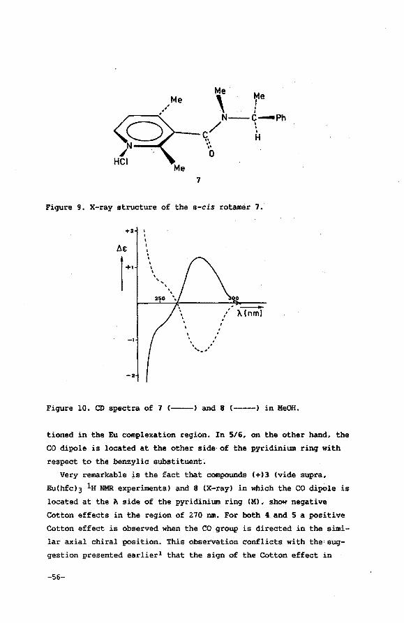
Me
' re Me
N-C....,..Ph
----c/ \ •• H
7
.. .. 0
Figure 9. X-ray structure of the s-cis rotamer 7.
+2 • • i
AE I
• • l +•
• I \ \ \
-I
-2
~
' '
I \ I I
I ' ' I
' ' ,_,
0 --/ 'A lnm) I ,
Figure 10. CD spectra of 7 c--) and 8 c----) in MeOH.
tioned in the Eu complexation region. In 5/6, on the other hand, the
CO dipole is located at the other side of the pyridinium ring with
respect to the benzylic substituent;
Very remarkable ~s the fact that compounds (+)3 (vide supra,
Eu(hfc)3 lH NMR experiments) and 8 (X-ray) in which the CO dipole is
located at the A side of the pyridinium ring (M), show negative
Cotton effects in the region of 270 nm. For both 4 and 5 a positive
Cotton effect is observed when the CO group is directed in the simi
lar axial chiral position. This observation conflicts with the'sug
gestion presented earlier1 that the sign of the Cotton effect in
-56-
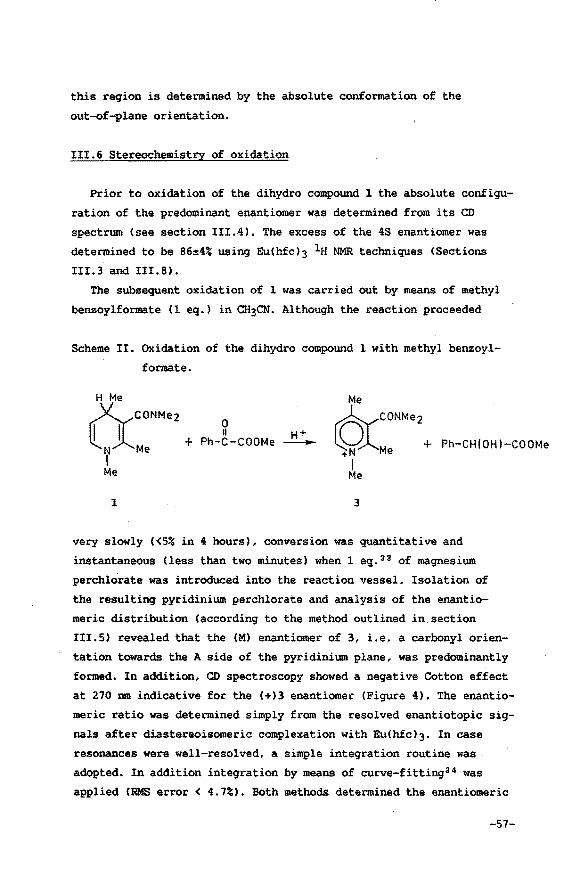
this region is determined by the absolute conformation of the
out-of-plane orientation.
III.6 Stereochemistry of oxidation
Prior to oxidation of the dihydro compound 1 the absolute configu
ration of the predominant enantiomer was determined from its CD
spectrum (see section III.4). The excess of the 48 enantiomer was
determined to be 86~4% using Eu(hfc)3 la NMR techniques (Sections
III.3 and III.8).
The subsequent oxidation of 1 was carried out by means of methyl
benzoylformate (l eq.) in CH3CN. Although the reaction proceeded
Scheme II. Oxidation of the dihydro compound 1 with methyl benzoyl
formate.
H Me
0 aCONMe2 +
N Me
II Ph-C-COOMe
Me
t6:cCONMe2+
+N Me Ph-CH [ OH I-CO OMe
I Me I
Me
1 3
very slowly (<5% in 4 hours), conversion was quantitative and
instantaneous (less than two minutes) when 1 eq. 33 of magnesium
perchlorate was introduced into the reaction vessel. Isolation of
the resulting pyridinium perchlorate and analysis of the enantio
meric distribution (according to the method outlined in.section
III.S) revealed that the (M) enantiomer of 3, i.e, a carbonyl orien
tation towards the A side of the pyridinium plane, was predominantly
formed. In addition, CD spectroscopy showed a negative Cotton effect
at 270 nm indicative for the (+)3 enantiomer (Figure 4). The enantio
meric ratio was determined simply from the resolved enantiotopic sig
nals after diastereoisomeric complexation with Eu(hfc)3· In case
resonances were well-resolved, a simple integration routine was
adopted. In addition integration by means of curve-fitting34 was
applied (RMS error< 4.7%). Both methods determined the enantiomeric
-57-
distribution to be 100:25, which is equivalent to an enantiomeric
excess of 60%. The optical yield in the oxidative generation of the
pyridinium cation therefore amounts to 70%.
The product of conversion of the methyl benzoylformate, the
methyl mandelate was analyzed using Eu(hfc)3 in CCl4 (10% CDCl3).
Resolution was rather poor. Normal integration routines were there
fore inadequate in establishing the optical purity of the alcohoL
Curve-fitting3 4 (RMS error of 9.0%) revealed normalized integrals in
the ratio of 100 to 30 (e.e. 54%). The optical yield in the produc
tion of methyl mandelate therefore equals 63%. Comparing the resolu
tion pattern with the one obtained from partially resolved (R)-me
thyl mandelate (synthesized from R-mandelic acid) showed that the
enantiomer predominantly formed bears S chirality.
III.7 Conclusions
Extensive studies have been performed in recent years on hydride
transfer reactions from chiral 1,4-dihydropyridines to prochiral
carbonyls rendering optical active alcohols35 • All the dihydro
compounds used in these studies bear additional configurational
chirality. In this chapter, however, a compound is presented which
is devoid of any configurational stereochemical element other than
chirality on C(4). It was established that methyl benzoylformate
reduction using the 4S enantiomer of 1 afforded the S methyl mande
late in 63% optical yield. This result is in complete accordance
with the results obtained by Meyers and co-workers 4 , who reported
that reduction of the same substrate with N-benzyl-5-(N,N-dimethyl
carbamoyl)-3-hydroxymethyl-4(Rl-methyl-1,4-dihydropyridine yielded
predominantly the 4R alcohol (62% e.e.). In case the 3-methoxymethyl
derivative was adopted 52% e.e. of the R alcohol was obtained.
Meyers and coworkers disputed the role of the carbonyl dipole of
these NADH model compounds. These authors presented a mechanism in
which the amide group is indeed rotated out of plane, but with the
dimethylamino group facing the ester carbonyl. This feature is
however purely speculative since these authors do not have any
substantial evidence for this type of interaction. Chiral stability
-58-
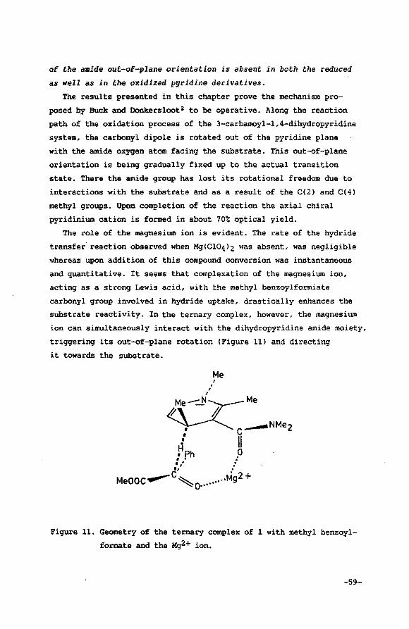
of the amide out-of-plane orientation is absent in both the reduced
as well as in the oxidized pyridine derivatives.
The results presented in this chapter prove the mechanism pro
posed by Buck and Donkersloot2 to be operative. Along the reaction
path of the oxidation process of the 3-carbamoyl-1,4-dihydropyridine
system, the carbonyl dipole is rotated out of the pyridine plane
with the amide oxygen atom facing the substrate. This out-of-plane
orientation is being gradually fixed up to the actual transition
state. There the amide group has lost its rotational freedom due to
interactions with the substrate and as a result of the C(2) and C(4) ·
methyl groups. Upon completion of the reaction the axial chiral
pyridinium cation is formed in about 70% optical yield.
The role of the magnesium ion is evident. The rate of the hydride
transfer reaction observed when Mg(Cl04>2 was absent, was negligible
whereas upon addition of this compound conversion was instantaneous
and quantitative. It seems that complexation of the magnesium ion,
acting as a strong Lewis acid, with the methyl benzoylformiate
carbonyl group involved in hydride uptake, drastically enhances the
substrate reactivity. In the ternary complex, however, the magnesium
ion can simultaneously interact with the dihydropyridine amide moiety,
triggering its out-of-plane rotation (Figure 11) and directing
it towards the substrate.
Me ,
Figure 11. Geometry of the ternary complex of 1 with methyl benzoyl
formate and the Mg2+ ion.
-59-
It should be emphasized that an optical yield of 70% of the axial
chiral 3-carbamoyl pyridinium cation is to be considered quite high
since both substrate and dihydropyridine compound are very reactive
(the ~+ catalyzed reaction reported here is instantaneous, whereas
conversions reported in literature33 are allowed to proceed for seve
ral days). It is to be expected that tempering the rate of conver~
sion by conducting the experiments at low temperature, will enhance
the optical yield. Preliminary experiments show that the optical
yields of the axial chiral pyridinium derivative up to 90% are
obtainable at 0°C.
The use of moderately activated or deactivated carbonylic sub
strates should also temper the rate of conversion. This could pro
bably enhance the optical yield of the process as well •. In this
context it should be noted that the dihydropyridine derivative 1 is
stable when dissolved in acetone (after four days no degradation or
conversion was observed). Preliminary experiments show that upon
addition of MgCCl04)2 a reaction is initiated (~50% conversion in
four hours) generating the corresponding pyridinium cation and
i-PrOH. Especially in this type of slow-conversion reactions built
up of a correct and well-defined ternary complex is most likely.
The optical yield in the production of the methyl mandelate at
22°C is 63% and 80% when the reaction is carried out at 0°C. These
values are somewhat lower than the optical yields obtained for the
axial chiral pyridinium system at these temperatures. The origin of
this phenomenon can easily be denoted. In addition to the ~+
interaction, stereospecificity in the reduction of the ketone is
determined by the relative positioning of the phenyl and ester
groups. Figure 11 depicts a situation in which stacking of the
dihydropyridine moiety and the substrate phenyl group is present,
originating from a donor (dihydropyridine) acceptor (phenyl group)
interaction. This geometry accounts for.the observation that the
methyl benzoylformate reduction by C4S)l yields the S alcohol.
However, one should bear in mind that rotation about the CC4)
(dihydropyridine)-H-carbonyl (substrate) axis does not change the
configurations of the reaction products. Inspection of Ohno's
results (see ref. 10) seems to indicate that. substrate orientation,
and therefore the stereoselectivity of the hydride uptake, is
-60-
governed by interaction between the free4!lectron pair(s) of a
carbonyl substituent with either ~+ or the dihydro compound.
Ohno et al. 3 observed that in the oxidation reactions of dihydro
quinoline derivatives, using strong oxidizing agents (p-chloranil
and DDQ), the orientation of the amide group in~ resulting
3-carbamoyl quinolinium cation is reversed (Section II.4). The four
electron-withdrawing quinone substituents will probably prevent
formation of interaction between the Mg2+ ion and the. substrate
carbonyl group. In order to minimize electrostatic repulsion forces
between the amide oxygen atom and the four negatively charged
quinone substituents, the amide oxygen atom is rotated away from the
substrate. In accordance with what is expected, the absolute confor
mation of the CO out-of-plane orientation generated during the oxida
tion process using p-chloranil or DDQ is identical when Mg(Cl04l2 is
present or absent3 6 ,
The importance of the proposed mechanisme of CO mediated stereo
chemistry of hydride incorporation in NM>+ model compounds for the
"in vivo" enzyme catalyzed reactions is evident. From the results
presented here it is clear that the amide oxygen atom within the
3-carbamoyl pyridinium cation, interacts with the substrate. It
seems highly unlikely that the enzyme, evolved to an optimal refined
catalyst, overlooks this kind of interaction. X-ray studies per
formed on a ternary complex of LADHtNAD+tDMS037 and a binary complex
of GAPDHtNAD+38 strongly support the proposed mechanisme. In both
complexes the carbonyl dipole is orientated out of the pyridinium
plane, towards the substrate binding region. This orientation is not
the result of interactions between the NAD+ amide moiety and the
substrate, but predesigned by NAD+ interactions with active site
amino acids in order to optimize the NAD+tsubstrate interactions in
the transition state of the hydride transfer process2 •
III.8 Experimental section
lu NMR spectra were run in the FT mode at 200 MHz on a Bruker
AC200 spectrometer using TMS as internal standard. CD spectra were
obtained on a Jobin Yvon Dichrograph Mark III-S spectrometer.
-61-
Solvents and reagents used were of reagent grade and were when
applied .in the synthesis of dihydropyridine compounds degassed and
extensively flushed with argon.
N ,N-dimethyl acetoacetamide
This compound was prepared according to the procedure outlined in
literature39 (68%1.
Bp lll°C/12mm. lH NMR (CDCl3): S 2.30(s,3H,CH3), 2.93(s,6H,
N(CH3)2), 3.55(s,2H,CHzl.
3-(N-methylamino)-R,N-dimethyl crotonamide4°
N,N-dimethyl acetoacetamide (5.5 g, 43.3 mmol) was kept at 120°C
for approximately 1% h during which a stream of monomethylamine was
led through the pure material. The reaction could easily be moni
tored by means of lH NMR. Collecting the colourless solid material
yielded the pure product (5.68 g, 92.3%). 1H NMR (CDCl3l: S L93(s,3H,CH31, 2.88(d,3H,NCH3), 2.95(s,6H,
N(CH3l2l, 4.68(s,lH,olef H), 9.37(br q,lH,NH)
3-(N,N-dimethylcarbamoyl)-1,2,4-trimethyl-1,4-dihydropyridine
To a solution of 3-(N-methylamino)-N,N~·dimethylcrotonamide (1.0
g, 7.03 mmol) in methanol (5 ml, p.a.), piperidine (30 ~1, 0.3 mmol)
and crotonaldehyde (0.63 g, 9.06 mmol) were subsequently added. The
reaction mixture was stirred under an argon atmosphere for
approximately 14 h after which the solvent was evaporated.
Chloroform was added (30 ml) and thermal elimination of water from
the intermediate 6-hydroxy-1,4,5,6-tetrahydro derivative was accom
plished by distilling off the organic solvent. Chromatography on
aluminium oxide (active, basic type E, Merck 1067, previously
degassed and saturated with argon) using chloroform as eluent
(Rf=0.60) and treat~nt of the collected product with dry ether,
afforded the 1,4-dihydro compound (0.86 g, 63%1. The product proved
to be highly sensitive to air. Generation of an enantiomeric excess
was accomplished chromatographically using a column packed with
cellulose-triacetate (Merck 16363, 25-40 ~~ upon elution with
i-PrOH (flushed with argon>.
-62-
Qe~e~!~tio~ Qf_the_d!chrQi~ ~!aE !b!O£P~iyi~_of !: Upon addition of tris-[3-{heptafluoropropylhydroxymethylene)-D
camphorato] Eu(III) derivative (Eu(hfc)3, concentrations ranging
from 48 mM to 370 mM) to a solution of 1 (20.7 mg) in acetone-d6
(500 ~1) the individual 1-Me signals of the enantiomers could be
sufficiently resolved. Ac:etone-d6 was used as a solvent in order to
prevent formation of a precipitate which would seriously hamper reso
lution. Integration of the. individual resonances of both enantiomers
could be achieved using the standard integration routine since both
peaks were sufficiently resolved. In addition, a curve-fit program34
was adopted. Spectra obtained on a Bruker AC200 spectrometer, run in
the FT mode, were collected in a 16K data base, which was zero-fil
led to 64K prior to Fourier transformation, in order to obtain
well-defined signals. Integration and curve-fitting {RMS error
<4.7%) determined the integrals to be in the ratio of 100 to 25. In
consequence the enantiomeric excess amounted 86~4%.
From a different sample of the same batch CD spectra were run
(total concentration 1.25 mg ml-1, sens 5*10-6, cuvette of 0.1 em).
Correcting for the enantiomeric excess resulted in a 4&=2.55 at 260
nm for the enantiomerically pure material.
lH NMR ICDCl3l: & 1.03{d,3H,C{4)CH3), 1.77(s,3H,C(2)CH3), 2.91(s,
3H,N(llCH3), 2.95 to 3.05(br s,6H,N(8)(CH3lz), 3.32(m,1H,H(4)),
4.44(dd,lH,H(5)), 5.73(d,lH,H(6)).
In a 60 MHz NMR spectrum the s-trans and s-cis N(8)Me signals can
be separately observed <& 2.91 and 3.01 ppm). Coalescense of the
four N(8)Me resonances resulting from the hindered CO out-of-plane
rotation is complete.
Oxidation with methyl benzoylformate
A batch of dihydropyridine compound 1 (approximately 40 mg)
eluted from the cellulose-triacetate column was collected. CD
revealed that the predominant enantiomer beared 4S chirality. lH NMR
chiral shift reagent experiments in acetone-d5·determined the enan
tiomeric excess to be 86%. The dihydro compound was placed in an NMR
tube under an argon atmosphere. To this, dry acetonitrile-d3 (0.5
mil and methyl benzoylformate (30 mg, 1 eq., as determined by inte
gration of the appropriate resonances in its NMR spectrum) were
-63-
added. MgCCl04l2 (approximately 1 eq.) was introduced (exothermic
process). After evaporation of the solvent, the residue was dis
solved in water/methylene chloride. The organic layer was subsequen
tly scrubbed with water, dried and filtered. Evaporation of the sol
vent resulted in the crude product. 1H NMR established the absence
of unreacted methyl benzoylformate and a quantitative yield of the
alcohol. lH NMR experiments using Eu(hfcl3 in CCl4 (10% CDCl3)
showed an e.e. of 54% (optical yield 63%) of the predominant S
isomer. The optical yield in the production of methyl mandelate
at 0°C is 80%.
The aqueous solution resulting from extraction, showing a
negative Cotton effect in the region of 270 nm, was evaporated
(p<O.OOS mm, T<27°C). EuChfcl3 1H NMR techniques were again applied
to determine the enantiomeric excess of as well as the CO
orientation in the predominant enantiomer (60% e.e. of the (M)
enantiomer, optical yield 70%). At 0°C, the axial chiral
pyridinium cation is formed in about 90% optical yield.
References and notes
1. de Kok, P.M.T.; Donkersloot, M.C.A.; v. Lier, P.M.; Meulendijks,
G.H.W.M.; Bastiaansen, L.A.M.; v. Hooff, H.J.G.; Kanters, J.A.;
Buck, H.M. Tetrahedron 1986,42,941.
2. Donkersloot, M.C.A.; Buck, H.M. J. Am. Chem. Soc. 1981,103,6554.
3. a. Ohno, A.; Kashiwagi, M.; Ishihara, Y.; Ushida, S.; Oka, s. Tetrahedron 1986,42,961.
b. Ohno, A.; Ohara, M.; Oka, S. J. Am. Chem. Soc. 1986,108,6438.
4. Meyers, A.I.; Op?en1aender, T. J. Am. Chem. Soc. 1986,108,1989.
5. Hanstein, W.; Wallenfe1s, K. Tetrahedron 1967,23,585.
6. Ohnishi, Y.; TaniMoto, S. Tetrahedron Lett. 1977,22,1909.
-64-
7. Several compounds of Table IV of Chapter II have been reduced
under these conditions yielding the corresponding 1,2-dihydro
derivatives in good yields (upto 90%).
8. Acetoacetamides with different·substituents in the amide group
can easily be prepared from 2,2,6-trimethyl-1,3-dioxen-4-one (di
ketene aceton adduct) according to the method outlined by
Clemens, R.J.; Hyatt, J.A. J. Org. Chem. 1985,50,2431.
9. a. Eigsperger, A.; Kastner, F. ; Mannschreck, A. J. Mol. Struct.
1985,126,421.
b. Mannschreck, A.; Koller, H.; Wernicke, R. Kontakte <Darmstadt)
1985,1,40.
10. The absolute configuration at C(4) of one of the diastereoisomers
of the 3-(N-«-methylbenzy1carbamoyl)-l-n-propyl-2,4-dimethyl-1,4-
dihydropyridine (2) was determined by chemical degradation of one
of the enantiomers without affecting the configurational chirality
at C(4) and converting it to dimethyl methylsuccinate. Comparing
the CD spectrum with that of an authentic sample derived from
partially resolved (R) methylsuccinic acid proved the enantiomer
subjected to the chemical degradation to have an (R) con
figuration at C(4).
Ohno, A.; Ikequchi, M.; Kimura, T.; Oka, S. J. Am. Chem. Soc.
1979,101,7036.
11. This compound does not bear the C(2) and C(4) methyl groups.
Chirality at C(4) is therefore absent.
12. v. Lier, P.M.; Donkersloot, M.C.A.; Koster., A.S.; v. Hooff,
H.J.G.; Buck, H.M. Reel. Trav. Chim. Pays~Bas 1982,101,119.
13 • Conversion of 3-(( R) -liT-«-methy lbenzy lcarbamoy 1) -2 , 4-dimethy 1
pyridine into the thioamide was accomplished with P2S5 according
to the procedure outlined in Section II.S, synthesis of the
3-(N,N-dimethylthiocarbamoyl)-2,4-dimethyl pyridine.
-65-
14. Bastiaansen, L.A.M.; Vermeulen, T.J.M.; Buck, H.M.; Smeets,
W.J.J.; Kanters', J·?'-•; Spek, A.L. J. Chem. Soc. r Chem. Commun.
1988,230.
15. Sweeting, L.M.; Crans, D.C.; Whitesides, G.M. J. Org. Chem.
1987,52,2273.
16. The use of less polar solvents such as CDCl3 is restricted due to
the poor solubility of the pyridinium salts.
17. Schurig, w. Kontakte <Darmstadt> 1985,2,22.
18. Kabuto, K.; Sakaki, T. J. Chem. Soc., Chem. Commun. 1987,670.
19. a. Ajisaka, K.; Kamisaku, Kainosho, M. Chem. Lett. 1972,857.
b, Mitte, F.; Meusinger, R.;· Borsdarf, R. Z. Chem. 1986,26,107.
20. Reich, C.J.; Sullivan, G.R.; Mosher, H.S. Tetrahedron Lett.
1973,1505.
21. Cervinka, 0.; Malon, P.; Trska, P. Collect. Czech. Chem. Commun.
1973,38,3299.
22. Sullivan, G.R.; Ciavarella, D.; Mosher, H.S. J. Org. Chem.
1974,39,2411.
23. capillon, J.; Lacombe, L. Can J. Chem. 1979,57,1446.
24. a. Gaudener, A. "Stereochemistry" (H.B. Kagan, Ed.) Vol 1, p 117,
Thieme Stuttgart, 1977.
b. Sullivan, G.~. Top. Stereochem. 1976,10,287.
c. Fraser, R.R. "Asymmetric Synthesis" (J.D. Morrison, Ed.) Vol
1, p 173, Academic Press, London, 1983.
25. Crystal data for 4: C17H21NzOI, M=396.27, orthorhombic, space
group P212121, a=7.183(3), b=8.769(2), c=28.093(6) A, U=l769.4(9)
-66-
A3, Dc=l.487 g cm-3, Z=4.
Crystal data for 5: C1sH23N20I, M=410.37, orthorhombic, space
group P212121, a=7.209(7), b=8.877(5), c=28.698(1) A, U=l836(2)
)1.3, Dc=l.484 g cm-3, Z=4.
26. Bastiaansen, L.A.M.; Kanters, J.A.; v.d. Steen, F.H.; de Graaf,
J.A.C., Buck, H.M. J. Chem. Soc., Chem. Commun. 1986,536.
27. The A side is the side of the nicotinamide moiety at which HA is
located in NADH.
28. Krow,G. "Topics in Stereochemistry" (E.L. Eliel, N.L. Allinger,
Eds.) vol 5, p 31, Willey-Interscience, New York, 1970.
29. The exact amount of chemical shift difference induced upon addi
tion of Eu(hfc)3 is highly dependent of the amount of water in
the sample or in the solvent.
30. v. Lier, P.M.; Meulendijks, G.H.W.M.; Buck, H.M. Reel. Trav.
Chim. Pays-Bas 1983,102,337.
31. Eu(fod)3 experiments performed on the pyridinophanic compound
which will be introduced in chapter IV, shows that the amide
hydrogen atom undergoes an extreme large down-field shift whereas
the 'chemical shift of the methylene group in the s-trans position
relative to the carbonyl dipole is hardly affected. The reversed
effect is observed for'the 3-(N-hexylcarbamoyl)-l~ethyl pyridi
nium cation. These results justify the conclusion that the Eu
shift reagent complexes at one side of the pyridinium cation at
which the carbonyl oxygen atom is lokated. ,
32. Crystal data for 7: C17H21N20Cl•H20 M=322.83, monoclinic, space
group P21, a=l3.677UL b=8.585UL c=l6.106(4) A, IS=l06.09U) 0,
U=l815.7(5) A3, Dc=l.l81 q cm-3, Z=4.
...67-
33. a. reference 3.
b. reference 10.
c. Amano, M.; Baba, N.; Oda, J.; Inouye, Y. Bioorganic Chemistry
1984,12,299.
d. Inouye, Y.; Oda, J.; Baba, N. "Asymmetric Synthesis" (J.D.
Morrison, Ed.) Vol 2, p 92, London, 1983.
34. Glinfit program: Copyright Bruker Spectrospin AG, Switzerland.
35. a. Reference 33.
b. Talma, A.G.; Jouin, P.; de Vries, J.G.; Troostwijk, C.B.;
Werumeus Buning, G.H.W.; Waninge, J.K.; Visscher, J.; Kellogg,
R.M. J. Am. Chem. Soc. 1985,107,3981.
36. Ohno, A.; Ohara, M.; Oka, S. J. Am. Chem. Soc. 1986,108,6438.
in reference to ·ohno, A.; Yasui, S.; Nakamura, K.; Oka, S. Bull.
Chem. Soc. Jpn. 1978,51,290.
37. Eklund, H.; Samama, J.P.; Jones, T.A. Biochemistry 1984,23,5982.
38. Skarzynski, T.; Moody, P.C.E.; Wonacott, A.J. J. Mol. Biol.
1987,193,171.
39. a. Bredereck,' H. Gompper, R.; Klemm, K. Angew. Chemie 1959,71,32.
b. Bredereck, H. Gompper, R.; Klemm, K. Chem. Ber. 1959,92,1456.
40. Kato, T.; Yamanaka, H.; Kawamata, J.; Shimomura, H. Chem. Pharm.
Bull. 1969,11,1889.
-68-
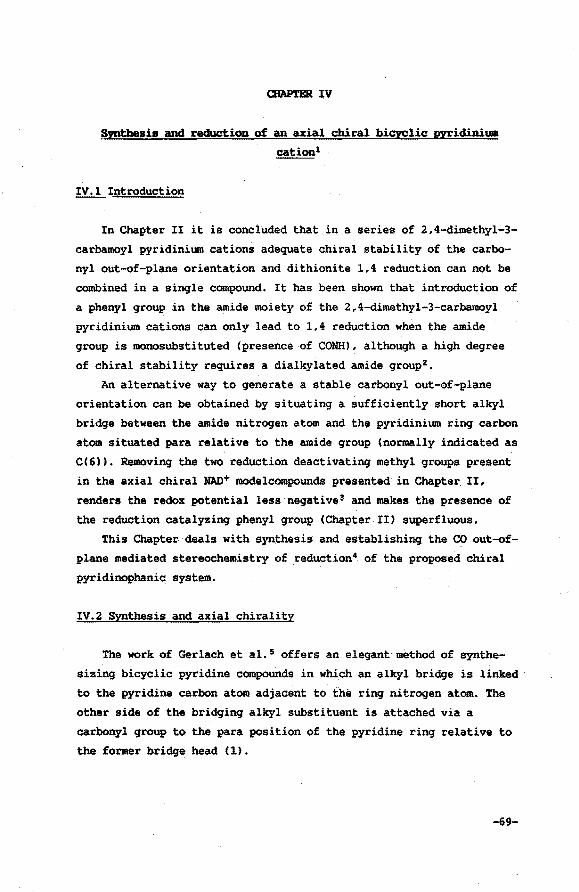
afAP'r.BR IV
Synthesis and reduction of. an axial chiral bicyclic pyridiniwa
cationl
IV.l Introduction
In Chapter II it is concluded that in a series of 2,4-dimethyl-3-
carbamoyl pyridinium cations adequate chiral stability of the carbo
nyl out-of-plane orientation and dithionite 1,4 reduction can not be
combined in a single compound. It has been shown that introduction of
a phenyl group in the amide moiety of the 2,4-dimethyl-3-carbamoyl
pyridinium cations can only lead tol,4 reduction when the amide
group is monosubstituted (presence of CONH), although a high degree
of chiral stability requires .. a dialkylated amide group2 •
An alternative way to generate a stable carbonyl out-of-plane
orientation can be obtained by situating a sufficiently short alkyl
bridge between the amide nitrogen atom and the pyridinium ring carbon
atom situated para relative to the amide group (normally indicated as
C(6)). Removing the two reduction deactivating methyl groups present
in the axial chiral HAD+ modelcompounds presented in Chapter II,
renders the redox potential less negative 3 and makes the presence of
the reduction catalyzing phenyl group (Chapter II) superfluous.
This Chapter deals with synthesis and establishing the CO out-of
plane mediated stereochemistry of reduction4 of the proposed chiral
pyridinophanic system.
IV.2 Synthesis and axial chirality
The work of Gerlach et al. 5 offers an elegant method of synthe
sizing bicyclic pyridine compounds in which an alkyl bridge is linked ·
to the pyridine carbon atom adjacent to the ring nitrogen atom. The
other side of the bridging alkyl substituent is attached via a
carbonyl group to the para position of the pyridine ring relative to
the former bridge head (1).
-69-
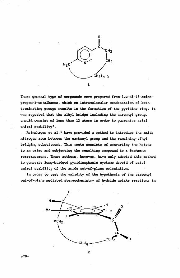
These general type of compounds were prepared from l,w-di-(3-amino
propen-1-on)alkanes, which on intramolecular condensation of both
terminating groups results in the formation of. the pyridine ring. It
was reported that the alkyl bridge including the carbonyl group,
should consist of less than 12 atoms in order. to guarantee axial
chiral stabili ty4.
Reinshagen et a1. 6 have provided a method to introduce the amide
nitrogen atom between the carbonyl group and the remaining alkyl
bridging substituent, This route consists. of converting the ketone
to an oxime and subjecting the resulting .compound to a Beclanann
rearrangement. These authors, however, have only adopted this method
to generate long-bridged pyridinophanic systems devoid of axial
chiral stability of the amide out-of-plane orientation.
In order to test the validity of the hypothesis of the carbonyl
out-of-plane mediated stereochemistry of hydride uptake reactions in
-:-70-
H ' '
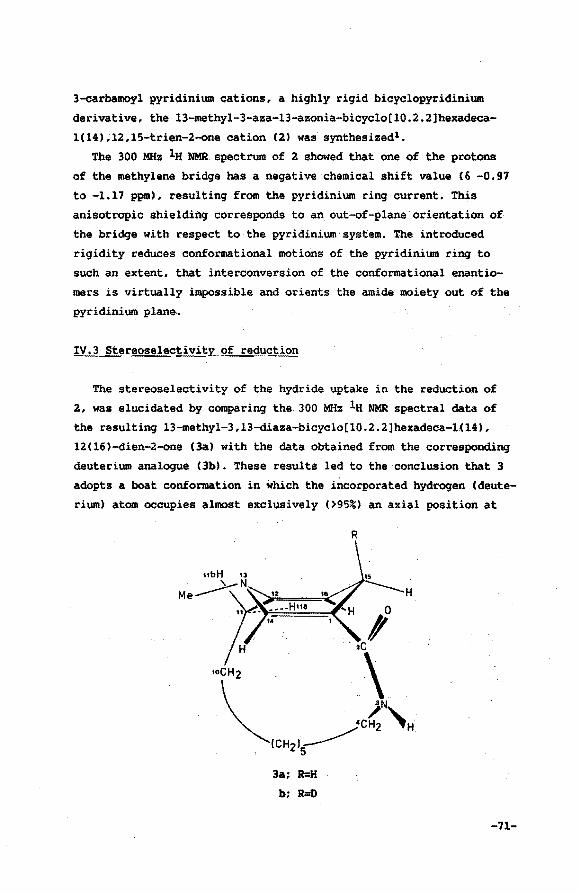
3-carbamoyl pyridinium cations, a highly rigid bicyclopyridinium
derivative, the 13-methyl-3-aza-l3-azonia-bicyclo[l0.2.2]hexadeca
l(l4),12,15~trien-2-one cation (2) was synthesized1 •
The 300 MHz 1a NMR.spectrum of 2 showed that one of the protons
of the methylene bridge has a negative chemical shift value C& -0.97
to -1.17 ppm), resulting from the pyridinium ring current. This
anisotropic shielding corresponds to an out-of-plane orientation of
the bridge with respect to the pyridinium system. The introduced
rigidity reduces conformational motions of the pyridinium ring to
such an extent, that interconversion of the conformational enantio
mers is virtually impossible and orients the amide moiety out of the
pyridinium plane.
IV.3 Stereoselectivity of reduction
The stereoselectivity of the hydride uptake in the reduction of
2, was elucidated by comparing the 300 MHz la NMR spectral data of
the resulting 13-methyl-3,13-diaza-bicyclo[l0.2.2lhexadeca-1H4),
12(16)-dien-2-one (3a) with the data obtained from the corresponding
deuterium analogue C3b). These results led to the conclusion that 3
adopts a boat conformation in which the incorporated hydrogen (deute
rium) atom occupies almost exclusively (>95%) an axial position at
3a: R=H
b; R=D
R
-71-
C(l5). The non-planarity was deduced from the coupling constants of
HC16). This hydrogen appeared in the lH NMR spectrum of 3a as an
eight-line pattern, owing to a vicinal coupling with Hax<lSl and
HeqC15), and an allylic coupling with H(lla). This clearly indicates
that the CC15) methylene protons are non-equivalent. Additional
proof was obtained from the NCH3 doublet, resulting from the coup
ling with HaxC15). Absence of puckering of the dihydropyridine ring,
however, would implicate a long-range coupling between the NCH3
group and both H(l5) methylene protons 7 •
Reduction of 2 in D20, led to 3b. The lack of coupling between
Hax<l5) 8 and NCH3, H(l4) and H(l6), respectively, clearly indicates"
the axial position of the deuteride. In addition, Dreiding models
unambiguously showed that the carbonyl group is rotated out of the
plane through C(l), C(l2), C(l4) and C(l6) which results in the fact
that the incorporated hydride is syn-positioned with respect to the
carbonyl group.
IV. 4. Conclusions
The observed unique stereoselectivity in the hydride uptake will
originate from three major features present in this system, i.e. the
out-of-plane orientation of the amide group with the carbonyl group
syn-positioned relative to the incoming hydride donor 9 , the shiel
ding effect of the methylene bridge which prevents equatorial attack
for steric reasons 10 and stereoelectronic factors resulting from
deformation of the pyridinium moiety11 induced by the relative short
bridging alkyl group.
With regards to the two aspects firstly mentioned, i.e. the car
bonyl out-of-plane orientation and the one-sided shielding, this
model system is in good agreement with recent crystallographic 3D
data obtained for the enzyme bound NAD+. The results for the ternary
complex of NAD+ bonded horse liver alcohol dehydrogenase (A specifi
city) obtained by Eklund et al. 12 show that the amide group (hydro
gen bonded to specific sites on the dehydrogenase) is 30° out of the
plane, with the carbonyl group directed towards the A side and shiel
ding the B side perfectly. In case of NAD+ bound to GAPDH1 3 (B speci-
-72-
ficity) the.'A side is shielded and the carbonyl dipole is rotated
22° out of plane pointing towards the B side.
The implications of the third feature, i.e. effects resulting
from the non-planarity of the pyridinium ring for the biological
process have already brought forward in as early as 195714 •
Puckering of the dihydropyridine ring, leading to a boat confor
mation, positions one of the dihydropyridine methylene protons in an
q.xial position. Stereoelectronic considerations explain the enhanced
reactivity of the axial.positioned hydrogen atom11 relative to the
equatorial hydrogen atom. Recently, Benner and co~workers 15 devised
a concept in which the stereochemistry of the enzymatic hydride
transfer is based on the syn or anti conformation of the nicotinami
de ring of the cofactor. This theory is also based on a distortion
of the dihydropyridine ring into a boat conformation. It has been
argued10 , however, that a relative strong perturbation of the dihy
dropyridine ring, necessary to explain the high degree of stereose
lectivity in NADH and NADH model compounds, should lead to a conside
rable U.V. hypochromic effect. Convincing evidence for this u.v. shift has not been reported10 • X-ray crystallographic results of
non-bridged dihydropyridine systems16 prove the dihydropyridine ring
puckering to be negligible. Still, this kind of coenzyme distortion
can be of importance during build-up of the transition state in the
(enzymatic) hydride transfer process.
The relative contributions of each of the three factors mentioned
above to the "in vitro" overall stereochemical behaviour of the
bicyclic pyridinium cation presented here, remain unclear.· Taking
into account the results of Ohno et al. 17 and the results presented
in the previous Chapter, the carbonyl out-of-plane orientation is
likely to be essential in catalysis of the hydride transfer process.
IV.S. Experimental section
Spectroscopy
60 MHz lH NMR spectra were recorded on a Varian EM360Aor a Hitachi
Perkin Elmer R-24B NMR spectrometer and 300 MHz lH NMR data were ob-
-73-

tained on a Bruker CXP 300 spectrometer. Me4Si was used as internal
standard (6 0.00). A mass spectrum (MS) was recorded on a Finnigan
4000 GC-MS instrument using electron ionization.
Synthesis
Decanedioic acid chloride (sebacyl chloride) was prepared with 89%
yield according to a procedure outlined in literature18 • Conversion of
the dichloride to the 12-aza-bicyclo[9.2.2]pentadeca-1(13),11,14-tri
en-2-one was adopted from the work of Gerlach et al. 4 The crude pro
duct was subsequently submitted to column chromatography (silicagel
60, Merck, eluent hexane/ethyl acetate=l/1, Rf=0.40) and to destina
tion. Total yield 22%.
Bp ll5°C/O.Ol mm. Anal. Calcd.: C,77.38; H,8.81; N,6.45. Found:
C,77.58; H,9.19; N,6.47. IRCCCl4l: v= 2915 cm-1; 2880 cm-1; 1705 cm-1
(C=OJ; 1590 cm-1. lH NMR CCDCl3l: & -0.40 to 0.48 (m,lH,Aliphatic HJ,
0.48 to 2.33(m, llH,Aliphatic Hs), 2.50 to 3.12(m,4H,2CH2), 7.22(d,
lH,pyrH), 7.67(dd,lH,pyrH), 8.68(d,lH,pyrH).
12-aza-bicyclo[9.2.2]pentadeca-1(13),ll,l4-trien-2-one oxime6
To a solution of the 12-aza-bicyclo[9.2.2]pentadeca-1(13),11,14-tri
en-2-one (5.51 g, 25.4 mmol, 1 n=8) in methanol (150 ml) was added
sodium acetate (10 g) and hydroxylamine hydrochloride (6 g). Water was
added until the reaction mixture eiarified. Stirring was continued at
~ temperature overnight. The product solidified on introducing dis
tilled water (300-ml). Collection of the solid material and rinsing it
with water, afforded the oxime (4.68 9i 82.5%) upon drying (50°C,
vacuo).
Mp 122.8-123. 7°C. Anal. Calcd.: C, 72.38; H,8.68; N,l2.06. Found:
C,72.42; H,9.15; N,l2.18. Rf CTLC, hexane/ethyl acetate=l/1)=0.26.
la NMR (CDCl3l: & -0.40 to 1.90(m, 12H,6CH2), 2.22 to 3.12(m,4H,2CH2),
7.15(d,lH,pyrH), 7.52(dd,lH,pyrH), 8.25(d,lH,pyrH), 11.22(br.s,lH,NOH).
3,13-diaza-bicyclo[l0.2.2]hexadeca-1(14),12,15-trien-2-one6
The powdered oxime (3.63 g, 15.6 mmol) was added over a period of
45 minutes to vigorously stirred polyphosphoric acid (100 ml) at
-74-
100°C. After stirring of the solution at this temperature for 6-7
hours, the resulting reaction mixture was quenched in water (1 1).
Basification of the aqueous solution with KzC03, thorough extraction
with chloroform, scrubbing the organic phases with water gave, upon
drying and evaporation of the solvent, the crude material (1.84 g).
Chromatography through silica (silicagel 60 Merck) using chloroform/
ethanol=95/5 as an eluent gave the pure product (650 mg, 18%).
Rf CTLC, chloroform/ethanol=95/5)=0.26. MSCm/z) 232 CC14HaoN20). IR
CKBr) 3220 <vmtL 1670 <ve=o> cm-1. lH NMR (300 MHz,CIX!l3): li -0.38 to
-0.04(m,2H,CH2), 0.73 to 0.92(m,2H,CH2), 0.98 to 1.37Cm,6H,3CH2), 1.51
to 1. 66 (m, lH,Aliphatic H), 1. 75 to 1. 90 (m, lH,Aliphatic H), 2. 80 to
3.02(m,3H,Aliphatic Hs), 3.07 to 3.23(m,1H,Aliphatic H), 6.43(br.s,
lH,NH), 7.30(d,lH,pyrH), 7.71 (dd,lH,pyrH), 8.66(d,lH,pyrH). 13c NMR
CCDCl3) li 24.87 to 28.98 (6t), 39.43(t), 42.53(t), 123.76Cd),
128.95(s), 153.55(d), 147.42(d), 164.54(s), 172.97(s). Using the
Eu(fod)3 shift reagent CCDCl3), we observed an increasing down-field
shift going from CC12)CH2, H(16), NCHz, H(14), H(15) to NH.
13-methyl-3-aza-13-azonia-bicyclo[l0.2.2]bezadeca-1(14),12,15-trien-2-
one iodide ( 2 >
The pyridine derivative (400 mg, 1.72 mmol) dissolved in methyl
iodide (10 ml) was refluxed for 16 h. Evaporation of the excess of
methyl iodide and sUbsequent trituration with dry ether gave the
desired product (520 mg, 1.39 mmol, 81%).
lH NMR CCD30D): & -1.17 to -0.97Cm,1H,Aliphatic H), 0.40 to 2.15(m,
llH,Aliphatic Hs), 2.80 to 3.60(m,4H,NCHz and pyrCH2>; 4.64(s,3H,
NCH3l, 8.2l(d,lH,H(16)), 8. 67(rA,lH,H(l5)), 9.88(d,lH,H(14)).
13-methyl-3,13-diaza-bicyclo[l0"2.2]bezadeca-1(14),12(16)-dien-2-one
(3a)
The pyridinium compound 2 (200 mg, 0.53 mmol) was added to a bi
layeric system consisting of an lN aqueous solution of sodium hydro
gencarbonate (100 ml) and methylene chloride (100 ml), both solvents
previously being degassed and saturated with argon. Sodium dithionite
(1.0 g, 5.7 mmol) was added in small portions. The reaction mixture
-75-
was thoroughly stirred for 6% h at room temperature under an argon
atmosphere. The organic layer was separated and scrubbed with water.
Drying of the methylene chloride on MgS04, filtration and evaporation
of the solvent gave the pure dihydro compound (140 mg, 95%). (The
product decomposes during elution from a silica column and oxidizes
rapidly upon exposure to air).
lH NMR CCDCl3l: & 0.90 to Z.OO(m,lZH,6CHzl, Z.53(m,ZH,CC12lCH2),
2.84Cm,lH,HeqC15)), 3.05(m,1H,Hax<15)), 3.06(d,3H,CH)Nl, 3.35(m,2H,
CH2NCOl, 4.47(m,1H,H(l6)), 5.95(br.t,lH,NH), 6.30(t,lH,HC14)).
Coupling constants (in Hz): J(H(l6l-Hax<l5)) 1.7, J(H(l6l-HeqC15ll
6.8, JCHC16l-HClla)) 1.1, JCHaxC15l-HeqC15)) 17.519 and
J(NCH3-HaxC15)) 0.8.
Suprisingly, for the NC13)Me group a doublet is found. This must be
attributed to coupling with one of the C(l5) methylene protons since
splitting of this resonance is absent in the mono-deutero analogue
C3b). Peak separation Cin Hz) has been shoWn to be independent of the
spectrometer frequency (300 MHz and 500 MHz lH NMR spectra were recor
ded) which proves splitting to be the result of spin-spin coupling.
13-methyl-3,13-diaza-15-monodeutero-bicyclo[l0.2.2]hexadeca-1(14),
12(16)-dien-2-one (3bl
The procedure outlined in the preparation of the non-deuterated
bicyclic dihydropyridine derivative 3a is adopted using 2 (100 mg,
0.27 mmoll, an lN sodium hydrogencarbonate solution in DzO (30 mll,
methylene chloride (30 ml) and sodium dithionite (0.42 g, 2.4 mmol).
Work-up yielded 67 mg <>95%).
lH NMR CCDCl3l: o 0.90 to 2.00(m,l2H,6CHzl, 2.52Cm,2H,CHzl,
2.82Cbr.d,lH,HeqC15)) 8 , 3.06(s,3H,CH3N), 3.37(m,ZH,CHzN), 4.46(dd,
1H,H(16)), 5.87Cbr.t,lH,NH), 6.30(d,lH,H(l4) ).
References and notes
1. de Kok, P.M.T.; Buck, H.M. J. Chem. _Soc., Chem. Commun. 1985,1009.
2. de Kok, P.M.T.; Donkersloot, M.C.A.; v. Lier, P.M.; Meulendijks,
G.H.W.M.; Bastiaansen, L.A.M.; v. Hooff, H.J.G.; Kanters, J.A.;
Buck, H.M. Tetrahedron 1986,42,941.
-76-
3. Introduction of electron-releasing methyl groups will lower the
redox potential. For instance, introducing methyl groups at the
C(2) and C(6) positions of the "Hantzsch" derivatives decreases the
standard redox potential by 100 mV.
Piepers, o: Thesis, Groningen 1981.
4. Donkersloot, M.C.A.; Buck, H.M. J. }lm. Chem. Soc. 1981.103,6554.
5. Gerlach, H.; Huber, E. Helv. Chim. }leta 1968,51,2027.
6. Reinshagen, H.; Stutz, A. Monatsh. Chem. 1979,110,567.
7. Compare with the results of the planar protonated 9-methyl
anthracene in which the methylene protons are equivalent as
reported by Colpa, J.P.; MacLean, C.; Mackor, .E.L. Tetrahedron
1963~19 supl. 2,65.
8. Presence of the gemina1 deuterium atom leads to a significant
broadening of the Heq<l5) signal as well as a small upfield shift
of 0.02 ppm. Rob et al. 10 report a deuterium induced shift of as 0.03 ppm in their mono-deutero 1,4-dihydropyridines.
9. a. Reference 2 and references cited herein.
b. Reference 4,
10. Rob, F.; v. Ramesdonk, H.J.; v. Gerresheim, W.; Bosma, P.;
Scheele, J.J.; Verhoeven, J.W. J. }lm, Chem. Soc. 1984,106,3826.
11. The enhanced reactivity of the axial over the equatorial posi
tion has been postulated earlier:
a. Kuthan, J.; Musil, L. Collect. Czech. Chem. Commun.
1977,42,867.
b. Alder and co-workers report a stereoelectronically controlled
protonation of l-dimethylamino-8-trimethy1ammonium naphtalene
tetrafluoroborate which occurs exclusively from an axial di
rection. Alder, R.W.; Bryce, M.R., Goode, N.C. J. Chem. Soc.,
Perkin II 1982,477.
-77-
c. Brounts, R.H.A.M.; Buck, H.M. J. ~m. Chem. Soc. 1983,105,1284.
d. Reference 10.
12. Eklund, H.; Samama, J.P.; Jones, T.A. Biochemistry 1984,23,5982.
13. Skarzynski, T.; Moody, P.C.E.; Wonacott, A.J. J. Mol. Biol.
1987,193,171.
14. Levy, H.R.; Vennes1and, B. J. Biol. Chem. 1957,228,85.
15. Benner, S.A.; Nambiar, P.K.; Chambers, G.K. J. Am. Chem. Soc.
1985,107,5513.
16. v. Lier, P.M.; Donkers1oot, M.C.A.; Koster, A.S.; v. Hoof£,
H.J.G.; Buck, H.M. Reel. Trav. Chim.·Pays~Bas 1982,101,119.
17. a. Ohno, A.; Kashiwagi, M.; Ishihara, Y.; Ushida, S.; Oka. S.
Tetrahedron 1986,42,961.
b. Ohno, A.; Ohara, M.; Oka, S. J. Am. Chem. Soc. 1986,108,6438.
18. Henecka, H. "Houben....Weyl, Methoden der Organischen Chemie" (E.
Muller Ed.) Vol VIII, p 467, Georg Thieme Verlag-Stutgart, 1952.
19. Gemina1 coupling of the methylene protons of C(4) in NADH is
18Hz (Sarma, R.H.; Kaplan, N.O. Biochemistry 1970,9,539) whereas
those reported for NADH-like cyclophanes are significantly
smaller (13-14Hz)lO,
-78~
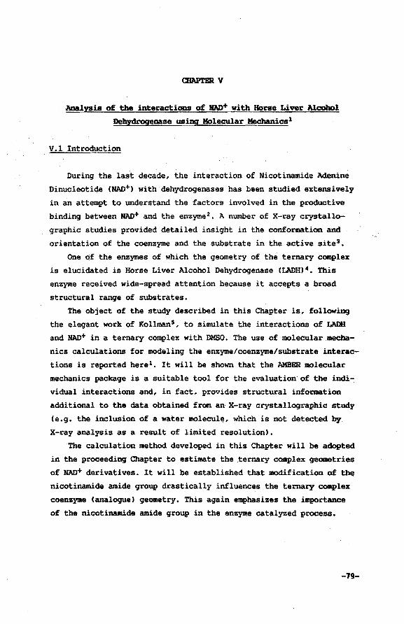
CHAP!BR v
Analysis of the interactions of JfAD+ with Horse Liver Alcohol
Dehydrogeaase using Molecular l!lechanics1
V.l Introduction
During the last decade, the interaction of Nicotinamide Adenine
Dinucleotide (NAD+) with dehydrogenase& has been studied extensively
in an attempt to understand the factors involved in the productive
binding between NAD+ and the enzyme 2 • A number of X-ray crystallo
graphic studies provided detailed insight in the conformation and
orientation of the coenzyme and the substrate in the active site3 •
One of the enzymes of which the geometry of the ternary complex
is elucidated is Horse Liver Alcohol Dehydrogenase (LADH) 4 • This
enzyme received wide-spread attention because i~ accepts a broad
structural range of substrates.
The object of the study described in this Chapter is, following
the elegant work of Kollman5, to simulate the interactions of LADH
and NAD+ in a ternary complex with DMSO. The use of molecular .mecha
nics calculations for modeling the enzyme/coenzyme/substrate interac
tions is reported here1 • It will be shown that the 1\MBER molecular
mechanics package is a suitable tool for the evaluation of the indi
vidual interactions and, in fact, provides structural information
additional to the data obtained from an X-ray crystallographic study
(e.g. the inclusion of a water molecule, which is not detected by
X-ray analysis as a result of limited resolution).
The calculation method developed in this Chapter will be adopted
in the proceeding Chapter to estimate the xernary complex geometries
of HAD+ derivatives. It will be established that modification of the
nicotinamide amide group drastically influences the ternary complex
coenzyme (analogue) geometry. This again emphasizes the i~~portance
of the nicotinamide amide group in the enzyme catalyzed process.
-19-
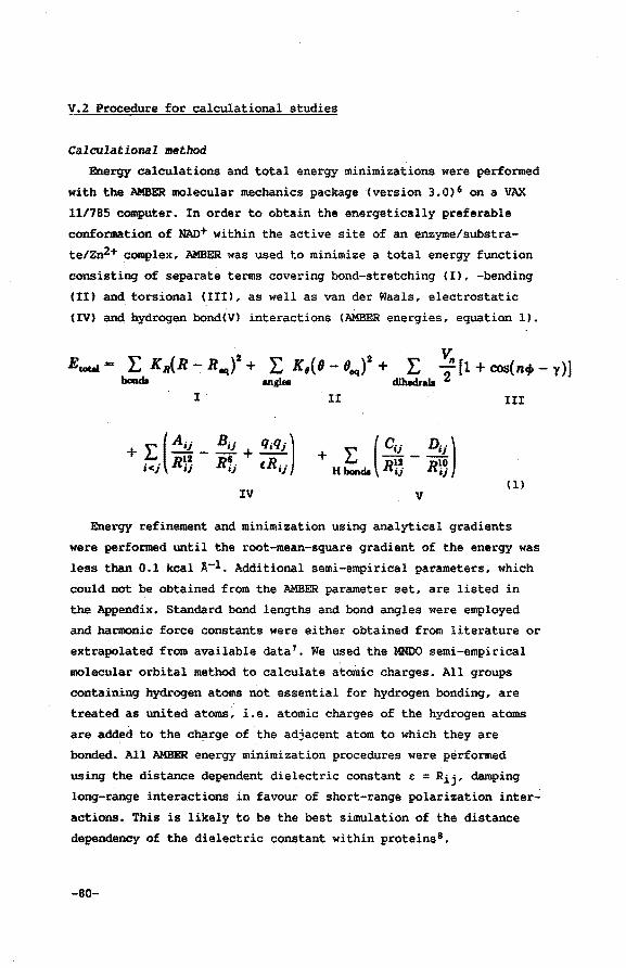
V.2 Procedure for calculational studies
Calculational method
Energy calculations and total energy minimizations were performed
with the AMBER molecular mechanics package (version 3.0)6 on a VAX
11/785 computer. In order to obtain the energetically preferable
conformation of NAO+ within the active site of an enzyme/substra
te/zn2+ complex, AMBER was used to minimize a total energy function
consisting of separate terms covering bond-stretching (I), -bending
(II) and torsional (III), as well as van der Waals, electrostatic
(IV) and hydrogen bond(V) interactions (AMBER energies, equation 1).
Etotat"'" L KR(R- Rfl4(+ L K 1(9- 8eq)2 + . L V,. [1 + cos(n+- y)] boada angl• dlheclrabl 2
I. . II III
IV (1)
Energy refinement and minimization using analytical gradients
were performed until the root-mean-square gradient of the energy was
less than 0.1 kcal A-1. Additional semi-empirical parameters, which
could not be obtained from the AMBER parameter set, are listed in
the Appendix. Standard bond lengths and bond angles were employed
and harmonic force constants were either obtained from literature or
extrapolated from available data 7 • We used the MNDO semi-empirical
molecular orbital method to calculate atomic charges. All groups
containing hydrogen atoms not essential for hydrogen bonding, are
treated as united atoms, i.e. atomic charges of the hydrogen atoms
are added to the c~rge of the adjacent atom to which they are
bonded. All AMBER energy minimization procedures were performed
using the distance dependent dielectric constant & = Rij• damping
long-range interactions in favour of short-range polarization inter
actions. This is likely to be the best simulation of the distance
dependency of the dielectric constant within proteins8 •
-80-
Examination of the resulting conformations was achieved using the
Anal module of AMBER in order to calculate the interaction energies
and an interactive computer graphics program (Chem-X. July 1987
update) 9 to generate stereodiagrams.
Initial conformations
AMBER calculations require starting geometries of the structures
to be calculated. For this, we employed the X-ray crystallographic
data for the ternary complex of LADH/NAD+tDMSO (2.9 A resolution~ crystallographic R factor of 0.22) reported by Eklund et al. 4a,
which were readily retrievable from the Brookhaven Protein Database.
Enzyme
Since AMBER can only perform its calculations with a limited num
ber of atoms, the core of the enzyme can only be represented by a
"cage" which is constructed of a relatively small number of amino
acids, each fixed at its initial (X-ray) position. In order to
obtain a "sealed" construction, 44 amino acids within a range of 6.0
A from the coenzyme/zn2+tDMSO complex, were taken into accountlO;
Enlargement of the cut-off distance did not result in any improve
ment of th~ results of the calculations. All parameters required
could be obtained from the standard AMBER data base.
Coenzyme
Since AMBER does not have all parameters needed in its initial
data base, we have introduced the parameters by creating additional
data bases (Appendix). Parameters for adenine, ribose and the phos
phate groups were retrieved directly from the AMBER data base, where
as parameters used for the nicotinamide moiety were obtained from
MNDO calculations (bond lengths, bond angles, torsion angles and
charges) or estimated in accordance with data reported in literature
(harmonic force constants) 7 • The geometry of NAD+ was taken from the
X-ray data. Hydrogen atoms that can be involved in hydrogen bonding
were added with the computer graphics modeling program Chem-X
(ribose OH's, nicotinamide NH's and adenine NH's).
-81-
DlfSO
The starting con£ormation was taken from the X-ray data. AMBER
parameters were added (Appendix) to the AMBER data base and charges
were calculated using MNDO.
Zinc
The charge of the zinc ion in the catalytic site of the ternary
complex is plus two. AMBER parameters (Appendix) were estimated
according to procedures reported in literature7 •
Initial studies, in which the zinc ion is not restricted to its
initial position, showed highly unlikely movements of DMSO bonded
zinc upon energy refinement. The origin of this shift can easily be
denoted; zinc is bonded in the active site of the enzyme by two
negatively charged deprotonated cysteine residues. In the AMBER
parameter set, however, parameters for the cysteine residues are
available only for the neutral state of the amino acid. This means
that interactions between zinc and the core of amino acids are
neglected. Two approaches were devised to overcome this problem. The
first comprises fixation of the zinc ion at its initial (X-ray)
position, whereas the second approach includes negatively charged
cysteine residues via modification of the AMBER data base. Both
methods have been applied and the results will be discussed.
V.3 Results and discussion
The first case studied was the energy refinement of the NAO+
coenzyme molecule within the core of amino acids, employing the
initial AMBER parameter set for all constituent amino acids of the
"cage" (including those of the neutral cysteine residues). The zinc
ion was fixed at its initial position. Conformational restraints on
the· position of the_ coenzyme molecule were not included. In order to
visualize the effects of energy minimization of. the geometry of
MAD+, both the initial NAD+ (X-ray) conformation as well as the
energy refined structure are depicted in Figure 1.
The torsion angles of the X-ray geometry of NAD+ and the con£orma
tion after energy refinement are included in Table I. The interac
tion energies are listed in Table II.
-82-
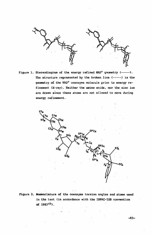
Figure 1. Stereodiagram of the energy refined MAD+ geometry (--).
The structure represented by the broken line (----~) is the
geometry of the MAD+ coenzyme molecule prior to energy re
finement (X~ray). Neither the amino acids, nor the zinc ion
are drawn since these atoms are not allowed to move during
energy refinement.
Figure z. Nomenclature of the coenzyme torsion angles and atoms used
in the text (in accordance with the IUPAC-IUB convention
of 19834b).
-83-
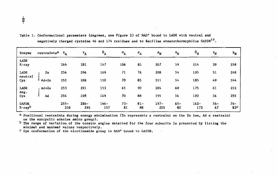
I co
"" I
Table I. Conformational parameters (degrees, see Figure 2) of NAD+ bound to LADH with neutral and
negatively charged cysteine 46 and 174 residues and to Bacillus stearothermophilus GAPDH12•
Enzyme restraintsa X~
LADH X-ray
LADH neutral Cys
LADH neg. Cys
GAPDH X-rayb
Zn
{ Ad+Zn
{ Ad:n
264
256
252
253
254
255-258
YA
281
296
288
291
298
286-295
f) A
147
169
150
153
169
146-157
ttA
106
71
70
65
70
73-81
PA
85
76
85
90
88
81-88
PN
207
208
211
204
195
197-205
~
59
54
54
60
56
65-80
f)N
214
195
185
175
193
162-172
YN
39
51
49
61
56
56-67
a Positional restraints during energy minimization (Zn represents a restraint on the Zn ion. Ad a restraint on the exocyclic adenine amino group).
b The range of variation of the torsion angles detected for the four subunits is presented by listing the minimal and maximal values respectively.
c Syn conformation of the nicotinamide group in NAD+ bound to GAPDH.
XN
258
248
244
252
255
76-83C
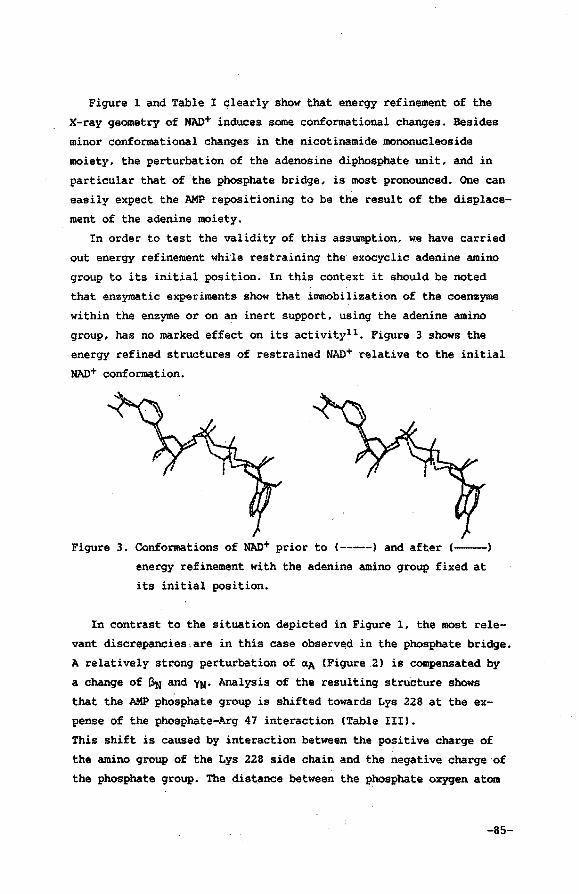
Figure 1 and Table I clearly show that energy refinement of the
X-ray geometry of NAD+ induces some conformational changes. Besides
minor conformational changes in the nicotinamide mononucleoside
moiety, the perturbation of the adenosine diphosphate unit, and in
particular that of the phosphate bridge, is most pronounced. One can
easily expect the AMP repositioning to be the result of the displace
ment of the adenine moiety.
In order to test the validity of this assumption, we have carried
out energy refinement whi'le restraining the exocyclic adenine amino
group to its initial position. In this context it should be noted
that enzymatic experiments show that immobilization of the coenzyme
within the enzyme or on an inert support, using the adenine amino
group, has no marked effect on its activity11 • Figure 3 shows the
energy refined structures of restrained NAD+ relative to the initial
NAD+ conformation.
Figure 3. Conformations of NAD+ prior to ( ----) and after (--)
energy refinement with the adenine amino group fixed at
its initial position.
In contrast to the situation depicted in Figure 1, the most rele
vant discrepancies,are in this case observed in the phosphate bridge.
A relatively strong perturbation of ~A (Figure.2) is compensated by
a change of ~ and YN· Analysis of the resulting structure shows
that the AMP phosphate group is shifted towards Lys 228 at the ex
pense of the phosphate-Arg 47 interaction (Table III).
This shift is caused by interaction between the positive charge of
the amino group of the Lys 228 side chain and the negative charge of
the phosphate group. The distance between the phosphate oxygen atom
-85-
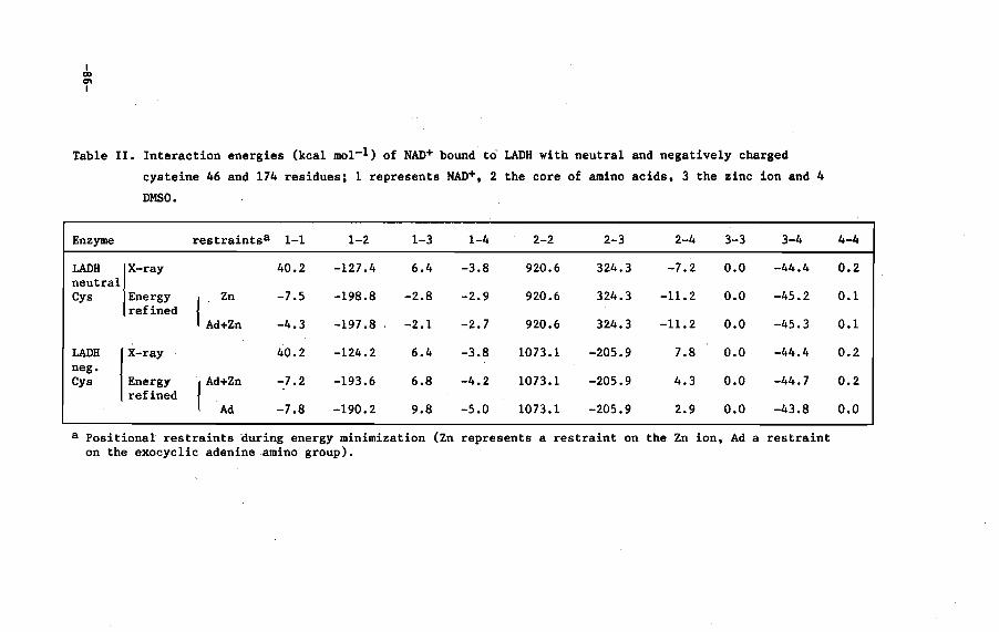
I CXI 0'1 I
Table II. Interaction energies (kcal mol-l) of NAD+ bound to LADH with neutral and negatively charged
cysteine 46 and 174 residues; 1 represents NAD+. 2 the core of amino acids. 3 the zinc ion and 4
DMSO.
Enzyme restraintsa 1-1 1-2 1-3 1-4 2-2 2-3 2-4 3-3 3-4
LADB rray 40.2 -127.4 6.4 -3.8 920.6 324.3 -7.2 0.0 -44.4 neutral Cys Energy { Zn -7.5 -198.8 -2.8 -2.9 920.6 324.3 -11.2 0.0 -45.2
refined Ad+Zn -4.3 -197.8 -2.1 -2.7 920.6 324.3 -11.2 0.0 -45.3
LADH r-ray 40.2 -124.2 6.4 -3.8 1073.1 -205.9 7.8 0.0 -44.4 neg. Cys Energy { Ad+Zn -7.2 -193.6 6.8 -4.2 1073.1 -205.9 4.3 0.0 -44.7
refined Ad -7.8 -190.2 9.8 -5.0 1073.1 -205.9 2.9 0.0 -43.8
a Positional restraints auring energy minimization (Zn represents a restraint on the Zn ion. Ad a restraint on the exocyclic adenine amino group).
4-4
0.2
0.1
0.1
0.2
0.2
0.0
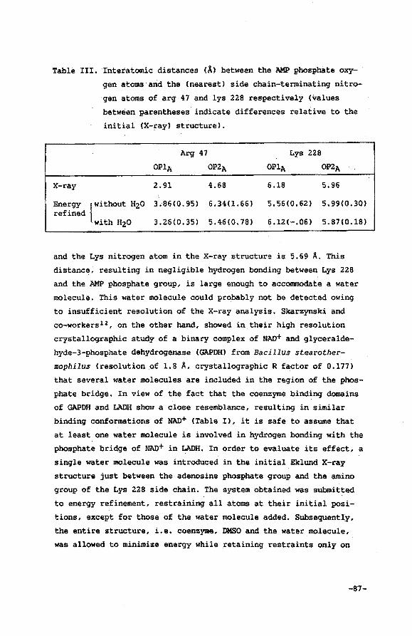
Table III. Interatomic distances (A) between the ~ phosphate oxy
gen atoms and the (nearest) side chain-terminatin9 nitro
gen atoms of arg 47 and lys 228 respectively (values
between parentheses indicate differences relative to the
initial (X-ray) structure).
A~ 47 Lys 228
OPlA OP2A OP1A OP2A
X-ray 2.91 4.68 6.18 5.96
Energy {without H20 3.86(0.95) 6.34(1.66) 5.56(0.62) 5.99(0.30) refined
with H20 3.26(0.35) 5.46(0.78) 6.12(-.06) 5.87(0.18)
and the Lys nitrogen atom in the X-ray structure is 5.69 A. This
distance; resulting in negligible hydrogen bonding between Lys 228
and the ~ phosphate group, is large enough to accommodate a water
molecule. This water molecule could probably not be detected owing
to insufficient resolution of the X-ray analysis. Skarzynski and
co-workersl2, on the other hand, showed in their high resolution
crystallographic study of a binary complex of NAD+ and glyceralde
hyde-3-phosphate dehydrogenase (GAPDH) from Bacillus stearother
mophilus (resolution of 1.8 A, crystallographic R factor of 0.177)
that several water molecules are included in the region of the phos
phate bridge. In view of the fact that the coenzyme bindin9 domains
of GAPDH and LADH show a close resemblance, resulting in similar
bindin9 conformations of NAD+ (Table I), it is safe to assume that
at least one water molecule is involved in hydrogen bonding with the
phosphate bridge of NAD+ in LADH. In order to evaluate its effect, a
single water molecule was introduced in the initial Eklund X-ray
structure just between the adenosine phosphate group and the amino
group of the Lys 228 side chain. The system obtained was submitted
to energy refinement, restrainin9 all atoms at their initial posi
tions, except for those of the water molecule added. Subsequently,
the entire structure, i.e. coenzyme, DMSO and the water molecule,
was allowed to minimize energy while retaining restraints only on
-81-
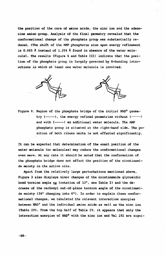
the position of the core of amino acids, the zinc ion and the adeno
sine amino group. Analysis of the final geometry revealed that the
conformational change of the phosphate group was substantially re
duced. (The shift of the AMP phosphorus atom upon energy refinement
is 0.665 A instead of 1.254 A found in absence of the water mole
cule). The results (Figure 4 and Table III) indicate that the posi
tion of the phosphate group is largely governed by H-bonding inter
actions in which at least one water molecule is involved.
Figure 4. Region of the phosphate bridge of the initial MAD+ geome
try (---), the energy refined geometries without ( .. ········)
and with (--) an additional water molecule. The AMP
phosphate group is situated at the right-hand side. The po
sition of both ribose units is not affected significantly.
It can be expected that determination of the exact position of the
water molecule (or molecules) may reduce the conformational changes
even more. At any rate it should be noted that the conformation of
the phosphate bridge does not affect the position of the nicotinami
de moiety in the active site.
Apart from the relatively large perturbations mentioned above,
Figure 3 also displays minor changes of the nicotinamide glycosidic
bond torsion angle XN (rotation of 13°, see Table I) and the de
crease of the carbonyl out-of-plane torsion angle of ~he nicotinami
de moiety (34° changing into 6°). In order to explain these confor
mational changes, we tabulated the relevant interaction energies
between MAO+ and the individual amino acids as well as the zinc ion
[Table IV). From the top half of Table IV, it appears that only the
interaction energies of NAD+ with the zinc ion and Val 292 are signi-
-88-
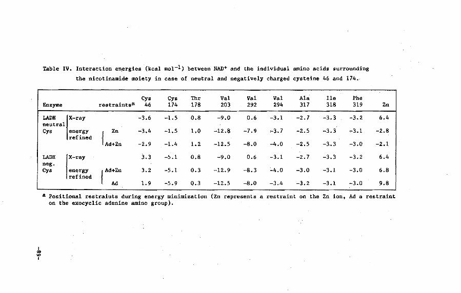
I co U)
I
Table IV. Interaction energies (kcal mol-l) between NAD+ and the individual amino acids surrounding
the nicotinamide moiety in case of neutral and negatively charged cysteine 46 and 174.
Cys Cys Thr Val Val Val Ala Ile Phe Enzyme restraints& 46 174 178 203 292 294 317 318 319 Zn --LADH rr"1 -3.6 -1.5 0.8 -9.0 0.6 -3.1 -2.7 -3.3 -3.2 6.4 neutral Cys energy { Zn -3.4 -1.5 1.0 -12.8 -7.9 ,..3.7 -2.5 -3.3 .,-3.1 -2.8
refined Ad+Zn -2.9 -1.4 1.2 -12.5 -8.0 -4.0 -2.5 -3.3 -3.0 -2.1
LADH rray 3.3 -5.1 0.8 -9.0 0.6 -3.1 -2.7 -3.3 -3.2 6.4 neg. Cys energy { Ad+Zn 3.2 -5.1 0.3 -12.9 -8.3 -4.0 -3.0 -3.1 -3.0 6.8
refined Ad 1.9 -5.9 0.3 -12.5 -8.0 -3.4 -3.2 -3.1 -3.0 9.8
a Positional restraints during energy minimization (Zn represents a restraint on the Zn ion. Ad a restraint on the exocyclic adenine amino group).
ficantly changed upon energy refinement. Apparently, the electrosta
tic repulsion between the positive charge of the zinc ion and the
positively charged CSN atom of the nicotinamide group induces a
slight torsion of the glycosidic bond (Table Il. This movement is
accompanied by an additional rotation around the C3~7N bond,
resulting in a diminished out-of-plane torsion angle of the amide
group. The latter rotation enables the amide oxygen C07Nl and the
hydrogen atom Csyn relative to the carbonyl dipole) to remain in
close contact with Phe 319 and Ala 317 respectively. Combination of
the above described interrelated rotational movements results in a
rather large displacement of one of the amide hydrogen atoms (anti
position to the carbonyl group), bringing it significantly closer to
the main-chain carbonyl group of Val 292. From this it is evident
that the magnitude of the carbonyl dipole out-of-plane rotation is
very much dependent on the exact positioning of Phe 319 and Val 292
and the extent of repulsion exerted by the zinc ion. The observed
repulsion effect stimulated us to investigate the influence of intro
ducing the negative charge on the cysteine 46 and 174 residues to
which the zinc ion is bound. This modification will diminish the
zinc-nicotinamide repulsion and, at the same time, settles with the
necessity of artificially restraining the zinc ion (vide supra).
AMBER parameters for the negatively charged cysteine residues
were directly taken from the AMBER parameter set except for the
atomic charges. The charges were derived from MNDO calculations of
neutral cysteine adding one formal negative charge to the sulfur
atoms, changing its value from +0.07 to -0.93. This method was adop
ted rather than using the calculated charges of deprotonated cys
teine itself, since in this case the extra negative charge is
neither stabilized by the S-H bond nor by the s-zn2+ bond. The
absence of a proton or zinc ion in the MNDO calculation will lead to
redistribution of t~e additional negative charge over all atoms,
including the main-chain atoms (The charge of the sulfur atom in
deprotonated cysteine in this case is -0.75). Figure 5 and 6 depict
the energy refined NAD+ geometry with and without the restraint on
the position of the zinc ion respectively.
-90-
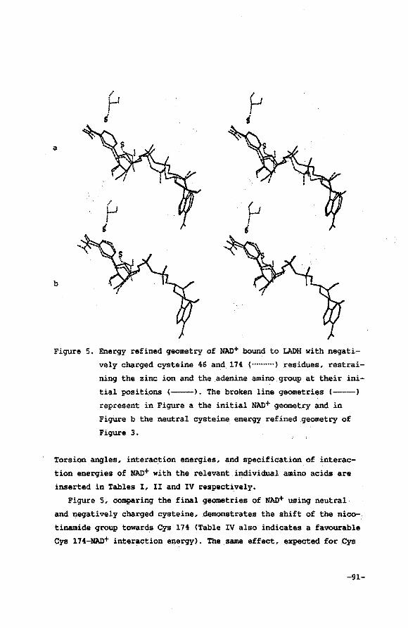
a
b
I I r r-·' i s
I l-1 I s
Figure 5. Energy refined geometry of NAD+ bound to LADH with negati-
vely charged cysteine 46 and. 174 ( .......... , residues, restrai-
ning the zinc ion and the .adenine amino group at their ini
tial positiqns (------). The broken line geometries (-----)
represent in Figure a the initial NAD+ geometry and in
Figure b the neutral cysteine energy refined geometry of
Figure 3.
Torsion angles, interaction energies, and specification of interac
tion energies of NAD+ with the relevant individual amino acids are
inserted in Tables I, II and IV respectively.
Figure 5, comparing the final geometries of NAO+ using neutral
and negatively charged cysteine, demonstrates the shift of the nico-.
tinamide group towards Cys 174 (Table IV also indicates a favourable
Cys 174-NAD+ interaction energy). The .same effect, expected for Cys
-91-
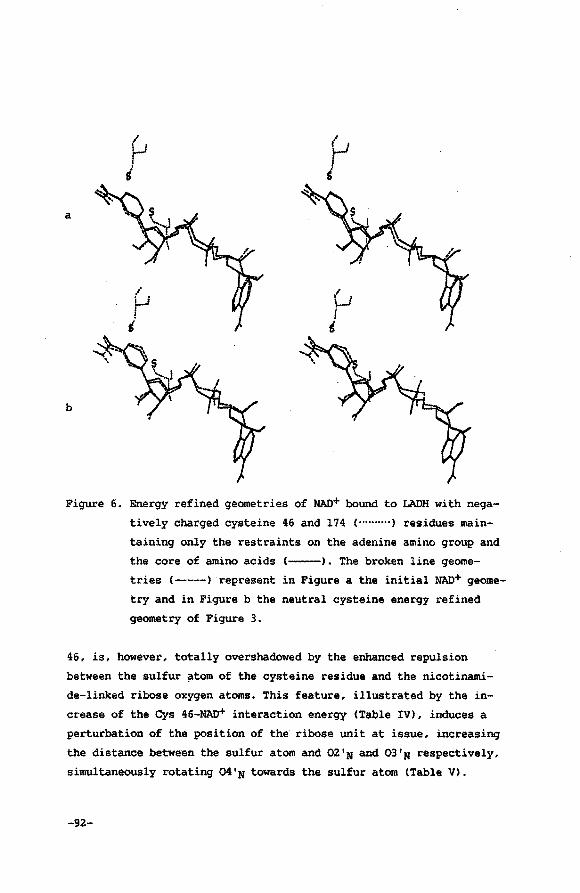
a
/
f-1
' 0 j
t
b
Figure 6. Energy refined qeometries of NAO+ bound to LADH with nega-
tively charged cysteine 46 and 174 ( .......... ) residues main-
taining only the restraints on the adenine amino group and
the core of amino acids (------). The broken line geome
tries (---) represent in Figure a the initial NAD+ geome
try and in Figure b the neutral cysteine energy refined
geometry of Figure 3.
46, is, however, totally overshadowed by the enhanced repulsion
between the sulfur ~tom of the cysteine residue and the nicotinami
de-linked ribose oxygen atoms. This feature, illustrated by the in
crease of the Cys 46-NAD+ interaction energy (Table IV), induces a
perturbation of the position of the ribose unit at issue, increasing
the distance between the sulfur atom and 02'N and 03'N respectively,
simultaneously rotating 04'N towards the sulfur atom (Table V).
-92-
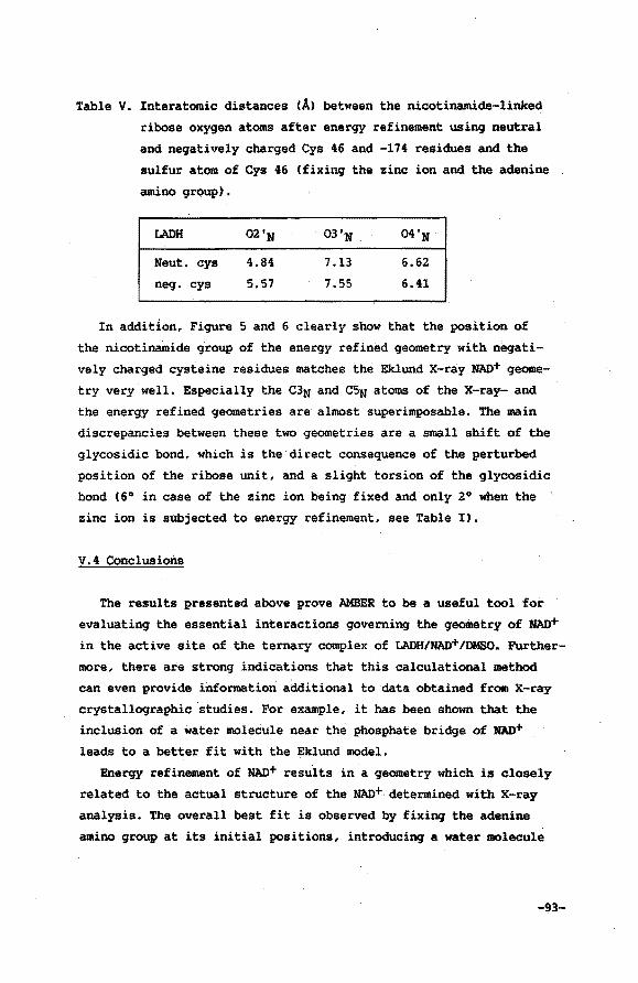
Table·V. Interatomic distances (A) between the nicotinamide-linked
ribose oxygen atoms after energy refinement using neutral
and negat~vely charged Cys 46 and -174 residues and the
sulfur atom of Cys 46 (fixing the zinc ion and the adenine
amino grQUP).
LADH
Neut. cys
neg. cys
02'N
4.84
5.57
7.13
7.55
04'N
6.62
6.41
In addition, Figure 5 and 6 clearly show that the position of
the nicotinamide group of the energy refined geometry with negati
vely charged cysteine residues matches the Eklund X-ray NAD+ geome
try very well. Especially the C3N and C5N atoms of the X-ray- and
the energy refined geometries are almost superimposable. The main
discrepancies between these two geometries are a small shift of the
glycosidic bond, which is the direct consequence of the perturbed
position of the ribose unit, and a slight torsion of the glycosidic
bond (6° in case of the zinc ion being fixed and only 2° when the
zinc ion is sUbjected to energy refinement, see Table I).
v.4 Conclusions
The results presented above prove AMBER to be a useful tool for
evaluating the essential interactions governing the geometry of NAD+
in the active site of the ternary complex of LADH/NAD+/DMSO. Further
more, there are strong indications that this calculational method
can even provide information additional to data obtained from X-ray
crystallographic studies. For example, it has been shown that the
inclusion of a water molecule near the phosphate bridge of NAD+
leads to a better fit with the Eklund model.
Energy refinement of NAD+ results in a geometry which is closely
related to the actual structure of the NAO+ determined with X-ray
analysis. The overall best fit is observed by fixing the adenine
amino group at its initial positions, introducing a water molecule
-93-
between the nicotinamide mononucleotide phosphate group and Lys 228,
and using negatively charged deprotonated Cys 46 and Cys 174 resi
dues. High resolution X-ray data obtained for a binary complex of
NAD+ and GAPDH12 (vide supra) reveal an NAD+ geometry which resem
bles the calculational geometry even better (Table I). This obser
vation becomes quite relevant considering the fact that the coenzyme
domains of several NAD+-dependent dehydrogenases (including LADH and
GAPDH) are closely related13 •
Comparing the NAD+ geometries in Table I, one should bear in mind
that torsion angles are highly sensitive to small deviations in
positional parameters of the atoms. Although one may expect errors
in the X-ray data of the coenzyme to be corrected by the energy
refinement procedure, inaccuracies in the positions of atoms within
the core of amino acids inevitably leads to the introduction of
systematic errors in the energy refined coenzyme geometry. Since
resolution of the x-ray data is limited to·2.9 A, the decrease of
the out-of-plane torsion angle of the nicotinamide carbonyl dipole,
for instance CLADH:30°, GAPDH:22° diminished to 6° upon energy
refinement), is probably due to relatively small perturbation of Val
292 or Phe 319 14 and to the torsional harmonic force constant added.
It can also be concluded that conformational changes of the
phosphate bridge and the AMP subunit do not affect the geometry of
the nicotinamide group. This finding is in accordance with obser
vations of Sicsic and co-workers15 , who showed that seve~al frag
ments of NAD+(H), nicotinamide mononucleotide and -mononucleoside
(in the presence of AMP) are functional in enzyme-catalyzed redox
reactions. In addition, immobilization using the exocyclic amino
group of adenine does not interfere with activity of NAD+ll.
In view of all these considerations AMBER looks capable of g1v1ng
a reasonably accurate description of the geometry of NAD+ bound in
the active site of ~ enzyme.
-94-
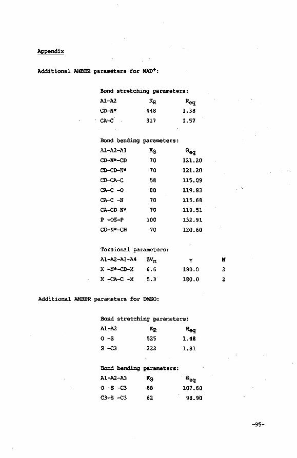
Appendix
Additional AMBER parameters for NAD+:
Bond stretching parameters:
Al-A2
CD-N*
CA'-C
Bond bending parameters:
A1-A2-A3 Ke CD-N*-cD 70
CD-cD-N* 70
CD-cA-c 58
CA-c -o 80
CA-c -N 70
CA-cD-N* 70
p -QS-P 100
CD-N*-cH 70
Torsional parameters:
A1-A2-A3-A4 %Vn
X -N*-cD-X 6.6
X -cA-c -X 5.3
Additional AMBER parameters for DMSO:
Reg:
1.38
1.57
E>eq 121.20
121.20
115.09
119.83
115.68
119.51
132.91
120.60
y
180.0
180.0
Bond stretchinq parameters:
A1-A2
o -s s ...C3
KR 525
222
Bond bending parameters:
A1-A2-A3 Ke 0 -s -c3 68
C3-S ...C3 62
Req 1.48
1.81
E>eq 107.60
98.90
N
2
2
-95'-'
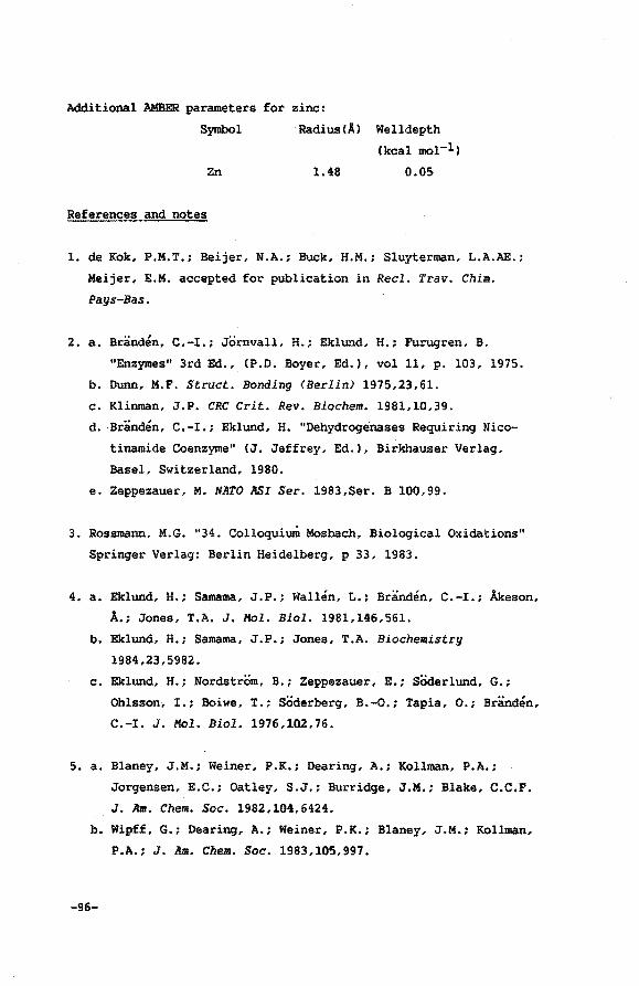
Additional AMBER parameters for zinc:
Symbol Radius!A) Welldepth
(kcal mol-l)
Zn 1.48 0.05
References and notes
1. de Kok, P.M.T.; Beijer, N.A.; Buck, H.M.; Sluyterman, L.A.AE.;
Meijer, E.M. accepted for publication in Reel. Trav. Chim.
Pays-Bas.
z. a. Branden, C.-I.; Jornvall, H.; Eklund, H.; Furugren, B.
"Enzymes" 3rd Ed., (P.D. Boyer, Ed.), vol 11, p. 103, 1975.
b. Dunn, M.F. Struct. Bonding <Berlin) 1975,23,61.
c. Klinman, J.P. CRC Crit. Rev. Biochem. 1981,10,39.
d. Branden, C.-I.; Eklund, H. "Dehydroge'nases Requiring Nico
tinamide Coenzyme" (J. Jeffrey, Ed.), Birkhauser Verlag,
Basel, Switzerland, 1980.
e. Zeppezauer, M. NATO ASI Ser. 1983,Ser. B 100,99.
3. Rossmann, M.G. "34. Colloquium Mosbach, Biological Oxidations"
Springer Verlag: Berlin Heidelberg, p 33, 1983.
4. a. Eklund, H.; Samama, J.P.; Wallen, L.; Branden, C.-I.; Akeson,
A.; Jones, T.A. J. Mol. Biol. 1981,146,561.
b. Eklund, H.; Samama, J.P.; Jones, T.A. Biochemistry
1984,23,5982.
c. Eklund, H.; Nordstrom, B.; Zeppezauer, E.; Soderlund, G.;
Ohlsson, I.; Boiwe, T.; SOderberg, B.-0.; Tapia, 0.; Branden,
C.-I. J. Mol. Biol. 1976,102,76.
5. a. Blaney, J.M.; Weiner, P.K.; Dearing, A.; Kollman, P.A.;
Jorgensen, E.C.; Oatley, S.J.; Burridge, J.M.; Blake, C.C.F.
J. Am. Chem. Soc. 1982,104,6424.
b. Wipff, G.; Dearing, A.; Weiner, P.K.; Blaney, J.M.; Kollman,
P.A.; J. Am. Chem. Soc. 1983,105,997.
-96-
c. Oatley, S.J.; Blaney, J.M.; Langridge, R.; Kollman, P.A.
Biopolymers 1984,23,2931.
d. Tilton, R.F.; Singh, U.C.; Weiner, S.J.; Connolly, M.L.;
Kuntz, I.D.; Kollman, P.A.; Max, N.; Case; D.A. J. Mol. Biol.
1986,192,443.
6. Weiner, P.K:; Kollman, P.A. J. Comp; Chem. 1981,2,287.
7. a. Weiner, S.J.; Kollman, P.A.; Case, D.A.; Singh, U.C.; Ghio,
C.; Alagona, G.; Profeta, S.; Weiner, P.K. J. Am. Chem. Soc.
1984,106,765.
b. Weiner, S.J.; Kollman, P.A.; Nguyen, D.T.; Case, D.A. J. Comp.
Chem. 1986,7,230.
8. a. Reference 7b.
b. Warshel, A. J. Phys. Chem. 1979,83,1640.
c. Rees, D.C. J. Mol. Biol. 1980,141,323.
9. Chem-X, july 1987 update, copyright Chemical Design Oxford Ltd.,
Oxford.
10. Residue numbers of the amino acids taken into account are:
46-48,51,56,57,67,68,93,94,116,140,141,173-175,178,182,~00-204,
222-225,228,268,269,271,274,291-295,316-320,362 and 369.
ll. Mosbach, K. "Enzymes in Organic Synthesis, Ciba Foundation
Symposium lll" (R. Porter and s. Clark, Eds.), p 57, Pitman,
London, 1985 and references cited therein.
12. Skarzynski, T.; Moody, P.C.E.; Wonacott, A.J. J. Mol. Biol.
1987,193,171.
13. a. Ohlsson, I.; Nordstrom, B.; Branden, c.-I. J. Mol. Biol.
1974,89,339.
b. Rossmann, M.G.; Moras, D.; Olsen, K.W. Nature (London>
1974,250,194.
-97-
c. Rossmann, M.G.; Liljas, A.; Branden, C.-I.; Banaszak. L.J.
"Enzymes" 3rd. Ed. (P.D. Boyer, Ed.) vol. 11, p. 61, 1975.
14. a. Chapters II, III and IV.
b. Donkers1oot, M.C.A.; Buck, H.M. J. Am. Chem. Soc.
1981,103,6554.
c. de Kok, P.M.T.; Donkersloot, M.C.A.; van Lier, P.M.;
Meulendijks, G.H.W.M.; Bastiaansen, L.A.M.; van Hoof£,
H.J.G.; Kanters, J.A.; Buck, H.M. Tetrahedron 1986,42,941.
15. a. Sicsic, S.; Durand, P.; Langrene, S.; Le Goffic, F. FEBS
Lett. 1984,176,321.
b. Sicsic, S.; Durand, P.; Lanqrene, S.; Le Goffic, F. Eur. J.
Biochem. 1986,155,403.
-98-
Molecular Mechanics calculation o£ geometries o£ ao+ derivatives,
IDOdified in the nicotinamide group, in a ternary COIIIplez with
Horse Liver Alcohol Dehydroqenasel
VI.l Introduction
In the previous Chapter AMBER molecular mechanics calculations
and energy minimi.zations2 .were shown to be very useful to obtain·.
insight in the relevant interactions between the coenzyme Nicotin
amide Adenine Dinucleotide (NAD+) and the enzyme Horse Liver Alcohol
Dehydrogenase (LADH) in a ternary complex with DMS03 • It has been
shown that this calculational method can give a reasonably good
estimate of the coenzyme geometry in the active site of the complex.
Moreover it permits analysis of individual interactions between all
constituent units of the ternary complex and it gives a strong indi
cation of their relative importance to the actual coenzyme geometry.
The results prompted us to test whether AMBER can be used to ob
tain a description of the geometry of NAD+ analogues in the ternary
complex. In this Chapter the AMBER molecular modelling package will
be applied to minimize the conformational energy of success~vely
sNAD+ (Figure 1) 4 ac3PdAD+, clac3PdAD+, pp3pdAD+, PdAD+, m4NAD+ and
cn3PdAD+, within a cage constructed by a limited number of amino
acidS of the enzyme.
Kinetic data for the enzyme-catalyzed reactions with several of
the previously mentioned coenzyme analogues have been reported&.
Interpretation of these results in terms of coenzyme geometries
remains speculative so far. This study presents a novel approach in
estimating ternary complex coenzyme geometries independent of the
availability of additional X-ray data. It will be shown that using
this technique one is able to rationalize individual substituent
effects. The results presented here clearly demonstrate that the
overall reactivity of the NAD+ derivatives is in most cases directly
... 99-
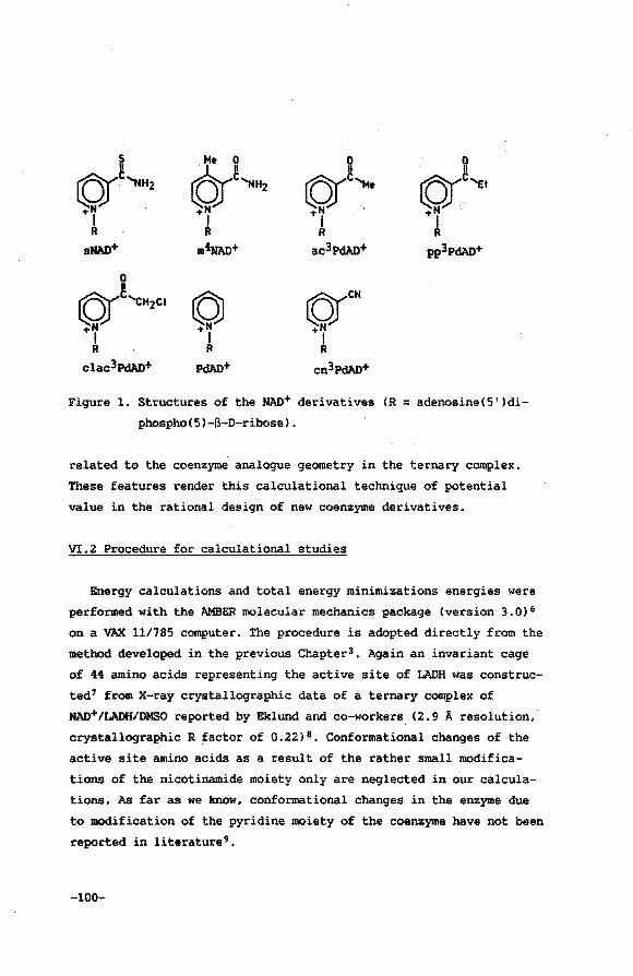
I 6J~,, tOr""' 0
©r~'Et !Of· 'NH2
+N +N +I A
+I I R R R
sHAD+ m4NAD+ ac3pdAI)+ pp3pdAI)+
©(~'cH2c• © ©rCN +N -tN -tN
I I I R R R
clac3pdAI)+ pdAI)+ cn3pdAI)+
Figure 1. Structures of the NAO+ derivatives (R = adenosine(5')di
phospho(5)-~-D-ribose).
related to the coenzyme analogue geometry in the ternary complex.
These features render this calculational technique of potential
value in the rational design of new coenzyme derivatives.
VI.2 Procedure for calculational studies
Energy calculations and total energy minimizations energies were
performed with the AMBER molecular mechanics package (version 3.0) 6
on a ~ 11/785 computer. The procedure is adopted directly from the
method developed in the previous Chapter3 • Again an invariant cage
of 44 amino acids representing the active site of LADH was construc
ted7 from X-ray crystallographic data of a ternary complex of
NAD+/LADH/DMSO reported by Eklund and co-workers. (2.9 A resolution,
crystallographic R ~actor of 0.22) 8 • Conformational changes of the
active site amino acids as a result of the rather small modifica
tions of the nicotinamide moiety only are neglected in our calcula
tions. As far as we know, conformational changes. in the enzyme due
to modification of the pyridine moiety of the coenzyme have not been
reported in literature9.
-100-
The 3D structure of the NAD+ analogues was derived directly from
the X-ray NAD+ geometry using aq interactive computer graphics pro
gram (Chem-X)lO. Structural parameters not available in the AMBER
data base were added (Appendix) using standard bond-lengths and
-angles. Harmonic force constants were either obtained from litera
ture or extrapolated from available data11 • Atomic charges were
calculated using the MNDO semi-empirical molecular orbital method.
All energy minimizations were performed until the RMS gradient
value of the energy was less than 0.1 kcal A-1, using the distance
dependent dielectric constant 12 and treating all CH, CH2 and CH3
groups as united atoms. Since the actual structure of the phosphate
bridge has shown to be of minor importance to the conformation of
the nicotinamide group 3 , it is permitted to ignore the presumed
inclusion of a water molecule near the phosphate bridge binding
region. The same holds for the adenosine part of the coenzyme which
justifies the introduction of a positional restraint on the exo
cyclic adenine NH2 group in order to reduce the c.omputational time.
In the preceding Chapter the necessity of either fixing the zinc
ion at its initial position or simulating the enzyme-zo2+ inter
actions by deprotonating the Cys 46 and Cys 174 residues, including
formal negative charges to both residues via modification of the
AMBER data base, has been mentioned. The latter method has shown to
lead to the introduction of repulsion forces between the negatively
charged cysteine 46 sulfur atom and the 02• and 04• atoms of the
ribose unit adjacent to the nicotinamide group. These newly intro
duced interactions primarily determine the position of the ribose
unit concerned. As a result of the repositioning of the ribose unit,
the nicotinamide moiety of the energy refined NAD+ geometry is
somewhat shifted leading to a slightly better fit with the X-ray
structure. In order to prevent build-up of extra tension in the
glycosidic bond we have applied the method of fixing the zinc ion.
In this context it should be noted that the arbitrary choice between
these two methods only introduces a different· systematic error in
the energy refined geometries of NAD+ and its analogues, probably
even smaller than the deviations resulting from inaccuracies in the
-101-
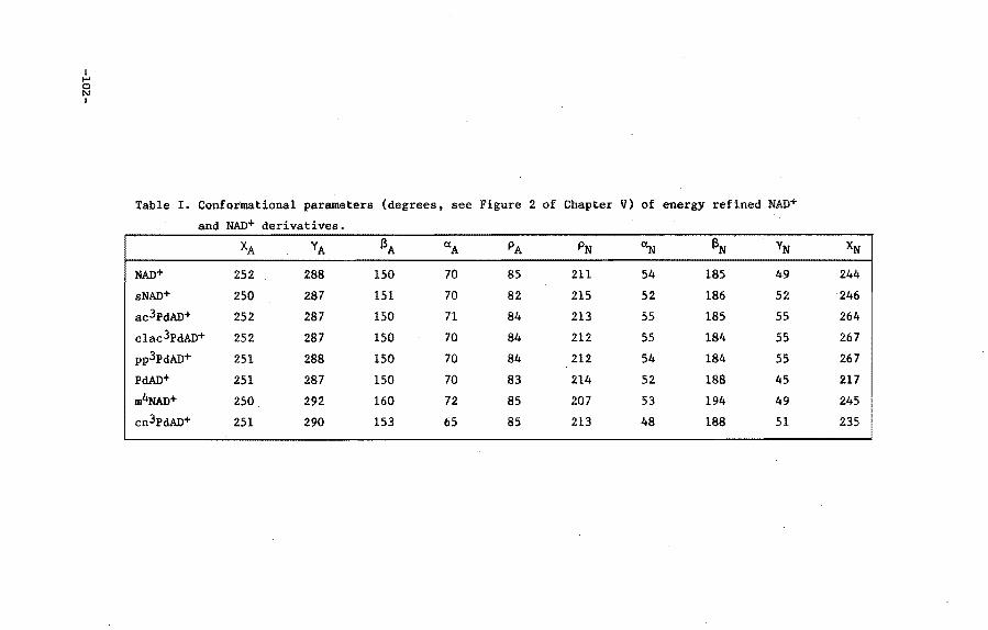
I ..... 0 N I
Table I. Conformational parameters (degrees. see Figure 2 of Chapter V) of energy refined NAD+
and NAD+ derivatives.
XA YA a A a. A PA PN ~ aN YN
NAD+ 252 288 150 70 85 211 54 185 49
sNAD+ 250 287 151 70 82 215 52 186 52
ac3PdAD+ 252 287 150 71 84 213 55 185 55
clac3PdAD+ 252 287 150 70 84 212 55 184 55
pp3PdAD+ 251 288 150 70 84 212 54 184 55
PdAD+ 251 287 150 70 83 214 52 188 45
m4NAo+ 250 292 160 72 85 207 53 194 49
cn3PdAD+ 251 290 153 65 85 213 48 188 51
XN
244
246
264
267
267
217
245
235
amino acids X-ray data. Using slightly erroneous positional parame
ters for the core of amino acids will inevitably perturb the optimal
NAD+ and MAO+ analogue geometries. For this reason it has no sence
to give a highly detailed description of the coenzyme (analogue)
geometry using X-ray data of limited resolution (e.g. the NAD+-carbo
nyl out-of-plane orientation13 ). Since such deviations of the core
of amino acids are systematically present in all minimization proce
dures, effects tend to cancel when two energy refined structures are
compared. In addition, the initial NAD+ geometry may be somewhat
erroneous owing to the limited resolution of the X-ray data. These
inaccuracies however are likely to be systematically corrected with
in the energy minimization procedures. From the foregoing, it is evi
dent that in order to evaluate the effect of substituents upon the
NAD+ geometry, the energy refined MAD+ geometry should be used as a
reference.
VI.3 Results
During energy minimization of the NAD+ analogues, the total ener
gy drops within a the range of 120 to 940 kcal mol-l, all converging
to a value of 920 kcal mol-l. The smallest total energy decrease is
observed for PdAD+. On substituting the pyridinium moiety the energy
drop increases upto a maximum value obtained for sNAO+. Upon energy
refinement, a similar pattern for the interaction energy between the
coenzyme derivatives and the core of amino acids (the energy drop
varies from 60 to 460 kcal mol-l) is observed.
Computer generated stereodiagrams 10 of the nicotinamide moiety of
the optimized geometries, relative to the energy refined geometry of
the active part of NAD+ itself, are presented in Figure 2. Since con
formational discrepancies are restricted to the nicotinamide moiety,
only these regions of the NAD+ structures are drawn. Neither the
zinc ion nor the amino acids are depicted since they are invariant
during all calculations.
Table I lists the numerical values of the torsion angles of the
coenzyme analogues. Total interaction energies are summarized in
Table II.
-103-
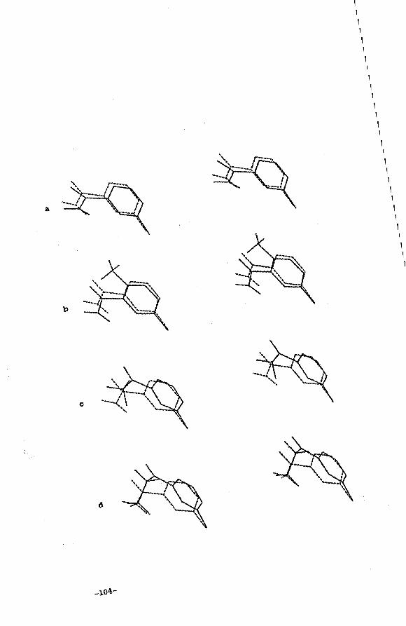
1 1 1 1 1 1 1 1 1 1 1 1 1 1 1 1 1 1 1 1 1 1 1 1 1
a 1 1 1 1 1
c
1 1 1 1 1 1 1 1 1 1 1 1 1 1 1 1 1 1 1 1 1 1 1 1 1 1 1 1 1 1 1 1 1 1 1
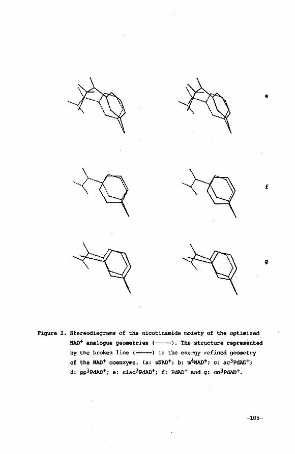
Figure 2. Stereodiagrams of the nicotinamide moiety of the optimized
NAD+ analogue geometries <--). The structure represented
by the broken line (-----) is the energy refined geometry
of the NAD+ coenzyme. (a: sHAD+; b: m~+; c: ac3PdAD+;
d: pp3pdAD+; e: clac3PdAD+; f: PdAD+ and g: cn3pdAD+.
-105-
e
f
9
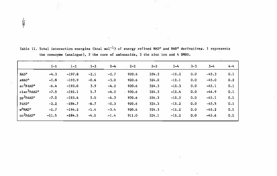
I ..... 0 en I
Table II. Total interaction energies (kcal mol-l) of energy refined NAD+ and NAD+ derivatives. 1 represents
the coenzyme (analogue), 2 the core of aminoacids, 3 the zinc ion and 4 DMSO.
1-1 1-2 1-3 1-4 2-2 2-3 2-4 3-3 3-4 4-4
NAD+ -4.3 -197.8 -2.1 -2.7 920.6 324.3 -11.2 0.0 -45.3 0.1
sNAD+ -1.8 -193.9 -0.6 -3.0 920.6 324.0 -11.1 0.0 -45.0 0.2
ac3PdAD+ -6.4 -193.0 3.9 -4.2 920.6 324.3 -11.3 0.0 -45.1 0.1
clac3PdAD+ -7.9 -192.1 5.7 -4.2 920.6 324.3 -11.4 0.0 -44.9 0.1
pp3PdAD+ -7.0 -193.4 5.0 -4.3 920.6 324.3 -11.3 0.0 -45.1 0.1
PdAD+ -2.2 -184.7 -6.7 -0.3 920.6 324.3 -11.2 o.o -45.5 0.1
m4NAD+ -1.7 -194.2 -1.4 -3.4 920.6 324.3 -11.2 0.0 -45.2 0.1
cn3PdAD+ -11.5 -184.5 -4.1 -1.4 911.0 324.1 -11.2 0.0 -45.6 0.1
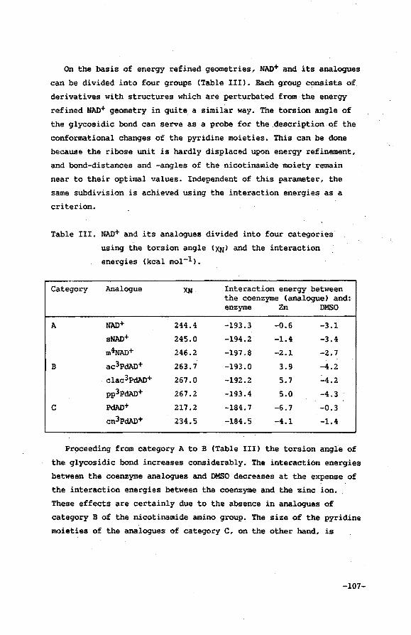
On the basis of energy refined.geometries, NAD+ and its analogues
can be divided into four groups (Table III). Each group consists of
derivatives with structures which are perturbated from the energy
refined NAD+ geometry in quite a similar way. The torsion angle of
the glycosidic bond can serve as a probe for the description of the
conformational changes of the pyridine moieties. This can be done
because the ribose unit is hardly displaced upon energy refinement,
and bond-distances and -angles of the nicotinamide moiety remain
near to their optimal values. Independent of this parameter, the
same subdivision is achieved using the interaction energies as a
criterion.
Table III. NAD+ and its analogues divided into four categories
using the torsion angle (XN) and the interaction
energies (kcal mol-l).
Category Analogue XN Interaction energy between the coenzyme (analogue) and: enzyme Zn DMSO
A NAD+ 244.4 -193.3 -0.6 -3.1 sNAD+ 245.0 -194.2 -1.4 -3.4 m4NAD+ 246.2 -197.8 -2.1 -2.7
B ac3PdAD+ 263.7 -193.0 3.9 -4.2 clac3PdAD+ 267.0 -192.2 5.7 -4.2 pp3pdAD+ 267.2 -193.4 5.0 -4.3
c PdAD+ 217.2 -184.7 -6.7 -0.3 cn3PdAD+ 234.5 -184.5 -4.1 -1.4
Proceeding from category A to B (Table III) the torsion angle of
the glycosidic bond increases considerably. The interaction energies
between the coenzyme analogues and DMSO decreases at the expense of
the interaction energies between the coenzyme and the zinc ion.
These effects are certainly due to the absence in analogues of
category B of the nicotinamide amino group. The size of the pyridine
moieties of the analogues of category C, on the other hand, is
-107-
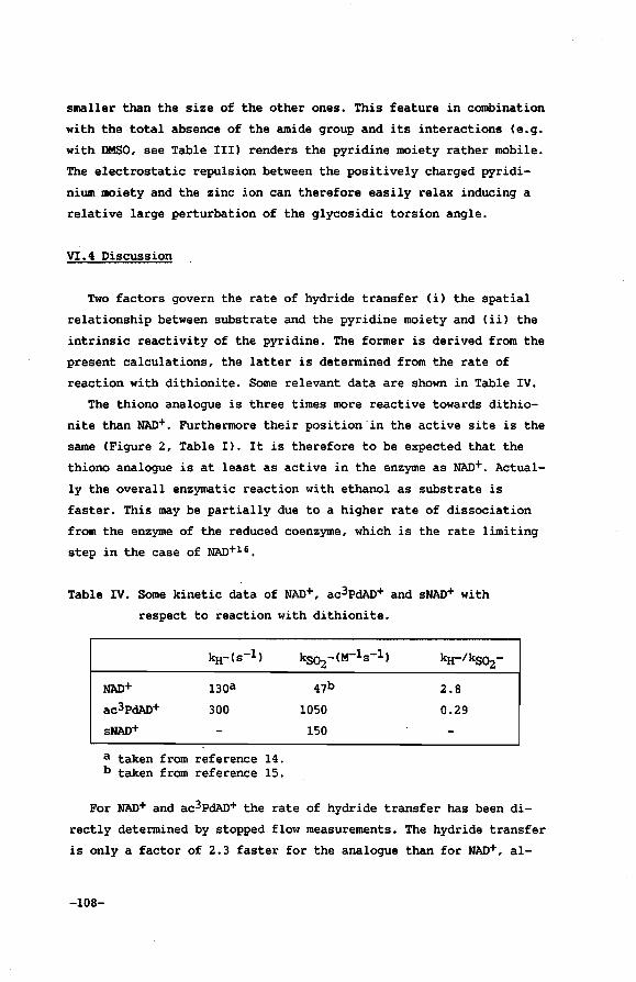
smaller than the size of the other ones. This feature in combination
with the total absence of the amide group and its interactions (e.g.
with DMSO, see Table III) renders the pyridine moiety rather mobile.
The electrostatic repulsion between the positively charged pyridi
nium moiety and the zinc ion can therefore easily relax inducing a
relative large perturbation of the glycosidic torsion angle.
VI.4 Discussion
Two factors govern the rate of hydride transfer (il the spatial
relationship between substrate and the pyridine moiety and (iil the
intrinsic reactivity of the pyridine. The former is derived from the
present calculations, the latter is determined from the rate of
reaction with dithionite. Some relevant data are shown in Table IV.
The thiono analogue is three times more reactive towards dithio
nite than NAD+. Furthermore their position'in the active site is the
same (Figure 2, Table Il. It is therefore to be expected that the
thiono analogue is at least as active in the enzyme as NAD+. Actual
ly the overall enzymatic reaction with ethanol as substrate is
faster. This may be partially due to a higher rate of dissociation
from the enzyme of the reduced coenzyme, which is the rate limiting
step in the case of NAD+l6.
Table IV. Some kinetic data of NAD+, ac3PdAD+ and sNAD+ with
respect to reaction with dithionite.
130a
300
a taken from reference 14. b taken from reference 15.
47b
1050
150
2.8
0.29
For NAD+ and ac3PdAD+ the rate of hydride transfer has been di
rectly determined by stopped flow measurements. The hydride transfer
is only a factor of 2.3 faster for the analogue than for NAD+, al-
-108-
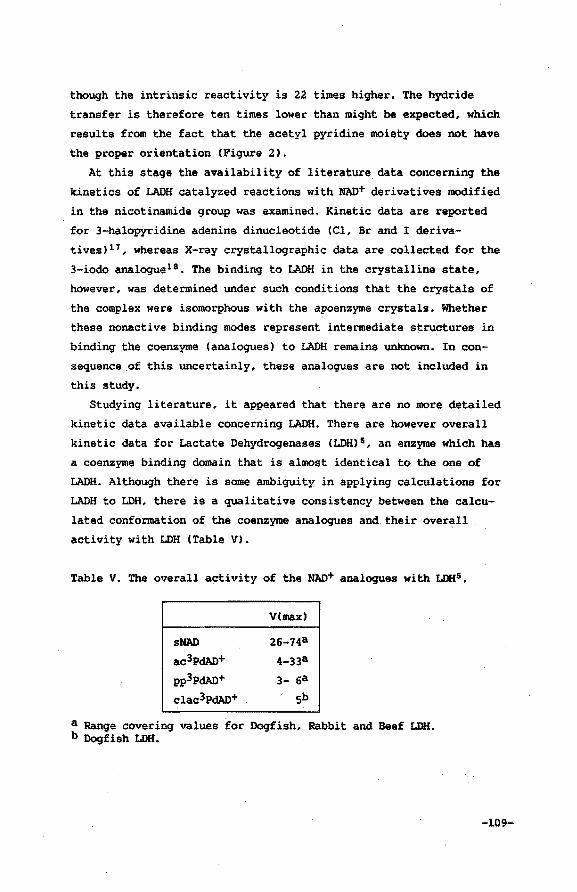
though the intrinsic reactivity is 22 times higher. 7he hydride
transfer is therefore ten times lower than might be expected, which
results from the fact that the acetyl pyridine moiety does not have
the proper orientation (Figure 2).
At this stage the availability of literature data concerning the
kinetics of LADH catalyzed reactions with MAD+ derivatives modified
in the nicotinamide group was examined. Kinetic data are reported
for 3-halopyridine adenine dinucleotide (Cl, Br and I deriva
tives)17, whereas X-ray crystallographic data are collected for the
3-iodo analogue18 • 7he binding to LADH in the crystalline state,
however, was determined under such conditions that the crystals of
the complex were isomorphous with the apoenzyme crystals. Whether
these nonactive binding modes represent intermediate structures in
binding the coenzyme (analogues) to LADH remains unknown. In con
sequence of this uncertainly, these analogues are not included in
this study.
Studying literature, it appeared that there are no more detailed
kinetic data available concerning LADH. There are however overall
kinetic data for Lactate Dehydrogenases (LDH) 5 , an enzyme which has
a coenzyme binding domain that is almost identical to the one of
LADH. Although there is some ambiguity in applying calculations for
LADH to LDH, there is a qualitative consistency between the calcu
lated conformation of the coenzyme analogues and their overall
activity with LDH (Table V).
Table V. The overall activity of the NAD+ analogues with LDHs.
sNAD
ac3PdAD+
pp3pdAD+
clac3PdAD+
V(max)
26-74a
4-33a
3- 6a
sb
a Range covering values for Dogfish, Rabbit and Beef LDH. b Dogfish LDH.
-109-
MAD+ and sNAD+, both category A, are fully active. Compounds of
category B are also active, though to a smaller extent. Those of
category C, deviating most from MAD+ conformation, exhibit no acti
vity. Nevertheless they are competitive inhibitors with respect to
MAD+, implying that they bind to the same enzyme site. m4NAD+ is a
special case, it is a strong competitive inhibitor because it binds
to LADH in the same way as NAD+ does. The 4-methyl group, owing to
its electron releasing capacity however, renders the redox potential
too negative for coenzyme activityla,
VI. 5 Conclusions
The overall reactivity of MAO+ and MAD+ derivatives in reactions
catalyzed by dehydrogenases depends on several factors. Except for
the intrinsic reactivity of the coenzyme (analogue) used, the disso
ciation rate of the reduced species is knoWn to be of importance to
the kcat 15 • The intrinsic reactivity can be determined studying the
non-enzymatic reduction of an NAD+ analogue. The relative importance
of the coenzyme dissociation. rate can be deduced from pre-steady
state kinetics. By far the most difficult problem in quantifying the
overall reactivity is estimating the influence of coenzyme geometry
perturbations resulting from the introduction of substituents.
Insight in these effects is very much dependent on the availability
of crystallographic data concerning ternary complexes with NAD+ deri
vatives. Not only is this technique very laborious, it cannot be
applied under all circumstances; the preparation of single crystals
is in most cases the limiting factor. For this reason explanation of
effects resulting from coenzyme modification remain rather specula
tive.
This study presents a new approach in estimating the ternary com
plex coenzyme analogue geometry independent from the availability of
additional X-ray data. Substituent effects, resulting in perturba
tions of the positioning of the pyridine moiety, can be rationalized
and are in most cases of crucial importance in explaining the over
all enzymatic rate. In some cases, however, this study clearly indi
cated the redox potential to determine the reactivity of the NAD+
-110-
derivative. For instance the methyl group at the 4 position of
m4sNAD+ has been said to cause considerable overcrowding5 • This
study indicates, however, that on introducing the 4-methyl substi
tuent, the NAD+ geometry is well-preserved. The lack of activity of
m4NAD+ derivatives can be easily explained by the electron releasing
capacity of the 4--methyl groupl3,
Owing to the limited resolution of the X-ray data, the calculatio
nal method outlined in this Chapter can only, as it is, be applied
to determine substituent effects, resulting from coenzyme modifica
tions, upon the ternary complex geometry. Table I and Figure 2
clearly demonstrate that the amide group of the nicotinamide moiety
is of vital importance to the coenzyme geometry. It should be empha
sized that in order to elucidate the exact role of the orientation
of the amide group (CO out-of-plane orientation), the accuracy of
the calculational method should be enhanced. This can probably be
accomplished by applying high-resolution X-ray data for the initial
LADH/NAD+/DMSO complex. Presently, these data are, however, not
available.
Still, the calculation method presented here proves to be able to
establish qualitatively the relation between the measured enzymatic
rates and the calculated conformations of the coenzyme used. It is
capable of rationalizing individual substituent effects and of evalu
ating the essential NAD+ analogue/enzyme/DMSO/Zn2+ interact~ons.
Appendix
Additional data used for the modified NAD+ analogues:
(AMBER parameters for NAD+ presented in the Appendix of Chapter v are also included)
Bond bending parameters
Al-A2-A3 ·Ke 9eq
CA-c -cT 85 120. 0
C -cT-HC 35 109.5
-111-
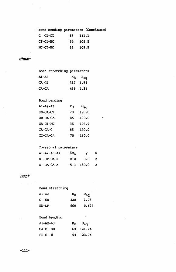
Bond bending parameters (Continued}
c -CT-CT 63 111.1
CT-CT-HC 35 109.5
HC-CT-HC 36 109.5
m~AD+
Bond stretching parameters
A1-A2 KR Req CA-cT 317 1.51
CA-CA 469 1.39
Bond bending
A1-A2-A3 Ke eeq CD-CA-cT 70 120.0
CD-CA-CA 85 120.0
CA-cT-HC 35 109.9
CA-CA-c 85 120.0
CT-CA-CA 70 120.0
Torsional parameters
A1-A2-A3-A4 Y.Vn y N
X -CT-CA-X o.o 0.0 2
X -CA-CA-X 5.3 180.0 2
sNAD+
Bond stretching
A1-A2 KR Req C -SD 328 1. 71
SD-LP 600 0.679
Bond bending
A1-A2-A3 Ke eeq CA-c -SD 64 121.24
so-c -N 64 123.74
-112-
cn3PdAD+
Bond bending (Continued)
C -SD-LP 600 96.70
LP-SD-LP 600 160.00
Torsional parameters
Al-A2-A3-A4 Y.Vn
x -c -so-x 1. o
Bond stretching·
Al-A2 KR
CA-c' 444
C'-N' 720
Bond bendinq
Al-A2-A3 Ke CD-cA-c.• 85
CA-c'-N' 70
y N 0.0 3
Req 1.42
1.16
eeq 119.42
180.0
clac3pdAJl+
Bond stretching
Al-A2 KR Req CT-cL 465 1.78
Bond bending
Al-A2-A3 Ke eeq HC-cT-cL 71 106.9
C -cT-cL 82 111.3
References and notes
1. de Kok, P.M.T.; Beijer, N.A.; Buck, H.M.; Sluyterman, L.A.AE.;
Meijer, E.M. submitted for publication in Eur. J. Biochem.
-113-
2. a. Blaney, J.M.; Weiner, P.K.; Dearing, A.; Kollman, P.A.;
Jorgensen, E.C.; Oatley, S.J.; Burridge, J.M.; Blake, C.C.F.
J. Am. Chem. Soc. 1982,104,6424.
b. Wipff, G.; Dearing, A.; Weiner, P.K.; Blaney, J.M.; Kollman,
P.A. J. Am. Chem. Soc. 1983,105,997.
c. Oatley, S.J.; Blaney, J.M.; Langridge, R.; Kollman, P.A.
Biopolymers 1984,23,2931.
d. Tilton, R.F.; Singh, U.C.; Weiner, S.J.; Connolly, M.L.;
Kuntz, I.D.; Kollman, P.A.; Max, N.; Case, D.A. J. Mol. Biol.
1986,192,443.
3. de Kok, P.M.T.; Beijer, M.A.; Buck, H.M.; Sluyterman, L.A.AE.;
Meijer, E.M. accepted for publication in Reel. Trav. Chim.
Pays-Bas.
4. Abbreviations: sNAD+, thionicotinamide adenine dinucleotide;
ac3PdAD+, 3-acetylpyridine adenine dinucleotide; clac3PdAD+,
3-chloroacetylpyridine adenine dinucleotide; pp3pdAD+, 3-propi
onylpyridine adenine dinucleotide; PdAD+, pyridine adenine dinu
cleotide; m4NAD+, 4-methyl nicotinamide adenine dinucleotide;
cn3pdAD+, 3-cyanopyridine adenine dinucleotide; m4sNAD+, 4-methyl
thionicotinamide adenine dinucleotide.
5. Samama, J.-P.; Marchal-Rosenheimer, N.; Biellmann, J.-F.;
Rossmann, M.G. Eur. J. Biochem. 1981,120,563.
6. Weiner, P.K.; Kollman, P.A. J. Comp. Chem. 1981,2,287.
7. Residue numbers of the amino acids taken into account are: 46-48,
51,56,57,67,68,93,94,116,140,141,173-175,178,182,200-204,222-225,
228,268,269,271,~74,291-295,316-320,362 and 369.
8. Eklund, H.; Samama, J.-P.; Wallen, L.; Branden, c.-I.; Akeson,
A.; Jones, T.A. J. Mol. Biol. 1981,146,561.
9. Biellmann, J.-F. Ace. Chem. Res. 1986,19,321.
-114-
10. Chem-X, july 1987 update, copyright Chemical Design Oxford Ltd.,
Oxford.
11. a. Weiner, S.J.; Kollman, P.A.; Case, D.A.; Singh, U.C.; Ghio, ·
C.; Ala9ona, G.; Profeta, S.; Weiner, P.K. J. Jim. Chem. Soc •.
1984,106,765.
b. Weiner, S.J.; Kollman, P.A.; Nguyen, D.T.; Case, D.A. J.
Comp. Chem. 1986,7,230.
12. a. Reference 11b.
b. Warshel, A. J. PhyS'. Chem. 1979,83,1640.
c. Rees, D.C. J. Hol. Biol. 1980,141,323.
13. a. Chapters II, III and IV.
b. Donkersloot, M.C.A.; Buck, H.M. J. Am. Chem. Soc.
1981,103,6554.
c. de Kok, P.M.T.; Donkersloot, M.C.A.; van Lier, P.M.;
Meulendijks, G.H.W.M.; Bastiaansen, L.A.M.; van Hooff,
H.J .G.; Kanters, J .A.; Buck, H.M. Tetrahedron 1986,42,941.
14. Shore, J.D.; Brooks, R.L. Brch. Biochem. Biophys. 1971.1t7,825.
15. Blankenhorn, G.; Moore, E.G. J. 1111. Chem. Soc. 1980,102,1092.
16. Joppich-Kuhn, R.; Luisi, P.L. Eur. J. Biochem. 1978,83,593.
17. a. Fischer, T.L.; Vercellotti, S.V.; Anderson, B.M. J. Biol.
Chem. 1973.,2t8, 4293.
b. Abdallah, M.A.; Biellmann, J.-F.; Samama, J.-P.; Wrixon, A.D.
Eur. J. Biochem. 1976,64,351.
c. Abdallah, M.A.; Biellmann, J.-F. Eur. J. Biochem.
1980,112,331.
d. Abdallah, M.A.; Cedergren-Zeppezauer, E.; Gerber, M.;
Dietrich, H.; Zeppezauer, M.; .Koerber, S.C.; MacGibbon,
A.K.H.; Dunn, M.F. Biochemistry 1984,23,1003.
18. Samama, J.-P.; Zeppezauer, E.; Biellmann, J.-F.; Branden, C.-I.
Eur. J. Biochem. 1977,81,403. -us-
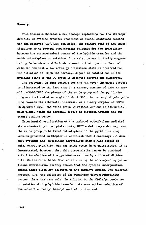
This thesis ,elaborates a new concept explaining how the stereope
cificity in hydride transfer reactions of (model compounds related
to) the coenzyme NAD+tNADH can arise. The primary goal of the inves
tigations is to provide experimental evidence for the correlation
between the stereochemical course of the hydride transfer and the
amide out-of-plane orientation. This relation was initially sugges
ted by Donkersloot and Buck who showed in their quantum chemical
calculations that a low-enthalpy transition state is observed for
the situation in which the carbonyl dipole is rotated out of the
pyridine plane if the CO group is directed towards the substrate.
The relevancy of this concept for the "in vivo" enzymatic process
is illustrated by the fact that in a ternary complex of LADH (A spe
cific)tNAD+tDMSO the planes of the amide group and the pyridinium
ring are inclined at an angle of about 30°, the carbonyl dipole poin
ting towards the substrate. Likewise, in a binary complex of GAPDH
(B specific)tNAO+ the amide group is rotated 22° out of the pyridi
nium plane. Again the carbonyl dipole is directed towards the sub
strate binding region.
Experimental verification of the carbonyl out-of-plane mediated
stereochemical hydride uptake, using NAD+ model compounds, requires
the amide group to be fixed out-of-plane of the pyridinium ring.
Results presented in Chapter II establish that 3-carbamoyl-2,4-dime
thyl pyridine and -pyridinium derivatives show a high degree of
axial chiral stability when the amide group is di-substituted. It is
demonstrated, however, that this prerequisite cannot be combined
with 1,4-reduction of the pyridinium cations by action of dithio
nite. On the other hand, Ohno et al., using the corresponding quino
linium derivatives, clearly showed that the hydride incorporation
indeed takes place,syn relative to the carbonyl dipOle. The reversed
process, i.e. the oxidation of the resulting dihydroquinolinium
system, obeys the same rule. In addition to the C(4)H/amide-co syn
orientation during hydride transfer, stereoselective reduction of
the substrate (methyl benzoylformate) is observed.
-116-
In Chapter III a method is developed for the direct synthesis of
the 1,4-dihydropyridine compounds derived from the axial chiral pyri
dinium compounds introduced in Chapter II. Enantiomeric separation
of the 3-(N,N-dimethylcarbamoyl)-1,2,4-trimethyl-1,4-dihydropyridine
was accomplished chromatographically using cellulose-triacetate as
the stationary phase. Determination of the absolute configuration at
C(4) of the predominant enantiomer was achieved using CD spectros
copy. Oxidative generation of the corresponding axial chiral pyridi
nium cation proceeded with 70% optical yield. Preliminary experi
ments show that the optical yield increases. to 90% when the experi
ment is conducted at 0°C. ·Eu(hfc)3 la NMR techniques were applied to
elucidate the absolute conformation of the CO out-of-plane orienta
tion. The results prove the mechanism proposed by Donkersloot and
Buck to be operative. In addition, the substrate (methyl benzoylfor
mate) is .converted to the methyl mandelate in 63% optical yield at
22°C. This value increased to 80% when the reaction is carried out
at 0°C (4R dihydropyridine induces predominantly R chirality in the
alcohol). A mechanistical interpretation is offered.
Chapter IV describes the dithionite reduction of an axial chiral
pyridinophanic system, the 13-methyl-3-aza-13-azonia-bicyclo[10.2.2]
hexadeca-1(14) ,12,15-trien-2-one iodide. Reduction resulted in its
boat-shaped 13,15-dihydro analogue in which the incorporated hydride
occupies almost exclusively (>95%) an axial position, i.e. ~ syn
orientation relative to the out-of-plane rotated CO dipole.
Chapters V and VI deal with molecular mechanics CAMBER) calcula~ tion of the geometry of HAD+ and HAD+ derivatives within a complex
of LADH and DMSO. The calculational method used is able to rationa
lize individual substituent effects and to evaluate essential inter
actions. The results reported demonstrate that the reactivity of the
NAD+ derivatives as reported in literature, can be qualitatively
related to the position of .the pyridine moiety in the active site,
which in turn is highly sensitive to modification of the nicotinami
de amide group. However, the accuracy of the method , as it is, is
insufficient to elucidate the exact role of the orientation of the
amide group.
-117-
Samenvatting
Dit pro~fschrift beschrijft een studie naar de mechanistische achter
grond van de stereospecificiteit in hydride overdracht reacties van
(modelverbindingen voor) het coenzym NAD+/NADH. Het onderzoek stelt
zich ten doel de correlatie tussen de "CO out-of-plane" orientatie
en de stereochemie in de hydride-overdrachts reacties experimenteel
te verifieren. Het belang van de orientatie van de amide groep voor
de stereochemie werd voor het eerst gepostuleerd door Donkersloot en
Buck. Met behulp van quantummechanische berekeningen toonden zij aan
dat indien de amide groep uit het pyridinium vlak wordt gefixeerd,
een transition state met een lage vormingsenthalpie wordt verkregen
als de carbonyl dipool gericht is naar het substraat. Biologische
implicaties van dit model lijken duidelijk aanwezig. In het ternaire
complex van LADH (A specifiekl/NAD+/DMSO blijkt de amide groep inder
daad uit het vlak van de pyridinium ring te worden gefixeerd (30°)
waarbij de carbonyl dipool naar het substraat gericht is. Evenzo
blijkt in een binair complex van GAPDH (B specifiekl/NAD+ een "CO
out-of-plane" orientatie op te treden (22°). Ook hierbij geldt dat
de CO dipool gericht is naar dat deel van het actieve centrum waar
substraat-binding plaatsvindt.
Experimentele verificatie van dit concept aan de hand van model
verbindingen waarin de stereochemie van hydride-overdrachts reacties
wordt gerelateerd aan de orientatie van de CO groep, kan aileen dan
worden uitgevoerd als de amide groep in dit type verbindingen kunst
matig uit het vlak van de pyridine ring wordt gefixeerd. Hoofdstuk
II behandelt de axiaal-chirale stabiliteit van 3-carbamoyl-2,4-dime
thyl pyridine en -pyridinium verbindingen. Het blijkt dat een hoge
mate van chirale stabiliteit aileen dan kan worden verkregen indien
het amide stikstof atoom di-gesubstitueerd is. Deze voorwaarde is in
dit type pyridinium verbindingen echter niet te combineren met 1,4-
reduceerbaarheid. Uit het werk van Ohno et al. blijkt daarentegen
dat de overeenkomstige chinolinium derivaten dit nadeel niet bezit
ten. Deze auteurs tonen aan dat reductie inderdaad aan die zijde van
het chinolinium systeem plaatsvindt, waar het carbonyl zuurstof
atoom is gelokaliseerd. Bovendien blijkt bij re-oxidatie met behulp
-118-
van methyl benzoylformiaat de axiale chiraliteit in het chinolinium
systeem eenduidig te worden vastgelegd door het optreden van de
C(4)H/CO syn-orientatie. Het substraat wordt hierbij stereospecifiek
gereduceerd.
Hoofdstuk III beschrijft de direkte synthese van de dihydro
derivaten afgeleid van de_axiaal-chirale pyridinium kationen uit
hoofdstuk II. Enantiomere scheiding van 3-(N,N-dimethylcarbamoyl)-
1,2-4-trimethyl-1,4-dihydropyridine blijkt realiseerbaar met behulp
van kolomchromatografie waarbij gebruik wordt gemaakt van cellulose
triacetaat als stationaire fase. De absolute configuratie op C(4) in
het enantiomeer dat in overmaat aanwezig is, werd vastgelegd met be
hulp van CD spectroscopie. De hierop volgende oxidatie reaktie die
met behulp van methyl benzoylformiaat wordt uitgevoerd, blijkt met
een optisch rendement van 70% te verlopen. Orienterende experimenten
wijzen erop dat het optisch rendement wordt verhoogd tot 90% indien
de reactie wordt uitgevoerd bij 0°C. De absolute conformatie van de
CO dipool in het resulterende axiaal-chirale pyridinium kation werd
bepaald aan de hand van EuChfc)3 lH NMR experimenten. Hieruit blijkt
dat de oxidatie inderdaad is verlopen volgens het protocol opgesteld
door Donkersloot en Buck. Bovendien geldt dat het methyl benzoylfor
miaat stereospecifiek is gereduceerd met een optisch rendement van
63% indien de reaktie wordt uitgevoerd bij 22°C. Ook hier wordt waar
genomen dat verlaging van de reaktietemperatuur (tot 0°C) een verho
ging van het optisch rendement (80%) tot gevolg had. < 4R dihydropyri
dine genereert een overmaat aan het R alkohol). Een mechanistische
verklaring van de resultaten is beschreven.
Hoofdstuk IV beschrijft de dithioniet reductie van een axiaal-chi
rale bicyclische MAO+ modelverbinding, het 13-methyl-3-aza-13-azo
nia-bicyclo[l0.2.2]hexadeca-1(14),12,15-trieen-2-on jodide. Reductie
resulteert in de vorming van de corresponderende 13,15-dihydro ver
binding, die duidelijk in een boot conformatie wordt gedwongen. Het
als hydride opgenomen waterstof atoom neemt hierbij een axiale posi
tie in. Oit impliceert een syn hydride aanval op het pyridinium sys
teem ten opzichte van de CO dipool.
De hoofdstukken V en VI hebben betrekking op het gebruik van mole
culaire mechanica CAMBER) berekeningen om daarmee de geometrie van
-119-
HAD+ en HAD+ derivaten te kunnen bepalen in bet actieve centrum van
het ternaire complex met LADH en DMSO. Het blijkt dat de methode in
staat is de invloed van afzonderlijke substituent effecten op de co
enzym geometrie in het ternaire complex te verklaren terwijl verde'r
de coenzym-substraat/enzym interacties kunnen worden bestudeerd. Uit
de berekeningen komt naar voren dat de reactiviteit van de NAD+
derivaten op een kwalitatieve wijze kan worden gerelateerd aan de
berekende positie van de nicotinamide kopgroep in het aktieve cen
trum, die op zijn beurt sterk afhangt van modificaties aangebracht
in de amide groep. Het blijkt echter dat de berekeningsmethode zoals
deze hier is beschreven niet nauwkeurig genoeg is om de rol van de
amide orientatie ("CO out-of-plane") eenduidig te beschrijven.
-120-
Curriculua vitae
De auteur van dit proefschrift werd qeboren op 10 oktober 1957 te
Tilburq. Hij bezocht aldaar het St. Paulus Lyceum waar in 1976 het
VWO-B diploma werd behaald. In datzelfde jaar werd een begin qemaakt
met de studie Scheikundiqe Technologie aan de Technische Hogeschool
Eindhoven. Onder leiding van prof. dr. H.M. Buck, P.M. van Lier en
drs. H.J.M. Kocken werd bij de vakgroep Organische Chemie het afstu
deerwerk verricht. Het doctoraal examen in de Scheikundige Technolo
qie werd met lof afqeleqd in februari 1984. Van l maart 1984 tot 1
maart 1988 was hij in de functie van onderzoekmedewerker werkzaam
bij de Neder1andse Orqanisatie voor Wetenschappe1ijk onderzoek
<SONINWO). In deze periode werd het onderzoek, beschreven in dit
proefschrift, uitqevoerd onder leidinq van prof. dr. H.M. Buck. Per
15 maart .1988 is de auteur van .dit proefschrift in dienst qetreden
van Nederlandse Unilever Bedrijven B.V. als researchmedewerker bij
Unilever Research Laboratorium te Vlaardinqen.
-121-
Dankwoord
Aan de totstandkoming van dit proefschrift hebben velen hun mede
werking verleend. Hiervoor ben ik hen bijzonder dankbaar.
Dit geldt in het bijzonder voor mijn beide promotoren die een
belangrijke rol hebben gespeeld in het onderzoek dat ten grondslag
ligt aan dit proefschrift.
Zij, die in het kader van dit promotieonderzoek hun afstudeerwerk
hebben verricht, ben ik dank verschuldigd: Ir. R.C.J. Schiepers, Ir.
P.S.M. Derks, Ir. P.J.M. Loomans en Ir. J.A. Kremers.
Een speciaal woord van dank gaat uit naar P.M. van Lier die
gedurende een lange reeks van jaren een essentiele bijdrage heeft
geleverd aan het onderzoek.
Ook ir. N.A. Beijer (in het kader van een 2e fase onderzoekersop
leiding) en dr. J.A.J.M. Vekemans wil ik bijzonder danken voor het
vele werk dat zij hebben verricht.
Diegene die door de vele plezierige discussies hebben bijgedragen
aan de totstandkoming van dit proefschrift ben ik zeer erkentelijk:
ir. Henk de Keijzer, dr. ir. Leo Koole, dr. ir. Berry Meulendijks,
ir. Michel Verhoeven en dr. ir. Koen de Vries.
Niet in de laatste plaats ben ik dank verschuldigd aan alle co
auteurs die hebben bijgedragen aan het Tetrahedron Symposium-in
Print. Hun wetenschappelijke bijdrage is van zeer groot belang.
Henk Eding wil ik mijn dank betuigen voor het conscientieus
verzorgen van alle tekeningen en de lay-out.
Tenslotte wil ik mijn vrouw, Dorry, bijzonder hartelijk bedanken
voor haar steun en stimulans, die mij in staat hebben gesteld om dit
werk, zoals dit nu voor U ligt, te voltooien.
The work described in this thesis was supported by the Netherilands
Foundation for Chemical Research (S.O.N.) with financial aid from
the Netherlands Organization for Scientific Research (N.W.O.).
-122-
Stellingen
1. De bereidingsmethode voor 4-fenyl-2-butenal volgens Julia et al. is niet correct en dient kritisch herzien te worden.
Julia, H.; Badet, B. Bull. Soc. Chim. Fr. 1975,1363.
2. Het verdient aanbeveling om alvorens spectrale gegevens van verbindingen op te nemen in databanken, de producent van de betreffende verbinding,
1te raadplegen. Verzuim hiervan kan,
zoals in het geval van H NHR toekenning van de beide amidemethylgroepen in 3-(N,N-dimethylcarbamoyl)-2,4-dimethyl pyridine zoals weer-gegeven in NHR Sadtler Standard Spectra, leiden tot foutieve interpretatie.
NMR Sadtler Standard Spectra 43343 M (1986}.
3. De procedure, die door Verhoeven et al. wordt gebruikt om de transition state voor hydride overdracht in het systeem 1,4-dihydropyridine/1,1-dicyanoetheen te localiseren, is in het geval van een parallelle-exo opstelling van beide reagentia te onnauwkeurig om een adequate berekening van de activeringsenergie te waarborgen.
Verhoeven, J.W.; van Gerresheim, W.; Martens, F.M.; van der Kerk, S.M. Tetrahedron 1986,42,975.
4. Het verdient aanbeveling de notatie H (minus) en P (plus) te reserveren voor specificering van axiale chiraliteit. Het Cahn-Ingold-Prelog R/S systeem is in dit verband ondoorzichtig, hetgeen mede in de hand wordt gewerkt doordat ten gevolge van revisie van de selectieregels de R/S specificatie is veranderd.
Krow, G. "Topics in Stereochemistry" (E.l. Eliel, N.l. Allinger, Eds.) Vol 5, p 31, Willey-Interscience, New York 1970. Ohno, A.; Ohara, H.; Oka, S. J. Am. Chern. Soc. 1986,108, 6438.
5. De vaststelling dat de stereochemie van enzymatische hydride overdrachtsreacties, die verlopen onder invloed van het 3-cyano derivaat van NAO+/NADH overeenkomt met de stereochemie van het natuurlijk coenzym, wordt ten onrechte als bewijs opgevat dat de amide groep geen rol speelt in de stereochemie van het enzymatisch proces.
Biellmann, J.-F.; Hirth, C.G.; Jung, M.J.; Rosenheimer, N.; Wrixton, A.D.; Eur. J. Biochem. 1974,41,517. Rob, F.; van Ramesdonk, H.J.; van Gerresheim, W.; Bosma, P.; Scheele, J.J.; Verhoeven, J.W. J. Am. Chern. Soc. 1984,106,3826.
6. Resultaten beschreven in dit proefschrift in combinatie met het gegeven dat activering van N-carboxybiotine voor C02-transfer wordt ge1nitieerd door het uit het vlak draaien van de carbonylgroep ten opzichte van het ureum gedeelte van het carboxybiotine, doet vermoeden dat dit type conformationele effecten een katalytische rol speelt in een groot aantal groepstransfer processen.
Thatcher, G.R.J.; Poirier, R.; Kluger, R. J. Am. Chern. Soc. 1986,108,2699. Dit proefschrift, hoofdstukken I,III en IV.
7. De toenemende mogelijkheid om via moderne en goedkope telecommunicatie apparatuur vanuit het eigen huis vele handelingen te verrichten vormt een ernstige bedreiging voor een goed-ontwikkeld sociaal omgangsverkeer.
8. Het niet op tijd informeren door de verhuurder van huurdersorganisaties van op handen zijnde beleidsmaatregelen leidt voor beide partijen tot vergaande inefficientie van tijdsbesteding.
9. "Geloof is vrijwillig blind~ het sluit de~gen om beter te kunnen zien" (naar Felicite de Lamennais) lijkt voor wat betreft de religieuze leiders veelvuldig hand in hand te gaan met "in het land der bl inden is eenoog koning".
P.M.T. de Kok, Eindhoven, 10 april 1988


