
Jpn J Ophthalmol 46, 668–672 (2002)© 2002 Japanese Ophthalmological Society 0021-5155/02/$–see front matterPublished by Elsevier Science Inc. PII S0021-5155(02)00550-6
Pseudotumor Cerebri Inducedby Minocycline Therapy for Acne Vulgaris
Kiyofumi Mochizuki*, Tomoko Takahashi*,Michihisa Kano
†
, Keiichi Terajima
†
and Nobuhide Hori
‡
*Department of Ophthalmology, JA Gifu Koseiren, Chuno General Hospital, Gifu;
†
Department of Neurosurgery, JA Gifu Koseiren, Chuno General Hospital, Gifu;and
‡
Department of Ophthalmology, Gifu University School of Medicine, Gifu, Japan
Background:
We report a case of a young girl who developed pseudotumor cerebri whiletaking minocycline for acne vulgaris.
Case:
A 16-year-old girl without a history of menstrual irregularity, weighing 60 kg (bodymass index: 26.0%) presented with a history of 1 week of headache and sudden onset of ahorizontal diplopia.
Observation:
Examination revealed bilateral papilledema and an abduction deficit in herright eye. Her cerebrospinal fluid had an opening pressure of 400 mm H
2
O and a normalcomposition. Following normal findings on computed tomography and magnetic resonanceimaging, a diagnosis of pseudotumor cerebri was made. She reported receiving minocyclineto treat acne vulgaris during the previous 3 weeks. The headache resolved with withdrawal ofminocycline. The diplopia and papilledema resolved after two lumbar punctures, althoughthe visual field defects persisted. The minocycline concentrations in the serum and cere-brospinal fluid taken after cessation of the drug were below the detectable level.
Conclusion:
The role of minocycline should be considered and routine ophthalmologic ex-amination during minocycline treatment should be performed when pseudotumor cerebri oc-curs in patients treated for acne vulgaris.
Jpn J Ophthalmol 2002;46:668–672
© 2002 Jap-anese Ophthalmological Society
Key Words:
Minocycline, pseudotumor cerebri, sixth nerve palsy, visual field.
Introduction
Pseudotumor cerebri is uncommon in Japan. A re-cent study of 30 institutions reported 20 cases ofpseudotumor cerebri in the past 5 years.
1
However,in Europe and the United States, the incidence ofpseudotumor cerebri is 1 or 2 per 100,000 persons.
2
Pseudotumor cerebri is characterized by an elevatedopening pressure on lumbar puncture examinationwith a normal cerebrospinal fluid composition, anormal brain scan, and papilledema.
2
Several medi-cations have been implicated as causative agents of
this condition, including systemic corticosteroids, vi-tamin A, nalidixic acid, tetracycline, and ciprofloxa-cin, among others.
2,3
There have been several reports of minocycline-related pseudotumor cerebri since 1978;
4
however,to our knowledge, there have been no cases reportedin Japan. We report the first case of minocycline-re-lated pseudotumor cerebri in Japan, which devel-oped in a young girl.
Case Report
A 16-year-old girl without a history of menstrualirregularity, weighing 60 kg (body mass index:26.0%) had been treated with minocycline (200 mgper day) for acne vulgaris since December 30, 1999.After 3 weeks of this therapy, she presented withheadache that had persisted for 1 week and a sudden
Received: October 20, 2001Correspondence and reprint requests to: Kiyofumi MOCHI-
ZUKI, MD, Department of Ophthalmology, JA Gifu Koseiren,Chuno General Hospital 5-1 Wakakusa-dori, Seki-shi, Gifu 501-3802, Japan

K. MOCHIZUKI ET AL.
669
MINOCYCLINE-RELATED PSEUDOTUMOR CEREBRI
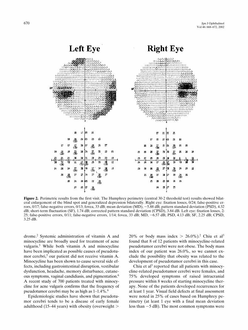
onset of horizontal diplopia on the morning of Feb-ruary 4th, 2000. She denied visual loss, nausea, vom-iting, and trauma. On examination in our hospital onFebruary 7, her visual acuity was 1.0 in the right eyeand 1.0 in the left eye. Her intraocular pressure was20 mm Hg in the right eye and 19 mm Hg in the lefteye. There was an abduction deficit of the right eyeand full motility of the left eye. An afferent pupillarydefect was not noted, and fundus examinationshowed bilateral papilledema with a single streakhemorrhage adjacent to the disc (Figure 1). The crit-ical flicker fusion frequency was normal, as was hercolor vision according to a Panel D-15 test. Auto-mated perimetry (Humphrey perimetry, central 30-2threshold test) showed enlarged blind spots in botheyes, with mean deviations of
�
5.88 dB in the righteye and
�
6.57 dB in the left eye (Figure 2). No ab-normal findings were seen by computed tomographyand magnetic resonance imaging of the brain onFebruary 9. Her cerebrospinal fluid had an openingpressure of 400 mm H
2
O, was acellular, had a pro-tein level of 17 mg/dL and a glucose level of 101 mg/dL. The culture was negative. The results of sero-logic studies for syphilis and endocrine disorders(Addison disease, Cushing disease, and hypopara-
thyroidism) were negative. Testing for antinuclearantibodies showed negative results. Minocycline in-take was discontinued on February 9. Seven days af-ter the first lumbar puncture, the sixth nerve palsyhad disappeared and her headache resolved. Visualacuity was 1.0 in the right eye and 1.0 in the left eye,with mild resolution of the papilledema bilaterally.A second lumbar puncture was performed on thesame day, and revealed an opening pressure of 230mm H
2
O. Her fundus oculi had returned to normalby March 11, and a third lumbar puncture revealedan opening pressure of 160 mm H
2
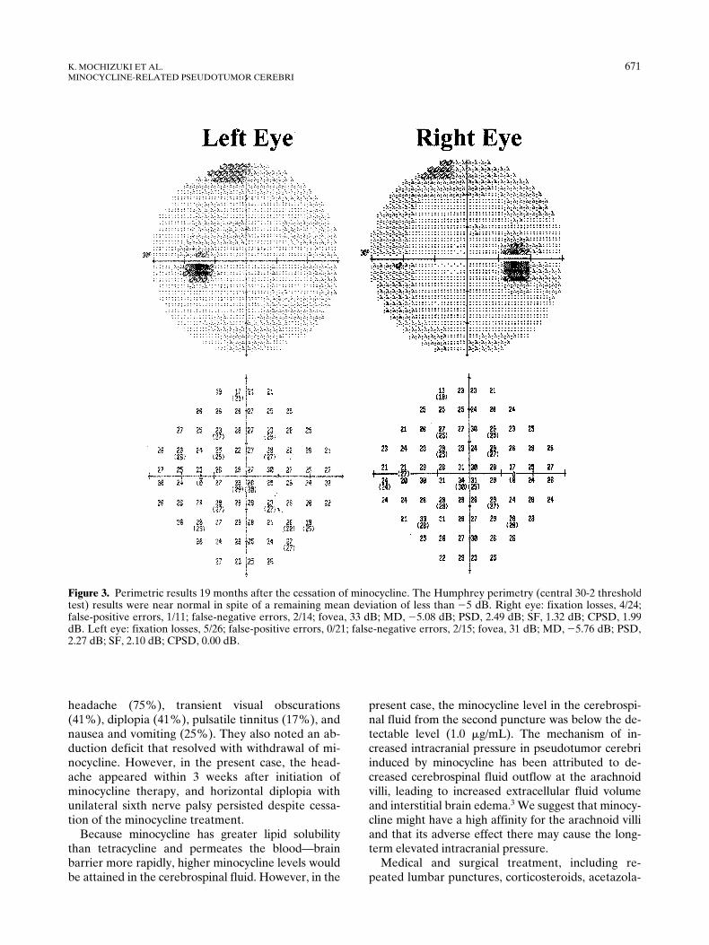
O. On August 29,2001, the final Humphrey mean deviations were
�
5.08 dB in the right eye and
�
5.76 dB in the lefteye (Figure 3). After the last lumbar puncture, no re-currence has been documented for at least 1.5 years.
We found that the minocycline concentrations inthe serum and cerebrospinal fluid on February 16were below the detectable level (1.0
�
g/mL).
Discussion
This case seems to fulfill the criteria for pseudotu-mor cerebri syndrome. Several conditions as well asmedications have been associated with this syn-
Figure 1. Fundus photographs taken during the first visit of a 16-year-old girl who developed pseudotumor cerebri whiletaking minocycline for acne vulgaris. Edema of the optic disc with adjacent hemorrhage was seen bilaterally.
670
Jpn J OphthalmolVol 46: 668–672, 2002
drome.
2
Systemic administration of vitamin A andminocycline are broadly used for treatment of acnevulgaris.
5
While both vitamin A and minocyclinehave been implicated as possible causes of pseudotu-mor cerebri,
5
our patient did not receive vitamin A.Minocycline has been shown to cause several side ef-fects, including gastrointestinal disruption, vestibulardysfunction, headache, memory disturbance, cutane-ous symptoms, vaginal candidiasis, and pigmentation.
6
A recent study of 700 patients treated with minocy-cline for acne vulgaris confirms that the frequency ofpseudotumor cerebri may be as high as 1–1.4%.
6
Epidemiologic studies have shown that pseudotu-mor cerebri tends to be a disease of early femaleadulthood (15–44 years) with obesity (overweight
�
20% or body mass index
�
26.0%).
2
Chiu et al
3
found that 8 of 12 patients with minocycline-relatedpseudotumor cerebri were not obese. The body massindex of our patient was 26.0%, so we cannot ex-clude the possibility that obesity was related to thedevelopment of pseudotumor cerebri in this case.
Chiu et al
3
reported that all patients with minocy-cline-related pseudotumor cerebri were females, and75% developed symptoms of raised intracranialpressure within 8 weeks of starting minocycline ther-apy. None of the patients developed recurrences forat least 1 year. Visual field defects at final assessmentwere noted in 25% of cases based on Humphrey pe-rimetry (at least 1 eye with a final mean deviationless than
�
5 dB). The most common symptoms were
Figure 2. Perimetric results from the first visit. The Humphrey perimetry (central 30-2 threshold test) results showed bilat-eral enlargement of the blind spot and generalized depression bilaterally. Right eye: fixation losses, 0/24; false-positive er-rors, 0/17; false-negative errors, 0/13; fovea, 33 dB; mean deviation (MD), �5.88 dB; pattern standard deviation (PSD), 4.32dB; short-term fluctuation (SF), 1.74 dB; corrected pattern standard deviation (CPSD), 3.84 dB. Left eye: fixation losses, 2/25; false-positive errors, 0/11; false-negative errors, 1/14; fovea, 33 dB; MD, �6.57 dB; PSD, 4.13 dB; SF, 2.25 dB; CPSD,3.25 dB.
K. MOCHIZUKI ET AL.
671
MINOCYCLINE-RELATED PSEUDOTUMOR CEREBRI
headache (75%), transient visual obscurations(41%), diplopia (41%), pulsatile tinnitus (17%), andnausea and vomiting (25%). They also noted an ab-duction deficit that resolved with withdrawal of mi-nocycline. However, in the present case, the head-ache appeared within 3 weeks after initiation ofminocycline therapy, and horizontal diplopia withunilateral sixth nerve palsy persisted despite cessa-tion of the minocycline treatment.
Because minocycline has greater lipid solubilitythan tetracycline and permeates the blood—brainbarrier more rapidly, higher minocycline levels wouldbe attained in the cerebrospinal fluid. However, in the
present case, the minocycline level in the cerebrospi-nal fluid from the second puncture was below the de-tectable level (1.0
�
g/mL). The mechanism of in-creased intracranial pressure in pseudotumor cerebriinduced by minocycline has been attributed to de-creased cerebrospinal fluid outflow at the arachnoidvilli, leading to increased extracellular fluid volumeand interstitial brain edema.
3
We suggest that minocy-cline might have a high affinity for the arachnoid villiand that its adverse effect there may cause the long-term elevated intracranial pressure.
Medical and surgical treatment, including re-peated lumbar punctures, corticosteroids, acetazola-
Figure 3. Perimetric results 19 months after the cessation of minocycline. The Humphrey perimetry (central 30-2 thresholdtest) results were near normal in spite of a remaining mean deviation of less than �5 dB. Right eye: fixation losses, 4/24;false-positive errors, 1/11; false-negative errors, 2/14; fovea, 33 dB; MD, �5.08 dB; PSD, 2.49 dB; SF, 1.32 dB; CPSD, 1.99dB. Left eye: fixation losses, 5/26; false-positive errors, 0/21; false-negative errors, 2/15; fovea, 31 dB; MD, �5.76 dB; PSD,2.27 dB; SF, 2.10 dB; CPSD, 0.00 dB.

672
Jpn J OphthalmolVol 46: 668–672, 2002
mide, glycerol, optic nerve fenestration, and a lum-boperitoneal shunt, have been tried in patients withpseudotumor cerebri.
7
We performed three lumbarpunctures within 4 weeks in the present case andachieved rapid visual improvement and resolution ofthe papilledema and diplopia, which was not likelyto have been due to the lumbar punctures alone.
According to a previous study,
3
the long-term out-come of minocycline-related pseudotumor cerebrimay not be entirely benign. In the present study, thepatient had a residual Humphrey mean deviation ofless than
�
5 dB in both eyes. Significant visual fieldloss as a result of minocycline therapy has also beenreported.
8,9
The possibility of drug-induced pseudo-tumor cerebri should be considered in any patientpresenting with unexplained headache, especially ifassociated with visual symptoms.
We want to emphasize the possible role of mi-nocycline when pseudotumor cerebri occurs in pa-tients treated for acne vulgaris, and we recommend aperiodic ophthalmologic examination during any mi-nocycline treatment.
References
1. Mukuno K, Ishikawa S. Informal questionnaire on the inci-dence of pseudotumor cerebri in Japan. Neuroophthalmol Jpn1994;11:52–54.
2. Radhakrishnan K, Ahlskog JE, Garrity JA, Kurland LT. Idio-pathic intracranial hypertension. Mayo Clin Proc 1994;69:169–180.
3. Chiu AM, Chuenkongkaew WL, Cornblath WT, et al. Minocy-cline treatment and pseudotumor cerebri syndrome. Am JOphthalmol 1998;126:116–121.
4. Monaco F, Agnetti V, Mutani R. Benign intracranial hyperten-sion after minocycline therapy. Eur Neurol 1978;17:48–49.
5. Lascari AD, Bell WE. Pseudotumor cerebri due to hypervita-minosis A: toxic consequence of self-medication for acne in anadolescent girl. Clin Pediatr 1970;9:627–628.
6. Goulden V, Glass D, Cunliffe WJ. Safety of long-term high-dose minocycline in treatment of acne. Br J Dermatol 1996;134:693–695.
7. Liu GT, Glaser JS, Schatz NJ. High-dose methylprednisoloneand acetazolamide for visual loss in pseudotumor cerebri. Am JOphthalmol 1994;118:88–96.
8. Lander CM. Minocycline-induced benign intracranial hyper-tension. Clin Exp Neurol 1989;26:161–167.
9. Donnet A, Dufour H, Graziani N, Grisoli F. Minocycline andbenign intracranial hypertension. Biomed Pharmacother 1992;46:171–172.


