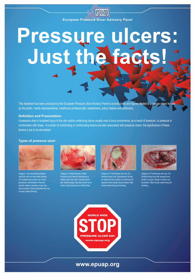
Category I. Non-blanchable erythema. Intact skin with non-blanchable redness of a localised area usually over a bony prominence. Discoloration of the skin, warmth, oedema, hardness or pain may also be present. Darkly pigmented skin may not have visible blanching.
Category II. Partial thickness. Partial thickness loss of dermis presenting as a shallow open ulcer with a red pink wound bed, without slough. May also present as an intact or open/ruptured serum-filled blister.
Category III. Full thickness skin loss. Full thickness tissue loss. Subcutaneous fat may be visible but bone, tendon or muscle are not exposed. Some slough may be present. May include undermining and tunneling.
Category IV. Full thickness skin loss. Full thickness tissue loss with exposed bone, tendon or muscle. Slough or eschar may be present. Often include undermining and tunneling.
This factsheet has been produced by the European Pressure Ulcer Advisory Panel to provide facts and figures related to pressure ulcers for use by the public, media representatives, healthcare professionals, researchers, policy makers and politicians.
Definition and PresentationA pressure ulcer is localised injury to the skin and/or underlying tissue usually over a bony prominence, as a result of pressure, or pressure in combination with shear. A number of contributing or confounding factors are also associated with pressure ulcers; the significance of these factors is yet to be elucidated1.
Types of pressure ulcer
www.epuap.org
Pressure ulcers: Just the facts!
European Pressure Ulcer Advisory Panel

Common locations for pressure ulcers. Pressure ulcers can occur at any location on the body but are most common in areas which regularly bear the weight of the body such as the sacrum (base of the spine), buttocks and heels.
Sources of information1. European Pressure Ulcer Advisory Panel and National Pressure Ulcer Advisory Panel. Prevention and treatment of pressure ulcers: quick reference guide. Washington DC: National Pressure Ulcer Advisory Panel; 2009.
(available at www.epuap.org)2. Vanderwee K, Clark M, Dealey C, Gunningberg L, Defloor T. Pressure ulcer prevalence in Europe: a pilot study Journal of Evaluation in Clinical Practice. 2007, 13(2): 227–235 3. Vowden KR, Vowden P. The prevalence, management, equipment provision and outcome for patients with pressure ulceration identified in a wound care survey within one English health care district. Journal of Tissue
Viability. 2009, 18(1): 20-64. Essex HN, Clark M, Sims J, Warriner A, Cullum N. Health-related quality of life in hospital in-patients with pressure ulceration: assessment using generic health-related quality of life measures. Wound Repair
Regeneration. 2009, 17(6): 797-805.5. Davies K, Strickland J, Lawrence V, Duncan A, Rowe J. The hidden mortality from pressure sores. Journal of Tissue Viability. 1991, 1(1): 18.6. Posnett J, Franks PJ. The costs of skin breakdown and ulceration in the UK. In Skin Breakdown: the silent epidemic. Smith & Nephew Foundation, Hull, 2007, pp 6-12.7. Bennett G, Dealey C, Posnett J. The costs of pressure ulcers in the UK. Age and Ageing. 2004; 33: 230-35.
All content was created by the EPUAP and printed under an unrestricted educational grant from
© ArjoHuntleigh, 2012
Pressure Ulcer Sites
Numbers of people with pressure ulcers.In 2007 Vanderwee and colleagues2 reported a pilot pressure ulcer prevalence survey conducted across 26 hospitals in Belgium, Italy, Portugal, Sweden and the UK. The survey included 5947 patients with 1078 (18.3%) having pressure ulcers. By country, the proportion of patients surveyed who had pressure ulcers varied - Italy (8.3%), Portugal (12.5%), Belgium (21.0%), UK (21.9%), Sweden (22.9%).
Most of the pressure ulcers reported by Vanderwee et al2 were Category I (n=454) or Category II pressure ulcers (n=282). Full thickness pressure ulcers were less common - Category III (n=199) and Category IV (n=143).
Pressure ulcers most commonly occurred at the sacrum (n=532) and the heels (n=484). Other common sites for pressure ulcer development were the ischial tuberosities (buttocks) (n=186), the ankles (n=149), elbows (n=143) and the hips (n=136).
In an audit across acute and community services in Bradford UK, Vowden and Vowden3 reported that the prevalence of pressure ulceration within the population receiving healthcare was 0.74 people with a pressure ulcer per 1000 population. Of the people with pressure ulcers few (n=40, 11%) were located in hospital suggesting that current pressure ulcer epidemiology and costs may be understated given their reliance on hospital based surveys of pressure ulcers.
Across all the surveys described in this fact sheet pressure ulcers were common, affecting almost 20% of all patients.
The consequences of having a pressure ulcer.Essex and colleagues4 have reported a number of consequences for people who have pressure ulcers - these included increased pain, reduced vitality, reduced physical activity and generally reduced quality of life. Pressure ulcers may also
lead to death with 171 death certificates in the UK in 1986 listing pressure ulcers as the cause of death with a further 1929 certificates citing pressure ulcers as contributing to death5.
The costs of pressure ulcers.There are no European wide estimates of the total cost of pressure ulcer prevention and treatment. Within specific countries the high cost of pressure ulcers has been identified. In the Netherlands, 1% of all health care expenditure was calculated to be spent on pressure ulcer care.
Posnett and Franks6 considered the UK national cost of pressure ulcers to the NHS to be between £1,760 million and £2,640 million each year, making pressure ulcers the single most costly chronic wound to the Health Service. This calculation was based upon the original modelling of Bennett and colleagues7 who estimated that the typical cost of treating Category I pressure ulcers would be £1064, rising to £4402 for a Category II ulcer then to £7313 and £10,551 for Category III and IV pressure ulcers respectively.


