
1139Clin. Invest. (Lond.) (2014) 4(12), 1139–1154 ISSN 2041-6792
Clinical Trial Outcomes
part of
Overview on clinical trials in Waldenstrom’s macroglobulinemia
Alessandra Tedeschi*,1, Anna Maria Frustaci1, Paola Picardi1 & Enrica Morra1
1Division of Hematology,
Niguarda Ca’ Granda Hospital,
Milano, Italy
*Author for correspondence:
Tel.: +39 0264 442 668
Fax: +39 0264 443 019
10.4155/CLI.14.96 © 2014 Future Science Ltd
Clin. Invest. (Lond.)
10.4155/CLI.14.96
Clinical Trial Outcomes
Tedeschi, Frustaci, Picardi & MorraOverview on clinical trials in Waldenstrom’s
macroglobulinemia
4
12
2014
Waldenstrom’s macroglobulinemia is characterized by lymphoplasmocytic cells accumulation predominantly in bone marrow, secreting immunoglobulin M monoclonal protein. There is not a standard of care as disease is rare and there are no large randomized trials to address treatment. Asymptomatic patients should be observed. In symptomatic patients treatment should be individualized considering patient fitness and disease characteristics. In elderly unfit patients single agent treatment may be still considered an option. In younger and fit patients immunochemotherapy should be considered the standard of care as recent data showed an improvement in quality of responses, progression-free and overall survival. In this overview are reported the most significant clinical trials that may help in treatment decision.
Keywords: bendamustine • immunochemotherapy • immunomodulatory drugs • monoclonal antibodies • proteasome inhibitors • treatment • Waldenstrom’s macroglobulinemia
The 2008 WHO classification of tumors of Hematopoietic and Lymphoid Tissues defines Waldenstrom’s macroglobulinemia (WM) as: a type of lymphoplasmocytic lym-phoma that involves bone marrow and is associated with a monoclonal immunoglobu-lin of the immunoglobulin M (IgM) class in the serum [1].
It is a very rare disease which comprises about 2% of all non-Hodgkin’s lymphomas with about twofold higher in men compared with women. Data from the US Surveillance, Epidemiology and End Results Registry (SEER) estimate the US incidence of WM to be 3.8 cases per million people per year [2]. WM has a distinct pattern reflecting racial disparity; incidence is higher in whites com-pared with blacks or Asians. Like other lym-phoprolipherative disorders, WM is a disease of the older population with an incidence rate increasing with age, median age at diagnosis of 73 years.
At least 25% of patients are asymptom-atic at diagnosis, some of them may remain asymptomatic and will never need specific
treatment, 50% of asymptomatic patients who are observed will not require therapy within 3 years [3,4]. This highlights the importance of careful determination for the need of treatment.
There is not a standard of care in WM. As the disease is rare there are limited large Phase III randomized trials, and treatment decisions are made basically on results from Phase II trials and expert recommendations. Recently, SEER indicated an improvement in overall survival (OS) in patients of all ages [5], which may be related to a better understand-ing of disease biology translating in new therapeutic approaches, a better risk strati-fication of patients, and an improvement of supportive care. Even though, an outcome improvement has not been observed in all series [6,7].
Usually WM follows an indolent course. In different series, OS reported ranges from 60 to 120 months [4,6–8]. In some cases dis-ease may be more aggressive, leading to treat-ment refractoriness and death within few months. Many studies analyzed clinical and
1140 Clin. Invest. (Lond.) (2014) 4(12) future science group
Clinical Trial Outcomes Tedeschi, Frustaci, Picardi & Morra
disease characteristics associated with a worse prog-nosis. Several risk factors have been found to predict outcome, but all these studies should be critically ana-lyzed. Results are not uniform depending on: study endpoint (OS or response to treatment), cut-off val-ues considered and patients characteristics, as in some series both asymptomatic or symptomatic patients are considered. Furthermore, as we are dealing with an elderly population the poorest survival of patients over 65 years may be related to a higher number of non-WM related deaths [9]. The main adverse prognostic factors found to be significant are older age, low albu-min, β2-microglobulin and cytopenias. In some stud-ies adverse prognosis has been associated with high IgM concentrations, whereas in other studies adverse prognosis was related to low IgM concentration [6,10–11].
The update of the Southwest Oncology Group (SWOG) S9003 study indicated that lactate dehydro-genase (LDH) was an independent prognostic factor for survival after fludarabine therapy. The prognos-tic role of LDH for OS has also been confirmed by Kastritis et al. [12,13].
Other features found to be significant for progno-sis are: constitutional symptoms, hepato-splenomeg-aly, hyperviscosity, cryoglobulinemia, male gender and urine monoclonal component [3,6,11]. Recently, an International Prognostic Scoring System for WM (IPSSWM) was designed only to predict survival after first-line therapy in symptomatic patients. This score has been initially validated for patients treated frontline with alkylating agents and purine analogs [4]. Subse-quently, Dimopoulous et al. showed in a large series of 93 patients that IPSSWM is also applicable in patients who received primary treatment with rituximab-based regimens [14]. The scoring system stratifies patients into three different risks categories based on five adverse covariates: age >65 years, hemoglobin 11.5 g/dl, plate-let count 100 × 109/l β2-microglobulin >3 mg/l and serum monoclonal protein concentration >70 g/l.
As the IPSSWM is designed only for symptomatic patients needing treatment, it should not be applied to determine whether a patient requires treatment.
Several studies in recent years have shown the importance of the immunoglobulin-free light chain assay in predicting response and survival, but its role remains to be defined [15,16].
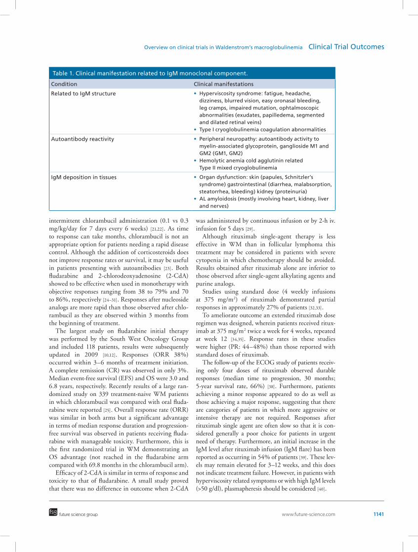
For asymptomatic patients, follow-up is recom-mended and the watch and wait strategy is still con-sidered a standard while treatment should be reserved only to symptomatic patients [17]. Symptoms may be either related to the IgM monoclonal component or to the expansion of the neoplastic clone resulting in tissue infiltration. Clinical manifestations induced by IgM monoclonal component are listed in Table 1.
Treatment should be individualized, and patient fitness and disease characteristics must be taken in account before initiating therapy. Most WM patients are aged >70 years with nonlymphoma-associated comorbidities. No studies have been addressed for this category of patients and they are clearly under-rep-resented in clinical trials. In older and unfit patients intensive immunochemotherapy should be avoided, and single-agent treatment may still be a valid option.
The choice of treatment is dependent not only on patient age and comorbidities, but is also strictly dependent on the need of: rapid disease control, associ-ated cytopenias, neuropathies, autoimmune phenom-ena, candidacy for autologous transplantation and long-term treatment toxicity. In patients presenting with hyperviscosity syndrome, plasmapheresis should be promptly instituted. Plasmapheresis exerts a tran-sient effect and can promptly reverse most clinical manifestations [18] but does not affect the underlying disease process so that systemic treatment should be administered concomitantly.
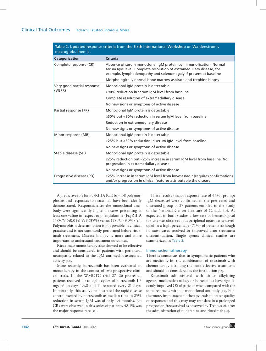
Response criteria in WMBefore the second International Workshop in WM, held in 2002, response criteria had not been standard-ized. In most of the studies responses were generally based on monoclonal IgM reduction and/or improve-ment of nodal involvement. In a small number of studies marrow evaluation had been performed to assess response. Considering the heterogeneity of the disease determining categorical responses only on the basis of the change of M protein may not be appropri-ate. Furthermore not always IgM reduction correlates with symptoms improvement and often discrepancies between IgM and bone marrow responses are found. The consensus panel in 2002 proposed the first rec-ommendations for specific tests to document response and guidelines for standardized response criteria. [19]. To better define responses and quality of responses recently an update of response assessment criteria has been published (Table 2) [20].
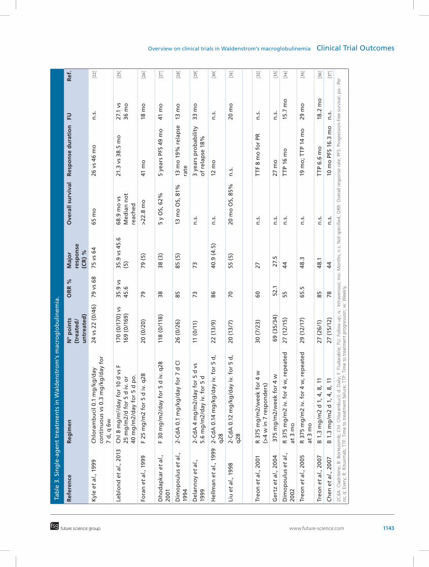
Single agents treatmentAlkylating agents in monotherapy and subsequently purine analogs have been extensively used. Single-agent chlorambucil may still be a valid option in non-fit patients [17]. An objective disease improvement, after chlorambucil treatment, has been observed in 50–80% of patients. Results of these studies should be carefully evaluated as most of the clinical trials are small Phase II studies with widely differing inclusion criteria; furthermore, they were performed before WM response criteria were standardized. No differences in outcome have been observed when comparing daily or
www.future-science.com 1141future science group
Overview on clinical trials in Waldenstrom’s macroglobulinemia Clinical Trial Outcomes
intermittent chlorambucil administration (0.1 vs 0.3 mg/kg/day for 7 days every 6 weeks) [21,22]. As time to response can take months, chlorambucil is not an appropriate option for patients needing a rapid disease control. Although the addition of corticosteroids does not improve response rates or survival, it may be useful in patients presenting with autoantibodies [23]. Both fludarabine and 2-chlorodeoxyadenosine (2-CdA) showed to be effective when used in monotherapy with objective responses ranging from 38 to 79% and 70 to 86%, respectively [24–31]. Responses after nucleoside analogs are more rapid than those observed after chlo-rambucil as they are observed within 3 months from the beginning of treatment.
The largest study on fludarabine initial therapy was performed by the South West Oncology Group and included 118 patients, results were subsequently updated in 2009 [10,12]. Responses (ORR 38%) occurred within 3–6 months of treatment initiation. A complete remission (CR) was observed in only 3%. Median event-free survival (EFS) and OS were 3.0 and 6.8 years, respectively. Recently results of a large ran-domized study on 339 treatment-naive WM patients in which chlorambucil was compared with oral fluda-rabine were reported [25]. Overall response rate (ORR) was similar in both arms but a significant advantage in terms of median response duration and progression-free survival was observed in patients receiving fluda-rabine with manageable toxicity. Furthermore, this is the first randomized trial in WM demonstrating an OS advantage (not reached in the fludarabine arm compared with 69.8 months in the chlorambucil arm).
Efficacy of 2-CdA is similar in terms of response and toxicity to that of fludarabine. A small study proved that there was no difference in outcome when 2-CdA
was administered by continuous infusion or by 2-h iv. infusion for 5 days [29].
Although rituximab single-agent therapy is less effective in WM than in follicular lymphoma this treatment may be considered in patients with severe cytopenia in which chemotherapy should be avoided. Results obtained after rituximab alone are inferior to those observed after single-agent alkylating agents and purine analogs.
Studies using standard dose (4 weekly infusions at 375 mg/m2) of rituximab demonstrated partial responses in approximately 27% of patients [32,33].
To ameliorate outcome an extended rituximab dose regimen was designed, wherein patients received ritux-imab at 375 mg/m2 twice a week for 4 weeks, repeated at week 12 [34,35]. Response rates in these studies were higher (PR: 44–48%) than those reported with standard doses of rituximab.
The follow-up of the ECOG study of patients receiv-ing only four doses of rituximab observed durable responses (median time to progression, 30 months; 5-year survival rate, 66%) [38]. Furthermore, patients achieving a minor response appeared to do as well as those achieving a major response, suggesting that there are categories of patients in which more aggressive or intensive therapy are not required. Responses after rituximab single agent are often slow so that it is con-sidered generally a poor choice for patients in urgent need of therapy. Furthermore, an initial increase in the IgM level after rituximab infusion (IgM flare) has been reported as occurring in 54% of patients [39]. These lev-els may remain elevated for 3–12 weeks, and this does not indicate treatment failure. However, in patients with hyperviscosity related symptoms or with high IgM levels (>50 g/dl), plasmapheresis should be considered [40].
Table 1. Clinical manifestation related to IgM monoclonal component.
Condition Clinical manifestations
Related to IgM structure • Hyperviscosity syndrome: fatigue, headache, dizziness, blurred vision, easy oronasal bleeding, leg cramps, impaired mutation, ophtalmoscopic abnormalities (exudates, papilledema, segmented and dilated retinal veins)
• Type I cryoglobulinemia coagulation abnormalities
Autoantibody reactivity • Peripheral neuropathy: autoantibody activity to myelin-associated glycoprotein, ganglioside M1 and GM2 (GM1, GM2)
• Hemolytic anemia cold agglutinin related Type II mixed cryoglobulinemia
IgM deposition in tissues • Organ dysfunction: skin (papules, Schnitzler’s syndrome) gastrointestinal (diarrhea, malabsorption, steatorrhea, bleeding) kidney (proteinuria)
• AL amyloidosis (mostly involving heart, kidney, liver and nerves)
1142 Clin. Invest. (Lond.) (2014) 4(12) future science group
Clinical Trial Outcomes Tedeschi, Frustaci, Picardi & Morra
A predictive role for FcγRIIIA (CD16)-158 polymor-phisms and responses to rituximab have been clearly demonstrated. Responses after the monoclonal anti-body were significantly higher in cases presenting at least one valine in respect to phenylalanine (FcγRIIIA 158V/V (40.0%) V/F (35%) versus 158F/F (9.0%) [41]. Polymorphism determination is not possible in clinical practice and is not commonly performed before ritux-imab treatment. Disease biology is more and more important to understand treatment outcomes.
Rituximab monotherapy also showed to be effective and should be considered in patients with peripheral neuropathy related to the IgM antimyelin associated activity [42].
More recently, bortezomib has been evaluated in monotherapy in the context of two prospective clini-cal trials. In the WMCTG trial 27, 26 pretreated patients received up to eight cycles of bortezomib 1.3 mg/m2 on days 1,4,8 and 11 repeated every 21 days. Importantly, this study demonstrated the rapid disease control exerted by bortezomib as median time to 25% reduction in serum IgM was of only 1.4 months. No CRs were observed in this series of patients, 48.1% was the major response rate [36].
These results (major response rate of 44%, prompt IgM decrease) were confirmed in the pretreated and untreated group of 27 patients enrolled in the Study of the National Cancer Institute of Canada [37]. As expected, in both studies a low rate of hematological toxicity was observed, but peripheral neuropathy devel-oped in a high percentage (74%) of patients although in most cases resolved or improved after treatment discontinuation. Single agents clinical studies are summarized in Table 3.
ImmunochemotherapyThere is consensus that in symptomatic patients who are medically fit, the combination of rituximab with chemotherapy is among the most effective treatments and should be considered as the first option [43].
Rituximab administered with either alkylating agents, nucleoside analogs or bortezomib have signifi-cantly improved OS of patients when compared with the same regimens without monoclonal antibody [44]. Fur-thermore, immunochemotherapy leads to better quality of responses and this may may translate in a prolonged progression-free survival as observed by Treon et al. after the administration of fludarabine and rituximab [45].
Table 2. Updated response criteria from the Sixth International Workshop on Waldenstrom’s macroglobulinemia.
Categorization Criteria
Complete response (CR) Absence of serum monoclonal IgM protein by immunofixation. Normal serum IgM level. Complete resolution of extramedullary disease, for example, lymphadenopathy and splenomegaly if present at baseline
Morphologically normal bone marrow aspirate and trephine biopsy
Very good partial response (VGPR)
Monoclonal IgM protein is detectable
≥90% reduction in serum IgM level from baseline
Complete resolution of extramedullary disease
No new signs or symptoms of active disease
Partial response (PR) Monoclonal IgM protein is detectable
≥50% but <90% reduction in serum IgM level from baseline
Reduction in extramedullary disease
No new signs or symptoms of active disease
Minor response (MR) Monoclonal IgM protein is detectable
≥25% but <50% reduction in serum IgM level from baseline.
No new signs or symptoms of active disease
Stable disease (SD) Monoclonal IgM protein is detectable
≤25% reduction but <25% increase in serum IgM level from baseline. No progression in extramedullary disease
No new signs or symptoms of active disease
Progressive disease (PD) ≥25% increase in serum IgM level from lowest nadir (requires confirmation) and/or progression in clinical features attributable the disease
www.future-science.com 1143future science group
Overview on clinical trials in Waldenstrom’s macroglobulinemia Clinical Trial OutcomesTa
ble
3. S
ing
le-a
gen
t tr
eatm
ents
in W
ald
enst
rom
’s m
acro
glo
bu
linem
ia.
Ref
eren
ceR
egim
enN
° p
oin
ts
(tre
ated
/u
ntr
eate
d)
OR
R %
Maj
or
resp
on
se
(CR
) %
Ove
rall
surv
ival
Res
po
nse
du
rati
on
FU R
ef.
Kyl
e et
al.,
199
9C
hlo
ram
bu
cil 0
.1 m
g/k
g/d
ay
con
tin
uo
us
vs 0
.3 m
g/k
g/d
ay f
or
7 d
, q 6
w
24 v
s 22
(0
/46
)79
vs
68
75 v
s 6
465
mo
26 v
s 4
6 m
on
.s.
[22]
Leb
lon
d e
t al
., 20
13C
hl 8
mg
/m2/d
ay f
or
10 d
vs
F
25 m
g/m
2/d
fo
r 5
d iv
. or
4
0 m
g/m
2/d
ay f
or
5 d
po
.
170
(0/1
70)
vs
169
(0/1
69)
35.9
vs
45.6
35.9
vs
45.6
(5
)6
8.9
mo
vs
Med
ian
no
t re
ach
ed
21.3
vs
38
.5 m
o27
.1 v
s 36
mo
[25]
Fora
n e
t al
., 19
99F
25 m
g/m
2 fo
r 5
d iv
. q28
20 (
0/2
0)
7979
(5
)>
22.8
mo
41 m
o18
mo
[26]
Dh
od
apka
r et
al.,
20
01F
30 m
g/m
2/d
ay f
or
5 d
iv. q
2811
8 (0
/118
)3
83
8 (3
)5
y O
S, 6
2%5
year
s PF
S 49
mo
41 m
o[27]
Dim
op
ou
lus
et a
l.,
199
42-
Cd
A 0
.1 m
g/k
g/d
ay f
or
7 d
Cl
26 (
0/2
6)
8585
(5
)13
mo
OS,
81%
13 m
o 1
9%
rel
apse
ra
te13
mo
[28]
Del
ann
oy
et a
l.,
1999
2-C
dA
4 m
g/m
2/d
ay f
or
5 d
vs
5.
6 m
g/m
2/d
ay iv
. fo
r 5
d11
(0
/11)
7373
n.s
.3
year
s p
rob
abili
ty
of
rela
pse
18%
33 m
o[29]
Hel
lman
et
al.,
1999
2-C
dA
0.1
4 m
g/k
g/d
ay iv
. fo
r 5
d,
q28
22 (
13/9
)8
64
0.9
(4.5
)n
.s.
12 m
on
.s.
[30]
Liu
et
al.,
199
82-
Cd
A 0
.12
mg
/kg
/day
iv. f
or
5 d
, q
2820
(13
/7)
7055
(5
)20
mo
OS,
85%
n.s
.20
mo
[31]
Treo
n e
t al
., 20
01R
375
mg
/m2
/wee
k fo
r 4
w
(>4
w in
7 r
esp
on
der
s)30
(7/
23)
6027
n.s
.T
TF 8
mo
fo
r PR
n.s
.[32]
Ger
tz e
t al
., 20
04
375
mg
/m2
/wee
k fo
r 4
w 6
9 (3
5/3
4)
52.
1 2
7.5
n.s
.27
mo
n.s
.[33]
Dim
op
ou
lus
et a
l.,
2002
R 3
75 m
g/m
2 iv
. fo
r 4
w, r
epea
ted
at
3 m
o27
(12
/15
)55
44
n.s
.T
TP 1
6 m
o15
.7 m
o[34]
Treo
n e
t al
., 20
05R
375
mg
/m2
iv. f
or
4 w
, rep
eate
d
at 3
mo
29 (
12/1
7)65
.54
8.3
n.s
.19
mo
; TTP
14
mo
29 m
o[35]
Treo
n e
t al
., 20
07B
1.3
mg
/m2
d 1
, 4, 8
, 11
27 (
26/1
)85
48
.1n
.s.
TTP
6.6
mo
18.2
mo
[36]
Ch
en e
t al
., 20
07B
1.3
mg
/m2
d 1
, 4, 8
, 11
27 (
15/1
2)
784
4n
.s.
10 m
o P
FS 1
6.3
mo
n.s
.[37]
2Cd
A: C
lad
rib
ine;
B: B
ort
ezo
mib
; Chl
: Chl
ora
mb
uci
l; d
: Dai
ly; F
: Flu
dar
abile
; FU
: Fo
llow
up
; iv.
: Int
rave
no
us;
mo
: Mo
nths
; n.s
: Not
sp
ecifi
ed; O
RR
: Ove
rall
resp
ons
e ra
te; P
FS: P
rog
ress
ion
-fre
e su
rviv
al; p
o.: P
er
os;
q: E
very
; R: R
itu
xim
ab; T
TF: T
ime
to t
reat
men
t fa
ilure
; TTP
: Tim
e to
tre
atm
ent
pro
gre
ssio
n; w
: Wee
kly.
1144 Clin. Invest. (Lond.) (2014) 4(12) future science group
Clinical Trial Outcomes Tedeschi, Frustaci, Picardi & Morra
Cyclophosphamide basedSeveral randomized trials in lymphoprolipherative disorders have shown that the inclusion of rituximab in cyclophosphamide-based regimens such as CVP (cyclophosphamide, vincristine and prednisone) and CHOP (cyclophosphamide, doxorubicin, vincristine and prednisone) improves response rate as well as response duration and OS [46,47].
Similar results have also been achieved in WM; in fact, in the German randomized trial in which untreated patients with WM were included, the addi-tion of monoclonal antibody to CHOP schedule led to a higher objective response rate (91 vs 60%). Further-more, R-CHOP induced a significantly longer time to treatment failure [48]. Although treatment was well tolerated R-CHOP may be considered to be too toxic because of the high incidence of myelosuppression.
The importance of doxorubicin and vincristine inclusion in the chemotherapeutic regimen since the introduction of rituximab has not yet been still clari-fied. Furthermore, the use of anthracyclines is associ-ated to adverse events such as alopecia, cardiopathies and cytopenia while vincristine should be avoided in patients presenting with neuropathies.
A non-randomized comparison of patients treated at the Dana Farber Institute showed that omitting doxorubicin or doxorubicin plus vincristine did not significantly decrease response rate with outcomes very similar to those observed after R-CHOP in the Ger-man study. Furthermore, these schedules were much less toxic compared with R-CHOP [49].
To avoid toxicity related to anthracyclines and vin-cristine, Dimopoulos et al. designed a regimen consist-ing of dexamethasone 20 mg followed by rituximab 375 mg/m2 intravenously on day 1 and cyclophospha-mide administered orally for 5 days at the dosage of 200 mg/m2 (DRC) [50]. Only untreated patients were included in the study, major response rate was 83%, including 7% with CR. Median time to response was 4.1 months.
Importantly, treatment was very well tolerated as only 7 of the 72 enrolled patients (9%) developed grade 3–4 neutropenia. The updated results of this trial showed a favorable median time to progres-sion of about 3 years, most patients with progression responded again to rituximab based regimens. After a minimum follow-up >6 years long-term toxicity was limited [51].
Currently in clinical practice, cyclophosphamide-based regimens and in particular DRC, which avoid unnecessary toxicity, are considered the standard of treatment for untreated patients. These regimens may also be preferable in younger patients eligible for stem cell collection [40,43].
Nucleoside analogs basedIn vitro studies demonstrated strong evidence of synergy between nucleoside analog and alkylating agents. This translated in increase of ORR and response duration when fludarabine or cladribine were administered in combination in untreated or pretreated patients [52,53].
Furthermore, preclinical data indicated that ritux-imab sensitized cells to both fludarabine and cyclo-phosphamide (FC); thus enhancing their cytotoxic activity [54,55].
The addition of riruximab to nucleoside analog-based chemotherapy allowed to obtain an amelioration of quality of response with a prolonged progression-free survival (Table 4).
The results of a study on 43 patients demonstrated that the combination of rituximab and fludarabine (FR) is highly active leading to an ORR of 95.3% and median TTP of 51.2 months being longer in previ-ously untreated patients and in those achieving at least a very good partial response [45]. Favorable responses and rapid disease control have also been obtained with the combination of rituximab and fludarabine plus cyclophosphamide (FCR) [56]. Authors conclude that although FR and FCR are highly effective, short- and long-term toxicities should be carefully considered. In both studies myelosuppression rate was high, leading to treatment dose reduction or discontinuation; long-lasting episodes of neutropenia were observed after the end of FCR treatment. Furthermore, in both studies during follow-up cases of myelodipslastic syndromes (MDS) and acute myeloid leukemia (AML) were reported (three in both studies), and three cases of diffuse large cell lymphoma after FR.
The safety of nucleoside analogs treatment in WM was the subject of investigation in a metanalysis by Leleu et al. [58,59].
The analysis of data showed a crude incidence of 6.6–10% for the development of disease transforma-tion and an incidence of 1.4–8.9% for the development of MDS or AML in patients treated with fludarabine or cladribine, including patients who had previously received purine analogs. These results were not con-firmed in the randomized study comparing the efficacy of fludarabine alone with that of chlorambucil [26]. In fact, in this study after 6 years the incidence of disease transformation was 7.7% in the fludarabine arm ver-sus 11.1% in the chlorambucil arm, and MDS/AMLs were observed only in patients treated with the alkyl-ating agent. These data suggest that the risk of these long-term complications are more frequent in patients treated with fludarabine–alkylator combinations.
The first experience with cladribine, cyclophos-phamide and rituximab combination was reported by Weber et al. in 2003 in a small series of 17 patients [53].

www.future-science.com 1145future science group
Overview on clinical trials in Waldenstrom’s macroglobulinemia Clinical Trial OutcomesTa
ble
4. I
mm
un
och
emo
ther
apy
in W
ald
enst
rom
’s m
acro
glo
bu
linem
ia.
Ref
eren
ceR
egim
enPa
tien
ts (
trea
ted
/u
ntr
eate
d)
Res
po
nse
(%
)Su
rviv
alD
ura
tio
n o
f re
spo
nse
FUR
ef.
Bu
ske
C e
t al
., 20
09R
-CH
OP
(R 3
75 m
g/s
qm
, C75
0 m
g/m
2, D
x 50
mg
/m
2, V
1.4
mg
/m2
d1,
P 1
00
mg
/m2
d1–
5)
23 (
0/2
3)
OR
R: 9
1 C
R: 9
n.s
.T
TF 6
3 m
on
.s.
[48]
C
HO
P (C
750
mg
/m2, D
x 50
mg
/m2, V
1.4
mg
/m2
D1,
P
100
mg
/m2
d1–
5)
25 (
0/2
5)
OR
R: 6
0 C
R: 4
n.s
.T
TF 2
2 m
o
Ioak
imid
is L
et
al.,
2009
R-C
HO
P (R
375
mg
/m2, C
750
mg
/m2, D
x 50
mg
/m2,
V 1
.4 m
g/m
2 D
1, P
10
0 m
g/m
2 d
1–5
)23
(0
/23
)O
RR
: 96
CR
: 17
VG
PR: 9
PR
: 44
mR
: 26
n.s
.T
TP 1
8 m
o25
mo
[49]
R
-CV
P (R
375
mg
/m2, C
750
–10
00
mg
/m2,
V 1
.4 m
g/m
2 d
1, P
10
0 m
g/m
2 d
1–5
)16
(0
/16
)O
RR
: 88
CR
: 12
VG
PR: 7
PR
: 44
mR
: 25
n.s
.T
TP m
edia
n
NR
15 m
o
R
-CP
(R 3
75 m
g/m
2, C
10
00
mg
/m2
d1,
P 1
00
mg
/m2
d1–
5)
19 (
0/1
9)
OR
R: 9
5 PR
: 74
mR
: 21
n.s
.T
TP m
edia
n
NR
9 m
o
Dim
op
ou
los
M e
t al
., 20
07D
RC
(D
20
mg
, R 3
75 m
g/m
2, C
10
0 m
g/m
2 d
1)72
(0
/72
) O
RR
: 83
CR
: 7
VG
PR: 6
7 PR
: 9
Med
ian
NR
PF
S m
edia
n
NR
23
.4
mo
[50]
Treo
n S
et
al.,
2009
FR (
F 25
mg
/m2, R
375
mg
/m2
d1)
43 (
16/2
7)O
RR
: 95
CR
: 4
VG
PR: 3
2 PR
: 48
T
TP 5
1,2
mo
40.
3 m
o[45]
Ted
esch
i et
al.,
2012
FCR
(F
25 m
g/m
2, C
250
mg
/m2, R
375
mg
/m2)
43 (
15/2
8)
OR
R: 7
9 C
R: 1
2 V
GPR
: 21
PR: 4
2 m
R: 4
Med
ian
NR
EFS
50 m
o37
.2
mo
[56]
Web
er e
t al
., 20
032C
DA
-CR
(2C
DA
1.5
mg
/m2
× 3
, C 4
0 m
g/m
2 ×
2
d1–
7, R
375
mg
/m2
wee
kly
for
4 w
eek)
17
OR
R: 9
3M
edia
n N
R
PFS
60n
.s.
[53]
Lasz
lo e
t al
., 20
112C
DA
-R (
2CD
A 0
,1 m
g/k
g d
1–5,
R 3
75 m
g/m
2 d
1)29
(13
/16
)O
RR
: 90
CR
: 24
PR: 5
5 m
R: 1
1n
.s.
TTF
med
ian
N
R49
.8
mo
[57]
2CD
A: 2
-Chl
oro
-2′-
deo
xyad
eno
sin
e; C
: Cyc
lop
ho
spha
mid
e; C
R: C
om
ple
te r
emis
sio
n; D
: Dex
amet
haso
ne;
Dx:
Dox
oru
bic
in; E
FS: E
vent
-fre
e su
rviv
al; F
: Flu
dar
abin
e; F
U: F
ollo
w u
p; m
o: M
ont
hs; m
R: M
ino
r re
spo
nse;
NR
: Not
rea
ched
; n.s
.: N
ot s
pec
ified
; OR
R: O
vera
ll re
spo
nse
rate
; P: P
red
niso
ne;
PFS
: Pro
gre
ssio
n-f
ree
surv
ival
; PR
: Par
tial
rem
issi
on
; R: R
itu
xim
ab; T
TP: T
ime
to p
rog
ress
ion
; TTF
: Tim
e to
tre
atm
ent
failu
re; V
: Vin
cris
tin
e; V
GPR
: Ver
y g
oo
d p
arti
al r
emis
sio
n.
1146 Clin. Invest. (Lond.) (2014) 4(12) future science group
Clinical Trial Outcomes Tedeschi, Frustaci, Picardi & Morra
Although combination treatment did not improve response rate the median remission duration was lon-ger (23 months for cladribine alone versus not reached after a median follow-up of 21 months in patients treated with immunochemotherapy).
The efficacy of rituximab and subcutaneous cladribine combination was evaluated in a larger study of 29 newly diagnosed/pretreated WM patients. ORR rate observed was 89.6% and was not influenced by previous treat-ment. Interestingly in this series after a median follow-up of 43 months none of the patients developed MDS/AML or transformation to aggressive lymphoma [57].
Although purine analogs-based immunochemother-apy of WM allows rapid and durable responses, stud-ies are still needed to optimize dosage, drug combina-tions and treatment duration. In fact, these regimens induce a prolonged immunosuppression and a sus-tained depletion of CD4+ and CD8+ T-lymphocytes that may translate in increased number of infections. There is a general consensus to avoid purine analogs-based treatment in first-line treatment and in younger patients not only due to the risk of MDS/AML devel-opment but also because they may hamper the ability of a subsequent stem cells collection for an autologous stem cell transplant [60].
Bendamustine basedBendamustine is effective in the treatment of chronic lymphocytic leukemia and other lymphoprolipherative disorders. A randomized trial comparing R-CHOP versus R-bendamustine (BR) in untreated low-grade non-Hodgkin’s lymphoma patients showed that the two regimens induce comparable response rates; however, PFS is significantly longer after BR (54.8 vs 31.2 months) [61]. The PFS benefit was confirmed also after the analysis of the subgroup of 40 WM enrolled patients (not reached vs 35 months after CHOP). Fur-thermore, BR regimen was better tolerated with signifi-cantly lower rates of hematological toxicity infections and peripheral neuropathy [62].
The first experience of bendamustine treatment in the relapsed/refractory patients was published in 2011 by Treon et al. Overall, 30 patients received bendamus-tine-based treatment, 24 in combination with ritux-imab [63]. Overall and major response rate was 83.3%. Treatment was well tolerated, and dose reduction and/or truncation of intended therapy was needed in 8/30 (26.6%) patients (with no difference in toxicity devel-opment when comparing younger to older patients). A retrospective Italian study on 54 patients showed similar results in terms of responses, major responses 83.4%, confirming also good tolerability. Longer fol-low-up and prospective trials are needed to evaluate the long-term safety of this combination [64].
Proteasome inhibitors basedConsidering the favorable results obtained with bort-ezomib monotherapy several studies aimed to evalu-ate the efficacy of the combination bortezomib and rituximab (Table 5).
Bortezomib administered at the dose of 1.3 mg/m2 in combination with dexamethasone 40 mg on days 1, 4, 8 and 11, and rituximab 375 mg/m2 led to a higher major response rate, 78%, compared with bortezomib monotherapy [65]. However, after this schedule the development of grade 3 peripheral neuropathy was very high (30%), suggesting that in WM the weekly bort-ezomib administration would be preferred. Similar results with manageable toxicity, 5% of grade 3–4 neu-tropathy and 78% of patients concluding the intended therapy, were achieved after the weekly bortezomib administration at the higher dosage of 1.6 mg/m2 (days 1, 8,15 in a 28-day cycle for six cycles) in combination with rituximab [66]. Median time to progression in this population of 37 heavily pretreated patients (3 median number of prior lines of treatment) resulted of 16.4 months. The same regimen administered to untreated patients led to a 100% of at least minor response or better, with a major response rate of 66%. Again with the weekly administration of bortezomib none of the patients developed grade 3 or 4 neuropathy [67].
Recently, a larger study of the European Myeloma Network confirmed the efficacy and low toxic profile of weekly bortezomib 1.6 mg/m2 (from the second course) followed by dexamethasone (40 mg) and iv. rituximab (375 mg/m2) in cycles 2 and 5 [68]. Major response rate resulted 68% with a median progres-sion-free survival of 42 months and a 3-year dura-tion of response for patients obtaining at least a PR of 70%. Even in this case peripheral neuropathy grade 3–4 developed in 7% of patients.
Carfilzomib is a ‘second generation’ proteasome inhibitor that specifically irreversibly binds the chy-motripsine-like site of the proteasome and is associated with lower rates of polyneuropathy when compared with bortezomib [71]. An in vitro model of neurodegen-eration demonstrated that bortezomib, but not carfilzo-mib, reduced neurite length and neuronal cell survival despite equivalent levels of proteasome inhibition with both agents. A nonproteasomal mechanism has been suggested; in fact, in cell lysates bortezomib, in con-trast to carfilzomib, significantly inhibited the serine proteases cathepsin G (CatG), cathepsin A, chymase, dipeptidyl peptidase II and HtrA2/Omi at potencies near or equivalent to that for the proteasome [72].
Carfilzomib has been approved for the treatment of relapsed/refractory multiple myeloma, and its activity has been examined in combination with rituximab and dexamethasone (CARD) in WM patients in a

www.future-science.com 1147future science group
Overview on clinical trials in Waldenstrom’s macroglobulinemia Clinical Trial Outcomes
prospective Phase II trial [73]. Thirty-one patients, most of them untreated, received an induction treat-ment consisting of six courses of carfilzomib 20 mg/m2 cycle 1 then 36 mg/m2 from cycle 2 and beyond) with iv. dexamethasone 20 mg given on days 1, 2, 8, 9 and rituximab 375 mg/m2 on days 2, 9 of each 21 day cycle. The best ORR obtained was 81% with 21 patients achieving a major response, responding patients sub-sequently received eight maintenance cycles. Median time to response was very short (2.1 m). As major toxicity of grade >2 was an asymptomatic increase in elevation of lipase, authors conclude that CARD is highly active and is a neuropathy sparing approach as no grade 2 or greater neuropathies were recorded.
Immunomodulatory agents basedConsidering the in vitro synergistic effect of rituximab with immunomodlatory agents, two clinical trials were designed by the Waldenstrom’s Macroglobulinemia Clinical Trials Group (WMCTG) with either thalido-mide or lenalidomide [69,70]. The intended therapy of thalidomide consisted of: 200 mg thalidomide for 2 weeks, followed by 50 weeks of 400 mg thalidomide, rituximab 375 mg/m2 was combined intravenously from weeks 2–5 and 13–16. Even if responses in the 25 symptomatic enrolled patients were encouraging, with a major response of 64% and median time to treatment failure of 34.8 months, high doses of thalidomide were poorly tolerated. All patients needed a dose reduction, and in 11 it was necessary to discontinue treatment. The poor tolerability to high doses of thalidomide had also been reported by Dimopoulous when the drug was administered in monotherapy [74]. Furthermore, thalidomide did not prevent the rituximab flare. Inter-estingly, responses were unaffected by FcγRIIIA-IgM levels. Lenalidomide was administered at the dosage of 25 mg in combination with rituximab in 16 patients. The ORR in the 12 evaluable patients was 67% most characterized by a minor response (four PRs). During the study an acute decrease of hematocrit, without any signs of hemolysis, was observed in 81% of cases result-ing in hospitalization in four patients. The underlying mechanism for anemia development is not completely known but it persisted despite reducing the dosage to 5 mg/daily. Thus, the use of this agent among WM is considered still investigational [17].
While there is a consensus of the role of adding rituximab to chemotherapy or novel agents, the use of rituximab maintenance therapy in WM is controver-sial. In follicular lymphomas rituximab maintenance in randomized trials determined a prolongation of PFS, longer time to next treatment translating also in a longer OS [75]. There are no randomized main-tenance trials designed for WM patients. A retrospec-
Tab
le 5
. Pro
teas
om
e in
hib
ito
rs a
nd
imm
un
om
od
ula
tory
ag
ents
in W
ald
enst
rom
’s m
acro
glo
bu
linem
ia.
Ref
eren
ceR
egim
enPa
tien
ts
(tre
ated
/u
ntr
eate
d)
Res
po
nse
(%
)Su
rviv
alD
ura
tio
n o
f re
spo
nse
FU R
ef.
Treo
n e
t al
., 20
09B
DR
(B
1.3
mg
/m2
d 1
–4–8
–11,
D 4
0 m
g/d
1–
4–8
–11,
R 3
75 m
g/m
2 d
11)
23 (
0/2
3)
OR
R 9
6 C
R: 1
3 n
CR
: 9
VG
PR: 1
3 PR
: 48
MR
: 13
n.s
.T
TP m
edia
n N
R22
.8
mo
[65]
Gh
ob
rial
et
al.,
2010
BR
(B
1.6
mg
/m2
d1–
8–1
5, R
375
mg
/m2
wee
kly
cycl
e 1
and
4)
37 (
37/0
)O
RR
81
CR
: 3 n
CR
: 3
PR: 4
5 m
R: 3
0M
edia
n N
R19
.5 m
o P
FS 1
5.6
mo
TN
T 17
.6 m
o16
mo
[66]
Gh
ob
rial
et
al.,
2010
BR
(B
1.6
mg
/m2
d 1
–8–1
5, R
375
mg
/m2
wee
kly
cycl
e 1
and
4)
26 (
0/2
6)
OR
R 8
8 C
R: 4
nC
R:
4 PR
: 58
mR
: 22
Med
ian
NR
Med
ian
NR
PFS
m
edia
n N
R14
mo
[67]
Dim
op
ou
los
et a
l., 2
013
BD
R (
B 1
.3 m
g/m
2 d
1–4
–8–1
1 1°
cyc
le B
1.
6 m
g/m
2 d
1–8
–15
–22
cycl
es 2
–5 -
D 4
0 m
g d
1–
8–1
5–2
2 cy
cles
2–5
, R 3
75 m
g/m
2 d
1–8
–15
–22
cycl
es 2
–5)
59 (
0/5
9)
OR
R 8
5 C
R: 3
VG
PR:
7 PR
: 58
mR
: 17
n.s
.PF
S m
edia
n 4
2 m
o42
mo
[68]
Treo
n e
t al
., 20
08
TR (
T 50
20
0 m
g/d
fo
r 2
w, 4
00
mg
/day
fo
r 4
w, R
37
5 m
g/m
2 w
eekl
y o
n w
2–5
, 13
–16
)25
(5
/20
)O
RR
: 72
CR
: 4 M
R:
64
MR
: 8n
.s.
PFS
med
ian
34
.8
mo
47.1
mo
[69]
Treo
n e
t al
., 20
09LR
(L
25 m
g/d
d 1
–21,
R 3
75/m
2/w
eekl
y o
n w
2–5
, 13
–16
)16
(12
/4)
OR
R: 5
0 C
R: 0
MR
: 25
mR
: 25
n.s
.PF
S m
edia
n 1
7.1
mo
31.3
m
o[70]
B: B
ort
ezo
mib
; CR
: Co
mp
lete
rem
issi
on
; d: D
ay; D
: Dex
amet
haso
ne;
FU
: Fo
llow
up
; L: L
enal
ido
mid
e; m
o: M
ont
hs; M
R: M
ajo
r re
spo
nse;
mR
: Min
or
resp
ons
e; N
R: N
ot r
each
ed; O
RR
: Ove
rall
resp
ons
e ra
te;
PFS:
pro
gre
ssio
n-f
ree
surv
ival
; R: R
itu
xim
ab; T
: Tha
lido
mid
e; T
NT:
Tim
e to
nex
t tr
eatm
ent;
TTP
: Tim
e to
pro
gre
ssio
n; w
: Wee
k.

1148 Clin. Invest. (Lond.) (2014) 4(12) future science group
Clinical Trial Outcomes Tedeschi, Frustaci, Picardi & Morra
tive analysis showed an amelioration of PFS and OS in the group of patients treated with rituximab mainte-nance over patients who were not selected for mainte-nance treatment [76]. Even though treatment was very well tolerated the group of patients receiving further monoclonal antibody treatment showed lower nor-mal Ig levels translating in an increase of infections. Although infections were mostly non severe the role of rituximab in WM should be better clarified possibly in prospective randomized trials.
New monoclonal antibodiesOfatumumab is a fully human monoclonal antibody targeting both the large and small extracellular loops of CD20. Considering the promising results obtained in chronic lymphocytic leukemia and other lymphop-roliferative disorders, a Phase II study for WM has been designed. Preliminary data on 37 patients showed that in WM ofatumumab has an acceptable toxicity and a low incidence of IgM flare (5%) (Table 6) [77]. The monoclonal antibody showed to be effective (ORR 59%) even in those patients relapsing after rituximab treatment.
An alternative target in the treatment of WM may be CD52. CD52 is expressed on WM mast cells which are typically increased in WM and support the growth and survival of the neoplastic clone through CD40 ligand [83]. Alemtuzumab is a fully humanized IgG1 monoclonal antibody that targets CD52 inducing antibody-dependent cell-mediated cytotoxicity againts mast cells. In 28 WM symtomatic patients alemtu-zumab led to an ORR of 76% with 32% achieving a major response [78]. The median time to progression was 14.5 months. As expected with alemtuzumab a high rate of neutropenia, infections and CMV reac-tivation were observed being more common in heavily pretreated patients. Authors conclude that despite the fact that alemtuzumab may be considered active, short- and long-term toxicities should be weighed against other available treatment options.
Signal transduction inhibitorsIt is well known that lymphoma cells’ survival and growth are strictly dependent on signal transduction pathways. Furthermore, there is strong evidence of the role of the tumor microenvironment in supporting the expansion of the malignant clone [84]. In recent years, better understanding of disease biology has led to the development of new agents that specifically target some of these signal transduction pathways leading to apop-tosis and inhibition of proliferation. Results obtained with this new compounds are summarized in Table 6.
MYD88 L265P is a common recurring muta-tion among patients with WM which has been rarely
observed in other lymprolipherative disorders [85]. Nor-mally MYD88 is directly activated after Toll-like recep-tor or IL-1 receptor binds to its ligand. Dimerization of MYD88 triggers autoplhosphorilation of IL-1 receptor associated kinase and bruton tyrosine kinase (BTK), resulting in a signal propagation that determines the activation of NF-κB [86]. L265P mutation exerts an oncogenic effect as it determines a constitutively acti-vating signal resulting in survival and proliferation of the malignant clone. BTK is highly expressed in cells from patients with WM and moreover overexpression of L265P leads to more robust BTK activation [87]. Considering that ibrutinib inhibits BTK activity there is a strong rationale for investigating its role in WM.
The administration of 420 mg of ibrutinib to 63 pretreated patients including 17 with refractory disease determined a rapid decrease in IgM level [79]. After a median follow-up of six cycles the best ORR resulted in 81% with a PR or better in 57%. The rate of >2 grade recorded is low and consisted mostly of neutro-penia (19%), thrombocytopenia (14%). Interestingly a higher response rate was observed in patients with wild-type CXCR4 (77%) when compared with patients showing WHIM-like CXCR4 mutation (30%).
Akt, which is upregulated in patients with WM, plays an important role in lymphomagenesis as it regulates multiple signaling pathways controlling, proliferation, cell cycle and apoptosis [88]. Perifosfine is a novel Akt inhibitor, preclinical studies demon-strated that is effective in inhibiting Akt in WM pri-mary cells and cells line [89]. A Phase II clinical trial in which perifosfine was administered orally 150 mg/daily was conducted in 37 heavily pretreated patients. At least a minor response was rapidly obtained in 35% of patients while the majority of them (54%) remained in stable disease [80]. The median PFS was 12.6 months superior to other targeted agents used in monotherapy such as bortezomib. The main toxicity was gastrointes-tinal of grade 1 and 2, neutropenia grade 3–4 was only 11%. The good tolerability of treatment and the in vitro evidence of a synergistic effect of perifosfine with rituximab warrants further studies using combination treatment.
Everolimus (RAD001) is a TORC 1 inhibitor that is effective in tumors that are dependent from PI3K/AKT/mTOR/ pathway. Everolimus induces direct cell cytotoxicity with induction of caspase cleavage and cell cycle arrest and furthermore inhibits angiogenesis [90]. RAD001 cytotoxicity has also been demonstrated in WM cell lines even if there are no reported spe-cific mutations in the PI3K/mTOR pathway [91]. The administration of RAD001 in monotherapy as salvage regimen in 50 pretreated patients with a median of three lines of therapy led to an ORR of 70% with 42%

www.future-science.com 1149future science group
Overview on clinical trials in Waldenstrom’s macroglobulinemia Clinical Trial Outcomes
PRs and 28% of minor responses [81]. Grade 3–4 tox-icity recorded was mostly hematologic. At the time of publication median PFS was not reached and 62% of patients were alive and progression free at 12 months. In the setting of first-line treatment everolimus showed to be active in 22 patients allowing to achieve an OR rate of 77% [92]. As observed in pretreated patients the drug induced a rapid reduction in serum IgM levels with a discordance to underlying bone marrow disease burden. This suggests that serial bone marrow evalua-tions are necessary for response assessments in patients treated with everolimus. Considering the high effec-tiveness of this drug administered as single agent evero-limus is considered a potential new therapeutic strat-egy. Preliminary results of a Phase I/II study including 46 heavily pretreated patients showed that everolimus given in combination with weekly bortezomib and rituximab is well tolerated with a low rate of grade 3–4 adverse events, and no grade 3–4 neuropathies. Major responses were recorded in 50% of patients [82].
Conclusion & future perspectiveTreatment options for WM are derived from other lymphoprolipherative disorders. As there is not a stan-dard of therapy in this disease, randomized controlled trials to assess the efficacy and toxicity of the different therapeutic options should be recommended. Most of the studies published up to now are based on Phase II studies on small series of patients so that many issues still remain open.
Phase II studies revealed that immunochemo-therapy led to the achievement of better quality of responses; however, randomized trials are warranted to determine whether the higher response rate will translate into survival improvement and to determine late treatment toxicities. Nucleoside analogs are effec-tive in WM treatment even though there is a general consensus to avoid their use in younger patients on the basis of retrospective data demonstrating an increased incidence of disease transformation to high-grade non-Hodgkin’s lymphoma and the potential development of tMDS/AML [58]. Optimal dosage and duration of fludarabine treatment has not yet been established.
BR demonstrated a better toxic profile in respect to R-CHOP in first line treatment in WM but there is not a general consensus to consider BR as the stan-dard front line treatment as data published in litera-ture specifically for WM are scanty (22 patients) [62]. Studies are needed to better understand the role of bendamustine treatment.
Rituximab has become part of treatment in combi-nation with chemotherapy in most patients with WM allowing to obtain benefits in response and progres-sion-free survival [44]. Results of the efficacy of ritux- Ta
ble
6. N
ew a
gen
t tr
eatm
ents
in W
ald
enst
rom
’s m
acro
glo
bu
linem
ia.
Ref
eren
ceR
egim
enPa
tien
ts (
trea
ted
/u
ntr
eate
d)
Res
po
nse
%D
ura
tio
n o
f re
spo
nse
Surv
ival
FU R
ef.
Furm
an R
et
al.,
2011
Ofa
tum
um
ab 3
00
mg
w1‐‐>
10
00
mg
w 2
-4 v
s 20
00
mg
w
2-5
iv. P
oss
ible
fu
rth
er 5
w
ther
apy
in S
D o
r M
R
37 (
28/9
)O
RR
: 59
PR: 3
5 m
R:
24 (
all)
OR
R: 4
7 vs
68
(AR
M a
vs
B)
n.s
.n
.s.
n.s
.[77]
Treo
n e
t al
., 20
11A
lem
tuzu
mab
30
mg
iv. t
hre
e-
tim
es/w
up
to
12
w28
(23
/5)
OR
R: 7
5 C
R: 5
MR
: 31
mR
: 39
TTP
14
.5 m
on
.s.
64
mo
med
ian
[78]
Treo
n e
t al
., 20
13Ib
ruti
nib
420
mg
/d p
o.
63 (
17 r
el; 4
6 re
fr)
OR
R: 8
1 V
GPR
: 6 P
R: 5
1 M
r: 2
4n
.s.
n.s
.6
mo
med
ian
[79]
Gh
ob
rial
et
al, 2
010
Peri
fosfi
ne
150
mg
/d p
o.
37 (
18 r
el; 1
9 re
fr)
OR
R: 3
5 PR
: 11
Mr:
24
TTP
, PFS
12,
6 m
edia
n26
mo
19.5
mo
med
ian
[80]
Gh
ob
rial
et
al.,
2010
Ever
olim
us
10 m
g/d
po
.50
(0
/50
)O
RR
: 70
PR: 4
2 m
R: 2
8M
edia
n N
R
Esti
mat
ed P
FS 6
2%
at 1
2 m
o
Med
ian
N
R11
.5 m
o M
edia
n
(aliv
e p
oin
ts
on
ly)
[81]
Gh
ob
rial
et
al.,
2013
Ever
olim
us
+ b
ort
ezo
mib
+
ritu
xim
ab4
6 (0
/46
)O
RR
: 87
CR
: 13
PR:
68
mR
: 6 (
Phas
e II
, 16
eval
uab
le p
ts)
n.s
.n
.s.
n.s
.[82]
CR
: Co
mp
lete
rem
issi
on
; d: d
ay; F
U: F
ollo
w u
p; i
v.: I
ntra
ven
ou
s; m
o: M
ont
hs; m
R: M
ino
r; n
.s: N
ot s
pec
ified
; OR
R: O
vera
ll re
spo
nse
rate
, po.
: Per
os;
PR
: Par
tial
rem
issi
on
; q: E
very
; ref
r: R
efra
cto
ry; r
el: R
elap
sed
; V
GPR
: Ver
y g
oo
d p
arti
al r
emis
sio
n; w
: wee
k.

1150 Clin. Invest. (Lond.) (2014) 4(12) future science group
Clinical Trial Outcomes Tedeschi, Frustaci, Picardi & Morra
imab in maintenance treatment have been reported by Trreon et al. in a retrospective study and are consistent with that observed in other lymphoproliferative disor-ders. A prospective trial to better understand the role of maintenance treatment and late toxic effects should be considered in the future.
Considering the high efficacy obtained with bort-ezomib second generation proteasome inhibitors are under investigation in WM [73]. Studies with carfil-zomib are ongoing and other proteasome inhibitors showing a synergistic effect with bortezomib are in development.
The understanding of the disease biology at the molecular and cellular level, the discovery of MYD88 L265P mutation, allowed the development of new pharmacological compounds. Results that will be obtained from the ongoing and future clinical trials on PKC inhibitors, histone deacetylase inhibitors, new anti-CD20 monoclonal antibodies will revolutionize the options for patients with WM. Preliminary studies showed that these target therapies have a lower toxic profile when compared with chemotherapy allowing the possible use in combination treatment to obtain higher and durable responses.
Multilevel genetic characterization of WM will provide in the near future the development of new
targeted therapies. Whole genome sequencing revealed activating somatic mutations in MYD88 (L265P) and CXCR4, both are important determi-nants of clinical presentation and impact OS [93,94]. Targeted therapies directed against MYD88 and/or CXCR4 signaling may provide a personalized treat-ment approach to WM. Furthermore, microRNAs have shown to play an important in supporting WM pathogenesis and represent an important prognostic marker. In particular, miRNA-155 levels showed to be elevated in stromal cells from WM patients com-pared with control samples and stromal cells from miRNA-155-knockout mice led to significant inhibi-tion of WM tumor growth [95,96]. These data indicate the potential role of miRNA-155 inhibition for WM treatment.
Financial & competing interests disclosureThe authors have no relevant affiliations or financial involve-
ment with any organization or entity with a financial inter-
est in or financial conflict with the subject matter or mate-
rials discussed in the manuscript. This includes employment,
consultancies, honoraria, stock ownership or options, expert
testimony, grants or patents received or pending, or royalties.
No writing assistance was utilized in the production of this
manuscript.
References1 Campo E, Swerdlow SH, Harris NL et al. The 2008 WHO
classification of lymphoidneoplasms and beyond: evolving concepts and practicalapplications. Blood 117, 5019–5032 (2011).
2 Wang H, Chen Y, Li F et al. Temporal and geographic variations of Waldenstrom’s Macroglobulinemia incidence. A large population-based study. Cancer 118, 3793–3800 (2012).
3 Garcia-Sanz R, Montoto S, Torrequebrada A et al. Waldenström macroglobulinaemia: presenting featuRes. and outcome in a series with 217 cases. Br. J. Haematol. 115(3), 575–582 (2001).
4 Morel P, Monconduit M, Jacomy B et al. Prognostic factors in Waldenström macroglobulinemia: a report on 232 patients with the description of a new scoring system and its validation on 253 other patients. Blood 96(3), 852–858 (2000).
5 Nelson S, Boise NH, Kaufman JL et al. Changing Epidemiology and Improved Survival In Patients With
Executive summary
• Not all patients with Waldenstrom’s macroglobulinemia (WM) require therapy.• Asymptomatic patients with WM should be observed and monitored and do not require treatment until
symptoms develop.• There are not randomized trials to define which first-line treatment should be considered as standard therapy.• Treatment should be individualized and patient fitness and disease characteristics must be taken into account
before initiating therapy.• Rituximab-based therapies should be considered as initial treatment for most patients with WM.• Long-term complications after nucleoside analogs therapy need to be better evaluated in large prospective
studies especially in younger patients. Considering the expanding options treatment risk versus benefit should be carefully evaluated when administering nucleoside analogs in first line treatment.
• Although a retrospective study demonstrated a better outcome in patients receiving rituximab as maintenance there is still no clear evidence for supporting its use.
• The introduction of novel agents for multiple myeloma such as proteasome inhibitors provided benefits in the treatment of WM.
• A better understanding of disease biology determined the use of small targeted molecules that are currently tested in Phase II studies.

www.future-science.com 1151future science group
Overview on clinical trials in Waldenstrom’s macroglobulinemia Clinical Trial Outcomes
Waldenstrom Macroglobulinemia: Review Of Surveillance, Epidemiology, and End Results (SEER) Data. Presented at: 56th ASH Annual Meeting.New Orleans, LA, USA, 07–10 December 2013.
6 Kastritis E, Kyrtsonis MC, Hatjiharissi E et al. No significant improvement in the outcome of patients with Waldenström macroglobulinaemia treated over the last 25 years. Am. J. Ematol. 86 479–483 (2011).
7 Ghobrial IM, Fonseca R, Gertz MA et al. Prognostic model for disease-specific and overall mortality in newly diagnosed symptomatic patients with Waldenström macroglobulinaemia. Br. J. Haematol. 133(2), 158–164 (2006).
8 Kristnisson SY, Eloranta S, Dickman PW et al. Patterns of survival in lymphoplasmocytic lymphoma/ Waldenström Macroglobulinaemia. A population based study of 1555 patients diagnosed in Sweden from 1980 to 2005. Am. J. Hematol. 88, 60–65 (2013).
9 Ricci F, Tedeschi A, Vismara E et al. The impact of advanced age according to IPSSWM cut-off on the outcome of symptomatic and asymptomatic Waldenstrom’s Macroglobulinemia at diagnosis. Clin. Lymphoma Myeloma Leuk. 11, 124–126 (2011).
10 Dhodapkar MV, Jacobson JL, Gertz MA et al. Prognostic factors and response to fludarabine therapy in patients with Waldenström macroglobulinemia: results of United States intergroup trial (Southwest Oncology Group S9003). Blood 98(1), 41–48 (2001).
11 Morel P, Merlini G. Risk stratification in Waldenström macroglobulinemia. Expert Rev. Hematol. 5(2), 187–199 (2012).
12 Dhodapkar MV, Hoering A, Gertz MA et al. Long-term survival in Waldenström macroglobulinemia: 10-year follow-up of Southwest Oncology Group-directed intergroup trial S9003. Blood 113(4), 793–796 (2009).
13 Kastritis E, Kyrtsonis MC, Hatjiarissi E et al. Greek Myeloma Study Group. Validation of the International prognostic Scoring System (IPSS) for Waldenström macroglobulinaemia and the importance of serum lactate dehydrogenase. Clin. Lymphoma Myeloma 9, 50–52 (2009).
14 Dimopoulos M, Kastritis E, Delimpassi S et al. The International Prognostic Scoring System for Waldenstrom’s macroglobulinemia is applicable in patients treated with rituximab-based regimens. Haematologica 93, 1420–1422 (2008).
15 Leleu X, Xie W, Bagshaw M et al. The role of serum immunoglobulin free-light chain in response and progression in Waldenström macroglobulinaemia. Clin. Cancer Res. 17, 3013–3018 (2011).
16 Itzykson R, Le Garff-Tavernier M, Katsahian S et al. Serum-free light chain elevation is associated with a shorter time to treatment in Waldenstrom’s macroglobulinemia. Haematologica 93(5), 793–794 (2008).
17 Dimopoulos MA, Gertz MA, Kastritis E et al. Update on treatment recommendations from the Fourth International Workshop on Waldenstrom’s Macroglobulinemia. J. Clin. Oncol. 27, 120–126 (2009).
18 Stone MJ, Bogen AA. Role of plasmapheresis in Waldenstrom’s Macroglobulinemia. Clin. Lymphoma Myeloma Leuk. 13, 238–240 (2013).
19 Weber D, Treon SP, Emmanouilides C et al. Uniform response criteria in Waldenstrom’s Macroglobulinemia: Consensus panel recommendations from the Second International Workshop on Waldenstrom’s Macroglobulinemia. Semin. Oncol. 30, 127–131 (2003).
20 Owen RG, Kyle RA, Stone MJ et al. VIth International Workshop on Waldenstrom’s Macroglobulinemia. Response assessment in Waldenstrom’s Macroglobulinemia from the VIth International Workshop. Br. J. Haematol. 160, 171–176 (2013).
21 Dimopoulous MA, Alexanian R. Waldenstrom’s Macroglobulinemia Blood 83, 1452–1459 (1994).
22 Kyle RA, Greipp PR, Gertz MA et al. Waldenstrom’smacroglobulinaemia: a prospective study comparing daily with intermittent oral chlorambucil. Br. J. Haematol. 108, 737–774 (2000).
23 Kyle RA, Treon SP, Alexanian L et al. Prognostic markers and criteria to initiate therapy in Waldenstrom’s Macroglobulinemia: consensus panel recommendations from the Second International Workshop on Waldenstrom Macroglobulinemia. Semin. Oncol. 30(2), 116–120 (2003).
24 Souchet-Compain L, Nguyen S, Choquet S et al. Primary therapy of Waldenström Macroglobulinemia with nucleoside analogue-based therapy. Clin. Lymphoma Myeloma Leuk. 13(2), 227–230 (2013).
25 Leblond V, Johnson S, Chevret S et al. Results of a randomized trial of chlorambucil versus fludarabine for patients with untreated Waldenstrom macroglobulinemia, marginal zone lymphoma, or lymphoplasmacyticlymphoma. J. Clin. Oncol. 31(3), 301–307 (2013).
26 Foran JM, Rohatiner AZS, Coiffier B et al. Multicenterphase II study of fludarabine phosphate for patients with newly diagnosed lymphoplasmacytoid lymphoma, Waldenström’s macroglobulinemia, and mantle-celllymphoma. J. Clin. Oncol. 17, 546–553 (1999).
27 Dhodapkar MV, Jacobson JL, Gertz MA et al. Prognostic factors and response to Fludarabine therapy in patients with Waldenstrom Macroglobulinemia: results of United Staes intergroup trial (Southwest Oncology Group S9003). Blood 98, 41–48 (2001).
28 Dimopoulus MA, Kantarjian H, Weber D et al. Primary therapy of Waldenström’s Macroglobulinemia with 2-chlorodeoxyadenosine. J. Clin. Oncol. 12(12), 2694 – 2698 (1994).
29 Dellanoy A, Van Den Neste E, Michaux JL et al. Cladribine for Waldenström’s Macroglobulinaemia. Br. J. Haematol. 104(4), 933–934 (1999).
30 Hellmann A, Lewandowski K, Zaucha JM et al. Effect of a 2-hour infusion of 2-chlorodeoxyadenosine in the treatment of refractory or previously untreated Waldenstrom’s Macroglobulinemia. Eur. J. Haematol. 63(1), 35–41 (1999).
31 Liu ES, Burian C, Miller WE et al. Bolus administration of cladribine in the treatment of Waldenstrom Macroglobulinemia. Br. J. Haematol. 103(3), 690–695 (1998)
1152 Clin. Invest. (Lond.) (2014) 4(12) future science group
Clinical Trial Outcomes Tedeschi, Frustaci, Picardi & Morra
32 Treon SP, Agus TB, Link B et al. CD20-directed antibody-mediated immunotherapy induces responses and facilitates hematologic recovery in patients with Waldenstrom’s Macroglobulinemia J. Immunother. 24 (3), 272–279 (2001).
33 Gertz MA, Rue M, Blood E et al. Multicenterphase 2 trial of rituximab for Waldenstrom macroglobulinemia (WM): an Eastern Cooperative Oncology Group Study (E3A98). Leuk. Lymphoma 45(10), 2047–2055 (2004).
34 Dimopoulos MA, Zervas C, Zomas A et al. Treatment of Waldenstrom’s Macroglobulinemia with rituximab. J. Clin. Oncol. 20(9), 2327–2333 (2002).
35 Treon SP, Emmanouilides C, Kimby E et al. Extended rituximab therapy in Waldenstrom’s macroglobulinemia. Ann. Oncol. 16, 132–138 (2005).
36 Treon SP, Hunter ZR, Matous J et al. Multicenter clinical trial of bortezomib in relapsed/refractory Waldenstrom’s macroglobulinemia: results of WMCTG Trial 03–248. Clin. Cancer Res. 13(11), 3320–3325 (2007).
37 Chen CI, Kouroukis CT, White D et al. Bortezomib is active in patients with untreated or relapsed Waldenstrom’s macroglobulinemia: a phase II study of the National Cancer Institute of Canada Clinical Trials Group. J. Clin. Oncol. 25(12), 1570–1575 (2007).
38 Gertz MA, Abonour R, Heffner LT et al. Clinical value of minor responses after 4 doses of rituximab in Waldenström macroglobulinaemia: a follow-up of the Eastern Cooperative Oncology Group E3A98 trial. Br. J. Haematol. 147(5), 677–680 (2009).
39 Ghobrial IM, Fonseca R, Greipp PR et al. Initial immunoglobulin M ‘flare’ after rituximab therapy in patients diagnosed with Waldenstrom macroglobulinemia: an Eastern Cooperative Oncology Group Study. Cancer 101(11), 2593–2598 (2004).
40 Gertz MA Waldenstrom’s Macroglobulinemia: 2013 update on diagnosis risk stratification and management. Am. J. Hematol. 88(8), 703–711 (2013).
41 Treon SP, Hansen M, Branagan AR et al. Polymorphisms in FcgammaRIIIA (CD16) receptor expression are associated with clinical response to rituximab in Waldenström’s Macroglobulinemia. J. Clin. Oncol. 23(3), 474–481 (2005).
42 Ramchandren S, Lewis RA. Monoclonalgammopathy and neuropathy. Curr. Opin. Neurol. 22(5), 480–485 (2009).
43 Buske C, Leblond V, Dimopoulos M et al. Waldenström’s macroglobulinaemia: ESMO Clinical Practice Guidelines for diagnosis, treatment and follow-up. Ann. Oncol. 24(Suppl. 6), 155–159 (2013).
44 Thomas SK, Delasalle KB, Shah JJ et al. Impact of rituximab on the treatment of Waldenström’s Macroglobulinemia (WM). Presented at: 54th ASH Annual Meeting and Exposition. Atlanta, GA, USA, 8–10 December 2012.
45 Treon P, Branagan R, Ioakimidis L et al. Long-term outomes to fludarabine and rituximab in Waldenström macroglobulinemia. Blood 113, 3673–3678 (2009).
46 Marcus R, Imrie K, Solal-Celigny P et al. Phase III study of R-CVP compared with cyclophosphamide, vincristine, and prednisone alone in patients with previously untreated advanced follicular lymphoma. J. Clin. Oncol. 26(28), 4579–4586 (2008).
47 Hiddeman W, Kneba M, Dreyling M et al. Frontline therapy with rituximab added to the combination of cyclophosphamide, doxorubicin, vincristine, and prednisone (CHOP) significantly improve the outcome for patients with advanced-stage follicular lymphoma compared with therapy with CHOP alone: results of a prespective randomized study of the German Low-Grade Lymphoma Study Group. Blood 106(12), 3725–3732 (2005).
48 Buske C, Hoster E, Dreyling M et al. The addition of Rituximab to front-line therapy with CHOP (R-CHOP) results in a higher response rate and longer time to treatment failure in patients with lymphoplasmacytic lymphoma: results of a randomized trial of the German Low-Grade Lymphoma Study Group (GLSG). Leukemia 23(1), 153–161 (2009).
49 Ioakimidis L, Patterson C, Hunter Z et al. Comparative outcomes following CP-R, CVP-R, and CHOP-R in Waldenström’s macroglobulinemia. Clin. Lymphoma Myeloma 9(1), 62–66 (2009).
50 Dimopoulos M, Anagnostopoulos A, Kyrtsonis MC et al. Primary treatment of Waldenström macroglobulinemia with dexamethasone, Rituximab, and Cyclophosphamide. J. Clin. Oncol. 25, 3344–3349 (2007).
51 Dimopoulos M, Roussou M, Kastritis E et al. Primary Treatment of Waldenstrom’s Macroglobulinemia with Dexamethasone, Rituximab and Cyclophosphamide (DRC): Final Analysis of a Phase II Study. Presented at: 54th ASH Annual Meeting and Exposition.Atlanta, GA, USA, 8–11 December 2012.
52 Tamburini J, Levy V, Chaleteix C et al. Fludarabine plus cyclophosphamide in Waldenstrom’s macroglobulinemia: results in 49 patients. Leukemia 19, 1831–1834 (2005).
53 Weber DM, Dimopoulous MA, Delasalle K et al. Chlorodeoxyadenosine alone and in combination for previously untreated Waldesntrom’s Macroglobulnemia. Semin. Oncol. 30(2), 243–247 (2003).
54 Demidem A, Lam T, Alas S et al. Chimeric anti-CD20 (IDEC-C2B8) monoclonal antibody sensitizes a B-cell lymphoma cell line to cell killing by cytotoxic drugs. Cancer Biother. Radiopharm.12, 177–186 (1997).
55 Alas S, Bonavida B, Emmanouilides C et al. Potentiation of fludarabine cytotoxicity on non-Hodgkin’s lymphoma by pentoxifylline and rituximab. Anticancer Res. 20, 2961–2966 (2000).
56 Tedeschi A, Benevolo G, Varettoni M et al. Fludarabine plus cyclophosphamide and rituximab in Waldenstrom macroglobulinemia: an effective but myelosuppressive regimen to be offered to patients with advanced disease. Cancer 118, 434–443 (2012).
57 Laszlo D, Andreola G, Rigacci L et al. Rituximab and subcutaneous 2-chloro-2′-deoxyadenosine combination treatment for patients with waldenström macroglobulinemia: clinical and biologic results of a phase II multicenter study. J. Clin. Oncol. 28, 2233–2238 (2010).
58 Leleu X, Soumerai J, Roccaro A et al. Increased incidence of transformation and myelodysplasia/acute leukemia in patients with Waldenstrom macroglobulinemia treated with nucleoside analogs. J. Clin. Oncol. 27, 250–255 (2009).

www.future-science.com 1153future science group
Overview on clinical trials in Waldenstrom’s macroglobulinemia Clinical Trial Outcomes
59 Leleu X, Tamburini J, Roccaro A et al. Balancing risk versus benefit in the treatment of Waldenstrom’s macroglobulinemia patients with nucleoside analogue-based therapy. Clin. Lymphoma Myeloma 9, 71–73 (2009).
60 Michallet M, Thièbaut A, Dreger P et al. Peripheral blood stem cell (PBSC) mobilization and transplantation after fludarabine therapy in chronic lymphocitic leukaemia (CLL): a report of the European Blood and Marrow Transplantation (EBMT) CLL subcommittee on behalf of the EBMT Chronic Leukaemias Working Party (CLWP). Br. J. Haematol. 108(3), 595–601 (2000).
61 Rummel MJ, Niederle N, Maschmeyer G et al. Bendamustine plus rituximab versus CHOP plus rituximab as first-line treatment for patients with indolent and mantle-cell lymphomas: an open-label, multicenter, randomized, phase 3 non-inferiority trial. Lancet 6381(9873), 1203–1210 (2013).
62 Rummel MJ, von Gruenhagen U, Niederle N et al. Bendamustine plus rituximab versus CHOP plus rituximab in the first-line treatment of patients with Waldenström macroglobulinemia – first interim results of a randomized phase III study of the Study-group Indolent Lymphomas (StiL). Presented at: 6th International Workshop on Waldenström’s Macroglobulinemia.Newport, USA, 26–26 August 2012.
63 Treon SP, Hanzis C, Tripass C et al. Bendamustine therapy in patients with relapsed or refractory Waldentröm’s macroglobulinemia. Clin. Lymphoma Myeloma Leuk. 11(1), 133–135 (2011).
64 Tedeschi A, Franceschetti S, Benevolo G et al. Bendamustine and rituximab combination is safe and effective as salvage regimen in Waldenström’s Macroglobulinemia. Presented at: 19th EHA European Haematology Association. Milan, Italy, 12-15 June 2014.
65 Treon SP, Ioakimidis L, Soumerai JD et al. Primary therapy of Waldenstrom macroglobulinemia with bortezomib, dexamethasone, and rituximab: WMCTG clinical trial 05–180. J. Clin. Oncol. 27, 3830–3835 (2009).
66 Ghobrial IM, Hong F, Padmanabhan S et al. Phase II trial of weekly bortezomib in combination with rituximab in relapsed or relapsed and refractory Waldenstrom macroglobulinemia. J. Clin. Oncol. 28(8), 1422–1428 (2010).
67 Ghobrial IM, Xie W, Padmanabhan S et al. Phase II trial bortezomib in combination with rituximab in untreated patients with Waldenström macroglobulinemia. Am. J. Hematol. 85(9), 670–674 (2010).
68 Dimopoulos MA, García-Sanz R, Gayriatopoulou M et al. Primary therapy of Waldenström macroglobulinemia (WM) with weekly bortezomib, low-dose dexamethasone, and rituximab (BDR): long-term results of a phase 2 study of the European Myeloma Network (EMN). Blood 122(19), 3276–3282 (2013).
69 Treon SP, Soumerai JD, Branagan AR et al. Thalidomide and rituximab in Waldenström macroglobulinemia. Blood 112(12), 4452–4457 (2008).
70 Treon SP, Soumerai JD, Branagan AR et al. Lenalidomide and rituximab in Waldenström macroglobulinemia. Clin. Cancer Res. 15(1), 355–360 (2009).
71 Kortuem KM, Stewart AK. Carfilzomib. Blood 121(6), 893–897 (2013).
72 Kapur SA, Anderl JL, Kraus M et al. Nonproteasomal targets of the proteasome inhibitors bortezomib and carfilzomib: a link to clinical adverse events. Clin. Cancer Res. 17 (7), 1753–1764 (2011).
73 Treon SP, Tripsas CK, Meid K et al. Carfilzomib, Rituximab and Dexamethasone (CaRD) is highly active and offers a neuropathy sparing approach for proteasome-inhibitor based therapy in Waldenström’s macroglobulinemia. Presented at: 55th ASH Annual Meeting and Exposition. New Orleans, USA, 7–10 December 2013.
74 Dimopoulos MA, Zomas A, Viniou NA et al. Treatment of Waldenstrom’s macroglobulinemia with thalidomide. J. Clin. Oncol. 19(16), 3596–3601 (2001).
75 Lunning MA, Vose JM. Management of indolent lymphoma: where are we now and where are we going. Blood Rev. 26(6), 279–288 (2012).
76 Treon SP, Hanzis C, Manniung RJ et al. Maintenance rituximab is associated with improved clinical outcome in rituximab naïve patients with Waldenstrom Macroglobulinemia who responde rto rituximab containing regimen. Br. J. Hematol. 154, 357–362 (2011).
77 Furman R, Eradat H. A Phase II Trial of Ofatumumab in Subjects with Waldenstrom’sMacroglobulinemia. Presented at: 54th ASH Annual Meeting and Exposition. San Diego, USA, 10–13 December 2011.
78 Treon SP, Soumerai JD, Hunter ZR et al. Long-term follow-up of symptomatic patents with lymphoplasmacytic lymphoma/Waldenstrom macroglobuinemia treated with the anti-CD52 monoclonal antibody alemtuzumb. Blood 118(2), 276–281 (2011).
79 Treon SP, Tripsas CK, Yang G et al. A prospective multicenter study of the bruton’s tyrosine kinase inhibitor ibrutinib in patients with relapsed or refractory Waldenström’s macroglobulinemia. Presented at: 56th ASH Annual Meeting and Exposition. New Orleans, LA, USA, 7–10 December 2013.
80 Ghobrial IM, Roccaro A, Hong F et al. Clinical and translational studies of a phase II trial of the novel oral AKT inhibitor perifosine in relapsed or relapsed/refractory Waldenström’s macroglobulinemia. Clin. Cancer Res. 16(3), 1033–1041 (2010).
81 Ghobrial IM, Gerts M, Laplant B et al. Phase II trial of the oral mammalian target of rapamycin inhibitor everolimus in relapsed or refractory Waldenström macroglobulinemia. J. Clin. Oncol. 28(8), 1408–1414 (2010).
82 Ghobrial IM, Boswell EN, Chuma S et al. Phase I/II trial of everolimus, bortezomib and rituximab in relapsed or relapsed/refractory Waldenström’s Macroglobulinemia. Presented at: 56th ASH Annual Meeting Meeting and Exposition. New Orleans, LA, USA, 7–10 December 2013.
83 Ho AVV, Hatjiharissi E, Ciccarelli BT et al. CD27-CD70 interactions in the pathogenesis of Waldenström macroglobulinemia. Blood 112(12), 4683–4689 (2008).
84 Witzig TE, Gupta M. Signal Transduction inhibitor therapy for lymphoma. Hematol. Am. Soc. Hematol. Educ. Program 265–270 (2010).

1154 Clin. Invest. (Lond.) (2014) 4(12) future science group
Clinical Trial Outcomes Tedeschi, Frustaci, Picardi & Morra
85 Treon SP, Xu L, Yang G et al. MYD88 L265P somatic mutatin in Waldenstrom’s macroglobulinemia. N. Engl. J. Med. 367, 826–833 (2012).
86 Janz S. Waldenström Macroglobulinemia: Clinical and immunological aspects, natural history, cell of origin, and emerging mouse models. ISRN Hematol. 2013, 815325 (2013).
87 Yang G, Zhou Y, Liu X et al. A mutation in MYD88 (L265P) supports the survival of lymphoplasmacytic cells by activation of Bruton tyrosine kinase in Waldenström macroglobulinemia. Blood 122(7), 1222–1232 (2013).
88 Leleu X, Jia X, Runnels J et al. The Akt pathway regulates survival and homing in Waldenstrom macroglobulinemia. Blood 110(13), 4417–4426 (2007).
89 Ruiter GA, Zerp SF, Bartelink H et al. Anti-cancer alkyl-lysophospholipids inhibit the phosphadylinositol 3-kinase-Akt/PBK survival pathway. Anticancer Drugs 14(2), 167–173 (2003).
90 Haritunians T, Mori A, O’Kelly J et al. Antiproliferative activity of RAD001 (everolimus) as a single agent and combined with other agents in mantle cell lymphoma. Leukemia 21(2), 333–339 (2007).
91 Roccaro AM, Sacco A, Jia X et al. Mechanism of activity of the TORC1 inhibitor everolimus in Waldenström
macroglobulinemia. Clin. Cancer Res. 18(24), 6690–6622 (2012).
92 Treon SP, Tripsa CK, Meid K et al. Prospective multicenter study of the Everolimus inhibitor (RAD001) as primary therapy in Waldenstrom’s Macroglobulinemia. Presented at: 56th ASH Annual Meeting and Exposition . New Orleans, LA, USA, 7–10 December 2013.
93 Treon SP, Cao Y, Xu L et al. Somatic mutations in MYD88 and CXCR4 are determinants of clinical presentation and overall survival in Waldenstrom macroglobulinemia. Blood 123(18), 2791–2796 (2014).
94 Cao Y, Hunter ZR, Liu X et al. The WHIM-like CXCR4S338X somatic mutation activates AKT and ERK, and promotes resistance to ibrutinib and other agents used in the treatment of Waldenstrom’s Macroglobulinemia. Leukemia doi: 10.1038/leu (2014) (Epub ahead of print).
95 Zhang Y, Roccaro AM, Rombaoa C et al. LNA-mediated anti-miR-155 silencing in low-grade B-cell lymphomas. Blood 120(8), 1678–1686 (2012).
96 Roccaro AM, Sacco A, Chen C et al. microRNA expression in the biology, prognosis, and therapy of Waldenström macroglobulinemia. Blood 113(18), 4391–4402 (2009).


