Novel insights into G protein and G protein-coupled receptorsignaling in cancerMorgan O’Hayre, Maria S Degese and J Silvio Gutkind
Available online at www.sciencedirect.com
ScienceDirect
G protein-coupled receptors (GPCRs) play a central role in
signal transmission, thereby controlling many facets of cellular
function. Overwhelming evidence now implicates GPCRs, G
proteins and their downstream signaling targets in cancer
initiation and progression, where they can influence aberrant
cell growth and survival, largely through activation of AKT/
mTOR, MAPKs, and Hippo signaling pathways. GPCRs also
play critical roles in the invasion and metastasis of cancer cells
via activation of Rho GTPases and cytoskeletal changes, and
angiogenesis to supply the tumor with nutrients and provide
routes for metastasis. Lastly, GPCRs contribute to the
establishment and maintenance of a permissive tumor
microenvironment. Understanding GPCR involvement in
cancer malignancy may help identify novel therapeutic
opportunities for cancer prevention and treatment.
Addresses
Oral and Pharyngeal Cancer Branch, Dental and Craniofacial Research,
National Institutes of Health, Bethesda, MD 20892, USA
Corresponding author: Gutkind, J Silvio ([email protected])
Current Opinion in Cell Biology 2014, 27:126–135
This review comes from a themed issue on Cell regulation
Edited by Jeffrey L Benovic and Mark von Zastrow
Available online XXX
0955-0674/$ – see front matter, Published by Elsevier Ltd.
http://dx.doi.org/10.1016/j.ceb.2014.01.005
IntroductionAgonist binding to G protein coupled receptors (GPCRs)
results in rapid conformational changes that lead to the
activation of heterotrimeric G proteins, comprised of Ga, b
and g subunits, and the recruitment of proteins responsible
for receptor internalization and desensitization, including
arrestins and GPCR kinases (GRKs) [1�,2]. A novel family
of highly evolutionarily conserved a-arrestins has recently
received attention due their implicated roles in GPCR
trafficking and degradation [3]. Most GPCRs will activate
one or multiple Ga proteins, which can be subdivided into
four major families: Gai, Ga12, Gas, and Gaq, with each
family activating distinct signaling pathways [4]. GPCRs
can also trigger G protein-independent mechanisms, in-
cluding signaling through b-arrestins and interactions with
PDZ containing proteins and other GPCR-regulators/
scaffolding proteins [5]. GPCRs act more as molecular
Current Opinion in Cell Biology 2014, 27:126–135
rheostats rather than on-off switches, so the engagement
of different G proteins and strength/duration of signaling
may not only differ between GPCRs, but also for a given
GPCR, depending on the ligand and cellular environment.
Early indications that GPCRs could function as oncogenes
include characterization of the transforming capacity of the
mas proto-oncogene and other GPCRs in the presence of
excess ligand availability, the identification of activating
oncogenic mutations in thyroid stimulating hormone re-
ceptor (TSHR), and the association of virally encoded
GPCRs with tumorigenesis [4]. Since then, many GPCRs
were shown to be overexpressed in a variety of cancer types
and linked to tumor-cell growth when activated by circu-
lating or locally produced ligands. Yet, despite the associ-
ation of GPCRs with cancer progression and the fact that
GPCRs represent one of the most ‘druggable’ classes of
molecules, representing approximately 25% of all thera-
peutics on the market, there are relatively few cancer
treatments targeting GPCRs [6]. By better understanding
the molecular mechanisms underlying GPCR function in
cancer, we can identify the best therapeutic targets for
cancer prevention and treatment.
GPCRs signaling in normal and cancer cellproliferation and survivalCell growth promotion has been traditionally associated
with the activation of tyrosine kinase growth factor recep-
tors (RTKs) [7]. The discovery and use of bacterial toxins
inhibiting G protein ai subunits [8] established that
multiple mitogens transduce proliferative signals through
GPCRs, including thrombin and lysophosphatidic acid
(LPA) [9��,10,11�]. Subsequent studies revealed that
many mitogens act on GPCRs linked to the Gq and
G12 G protein families, including many peptide hor-
mones, bioactive lipid mediators, and neurotransmitters
[4,12], supporting the involvement of GPCRs in cell
proliferation in a variety of cell types [4,13,14]. The
molecular mechanisms underlying cell growth promotion
by GPCRs is still an active area on investigation, as it
involves the coordinated activation of traditional second
messenger generating systems with the regulation of
protein–protein interaction based networks. Some of
these signaling circuits may act in cell type specific
manners to initiate or sustain cancer cell growth and
the metastatic spread of primary tumor lesions.
Second messenger generating systems
GPCR stimulation triggers the activation of heterotri-
meric G proteins as GTP replaces GDP on the Ga
www.sciencedirect.com
G protein, G protein-coupled receptor signaling in cancer O’Hayre, Degese and Gutkind 127
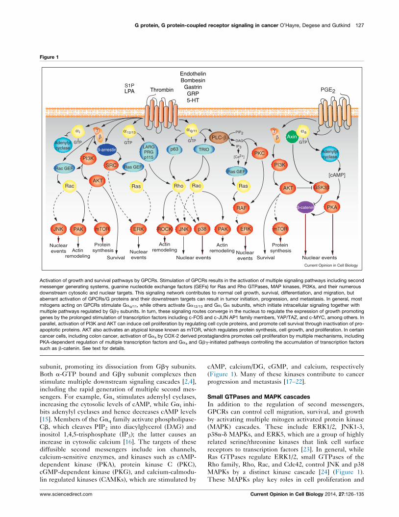
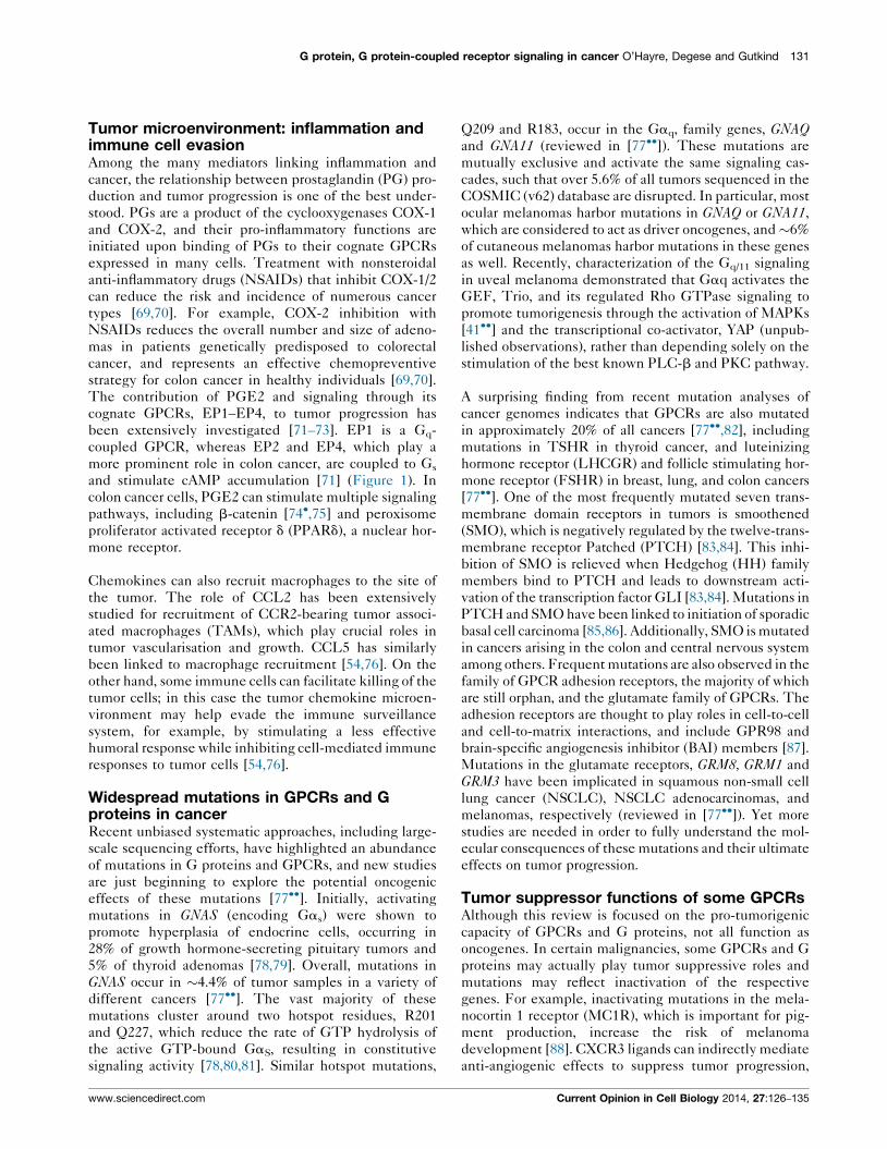
Figure 1
LPA PGE2
βγ
GTP
αq/11
β-arrestin
PI3K
GTP
αi
Adenylylcyclase
Ras GEFRac GEF
Rac
PAK
Nuclearevents
JNK
SRC
Ras
ERK
AKT
mTOR
Nuclearevents
Proteinsynthesis
Survival
LARGPRGp115
TRIOp63
Rho
ROCK PAK
Rac
JNK p38 ERK
Ras
Ras GEF
PKC
Actinremodeling
Nuclear events
PLC-β
Adenylylcyclase
Axin
β-catenin
GSK3β
[cAMP]
PKA
EndothelinBombesin
GastrinGRP5-HT
Actinremodeling Nuclear events
GTP
αsβ
γ
PI3K
AKT
mTOR
Proteinsynthesis
PIP2
DAG
IP3
[Ca2+]
GTP
α12/13
Actinremodeling Nuclear
events
RAF
S1PThrombin
Survival
Current Opinion in Cell Biology
Activation of growth and survival pathways by GPCRs. Stimulation of GPCRs results in the activation of multiple signaling pathways including second
messenger generating systems, guanine nucleotide exchange factors (GEFs) for Ras and Rho GTPases, MAP kinases, PI3Ks, and their numerous
downstream cytosolic and nuclear targets. This signaling network contributes to normal cell growth, survival, differentiation, and migration, but
aberrant activation of GPCRs/G proteins and their downstream targets can result in tumor initiation, progression, and metastasis. In general, most
mitogens acting on GPCRs stimulate Gaq/11, while others activate Ga12/13 and Gai Ga subunits, which initiate intracellular signaling together with
multiple pathways regulated by Gbg subunits. In turn, these signaling routes converge in the nucleus to regulate the expression of growth promoting
genes by the prolonged stimulation of transcription factors including c-FOS and c-JUN AP1 family members, YAP/TAZ, and c-MYC, among others. In
parallel, activation of PI3K and AKT can induce cell proliferation by regulating cell cycle proteins, and promote cell survival through inactivation of pro-
apoptotic proteins. AKT also activates an atypical kinase known as mTOR, which regulates protein synthesis, cell growth, and proliferation. In certain
cancer cells, including colon cancer, activation of Gas by COX-2 derived prostaglandins promotes cell proliferation by multiple mechanisms, including
PKA-dependent regulation of multiple transcription factors and Gas and Gbg-initiated pathways controlling the accumulation of transcription factors
such as b-catenin. See text for details.
subunit, promoting its dissociation from Gbg subunits.
Both a-GTP bound and Gbg subunit complexes then
stimulate multiple downstream signaling cascades [2,4],
including the rapid generation of multiple second mes-
sengers. For example, Gas stimulates adenylyl cyclases,
increasing the cytosolic levels of cAMP, while Gai inhi-
bits adenylyl cyclases and hence decreases cAMP levels
[15]. Members of the Gaq family activate phospholipase-
Cb, which cleaves PIP2 into diacylglycerol (DAG) and
inositol 1,4,5-trisphosphate (IP3); the latter causes an
increase in cytosolic calcium [16]. The targets of these
diffusible second messengers include ion channels,
calcium-sensitive enzymes, and kinases such as cAMP-
dependent kinase (PKA), protein kinase C (PKC),
cGMP-dependent kinase (PKG), and calcium-calmodu-
lin regulated kinases (CAMKs), which are stimulated by
www.sciencedirect.com
cAMP, calcium/DG, cGMP, and calcium, respectively
(Figure 1). Many of these kinases contribute to cancer
progression and metastasis [17–22].
Small GTPases and MAPK cascades
In addition to the regulation of second messengers,
GPCRs can control cell migration, survival, and growth
by activating multiple mitogen activated protein kinase
(MAPK) cascades. These include ERK1/2, JNK1-3,
p38a-d MAPKs, and ERK5, which are a group of highly
related serine/threonine kinases that link cell surface
receptors to transcription factors [23]. In general, while
Ras GTPases regulate ERK1/2, small GTPases of the
Rho family, Rho, Rac, and Cdc42, control JNK and p38
MAPKs by a distinct kinase cascade [24] (Figure 1).
These MAPKs play key roles in cell proliferation and
Current Opinion in Cell Biology 2014, 27:126–135

128 Cell regulation
metastasis, and their deregulation is a frequent event in
human malignancies. Hence, how GPCRs regulate
MAPKs, particularly through Ras and Rho GTPases,
has been explored under multiple physiological and
pathological conditions.
Specifically, many GPCRs coupled to Gi activate Rac and
JNK through the direct interaction of Gbg subunits with
the P-REX1/2 family of Rac guanine nucleotide exchange
factors (GEFs) [25,26]. Gaq activates Rho GTPases
through p63-RhoGEF and Trio [27�]. Receptors coupled
to Ga12 and Ga13 activate Rho by stimulating a family of
Rho GEFs, comprised of p115, PDZRhoGEF and LARG
[28]. The JNK cascade is activated downstream of Rac
and Cdc42 [24], which can mediate signaling from Gbg
dimers and Ga12, Ga13, Gaq and Gai (reviewed in [4]).
Activation of the ERK1/2 pathway by GPCRs is achieved
in a highly cell-specific fashion (reviewed in [4]), pro-
moted by Ras, tyrosine kinases, PI3Ks, PKCs, and/or
arrestins. How GPCRs activate p38 and ERK5 is much
less clear, but in general these MAPKs are activated
primarily by Gaq, Ga12/13, and Gbg dimers [4]. Activation
of MAPK pathways stimulates the expression of growth
promoting early-immediate responsive genes, including
those encoding the AP-1 family of transcription factors.
MAPKs are involved in the regulation of both gene
expression and transactivating activity of AP-1 members
by a complex and not fully understood mechanism
(Figure 1).
Activation of the PI3K, AKT, and mTOR pathway
Activation of the PI3K–AKT–mTOR pathway plays a
central role in cell metabolism, migration, growth and
survival [29,30]. PI3K generates PIP3 inducing activation
of AKT and mTOR [29,30]. PI3Kg exhibits restricted
tissue distribution and is activated by the direct interaction
of its catalytic (p110g) and regulatory subunit (p101) with
Gbg subunits [31]. PI3Kg is involved in chemokine-
induced migration of leukocytes, and plays significant roles
in innate immunity [32]. In cells lacking PI3Kg expression,
GPCRs can utilize PI3Kb to stimulate PIP3 synthesis
[33,34]. One of the most studied PI3K-regulated events
is the activation of the kinases AKT and mTOR, which
phosphorylate multiple substrates involved in cell
migration, survival, and metabolism [33,34] (Figure 2).
Regulation of the Hippo signaling pathway
GPCRs involved in cell proliferation stimulate the
activity of the transcriptional co-activator YAP [35��],which is a critical component of the Hippo signaling
pathway that controls organ size in mammals [36–39].
YAP (and related TAZ), is active in proliferating cells, but
cell confluence triggers the activation of the growth-
inhibitory Hippo kinase cascade. This causes the acti-
vation of two kinases known as LATS1/LATS2, which
phosphorylate and thereby inhibit YAP [40]. GPCRs
linked to Ga12/13 inhibit the activity of LATS, thus
Current Opinion in Cell Biology 2014, 27:126–135
relieving YAP from LATS-dependent inhibition [35��],while receptors activating Gas promote LATS activation
thus inhibiting YAP [35��]. Recent work in our laboratory
indicates that oncogenic mutations in the gene encoding
Gaq activate YAP by a mechanosensing pathway initiated
upon actin polymerization rather than by the inhibition of
the Hippo pathway (unpublished results). YAP activation
may represent a key pro-tumorigenic pathway activated
by GPCRs, thereby representing a novel target for cancer
treatment.
GPCR signal integration and crosstalk
While GPCRs can stimulate multiple diffusible second-
messenger generating systems, their ability to promote
normal and aberrant cell proliferation often relies on the
persistent activation of PI3K/AkKT/mTOR, Ras and Rho
GTPases, and MAPK cascades, thereby regulating the
activity of nuclear transcription factors and co-activators,
such as JUN, FOS and YAP [35��,41��]. Additionally,
arrestin proteins contribute to G protein-dependent
and G protein-independent events, initiating signaling
and regulating receptor internalization and degradation/
recycling kinetics [42,43]. b-Arrestins are now believed to
scaffold a wide variety of signaling complexes [44,45].
Some b-arrestin-biased GPCR agonists initiate intracellu-
lar signaling independently of the activation of hetero-
trimeric G proteins [45]. By forming multimeric signaling
complexes with ERK1/2 and JNK, b-arrestins can retain
these MAPKs in the cytosol, thus restricting their nuclear
translocation and leading to interaction with cytosolic
substrates instead [45].
A more global view of the general systems by which
GPCRs exert their numerous physiological and patho-
logical roles is necessary to appreciate the overall implica-
tions to tumorigenesis. In particular, extensive cross-talk
and co-regulation may occur between GPCR-initiated
and RTK-initiated signaling pathways and through re-
ceptor transactivation [46,47]. Therefore, the final bio-
logical outcome of GPCR activation results from the
integration of the network of GPCR-initiated bio-
chemical responses in each cellular and environmental
context. Such systems level understanding may provide a
molecular framework for the development of novel
approaches for therapeutic intervention in some of the
most prevalent human diseases.
Viral GPCRsEarly studies of virally encoded oncogenes provided the
foundation of our current understanding of cancer
biology. At least seven human viruses, Epstein-Barr virus
(EBV/HHV-4), hepatitis B virus (HBV), hepatitis C virus
(HCV), human papilloma virus (HPV), human T-cell
lymphotropic virus (HTLV-1), and Kaposi’s associated
sarcoma herpes virus (KSHV/HHV-8), and Merkel cell
polyomavirus, contribute to 10–15% of cancers [48,49].
Surprisingly, many human viruses harbor open reading
www.sciencedirect.com
G protein, G protein-coupled receptor signaling in cancer O’Hayre, Degese and Gutkind 129
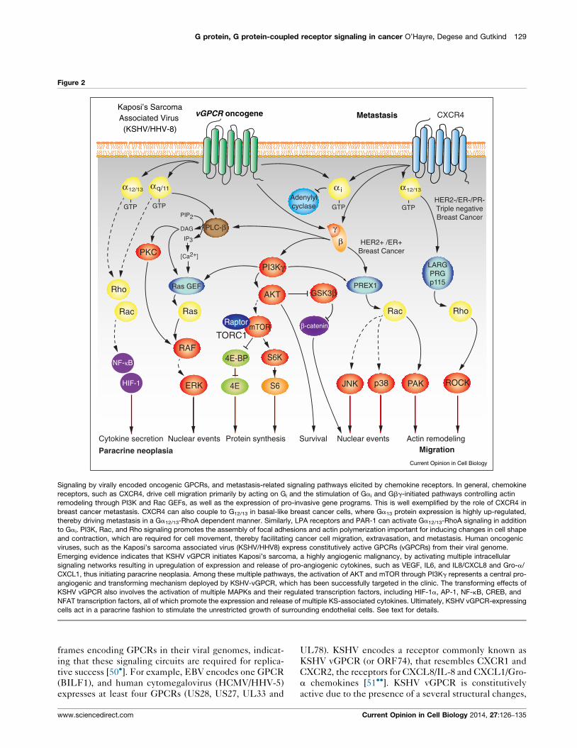
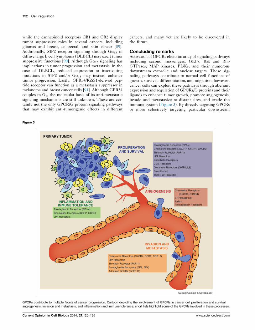
Figure 2
Current Opinion in Cell Biology
Kaposi’s SarcomaAssociated Virus (KSHV/HHV-8)
CXCR4
GTP
αq/11
GTP
α iAdenylylcyclase
PREX1
PAK
Rac
JNK p38
Actin remodeling
β
γ
Protein synthesis
LARGPRGp115
Rho
ROCK
GTP
α12/13
PKC
Metastasis
GTP
α12/13
Paracrine neoplasia
Cytokine secretion
β-catenin
GSK3β
PI3Kγ
AKT
mTORRaptor
TORC1
Survival
4E-BP S6K
4E S6
Nuclear events
ERK
Ras
Ras GEF
PLC-β
PIP2
DAG
IP3
[Ca2+]
RAF
Nuclear events Migration
HER2-/ER-/PR-Triple negativeBreast Cancer
Rac
Rho
NF-κB
HIF-1
HER2+ /ER+Breast Cancer
vGPCR oncogene
Signaling by virally encoded oncogenic GPCRs, and metastasis-related signaling pathways elicited by chemokine receptors. In general, chemokine
receptors, such as CXCR4, drive cell migration primarily by acting on Gi and the stimulation of Gai and Gbg-initiated pathways controlling actin
remodeling through PI3K and Rac GEFs, as well as the expression of pro-invasive gene programs. This is well exemplified by the role of CXCR4 in
breast cancer metastasis. CXCR4 can also couple to G12/13 in basal-like breast cancer cells, where Ga13 protein expression is highly up-regulated,
thereby driving metastasis in a Ga12/13-RhoA dependent manner. Similarly, LPA receptors and PAR-1 can activate Ga12/13-RhoA signaling in addition
to Gai. PI3K, Rac, and Rho signaling promotes the assembly of focal adhesions and actin polymerization important for inducing changes in cell shape
and contraction, which are required for cell movement, thereby facilitating cancer cell migration, extravasation, and metastasis. Human oncogenic
viruses, such as the Kaposi’s sarcoma associated virus (KSHV/HHV8) express constitutively active GPCRs (vGPCRs) from their viral genome.
Emerging evidence indicates that KSHV vGPCR initiates Kaposi’s sarcoma, a highly angiogenic malignancy, by activating multiple intracellular
signaling networks resulting in upregulation of expression and release of pro-angiogenic cytokines, such as VEGF, IL6, and IL8/CXCL8 and Gro-a/
CXCL1, thus initiating paracrine neoplasia. Among these multiple pathways, the activation of AKT and mTOR through PI3Kg represents a central pro-
angiogenic and transforming mechanism deployed by KSHV-vGPCR, which has been successfully targeted in the clinic. The transforming effects of
KSHV vGPCR also involves the activation of multiple MAPKs and their regulated transcription factors, including HIF-1a, AP-1, NF-kB, CREB, and
NFAT transcription factors, all of which promote the expression and release of multiple KS-associated cytokines. Ultimately, KSHV vGPCR-expressing
cells act in a paracrine fashion to stimulate the unrestricted growth of surrounding endothelial cells. See text for details.
frames encoding GPCRs in their viral genomes, indicat-
ing that these signaling circuits are required for replica-
tive success [50�]. For example, EBV encodes one GPCR
(BILF1), and human cytomegalovirus (HCMV/HHV-5)
expresses at least four GPCRs (US28, US27, UL33 and
www.sciencedirect.com
UL78). KSHV encodes a receptor commonly known as
KSHV vGPCR (or ORF74), that resembles CXCR1 and
CXCR2, the receptors for CXCL8/IL-8 and CXCL1/Gro-
a chemokines [51��]. KSHV vGPCR is constitutively
active due to the presence of a several structural changes,
Current Opinion in Cell Biology 2014, 27:126–135
130 Cell regulation
and contributes to KS development due to its potent
transforming and pro-angiogenic properties. It promotes
sarcomagenesis by increasing the activity of a complex
signaling network, among which the activation of the
PI3Kg/AKT/mTOR pathway represents a clinically
relevant target for KS treatment (Figure 2). Ultimately,
KSHV vGPCR-expressing cells act in a paracrine fashion
to stimulate the unrestricted growth of surrounding and
distant endothelial cells, thereby representing an excel-
lent example paracrine neoplasia.
GPCRs in migration, invasion and metastasisOne of the most serious challenges facing cancer treat-
ment is metastasis, the spread of cancer cells through
blood or lymphatic vessels to distant organs [52]. Rather
than spreading randomly, cancer cells metastasize pre-
ferentially to specific organs, with a greater incidence than
would be expected from the circulatory pattern between
the primary tumor site and secondary organs [53��]. The
GPCR family of chemokine receptors is centrally linked
to the organ-specific metastasis of a number of cancers, in
line with their normal immune cell function of directing
receptor-bearing leukocytes towards sites of chemokine
production. Similarly, tumor cells aberrantly expressing
chemokine receptors can co-opt the migratory activity of
chemokines, facilitating metastasis to other organs [54].
Chemokines locally released into the tumor microenvir-
onment can also enhance the motility and survival of
cancer cells in an autocrine and paracrine fashion [54].
Chemokines direct cell movement by inducing changes
in cytoskeletal structure and dynamics of receptor-bear-
ing cells. Actin polymerization leads to formation of
protrusions, or pseudopods, and with the help of integrins,
form focal adhesions with the extracellular matrix (ECM)
to help propel the cell forward [55].
CXCR4 represents one of the best established chemokine
receptors driving cancer metastasis. Tumor cells fre-
quently exhibit aberrant CXCR4 expression, which has
proliferative, pro-survival, and pro-migratory effects;
additionally, the organs that are the most frequent sites
of metastasis, including lymph nodes, lungs, bone marrow
and liver, express its chemokine ligand, CXCL12/SDF-1
[53��]. Several factors may contribute to the observed
overexpression of CXCR4 in many tumors. For example,
the HER2/Neu oncogene, which occurs in �30% of
breast cancers, limits the degradation of CXCR4, leading
to its increased expression [56]. Additionally, hypoxia
through activation of hypoxia-inducible factor-1 (HIF-
1a) induces transcription of CXCR4 [57]. As such,
CXCR4 is highly expressed in breast cancer cells but
not in normal breast tissues, and the inhibition of CXCR4
prevents the metastatic spread of breast cancer cells [53��]and many other cancer types [52,54,58]. Although
CXCR4 would represent an attractive target for thera-
peutic development, the use of CXCR4 inhibitors leads
to the mobilization of stem cell progenitors from the bone
Current Opinion in Cell Biology 2014, 27:126–135
marrow, thus limiting their use clinically for cancer treat-
ment [59]. However, targeting molecules involved in the
regulation of CXCR4 expression on cancer cells or their
downstream signaling may offer alternative approaches
for therapeutic intervention. In this regard, CXCR4 acti-
vates Rac1 through P-REX1, which plays a central role in
metastasis in most breast cancer types [60]. CXCR4 can
also couple to G12/13 in basal-like breast cancer cells,
where Ga13 protein expression is highly upregulated,
thus driving metastasis through a Ga12/13-RhoA depend-
ent manner [61�], similarly to LPA and PAR-1 receptors
(Figure 2), all of which can be considered potential targets
for metastasis prevention and treatment.
Other chemokine receptors, including CCR7 and CCR10,
have also been shown to play direct roles in metastatic
homing of cancer cells types [62] and cancer cell survival
and growth [63]. Local chemokine production in the
tumor microenvironment can attract macrophages and
leukocytes that may enhance the cytokine-rich microen-
vironment and induce the release of matrix metallopro-
teases (MMPs) that enable tumor cells to survive,
proliferate and invade. In addition, recent functional
screens demonstrated a role for GPR116, a member of
the poorly characterized family of adhesion GPCRs, in
invasion and migration of breast cancer cells, acting in a
Gaq-RhoA/Rac1-dependent manner. Many new efforts
are now focused on exploring the adhesion family of
GPCRs, and their potential implications in tumor growth
and metastasis [64].
GPCRs in tumor-induced angiogenesisSolid tumors produce angiogenic factors promoting the
migration and proliferation of endothelial cells, thus
resulting in the formation of new vessels in response to
the increasing nutrients and oxygen demands of the
tumor cells. Many angiogenic factors act on GPCRs
expressed on endothelial cells, including thrombin, pros-
taglandins, S1P, and chemokines [65–67]. Many chemo-
kines, including CCL2, CCL5, and CXCL8/IL-8, recruit
leukocytes and macrophages to the tumor site, which in
turn can release VEGF and other angiogenic factors that
contribute to the growth of new blood vessels [66]. In
addition, inflammatory cytokines released in the tumor
microenvironment promote the expression of COX-2 and
the local release of prostaglandin E2 (PGE2), which
increases the expression of pro-angiogenic VEGF,
CXCL8 and CXCL5 by tumor and stromal cells [68].
Overall, GPCRs and their cognate ligands can promote
angiogenesis directly by inducing proliferation of endo-
thelial cells or indirectly by promoting release of VEGF
and other angiogenic factors from stromal, immune, or
cancer cells. Tumor vascularization provides nutrients to
promote tumor outgrowth, and routes for invasion and
metastasis.
www.sciencedirect.com
G protein, G protein-coupled receptor signaling in cancer O’Hayre, Degese and Gutkind 131
Tumor microenvironment: inflammation andimmune cell evasionAmong the many mediators linking inflammation and
cancer, the relationship between prostaglandin (PG) pro-
duction and tumor progression is one of the best under-
stood. PGs are a product of the cyclooxygenases COX-1
and COX-2, and their pro-inflammatory functions are
initiated upon binding of PGs to their cognate GPCRs
expressed in many cells. Treatment with nonsteroidal
anti-inflammatory drugs (NSAIDs) that inhibit COX-1/2
can reduce the risk and incidence of numerous cancer
types [69,70]. For example, COX-2 inhibition with
NSAIDs reduces the overall number and size of adeno-
mas in patients genetically predisposed to colorectal
cancer, and represents an effective chemopreventive
strategy for colon cancer in healthy individuals [69,70].
The contribution of PGE2 and signaling through its
cognate GPCRs, EP1–EP4, to tumor progression has
been extensively investigated [71–73]. EP1 is a Gq-
coupled GPCR, whereas EP2 and EP4, which play a
more prominent role in colon cancer, are coupled to Gs
and stimulate cAMP accumulation [71] (Figure 1). In
colon cancer cells, PGE2 can stimulate multiple signaling
pathways, including b-catenin [74�,75] and peroxisome
proliferator activated receptor d (PPARd), a nuclear hor-
mone receptor.
Chemokines can also recruit macrophages to the site of
the tumor. The role of CCL2 has been extensively
studied for recruitment of CCR2-bearing tumor associ-
ated macrophages (TAMs), which play crucial roles in
tumor vascularisation and growth. CCL5 has similarly
been linked to macrophage recruitment [54,76]. On the
other hand, some immune cells can facilitate killing of the
tumor cells; in this case the tumor chemokine microen-
vironment may help evade the immune surveillance
system, for example, by stimulating a less effective
humoral response while inhibiting cell-mediated immune
responses to tumor cells [54,76].
Widespread mutations in GPCRs and Gproteins in cancerRecent unbiased systematic approaches, including large-
scale sequencing efforts, have highlighted an abundance
of mutations in G proteins and GPCRs, and new studies
are just beginning to explore the potential oncogenic
effects of these mutations [77��]. Initially, activating
mutations in GNAS (encoding Gas) were shown to
promote hyperplasia of endocrine cells, occurring in
28% of growth hormone-secreting pituitary tumors and
5% of thyroid adenomas [78,79]. Overall, mutations in
GNAS occur in �4.4% of tumor samples in a variety of
different cancers [77��]. The vast majority of these
mutations cluster around two hotspot residues, R201
and Q227, which reduce the rate of GTP hydrolysis of
the active GTP-bound GaS, resulting in constitutive
signaling activity [78,80,81]. Similar hotspot mutations,
www.sciencedirect.com
Q209 and R183, occur in the Gaq, family genes, GNAQand GNA11 (reviewed in [77��]). These mutations are
mutually exclusive and activate the same signaling cas-
cades, such that over 5.6% of all tumors sequenced in the
COSMIC (v62) database are disrupted. In particular, most
ocular melanomas harbor mutations in GNAQ or GNA11,
which are considered to act as driver oncogenes, and �6%
of cutaneous melanomas harbor mutations in these genes
as well. Recently, characterization of the Gq/11 signaling
in uveal melanoma demonstrated that Gaq activates the
GEF, Trio, and its regulated Rho GTPase signaling to
promote tumorigenesis through the activation of MAPKs
[41��] and the transcriptional co-activator, YAP (unpub-
lished observations), rather than depending solely on the
stimulation of the best known PLC-b and PKC pathway.
A surprising finding from recent mutation analyses of
cancer genomes indicates that GPCRs are also mutated
in approximately 20% of all cancers [77��,82], including
mutations in TSHR in thyroid cancer, and luteinizing
hormone receptor (LHCGR) and follicle stimulating hor-
mone receptor (FSHR) in breast, lung, and colon cancers
[77��]. One of the most frequently mutated seven trans-
membrane domain receptors in tumors is smoothened
(SMO), which is negatively regulated by the twelve-trans-
membrane receptor Patched (PTCH) [83,84]. This inhi-
bition of SMO is relieved when Hedgehog (HH) family
members bind to PTCH and leads to downstream acti-
vation of the transcription factor GLI [83,84]. Mutations in
PTCH and SMO have been linked to initiation of sporadic
basal cell carcinoma [85,86]. Additionally, SMO is mutated
in cancers arising in the colon and central nervous system
among others. Frequent mutations are also observed in the
family of GPCR adhesion receptors, the majority of which
are still orphan, and the glutamate family of GPCRs. The
adhesion receptors are thought to play roles in cell-to-cell
and cell-to-matrix interactions, and include GPR98 and
brain-specific angiogenesis inhibitor (BAI) members [87].
Mutations in the glutamate receptors, GRM8, GRM1 and
GRM3 have been implicated in squamous non-small cell
lung cancer (NSCLC), NSCLC adenocarcinomas, and
melanomas, respectively (reviewed in [77��]). Yet more
studies are needed in order to fully understand the mol-
ecular consequences of these mutations and their ultimate
effects on tumor progression.
Tumor suppressor functions of some GPCRsAlthough this review is focused on the pro-tumorigenic
capacity of GPCRs and G proteins, not all function as
oncogenes. In certain malignancies, some GPCRs and G
proteins may actually play tumor suppressive roles and
mutations may reflect inactivation of the respective
genes. For example, inactivating mutations in the mela-
nocortin 1 receptor (MC1R), which is important for pig-
ment production, increase the risk of melanoma
development [88]. CXCR3 ligands can indirectly mediate
anti-angiogenic effects to suppress tumor progression,
Current Opinion in Cell Biology 2014, 27:126–135
132 Cell regulation
while the cannabinoid receptors CB1 and CB2 display
tumor suppressive roles in several cancers, including
gliomas and breast, colorectal, and skin cancer [89].
Additionally, SIP2 receptor signaling through Ga13 in
diffuse large B cell lymphoma (DLBCL) may exert tumor
suppressive functions [90]. Although Ga13 signaling has
implications in tumor progression and metastasis, in the
case of DLBCL, reduced expression or inactivating
mutations in S1P2 and/or Ga13 may instead enhance
tumor progression. Lastly, GPR54/KiSS1-derived pep-
tide receptor can function as a metastasis suppressor in
melanoma and breast cancer cells [91]. Although GPR54
couples to Gq, the molecular basis of its anti-metastatic
signaling mechanisms are still unknown. These are cer-
tainly not the only GPCR/G protein signaling pathways
that may exhibit anti-tumorigenic effects in different
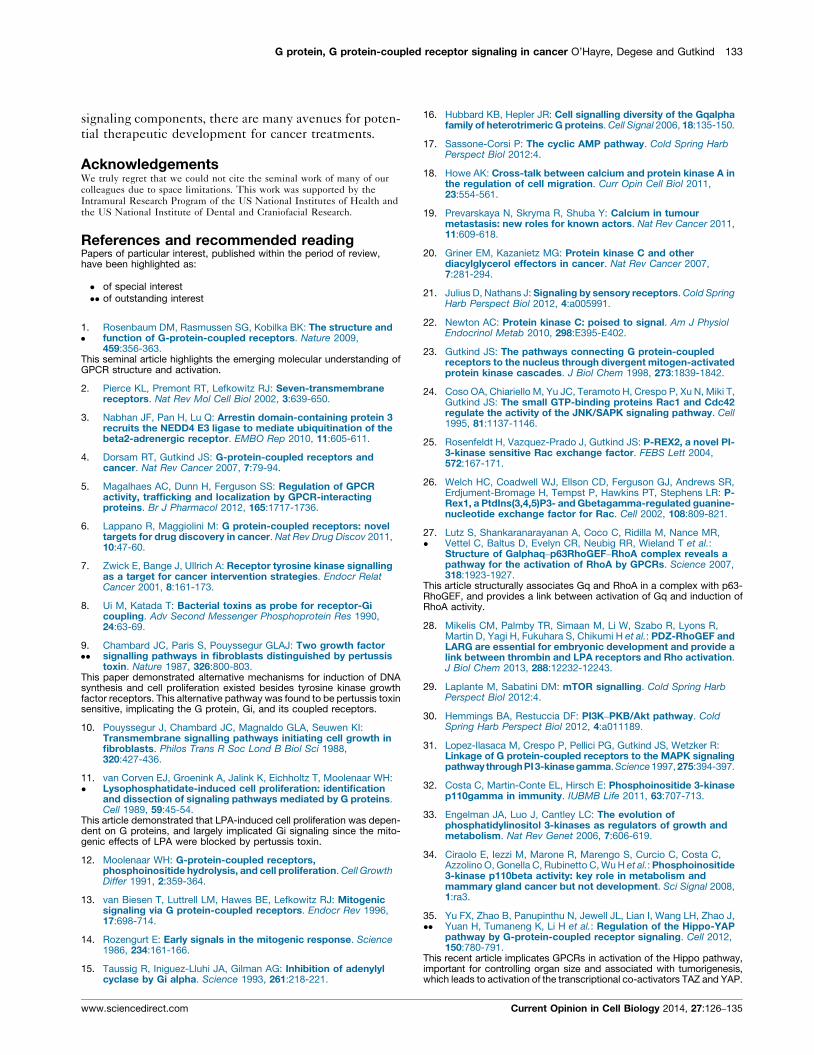
Figure 3
PROLIFAND S
PRIMARY TUMOR
IMMUNE TOLERANCEINFLAMMATION AND
Chemokine Recep LPA Recep tors
Thrombin Recep to
Prostaglandin Rec
Adhesion GPCRs
Prostaglandin Recep tors (EP1-4)
Chemokine Recep tors (CCR2, CCR5)
LPA Recep tors
GPCRs contribute to multiple facets of cancer progression. Cartoon depicti
angiogenesis, invasion and metastasis, and inflammation and immune toleran
Current Opinion in Cell Biology 2014, 27:126–135
cancers, and many yet are likely to be discovered in
the future.
Concluding remarksActivation of GPCRs elicits an array of signaling pathways
including second messengers, GEFs, Ras and Rho
GTPases, MAP kinases, PI3Ks, and their numerous
downstream cytosolic and nuclear targets. These sig-
naling pathways contribute to normal cell functions of
growth, survival, differentiation, and migration; however,
cancer cells can exploit these pathways through aberrant
expression and regulation of GPCRs/G proteins and their
ligands to enhance tumor growth, promote angiogenesis,
invade and metastasize to distant sites, and evade the
immune system (Figure 3). By directly targeting GPCRs
or more selectively targeting particular downstream
ANGIOGENESIS
INVASION ANDMETASTASIS
ERATIONURVIVAL
tors (CXCR4, CCR7, CCR10)
r (PAR-1)
ep tors (EP2, EP4)
(GPR116)
Chemokine Recep tors
(CXCR2, CXCR4)
S1P Recep tors
PAR-1Prostaglandin Recep tors
Prostaglandin Recep tors (EP1-4)
Chemokine Recep tors (CCR7, CXCR4, CXCR2)
Thrombin Recep tor (PAR-1)
LPA Recep tors
Endothelin Recep tors
CCK Recep tors
Glutamate Recep tors (GMR1,3,8)
Smoothened
TSHR, LH Recep tor
Current Opinion in Cell Biology
ng the involvement of GPCRs in cancer cell proliferation and survival,
ce; short lists highlight some of the GPCRs involved in these processes.
www.sciencedirect.com
G protein, G protein-coupled receptor signaling in cancer O’Hayre, Degese and Gutkind 133
signaling components, there are many avenues for poten-
tial therapeutic development for cancer treatments.
AcknowledgementsWe truly regret that we could not cite the seminal work of many of ourcolleagues due to space limitations. This work was supported by theIntramural Research Program of the US National Institutes of Health andthe US National Institute of Dental and Craniofacial Research.
References and recommended readingPapers of particular interest, published within the period of review,have been highlighted as:
� of special interest�� of outstanding interest
1.�
Rosenbaum DM, Rasmussen SG, Kobilka BK: The structure andfunction of G-protein-coupled receptors. Nature 2009,459:356-363.
This seminal article highlights the emerging molecular understanding ofGPCR structure and activation.
2. Pierce KL, Premont RT, Lefkowitz RJ: Seven-transmembranereceptors. Nat Rev Mol Cell Biol 2002, 3:639-650.
3. Nabhan JF, Pan H, Lu Q: Arrestin domain-containing protein 3recruits the NEDD4 E3 ligase to mediate ubiquitination of thebeta2-adrenergic receptor. EMBO Rep 2010, 11:605-611.
4. Dorsam RT, Gutkind JS: G-protein-coupled receptors andcancer. Nat Rev Cancer 2007, 7:79-94.
5. Magalhaes AC, Dunn H, Ferguson SS: Regulation of GPCRactivity, trafficking and localization by GPCR-interactingproteins. Br J Pharmacol 2012, 165:1717-1736.
6. Lappano R, Maggiolini M: G protein-coupled receptors: noveltargets for drug discovery in cancer. Nat Rev Drug Discov 2011,10:47-60.
7. Zwick E, Bange J, Ullrich A: Receptor tyrosine kinase signallingas a target for cancer intervention strategies. Endocr RelatCancer 2001, 8:161-173.
8. Ui M, Katada T: Bacterial toxins as probe for receptor-Gicoupling. Adv Second Messenger Phosphoprotein Res 1990,24:63-69.
9.��
Chambard JC, Paris S, Pouyssegur GLAJ: Two growth factorsignalling pathways in fibroblasts distinguished by pertussistoxin. Nature 1987, 326:800-803.
This paper demonstrated alternative mechanisms for induction of DNAsynthesis and cell proliferation existed besides tyrosine kinase growthfactor receptors. This alternative pathway was found to be pertussis toxinsensitive, implicating the G protein, Gi, and its coupled receptors.
10. Pouyssegur J, Chambard JC, Magnaldo GLA, Seuwen KI:Transmembrane signalling pathways initiating cell growth infibroblasts. Philos Trans R Soc Lond B Biol Sci 1988,320:427-436.
11.�
van Corven EJ, Groenink A, Jalink K, Eichholtz T, Moolenaar WH:Lysophosphatidate-induced cell proliferation: identificationand dissection of signaling pathways mediated by G proteins.Cell 1989, 59:45-54.
This article demonstrated that LPA-induced cell proliferation was depen-dent on G proteins, and largely implicated Gi signaling since the mito-genic effects of LPA were blocked by pertussis toxin.
12. Moolenaar WH: G-protein-coupled receptors,phosphoinositide hydrolysis, and cell proliferation. Cell GrowthDiffer 1991, 2:359-364.
13. van Biesen T, Luttrell LM, Hawes BE, Lefkowitz RJ: Mitogenicsignaling via G protein-coupled receptors. Endocr Rev 1996,17:698-714.
14. Rozengurt E: Early signals in the mitogenic response. Science1986, 234:161-166.
15. Taussig R, Iniguez-Lluhi JA, Gilman AG: Inhibition of adenylylcyclase by Gi alpha. Science 1993, 261:218-221.
www.sciencedirect.com
16. Hubbard KB, Hepler JR: Cell signalling diversity of the Gqalphafamily of heterotrimeric G proteins. Cell Signal 2006, 18:135-150.
17. Sassone-Corsi P: The cyclic AMP pathway. Cold Spring HarbPerspect Biol 2012:4.
18. Howe AK: Cross-talk between calcium and protein kinase A inthe regulation of cell migration. Curr Opin Cell Biol 2011,23:554-561.
19. Prevarskaya N, Skryma R, Shuba Y: Calcium in tumourmetastasis: new roles for known actors. Nat Rev Cancer 2011,11:609-618.
20. Griner EM, Kazanietz MG: Protein kinase C and otherdiacylglycerol effectors in cancer. Nat Rev Cancer 2007,7:281-294.
21. Julius D, Nathans J: Signaling by sensory receptors. Cold SpringHarb Perspect Biol 2012, 4:a005991.
22. Newton AC: Protein kinase C: poised to signal. Am J PhysiolEndocrinol Metab 2010, 298:E395-E402.
23. Gutkind JS: The pathways connecting G protein-coupledreceptors to the nucleus through divergent mitogen-activatedprotein kinase cascades. J Biol Chem 1998, 273:1839-1842.
24. Coso OA, Chiariello M, Yu JC, Teramoto H, Crespo P, Xu N, Miki T,Gutkind JS: The small GTP-binding proteins Rac1 and Cdc42regulate the activity of the JNK/SAPK signaling pathway. Cell1995, 81:1137-1146.
25. Rosenfeldt H, Vazquez-Prado J, Gutkind JS: P-REX2, a novel PI-3-kinase sensitive Rac exchange factor. FEBS Lett 2004,572:167-171.
26. Welch HC, Coadwell WJ, Ellson CD, Ferguson GJ, Andrews SR,Erdjument-Bromage H, Tempst P, Hawkins PT, Stephens LR: P-Rex1, a PtdIns(3,4,5)P3- and Gbetagamma-regulated guanine-nucleotide exchange factor for Rac. Cell 2002, 108:809-821.
27.�
Lutz S, Shankaranarayanan A, Coco C, Ridilla M, Nance MR,Vettel C, Baltus D, Evelyn CR, Neubig RR, Wieland T et al.:Structure of Galphaq–p63RhoGEF–RhoA complex reveals apathway for the activation of RhoA by GPCRs. Science 2007,318:1923-1927.
This article structurally associates Gq and RhoA in a complex with p63-RhoGEF, and provides a link between activation of Gq and induction ofRhoA activity.
28. Mikelis CM, Palmby TR, Simaan M, Li W, Szabo R, Lyons R,Martin D, Yagi H, Fukuhara S, Chikumi H et al.: PDZ-RhoGEF andLARG are essential for embryonic development and provide alink between thrombin and LPA receptors and Rho activation.J Biol Chem 2013, 288:12232-12243.
29. Laplante M, Sabatini DM: mTOR signalling. Cold Spring HarbPerspect Biol 2012:4.
30. Hemmings BA, Restuccia DF: PI3K–PKB/Akt pathway. ColdSpring Harb Perspect Biol 2012, 4:a011189.
31. Lopez-Ilasaca M, Crespo P, Pellici PG, Gutkind JS, Wetzker R:Linkage of G protein-coupled receptors to the MAPK signalingpathway through PI 3-kinase gamma. Science 1997, 275:394-397.
32. Costa C, Martin-Conte EL, Hirsch E: Phosphoinositide 3-kinasep110gamma in immunity. IUBMB Life 2011, 63:707-713.
33. Engelman JA, Luo J, Cantley LC: The evolution ofphosphatidylinositol 3-kinases as regulators of growth andmetabolism. Nat Rev Genet 2006, 7:606-619.
34. Ciraolo E, Iezzi M, Marone R, Marengo S, Curcio C, Costa C,Azzolino O, Gonella C, Rubinetto C, Wu H et al.: Phosphoinositide3-kinase p110beta activity: key role in metabolism andmammary gland cancer but not development. Sci Signal 2008,1:ra3.
35.��
Yu FX, Zhao B, Panupinthu N, Jewell JL, Lian I, Wang LH, Zhao J,Yuan H, Tumaneng K, Li H et al.: Regulation of the Hippo-YAPpathway by G-protein-coupled receptor signaling. Cell 2012,150:780-791.
This recent article implicates GPCRs in activation of the Hippo pathway,important for controlling organ size and associated with tumorigenesis,which leads to activation of the transcriptional co-activators TAZ and YAP.
Current Opinion in Cell Biology 2014, 27:126–135
134 Cell regulation
36. Pan D: The hippo signaling pathway in development andcancer. Dev Cell 2010, 19:491-505.
37. Ramos A, Camargo FD: The Hippo signaling pathway and stemcell biology. Trends Cell Biol 2012, 22:339-346.
38. Zhao B, Li L, Lei Q, Guan KL: The Hippo-YAP pathway in organsize control and tumorigenesis: an updated version. Genes Dev2010, 24:862-874.
39. Sudol M, Bork P, Einbond A, Kastury K, Druck T, Negrini M,Huebner K, Lehman D: Characterization of the mammalian YAP(Yes-associated protein) gene and its role in defining a novelprotein module, the WW domain. J Biol Chem 1995,270:14733-14741.
40. Yu FX, Guan KL: The Hippo pathway: regulators andregulations. Genes Dev 2013, 27:355-371.
41.��
Vaque JP, Dorsam RT, Feng X, Iglesias-Bartolome R,Forsthoefel DJ, Chen Q, Debant A, Seeger MA, Ksander BR,Teramoto H et al.: A genome-wide RNAi screen reveals a Trio-regulated Rho GTPase circuitry transducing mitogenic signalsinitiated by G protein-coupled receptors. Mol Cell 2013,49:94-108.
This article demonstrates molecular mechanisms by which activation ofthe G proteins, Gq and G11, leads to cell proliferation through activationof the GEF, Trio, and subsequent activation of Rho and Rac and down-stream p38 and JNK MAPK pathways. It supports the emerging notionthat cell growth promotion by GPCRs depends on a hard wired signalingnetwork based on localized protein–protein interactions rather than solelyon diffusible second messenger systems.
42. Lefkowitz RJ, Shenoy SK: Transduction of receptor signals bybeta-arrestins. Science 2005, 308:512-517.
43. Luttrell LM, Lefkowitz RJ: The role of beta-arrestins in thetermination and transduction of G-protein-coupled receptorsignals. J Cell Sci 2002, 115:455-465.
44. Luttrell LM, Gesty-Palmer D: Beyond desensitization:physiological relevance of arrestin-dependent signaling.Pharmacol Rev 2010, 62:305-330.
45. Rajagopal S, Rajagopal K, Lefkowitz RJ: Teaching old receptorsnew tricks: biasing seven-transmembrane receptors. Nat RevDrug Discov 2010, 9:373-386.
46. Prenzel N, Zwick E, Daub H, Leserer M, Abraham R, Wallasch C,Ullrich A: EGF receptor transactivation by G-protein-coupledreceptors requires metalloproteinase cleavage of proHB-EGF.Nature 1999, 402:884-888.
47. Natarajan K, Berk BC: Crosstalk coregulation mechanisms of Gprotein-coupled receptors and receptor tyrosine kinases.Methods Mol Biol 2006, 332:51-77.
48. Feng H, Shuda M, Chang Y, Moore PS: Clonal integration of apolyomavirus in human Merkel cell carcinoma. Science 2008,319:1096-1100.
49. Martin D, Gutkind JS: Human tumor-associated viruses andnew insights into the molecular mechanisms of cancer.Oncogene 2008, 27(Suppl 2):S31-S42.
50.�
Montaner S, Kufareva I, Abagyan R, Gutkind JS: Molecularmechanisms deployed by virally encoded G protein-coupledreceptors in human diseases. Annu Rev Pharmacol Toxicol2013, 53:331-354.
This review summarizes the currently known GPCRs encoded by differentviruses and their implications in human disease, including cancer.
51.��
Arvanitakis L, Geras-Raaka E, Varma A, Gershengorn MC,Cesarman E: Human herpesvirus KSHV encodes aconstitutively active G-protein-coupled receptor linkedto cell proliferation [see comments]. Nature 1997,385:347-350.
This article demonstrated that a virally encoded GPCR in Kaposi’ssarcoma herpes virus tumors contributes to cellular proliferation.
52. Chambers AF, Groom AC, MacDonald IC: Dissemination andgrowth of cancer cells in metastatic sites. Nat Rev Cancer 2002,2:563-572.
53.��
Muller A, Homey B, Soto H, Ge N, Catron D, Buchanan ME,McClanahan T, Murphy E, Yuan W, Wagner SN et al.: Involvement
Current Opinion in Cell Biology 2014, 27:126–135
of chemokine receptors in breast cancer metastasis. Nature2001, 410:50-56.
This paper demonstrated a direct link between the expression of thechemokine receptor, CXCR4, in tumor cells and organ specific metas-tasis to sites in which its ligand, CXCL12, is produced, with emphasis onbreast cancer.
54. Balkwill F: Cancer and the chemokine network. Nat Rev Cancer2004, 4:540-550.
55. Friedl P, Wolf K: Tumour-cell invasion and migration: diversityand escape mechanisms. Nat Rev Cancer 2003, 3:362-374.
56. Liang Z, Wu T, Lou H, Yu X, Taichman RS, Lau SK, Nie S,Umbreit J, Shim H: Inhibition of breast cancer metastasis byselective synthetic polypeptide against CXCR4. Cancer Res2004, 64:4302-4308.
57. Staller P, Sulitkova J, Lisztwan J, Moch H, Oakeley EJ, Krek W:Chemokine receptor CXCR4 downregulated by von Hippel-Lindau tumour suppressor pVHL. Nature 2003, 425:307-311.
58. Burger JA, Kipps TJ: CXCR4: a key receptor in the crosstalkbetween tumor cells and their microenvironment. Blood 2006,107:1761-1767.
59. Levesque JP, Hendy J, Takamatsu Y, Simmons PJ, Bendall LJ:Disruption of the CXCR4/CXCL12 chemotactic interactionduring hematopoietic stem cell mobilization induced by GCSFor cyclophosphamide. J Clin Invest 2003, 111:187-196.
60. Sosa MS, Lopez-Haber C, Yang C, Wang H, Lemmon MA,Busillo JM, Luo J, Benovic JL, Klein-Szanto A, Yagi H et al.:Identification of the Rac-GEF P-Rex1 as an essential mediatorof ErbB signaling in breast cancer. Mol Cell 2010, 40:877-892.
61.�
Yagi H, Tan W, Dillenburg-Pilla P, Armando S, Amornphimoltham P,Simaan M, Weigert R, Molinolo AA, Bouvier M, Gutkind JS: Asynthetic biology approach reveals a CXCR4–G13–Rhosignaling axis driving transendothelial migration of metastaticbreast cancer cells. Sci Signal 2011, 4:ra60.
This paper implicates a link between the chemokine receptor, CXCR4,and Ga13 signaling to Rho and ROCK kinase, which can contribute to themetastasis of triple negative breast cancer cells that overexpress G13 asubunit.
62. Zlotnik A, Burkhardt AM, Homey B: Homeostatic chemokinereceptors and organ-specific metastasis. Nat Rev Immunol2011, 11:597-606.
63. O’Hayre M, Salanga CL, Handel TM, Allen SJ: Chemokines andcancer: migration, intracellular signalling and intercellularcommunication in the microenvironment. Biochem J 2008,409:635-649.
64. Tang X, Jin R, Qu G, Wang X, Li Z, Yuan Z, Zhao C, Siwko S, Shi T,Wang P et al.: GPR116, an adhesion G-protein-coupledreceptor, promotes breast cancer metastasis via theGalphaq–p63RhoGEF–Rho GTPase pathway. Cancer Res 2013,73:6206-6218.
65. Moore BB, Keane MP, Addison CL, Arenberg DA, Strieter RM:CXC chemokine modulation of angiogenesis: the importanceof balance between angiogenic and angiostatic members ofthe family. J Investig Med 1998, 46:113-120.
66. Richard DE, Vouret-Craviari V, Pouyssegur J: Angiogenesis andG-protein-coupled receptors: signals that bridge the gap.Oncogene 2001, 20:1556-1562.
67. Wang D, Dubois RN: Prostaglandins and cancer. Gut 2006,55:115-122.
68. Iniguez MA, Rodriguez A, Volpert OV, Fresno M, Redondo JM:Cyclooxygenase-2: a therapeutic target in angiogenesis.Trends Mol Med 2003, 9:73-78.
69. Brown JR, DuBois RN: COX-2: a molecular target for colorectalcancer prevention. J Clin Oncol 2005, 23:2840-2855.
70. Gupta RA, Dubois RN: Colorectal cancer prevention andtreatment by inhibition of cyclooxygenase-2. Nat Rev Cancer2001, 1:11-21.
71. Hull MA, Ko SC, Hawcroft G: Prostaglandin EP receptors:targets for treatment and prevention of colorectal cancer?Mol Cancer Ther 2004, 10:1-1039.
www.sciencedirect.com
G protein, G protein-coupled receptor signaling in cancer O’Hayre, Degese and Gutkind 135
72. Hansen-Petrik MB, McEntee MF, Jull B, Shi H, Zemel MB,Whelan J: Prostaglandin E(2) protects intestinal tumors fromnonsteroidal anti-inflammatory drug-induced regression inApc(Min/+) mice. Cancer Res 2002, 62:403-408.
73. Sonoshita M, Takaku K, Sasaki N, Sugimoto Y, Ushikubi F,Narumiya S, Oshima M, Taketo MM: Acceleration of intestinalpolyposis through prostaglandin receptor EP2 in Apc(Delta716) knockout mice. Nat Med 2001, 7:1048-1051.
74.�
Castellone MD, Teramoto H, Williams BO, Druey KM, Gutkind JS:Prostaglandin E2 promotes colon cancer cell growth througha Gs–axin–beta-catenin signaling axis. Science 2005,310:1504-1510.
This article characterizes the signaling pathways induced by inflamma-tory COX2 production of PGE2, leading to activation of the Gs-coupledEP2 receptor and downstream activation of AKT, axin and beta-catenin incolon cancer.
75. Shao J, Jung C, Liu C, Sheng H: Prostaglandin E2 stimulates thebeta-catenin/T cell factor-dependent transcription in coloncancer. J Biol Chem 2005, 280:26565-26572.
76. Rollins BJ: Inflammatory chemokines in cancer growth andprogression. Eur J Cancer 2006, 42:760-767.
77.��
O’Hayre M, Vazquez-Prado J, Kufareva I, Stawiski EW, Handel TM,Seshagiri S, Gutkind JS: The emerging mutational landscape ofG proteins and G-protein-coupled receptors in cancer. Nat RevCancer 2013, 13:412-424.
This bioinformatic analysis of large cancer sequencing data sets revealedan unexpected and surprisingly high incidence of G protein and GPCRmutations in some of the most prevalent human neoplastic diseases.
78. Landis CA, Masters SB, Spada A, Pace AM, Bourne HR, Vallar L:GTPase inhibiting mutations activate the alpha chain of Gs andstimulate adenylyl cyclase in human pituitary tumours. Nature1989, 340:692-696.
79. Weinstein LS, Shenker A, Gejman PV, Merino MJ, Friedman E,Spiegel AM: Activating mutations of the stimulatory G proteinin the McCune-Albright syndrome [see comments]. N Engl JMed 1991, 325:1688-1695.
80. Drews RT, Gravel RA, Collu R: Identification of G protein alphasubunit mutations in human growth hormone (GH)- and GH/prolactin-secreting pituitary tumors by single-strandconformation polymorphism (SSCP) analysis. Mol CellEndocrinol 1992, 87:125-129.
www.sciencedirect.com
81. Wilson CH, McIntyre RE, Arends MJ, Adams DJ: The activatingmutation R201C in GNAS promotes intestinal tumourigenesisin Apc(Min/+) mice through activation of Wnt and ERK1/2MAPK pathways. Oncogene 2010, 29:4567-4575.
82. Kan Z, Jaiswal BS, Stinson J, Janakiraman V, Bhatt D, Stern HM,Yue P, Haverty PM, Bourgon R, Zheng J et al.: Diverse somaticmutation patterns and pathway alterations in human cancers.Nature 2010, 466:869-873.
83. Epstein EH: Basal cell carcinomas: attack of the hedgehog. NatRev Cancer 2008, 8:743-754.
84. Rubin LL, de Sauvage FJ: Targeting the Hedgehog pathway incancer. Nat Rev Drug Discov 2006, 5:1026-1033.
85. Lum L, Beachy PA: The Hedgehog response network: sensors,switches, and routers. Science 2004, 304:1755-1759.
86. Xie J, Murone M, Luoh SM, Ryan A, Gu Q, Zhang C, Bonifas JM,Lam CW, Hynes M, Goddard A et al.: Activating smoothenedmutations in sporadic basal-cell carcinoma. Nature 1998,391:90-92.
87. Paavola KJ, Hall RA: Adhesion G protein-coupled receptors:signaling, pharmacology, and mechanisms of activation. MolPharmacol 2012, 82:777-783.
88. Mitra D, Luo X, Morgan A, Wang J, Hoang MP, Lo J, Guerrero CR,Lennerz JK, Mihm MC, Wargo JA et al.: An ultraviolet-radiation-independent pathway to melanoma carcinogenesis in the redhair/fair skin background. Nature 2012, 491:449-453.
89. Velasco G, Sanchez C, Guzman M: Towards the use ofcannabinoids as antitumour agents. Nat Rev Cancer 2012,12:436-444.
90. Green JA, Suzuki K, Cho B, Willison LD, Palmer D, Allen CD,Schmidt TH, Xu Y, Proia RL, Coughlin SR et al.: The sphingosine1-phosphate receptor S1P(2) maintains the homeostasis ofgerminal center B cells and promotes niche confinement. NatImmunol 2011, 12:672-680.
91. Lee JH, Miele ME, Hicks DJ, Phillips KK, Trent JM, Weissman BE,Welch DR: KiSS-1: a novel human malignant melanomametastasis-suppressor gene. J Natl Cancer Inst 1996,88:1731-1737.
Current Opinion in Cell Biology 2014, 27:126–135


![signal transduction [Read-Only] - Hadassah · G-protein signaling Mechanisms of signal transduction. 13 G-protein signaling. 14 ... Target cell de sensitization and hyper sensitization](https://cdn.vdocuments.site/doc/165x107/5ed96e04f59b0f56f45f79d7/signal-transduction-read-only-g-protein-signaling-mechanisms-of-signal-transduction.jpg)


