
Functional Foods in Health and Disease 2014; 4(1):23-65 Page 23 of 65
Review Article Open Access
Neurodegenerative and Fatiguing Illnesses, Infections and Mitochondrial
Dysfunction: Use of Natural Supplements to Improve Mitochondrial
Function.
Garth L. Nicolson1, Robert Settineri
2 and Rita R. Ellithorpe
3
1Department of Molecular Pathology, The Institute for Molecular Medicine, Huntington Beach,
CA 92647 USA; 2Sierra Research, Irvine, CA, 92606 USA;
3Tustin Longevity Center, Tustin,
California 92780, USA
Corresponding Author: Prof. Emeritus Garth L. Nicolson, Department of Molecular Pathology,
Institute for Molecular Medicine, P. O. Box 9355, S. Laguna Beach, CA 92652
Submission date: November 08, 2013; Acceptance date: January 22, 2014; Publication date:
January 25, 2014
ABSTRACT
Background: Many chronic diseases and illnesses are associated with one or more chronic
infections, dysfunction of mitochondria and reduced production of ATP. This results in fatigue
and other symptoms that occur in most if not all chronic conditions and diseases.
Methods: This is a review of the published literature on chronic infections in neurodegenerative
diseases and fatiguing illnesses that are also typified by mitochondrial dysfunction. This
contribution also reviews the use of natural supplements to enhance mitochondrial function and
reduce the effects of chronic infections to improve overall function in various chronic illnesses.
Results: Mitochondrial function can be enhanced by the use of various natural supplements,
notably Lipid Replacement Therapy (LRT) using glyerolphospholipids and other mitochondrial
supplements. In various chronic illnesses that are characterized by the presence of chronic
infections, such as intracellular bacteria (Mycoplasma, Borrelia, Chlamydia and other infections)
and viruses, LRT has proven useful in multiple clinical trials. For example, in clinical studies on
chronic fatigue syndrome, fibromyalgia syndrome and other chronic fatiguing illnesses where a
large majority of patients have chronic infections, LRT significantly reduced fatigue by 35-43%
in different clinical trials and increased mitochondrial function. In clinical trials on patients with
multiple intracellular bacterial infections and intractable fatigue LRT plus other mitochondrial
supplements significantly decreased fatigue and improved mood and cognition.
Conclusions: LRT formulations designed to improve mitochondrial function appear to be useful
as non-toxic dietary supplements for reducing fatigue and restoring mitochondrial and other
cellular membrane functions in patients with chronic illnesses and multiple chronic infections.
Functional Foods in Health and Disease 2014; 4(1):23-65 Page 24 of 65
Key words: Neurodegenerative and fatiguing illnesses, infections and mitochondrial
dysfunction, ATP, Lipid Replacement Therapy (LRT)
Background
Patients with chronic neurodegenerative, neurobehavioral and fatiguing illnesses commonly test
positive for systemic and central nervous system (CNS) bacterial and viral infections [1-3]. In
addition, other chronic illnesses where neurological manifestations are routinely found, such as
autoimmune diseases and other chronic illnesses and disorders, also show evidence of systemic
bacterial and viral infections that could be important in disease inception, progression and/or
enhancing the types and severities of signs and symptoms [2, 3].
Evidence of bacterial infections, such as Mycoplasma species, Chlamydia pneumoniae,
Borrelia burgdorferi, among others, and viruses, such as human herpesvirus (HHV),
cytomegalovirus (CMV), human herpes viruses (HHV) and other viral infections, have revealed
high rates of infection in the illnesses listed above that were not found in control populations [1-
3]. Although the specific roles of chronic infections in various diseases and their pathogeneses
have not been carefully determined, the data suggest that chronic bacterial and/or viral infections
are common features of essentially all progressive chronic diseases [1-3].
Another common finding in chronic illness patients is mitochondrial dysfunction,
characterized by loss of efficiency in the electron transport chain, reductions in mitochondrial
inner membrane trans-membrane potential and reductions in the synthesis of high-energy
molecules such as ATP [4-6]. This is also a characteristic of aging, and it essentially occurs in
all chronic diseases, including cancer [5-7].
This review will concentrate on commonly acquired mechanisms that affect mitochondrial
function. To treat functional loss associated wtih chronic infections mitochondrial replacement
strategies with natural supplements and combinations of natural supplements have been used,
including vitamins, minerals, enzyme cofactors, antioxidants, metabolites, transporters,
membrane-type phospholipids and other natural supplements in order to improve mitochondrial
function.
Introduction
Chronic infections appear to be a common feature of various diseases, including
neurodegenerative, psychiatric, neurobehavioral diseases and other conditions [1-3, 8]. Chronic
infections are also associated with autoimmune diseases [9, 10] and fatiguing illnesses [1, 3, 11].
This will be discussed in various sections of this review. In addition, many chronic illnesses are
directly caused by chronic infections, such as Lyme disease, brucellosis, babesiosis, and other
infection-based chronic diseases [2, 12-14].
Chronic infections collectively result in induction of excess Reactive Oxygen Species (ROS)
and Reactive Nitrogen Species (RNS) that damage cellular structures, especially mitochondrial
membranes [15-17]. Mitochondria are especially sensitive to excess levels of ROS/RNS, and in
chronic illnesses there is extensive damage to mitochondria in the form of membrane oxidation,
damage to mitochondrial DNA (mtDNA) and loss of mitochondrial enzymatic function and inner
mitochondrial membrane potential [6-8, 18-20]. In this review we will discuss the use of
Functional Foods in Health and Disease 2014; 4(1):23-65 Page 25 of 65
comprehensive approaches to restore mitochondrial function damaged by infections and other
causes.
Patients with chronic illnesses (often caused by or exacerbated by chronic infections) are
particularly difficult to treat using single modality approaches, and this is particularly true for
patients who have multiple chronic infections [21, 22]. The multi-focal nature of chronic
diseases and the fact that often treatments are given to suppress adverse signs and symptoms,
rather than treat causes of the disease or its progression, have resulted in incomplete or
ineffective treatments. On the other hand, even if the causes of chronic diseases are known, by
the time therapeutic interventions are undertaken, it may be entirely too late to use approaches
that might work on the disease at an early stage or if chronic infections were not also present. At
the stage(s) of disease when patients usually seek medical care for their conditions, they usually
have multiple problems, including chronic infections, and each of these problems usually
requires complex therapeutic approaches. Their multiple chronic infections also cause additional
cellular damage [1, 2, 8].
Multiple alternations in mitochondrial membranes, proteins and mtDNA are thought to be
the cause for mitochondrial dysfunction, and this damage can also accumulate over time [6, 23].
By the time patients seek care, they usually have multiple defects in their mitochondria, and thus
there are no simple approaches that are effective in promoting functional recovery of their
mitochondria. With this in mind, we have begun this review by discussing the evidence for
chronic infections and mitochondrial dysfunction in selected chronic illnesses and diseases.
Then we will discuss the role that various supplements play in restoring mitochondrial function,
even in patients with multiple chronic infections that continue to degrade mitochondrial
components. Finally we will discuss the role of combination supplements for restoring
mitochondrial function in patients with chronic illnesses and multiple chronic infections.
Neurodegenerative Diseases
Neurodegenerative diseases, or chronic degenerative diseases of the central nervous system
(CNS) that cause dementia, are mainly diseases of the elderly [1, 2]. On the other hand,
neurobehavioral diseases are found mainly in young patients and include autism spectrum
disorders (ASD), such as autism, attention deficit disorder, Asperger’s syndrome and other
disorders [24]. For the most part, the causes of these neurological diseases remain largely
unknown but it is thought that multiple factors are involved in each disease [1, 2].
Neurodegenerative diseases are characterized by molecular and genetic changes in nerve
cells that result in nerve cell dysfunction, degeneration and ultimately cell death, resulting in
neurological signs and symptoms and eventually dementia [1, 2]. In contrast, neurobehavioral
diseases are related to fetal brain and early post-partum development but are less well
characterized at the cellular level. Both of these disease types involve genetic and environmental
factors [24, 25], and they also have multiple chronic infections [1-3, 26, 27]. Even less well
characterized at the cellular and genetic level are the psychiatric disorders, such as
schizophrenia, paranoia, bipolar disorders, depression and obsessive-compulsive disorders, but
these diseases are also associated with the presence of chronic infections [3].
Genetic alternations have been found in neurodegenerative and neurobehavioral diseases,
but the genetic changes as well as changes in gene expression that have been found are complex

Functional Foods in Health and Disease 2014; 4(1):23-65 Page 26 of 65
and usually not directly related to simple gene alterations, such as single mutations and deletions,
that lead to single protein molecule alterations [24, 25, 28]. Importantly, mutations that affect
mitochondrial function are known to be associated with neurodegenerative and neurobehavioral
diseases [29, 30]. These include mutations in mtDNA as well as nuclear DNA [29]. In addition
to chronic infections and genetic changes, environmental toxins, heavy metals, nutritional
deficiencies, autoimmune immunological responses, vascular diseases, head trauma (and
accumulation of fluid in the brain), changes in neurotransmitter concentrations, among other
elements, are thought to be collectively involved in the pathogenesis of various
neurodegenerative and neurobehavioral diseases [1, 2, 24-31]. These important topics will not
be discussed in detail in this review.
Chronic Infections are important factors in neurodegenerative and neurobehavioral diseases,
and infectious agents may enter the brain within infected migratory macrophages. Alternatively,
they can also gain access by direct penetration of the blood-brain-barrier or entry by
intraneuronal transfer from peripheral nerves [32]. Cell wall-deficient bacteria, such as species
of Mycoplasma, Chlamydia (Chlamydophila), Borrelia, Brucella, among others and various
viruses are candidate brain infectious agents, because they are capable of CNS penetration and
have been found routinely in neurodegenerative and neurobehavioral diseases [1-3, 26, 27, 32,
33]. Such infections are usually systemic and can affect immune systems and essentially any
organ system, resulting in a variety of systemic signs and symptoms that are not limited to the
CNS [10, 11, 26, 27, 32, 33].
Amyotrophic lateral sclerosis (ALS)
ALS is an adult-onset, progressive neurodegenerative disease of unknown etiology that affects
both central and peripheral motor neurons where patients show gradual progressive weakness
and paralysis of muscles due to destruction of upper motor neurons in the motor cortex and lower
motor neurons in the brain stem and spinal cord [34, 35]. Eventually this results in death, usually
by respiratory failure [35].
Chronic infections in ALS, such as the finding of enterovirus sequences in a majority of
ALS spinal cord samples by polymerase chain reaction (PCR) [36], have attracted widespread
attention. However, others have failed to detect enterovirus sequences in ALS spinal cord
samples [37]. Using PCR methods systemic mycoplasmal infections have been found in a high
percentage (83%) of ALS patients [38]. For example, all of the tested Gulf War veterans
diagnosed with ALS from three nations had systemic mycoplasmal infections [38]. In addition, a
majority of ALS patients in Lyme endemic areas show immunological evidence of Borrelia
infections [39], and some patients diagnosed with ALS were subsequently found to have
neuroborreliosis infections [40]. Although high rates of infection may occur in certain regions,
the overall rate of Borrelia infections in ALS is low (10% or less) in North America [41].
MacDonald [42], however, observed a high incidence of spirochetal forms in the brain tissues of
ALS patients and in patients with other neurodegenerative diseases, suggesting that the presence
of chronic bacterial infections in the CNS of neurodegenerative diseases patients is much more
common than previously assumed.
ALS patients also show evidence of other infections. These include: human herpes virus-6
(HHV-6), Chlamydia pneumoniae, cyanobacteria and other infections [43-45]. Chronic

Functional Foods in Health and Disease 2014; 4(1):23-65 Page 27 of 65
infections plus other defects (accumulation of glutamate causing excitotoxicity, deficiency of
nerve growth factor, autoimmune reactions against motor neurons and dysfunction of
mitochondrial superoxide dismutase) have been proposed to be important in ALS pathogenesis
[review: 2].
Mitochondrial dysfunction is a common feature of ALS and animal models of ALS [46,
47]. Evidence from patients with sporadic and familial ALS and from ALS models based on the
over-expression of mutant SOD1 found in a small subset of patients, clearly point to
mitochondrial damage as a relevant facet of this neurodegenerative condition [46]. In addition to
mutations in superoxide dismutase genes, some ALS patients present with mutations in
mitochondrial transport genes and misfolding in inclusion proteins, ubiquilin-2 and other
mitochondrial associated proteins [review: 47]. Dysfunction in several other cellular
mechanisms, including mitophagy, oxidative stress, lipid peroxidation and cholesterol
esterification, protein and neurofilament aggregation, impaired axonal transport, among other
changes in ALS patients have been reviewed recently [47, 48].
Multiple Sclerosis (MS)
The most common demyelinating neurological disease is MS [49]. MS can occur in all age
groups as a cyclic (relapsing-remitting) or a progressive disease that continues progressing
without remitting [49]. Inflammation and the presence of autoimmune antibodies against myelin
and other nerve cell antigens are thought to cause myelin sheath breakdown, resulting in
decrease or loss of electrical impulses along nerve fibers [49, 50]. In the MS patients with
progressive neurological symptoms damage occurs additionally by the deposition of plaques on
nerve cells to the point where nerve cell death occurs. Importantly, breakdown of the blood-
brain barrier in the CNS of MS patients is associated with local inflammation caused by
activated glial cells [49, 50]. The combination of demyelinization, plaque damage and blood-
brain barrier disruption causes multiple, variable symptoms, but they usually include impaired
vision, alterations in motor, sensory and coordination nerve systems along with cognitive
dysfunction [50].
MS is a disease in which environmental, genetic and epigenetic factors determine the risk of
developing MS, its progression and responsiveness to treatment [51, 52]. Just as in ALS, there
are multiple genetic components in MS [51, 53]. Although it has been established that there is a
genetic basis to MS susceptibility, epidemiological and twin studies suggest that MS is basically
an acquired disease with some genetic and environmental components [54].
The molecular mechanisms through which environmental signals are translated into changes
in gene expression include: DNA methylation, post-translational modification of nucleosomal
histones, and non-coding RNAs. These mechanisms are regulated by families of specialized
enzymes that are tissue-selective and cell-type specific [54].
Chronic infections have been linked to the pathophysiology of MS [55, 56]. For example,
MS patients show immunological and cytokine elevations consistent with chronic infections [57,
58]. An infectious basis for MS has been under examination for some time, and patients have
been tested for various viral and bacterial infections [1, 3, 53, 55, 56].
One of the most consistent findings in MS patients has been the presence of C. pneumoniae
antibodies and DNA in their cerebrospinal fluid [59-61]. By examining relapsing-remitting and

Functional Foods in Health and Disease 2014; 4(1):23-65 Page 28 of 65
progressive MS patients for the presence of C. pneumoniae in cerebrospinal fluid by culture,
PCR and immunoglobulin reactivity Sriram et al. [60] were able to identify C. pneumoniae in
64% of MS cerebrospinal fluid versus 11% of patients with other neurological diseases. They
also found high rates of PCR-positive MOMP gene (97%) in MS- patients (versus 18% in other
neurological diseases), and this correlated with a high rate of patients being serology-positive
(86%) for Chlamydia antigens by ELISA and Western blot analysis [60]. MS patients examined
for oligoclonal antibodies against C. pneumoniae revealed that 82% of MS patients were positive
compared to none of the control non-MS neurological patients [61]. Similarly, C. pneumoniae
RNA and DNA transcripts were found in mononuclear cells and cerebrospinal fluids of 64.2% of
MS patients but in only 3 controls [62].
The brain tissues of MS and non-MS neurological patients have also been examined for C.
pneumoniae antigens [63]. Using immunohistochemistry to find C. pneumoniae antigens in
formalin-fixed brain tissue Sriram et al. [63] found that in a subset of MS patients (35%)
Chlamydial antigens were localized to ependymal surfaces and pariventricular regions. Positive
reactions were not found in brain tissue samples from other neurological diseases. PCR
amplification of C. pneumoniae genes was accomplished in 63% of brain tissue samples from
MS patients but none in frozen brain tissues from other neurological diseases. In addition, using
immuno-electron microscopy the sediment from cerebrospinal fluid was examined for
Chlamydial antigens [63]. Sriram et al. [63] found that the electron dense bodies resembling
bacterial structures that were positive by immuno-electron microscopy correlated with tissue
PCR-positive MS cases (91% positive using both methods).
Using different nested PCR methods to examine additional C. pneumoniae gene sequences
in the cerebrospinal fluid of 72 MS patients Contini et al. [64] were able to match these results to
MS-associated lesions seen by MRI. Grimaldi et al. also used MRI to link the presence of C.
pneumoniae infection with abnormal MRI results and found linkage in 21% in MS patients [65].
The MS patients with C. pneumoniae infections were also the MS patients with more progressive
disease. Indeed, higher rates of C. pneumoniae transcription were found in the cerebrospinal
fluid of 84 patients with the more progressive form of MS [66]. These studies strongly support
the presence of C. pneumoniae in the brains of MS patients with progressive disease [67, 68].
Not all researchers have found C. pneumoniae or other bacteria
in the brains of MS patients
[69, 70]. For example, Hammerschlag et al. used nested PCR and culture to examine frozen
brain samples from MS patients but could not find any evidence for C. pneumoniae gene
sequences [71]. Thus the evidence linking C. pneumoniae infection with MS is not universally
accepted, and other genetic changes may be necessary to complete the link between such
infections and the etiology of MS [72].
Multiple infections in MS patients may complicate the evidence linking MS with specific
chronic infections. Thus other infections similar to C. pneumoniae could be involved rather than
just one specific infection [1]. In addition to C. pneumoniae, MS patients could also have
Mycoplasma species, B. burgdorferi and other bacterial infections as well as viral infections [73].
When multiple infections are considered, it is likely that >90% of MS patients have obligate
intracellular bacterial infections.
Various viruses have also been found in MS patients. For example, HHV-6 has been found
at higher frequencies in MS patients, but this virus has also been found at lower incidence in

Functional Foods in Health and Disease 2014; 4(1):23-65 Page 29 of 65
control samples [74]. PCR was used to examine postmortem brain tissue and controls for the
presence of various neurotrophic viruses [74]. These studies revealed that 57% of MS cases and
43% of non-MS neurological disease controls contain sequences for HHV-6, whereas 37%, 28%,
and 43%, respectively, contained sequences for herpes simplex virus (HSV)-1 and –2 and
varicella zoster virus. Although impressive, the data did not achieve statistical significance.
They also found that 32% of the MS active plaques and 17% of the inactive plaque areas were
positive for HHV-6 [74].
Using sequence difference analysis Challoner et al. searched for pathogens in MS brain
specimens and found that >70% of MS patients were positive for infection-associated sequences
[76]. They also used immunocytochemistry and found positive staining around MS plaques
more frequently than around surrounding white matter. Additionally, HHV-6 DNA was found in
peripheral leukocytes in the systemic circulation of MS patients [77] but not in every study [78].
Examination of the literature strongly suggests an infectious process in MS [1, 55, 56, 79,
80]. In most studies the more progressive forms of MS rather than the relapsing-remitting forms
of MS were associated with chronic infections. Thus chronic infections may play a role in
progression of MS. If infections like C. pneumoniae and Mycoplasma species are important in
MS, then antibiotics effective against these infections should improve clinical status [1]. This
has, in fact, been seen in most but not in all MS patients [81]. As in other neurodegenerative
diseases, multiple factors appear to be involved in the pathogenesis of MS [1, 49, 55, 56, 82].
One of the factors in MS appears to be mitochondrial dysfunction due to oxidative injury
[83, 84]. Broadwater et al. have identified several MS-related damaged mitochondrial proteins
that are involved in respiration, including cytochrome c oxidase subunit 5b, an isozyme of
appears to be damage to the permeability transition pore (PTP) by excess reactive oxygen species
(ROS) [86]. This critical structure is central to mitochondrial dysfunction by allowing ion
dysregulation within neural cells that drives neurodegeneration by allowing the PTP to change
the ion gradients inside mitochondria, lowering inner membrane trans-membrane potential (thus
reducing oxidative phosphorylation [87]), promoting matrix expansion leading to release of
cytochrome c and initiating cell death programs [86]. In addition, the energy and calcium
balance in neurons plays an important role in maintaining a healthy myelin sheath, and a
hallmark of MS is axon demyelation due to mitochondrial dysfunction [83], which drives an
inflammatory response characteristic of MS progression [88].
Alzheimer’s disease (AD)
AD is characterized by distinct pathological changes in brain cells and tissues [1, 2]. Among the
most notable are the appearance of plaques and tangles of neurofibrils in brain nerve cells that
affect synapses and nerve-nerve cell communication. These alterations involve the deposition of
altered amyloid proteins [89, 90]. Although the origins of AD are not known for certain, the
formation of the amyloid plaques and neurofiber tangles found in AD may be due to genetic
defects and resulting changes in the structure of beta amyloid proteins, which may be caused by
chemicals or other toxic events, inflammatory responses, excess oxidative stress and increases in
ROS, loss of nerve trophic factors and reductions in nerve cell transmission [89-92].

Functional Foods in Health and Disease 2014; 4(1):23-65 Page 30 of 65
Infections are potentially important in the AD disease process [93, 94]. One pathogen that
has attracted considerable attention because of its neurotropism is C. pneumoniae [95, 96]. This
intracellular bacterium has been found at high incidence in the brains of AD patients by PCR and
immunohistochemistry [96]. C. pneumoniae has also been found localized in nerve cells in close
proximity to neurofibrillary tangles, a characteristic of AD [96, 97].
C. pneumoniae can invade endothelial cells and promote the transmigration of monocytes
through human brain endothelial cells into the brain parenchyma [98]. C. pneumoniae has been
found in the brains of most AD patients [95], and it has been cultured from the brain tissue of AD
patients [99]. Immunohistological detection of C. pneumoniae was observed inside and outside
cells in the frontal and temporal cortices of AD brains [100]. Indeed, in experiments with mice
injection of C. pneumoniae stimulated brain beta amyloid plaque formation [101]. The data are
compelling, but some investigators have not been able to duplicate the findings on infections in
AD [102].
In addition to C. pneumoniae investigators have found other infections in AD patients, such
as B. burgdorferi [103, 104]. Using serology, culture, Western blot and immunofluorenscence
methods this infection has been examined in AD patients (with or without a diagnosis of Lyme
disease) and found to be present [104, 105]. The presence of intracellular infections like B.
burgdorferi in AD patients has been proposed to be a primary event in the formation of AD beta
amyloid plaques, which are thought to occur by the formation of ―congophilic cores‖ that attract
beta amyloid materials [106]. In fact, exposure of glial and neuronal cells in vitro to Borrelia
burgdorferi spirochetes and to the inflammatory bacterial lipopolysaccharide LPS caused
morphological changes analogous to those found in deposits in AD brains [107]. Also detected
were increases in beta amyloid precursor protein and hyperphosphorylated tau protein
characteristic of AD [107]. Several reports indicate that AD nerve cells are often positive for B.
burgdorferi, indicating that this intracellular bacteria could be important in the pathogenesis of
AD [103-106, 108]. However, there are reports that could not find evidence for the presence of
Borrelia in AD brain tissue [109].
Miklossy has reviewed the data indicating that chronic infections, including B. burgdorferi,
are commonly found in AD patients and has concluded that intracellular bacteria contain
amyloidogenic proteins that can induce amyloid beta deposition and tau phosphorylation [110,
111]. In addition, specific bacterial ligands and bacterial and viral DNA and RNA increase the
expression of proinflammatory molecules that activate the innate and adaptive immune systems.
Evasion of brain pathogens from destruction by the host immune system can result in persistent
infection, chronic inflammation, neuronal destruction and beta amyloid deposition [111].
The hypothesis that intracellular microorganisms or their protein products can induce beta
amyloid protein and then provide ―nucleation sites‖ for the attraction of beta amyloid materials is
attractive [111], but other factors, including the induction of reactive oxygen species, lipid
peroxidation and the breakdown of the lysosomal membranes releasing lysosomal hydrolases,
are also thought to be important in beta amyloid deposition [108]. An infectious basis in AD
pathogenesis is attractive; however, although some negative reports imply that infections like B.
burgdorferi are not essential in AD pathogenesis [109]. On the other hand, other intracellular
bacterial infections (Mycoplasma, Chlamyda, Helicobacter etc.) have been found in AD patients
and could be present in those patients who are negative for Borrelia infections [111-113]. It has
Functional Foods in Health and Disease 2014; 4(1):23-65 Page 31 of 65
been proposed that chronic infections may be important cofactors in AD and contribute to the
pathogenic process [113].
Viral infections may also play a role in AD pathogenesis. Herpes virus infections,
especially HSV-1, have been found in AD patients [114, 115]. Previously it was determined that
HSV-1 but not a related neurotrophic virus (varicella zoster virus) was present more often in AD
brains, and this could be linked to patients who have the AD risk factor ApoE e4 allele [116,
117]. Similar to bacterial proteins, HSV-1 proteins may also be involved in the abnormal
aggregation of beta amyloid fragments within the AD brain, but in this case by reducing the
amount of full-length beta amyloid precursor protein and increasing the amounts of their
fragments [118]. HSV-1 infection of glial and neuronal cells resulted in a dramatic increase in
the intracellular levels of beta amyloid forms, whereas the levels of native beta amyloid
precursor protein decreased [119]. This has been found in mice infected with HSV-1, indicating
that HSV-1 is probably involved directly in the development of senile-associated plaques. Other
herpes viruses, such as HHV-6, have also been found in AD patients, but it is thought that this
virus is not directly involved in AD pathogenesis. Another virus that has been implicated in AD
is cytomegalovirus [120]. A high proportion of brains from vascular dementia patients show
evidence of both HSV-1 and cytomegalovirus [120].
Mitochondrial dysfunction may be an early event in the pathogenesis of AD [121-123]. AD
patients show impairments in mitochondrial function that start early in process of
neurodegeneration [121, 123]. Mutations in the AbetaPP and tau genes induce oxidative stress
and mitochondrial dysfunction leading eventually to apoptotic cell death [124]. Indeed,
transgenic mouse models of AD point to impairments in oxidative phosphorylation as an
important aspect of AD pathogenesis [125]. The oxidative stress is thought to cause protein
alterations that have synergistic effects on mitochondria, leading to synaptic dysfunction and
apoptotic cell death [124].
Parkinson’s Disease (PD)
PD is characterized by akinesia, muscular rigidity and tremor. In addition, autonomic
dysfunction, olfactory disturbances, depression, sensory and sleep disturbances and frequently
dementia characterize this disease [126]. The pathology indicates a progressive loss of the
-
synuclein. Extensive brain degeneration also occurs in PD [127]. Inclusion bodies and protein
aggregations or defects in their degradation characteristically are characteristic of PD, but their
role in PD pathogenesis is unclear [127, 128]. Available evidence suggests a relationship
between PD and specific genetic changes, such as changes in the genes in mitochondria, those
affecting protein degradation, organelle trafficking and vesicular fusion, and in proteins involved
in oxidative stress or antioxidant function [129. 130]. Inflammation has also been associated
with PD [131].
PD has been proposed to be due to neurotoxic events in genetically susceptible individuals
that are especially sensitive to neuro-oxidative damage [132]. Multiple environmental factors
and genetic background are also statistically related risk factors for PD [133]. The mitochondria
in neuromelanin-containing dopaminergic neurons of the substantia nigra are the targets for
oxidative damage [128, 133, 134], and early life exposures are also important [135]. For
Functional Foods in Health and Disease 2014; 4(1):23-65 Page 32 of 65
example, early life exposure to brain injury, chemicals and/or infections may initiate a cyclic
inflammatory process involving oxidative damage, excitotoxicity, mitochondrial dysfunction and
altered proteolysis that later in life results in neuron death in the substantia nigra [136, 137].
Chronic infections have been proposed as important in PD pathogenesis [136, 137]. In fact,
regression analysis of a case-control study on infections in PD patients clearly showed that
infectious processes are an important risk factor in PD [138]. One infection found in PD that has
aroused considerable interest is the presence of chronic gastrointestinal Helicobacter pylori
infections [139]. Treatment of this infection in PD patients offered relief from late stage
cachexia [140]. Helicobacter pylori-infected PD patients also showed reduced L-dopa
absorption and increased clinical disability [141], and in antimicrobial-treated PD patients there
was increased L-dopa absorption and decreased clinical disability [142]. Although H. pylori
may not be directly involved in the pathogenesis of PD, its systemic presence has been proposed
to affect the progression and treatment of PD [141].
PD patients’ chronic infections have been linked to autoimmunity and inflammation [143-
145], and the role of neuro-inflammatory and oxidative processes in nigral degeneration has
gained increasing attention [145, 146]. Moreover, experimental models of PD have been
developed using viral or bacterial infections to initiate the pathogenic process [147, 148]. In
examining PD patients various infections have been found, especially bacterial and viral
infections [144, 149]. For example, spirochetes have also been found in the brain Lewy bodies
of Lyme-associated PD patients [150]. Other infections, such as viral encephalitis [151],
cornavirus [152], Mycoplasma pneumoniae [153], AIDS-associated infections of the basal
ganglia [154], HIV [155], among other infections, have been found in PD patients [144, 149,
155]. Additional research will be necessary to establish whether a causal link exists between PD
and chronic infections [143, 155, 156].
A common link between oxidative stress, mitochondrial dysfunction and PD exists [157,
158]. Although the underlying mechanisms for selective dopaminergic nerve degeneration in PD
are not completely known, the increase in ROS in Parkinson’s substantia nigra neurons results in
increased DNA mutation, especially in mtDNA, reduced efficiency of the electron transport
chain, and changes in protein aggregation and lipid oxidation that contribute to mitochondrial
destruction (mitophagy) and neurodegeneration [132, 145, 156-158]. Mutations in genes that
protect neural cells from oxidative damage-mediated mitochondrial dysfunction, such as tensin
(PTEN) homologue-induced kinase-1 (PINK1), are known to be associated with recessively
inherited PD, and this also points to mitochondrial damage as an underlying defect in PD [159].
PINK1 is involved in mitochondrial quality control, and under steady state conditions PINK1 is
rapidly and constitutively degraded in a mitochondrial membrane potential-dependent manner
[160]. Loss of mitochondrial inner membrane potential stabilizes PINK1 mitochondrial
accumulation and stimulates the initiation of autophagic degradation and removal of damaged
mitochondria (mitophagy), but mutations in PINK1 inhibit this process [161]. This implicates
loss of mitochondrial integrity and mitophagy in the pathogenesis of PD.
Neurobehavioral Diseases
Autism spectrum disorders (ASD)

Functional Foods in Health and Disease 2014; 4(1):23-65 Page 33 of 65
ASD includes autism, Asperger’s syndrome, among other disorders. These diseases affect
primarily young patients who generally suffer from an inability to properly communicate, form
relationships with others and respond appropriately to their environment. ASD patients do not
all share the same signs and symptoms but tend to have in common certain social,
communication, motor and sensory problems (non-compliance, hyperactivity, sensory
defensiveness, self-injury, among others) that affect their behavior. They can display repetitive
actions and develop troublesome fixations with specific objects, and they are often painfully
sensitive to certain sounds, tastes and smells [162, 163].
Multiple factors appear to be involved in ASD, including genetic factors, environmental
exposures, such as heavy metals and chemicals and biological exposures, which are probably
different in each patient [27, 28, 164-169]. ASD patients appear to have similarities in genetic
defects and environmental exposures that have been proposed to play interactive roles that are
probably important in patient morbidity or in illness progression [170, 171].
Chronic infections appear to be an important element in the development of ASD [2, 26, 27,
168, 169]. In ASD patients more than 50 different bacterial, viral and fungal infections have
been documented [27]. A few of these occur at high incident rates and may be more important
than others in causing ASD symptoms [2, 169]. ASD patients also present with a number of
nonspecific chronic signs and symptoms that suggest infections, such as fatigue, headaches,
gastrointestinal and vision problems as well as intermittent low-grade fevers and other signs and
symptoms [169, 172]. Increased titers to various viruses as well as bacterial and fungal
infections have been commonly seen in ASD patients [27, 169, 172-174].
Infections along with environmental exposures to chemicals and heavy metals is
controversial but in some cases may be important in the development of ASD in genetically
susceptible children [27, 28,166-174]. The relationship between ASD and heavy metals is
controveral but could be linked to the multiple vaccines given during pre-school years [166,
167]. ASD often developes only after multiple childhood immunizations, and the sharp increase
in Autism may be linked to vaccines after they came into widespread use [167]. Many of these
vaccines contain mercury and other toxic preservatives, and some may also contain
contaminating bacteria, as found in 6% of veterinary vaccines [175]. ASD is also related to
environmental infections, such as Lyme Borrelia and associated co-infections [21, 27, 169, 174].
An interesting study on the transmission of infections and subsequent ASD has come from
the families of veterans of the Gulf War [176, 177]. After veterans with Gulf War Illness
returned to the home, their children subsequently became symptomatic, and these children were
often diagnosed with ASD [178]. Symptomatic children with ASD were infected with the same
Mycoplasma species, M. fermentans, that was found in the veterans and their symptomatic
family members, and this was not seen in aged-matched control subjects or in military families
without GWI. In these families some non-symptomatic family members did have mycoplasmal
infections (~10%), but this was not significantly different from the incidence of mycoplasmal
infections in healthy control subjects [177, 178].
Non-military families were also examined for systemic mycoplasmal infections [26]. In the
ASD cases a majority were positive for mycoplasmal infections. In contrast to the children from
military families who for the most part had only M. fermentans, the civilian children tested
positive for a variety of Mycoplasma species [26]. For example, a large subset (>58%) of ASD
Functional Foods in Health and Disease 2014; 4(1):23-65 Page 34 of 65
patients showed evidence of Mycoplasma infections compared to age-matched control subjects
(Odds Ratio=13.8, p<0.001). ASD patients were also examined for C. pneumoniae (8.3%
positive, Odds Ratio=5.6, p<0.01) and HHV-6 (29.2% positive, Odds Ratio=4.5, p<0.01). The
results indicated that a large subset of ASD patients have bacterial and/or viral infections (Odds
Ratio=16.5, p<0.001) [26].
In addition to Mycoplasma infections, many ASD patients have B. burgdorferi infections
[179]. Various studies revealed that 22-30% of ASD patients have Borrelia infections [169].
The incidence of Borrelia infections in ASD patients may be related to Lyme disease
distribution, and other Lyme-associated infections, such as Bartonella, Babesia, Ehrlichia, may
also be present in ASD patients [169].
Mitochondrial dysfunction is a common finding in ASD [180, 181]. Many mitochondrial
biomarkers were significantly different between ASD patients and controls, and some markers
correlated with ASD signs and symptoms severity [181]. Because of the similarities in
symptoms, mitochondrial dysfunction in ASD patients may be related to non-ASD mitochondrial
disease [181]. In addition, there was also an association between ASD and immune
dysregulation and inflammation, oxidative stress, and toxicant exposures [182]. Palmieri and
Persico have proposed that mitochondrial dysfunction in ASD is a down-stream affect, since it
cannot be directly linked to many genetic or genomic defects found in ASD patients. Thus
mitochondrial dysfunction in ASD may be due to the consequences of dysreactive immunity or
altered calcium signaling [183].
Fatiguing Illnesses
Fatigue is usually understood as a subjective loss of energy and inability to perform even simple
tasks without exertion. It is the most common complaint of patients seeking general medical
care [184, 185]. Fatigue occurs naturally during aging, and it is also an important secondary
condition in many clinical illnesses, including respiratory, coronary, musculoskeletal, and bowel
conditions as well as infections [184-186].
Chronic Fatigue Syndrome (CFS)
Chronic fatigue lasting more than 6 months that is not reversed by normal sleep along with other
signs/symptoms (including neurophysiological) usually indicates CFS [187, 188]. CFS patients
also display immune abnormalities, inflammation, autonomic dysfunction and impaired
functioning of the hypothalamic-pituitary-adrenal axis [189-191]. This results in alternations in
immune cells, such as natural killer cells, and release of pro-inflammatory cytokines [191-195].
Most CFS patients have multiple chronic bacterial and viral infections [196-200]. A
common finding was systemic Mycoplasma species [197, 200]. For example, when patients
were examined for evidence of any multiple, systemic bacterial and viral infections, the odds
ratio for this was found to be 18 (CI 95% 8.5-37.9, p<0.001) [197]. In addition to Mycoplasma
species (OR=13.8, CI 95% 5.8-32.9, p<0.001), co-infections with C. pneumoniae (OR=8.6, CI
95% 1.0-71.1, p<0.01) and HHV-6 (OR=4.5, CI 95% 2.0-10.2, p<0.001) were also found [197].
The presence of these infections was also related to the number and severity of signs and
symptoms [201]. Similarly, Vojdani et al. also found Mycoplasma species in a majority of
CFS/ME patients [200], but this has not been seen in all studies [202]. Regional differences may

Functional Foods in Health and Disease 2014; 4(1):23-65 Page 35 of 65
be important, because when European CFS patients were examined for various Mycoplasma
species, the most common species found was M. hominis [203], whereas in North America the
most common species found was M. pneumoniae [197, 201].
CFS patients are also often found to be infected with B. burgdorferi [204], C. pneumoniae
[197, 201, 205], cytomegalovirus [206], B19 parvovirus [207] and HHV-6 [197, 201, 208].
However, not all studies on infections in CFS patients have been accurate. A recent finding of a
retrovirus (XMRV) in CFS patients has been proven to be an artifact [209].
As with other diseases that show high rates of chronic infections, CFS patients are also
dysfunctional in their mitochondria [210, 211]. Studies have shown deficiencies in ATP
production [210, 211] and reduced mitochondrial inner membrane potential [212] in white blood
cells from CFS patients. Myhill et al. have proposed that CFS patients have basically two types
of mitochondrial impairments: substrate or co-factor deficiencies or defects caused by exogenous
or endogenous mitochondrial toxic factors [213]. Intracellular infections are likely the most
common cause of the latter type of mitochondrial dysfunction.
Fibromyalgia (FM)
FM has many signs and symptoms in common with CFS, such as debilitating fatigue, mood and
cognitive changes and sleep disturbances, but FM patients also present with widespread pain and
abnormal pain processing [214, 215]. Among the risk factors for FM include genetic
predisposition, obesity, allergies, toxins, autoimmune responses, physical trauma and chronic
infections [215, 216]. Up to 70% of FM patients are also diagnosed with CFS [217].
As with CFS, a high incidence of chronic bacterial and viral infections have been found in
FM patients [2, 215]. Among the most commonly found infections are due to intracellular
bacteria, such as Mycoplasma, Chlamydia, Brucella and Borrelia (reviewed in [215]). Similarly,
viral infections have also been noted, such as cytomegalovirus, enteroviruses and HHV-6
(reviewed in [215]). These infections are often found at the same or higher incidence rates as
those found in CFS patients.
Autoantibodies are routinely found in FM patients. One study reported thyroid
autoantibodies in 41% of FM patients versus 15% of controls [218], and another report indicated
34.4% of FM patients were positive versus 18.8% in controls (p=0.025) [219]. Autoantibodies to
serotonin were also identified in 74% of 50 patients with FM compared with 6% of 32 controls
[220].
As with CFS, FM patients show dysfunctional mitochondria [221, 222]. Excess oxidative
stress was indicated by lipid peroxidation in blood mononuclear cells and plasma from FM
patients [222]. In addition, reduced inner mitochondrial membrane potential initiating
mitophagy and reductions in functional mitochondria were also found, suggesting that oxidative
stress and increased mitophagy may play a role in the pathophysiology of FM [222]. Similar to
CFS patients, FM patients also show reduced production of ATP but there were also some
differences between CFS and FM patients. In FM patients lower levels of citrate synthase and
other enzymes and lower amounts of mtDNA were found compared to CFS patients [223].
Functional Foods in Health and Disease 2014; 4(1):23-65 Page 36 of 65
Consequences of Mitochondrial Dysfunction
Mitochondrial dysfunction arises from an inadequate number of mitochondria within cells, an
inability to provide necessary substrates and cofactors to mitochondria, and dysfunction in their
electron transport or ATP synthesis machinery. The number and functional status of
mitochondria in a cell can be changed by the fusion of partially dysfunctional mitochondria and
mixing of undamaged components to improve overall function (mitophagy), the generation of
entirely new mitochondria (fission), and the removal and complete degradation of dysfunctional
mitochondria (autophagy) [224]. These events are controlled by complex cellular processes that
sense the deterioration of mitochondria, such as the loss of inner mitochondrial membrane
potential or the activation of certain transcription pathways [225].
The ability of mitochondria to produce high-energy molecules like ATP is directly related to
the ability of the electron transport chain to convert the energy of metabolites to transfer
electrons to the electron transport chain from NADH and eventually to molecular oxygen while
pumping protons from the mitochondrial matrix across the inner mitochondrial membrane to the
intermembrane space [225]. This creates a transmembrane proton gradient (∆p) and
elect m) across the mitochondrial inner membrane that is used by ATP
synthase to generate ATP [226].
As a consequence of the electron transport process highly reactive free radicals, such as
Reactive Oxygen Species (ROS), are produced as a byproduct of oxidative phosphorylation. The
main cellular sources of ROS and related Reactive Nitrogen Species (RNS) are mitochondria,
and when produced in excess over cellular antioxidant systems these free radicals can damage
cellular lipids, proteins and DNA [227-229]. There are cellular mechanisms to neutralize excess
ROS/RNS, such as dismutase enzymes and antioxidants [230]. Another mechanism to control
the amount of excess ROS is by a controlled leak of protons back across the inner mitochondrial
membrane via uncoupling proteins that allow protons to flow against the proton gradient [226,
231].
Excess oxygen consumption, controlled mitochondrial proton leak and resulting ROS
production can result in inappropriate damage to mitochondrial membrane lipids [228, 232], such
as the very ROS/RNS-sensitive inner mitochondrial phospholipid cardiolipin [232]. Oxidative
damage of inner mitochondrial membrane cardiolipin and other membrane phospholipids can
result in increased proton and ion leakage across the inner membrane and partial loss of the
electrochemical gradient. Cardiolipin is an important component of the electron transport chain,
providing stability for the cytochrome/enzyme complexes in the inner mitochondrial membrane
[232, 233]. If damaged by ROS/RNS, oxidized cardiolipin results in loss of electron transport
function [233].
Antioxidant defenses usually maintain ROS/RNS levels at concentrations that do not result
in oxidation of cellular molecules or stimulate adverse events like carcinogenesis [234, 235].
Endogenous cellular antioxidant defenses are essentual for protecting cellular molecules from
oxidative damage and are mediated by proteins, such as glutathione peroxidase, catalase and
superoxide dismutase, among others [236, 237]. There are also low molecular weight dietary
antioxidants that can modify anti-oxidant oxidant balance [238-240]. Some of these dietary
antioxidants have been used as natural preventive agents to shift the excess concentrations of
Functional Foods in Health and Disease 2014; 4(1):23-65 Page 37 of 65
oxidative molecules down to physiological levels that can be maintained by endogenous cellular
antioxidant systems [241].
Supplements for Fatigue and and Mitochondrial Dysfunction
Although mild fatigue can be related to psychological disturbances, moderate to severe fatigue is
almost always related to loss of mitochondrial function and diminished production of ATP [211,
212]. A number of natural supplements have been used to treat non-psychological fatigue and
mitochondrial dysfunction [6, 242-244]. These include supplements containing vitamins,
minerals, antioxidants, metabolites, enzyme inhibitors and cofactors, mitochondrial transporters,
herbs and membrane phospholipids (Table 1) [6, 243]. Although several natural supplements
have been used to reduce fatigue and improve mitochondrial function, few are considered
effective [245]. Among the most useful supplements are the following:
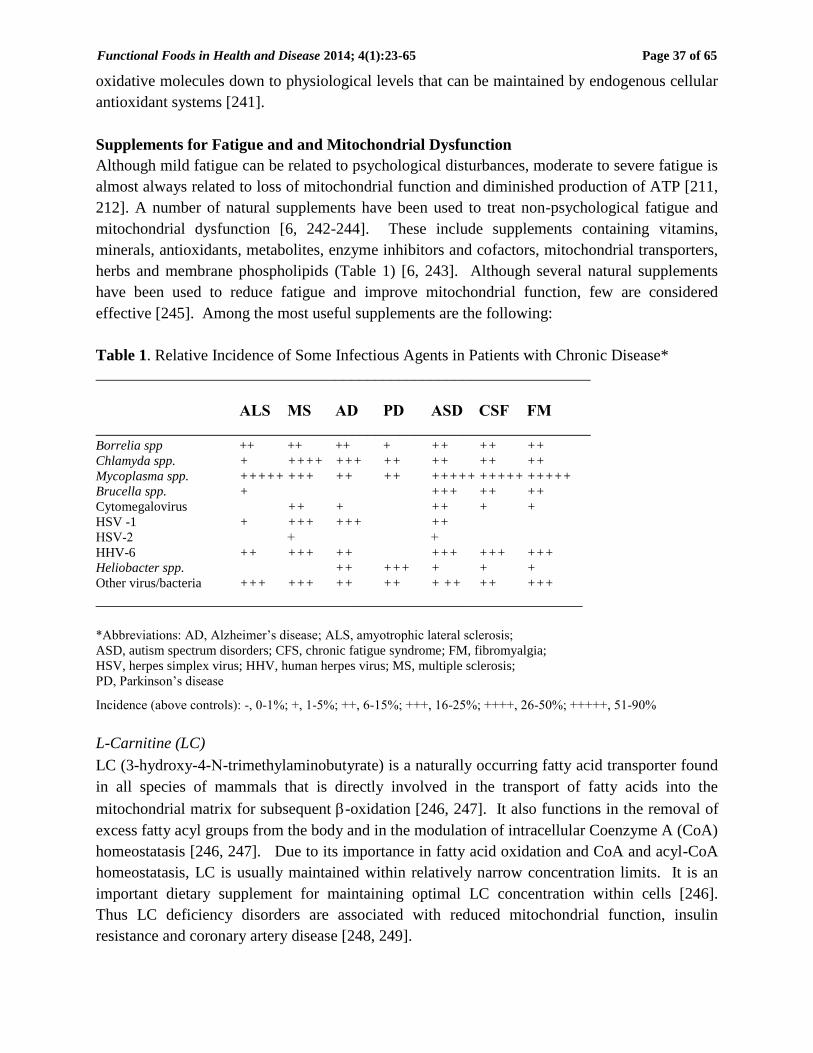
Table 1. Relative Incidence of Some Infectious Agents in Patients with Chronic Disease*
______________________________________________________________
ALS MS AD PD ASD CSF FM
______________________________________________________________ Borrelia spp ++ ++ ++ + ++ ++ ++
Chlamyda spp. + ++++ +++ ++ ++ ++ ++
Mycoplasma spp. +++++ +++ ++ ++ +++++ +++++ +++++
Brucella spp. + +++ ++ ++
Cytomegalovirus ++ + ++ + +
HSV -1 + +++ +++ ++
HSV-2 + +
HHV-6 ++ +++ ++ +++ +++ +++
Heliobacter spp. ++ +++ + + +
Other virus/bacteria +++ +++ ++ ++ + ++ ++ +++
_____________________________________________________________
*Abbreviations: AD, Alzheimer’s disease; ALS, amyotrophic lateral sclerosis;
ASD, autism spectrum disorders; CFS, chronic fatigue syndrome; FM, fibromyalgia;
HSV, herpes simplex virus; HHV, human herpes virus; MS, multiple sclerosis;
PD, Parkinson’s disease
Incidence (above controls): -, 0-1%; +, 1-5%; ++, 6-15%; +++, 16-25%; ++++, 26-50%; +++++, 51-90%
L-Carnitine (LC)
LC (3-hydroxy-4--trimethylaminobutyrate) is a naturally occurring fatty acid transporter found
in all species of mammals that is directly involved in the transport of fatty acids into the
mitochondrial matrix for subsequent -oxidation [246, 247]. It also functions in the removal of
excess fatty acyl groups from the body and in the modulation of intracellular Coenzyme A (CoA)
homeostatasis [246, 247]. Due to its importance in fatty acid oxidation and CoA and acyl-CoA
homeostatasis, LC is usually maintained within relatively narrow concentration limits. It is an
important dietary supplement for maintaining optimal LC concentration within cells [246].
Thus LC deficiency disorders are associated with reduced mitochondrial function, insulin
resistance and coronary artery disease [248, 249].

Functional Foods in Health and Disease 2014; 4(1):23-65 Page 38 of 65
The importance of LC in mitochondrial health has spurred the use of LC supplements to
potentially improve mitochondrial function and physical performance [250]. The justification is
that increased reliance on fat as the principle substrate for energy production during extreme
exercise should reduce the need for carbohydrates and delay the depletion of carbohydrate stores.
This should increase overall energy production and reduce exercise-induced fatigue. Increased
reliance on lipids requires increased levels of LC to transport fatty acids into mitochondria.
However, increasing intake of oral LC, even for a few weeks prior to extreme exercise, did not
increase skeletal muscle carnitine content, and therefore it is unlikely that increasing LC
supplementation alters muscle metabolism during extreme exercise [251].
LC supplementation has been used in disorders that are characterized by low LC
concentrations or impaired fatty acid oxidation, such as diabetes, sepsis, renal disease and
cardiomyopathy [252]. For example, in patients with congestive heart failure propionyl-LC
supplementation resulted in increased peak heart rate (increased mean by 12%), exercise
capacity (increased mean by 21%) and peak oxygen consumption (increased mean by 45%) in
the treatment group [253].
Since the rate of mitochondrial oxidative phosphorylation naturally declines with age an
important anti-aging use of LC has been to increase the rate of mitochondrial oxidative
phosphorylation in aged populations. Feeding old rats acetyl-LC was found to reverse age-
related decreases in LC levels while increasing fatty acid metabolism. It also reversed the age-
related decline in cellular glutathione levels and improved muscle mitochondrial complex IV
activity [251].
Dietary supplementation with LC and its various derivatives (up to 2 g per day) is a safe and
potentially useful method to increase mitochondrial function [254]. Multiple clinical trials
demonstrating its effectiveness in age-related chronic illnesses other than diabetes and
cardiovascular diseases have not been conducted. One exception was a randomized, controlled
clinical trial on 70 elderly subjects who were treated with LC for 6 months. At the beginning of
the trial the aged subjects were generally found to have muscle weakness, decreasing mental
health, impaired mobility and poor endurance. By the end of the study the treated group showed
significant improvements in physical fatigue, mental fatigue and fatigue severity. They also
displayed reductions in total fat mass, increased muscle mass and an increased capacity for
physical and cognitive activity through reduced fatigue and improved cognitive function [255].
Other clinical trials on alcoholism, hepatic encephalopathy, coronary heart diseases, Peyronie’s
disease, cerebral ischemia and infertility indicate that supplementation with LC can have positive
effects (reviewed in [254]).
Alpha-Lipoic Acid (ALA)
ALA (1,2-dithiolane-3-pentanoic acid) is a potent antioxidant, transition metal ion chelator,
redox transcription regulator and anti-inflammatory agent [257]. ALA acts as a critical cofactor
-ketoacid dehydrogenases, and it is important molecule in mitochondrial
oxidative decarboxylation [257, 258]. Clinically ALA has been used as an oral supplement in
the treatment of complications associated with diabetes mellitus, and it has been shown to bring
about improvements in various diabetic-associated neuropathies, inflammation and vascular

Functional Foods in Health and Disease 2014; 4(1):23-65 Page 39 of 65
health [245]. These effects have been attributed mainly to ALA having signal transduction
effects on gene regulation and glucose uptake and metabolism [259].
During aging and in many chronic diseases certain sphingolipids, especially ceramides and
in particular short-chain ceramides, accumulate in mitochondria due to hydrolysis of
sphingomyelin by sphingomyelinase. Eventually this retards electron transport activity [260,
261]. Ceramide accumulation in mitochondria is especially damaging in cardiac tissue, so in
eramide levels in the vascular endothelial cells of
cardiac muscle by inhibiting sphingomyelinase activity. This resulted in restoration of
mitochondrial glutathione levels and increasing electron transport function [262].
As previously discussed, in diabetes ALA has been used to reduce diabetic complications,
such as sensorimotor polyneuropathies [263]. A blinded study demonstrated its clinical utility
-Lipoic acid (but not nerve
conduction attributes) [264]. The long-term use of ALA has proven to be safe in diabetic patients
[264].
Given as an oral supplement ALA is rarely present in tissues above micromolar levels;
therefore, it is unlikely to be an important cellular antioxidant [258]. However, an important
property of ALA is its ability to increase cellular glutathione levels by regulating glutathione
synthesis and thus indirectly reducing oxidative stress [252]. ALA can also modify the
regulation of nuclear transcription factor NF-
transcriptional effects, resulting in the reduction of free radical and cytotoxic cytokine
production [265]. As a transition metal chelater ALA can remove excess copper, iron and other
metals that are involved in chronic diseases, such as hemochromatosis, end-stage renal failure,
AD and PD, and it is a potential therapeutic agent to prevent or mitigate heavy metal poisoning
[256].
ALA has been shown to improve cognitive function along with mitochondrial function,
suggesting a link between oxidative damage to mitochondria and congnition [266]. ALA has not
been used in clinical trials on chronic fatigue, but its widespread use as a safe supplement (at
doses of 200-600 mg/day) to support mitochondrial function and reduce oxidative stress has
justified its incorporation into various anti-aging and mitochondrial support supplements [264,
265].
Coenzyme Q10 (CoQ10)
Ubiquinone or CoQ10 is a key mitochondrial cofactor and component of the mitochondrial
electron transport chain and one of the most widely used natural supplements [243, 267]. It is
also a strong antioxidant in its reduced form, and it can modify the expression of certain genes
involved in cell signaling, metabolism and transport [267, 268]. The most important role of
CoQ10 is its involvement in the transfer of electrons along the multiple complexes of the
mitochondrial electron transport chain [267, 269]. It has been used in doses up to 1,200 mg per
day, but most studies used lower doses [267].
CoQ10 is an essential component of the mitochondrial oxidative phosphorylation system,
thus its supplementation in patients with reduced CoQ10 levels should result in increased
mitochondrial energy production and reduced fatigue [267, 269]. A systematic review of the
literature on the effects of CoQ10 on adaptive physical exercise, hypertension and heart failure

Functional Foods in Health and Disease 2014; 4(1):23-65 Page 40 of 65
revealed that most published studies showed modest improvements in exercise capacity in the
subjects given oral CoQ10 [270]. In addition, in eight publications on the effects of CoQ10 on
hypertension there was a mean decrease in systolic (-16 mm Hg) and diastolic (-10 mm Hg)
blood pressure. In nine randomized trials on the use of CoQ10 in heart failure patients there
were non-significant trends towards increased injection fraction and reduced mortality [270].
Rosenfeldt et al. performed their own three-month randomized, placebo-controlled trial on the
effects of oral CoQ10 in patients with heart failure [270]. They found that in the test arm but not
in the control arm patients showed significant improvements in symptoms and a trend towards
improvements in mean exercise times [270].
As mentioned above, the anti-fatigue effects of oral CoQ10 during physical exercise have
been examined in a blinded, cross-over trial [271]. Healthy subjects received CoQ10 or placebo
for eight days, and their performance was evaluated at fixed workloads on a bicycle ergometer
twice for two hr with a four hr rest in-between [271]. The subjects on CoQ10 were able to
achieve higher work outputs, they reported less fatigue, and their need for a recovery period was
alleviated compared to the placebo group [271]. This study indicated that CoQ10 is a useful
supplement to improve fatigue and performance.
In patients with various diagnoses, such as neurodegenerative disease, CoQ10 has been used
to reduce symptoms and delay progression [267, 269]. In AD models CoQ10 administration
significantly delayed brain atrophy and typical -amyloid plaque formation [272, 273]. In a
randomized, placebo-controlled clinical trial on Alzheimer’s patients that took an oral mixture of
CoQ10, vitamins C and E and ALA in the test arm showed significant reductions in oxidative
stress markers but failed to show significant changes in cerebrospinal fluid markers related to -
amyloid or tau pathology [273]. PD patients generally show increased oxidized-to-total CoQ10
ratios as well as significant increases in markers of oxidative damage in the cerebrospinal fluid,
but this can be partially reversed with CoQ10 supplementation [274]. In patients with early
Huntington’s disease CoQ10 administration for 30 months slowed the usual decline in total
functional capacity, but these differences did not reach statistical significance [275]. In contrast,
in a multi-center placebo-controlled phase II trial with amyotrophic lateral sclerosis patients
CoQ10 did not significantly modify functional decline over a nine-month period [276], and in
genetic-based mitochondrial diseases CoQ10 plus several vitamins was shown to be ineffective
[277].
Reduced Nicotinamide Adenine Dinucleotide (NADH)
NADH is a cellular redox cofactor in over 200 redox reactions and serves as substrate for certain
enzymes [278, 279]. Cells have a universal requirement for NADH, and its deficiency results in
a condition called pellagra, which is characterized by dermatitis, diarrhea, dementia and
eventually leads to death [279]. In the mitochondria NADH delivers electrons from lipid and
other metabolite hydrolysis to the electron transport chain, but in its reduced form NADH can
also act as a strong antioxidant [278, 279].
Historically dietary NADH supplementation has been via NADH precursors, such as niacin,
nicotinic acid or nicotinamide, but recently microcarriers have been used to stabilize oral NADH
so that it can be directly absorbed in the gastrointestinal tract. This turns out to be more effective
Functional Foods in Health and Disease 2014; 4(1):23-65 Page 41 of 65
than using large oral doses of uncomplexed NADH, which are prone to oxidation and
degradation and are generally considered ineffective [280].
In many chronic diseases oxidative damage is extensive, and various mitochondrial
antioxidants have been used to treat disease and delay progression [4-6, 243, 281-284]. Nowhere
has this been more apparent than in neurodegenerative diseases [4, 5, 84, 86, 134, 239]. For
example, in AD stabilized oral NADH has been used to improve cognitive functioning and
dementia [278]; however, in another clinical trial there was no evidence of improvements in
cognition or dementia using oral NADH [280]. In a controlled clinical trial AD patients were
given stabilized NADH or placebo for six months, and it was found that the test group had
significantly better performance scores than the placebo group (verbal fluency, visual
construction and a trend toward increased performance on abstract verbal reasoning) [285].
However, there was no evidence of better performance using other measures (attention, memory)
or on scores of dementia severity [285].
Stabilized oral NADH has also been used to reduce the symptoms of PD. In a preliminary
open label clinical trial the effects of IV and oral NADH in over 800 Parkinson disease patients
19.3% of patients showed 30-50% improvement in disability, 58.8% had moderate (10-30%)
improvement, and 21.8% did not respond to the therapy (p<0.01) [286]. Younger patients with a
shorter duration of disease responded better and showed more significant improvements than
older patients and patients with a longer duration of disease. The oral form was found to
comparable to IV NADH in its effects [286]. However, when this type of trial was repeated
statistically significant improvements in PD Rating scores were not found in patients treated with
NADH, and diferences were also not found in CSF clinical markers associated with PD severity
[287].
Oral NADH has also been used in a stabilized form to reduce symptoms in patients with
chronic fatigue. One such study on CFS patients used stabilized, oral NADH or placebo for four
weeks in a cross-over trial [288]. Eight of 26 patients (30.7%) responded positively to the
microencapsulated NADH compared with 2 of 26 (8%) in the placebo arm (p<0.05) [288].
There was clearly an effect but only in a subset of patients in the trial. These results were not
considered significant by Colquhoun and Senn [289]. A comparison of oral, stabilized NADH to
psychological/nutritional therapy in 31 chronic fatigue syndrome patients revealed that stabilized
NADH alone reduced fatigue in the first 4 months of a 12-month trial. After the first 4 months,
however, symptom scores were similar in the NADH and the psychological/nutritional arms of
the trial [290]. In another study stabilized NADH was given orally for two months to treat CFS
patients with extensive fatigue [291]. Alegre et al. found in decreases in anxiety and maximum
heart rate after a stress test, but there were little or no differences found in the functional impact
of fatigue, quality of life, sleep quality, exercise capacity, or functional reserve [291]. The
stabilized NADH alone has shown mixed results in various diseases and disorders, and not every
patient responded to the oral, stabilized supplement [6].
Lipid Replacement Theapy (LRT)
The dietary replacement of cellular membrane phospholipids (LRT) using food-derived
glyerolphosholipids to remove damaged, mainly oxidized, membrane lipids in mitochondria and
other cellular organelles has proved very effective at increasing mitochondrial function and

Functional Foods in Health and Disease 2014; 4(1):23-65 Page 42 of 65
reducing fatigue [6, 7, 212, 244].
To some degree antioxidant supplements can reduce
ROS/RNS levels and prevent some mitochondrial membrane phospholipid oxidation, but
antioxidants alone cannot repair the damage already done to cells, and in particular, to their
mitochondrial inner membranes [7, 244].
The use of oral membrane phospholipids plus antioxidants in doses ranging from 500-2,000
mg per day has been effective in the treatment of various clinical conditions, such as CFS and
other fatiguing illnesses (Table 2) [6, 7, 136, 212, 292, 293]. LRT results in the actual
replacement of damaged membrane phospholipids with undamaged (unoxidized) lipids to ensure
proper function of cellular and especially mitochondrial membranes. In these studies fatigue was
monitored by use of the Piper Fatigue Scale (PFS) to measure clinical fatigue and quality of life
[294].
Table 2. A partial list of ingredients/agents or supplements that have been used
or suggested to treat mitochondrial dysfuction1
Category Examples
__________________________________________________
Vitamins Vitamins C, D and E, Thiamine, Riboflavin
Minerals Magnesium, Calcium, Phosphate
Lipids Membrane Phospholipids, Unsaturated Fatty Acids
Metabolites Creatine, Pyruvate
Cofactors CoQ10,Lipoic acid, NADH, nicotinic acid
Transporters L-Carnitine, Membrane Phospholipids
Antioxidants CoQ10, -Lipoic acid, NADH, Glutathione
Enzyme inhibitors -Lipoic acid, Dichloroacetate
Herbs Curcimin, Schisandrin
_________________________________________________________ 1 Modified from Kerr [243] and Nicolson [6]
In a subsequent cross-over study the effects of LRT on fatigue and mitochondrial function
were monitored in patients with moderate to severe chronic fatigue [212]. There was good
correspondence between reductions in fatigue and gains in mitochondrial function. After 8
weeks of LRT with NTFactor, mitochondrial function was significantly improved, and after 12
weeks of NTFactor supplementation, fatigue was decreased by 35.5% (p<0.001), and
mitochondrial function was found to be similar to that found in young healthy adults (26.8%
increase, p<0.0001) [212]. After 12 weeks of supplement use, subjects were placed on placebo
for an additional 12 weeks, and their fatigue and mitochondrial function were again measured.
After the placebo period, fatigue and mitochondrial function were intermediate between the
initial values and those found after 8 or 12 weeks on the supplement, indicating that continued
supplementation is required to show improvements in mitochondrial function and maintain lower
fatigue scores [212].
Similar findings on fatigue reduction were observed in chronic fatigue syndrome and
fibromyalgia syndrome patients given oral membrane phospholipids (NT Factor) [293]. Using a

Functional Foods in Health and Disease 2014; 4(1):23-65 Page 43 of 65
new formulation of NT Factor plus vitamins, minerals and other supplements in patients with
moderate chronic fatigue resulted in a 36.8% reduction in fatigue within one week [295].
Vitamins and Minerals
Vitamins, minerals and other small molecules fall into the category of micronutrients. They are
essential in the support of mitochondrial function by providing antioxidants, cofactors, metal
ions, salts, and other molecules that are essential in supporting the functions of mitochondrial
enzymes, electron transport systems, mtDNA replication, fat and sugar metabolism, protein
synthesis and proper antioxidant balance [296-298]. Vitamins, such as B (multiple), D, E, C, and
ions, such as iron, magnesium, manganese, zinc, among other small molecules, are important n
this regard, and up to one-half of the aging North American population is deficient in these
vitamins, minerals and other micronutrients [299].
The use of micronutrients in helping to restore and/or maintain mitochondrial function has
proven useful in consert with other treatment modialties [296, 298, 300]. Although there are few
clinical trials in the literature that demonstrate the usefulness and utility of supplementation with
only vitamins, minerals, antioxidants and other micronutrients alone in supporting mitochondrial
function, the ones that have been conducted clearly show the importance of providing adequate
oral doses of vitamins, minerals, antioxidants and other micronutrients to maintain mitochondrial
energy functions [296. 300-302]. However, their sole use in the treatment of mitochondrial
diseases has proved disappointing [277]. But it is reassuring to find that many commercial
mitochondrial supplements contain adequate amounts of these important molecules (for example
[282, 292, 295, 302]). Thus in addition to vitamins, minerals, antioxidants and other
micronutrients, other supplement components are likely required for significant and lasting
effects on mitochondrial function.
Combination Supplements to Restore Mitochondrial Function
Oral supplements containing membrane phospholipids (NTFactor, 2,000 mg/day), CoQ10 (35
mg/day), microencapsulated NADH (35 mg/day), LC (160 mg/day), -ketoglutaric acid (180
mg/day) and other micronutrients have been combined into a dietary supplement (ATP Fuel®
) to
treat fatigue and mitochondrial dysfunction [303]. This formulation was used in a study to treat
long-term intractable fatigue in patients with a variety of diagnoses during a two-month trial.
The 58 participants in the ATP Fuel trial had moderate to severe intractable fatigue for an
average >17 years and had been to an average of >15 practitioners without resolution of their
fatigue. The study included 30 patients with chronic fatigue syndrome, 17 with chronic Lyme
disease; 16 with other fatiguing illnesses, including fibromyalgia syndrome and Gulf War illness;
4 with autoimmune disease, including rheumatoid arthritis; 2 cancer; and 2 diabetes. These
patients had tried unsuccessfully many drugs and supplements (average >35) to reduce their
fatigue without success [303].
Participants in the trial included chronic illness patients who took the combination LRT
supplement (ATP Fuel®) for 8 weeks, and their fatigue was scored monthly [303]. The Piper
Fatigue Score (PFS) is a validated instrument that measures four dimensions of subjective
fatigue: behavioral/severity, affective/ meaning, sensory, and cognitive/mood [294]. These were
used to calculate the four subscale/dimensional scores and the total fatigue scores. In this study
Functional Foods in Health and Disease 2014; 4(1):23-65 Page 44 of 65
the long-term chronic illness patients with intractable fatigue had initial PFS mean total fatigue
scores of 7.51 ± 0.29, and after 8 weeks of supplement the mean scores improved to 5.21 ± 0.28,
or a 30.7% reduction in fatigue (p<0.0001) [303].
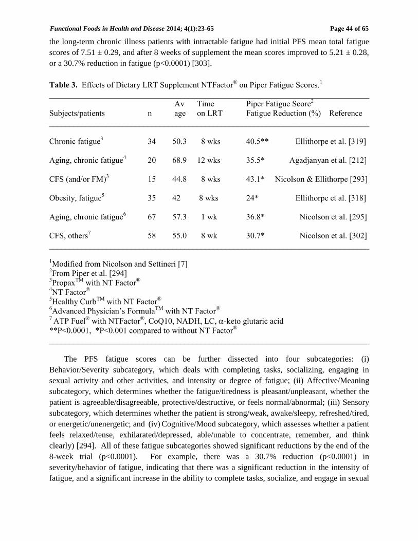
Table 3. Effects of Dietary LRT Supplement NTFactor® on Piper Fatigue Scores.
1
______________________________________________________________________________
Av Time Piper Fatigue Score2
Subjects/patients n age on LRT Fatigue Reduction (%) Reference
______________________________________________________________________________
Chronic fatigue3 34 50.3 8 wks 40.5** Ellithorpe et al. [319]
Aging, chronic fatigue4 20 68.9 12 wks 35.5* Agadjanyan et al. [212]
CFS (and/or FM)3 15 44.8 8 wks 43.1* Nicolson & Ellithorpe [293]
Obesity, fatigue5 35 42 8 wks 24* Ellithorpe et al. [318]
Aging, chronic fatigue6 67 57.3 1 wk 36.8* Nicolson et al. [295]
CFS, others7 58 55.0 8 wk 30.7* Nicolson et al. [302]
______________________________________________________________________________
1Modified from Nicolson and Settineri [7]
2From Piper et al. [294]
3Propax
TM with NT Factor
®
4NT Factor
®
5Healthy Curb
TM with NT Factor
®
6Advanced Physician’s Formula
TM with NT Factor
®
7 ATP Fuel
® with NTFactor
®, CoQ10, NADH, LC, -keto glutaric acid
**P<0.0001, *P<0.001 compared to without NT Factor®
______________________________________________________________________________
The PFS fatigue scores can be further dissected into four subcategories: (i)
Behavior/Severity subcategory, which deals with completing tasks, socializing, engaging in
sexual activity and other activities, and intensity or degree of fatigue; (ii) Affective/Meaning
subcategory, which determines whether the fatigue/tiredness is pleasant/unpleasant, whether the
patient is agreeable/disagreeable, protective/destructive, or feels normal/abnormal; (iii) Sensory
subcategory, which determines whether the patient is strong/weak, awake/sleepy, refreshed/tired,
or energetic/unenergetic; and (iv)
Cognitive/Mood subcategory, which assesses whether a patient
feels relaxed/tense, exhilarated/depressed, able/unable to concentrate, remember, and think
clearly) [294]. All of these fatigue subcategories showed significant reductions by the end of the
8-week trial (p<0.0001). For example, there was a 30.7% reduction (p<0.0001) in
severity/behavior of fatigue, indicating that there was a significant reduction in the intensity of
fatigue, and a significant increase in the ability to complete tasks, socialize, and engage in sexual
Functional Foods in Health and Disease 2014; 4(1):23-65 Page 45 of 65
and other activities. Also, there was also a 28.0% improvement (p<0.0001) in mood and
cognitive ability, such as the ability to concentrate, remember, and think clearly [303].
The benefits of combining various natural mitochondrial support components into
combination mitochondrial supplements should be obvious. Separate supplements are no longer
required, and the individual components can be adjusted in dosage to provide maximal
effectiveness. Also, with combined supplements patients who often have memory and other
defects are more likely to be compliant than with the requirement of taking several separate
supplements on a daily basis. New formulations can be designed to replace damaged
mitochondrial components and also address particular protective needs, such as preconditioning
to defend against oxidative stress, damage to ion channels and other needs [304].
Final Comments
There is ample evidence that intracellular infections are linked to mitochondria and
mitochondrial dysfunction. Mitochondria play essential roles in cellular homeostasis through
control of cell death and as sentinels of cellular danger, such as oxidative stress, loss of essential
growth and maintenance factors, physical damage and infection by foreign agents [305-307].
They also orchestrate cellular adaptive danger responses to sustain cell survival, especially when
infections threaten cellular systems [307, 308]. However, microorganisms have evolved with
different strategies to evade or circumvent host mechanisms to identify, neutralize and degrade
them [308-310].
During infection and cellular invasion by microorganisms coordination of multiple innate
immune signaling pathways occurs through different pathogen-associated molecular pattern
recognition receptors. The recognition of foreign invaders (and other danger molecules) by these
innate receptors (Patern Recognition Receptors) can initiate an intracellular signaling cascade
that results in activation of anti-microbial mechanisms to clear the infection [311, 312].
Mitochondria have emerged as critical regulators of innate immune responses to invading
pathogens as well as other stressors [311-313]. They initiate immune signaling modulators that
are tightly linked to mitochondrial energetics through intracellular danger-sensing multiprotein
platforms called inflammasomes [12, 313, 314]. Thus mitochondria operate as fundamental
―hubs‖ in the pathways from detection of intracellular pathogens to adaptive responses that
attempt to neutralize and eliminate pathogens as well as housing crucial signal transducers and
providing a structural scaffold that is involved in regulating anti-microbial activities [305, 306].
They also warn the host with danger signals by providing multiple mitochondrial damage
products (DAMPS) that are recognized by the innate immune system, resulting in the stimulation
of local or systemic immune responses [315].
Mitochondria are addionally implicated in pathways that result in the cellular destruction of
pathogens by autophagy through specialized autophagosomes [304, 307]. When pathogens
damage cells beyond their ability to be repaired, mitochondria are also involved in initiating and
removing the damaged cells by apoptosis [304, 316]. They not only modulate apoptosis
triggered by terminal alternations in intracellular homeostasis, but they can also participate in
apoptosis stimulated by responses to extracellular or external signals [317, 318]. Thus
mitochondria are intrinsically tied to cellular infections in a number of ways.

Functional Foods in Health and Disease 2014; 4(1):23-65 Page 46 of 65
Oral mitochondrial supplements containing various mitochondrial substrates, cofactors,
precursors, components and modulators have proven useful in maintaining and improving
mitochondrial function in various chronic diseases. In some examples cited in here this has
resulted in reduction of specific illness signs and symptoms, improvements in quality of life and
assistance in over-all recovery. Future efforts to refine and improve oral mitochondrial
supplements and apply them clinically should enhance our abilities to treat and care for chronic
illness patients as well as improve quality of life for aging populations.
List of Abbreviations Used
AD, Alzheimer’s disease; ALA,Lipoic acidALS, amyotrophic lateral sclerosis; ASD, autism
spectrum disorders; CFS, chronic fatigue syndrome; CI, confidence interval; CMV,
cytomegalovirus; CoQ10, coenzyme Q10; CSF, cerebrospinal fluid; CNS, central nervous
system; ELISA, enzyme linked immunoabsorbant assay; HHV human herpes virus; HSV, herpes
simplex virus; LC, L-carnitine; LRT, Lipid Replacement Therapy; MS, multiple sclerosis;
NADH, Reduced Nicotinamide Adenine Dinucleotide;PCR, polymerase chain reaction; PD,
Parkinson’s disease RNS, Nitrogen Reactive Species; ROS, Reactive Oxygen Species
Competing Interests
The authors have no financial interests or conflicts of interest.
Authors’ Contributions
All authors contributed to this review.
Acknowledgements and Funding
The authors would like to thank Nutritional Therapeutics, Inc. and Researched Nutritionals, Inc.
for research and clinical trial financial support.
References
1. Nicolson GL. Chronic infections in neurodegenerative and neurobehavioral diseases.
Lab Med 2008; 39(5): 291-299.
2. Nicolson GL, Haier J. Role of chronic bacterial and viral Infections in
neurodegenerative, neurobehavioral, psychiatric, autoimmune and fatiguing illnesses:
Part 1. Brit J Med Practit 2009; 2(4): 20-28.
3. Nicolson GL, Haier J. Role of chronic bacterial and viral Infections in
neurodegenerative, neurobehavioral, psychiatric, autoimmune and fatiguing illnesses:
Part 2. Brit J Med Practit 2010; 3(1): 301-311.
4. Reddy PH. Mitochondrial medicine for aging and neurodegenerative diseases.
Neuromolec Med 2008; 10(4): 291-315.
5. Reddy PH, Reddy TP. Mitochondria as a therapeutic target for aging and
neurodegenerative diseases. Curr Alzheimer Res 2011; 8(4): 393-409.
6. Nicolson GL. Mitochondrial dysfunction and chronic disease: treatment with natural
supplements. Alt Ther Health Med 2013; 19: at5027. [Epub ahead of print].

Functional Foods in Health and Disease 2014; 4(1):23-65 Page 47 of 65
7. Nicolson GL, Settineri R. Lipid Replacement Therapy: a functional food approach with
new formulations for reducing cellular oxidative damage, cancer-associated fatigue and
the adverse effects of cancer therapy. Funct Food Health Dis 2011; 1(4): 135-160.
8. De Chiara G, Marococci ME, Sgarbanti R, Civitelli L, Ripoli C, Placentini R, Garaci E,
Grassi C, Palamara AT. Infectious agents and neurodegeneration. Mol Neurobiol 2012;
46(3): 614-638.
9. Bach JF. Infections and autoimmune diseases. J Autoimmun 2005; 25(suppl): 74-80.
10. Sherbet G. Bacterial infections and the pathogenesis of autoimmune conditions. Brit J
Med Practit 2009; 2(1): 6-13.
11. Nicolson GL, Haier J, Nasralla M, Nicolson NL, Ngwenya R, De Meirleir K.
Mycoplasmal infections in Chronic Fatigue Syndrome, Fibromyalgia Syndrome and
Gulf War Illness. J Chronic Fatigue Syndr 2000; 6(3): 23-39.
12. Verdon ME, Sigal LH. Recognition and management of Lyme Disease. Am Family
Physician 1997; 56: 427-436.
13. Homer MJ, Aguilar-Delfin I, Telford SR 3rd
, Krause PJ, Persing DH. Babesiosis. Clin
Microbiol Rev 2000; 13: 451-469.
14. Kizilkilic O, Calli C. Neurobrucellosis. Neuroimaging Clin N Am 2011; 21: 927-937.
15. Galley HF. Oxidative stress and mitochondrial dysfunction in sepsis. Brit J Anaesth
2011; 107: 57-64.
16. Pessayre D, Fromenty B, Mansouri A. Mitochondrial injury in steatohepatitis. Eur J
gastroenterol Hepatol 2004; 16: 1-11.
17. Rusconi B, Greub G. Chlamydiales and the innate immune response: friend or foe?
FEMS Immunol Med Microbiol 2011; 61: 231-244.
18. Richter C, Par JW, Ames B. Normal oxidative damage to mitochondrial and nuclear
DNA is extensive. Proc Nat Acad Sci USA 1998; 85: 6465-6467.
19. Stadtman E. Introduction to serial reviews on oxidatively modified proteins in aging
and disease. Free Radical Biol Med 2002; 32: 789.
20. Spector AA, Yorek MA. Membrane lipid composition and cellular function. J Lipid
Res 1985; 26: 101-105.
21. Nicolson GL. Diagnosis and therapy of chronic systemic co-infections in Lyme
Disease and other tick-borne infectious diseases. Townsend Lett 2007; 285: 93-98.
22. Burrascano JJ. Diagnostic hints and treatment guidelines for Lyme and other tick-borne
diseases. In: Advanced Topics in Lyme Disease, Managing Lyme Disease, 16th
edition,
5-28, 2008.
23. Muravchick S, Levy RJ. Clinical implications of mitochondrial dysfunction.
Anesthesiol 2006; 105: 819-837.
24. Keen D, Ward S. Autistic Spectrum Disorder. Autism 2004; 8: 39-58.
25. Fatemi SH, Reutiman TJ, Folsom TD, Sidwell RW. The role of cerebellar genes in
pathology of autism and schizophrenia. Cerebellum 2008; 99: 56-70.
26. Nicolson GL, Gan R, Nicolson NL, Haier J. Evidence for Mycoplasma, Chlamydia
pneunomiae and HHV-6 co-infections in the blood of patients with Autism Spectrum
Disorders. J Neurosci Res 2007; 85: 1143-1148.
Functional Foods in Health and Disease 2014; 4(1):23-65 Page 48 of 65
27. Bransfield RC. Preventable cases of autism: relationship between chronic infectious
diseases and neurological outcome. Pediat Health 2009; 3(2): 125-140.
28. Muhle R, Trentacoste SV, Rapin I. The genetics of autism. Pediatr 2004; 113: 72-86.
29. Dhillon VS, Fenech M. Mutations that affect mitochondrial functions and their
association with neurodegenerative diseases. Mutat Res 2013; Sept 17 doi:
10.1016/j.mrrev.2013.09.001 [Epub ahead of print].
30. Frye RE, Delatorre R, Taylor H, Slattery J, Melnyk S, Chowdhury N, James SJ. Redox
metabolism abnormalities in autistic children associated with mitochondrial disease.
Transl Psychiatry 2013; 3: e273 [Epub ahead of print].
31. Deth R, Muratore C, Benzercry J, Power-Charnitsky VA, Waly M. How environmental
and genetic factors combine to cause autism: a redox/methylation hypothesis.
Neurotoxicol 2008; 29: 190-201.
32. Mattson MP. Infectious agents and age-related neurodegenerative disorders. Ageing
Res Rev 2004; 3: 105-120.
33. Bazala E, Renda J. Latent Chlamydial infections: the probably cause of a wide
spectrum of human diseases. Med Hypotheses 2005; 65: 578-584.
34. Williams DB, Windebank AJ. Motor neuron disease (Amyotrophic Lateral Sclerosis).
Mayo Clinic Proc 1991; 66: 54-82.
35. Walling AD. Amyotrophic Lateral Sclerosis: Lou Gehrig’s Disease. Am Family
Physician 1999; 59: 1489-1496.
36. Berger MM, Kopp N, Vital C, Redi B, Aymard M, Lina B. Detection and cellular
localization of enterovirus RNA sequences in spinal cord of patients with ALS.
Neurology 2000; 54: 20-25.
37. Walker MP, Schlaberg R, Hays AP, Bowser R, Lipkin WI. Absence of echovirus
sequences in brain and spinal cord of amyotrophic lateral sclerosis patients. Ann
Neurol 2001; 49: 249-253.
38. Nicolson GL, Berns P, Nasralla M, Haier J, Pomfret J. High frequency of systemic
mycoplasmal infections in Gulf War veterans and civilians with Amyotrophic Lateral
Sclerosis (ALS). J Clin Neurosci 2002; 9: 525-429.
39. Halperin JJ, Kaplan GP, Brazinsky S, Tsai TF, Cheng T, Ironside A, Wu P, Delfiner J,
Golightly M, Brown RH. Immunologic reactivity against Borrelia burgdorferi in
patients with motor neuron disease. Arch Neurol 1990; 47: 586-594.
40. Hansel Y, Ackerl M, Stanek G. ALS-like sequelae in chronic neuroborreliosis. Wien
Med Wochensch 1995; 147: 186-188.
41. Qureshi M, Bedlack RS, Cudkowicz ME. Lyme disease serology in amyotrophic lateral
sclerosis. Muscle Nerve 2009; 40: 626-628.
42. MacDonald AB. Spirochetal cyst forms in neurodegenerative disorders, hiding in plain
site. Med Hypotheses 2006; 67: 819-832.
43. Hugan J. ALS therapy: targets for the future. Neurol 1996; 47(suppl 4): S251-S254.
44. McCormick AL, Brown RH Jr, Cudkowicz ME, Al-Chalabi A, Garson JA.
Quantification of reverse transcriptase in ALS and elimination of a novel retroviral
candidate. Neurol 2008; 70: 278-283.
Functional Foods in Health and Disease 2014; 4(1):23-65 Page 49 of 65
45. Stipa G, Taiuti R, de Scisciolo G, Ametoli G, Tredici MR, Biondi N, Barsanti L, Lolli
F. Sporadic amyotrophic lateral sclerosis as an infectious disease: a possible role of
cyanobacteria? Med Hypotheses 2006; 67: 1363-1371.
46. Cozzolino M, Ferri A, Valle C, Carri MT. Mitochondria and ALS: implications from
novel genes and pathways. Mol Cell Neurosci 2013; 55: 44-49.
47. Cozzolino M, Pesaresi MG, Gerbino V, Grosskreutz J, Carr MT. Amyotrophic lateral
sclerosis: new insights into underlying molecular mechanisms and opportunities for
therapeutic intervention. Antiox Redox Signal 2012; 9: 1277-1330.
48. Parakh S, Spencer DM, Halloran MA, Soo KY, Atkin JD. Redox regulation in
Amyotrophic Lateral Sclerosis. Oxid Med Cell Longev 2013; 2013: 408681. doi:
10.1155/2013/408681.
49. Sobel RA. The pathology of multiple sclerosis. Neurol Clinic 1995; 13: 1-21.
50. Bruck W. Clinical implications of neuropathological findings in multiple sclerosis. J
Neurol 2005; 252(suppl 3): 10-14.
51. Muñoz-Culla M, Irizar H, Otaegui D. The genetics of multiple sclerosis: review of
current and emerging candidates. Appl Clin Genet 2013; 8: 63-73.
52. Huynh JL, Casaccia P. Epigenetic mechanisms in multiple sclerosis: implications for
pathogenesis and treatment. Lancet Neurol 2013; 12(2): 195-206.
53. Baranzini SE, Oksenberg JR. Genomics and new targets for multiple sclerosis.
Pharmacogenomics 2005; 6: 151-161.
54. Currier RD, Eldridge R. Possible risk factors in multiple sclerosis as found in a national
twin study. Arch Neurol 1982; 39: 140-44.
55. Greenlee JE, Rose JW. Controversies in neurological infectious diseases. Semin
Neurol 2000; 20: 375-386.
56. Gilden DH. Infectious causes of multiple sclerosis. Lancet Neurol 2005; 4: 195-202.
57. Malmone D, Gregory S, Arnason BG, et al. Cytokine levels in the cerebrospinal fluid
and serum of patients with multiple sclerosis. J Neuroimmunol 1991; 32: 87-74.
58. Woodroofe MN. Cytokine production in the central nervous system. Neurol 1995; 45
(Suppl 6): S6-S10.
59. Sriram S, Mitchell W, Stratton C. Multiple sclerosis associated with Chlamydia
pneumoniae infection of the CNS. Neurol 1998; 50: 571-572.
60. Sriram S, Stratton CW, Yao S, Tharp A, Ding L, Bannan JD, Mitchell WM.
Chlamydia pneumoniae infection of the central nervous system in multiple sclerosis.
Ann Neurol 1999; 46: 6-14.
61. Yao S-Y, Stratton CW, Mitchell WM, Sriram S. CFS oligoclonal bands in MS include
antibodies against Chamydophila antigens. Neurol 2001; 51: 1168-1176.
62. Contini C, Seraceni S, Castellazzi M, et al. Chlaymdophila pneumoniae DNA and
mRNA transcript levels in peripheral blood mononuclear cells and cerebrospinal fluid
of patients with multiple sclerosis, Neurosci Res 2008; 62: 58-61.
63. Sriram S, Ljunggren-Rose A, Yao S-Y, Whetsell WO Jr. Detection of chlamydial
bodies and antigens in the central nervous system of patients with multiple sclerosis. J
Infect Dis 2005; 192: 1219-1228.

Functional Foods in Health and Disease 2014; 4(1):23-65 Page 50 of 65
64. Contini C, Cultrera R, Seraceni S, Castellazzi M, Granieri E, Fainardi E. Cerebrospinal
fluid molecular demonstration of Chlamydia pneumoniae DNA is associated to clinical
and brain magnetic resonance imaging activity in a subset of patients with relapsing-
remitting multiple sclerosis. Multiple Sclerosis 2004; 10: 360-369.
65. Grimaldi LM, Pincherle A, Martinelli-Boneschi F, Filippi M, Patti F, Reggio A,
Franciotta D, Allegra L, Comi G, Blasi F. An MRI study of Chlamydia pneumoniae
infection in Italian multiple sclerosis patients. Multiple Sclerosis 2003; 9: 467-471.
66. Dong-Si T, Weber J, Liu YB, Buhmann C, Bauer H, Bendl C, Schnitzier P, Grond-
Ginsbach C, Grau AJ. Increased prevalence of and gene transcription by Chlamydia
pneumoniae in cerebrospinal fluid of patients with relapsing-remitting multiple
sclerosis. J Neurol 2004; 251: 542-547.
67. Stratton CW, Sriram S. Association of Chlamydia pneumoniae with central nervous
system disease. Microbes Infect 2003; 5: 1249-1253.
68. Stratton CW, Wheldon DB. Multiple sclerosis: an infectious syndrome in ving
Chlamydophia pneumoniae. Trends Microbiol 2006; 14: 474-479.
69. Boman J, Roblin PM, Sundström P, Sundström M, Hmmerschlag MR. Failure to detect
Chlamydia pneumoniae in central nervous system of patients with MS. Neurol 2000;
11: 265.
70. Pucci E, Taus C, Cartechini E, Morelli M, Giuliani G, Clementi M, Menzo S. Lack of
Chlamydia infection of the central nervous system in multiple sclerosis. Ann Neurol
2000; 48: 399-400.
71. Hammerschlag MR, Ke Z, Lu F, Roblin P, Boman J, Kalman B. Is Chlamydia
pneumoniae present in brain lesions of patients with multiple sclerosis? J Clin
Microbiol 2000; 38: 4274-4276.
72. Swanborg RH, Whittum-Hudson JA, Hudson AP. Infectious agents and multiple
sclerosis—Are Chlamydia pneumoniae and human herpes virus 6 involved? J
Neuroimmunol 2003; 136: 1-8.
73. Nicolson GL. Systemic intracellular bacterial infections (Mycoplasma, Chlamydia,
Borrelia species) in neurodegenerative (Alzheimers, MS, ALS) and behavioral diseases
(Autistic Spectrum Disorders). Townsend Lett 2008; 295: 74-84.
74. Sanders VJ, Felisan S, Waddell A, Tourtellotte WW. Detection of herpesviridae in
postmortem multiple sclerosis brain tissue and controls by polymerase chain reaction. J
Neurovirol 1996; 2: 249-258.
75. Sanders VJ, Felisan S, Waddell A, et al. Detection of herpesviridae in postmortem
multiple sclerosis brain tissue and controls by polymerase chain reaction. J Neurovirol
1996; 2: 249-58.
76. Challoner PB, Smith KT, Parker JD, MacLeod DL, Coulter SN, Rose TM, Schultz ER,
Bennett JL, Garber RL, Chang M. Plaque-associated expression of human herpesvirus
6 in multiple sclerosis. Proc Natl Acad Sci USA 1995; 92: 7440-7444.
77. Sola P, Merelli E, Marasca R, Poggi M, Luppi M, Montorsi M, Torelli G. Human
herpesvirus-6 and multiple sclerosis: survey of anti-HHV-6 antibodies by
immunofluorescence analysis and viral sequences by polymerase chain reaction. J
Neurol Neurosurg Psychiat 1993; 56: 917-919.

Functional Foods in Health and Disease 2014; 4(1):23-65 Page 51 of 65
78. Kuusisto H, Hyöty H, Kares S, Kinnunen E, Elovaara I. Human herpes virus 6 and
multiple sclerosis: a Finnish twin study. Mult Scler 2008; 14: 54-58.
79. Steiner I, Nisipianu P, Wirguin I. Infection and etiology and pathogenesis of multiple
sclerosis. Curr Neurol Neurosci Rep 2001; 1: 271-76.
80. Giraudon P, Bernard A. Chronic viral infections of the central nervous system: aspects
of specific to multiple sclerosis. Rev Neurol (Paris) 2009; 169: 789-795.
81. Frykholm BO. On the question of infectious aetiologies for multiple sclerosis,
schizophrenia and the chronic fatigue syndrome and the treatment with antibiotics.
Med Hypotheses 2009; 72: 736-739.
82. Lassmann H. Pathology and disease mechanisms in different stages of multiple
sclerosis. J Neurol Sci 2013; Jun 2. doi: 10.1016/j.jns.2013.05.010.
83. Campbell GR, Ohno N, Turnbull DM, Mahad DJ. Mitochondrial changes within axons
in multiple sclerosis: an update. Curr Opin Nuerol 2012; 4: 169. doi: 10.1097/WCO.
0b013e3283533a25.
84. Su K, Bourdette D, Forte M. Mitochondrial dysfunction and neurodegeneration in
multiple sclerosis. Front Physiol 2013; 4: 169. doi: 10.3389/fphys.2013.00169.
85. Broadwater L, Pandit A, Clements R, Azzam S, Vadnal J, Sulak M, Yong VW,
Freeman EJ, Gregory RB, McDonough J, Analysis of the mitochondrial proteome in
multiple sclerosis cortex. Biochim Biophys Acta 2011; 1812: 630-641.
86. Su K, Bourdette D, Fore M. Mitochondrial dysfunction and neurodegeneration in
multiple sclerosis. Front Physiol 2013; July 25 doi: 10.3389/fphys.2013.00169.
87. Nicholls DG. Mitochondrial ion circuits. Essays Biochem 2010; 47: 25-35.
88. Mahad D, Lassmann H, Turnbull D. Review: mitochondria and disease progression in
multiple sclerosis. Neuropathol Appl Neurobiol 2008; 34: 577-589.
89. Masters CL, Beyreuther K. Alzheimer’s centennial legacy: prospects for rational
therapeutic intervention targeting the Abeta amyloid pathway. Brain 2006; 129: 2823-
2839.
90. Drachman DA. Aging of the brain, entropy, and Alzheimer Disease. Neurol 2006; 67:
1340-1352.
91. Markesbery WR, Lovell MA. Damage to lipids, proteins, DNA and RNA in mild
cognitive impairment. Arch Neurol 2007; 64: 954-956.
92. Daly MP. Diagnosis and management of Alzheimer Disease. J Am Board Family Pract
1999; 12: 375-385.
93. Holmes C, El-Okl M, Williams AL, Cunningham C, Wilcockson D, Perry VH.
Systemic infection, interleukin 1-beta and cognitive decline in Alzheimer’s disease. J
Neurol Neurosurg Psychiat 2003; 74: 788-789.
94. Balin BJ, Appelt DM. Role of infection in Alzheimer’s Disease. J Am Osteopath
Assoc 2001; 101(suppl 12): S1-S6.
95. Yucesan C, Sriram S. Chlamydia pneumoniae infection of the central nervous system.
Curr Opin Neurol 2001; 14: 355-359.

Functional Foods in Health and Disease 2014; 4(1):23-65 Page 52 of 65
96. Balin BJ, Gerard HC, Arking EJ, Appelt DM, Branigan JP, Abrams JT, Whittum-
Hudson JA, Hudson AP. Identification and localization of Chlamydia pneumoniae in
the Alzheimer’s brain. Med Microbiol Immunol 1998; 187: 23-42.
97. Gerard HC, Dreses-Werringloer U, Wildt KS, Deka S, Oszust C, Balin BJ, Frey WH
2nd
, Bordayo EZ, Whittum-Hudson JA, Hudson AP. Chlamydophila (Chlamydia)
penumoniae in the Alzheimer’s brain. FEMS Immunol Med Microbiol 2006; 48: 355-
366.
98. MacIntyre A, Abramov R, Hammond CJ, Hudson AP, Arking EJ, Little CS, Appelt
DM, Balin BJ. Chlamydia pneumoniae infection promotes the transmigration of
monocytes through human brain endothelial cells. J Neurosci Res 2003; 71: 740-750.
99. Dreses-Werringloer U, Bhuiyan M, Zhao Y, Gérard HC, Whittum-Hudson JA, Hudson
AP. Initial characterization of Chlamydophila (Chlamydia) pneumoniae cultured from
the late-onset Alzheimer brain. Intern J Med Microbiol 2998; 299: 187-201.
100. Hammond CJ, Hallock LR, Howanski RJ, Appelt DM, Balin BJ. Immunohistological
detection of Chlamydia pneumoniae in the Alzheimer’s disease brain. BMC Neurosci
2010; 11: 121. doi: 10.1186.1471-2202-11-121.
101. Little CS, Hammond CJ, MacIntyre A, Balin BJ, Appelt DM. Chlamydia pneumoniae
induces Alzheimer-like amyloid plaques in brains of BALB/c mice. Neurobiol Aging
2004; 25: 419-429.
102. Gieffers J, Reusche E, Solbach W, Maass M. Failure to detect Chlamydia pneumoniae
in brain sections of Alzheimer’s Disease patients. J Clin Microbiol 2000; 38: 881-882.
103. Meer-Scherrer L, Chang-Loa C, Adelson ME, Mordechai E, Lobrinus JA, Fallon BA,
Tilton RC. Lyme disease associated with Alzheimer’s Disease. Curr Microbiol 2006;
52: 330-332.
104. Miklossy J, Khalili K, Gern L, Eircson RL, Darekar P, Bolle L, Hurlimann J, Paster BJ.
Borrelia burgdorferi persists in the brain in chronic Lyme neuroborreliosis and may be
associated with Alzheimer’s disease. J Alzheimers Dis 2004; 6: 639-649.
MacDonald AB. Alzheimer’s Disease Braak Stage progressions: reexamined and redefined
as Borrelia infection transmission through neural circuits. Med Hypotheses 2007; 68:
1059-1064.
106. MacDonald AB. Plaques of Alzheimer’s Disease originate from cysts of Borrelia
burgdorferi, the Lyme Disease spirochete. Med Hypotheses 2006; 67: 592-600.
107. Miklossy J, Kis A, Radenovic A, Miller L, Forro L, Martins R, Reiss K, Darbinian N,
Darekar P, Mihaly L, Khalili K. Beta-amyloid deposition and Alzheimer’s type changes
induced by Borrelia spirochetes. Neurobiol Aging 2006; 27: 228-236.
108. Glabe C. Intracellular mechanisms of amyloid accumulation and pathogenesis in
Alzheimer’s Disease. J Molec Neurosci 2001; 17: 137-145.
109. Marques AR, Weir SC, Fahle GA, Fischer SH. Lack of evidence of Borrelia
involvement in Alzheimer’s Disease. J Infect Dis 2000; 182: 1006-1007.
110. Miklossy J. Chronic inflammation and amyloidogenesis in Alzheimer’s disease—role
of spirochetes. J Alzheimers Dis 2008; 13: 381-391.
111. Miklossy J. Emerging roles of pathogens in Alzheimer disease. Expert Rev Mol Med
2011; 13: e30. doi: 10.1017/S1462399411002006.

Functional Foods in Health and Disease 2014; 4(1):23-65 Page 53 of 65
112. Kountouras J, Boziki JM, Gavalas E, Zavos C, Deretzi G, Grigoriadis N, Tsolaki M,
Chatzopoulos D, Katsinelos P, Tzilves D, Zabouri A, Michailidou I. Increased
cerebrospinal fluid Helicobacter pylori antibiody in Alzheimer’s disease. Intern J
Neurosci 2009; 119: 765-767.
113. Robinson SR, Dobson C, Lyons J. Challenges and directions for the pathogen
hypothesis of Alzheimer’s disease. Neurobiol Aging 2004; 25: 629-637.
114. Denaro FJ, Staub P, Colmer J, Freed DM. Coexistance of Alzheimer’s disease
neuropathology with herpes simplex encephalitis. Cell Molec Biol 2003; 49: 1233-
1240.
115. Itzhaki RF, Wozniak MA. Herpes simplex virus type 1 in Alzheimers’s disease: the
enemy within. J Alzheimers Dis 2008; 13: 393-405.
116. Lin WR, Shang D, Itzhaki RF. Neurotrophic viruses and Alzheimer’s Disease:
Interaction of Herpes Simplex type-1 virus and apolipoprotein E in the etiology of the
disease. Molec Chem Neuropathol 1996; 28: 135-141.
117. Itzhaki RF, Dobson CB, Lin WR, Wozniak MA. Association of HSV1 and
apolipoprotein E-varepsiolon4 in Alzheimer’s disease. J Neurovirol 2001; 7: 570-571.
118. Shipley SJ, Parkin ET, Itzhaki RF, Dobson CB. Herpes Simplex virus interferes with
amyloid precursor protein processing. BMC Microbiol 2005(Aug 18); 5: 48.
119. Wozniak MA, Itzhaki RF, Shipley SJ, Dobson CB. Herpes simplex virus infection
causes cellular beta-amyloid accumulation and secretase upregulation. Neurosci Lett
2007; 429: 95-100.
120. Lin WR, Wozniak MA, Wilcock GK, Itzhaki RF. Cytomegalovirus is present in a very
high proportion of brains from vscular dementia patients. Neurobiol Dis 2002; 9: 82-
87.
121. Reddy PH. Mitochondrial oxidative damage in aging and Alzheimer’s disease:
implications for mitochondrially targeted antioxidant therapies. J Biomed Biotechnol
2006; 2006: 31372.
122. Leuner K, Hauptmann S, Abdel-Kader R, Scherping I, Keil U, Strosznajder JB, Eckert
A, Muller WE. Mitochondrial dysfunction: the first domino in brain aging and
Alzheimer’s diseases? Antioxid Redox Signal 2007; 9: 1659-1675.
123. Hauptmann S, Keil U, Scherping I, Bonert A, Eckert A, Müller WE. Mitochondrial
dysfunction in sporadic and genetic Alzheimer’s disease. Exp Gerontol 2006; 41: 668-
673.
124. Keil U, Hauptmann S, Bonert A, Scherping I, Eckert A, Müller WE. Mitochondrial
dysfunction induced by disease relevant AbetaPP and tau protein mutations. J
Alzheimers Dis 2006; 9: 139-146.
125. Rhein V, Song X, Wiesner A, Ittner LM, Baysang G, Meier F, Ozmen L, Bluethmann
H, Drose S, Brandt U, Svaaskan E, Czech C, Gotz J, Eckert A. Amyloid-beta and tau
synergistically impair the oxidative phosphorylation system in triple transgenic Alzheimer’s
disease mice. Proc Natl Acad Sci USA 2009; 106: 20057-20062.
126. Wolters EC, Braak H. Parkinson’s disease: premotor clinico-pathological correlations.
J Neural Transmiss 2006; 70(suppl 1); 309-319.

Functional Foods in Health and Disease 2014; 4(1):23-65 Page 54 of 65
127. Klockgether T. Parkinson’s disease: clinical aspects. Cell Tissue Res 2004; 318: 115-
120.
128. Sulzer D. Multiple hit hypothesis for dopamine neuron loss in Parkinson’s disease.
Trends Neurosci 2007; 30: 244-250.
129. Puschmann A. Monogenic Parkinson’s disease and parkinsonism: clinical phenotypes
and frequencies of known mutations. Parkinsonism Relat Disord 2013; 19: 407-415.
130. Singleton AB, Farrer MJ, Bonifati V. The genetics of Parkinson’s disease: progress
and therapeutic implications. Mov Disord 2013; 28: 14-23.
131. Fahn S. Description of Parkinson’s disease as a clinical syndrome. Ann New York
Acad Sci 2003; 991: 1-14.
132. Zuo L, Motherwell MS. The impact of reactive oxygen species and genetic
mitochondrial mutations in Parkinson’s disease. Gene 2013; 532: 18-23.
133. Zorzon M, Capus L, Pellegrino A, Cazzato G, Zivadinov R. Familial and environmental
risk factors in Parkinson’s disease: a case control study in north-east Italy. Acta Neurol
Scand 2002; 105: 77-82.
134. Olanow CW, Arendash GW. Metals and free radicals in neurodegeneration. Curr Opin
Neurol 1994; 7: 548-558.
135. Logroscino G. The role of early life environmental risk factors in Parkinson disease:
what is the evidence? Environ Health Perspect 2005; 113: 1234-1238.
136. Stoessl AJ. Etiology of Parkinson’s disease. Can J Nuerol Sci 1999; 26(suppl 2): S5-
S12.
137. Liu B, Gao HM, Hong JS. Parkinson’s disease and exposure to infectious agents and
pesticides and the occurrence of brain injuries: role of neuroinflammation. Environ
Health Perspect 2003; 111: 1065-1073.
138. Vlajinac H, Dzoljic E, Maksimovic J, Marinkovic J, Sipetic S, Kostic V. Infections as
a risk factor for Parkinson’s disease: a case-control study. Intern J Neurosci 2013; 123:
329-332.
139. Jenner P, Olanow CW. The pathogenesis of cell death in Parkinson’s disease. Neurol
2006; 66(suppl 4): S24-S36, 2006.
140. Dobbs RJ, Dobbs SM, Bjarnason IT, Oxlade NL, Charlett A, Al-Janabi MA, Kerwin
RW, Mahler RF, Price AB. Role of chronic infection and inflammation in the
gastroinstestinal tract in the etiology and pathogenesis of idiopathic parkinsonism. Part
1: eradication of Helicobacter in the cachexia of idiopathic parkinsonism. Helicobacter
2005; 10: 267-275.
141. Pierantozzi M, Piertroiusti A, Sancesario G, Lunardi G, Fedele E, Giacomini P, Frasca
S, Galante A, Marciani MG, Stanzione P. Reduced L-dopa absorption and increased
clinical fluctuations in Helicobacter pylori-infected Parkinson’s disease patients.
Neurol Sci 2001; 22: 89-91.
142. Pierantozzi M, Piertroiusti A, Brusa L, Galati S, Stefani A, Lunardi G, Fedele E,
Sancesario G, Bernadi G, Bergamaschi A, Magrini A, Stanzione P, Galante A.
Helicobacter pylori eradication and L-dopa absorption in patients with PD and motor
fluctuations. Neurol 2006; 66: 1824-1829.

Functional Foods in Health and Disease 2014; 4(1):23-65 Page 55 of 65
143. Barker RA, Cahn AP. Parkinson’s disease: an autoimmune process. Intern J Neurosci
1988; 43: 1-7.
144. Arai H, Furuya T, Mizuno Y, Mochizuki H. Inflammation and infection in Parkinson’s
disease. Histol Histopathol 2006; 21: 673-678.
145. Wersinger C, Sidhu A. An inflammatory pathomechanism for Parkinson’s disease. Curr
Med Chem 2006; 13: 591-602.
146. Hald A, Lotharius J. Oxidative stress and inflammation in Parkinson’s disease: is there
a causal link? Exp Neurol 2005; 193: 279-290.
147. Ogata A, Tashiro K, Nukuzuma S, Nagashima K, Hall WW. A rat model of Parkinson’s
disease induced by Japanese encephalitis virus. J Neurovirol 1997; 3: 141-147.
148. Beaman BL, Canfield D, Anderson J, Pate B, Calne D. Site-specific invasion of the
basal ganglia by Nocaardia asteriodes GUH-2. Med Microbiol Immunol 2000; 188:
161-168.
149. De Chiara G, Marcocci ME, Sgarbanti R, Civitelli L, Ripoli C, Piacentini R, Garaci E,
Grassi C, Palamara AT. Infectious agents and neurodegeneration. Mol Neurobiol 2012;
46: 614-638.
150. Cassarino DS, Quezado MM, Ghatak NR, Duray PH. Lyme-associated parkinsonism: a
neuropathologic case study and review of the literature. Arch pathol Lab Med 2003;
127: 1204-1206.
151. Ickenstein GW, Klotz JM, Langohr HD. Virus encepthalitis with symptomatic
Parkinson syndrome, diabetes insipidus and panhypopituitarism. Fortschr Neurol
Psychiat 1999; 67: 476-481.
152. Fazzini E, Fleming J, Fahn S. Cerebrospinal fluid antibodies to coronavirus in patients
with Parkinson’s disease. Movment Disord 1992; 7: 153-158.
153. Kim JS, Choi IS, Lee MC. Reversible parkinsonism and dystonia following
Mycoplasma pneumoniae infection. Mov Disord 1995; 10: 510-512.
154. Maggi P, de Mari M, Moramarco A, Fiorentino G, Lamberti P, Angarano G.
Parkinsonism in a patient with AIDS and cerebral opportunistic granulomatous lesions.
Neurol Sci 2000; 21: 173-176.
155. Mattson MP. Infectious agents and age-related neurodegenerative disorders. Ageing
Res Rev 2004; 3: 105-120.
156. Alasia DD, Asekomeh GA, Unachuku CN. Parkinsonism induced by sepsis: a case
report. Niger J Med 2006; 15: 333-336.
157. Onyango IG. Mitochondrial dysfunction and oxidative stress in Parkinson’s disease.
Neurochem Res 2008; 33: 589-597.
158. Gandhi S, Wood NW. Molecular pathogenesis of Parkinson’s disease. Hum Mol Genet
2005; 14: 2749-2755.
159. Matsuda S, Kitagishi Y, Kobayashi M. Function and characteristics of PINK1 in
mitochondria. Oxid Med Cell Longev 2013; 2013: 601587. doi: 10.1155/2013/601587.
[Epub before print].
160. Narendra DP, Youle RJ. Targeting mitochondrial dysfunction: role for PINK1 and
Parkin in mitochondrial quality control. Antioxid Redox Signal 2011; 14: 1929-1938.

Functional Foods in Health and Disease 2014; 4(1):23-65 Page 56 of 65
161. Matsuda N, Sato S, Shiba K, Okatsu K, Saisho K, Gautier CA, Sou YS, Saiki S,
Kawajiri S, Sato F, Kimura M, Komatsu M, Hattori N, Tanaka K. PINK1 stabilized by
mitochondrial depolarization recruits Parkin to damaged mitochondria and activates
latent Parkin for mitophagy. J Cell Biol 2010; 189: 211-221.
162. Johnson NL, Rodriguez D. Children with autism spectrum disorder at a pediatric
hospital: a systematic review of the literature. Pediatr Nurs 2013; 39: 131-141.
163. Zwaigenbaum L, Bryson S, Garon N. Early identification of autism spectrum
disorders. Behav Brain Res 2013; 251: 133-146.
164. Persico AM, Napolioni V. Autism genetics. Behav Brain Res 2013; 251: 95-112.
165. Zafeiriou DI, Ververi A, Dafoulis V, Kayyva E, Vargiami E. Autism spectrum
disorders: the quest for genetic syndromes. Am J Med Genet Neuropsychiatr Genet
2013; 162B: 327-366.
166. Rimland B. The Autism epidemic, vaccinations and mercury. J Nut Environ Med
2000; 10: 261–266.
167. Downing D. Mercury again. J Nut Environ Med 2000; 10: 267–269.
168. Herbert MR. Contributions of the environment and environmentally vulnerable
physiology to autism spectrum disorders. Curr Opin Neurol 2010; 23: 103-110.
169. Bransfield RC, Wulfman JS, Harvey WT, Usman AI. The association between tick-
bourne infections, Lyme borreliosis and autism spectrum disorders. Med Hypotheses
2008; 70: 967-974.
170. Zahir FR, Brown CJ. Epigenetic impacts on neurodevelopment: pathophysiological
mechanisms and genetic modes of action. Pediatr Res 2011; 69: 92R-100R.
171. Gadad BS, Hewitson L, Young KA, German DC. Neuropathology and animal models
of autism: genetic and environmental factors. Autism Res Treat 2013; 2013: 731935
[Epub Sept 16, 2013].
172. Takahashi H, Arai S, Tanaka-Taya K, et al. Autism and infection/immunization
episodes in Japan. Jap J Infect Dis 2001; 54: 78-79
173. Libbey JE, Sweeten TL, McMahon WM, et al. Autistic disorder and viral infections. J
Neurovirol 2005; 11: 1-10.
174. Bransfield RC, Kuhn M. Autism and Lyme disease. JAMA 2013; 310: 856-857.
175. Thornton D. A survey of Mycoplasma detection in veterinary vaccines. Vaccine 1986;
4: 237–240.
176. Nicolson GL, Nicolson NL. Diagnosis and treatment of mycoplasmal infections in
Persian Gulf War Illness-CFIDS patients. Intern J Occupat Med Immunol Tox 1996; 5:
69–78.
177. Nicolson GL, Nasralla M, Nicolson NL, Haier J. High prevalence of mycoplasmal
infections in symptomatic (Chronic Fatigue Syndrome) family members of
mycoplasma-positive Gulf War Illness patients. J Chronic Fatigue Syndr 2003; 11(2):
21-36.
178. Nicolson GL, Berns P, Gan R, Haier J. Chronic mycoplasmal infections in Gulf War
veterans’ children and autism patients. Med Veritas 2005; 2: 383-387.

Functional Foods in Health and Disease 2014; 4(1):23-65 Page 57 of 65
179. Bransfield RC, Wulfman JS, Harvey WT, Usman AI. The association between tick-
borne infections, Lyme borreliosis and autism spectrum disorders. Med Hypotheses
2008; 70: 967-974.
180. Frye RE, Rossignol DA. Mitochondrial dysfunction can connect the diverse medical
symptoms associated with autism spectrum disorders. Pediatr Res 2011; 59: 41R-47R.
181. Rossignol DA, Frye RE. Mitochondrial dysfunction in autism spectrum disorders: a
systematic review and meta-anlaysis. Mol Psychiatry 2012; 17: 290-314.
182. Rossignol DA, Frye RE. A review of research trends in physiological abnormalities in
autism spectrum disorders: immune dysregulation, inflammation, oxidative stress,
mitochondrial dysfunction and environmental toxicant exposures. Mol Psychiatry 2012;
17: 389-401.
183. Palmieri L, Persico AM. Mitochondrial dysfunction in autism spectrum disorders:
cause or effect? Biochim Biophys Acta 2010; 1797: 1130-1137.
184. McDonald E, David AS, Pelosi AJ, Mann AH. Chronic fatigue in primary care
attendees. Psycholog Med 1993; 23: 987-998.
185. Morrison JD. Fatigue as a presenting complaint in family practice. J Family Pract
1980; 10: 795-801.
185. Kroenke K, Wood DR, Mangelsdorff AD, Meier NJ, Powell JB. Chronic fatigue
in primary care. Prevalence, patient characteristics, and outcome. JAMA 1988; 260:
929-934.
187. Fukuda K, Strauss SE, Hickie I, Sharpe MC, Dobbines JG, Komaroff A. The Chronic
Fatigue Syndrome, a comprehensive approach to its definition and study. Ann Internal
Med 1994; 121: 953-959.
188. Crothers BM, Jain AK, De Meirleir KL, Peterson DL, Klimas NG, Lerner AM, Bested
AC, Flor-Henry P, Joshi P, Powles ACP, Sherkey JA, van de Sande MI. Myalgic
encephalomyelitis/chronic fatigue syndrome: clinical working case definition,
diagnostic and treatment protocols. J Chronic Fatigue Syndr 2003; 11(1): 7-116.
189. Fletcher MA, Maher KJ, Klimas NG. Natural Killer cell function in chronic fatigue
syndrome. Clin Appl Immunol Rev 2002; 2: 129-139.
190. Nijs J, Fremont M. Intercellular immune dysfunction in myalgic
encephalomyelitis/chronic fatigue syndrome: state of the art and therapeutic
implications. Expert Opin Ther Targets 2008; 12: 281-289.
191. Tanriverdi F, Karaca Z, Unluhizarci K, Kelestimur F. The hypothalamo-pituitary-
adrenal axis in chronic fatigue syndrome and fibromyalgia syndrome. Stress 2007; 10:
13-25.
192. Patarca R. Cytokines and chronic fatigue syndrome. Ann New York Acad Sci 2001;
933: 185-200.
193. Silverman MN, Heim CM, Nater UM, Marques AH, Sternberg EM. Neuroendocrine
and immune contributors to fatigue. PMR 2010; 2: 338-346.
194. Lorusso L, Mikhaylova SV, Capelli E, Ferrari D, Ngonga GK, Ricevuti G.
Immunological aspects of chronic fatigue syndrome. Autoimmun Rev 2009; 8: 287-
291.

Functional Foods in Health and Disease 2014; 4(1):23-65 Page 58 of 65
195. Klimas NG, Broderick G, Fletcher MA. Biomarkers for chronic fatigue. Brain Behav
Immun 2012; 26: 1202-1210.
196. Nicolson GL, Nasralla M, Franco AR, Nicolson NL, Erwin R, Ngwenya R, Berns PA.
Diagnosis and integrative treatment of intracellular bacterial infections in chronic
fatigue
and fibromyalgia syndromes, Gulf War illness, rheumatoid arthritis and other chronic
illnesses. Clin Pract Alt Med 2000; 1(2): 92-102.
197. Nicolson, G.L., Nasralla, M., Gan, R., Haier, J. and De Meirleir, K. Evidence for
bacterial (Mycoplasma, Chlamydia) and viral (HHV-6) co-infections in chronic fatigue
syndrome patients. J Chronic Fatigue Syndr 2003; 11(2): 7-20.
198. Zhang L, Gough J, Christmas D, Mattey DL, Richards SC, Main J, Enlander D,
Honeybourne D, Ayres JG, Nutt DJ, Kerr JR. Microbial infections in eight genomic
subtypes of chronic fatigue syndrome/myalgic encepthalomyelitis. J Clin Pathol 2010;
63: 156-164.
199. Komaroff AL, Cho TA. Role of infection and neurologic dysfunction in chronic fatgue
syndrome. Semin Neurol 2011; 31: 325-337.
200. Vojdani A, Choppa PC, Tagle C, Andrin R, Samimi B, Lapp CE. Detection of
Mycoplasma genus and Mycoplasma fermentans by PCR in patients with Chronic
Fatigue Syndrome. FEMS Immunol Med Microbiol 1998; 22: 355-365.
201. Nicolson GL, Gan R, Haier J. Multiple co-infections (Mycoplasma, Chlamydia, human
herpesvirus-6) in blood of chronic fatigue syndrome patients: association with signs and
symptoms. APMIS 2003; 111: 557-566.
202. Vernon SD, Shukia SK, Reeves WC. Absence of Mycoplasma species DNA in chronic
fatigue syndrome. J Med Microbiol 2003; 52: 1027-1028.
203. Nijs J, Nicolson GL, De Becker P, Coomans D, De Meirleir K. High prevalence of
mycoplasmal infections among European Chronic Fatigue Syndrome patients.
Examination of four Mycoplasma species in Chronic Fatigue Syndrome patients.
FEMS Immunol Med Microbiol 2002; 34: 209-214.
204. Nicolson GL, Nicolson NL, Haier J. Chronic Fatigue Syndrome patients subsequently
diagnosed with Lyme Disease Borrelia burgdorferi: evidence for Mycoplasma species
co-infections. J Chronic Fatigue Syndr 2008; 15(4): 5-17.
205. Chia JKS, Chia LY. Chronic Chlamydia pneumoniae infection: a treatable cause of
Chronic Fatigue Syndrome. Clin Infect Dis 1999; 29: 452-453.
206. Beqaj SH, Lerner AM, Fitzheerald JT. Immunossay with cytomegalovirus early
antigens from gene products p52 and CM2 (UL44 and UL57) detects active infection in
patients with chronic fatigue syndrome. J Clin Pathol 2008; 61: 623-526.
207. Seishima M, Mizutani Y, Shibuya Y, Arakawa C. Chronic fatigue syndrome after
human parvovirus B19 infection without persistent viremia. Dermatol 2008; 216: 341-
346.
208. Wagner M, Krueger GRF, Ablashi DV, Whitman JE. Chronic fatigue syndrome (CFS):
a critical evaluation of testing for active human herpesvirus-6 (HHV-6) infection: a
review of data on 107 cases. J Chronic Fatigue Syndr 1996; 5: 3-16.
Functional Foods in Health and Disease 2014; 4(1):23-65 Page 59 of 65
209. Delviks-Frankenberry K, Cingöz O, Coffin JM, Pathak VK. Recombinant orgin,
contamination and de-discovery of XMRV. Curr Opin Virol 2012; 2: 499-507.
210. Myhill S, Booth NE, McLaren-Howard J. Chronic fatigue syndrome and mitochondrial
dysfunction. Intern J Clin Exp Med 2009; 2(1): 1-16.
211. Booth ME, Myhill S, McLaren-Howard J. Mitochonrial dysfunction and the
pathophysiology of myalgic ecephalomyelitis/chronic fatigue syndrome (ME/CFS).
Intern J Clin Exp Med 2012; 5: 208-220.
212. Agadjanyan M, Vasilevko V, Ghochikyan A, Berns P, Kesslak P, Settineri R, Nicolson
GL. Nutritional supplement (NTFactor) restores mitochondrial function and reduces
moderately severe fatigue in aged subjects. J Chronic Fatigue Syndr 2003; 11(3): 23-
26.
213. Myhill S, Booth NE, McLaren-Howard J. Targeting mitochondrial dysfunction in the
treatment of myalgic encepthalomyelitis/chronic fatigue syndrome (ME/CFS)—a
clinical audit. Intern J Clin Exp Med 2013; 6(1): 1-15.
214. Rehm SE, Koroschetz J, Gockel U, Brosz M, Freynhagen R, Tölle TR, Baron R. A
cross-sectional survey of 3035 patients with fibromyalgia: subgroups of patients with
typical comorbidities and sensory symptom profiles. Rheumatol 2010; 49: 1146-1152.
215. Breeding PC, Russell NC, Nicolson GL. An integrative model of chronically activated
immune-hormonal pathways important in the generation of fibromyalgia. Brit J Med
Practit 2012; 5(3): a524-a534.
216. Arnold LM, Hudson JI, Hess EV, Ware AE, Fritz DA, Auchenbach MD, Starck LO,
Keck PE Jr. Family study of fibromyalgia. Arthritis Rheum 2004; 50: 944-952.
217. Neumann L, Buskila D. Epidemiology of fibromyalgia. Curr Pain Headache Rep 2003;
7(5): 362-368.
218. Bazzichi L, Rossi A, Giuliano T, De Feo F, Giacomelli C, Consensi A, Ciapparelli A,
Consoli G, Dellosso L, Bombardieri S. Association between thyroid autoimmunity and
fibromyalgic disease severity. Clin Rheumatol 2007; 26: 2115-2120.
219. Ribeiro LS, Proietti FA. Interrelations between fibromyalgia, thyroid autoantibodies,
and depression. J Rheumatol 2004; 31: 2036-2040.
220. Klein R, Bansch M, Berg PA. Clinical relevance of antibodies against serotonin and
gangliosides in patients with primary fibromyalgia syndrome. Psychoneuroendocrinol
1992; 17(6): 593-598.
221. Cordero MD, de Miguel M, Carmona-López I, Bonal P, Campa F, Moreno-Fernández
AM. Oxidative stress and mitochondrial dysfunction in fibromyalgia. Neuro Endocrinol
Lett 2010; 31: 169-173.
222. Codero MD, De Miguel M, Moreno Fernández AM, Carmona López IM, Carrido
Maraver J, Cotán D, Gómez Izquierdo L, Bonal P, Campa F, Bullon P, Navas P,
Sánchez Alcázar JA. Mitochondrial dysfunction and mitophagy activation in blood
mononuclear cells of fibromyalgia patients: implications in the pathogenesis of the
disease. Arthritis Res Ther 2010; 12(1): R17 [Jan 28, 2010 Epub].
223. Castro-Marrero J, Cordero MD, Sáez-Francas N, Jimenez-Gutierrez C, Aguilar-
Montilla FJ, Aliste L, Alegre-Martin J. Could mitochondrial dysfunction be a

Functional Foods in Health and Disease 2014; 4(1):23-65 Page 60 of 65
differentiating marker between chronic fatigue syndrome and fibromyalgia? Antioxid
Redox Signal 2013; May 29 [Epub ahead of print].
224. Twig G, Shirihi OS. The interplay between mitochondrial dynamics and mitophagy.
Antiox Redox Signal 2011; 14: 1939-1951.
225. Rich PR, Marechal A. The mitochondrial respiratory chain. Essays Biochem 2010; 47:
1–23.
226. Nicholls DG. Mitochondrial ion circuits. Essays Biochem 2010; 47: 25-35.
227. Richter C, Par JW, Ames B. Normal oxidative damage to mitochondrial and nuclear
DNA is extensive. Proc Nat Acad Sci USA 1998; 85: 6465-6467.
228. Paradies G, Petrosillo G, Paradies V, Ruggiero FM. Mitochondrial dysfunction in brain
aging: role of oxidative stress and cardiolipin. Neurochem Intern 2011; 58: 447-457.
229. Stadtman E. Introduction to serial reviews on oxidatively modified proteins in aging
and disease. Free Radical Biol Med 2002; 32: 789.
230. Duchen MR, Szabadkai G. Roles of mitochondria in human disease. Essays Biochem
2010; 47: 115–137.
231. Divakaruni AS, Brand MD. The regulation and physiology of mitochondrial proton
leak. Physiology 2011; 26: 192-205.
232. Chicco AJ, Sparagna GC. Role of cardiolipin alterations in mitochondrial dysfunction
and disease. Am J Physiol Cell Physiol 2007; 292: C33-C44.
233. Houtkooper RH, Vaz FM. Cardiolipin, the heart of mitochondrial mechanisms. Cell
Molec Life Sci 2008; 65: 2493-2506.
234. Barber DA, Harris SR. Oxygen free radicals and antioxidants: a review. Am Pharmacol
1994; 34: 26-35
235. Sun Y. Free radicals, antioxidant enzymes and carcinogenesis. Free Radical Biol Med
1990; 8: 583-599.
236. Fridovich I. Superoxide radical and superoxide dismutases. Annu Rev Biochem 1995;
64: 97-112.
237. Marí M, Morales A, Colell A, Garcia-Ruiz C, Kaplowitz N, Fernández-Checa JC.
Mitochondrial glutathione: features, regulation and role in disease. Biochim Biophys
Acta 2013; 1830: 3317-3328.
238. Diplock AT, Charleux JL, Crozier-Willi G, Kok FJ, Rice-Evans C, Roberfroid M, Stahl
W, Viña-Ribes J. Functional food science and defence against reactive oxidation
species. Br J Nutr 1998; 80(Suppl 1): S77-S112.
239. Albarracin SL, Stab B, Casas Z, Sutachan JJ, Samudio I, Gonzalez J, Gonzalo L,
Capani F, Morales L, Bareto GE. Effects of natural antioxidants in neurodegenerative
disease. Nutr Neurosci 2012; 15: 1-9.
240. Maarchal J, Pifferi F, Aujard F. Reservatrol in mammals: effects on aging biomarkers,
age-related diseases and life span. Ann N Y Acad Sci 2013; 1290: 67-73.
241. Obrenovich ME, Li Y, Parvathaneni K, Yendluri BB, Palacios HH, Leszek J, Aliev G.
Antioxidants in health, disease and aging. CNS Neurol Discord Drug Targets 2011; 10:
192-207.

Functional Foods in Health and Disease 2014; 4(1):23-65 Page 61 of 65
242. DiMauro S, Rustin P. A critical approach to the therapy of mitochondrial respiratory
chain and oxidative phosphorylation diseases. Biochim Biophys Acta 2009; 1792:
1159-1167.
243. Kerr DS. Treatment of mitochondrial electron transport chain disorders: a review of
clinical trials over the past decade. Mol Genetics Metabol 2010; 99: 246-255.
244. Nicolson GL, Ash ME. Lipid Replacement Therapy: a natural medicine approach to
replacing damaged phospholipids in cellular membranes and organelles and restoring
function. Biochim Biophys Acta 2013; 1838: in press.
245. Chambers D, Bagnall A-M, Hempel S, Forbes C. Interventions for the treatment,
management and rehabilitation of patients with chronic fatigue syndrome/myalgic
encepthalomyelitis: an updated systematic review. J Royal Soc Med 2006; 99: 506–
520.
246. Reuter SE, Evans AM. Carnitine and acylcarnitines: pharmacokinetic, pharmacological
and clinical aspects. Clin Pharmacokinet 2012; 51: 553-572.
247. Marcovina SM, Sirtori C, Peracino A, Gheorghiade M, Borum P, Remuzzi G, Ardehali
H. Translating the basic knowledge of mitochondrial functions to metabolic therapy:
role of L-carnitine. Translat Res 2012; Nov 5, pii: S1931-5244(12)00367-2. doi:
10.1016/j.trsl2012.10.006.
248. Koves TR, Ussher JR, Noland RC, Slentz D, Mosedale M, Ilkayeva O, Bain J, Stevens
R, Dyck JR, Newgard CB, Lopaschuk GD, Muoio DM. Mitochondrial overload and
incomplete fatty acid oxidation contribute to skeletal muscle insulin resistance. Cell
Metab 2008; 7: 45-56.
249. Shah SH, Hauser ER, Bain JR, Muehlbauer MJ, Haynes C, Stevens RD, Wenner BR,
Dowdy ZE, Granger CB, Ginsburg GS, Newgard CB, Kraus WE. High heritability of
metabolomic profiles in families burdened with premature cardiovascular disease. Mol
Syst Biol 2009; 5: 258. doi:10.1038/msb.2009.11.
250. Spriet LL, Perrry CGR, Talanian JL. Legal pre-event nutritional supplements to assist
energy metabolism. Essays Biochem 2008; 44: 27-43.
251. Brass EP. Supplemental carnitine and exercise. Am J Clin Nutr 2000; 72; 618S-623S.
252. Yoshida K, Hirokawa J, Tagami S, Kawakami Y, Urata Y, Kondo T. Weakened
cellular scavenging activity against oxidative stress in diabetes mellitus: regulation of
glutathione synthesis and efflux. Diabetologia 1995; 38: 201-210.
253. Anand I, Chandrashekhan Y, De Giuli F, Pasini E, Mazzoletti A, Confortini R, Ferrari
R. Acute and chronic effects of propionyl-L-carnitine on the hemodynamics, exercise
capacity and hormones in patients with congestive heart failure. Cardiovasc Drugs
Ther 1998; 12: 291-299.
254. Acetyl-L-Carnitine Monograph. Altern Med Rev 2010; 15(1): 76-83.
255. Malaguarnera M, Cammalleri L, Gargante MP, Vacante M, Colonna V, Motta M. L-
carnitine treatment reduces severity of physicial and mental fatigue and increases
cognitive functions in centenarians: a randomized and controlled clinical trial. Am J
Clin Nutr 2007; 86: 1738-1744.
Functional Foods in Health and Disease 2014; 4(1):23-65 Page 62 of 65
256. Smith AR, Shenvi SV, Widlansky M, Suh JH, Hagen TM. Lipoic acid as a potential
therapy for chronic diseases associated with oxidative stress. Curr Medicinal Chem
2004; 11: 1135-1146.
257. Marczurek A, Hager K, Kenklies M, Sharman M, Martins R, Engel J, Carlson DA,
Munch G. Lipoic acid as an anti-inflammatory and neuroprotective treatment for
Alzheimer’s disease. Adv Drug Deliv Rev 2008; 60: 1463-1470.
258. Shay KP, Moreau RF, Smith EJ, Smith AR, Hagen TM. Alpha-lipoic acid as a dietary
supplement: molecular mechanisms and therapeutic potential. Biochim Biophys Acta
2009; 1790: 1149-1160.
259. Estrada DE, Ewart HS, Tsakiridis T, Volchuk A, ramtal T, Tritschler H, Klip A.
Stimulation by alpha-lipoic acid of glucose transport activity in skeletal muscle of lean
and obese Zucker rats. Life Sci 1997; 61: 805-812.
260. Gudz TI, Tserng KY, Hoppel CL. Direct inhibition of mitochondrial respiratory chain
complex III by cell-permeable ceramide. J Biol Chem 1997; 272: 24154-24158.
261. Di Paola M, Cocco T, Lorusso M. Ceramide interaction with the respiratory chain of
heart mitochondria. Biochem. 2000; 39: 6660-6668.
262. Monette JS, Gomez LA, Moreau RF, Dunn KC, Butler JA, Finlay LA, Michels AJ,
Shay KP, Smith EJ, Hagen TM. (R)- -Lipoic acid treatment restores ceramide
balance in aging rat cardiac muscle. Pharmacol Res 2011; 63: 23-29.
263. Ziegler D, Nowak H, Kempler P, Vargha P, Low PA. Treatment of symptomatic
diabetic polyneuropathy with the antioxidant alpha-lipoic acid: a meta-analysis. Diabet
Med 2004; 21: 114-121.
264. Ziegler D, Low PA, Litchy WJ, Boulton AJM, Vink AI, Freeman R, Samigullin R,
Tritschler H, Munzel U, Maus, J, Schute K, Dyck PJ. Efficacy and safety of
-Lipoic acid over 4 years in diabetic polyneuropathy.
Diabetes Care 2011; 34: 2054-2060.
265. Goraca A, Huk-Kolega H, Piechota A, Kleniewska P, Ciejka E, Skibska B. Lipoic
acid—biological activity and therapeutic potential. Pharmacol Rep 2011; 63: 849-858.
266. Head E, Nukala VN, Fenoglio KA, Muggenburg BA, Cotman CW, Sullivan PG.
Effects of age dietary and behavioral enrichment on brain mitochondria in a canine
model of human aging. Exp Neurol 2009; 220: 171-176.
267. Littarru GP, Tiano L. Clinical aspects of coenzyme Q10: an update. Nutr 2010; 26:
250–254.
268. Groneberg DA, Kindermann B, Althammer M, Klapper M, Vormann J, Littarru GP,
Doring F. Coenzyme Q10 affects expression of genes involved in cell signaling,
metabolism and transport in CaCo-2 cells. Intern J Biochem Cell Biol 2005; 37: 1208-
1218.
269. Orsucci D, Mancuso M, Ienco EC, LoGerfo A, Siciliano G. Targeting mitochondrial
dysfunction and neurodegeneration by means of coenzyme Q10 and its analogues. Curr
Medicinal Chem 2011; 18: 4053-4064.
270. Rosenfeldt F, Hilton D, Pepe S, Krum H. Systematic review of effect of coenzyme Q10
in physical exercise, hypertension and heart failure. Biofactors 2003; 18: 91-100.
Functional Foods in Health and Disease 2014; 4(1):23-65 Page 63 of 65
271. Mizuno K, Tanaka M, Nozaki S, Mizuma H, Ataka S, Tahara T, Sugino T, Shirai T,
Kajimoto Y, Kuratsune H, Kajimoto O, Watanabe Y. Antifatigue effects of coenzyme
Q10 during physical fatigue. Nutr 2008; 24: 293-299.
272. Li G, Jack CR, Yang XF, Yang ES. Diet supplement CoQ10 delays brain atrohy in
aged transgenic mice with mutations in the amyloid precursor protein: an in vivo
volume MRI study. Biofactors 2008; 32: 169-178
273. Yang X, Dai G, Li G, Yang ES. Coenzyme Q10 reduces beta-amyloid plaque in an
APP/PSI transgenic mouse model of Alzheimer’s disease. J Mol Neurosci 2010; 41:
110-113.
273. Galasko DR, Peskind E, Clark CM, Quinn JF, Ringman JM, Jicha GA, Cotman C,
Cotrell B, Montine TJ, Thomas RG, Aisen P. Antioxidants for Alzheimer’s disease: a
randomized clinical trial with cerebrospinal fluid biomarker measures. Arch Neurol
2012; 69: 836-841.
274. Isobe C, Abe T, Terayama Y. Levels of reduced and oxidized coenzyme Q10 and 8-
hydroxy-2’-deoxyguanosine in the cerebrospinal fluid of patients with living
Parkinson’s disease demonstrates that mitochondrial oxidative damage contributes to
the neurodegenerative process. Neurosci Lett 2010; 220: 41-48.
275. Kieburtz K, Huntington Study Group. A randomized, placebo-controlled trial of
coenzyme Q10 and remacemide in Huntington’s disease. Neurol 2001; 57: 397-404.
276. Thompson KP, Levy G, Buchsbaum R, Shefner J, Krivickas LS, Katz J, Rollins Y,
Barohn RJ, Jackson CE, Tiryaki E, Lomen-Hoerth C, Armon C, Tandan R, Rudnicki
SA, Rezania K, Sufit R, Pestronk A, Novella SP, Heiman-Patterson T, Kasarskis EJ,
Pioro EP, Montes J, Arbing R, Vecchio D, Barsdorf A, Mitsumoto H, Levin B. Phase II
trial of CoQ10 for ALS finds insufficient evidence to justify phase III. Ann Neurol
2009; 66: 235-244.
277. Mathews PM, Ford B, Dandurand RJ, Eidelman DH, O’Connor D, Sherwin A, Karpati
G, Andermann F, Arnold DL. Coenzyme Q10 with multiple vitamins is generally
ineffective in treatment of mitochondrial disease. Neurol 1993; 43: 884-890.
278. Kirkland JB. Niacin status, NAD distribution and ADP-ribose metabolism. Curr
Pharmaceut Design 2009; 15(1): 3-11.
279. Penberthy WT. Nicotinamide adenine dinucleotide biology and disease. Curr
Pharmaceut Design 2009; 15(1): 1-2.
280. Birkmayer JG. Coenzyme nicotinamide adenine dinucleotide: new therapeutic approach
for improving dementia of the Alzheimer type. Ann Clin Lab Sci 1996; 26(1): 1-9.
281. Swerdlow RH. Brain aging, Alzheimer’s disease and mitochondria. Biochim Biophys
Acta 2011; 1812: 1630-1639.
282. Reddy PH. Mitochondrial medicine for aging and neurodegenerative diseases.
Neuromolec Med 2008; 10: 291-315.
283. Victor VM, Apostolova N, Herance R, Hernandez-Mijares A, Rocha M. Oxidative
stress and mitochondrial dysfunction in atherosclerosis: mitochondria-targeted
antioxidants as potential therapy. Curr Med Chem 2009; 16: 4654-4667.
284. Reddy PH, Reddy TP. Mitochondria as a therapeutic target for aging and
neurodegenerative diseases. Curr Alzheimer Res 2011; 8: 393-409.
Functional Foods in Health and Disease 2014; 4(1):23-65 Page 64 of 65
285. Demarin V, Podobnik SS, Storga-Tomic D, Kay G. Treatment of Alzheimer’s disease
with stabilized oral nicotinamide adenine dinucleotide: a randomized, double-blind
study. Drugs Exp Clin Res 2004; 30(1):27-33.
286. Birkmayer JG, Vrecko C, Volc D, Birkmayer W. Nicotinamide adenine dinucleotide—
a new therapeutic approach to Parkinson’s disease. Comparison of oral and parenteral
application. Acta Neurol Scand Suppl 1993; 146: 32-35.
287. Dizdar N, Kagedal B, Lindvall B. Treatment of Parkinson’s disease with NADH. Acta
Neurol Scand 1994; 90: 345-347.
288. Forsyth LM, Preuss HG, MacDowell AL, Chiazze L jr, Birkmayer GD, Bellanti JA.
Therapeutic effects of oral NADH on the symptoms of patients with chronic fatigue
syndrome. Ann Allergy Asthma Immunol 1999; 82: 185–191.
289. Colquhoun D, Senn S. Is NADH effective in the treatment of chronic fatigue
syndrome? Ann Allergy Asthma Immunol 2000; 84: 639–640.
290. Santaella ML, Font I, Disdier OM. Comparison of oral nicotinamide adenine
dinucleotide (NADH) versus conventional therapy for chronic fatigue syndrome. Puerto
Rico Health Sci J 2004; 23: 89–93.
291. Alegre J, Rosés JM, Javierre C, Ruiz-Baqués A, Segundo MJ, de Sevilla TF.
Nicotinamide adenine dinucleotide (NADH) in patients with chronic fatigue syndrome.
Rev Clin España 2010; 210: 284–288.
292. Ellithorpe RR, Settineri R, Nicolson GL. Reduction of fatigue by use of a dietary
supplement containing glycophospholipids. J Am Nutraceut Assoc 2003; 6(1): 23-28.
293. Nicolson GL, Ellithrope R. Lipid replacement and antioxidant nutritional therapy for
restoring mitochondrial function and reducing fatigue in chronic fatigue syndrome and
other fatiguing illnesses. J Chronic Fatigue Syndr 2006; 13(1): 57-68.
294. Piper BF, Linsey AM, Dodd MJ. Fatigue mechanism in cancer. Oncol Nurs Forum
1987; 14: 17-23.
295. Nicolson GL, Ellithorpe RR, Ayson-Mitchell C, Jacques B, Settineri R. Lipid
Replacement Therapy with a glycophospholipid-antioxidant-vitamin formulation
significantly reduces fatigue within one week. J Am Nutraceutical Assoc 2010; 13(1):
10-14.
296. Kucharská J. Vitamins in mitochondrial function, in: Gvozdjáková A, ed.
Mitochondrial Medicine, Springer, Netherlands, 2008, pp. 367-384.
297. Ames BN, Atamna H, Killilea DW. Mineral and vitamin deficiencies can accelerate the
mitochondrial decay of aging. Mol Aspects Med 2005; 26: 363-378.
298. Scarpelli M, Cotelli MS, Mancuso M, Tomelleri G, Tonin P, Baronchelli C, Vielmi V,
Gregorelli V, Todeschini A, Padovani A, Filosto M. Current options in the treatment of
mitochondrial diseases. Recent Pat CNS Drug Discov 2010; 5: 203-209.
299. Ames BN. A role for supplements in optimizing health: the metabolic tune-up. Arch
Biochem Biophys 2004; 423: 227-234.
300. Sies H, Stahl W, Sundquist AR. Antioxidant functions of vitamins, mitamins E and C,
beta-carotene and other carotenoids. Ann N Y Acad Sci 1992; 669: 7-20.
301. Wojcik M, Burzynska-Pedziwiatr I, Wozniak LA. A review of natural and synthetic
antioxidants important for health and longevity. Curr Med Chem 2010; 17: 3268-3288.

Functional Foods in Health and Disease 2014; 4(1):23-65 Page 65 of 65
302. Huskisson E, Maggini S, Ruf M. Energy metabolism and well being. J Intern Med Res
2007; 35: 277-289.
303. Nicolson GL, Settineri R, Ellithorpe E. Lipid Replacement Therapy with a
glycophospholipid formulation with NADH and CoQ10 significantly reduces fatigue in
intractable chronic fatiguing illnesses and chronic Lyme disease. Intern J Clin Med
2012; 3(3): 163-170.
304. Perez-Pinzon MA, Stetler RA, Fiskum G. Novel mitochondrial targets for
neuroprotection. J Cerebral Blood Flow Met 2012; 32: 1362-1376.
305. Galluzzi L, Kepp O, Kroemer G. Mitochondria: master regulators of danger signaling.
Nat Rev Cell Mol Biol 2012; 13: 780-788.
306. Lartigue L, Faustin B. Mitochondria: metabolic regulators of innate immune responses
to pathogens and cell stress. Intern J Biochem Cell Biol 2013; 45: 2052-2056.
307. Arnoult D, Soares F, Tattoli I, Girardin SE. Mitochondria in innate immunity. EMBO
Rep 2011; 901-910.
308. Yuk J-M, Yoshimori T, Jo E-K, Autophagy and bacterial infectious disease. Exp Mol
Med 2012; 44: 99-108.
309. Campoy E, Colombo MI. Autophagy in intracellular bacterial infection. Biochim
Biophys Acta 2009; 1793: 1465-1477.
310. Orvedalh A, Levine B. Eating the enemy within: autophagy in infectious diseases. Cell
Death Differ 2009; 16: 57-69.
311. Jo EK, Yang CS, Choi CH, Harding CV. Intracellular signaling cascades regulating
innate immune responses to Mycobacteria: branchig out form Toll-like receptors. Cell
Microbiol 2007; 9: 1087-1098.
312. Brodsky IE, Medzhitov R. Targeting of immune signaling networks by bacterial
pathogens. Nat Cell Biol 2009; 11: 521-526.
313. Schroder K, J. Tschopp J. The inflammasomes. Cell 2010; 140: 821–832.
314. Krysko DV, Agostinis P, Krysko O, Garg AD, Bachert C, Lambrecht BN,
Vandenabeele P. Emerging role of damage-associated molecular patterns derived from
mitochondria in inflammation. Trends Immunol 2011; 32: 157-164.
315. Krysko DV, Agostinis P, Krysko O, Garg AD, Bachert C, Lambrecht BN,
Vandenabeele P. Emerging role of damage-associated molecular patterns derived from
mitochondria in inflammation. Trends Immunol 2011; 32: 157-164.
316. Kryso DV, Vandenabeele P. Clearance of dead cells: mechanisms, immune responses
and implication in the development of disease. Apoptosis 2010; 15: 995-997.
317. Kroemer G, Galluzzi L, Brenner C. Mitochondrial membrane permeabilization in cell
death. Physiol Rev 2007; 87: 99-163.
318. Tait SW, Green DR. Mitochondria and cell death: outer membrane permebilization and
beyond. Nat Rev Mol Cell Biol 2010; 11: 621-632.
319. Ellithorpe RA, Settineri R, Jacques B, Nicolson GL. Lipid Replacement Therapy
functional food with NT Factor for reducing weight, girth, body mass, appetite,
cravings for foods and fatigue while improving blood lipid profiles. Funct Food Health
Dis 2012; 2(1): 11-24.


