Download - Nephrotic syndrome
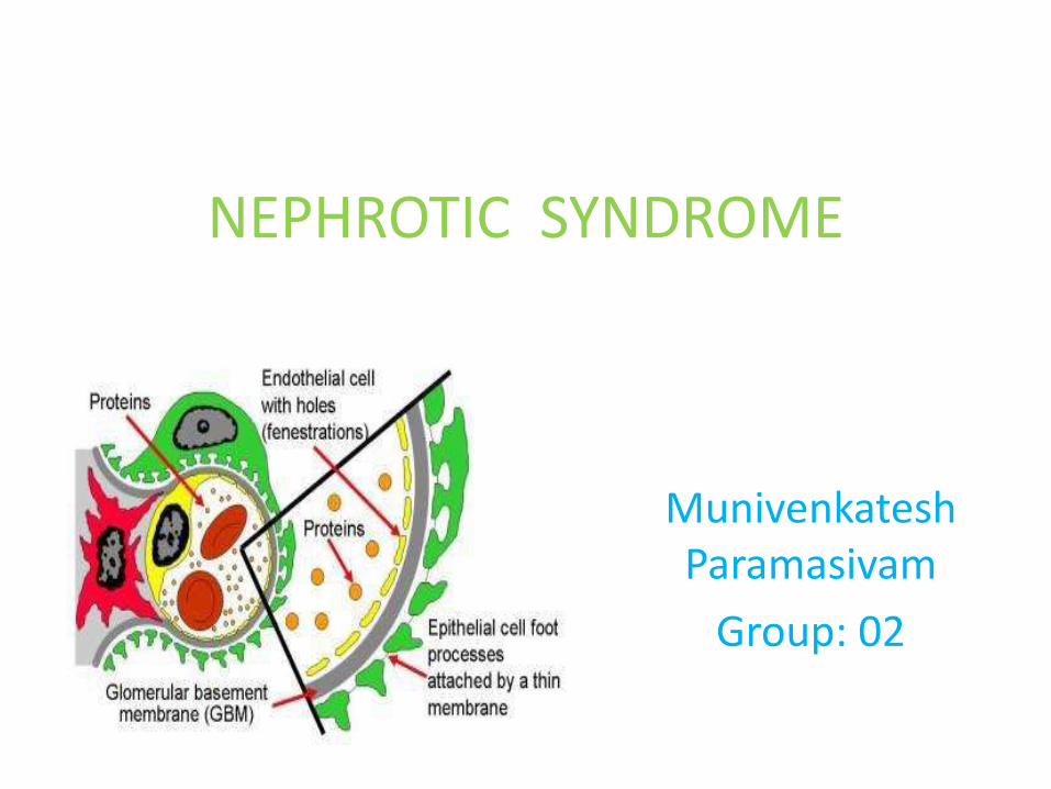
NEPHROTIC SYNDROME
MunivenkateshParamasivam
Group: 02
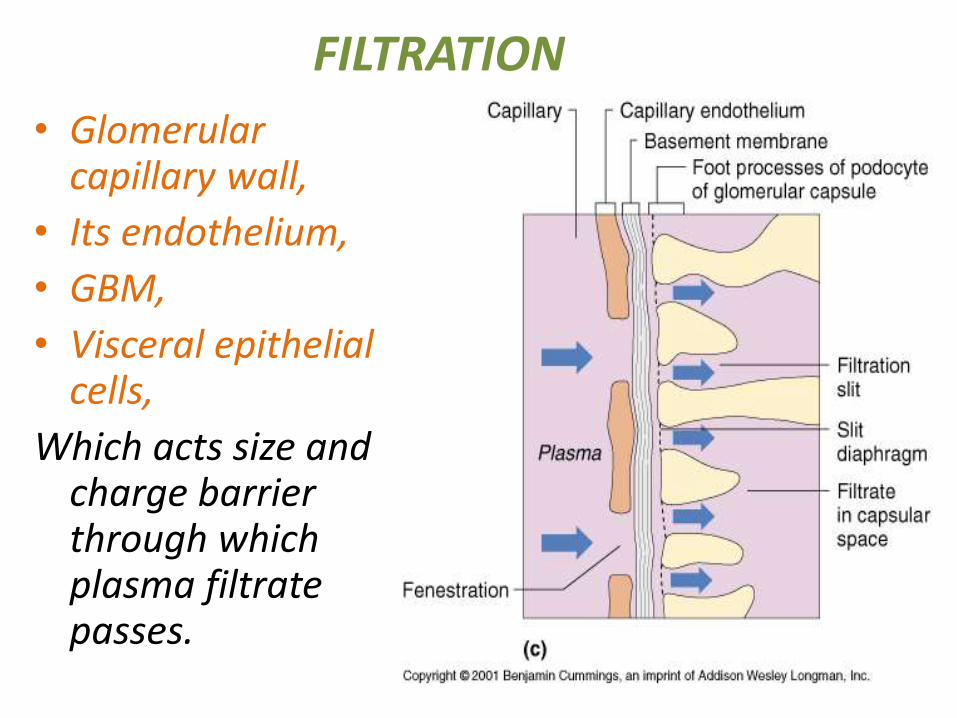
• Glomerularcapillary wall,
• Its endothelium,
• GBM,
• Visceral epithelial cells,
Which acts size and charge barrier through which plasma filtrate passes.
FILTRATION

Pathogenesis• The primary disorder
of incresaedglomerularpermeability to plasma proteins.
• DUE TO
# Derangement in capillary walls
# Change in GBM construction
# Loss of negative charge on GBM

Pathophysiology
NS is an accumulation of symptoms and signs and is characterized by
• Proteinuria (>3.5g/day),
• hypoproteinemia,
• Hypoalbuminemia ( serum albumin < 2.5 gm/dL ),
• Hyperlipidemia,
• Thrombophilia and
• Generalized Edema (anasarca) – begins in the face. Hypoproteinemia plasma oncotic pressure
so fluid goes to interstitial space.

• Puffiness around the eyes (Morning)
• Pitting edema over the legs.
• Pleural effusion and Pulmonary edema.
• Ascites,
• Hypertension,
• Anemia,
• Dyspnea,
• ESR >100mm/hr.
• Anorexia,
• Fatigue,
• Abdominal pain,
• Diarrhea

• Hyponatremia can also occur with a low fractional sodium excretion.
• Muehrcke's nails – white lines (leukonychia) that lie parallel to the lunula.

Classification
• Minimal change disease (Lipoid Nephrosis)
• Focal segmental glomerulosclerosis
• Membranous nephropathy (Membranous
glomerulonephritis)
• Membranoproliferative Glomerulonephritis
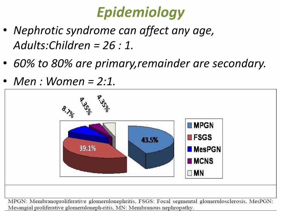
Epidemiology• Nephrotic syndrome can affect any age,
Adults:Children = 26 : 1.
• 60% to 80% are primary,remainder are secondary.
• Men : Women = 2:1.

CAUSES-SECONDARY
• Focal segmental glomerulosclerosis– Hypertensive nephrosclerosis
– HIV, Obesity, Kidney loss.
• Membranous nephropathy (MN):– Systemic lupus erythematosus (SLE)
– Diabetes mellitus, Sarcoidosis, Cancer
– Drugs (such as corticosteroids, gold, heroin)
– Bacterial infections, e.g. leprosy & syphilis.
• CAUSES
-Prophylactic immunization,
-Corticosteroid and immunosupperssive therapy,
-Atopic disorders (eczema, rhinitis)
- Hodgkin's lymphoma,
-Allergy, Bee sting
PATHOGENESIS
uniform and diffuse effacement of the foot processes of the podocytes
Minimal Change Disease
Minimal Change Disease
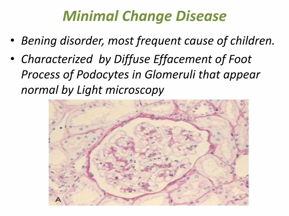
• Bening disorder, most frequent cause of children.
• Characterized by Diffuse Effacement of Foot Process of Podocytes in Glomeruli that appear normal by Light microscopy
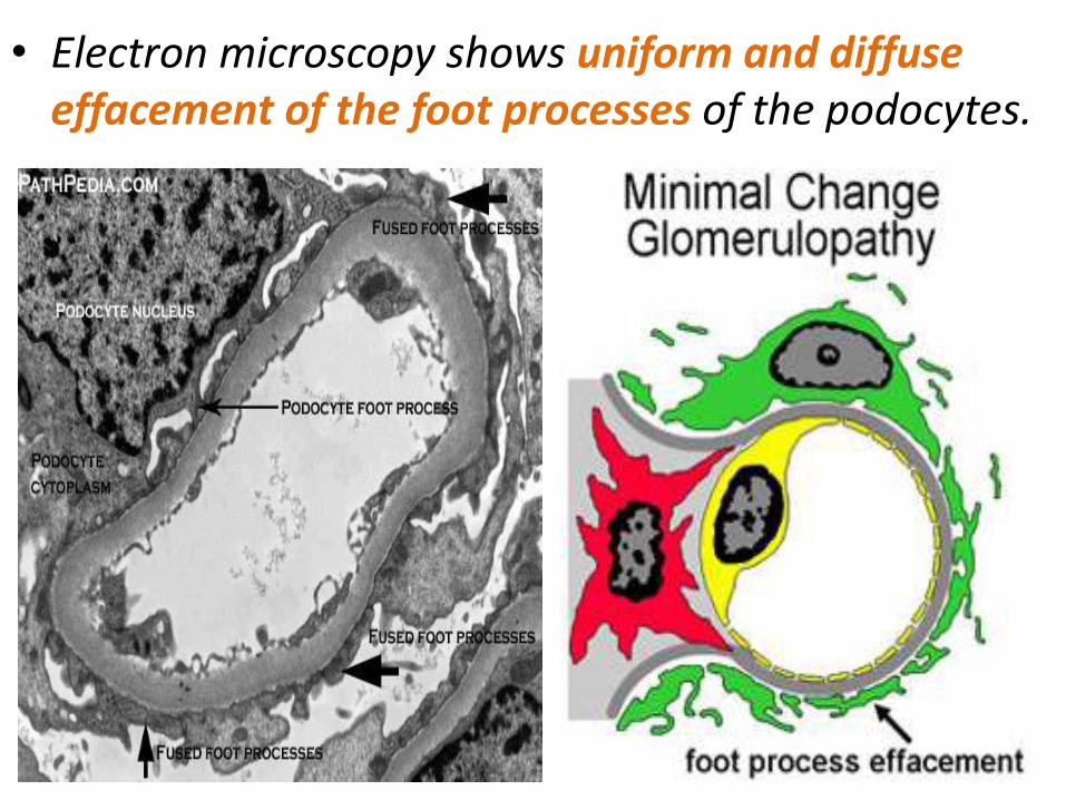
• Electron microscopy shows uniform and diffuse effacement of the foot processes of the podocytes.
Focal Segmental Glomerulosclerosis• As the name implies, this lesion is charecterized by sclerosis
of some glomeruli (Focal); and in the affected glomerulionly a portion of Capillary tuft is involved (segmental).
• CAUSES
Hypertensive nephrosclerosis
Idiopathic,
IgA nephropathy,
HIV,
Obesity,
Kidney loss,
Inherited.
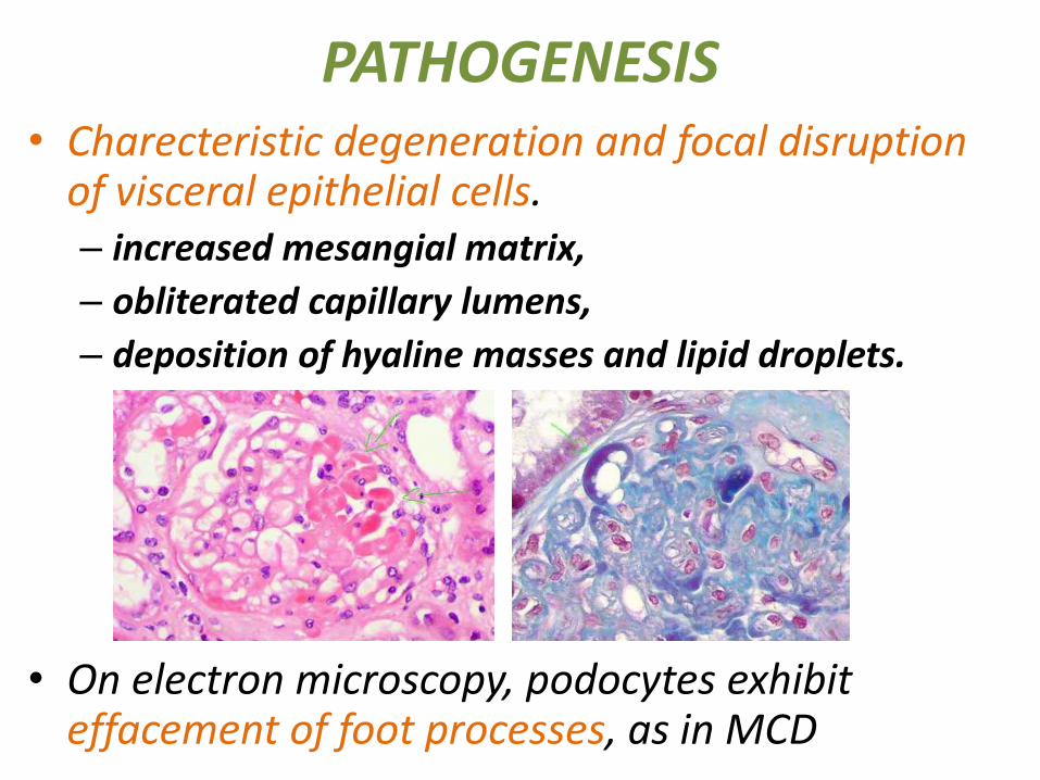
PATHOGENESIS• Charecteristic degeneration and focal disruption
of visceral epithelial cells.– increased mesangial matrix,
– obliterated capillary lumens,
– deposition of hyaline masses and lipid droplets.
• On electron microscopy, podocytes exhibit effacement of foot processes, as in MCD

Typical Lesions
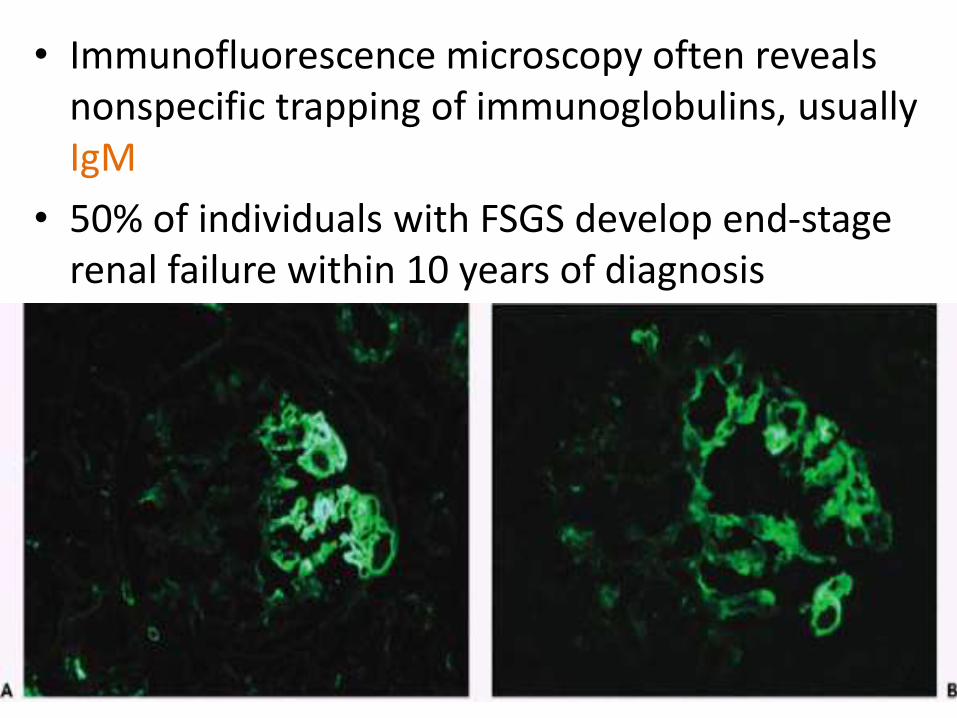
• Immunofluorescence microscopy often reveals nonspecific trapping of immunoglobulins, usually IgM
• 50% of individuals with FSGS develop end-stage renal failure within 10 years of diagnosis
Membranous Nephropathy
• Membranous Nephropathy is a common cause of the nephrotic sydrome in adults ( age 30 to 50 ).
CAUSES
-Drugs (NSAID),
-Underlying malignant tumors,
-SLE,
-Infections (chronic hepatitis B,C and malaria),
-Autoimmune disorders,
-Diabetes mellitus,
-Sarcoidosis.

• Characterised by Diffuse thickening of glomerularcapillary wall due to accumulation of electron dense, Ig containing deposits along subepithelial side of basement membarane.("spike and dome" pattern)

THICKENED BASEMENT MEMBRANE
• In addition, the podocytes show effacement of foot processes
• Later the incorporated deposits may be catabolized and eventually disappear, leaving cavities within the GBM.
• Further progression, the glomeruli can become sclerosed
• Immunofluorescence microscopy shows typical granular deposits of immunoglobulins and complement along the GBM


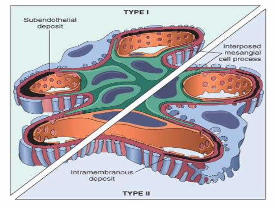
Membranoproliferative Glomerulonephritis
• Characterized histologically by alterations in the glomerular basement membrane, proliferation of glomerular cells, mesangial and endothelial cells and leukocyte infiltration.
• Two major types (I and II).
• Type I is far more common.
• Types I & II have different
ultrastructural &
Immunofluorescence
and pathological features.

Type I MPGN
• It is characterized by discrete subendothelialelectron-dense deposits.
• By immunofluorescence microscopy,
C3 is deposited in an irregular
granular pattern.
• IgG and early complement
components (C1q and C4)
are often also present,
indicative of an immune
complex pathogenesis.
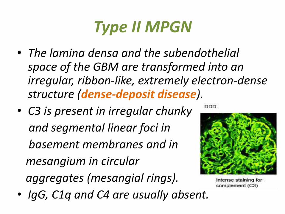
Type II MPGN
• The lamina densa and the subendothelialspace of the GBM are transformed into an irregular, ribbon-like, extremely electron-dense structure (dense-deposit disease).
• C3 is present in irregular chunky
and segmental linear foci in
basement membranes and in
mesangium in circular
aggregates (mesangial rings).
• IgG, C1q and C4 are usually absent.

Diagnosis• Urinalysis to test proteinuria
(>3.5 g per 1.73 m2 per 24 hours).
• Blood screen to look for hypoalbuminemia:
≤2.5 g/dL (normal=3.5-5 g/dL).
• Renal function Creatinine Clearance test to evaluate renal function particularly the glomerular filtration capacity.
• A lipid profile for hypercholesterolemia.
• A kidney biopsy may also be used.
• Further investigations include analysis of auto-immune markers or ultrasound.

Treatment• Symptomatic treatment for edema,
hypoalbuminemia, hyperlipidemia, thrombophilia.
• Treatment of kidney damage:
#Corticosteroids (Prednisone)
#Frequent relapses treated by: cyclophosphamide or nitrogen mustard or cyclosporin or levamisole.
#Immunosupressors (cyclophosphamide)
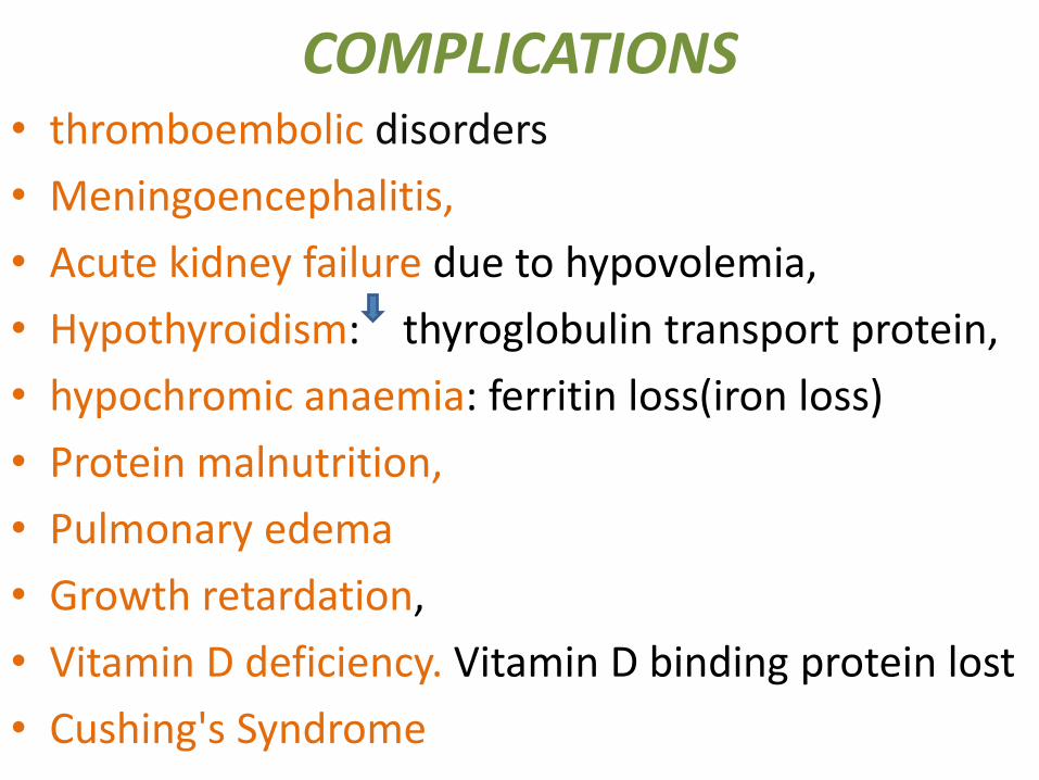
COMPLICATIONS• thromboembolic disorders
• Meningoencephalitis,
• Acute kidney failure due to hypovolemia,
• Hypothyroidism: thyroglobulin transport protein,
• hypochromic anaemia: ferritin loss(iron loss)
• Protein malnutrition,
• Pulmonary edema
• Growth retardation,
• Vitamin D deficiency. Vitamin D binding protein lost
• Cushing's Syndrome