
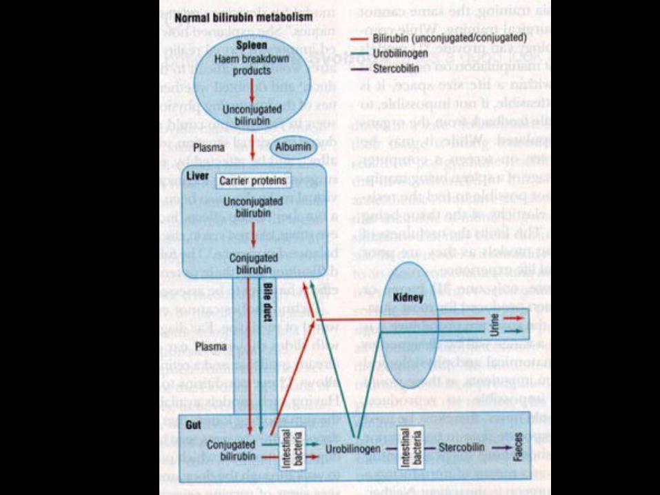
Neonatal Jaundice: Indirect
Hyperbilirubinemia

Objectives
• Identify risk factors for severe hyperbilirubinemia
• Understand the relationship between hyperbilirubinemia and the risk for neurologic or developmental injury
• Discuss ways to screen for infants who might develop severe hyperbilirubinemia
• Discuss guidelines for treatment

Epidemiology: Increased risk for neonatal jaundice
• Infant Factors– Blood group incompatibilities: Rh, ABO, others– Hemolysis (non-isoimmune): infection, drugs, T-antigen
exposure, coagulopathy, RBC enzyme deficiencies (G6PD, PK, HK), RBC structural defects (spherocytosis, elliptocytosis)
– Hemorrhage: cephalohematomas, intracranial bleeding, bruising
– Infection: sepsis, UTI– Endocrine: hypothyroidism, adrenal insufficiency

Epidemiology: Increased risk for neonatal jaundice
• Infant Factors– Prematurity– Male– Polycythemia– Breast feeding vs. formula feeding– Caloric deprivation, postnatal weight loss
• increased enterohepatic circulation– Delayed passage of meconium

Epidemiology: Increased risk for neonatal jaundice
• Race: – Increased production: East Asian, Native
American– G6PD: Greek, East Asian, African
• Genetic: – History of sibling with jaundice– G6PD, hexokinase, pyruvate kinase deficiency– Gilbert’s syndrome, Crigler Najjar Syndrome– Spherocytosis, Elliptocytosis

Epidemiology: Increased risk for neonatal jaundice
• Maternal diabetes mellitus:– Increased bilirubin production rate– Correlation with macrosomia and
polycythemia– Elevated beta-glucuronidase in breastmilk
• Maternal drugs: – epidural anesthesia (bupivacaine)– oxytocin
• Delayed cord clamping

Epidemiology: Increased risk for neonatal jaundice
• Environmental factors:– Phenolic detergents
– Naphthalene (moth balls)
• Short hospital stay– Failure to detect significant jaundice
– Failure to establish breastfeeding

What is a normal “physiologic” serum bilirubin?
• Dennery et al. NEJM 2001: average peak bilirubin in term newborn, 5-6 mg/dL
• Breast fed infants are on average about 2 mg/dL higher than bottle fed infants in the first days of life.
• Racial differences– Greek, Asian, Navajo reach higher peaks
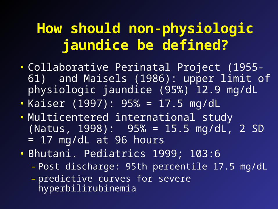
How should non-physiologic jaundice be defined?
• Collaborative Perinatal Project (1955-61) and Maisels (1986): upper limit of physiologic jaundice (95%) 12.9 mg/dL
• Kaiser (1997): 95% = 17.5 mg/dL• Multicentered international study (Natus,
1998): 95% = 15.5 mg/dL, 2 SD = 17 mg/dL at 96 hours
• Bhutani. Pediatrics 1999; 103:6– Post discharge: 95th percentile 17.5 mg/dL– predictive curves for severe hyperbilirubinemia

JCAHO Sentinel Alert: April 2001 Root causes for re-admission for
hyperbilirubinemia identified• Unreliability of visual assessment of jaundice• Failure to measure bilirubin before discharge or in
an infant with visible jaundice in the first 24 hours• Early discharge: especially <38 weeks GA infant• Failure to provide early f/u assessment post-
discharge• Failure to provide lactation support, information to
parents about jaundice or poor feeding• Failure to treat appropriately

Strategies to prevent severe jaundice
• Pre-discharge assessment (transcutaneous bilimeter or serum bilirubin) with use of Bhutani nomogram to predict risk
• Standardized policies for screening• Follow-up of all newborns in 24-48 hr• Informational materials for parents about jaundice• Lactation support• Optimal application of phototherapy

Bhutani: hour specific serum bilirubin. Pediatrics 1999;103:6-14

Predictive nomograms for severe hyperbilirubinemia: Bhutani 1991
• What is the risk for subsequent “severe hyperbilirubinemia” (i.e. bilirubin level in the high risk zone, 95th%)?– > 95th %: 39.5%
– 75-95th %: 21.6%
– 40-75th %: 11.6%
– < 40th %: virtually 0

Bilirubin follow-up policy
• Compare serum bilirubin or transcutaneous bilirubin to Bhutani curves– > 95th%: repeat serum bilirubin in 24-48 hours– 75-95th%: repeat serum bilirubin in 24-48 hours– 40-75th%: if risk factors present, serum bilirubin
in 24-48 hours– < 40th%: no follow-up needed

Bilirubin injury to the brain
• Bilirubin encephalopathy:– Acute reversible changes– Acute irreversible changes
• Kernicterus (yellow staining of the brain)• Neurodevelopmental sequelae
– Clinical correlations– Epidemiologic studies

Clinical features of acute bilirubin encephalopathy
• Acute form:– Early Phase 1 (1-2 days): poor suck, stupor, hypotonia,
seizures– Intermediate Phase 2 (mid 1st week): hypertonia of
extensor muscles, irritability, retrocollis-opisthotonus, fever
– Advanced Phase 3 (after 1st week): irreversible CNS damage, retrocollis-opisthotonus, hypertonia, shrill cry, seizures, coma, apnea, death

Clinical features of kernicterus
• Chronic form:– First year: hypertonia, active DTRs, obligatory tonic
neck reflexes, delayed motor skills– > 1 year: movement disorders (choreoathetosis,
ballismus, tremor), paralysis of upward gaze, hearing loss, mental retardation

Pathology of kernicterus
• Orth: described bilirubin pigmentation of the brain in infants with severe jaundice in 1875
• Kernicterus: German word meaning jaundice of the nuclei– Term was coined by Christian Schmorl in 1904
• Yellow staining of the brain (basal ganglia)• Neuronal swelling• Death of neurons

Pathophysiology of bilirubin encephalopathy
• Bilirubin monoanion binds to membrane– Causes changes in membrane characteristics– May affect membrane permeability
• P-glycoprotein (PGP): ATP mediated transport of bilirubin across membranes and out of the cell– Activity low in immature animal– Can be inhibited by drugs: e.g., ceftriaxone
• Membrane associated bilirubin oxidizing enzyme in the brain: activity low in immature animal
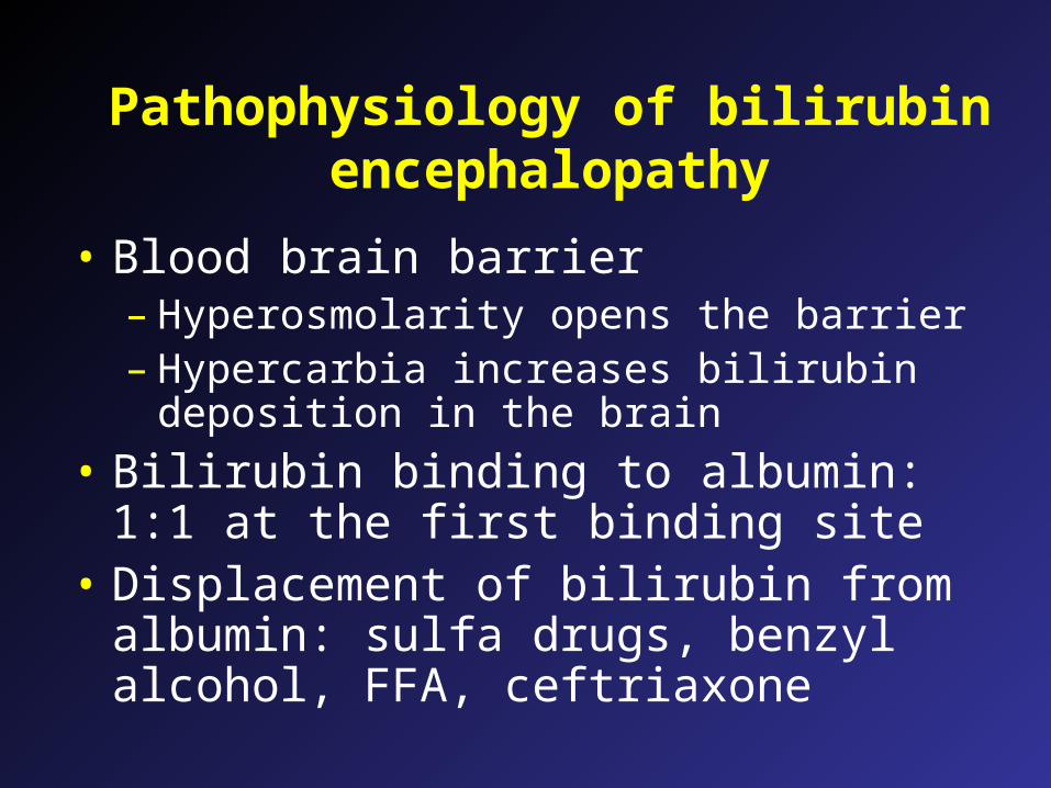
Pathophysiology of bilirubin encephalopathy
• Blood brain barrier– Hyperosmolarity opens the barrier– Hypercarbia increases bilirubin deposition in
the brain
• Bilirubin binding to albumin: 1:1 at the first binding site
• Displacement of bilirubin from albumin: sulfa drugs, benzyl alcohol, FFA, ceftriaxone

Cellular mechanisms of bilirubin toxicity
• Binding to cellular membranes
• Decreased Na-K exchange
• Cellular accumulation of water
• Axonal swelling
• Lowering of membrane potentials, decreased action potential
• Decreased amplitude and longer intervals in auditory response

Clinical factors which increase the risk for kernicterus or bilirubin encephalopathy
• Displacement of bilirubin from albumin• Hyperosmolarity• Hypoxemia, hyperoxemia• Asphyxia• Hypercarbia• Acidosis• Sepsis• Hemolysis• Prematurity
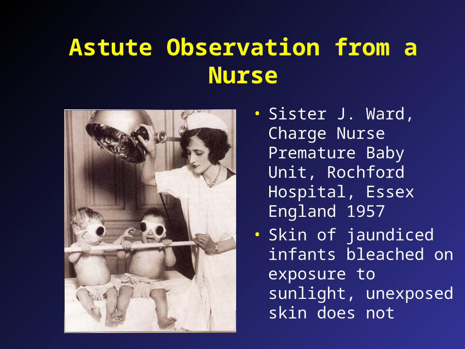
Astute Observation from a Nurse
• Sister J. Ward, Charge Nurse Premature Baby Unit, Rochford Hospital, Essex England 1957
• Skin of jaundiced infants bleached on exposure to sunlight, unexposed skin does not

The Science of Phototherapy
• Bilirubin is a yellow pigment, absorbs blue light spectrum
• Conversion of bilirubin into lumirubin, a water soluble compound
• Elimination by the GI tract and kidney

Can You “Overdose” With Phototherapy?
• “With existing equipment there is no such thing as an overdose of phototherapy” (Maisels2001)
• The saturation point (where higher irradiance levels don’t matter) is not known

Phototherapy devices
• White fluorescent tubes– Broad spectrum light exposure
• Blue fluorescent tubes– Blue light is more effective
• Blue LED lights (NeoBlue)• Halogen lamps
– More compact, bulbs are hot and can burn if too close
• Fiber optic blankets– small area of exposure

How Fast Can the Bilirubin Decline?
• 6-20% decrease in 24 hours-”standard phototherapy”
• 32% decrease in 18 hours- fiberoptic + bluelights
• 43% decrease in 24 hours- blue lights above and below

Fluorescent Phototherapy Lights
• Fluorescent lights cover more skin surface
• Deliver higher intensity without heating
• White lights effective, blue lights most effective
• Bulbs lose intensity long before they “burn out”

Fluorescent Bili Lights: 30-35 microwatts

LED Phototherapy: 25-50 microwatts

NeoBlue Mini: 30-40 microwatts
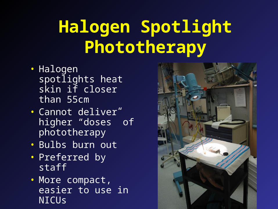
Halogen Spotlight Phototherapy
• Halogen spotlights heat skin if closer than 55cm
• Cannot deliver higher “doses” of phototherapy
• Bulbs burn out• Preferred by staff• More compact,
easier to use in NICUs

“Triple”Phototherapy:Halogen, Blanket
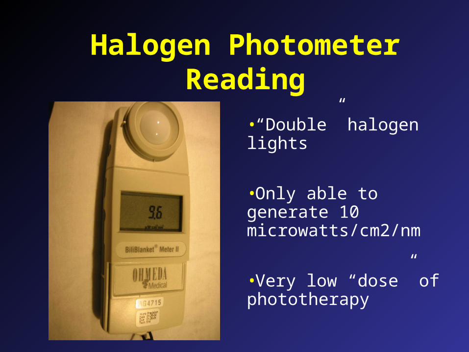
Halogen Photometer Reading
•“Double” halogen lights
•Only able to generate 10 microwatts/cm2/nm
•Very low “dose” of phototherapy

Fiberoptic Phototherapy
•Light from tungsten-halogen bulb through fiberoptic cable•Less effective than conventional phototherapy•Should not be used in VLBW infants, potential for skin injury

Skin Injury From Bili Blanket

Factors that determine dose and effectiveness of phototherapy
• Spectrum of light (blue is best)
• Irradiance of light source– power output of the lamp
• Design of phototherapy device– does it expose the maximal amount of
skin?
• Surface area exposed to light
• Distance of infant from light

Acute management of severe hyperbilirubinemia
• Phototherapy with fluorescent or LED blue lights: maximal surface exposure and dose
• Correct dehydration, acidosis (respiratory and metabolic), and hypotension
• Correct hypoalbuminemia (1 g/dL of albumin binds 8.3 mg/dL bilirubin): augments removal of bilirubin with exchange transfusion
• Reduce enterohepatic circulation of bilirubin: stop breast milk feedings, use formula feedings– PO charcoal and agar reported, but not commonly used

Acute management of severe hyperbilirubinemia
• Avoid drugs which displace bilirubin from albumin or affect P glycoprotein
• Avoid use of hyperosmolar drugs or infusions• Inhibitors of heme oxygenase (protoporphyrins):
– Reduces bilirubin production– Sn and Zn protoporphyrins reported to be useful, but
not yet FDA approved
• Extra-corporeal removal of bilirubin: – theoretical possible– extracorporeal charcoal binding used in Russia

Recommendations for treatment of hyperbilirubinemia (AAP practice
guideline)Age Consider Exchange if Exchange*
(hr) phototherapy phototherapy photoRx fails# transfusion
25-48 > 12 > 15 > 20 > 25
48-72 > 15 > 18 > 25 > 30
>72 > 17 > 20 > 25 > 30
#Phototherapy should result in a decline 1-2 mg/dL of total bilirubin within 4-6 hour, should continue to fall and remain below exchange transfusion levels.
*Intensive phototherapy, prepare for exchange, exchange if bilirubin does not fall below exchange transfusion levels.
Adapted from Pediatrics 1994;94:558
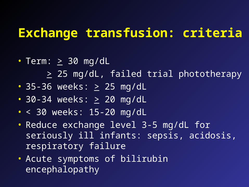
Exchange transfusion: criteria
• Term: > 30 mg/dL
> 25 mg/dL, failed trial phototherapy• 35-36 weeks: > 25 mg/dL• 30-34 weeks: > 20 mg/dL• < 30 weeks: 15-20 mg/dL• Reduce exchange level 3-5 mg/dL for seriously ill
infants: sepsis, acidosis, respiratory failure• Acute symptoms of bilirubin encephalopathy

Exchange Transfusion
• ABO type-specific Rh negative blood in cases with Rh incompatibility
• Type O Rh-specific cells in cases with cases with ABO incompatibility
• Whole blood diluted with FFP to Hct of 50-55%.
• Fresh blood < 24 hours old preferred.• Double volume exchange 160ml/kg

Technique for Exchange Transfusion
• Withdrawal thru UA catheter with simultaneous infusion thru UVC catheter
• 5-to 20-ml increments of warmed blood• Agitate blood every 10-15 minutes so
cells don’t settle.• Initial sample sent for bilirubin, Hct,
lytes, calcium, cultures

Things to Remember
• Monitor ECG, BP, and temperature during procedure
• Measure ABG at beginning, middle, and end of procedure.
• Measure glucose at 10, 30, 60 minutes post procedure.
• Measure calcium after each 100 ml of blood.• Warming blood > 37 degrees causes
hemolysis

Bilirubin After Double Volume Exchange
• Serum bilirubin is 45% to 60% of preexchange level

Potential complications Infant• Hypothermia
• Hyperkalemia
• Thrombocytopenia
• Low Ca++ and Mg++
• Reactive hypoglycemia
Action• Warm donor blood
• Use fresh blood, monitor ECG
• Transfuse platelets at end if < 75K
• Give CaGluconate 100mg/kg/d
• IV glucose 5mg/kg/min 10-30 minutes after end of exchange

Followup issues for hyperbilirubinemia
• Hearing screen• “Rebound” bilirubin
– AAP guideline: repeat bilirubin level not indicated in healthy term infants
– useful in premature infants, hemolysis (isoimmunization, G6PD)
• Infants with bilirubin encephalopathy– neurodevelopmental followup– hearing screen


