©2015 Waters Corporation 1
N-linked Glycan Characterization and
Profiling: Combining the Power of a Novel
Labeling Reagent and a Streamlined Analytical
Workflow
Ying Qing Yu Ph.D
Waters Corporation
Glycobiology World Congress 2015
©2015 Waters Corporation 2
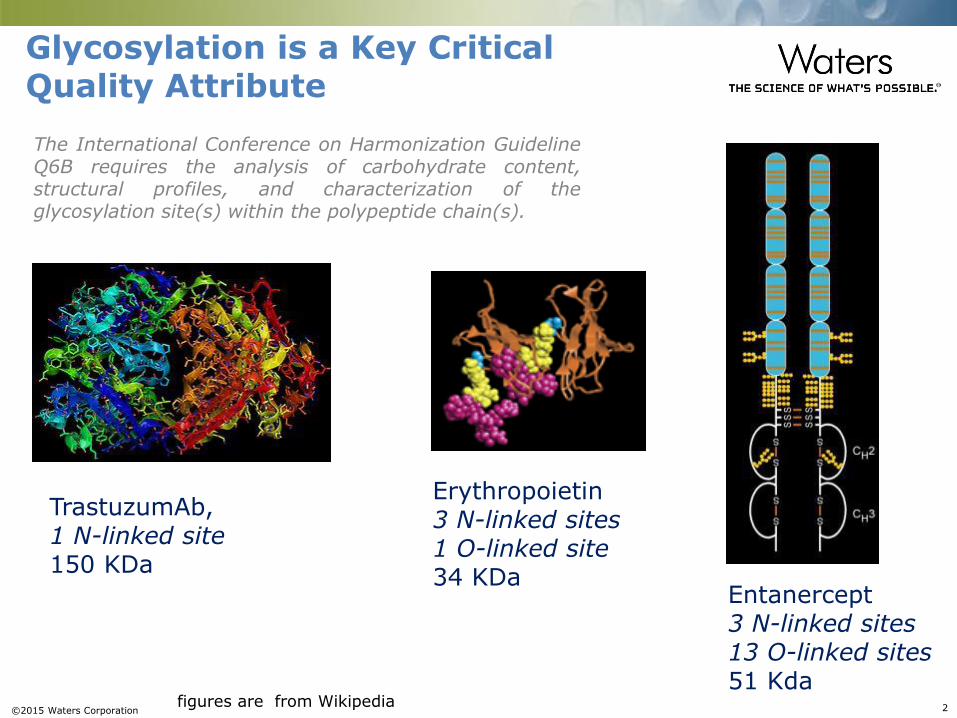
Glycosylation is a Key Critical Quality Attribute
TrastuzumAb,1 N-linked site150 KDa
Entanercept3 N-linked sites13 O-linked sites51 Kda
Erythropoietin3 N-linked sites1 O-linked site34 KDa
figures are from Wikipedia
The International Conference on Harmonization GuidelineQ6B requires the analysis of carbohydrate content,structural profiles, and characterization of theglycosylation site(s) within the polypeptide chain(s).
©2015 Waters Corporation 3
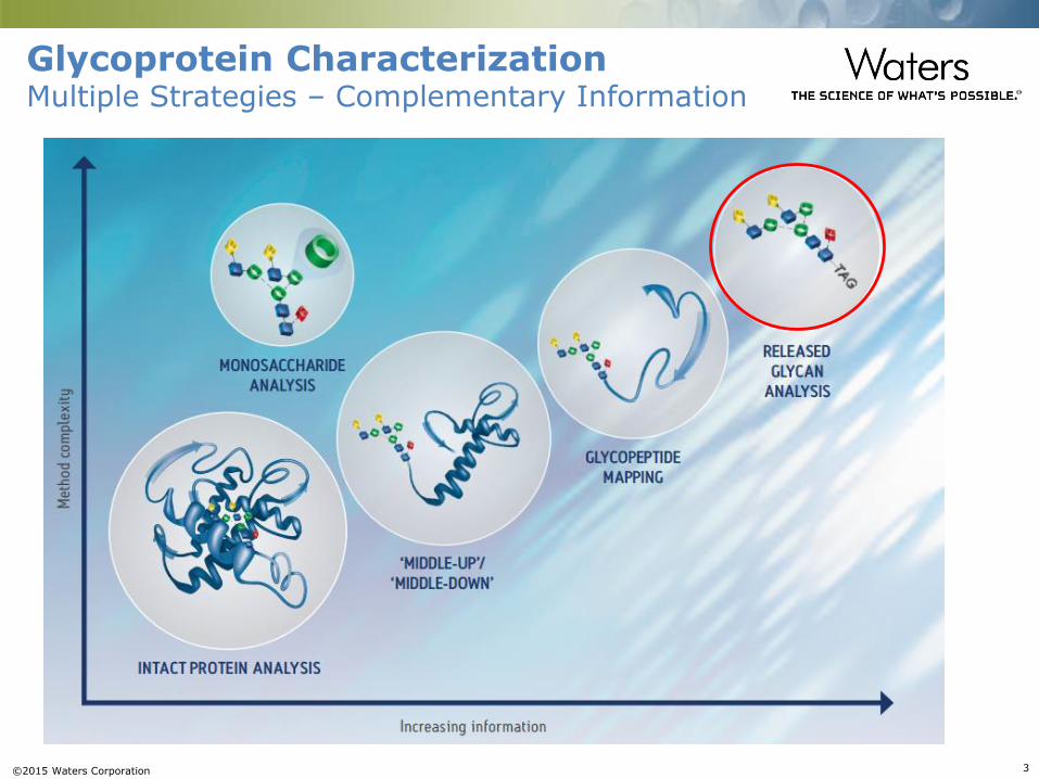
Glycoprotein CharacterizationMultiple Strategies – Complementary Information
©2015 Waters Corporation 4
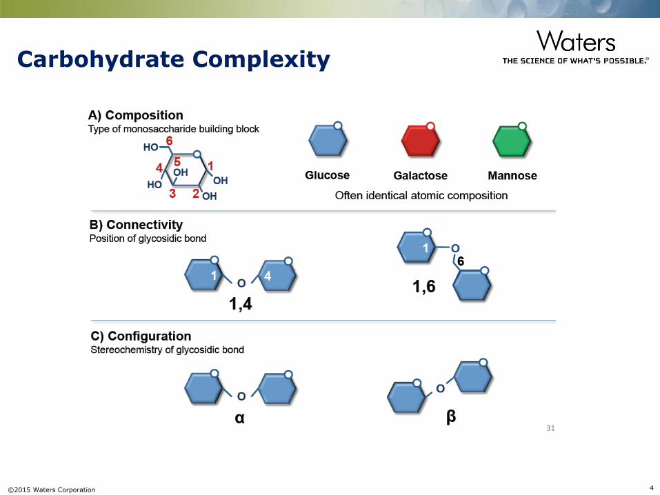
Carbohydrate Complexity
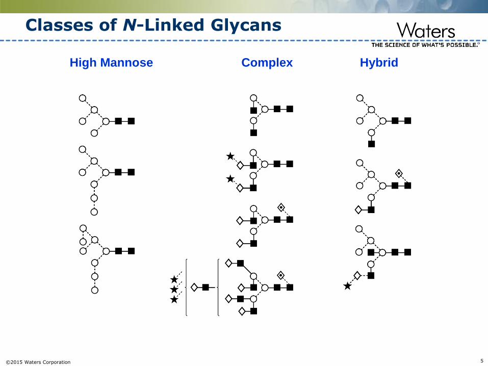
©2015 Waters Corporation 5
Classes of N-Linked Glycans
Complex HybridHigh Mannose
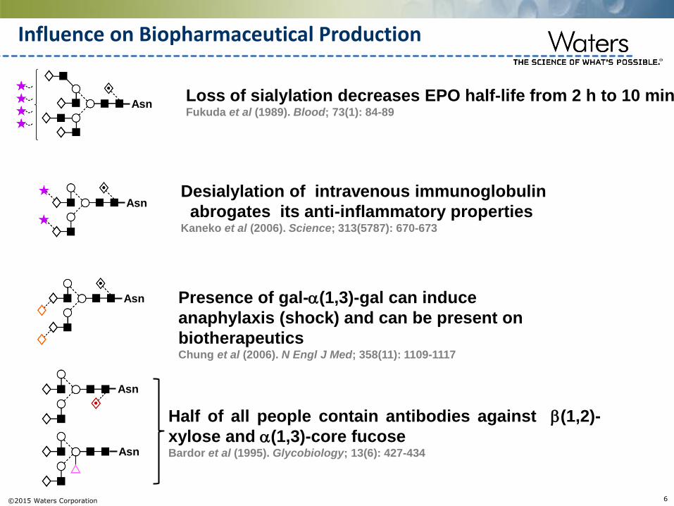
©2015 Waters Corporation 6
Asn
AsnDesialylation of intravenous immunoglobulin
abrogates its anti-inflammatory properties Kaneko et al (2006). Science; 313(5787): 670-673
Loss of sialylation decreases EPO half-life from 2 h to 10 minFukuda et al (1989). Blood; 73(1): 84-89
Asn Presence of gal-(1,3)-gal can induce
anaphylaxis (shock) and can be present on
biotherapeuticsChung et al (2006). N Engl J Med; 358(11): 1109-1117
Influence on Biopharmaceutical Production
Asn
Asn
Half of all people contain antibodies against (1,2)-
xylose and (1,3)-core fucoseBardor et al (1995). Glycobiology; 13(6): 427-434
©2015 Waters Corporation 7
Rapid Preparation
DeglycosylationLabeling
SPE
30 min
High Sensitivity
HILIC-Fluorescence-MSFluorophore
+MS Charge Tag
Fluorescence MS
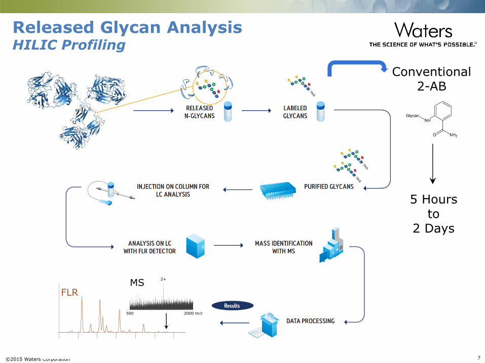
Released Glycan AnalysisHILIC Profiling
5 Hoursto
2 Days
0.0E+0
3.3E+6
3 4 5 6 7 8 9 10
FLR
Conventional2-AB
5.3e2
m/z500 1000 1500 2000
%
0
100
m/z500 1000 1500 2000
%
0
100
m/z500 1000 1500 2000
%
0
100
m/z500 1000 1500 2000
%
0
100
RapiFluorMS_hIgG_14pmol_18Nov14_04 283 (5.065)3.47e6
2AB_hIgG_halfload_20Nov14_10 235 (4.234)2.89e4
2AB_hIgG_halfload_20Nov14_10 495 (8.689)529
RapiFluorMS_hIgG_14pmol_18Nov14_04 507 (8.892)1.15e5
500 2000 m/z
2+
MS

©2015 Waters Corporation 8
Conventional Workflow
16 hrs
Conventional
3 hrs
0.1 hrs
(Pre-Labeling Clean-Up)(Drying)
1-3 hrs
(Drying)
2 hrs
Glycoprotein
Released N-Glycans+ Protein
LabeledN-Glycans+ Reagents
Labeled Glycans
LC-FLR LC-FLR-MS
Deglycosylation
Labeling
Clean-up
Analysis
ReactionByproducts
>24 HOURS

©2015 Waters Corporation 9
What is new?
Novel RapiFluor-MSTM (RFMS) Reagent
Patent Pending
©2015 Waters Corporation 10
+
+
H2O
+ + CO2
rapiFluor-MS
Monoisotopic mass shift (from glycosylamine)312.1586 Da
Glycosylamine+C17H20N4O2
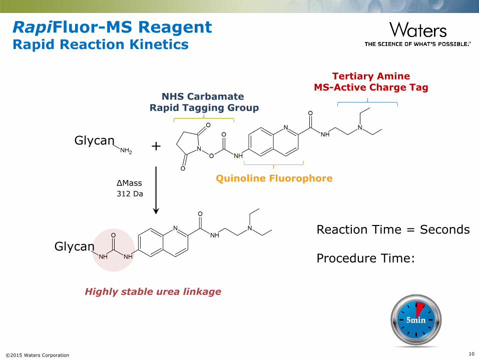
RapiFluor-MS ReagentRapid Reaction Kinetics
NHS CarbamateRapid Tagging Group
Tertiary AmineMS-Active Charge Tag
Reaction Time = Seconds
Procedure Time:
Quinoline Fluorophore
Glycan
Glycan
Highly stable urea linkage
ΔMass
312 Da

©2015 Waters Corporation 11
16 hrs
Conventional
3 hrs
0.1 hrs
(Pre-Labeling Clean-Up)(Drying)
1-3 hrs
(Drying)
2 hrs
<15 min
5 min
30 min
GlycoWorks RapiFluor-MS N-Glycan Kit
10 min
Glycoprotein
Released N-Glycans+ Protein
LabeledN-Glycans+ Reagents
Labeled Glycans
LC-FLR LC-FLR-MS
Deglycosylation
Labeling
Clean-up
Analysis
ReactionByproducts
Simplified Sample Preparation
>24 HOURSPatent Pending

©2015 Waters Corporation 12
RFMS vs. 2AB for MS sensitivity comparison
Greater than 100x MS response over 2AB labeling
BPI
MS
Sample: NIST RM 8670 mAb lot #3F1b
2AB
RFMS
©2015 Waters Corporation 13
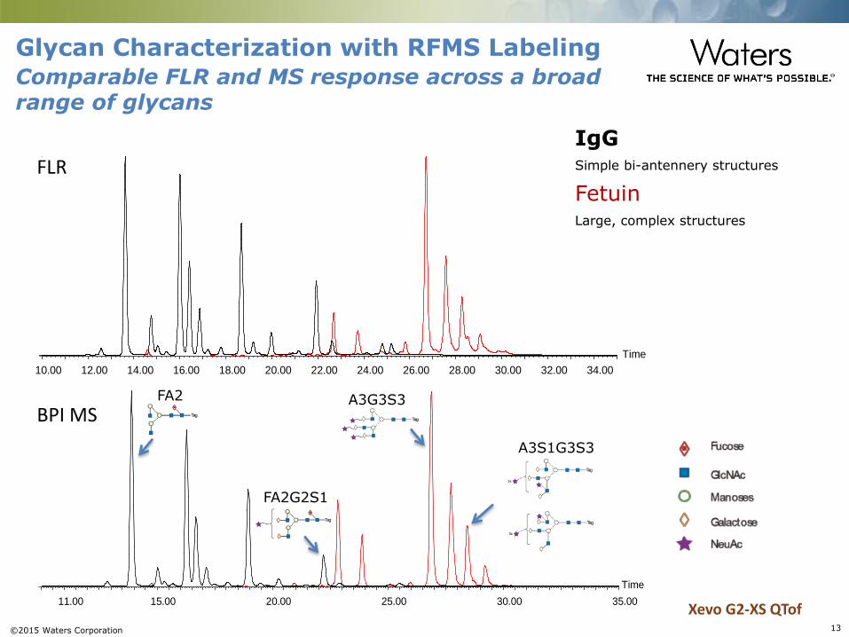
Time
11.00 15.00 20.00 25.00 30.00 35.00
Time
10.00 12.00 14.00 16.00 18.00 20.00 22.00 24.00 26.00 28.00 30.00 32.00 34.00
FLR
BPI MS
IgGSimple bi-antennery structures
FetuinLarge, complex structures
Glycan Characterization with RFMS Labeling
Xevo G2-XS QTof
Comparable FLR and MS response across a broad range of glycans
FA2
FA2G2S1
A3G3S3
A3S1G3S3
©2015 Waters Corporation 14
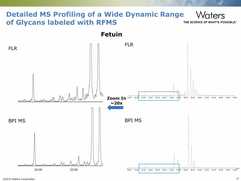
10.00 15.00 20.00 25.00 30.00 35.00 40.00
FLR
BPI MS
Detailed MS Profiling of a Wide Dynamic Range of Glycans labeled with RFMS
Time10.00 12.00 14.00 16.00 18.00 20.00 22.00 24.00 26.00 28.00 30.00 32.00 34.00 36.00 38.00 40.000
BPI MS
10.00 12.00 14.00 16.00 18.00 20.00 22.00 24.00 26.00 28.00 30.00 32.00 34.00 36.00 38.00 40.00
FLR
Zoom In~20x
Fetuin
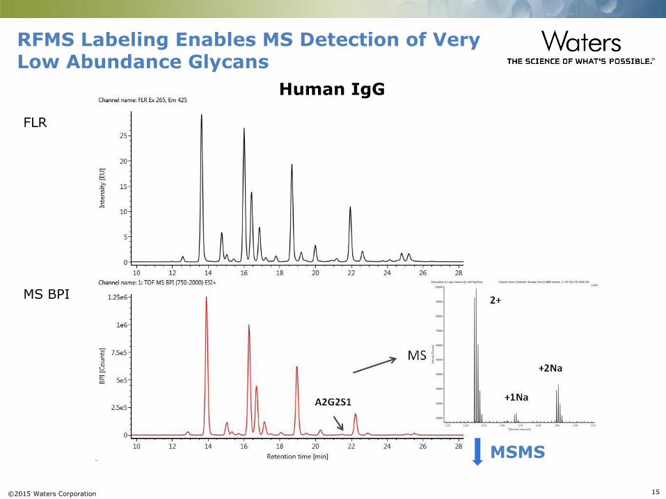
©2015 Waters Corporation 15
RFMS Labeling Enables MS Detection of Very Low Abundance Glycans
Human IgG
FLR
MS BPI
MSMS
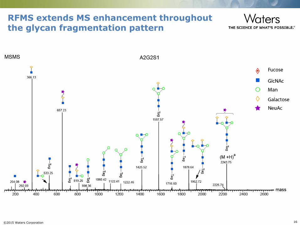
©2015 Waters Corporation 16
RFMS extends MS enhancement throughout the glycan fragmentation pattern

©2015 Waters Corporation 17
RFMS enables easy assignment of two isobaric glycans
RFMS labeled N-glycans from a murine IgG1 mAb
FLR
MS
A B FA2G2
FA2G1Ga1
FA2G1Ga1
FA2G2
Zoom 10x
2.1E+6
8.4E+6
0.0E+6
0.0E+6
10 20 min 100 2000 m/z
FLR
MS BPI
• The two isobaric glycans, FA2G2 and a minor shoulder peak, partially resolved by HILIC
• The minor peak represents only 0.7% of total FLR signal
?

©2015 Waters Corporation 18
FLR
MS
A B FA2G2
FA2G1Ga1
FA2G1Ga1
FA2G2
Zoom 10x
2.1E+6
8.4E+6
0.0E+6
0.0E+6
10 20 min 100 2000 m/z
FLR
MS
A B FA2G2
FA2G1Ga1
FA2G1Ga1
FA2G2
Zoom 10x
2.1E+6
8.4E+6
0.0E+6
0.0E+6
10 20 min 100 2000 m/z
Loss of GlcNAc
0.04
RFMS enables easy assignment of a minor shoulder peak of FA2G2 as FA2G1Ga1
• Structurally diagnostic ions: 1) predominant 528 m/z ion and 2) prominent GlcNAc loss
• High sensitivity and information rich fragmentation data support the identification of the isobaric, lower abundance species as an α-Gal containing FA2G1Ga1.
RFMS labeled N-glycans from a murine IgG1 mAb
FLR
MS
A B FA2G2
FA2G1Ga1
FA2G1Ga1
FA2G2
Zoom 10x
2.1E+6
8.4E+6
0.0E+6
0.0E+6
10 20 min 100 2000 m/z
FLR
MS BPI
• The two isobaric glycans, FA2G2 and a minor shoulder peak, partially resolved by HILIC
• The minor peak represents only 0.7% of total FLR signal
FA2G1Ga1
©2015 Waters Corporation 19
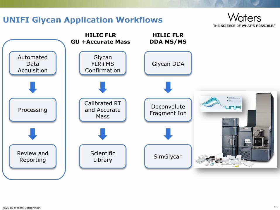
UNIFI Glycan Application Workflows
HILIC FLRGU +Accurate Mass
HILIC FLRDDA MS/MS
Automated Data
Acquisition
Processing
Review andReporting
Glycan FLR+MS
Confirmation
Calibrated RT and Accurate
Mass
Scientific Library
Glycan DDA
Deconvolute Fragment Ion
SimGlycan
©2015 Waters Corporation 20
The Utility of GU Values
What is a GU Value?
• GU stands for Glucose Unit
• A GU value is a normalized glycan structure retention time observed in HILIC for glycan peaks, obtained using a dextran ladder calibration
Why is the GU approach useful?
• GU Values assist in normalizing glycan retention time across days, instruments and laboratories, so data can be compared and shared easily.
• GU Values facilitate more routine glycan assignments by enabling the creation of a single glycan GU retention library.
©2015 Waters Corporation 21
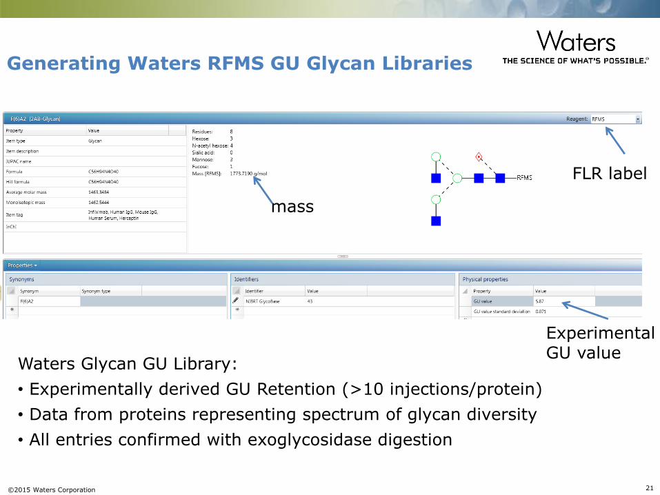
Generating Waters RFMS GU Glycan Libraries
Experimental GU value
FLR label
mass
Waters Glycan GU Library:
• Experimentally derived GU Retention (>10 injections/protein)
• Data from proteins representing spectrum of glycan diversity
• All entries confirmed with exoglycosidase digestion
©2015 Waters Corporation 22
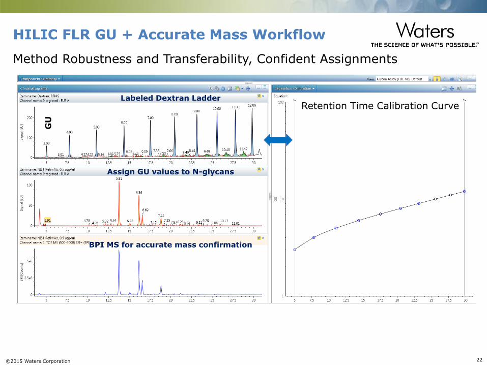
HILIC FLR GU + Accurate Mass Workflow
Labeled Dextran LadderRetention Time Calibration Curve
Assign GU values to N-glycans
BPI MS for accurate mass confirmation
GU
Method Robustness and Transferability, Confident Assignments

©2015 Waters Corporation 23
Glycan GU Scientific Library Search for Confident Glycan Assignments
Both 2-AB and RFMS labeled glycan performance test standards are now available to support this workflow
©2015 Waters Corporation 24
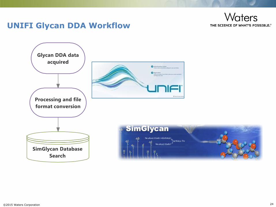
UNIFI Glycan DDA Workflow

©2015 Waters Corporation 25
UNIFI Acquire and Process Glycan DDA DataRFMS labeled N-glycans from mouse IgG
FLR
BPI MS
Raw MSMS Spectrum
~ 0.1% total FLR
Deconvolute & deeisotopeExport as .LCS (or mzml) format
MSMS fragmentation of a highlighted minor glycoform was displayed
©2015 Waters Corporation 26
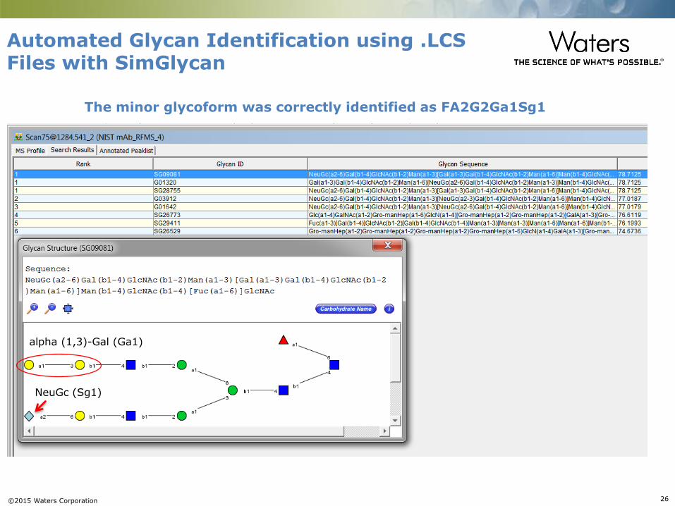
Automated Glycan Identification using .LCS Files with SimGlycan
The minor glycoform was correctly identified as FA2G2Ga1Sg1
alpha (1,3)-Gal (Ga1)
NeuGc (Sg1)
©2015 Waters Corporation 27
Work in progress
Developing a scientific library for RapiFluor-MS labeled glycans
for automated glycan assignment
Applying RapiFluor-MS label for more complex glycosylated
proteins

©2015 Waters Corporation 28
Professor Pauline Rudd (COI)
– Mark Hilliard (UNIFI Glycan Scientific Library)
– Giogio Carta (UNIFI Glycan Scientific Library)
Acknowledgements
Waters
Matt Lauber PhD
New England Biolabs
Paula Magnelli PhD


