
Muscular System:Histology and Physiology
Chapter 9


Muscular System Functions
Body movement Maintenance of posture Respiration Production of body heat Communication Constriction of organs and vessels Heart beat

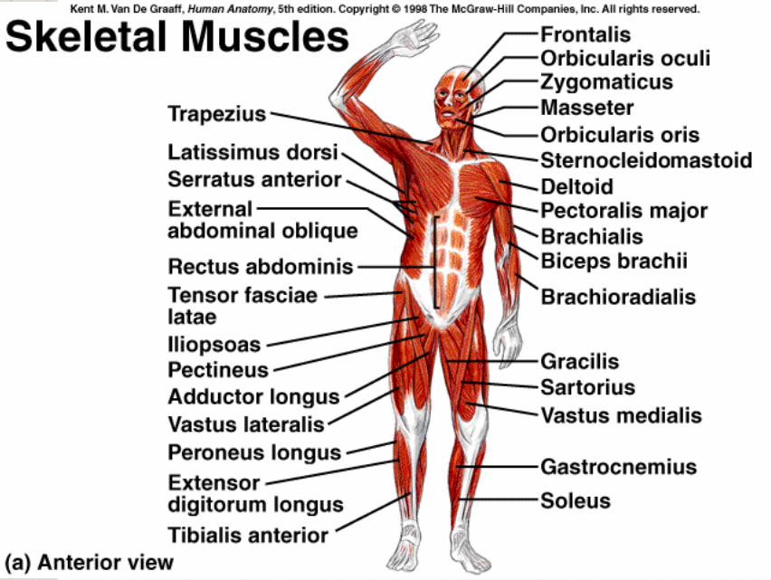
Criteria for Naming Muscles Shape: romboideus, trapezius, biceps Location: pectoralis (chest) intercostal (ribs) Attachment: zygomaticus, sternocleidomastoid Size: maximus, minimus, brevis, longis Orientation of fibers: rectus (straight), oblique
(slanting) Relative position (lateral, medial, internal,
external) Function: adductor, flexor, extensor, pronator


Properties of Muscle
Contractility Ability of a muscle to shorten with force
Excitability Capacity of muscle to respond to a stimulus
Extensibility Muscle can be stretched to its normal
resting length and beyond to a limited degree
Elasticity Ability of muscle to recoil to original resting
length after stretched
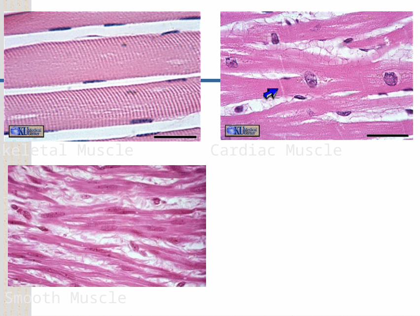
Skeletal Muscle
Smooth Muscle
Cardiac Muscle

Features Skeletal Muscle Smooth Muscle Cardiac Muscle
Location Attached to bone walls of hollow organs, blood vessels, eyes, glands and skin
heart
Cell shape very long, cylindrical
Spindle shaped Cylindrical and branched
Nucleus Multiple, peripherally located
Single centrally located
single centrally located
Special features
Gap junctions join visceral smooth muscle
Intercalated disks join cells
Control voluntary and involuntary reflexes
Involuntary Involuntary
Spontaneous contraction
No Yes Yes
Function Body movement Food movement, urinary bladder, blood vessels, glands and duct
pumps blood

Skeletal Muscle Structure
Muscle fibers or cells Develop from
myoblasts Numbers remain
constant Hypertrophy –
increase in the size of each fiber.
Connective tissue Nerve and blood
vessels


Connective Tissue, Nerve, Blood Vessels
Connective tissue External lamina Endomysium Perimysium Fasciculus Epimysium
Fascia Binds adjacent muscles
or overlying skin. Nerve and blood
vessels Abundant


Parts of a Muscle

Structure of Actin and Myosin

Components of Sarcomeres


Sliding Filament Model
Actin myofilaments sliding over myosin to shorten sarcomeres Actin and myosin do not change length Shortening sarcomeres responsible for
skeletal muscle contraction During relaxation, sarcomeres
lengthen

Sarcomere Shortening


Physiology of Skeletal Muscle
Nervous system Controls muscle
contractions through action potentials
Resting membrane potentials
Membrane voltage difference across membranes (polarized)
• Inside cell more negative and more K+
• Outside cell more positive and more Na+
Must exist for action potential to occur

Ion Channels
Types Ligand-gated
• Example: neurotransmitters
Voltage-gated• Open and close in
response to small voltage changes across plasma membrane

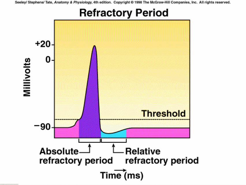
Action Potentials Phases
Depolarization• Inside plasma
membrane becomes less negative
Repolarization• Return of resting
membrane potential All-or-none principle
Like camera flash system
Propagate Spread from one
location to another Frequency
Number of action potential produced per unit of time


0013.exe


Action Potential Propagation

Neuromuscular Junction
Synapse or NMJ Presynaptic terminal Synaptic cleft Postsynaptic membrane or motor end-plate
Synaptic vesicles Acetylcholine: Neurotransmitter Acetylcholinesterase: A degrading enzyme in synaptic cleft
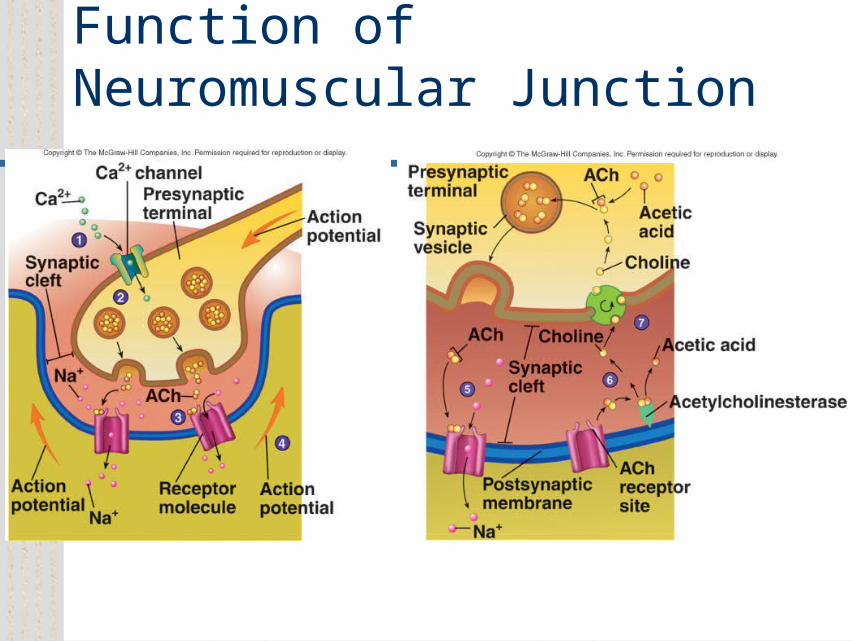
Function of Neuromuscular Junction

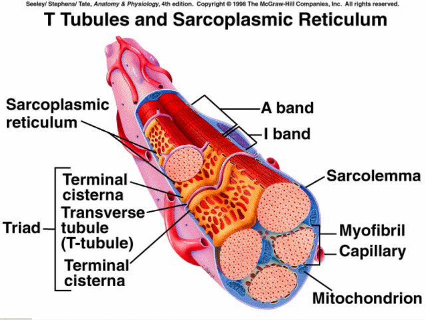
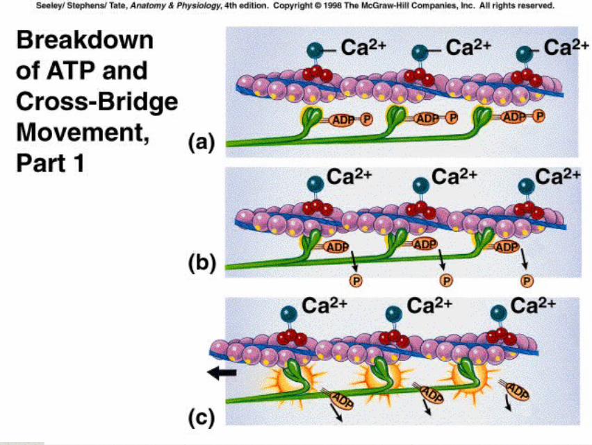
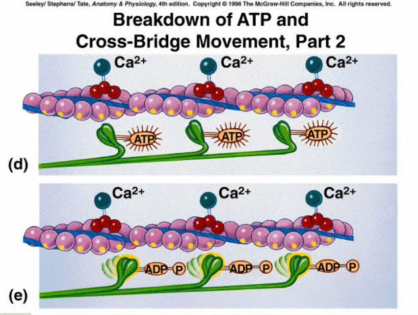
Excitation-Contraction Coupling
Mechanism by which an action potential causes muscle fiber contraction
Involves Sarcolemma Transverse or T
tubules Terminal cisternae Sarcoplasmic
reticulum Ca2+
Troponin

Action Potentials and Muscle Contraction
0010.exe

Cross-Bridge Movement0011.exe


Muscle Twitch
Muscle contraction in response to a stimulus that causes action potential in one or more muscle fibers
Phases Lag or latent Contraction Relaxation

Stimulus Strength and Muscle Contraction All-or-none law for
muscle fibers A motor unit contracts
with a consistent force in response to each action potential
• Sub-threshold stimulus• Threshold stimulus• Stronger than threshold
Motor units Single motor neuron and
all muscle fibers that it innervates
Graded for whole muscles Strength of contractions
range from weak to strong depending on stimulus strength

Multiple Motor Unit Summation
A whole muscle contracts with a small or large force depending on number of motor units stimulated to contract
Muscle performing delicate and precise movements have motor units with smaller numbers of fibers

Multiple-Wave Summation As frequency of action
potentials increase, frequency of contraction increases
Incomplete tetanus• Muscle fibers partially relax
between contraction Complete tetanus
• No relaxation between contractions
Multiple-wave summation Muscle tension increases
as contraction frequencies increase
Due to increased calcium concentration around myofibrils and more complete stretching of muscle elastic elements

Treppe Increase in the force of
contraction during the first few contractions of a rested muscle.
Occurs in muscle rested for prolonged period
Each subsequent contraction is stronger than previous until all equal after few stimuli
Due to Ca++ ion levels around myofibrils and increased temperature of muscle Enzymes for muscle
contraction respond more effectively at higher temperature.

Types of Muscle Contractions
Isometric: No change in length but tension increases Postural muscles of body
Isotonic: Change in length but tension constant Concentric: Overcomes opposing resistance
and muscle shortens Eccentric: Tension maintained but muscle
lengthens Muscle tone: Constant tension by
muscles for long periods of time

Muscle Length and Tension

Fatigue
Decreased capacity to work and reduced efficiency of performance Usually follows a period of activity
Types Psychological (in CNS)
• Depends on emotional state of individual• Perception that muscle is too tired (
• Home court advantage Muscular
• Results from ATP depletion in muscle Synaptic
• Occurs in NMJ due to lack of acetylcholine

Energy Sources ATP provides immediate energy for muscle
contractions from 3 sources Creatine phosphate
• During resting conditions stores energy to synthesize ATP• Exhausted quickly (10-15 sec.)
Anaerobic respiration• Occurs in absence of oxygen and results in breakdown of
glucose to yield ATP and lactic acid Aerobic respiration
• Requires oxygen and breaks down glucose to produce ATP, carbon dioxide and water
• More efficient than anaerobic
Oxygen Debt After anaerobic respiration, aerobic respiration is higher
than normal to replace creatine phosphate and convert lactic acid to glucose.
Slow and Fast Fibers Slow-twitch or high-oxidative
Contract more slowly, smaller in diameter, well developed blood supply, more mitochondria and high myoglobin content, more fatigue-resistant than fast-twitch
Fast-twitch or low-oxidative Respond rapidly to nervous stimulation, less blood
supply, fewer and smaller mitochondria, lower myoglobin content than slow-twitch, fatigue easily.
Two types:• Fast twitch fatigable fibers• Fast twitch fatigue resistant (highly trained muscle)
Distribution of fast-twitch and slow twitch Most muscles have both but varies for each muscle

Effects of Exercise Training muscle increases muscular size and strength
(Hypertrophy). Aerobic exercise can convert fast-twitch easily fatigued muscle
into fatigue-resistant fast-twitch muscle. • Change in myosin type, increase size and number of mitochondria
and increased blood supply Muscles that are not used Atrophy or decreases in muscle
size. Atrophy or hypertrophy are the result of changes in the size
of individual muscle cells not the number of muscle cells. Number of myofibrils and sacromeres changes. Blood vessels, mitochondria and connective tissues increase.
Trained athletes: Have the ability to recruit large numbers of motor units
simultaneously improving coordination. Have a greater capacity for nutrient uptake and ATP production
(increased metabolism) Have improved circulation and more efficient respiration.

Heat Production Heat is a biproduct of the chemical
reactions that occur in the body. As muscles are worked they produce
excess heat that must be disipated by other body systems (circulatory and integument)
When body temperature drops muscle shiver to generate more heat (up to 18 times that of resting muscle).

Smooth Muscle Characteristics
Spindle shaped Fewer actin and myosin
• Organized in loose bundles.
• Not striated. Dense bodies hold actin
filaments together and are attached to noncontractile intermediate filaments
Ca2+ required to initiate contractions
Sarcoplamic reticulum is not well developed.
Fig. 9.23

Smooth Muscle Contraction

Types of Smooth Muscle Visceral or Unitary Smooth Muscle
Found in digestive, urinary and reproductive tracts.
Contains gap junctions, contracts in waves and often has autorhythmicity.
Multiunit smooth muscle Found in iris, blood vessels, arrector pili. Fewer gap junctions, groups of cells act as
independent units, only contracts when stimulated by nerves or hormones.

Electrical Properties of Smooth Muscle

Functional Properties of Smooth Muscle
Some visceral muscle exhibits autorhythmic contractions
Tends to contract in response to sudden stretch but not to slow increase in length
Exhibits relatively constant tension: Smooth muscle tone
Amplitude of contraction remains constant although muscle length varies
Smooth Muscle Regulation
Innervated by autonomic nervous system
Neurotransmitter are acetylcholine and norepinephrine
Hormones important as epinephrine and oxytocin
Receptors present on plasma membrane which neurotransmitters or hormones bind determines response

Cardiac Muscle
Found only in heart Striated Each cell usually has one nucleus Has intercalated disks and gap junctions Autorhythmic cells Action potentials of longer duration and
longer refractory period Ca2+ regulates contraction


Types of Muscle Contraction Isometric
Increase in tension with no change in length during the contraction process (postural muscles)
Isotonic Tension produced by muscle remains constant while
length changes. Note - Both Isometric and Isotonic contractions are used in
most body movements
Concentric contractions Eccentric contractions


Effects of Aging on Skeletal Muscle
Reduced muscle mass Increased time for muscle to contract in
response to nervous stimuli Reduced stamina Increased recovery time Loss of muscle fibers Decreased density of capillaries in muscle


