
Modulating F-actin organization induces organgrowth by affecting the Hippo pathway
Leticia Sansores-Garcia1,6, Wouter Bossuyt1,6,Ken-Ichi Wada2, Shigenobu Yonemura3,Chunyao Tao1, Hiroshi Sasaki2 andGeorg Halder1,4,5,*1Department of Biochemistry and Molecular Biology, University ofTexas, MD Anderson Cancer Center, Houston, TX, USA, 2Laboratoryfor Embryonic Induction, RIKEN Center for Developmental Biology,Hyogo, Japan, 3Electron Microscope Laboratory, RIKEN Center forDevelopmental Biology, Hyogo, Japan, 4Program in DevelopmentalBiology, Baylor College of Medicine, Houston, TX, USA and 5Programin Genes and Development, University of Texas, MD Anderson CancerCenter, Houston, TX, USA
The Hippo tumour suppressor pathway is a conserved
signalling pathway that controls organ size. The core of
the Hpo pathway is a kinase cascade, which in Drosophila
involves the Hpo and Warts kinases that negatively reg-
ulate the activity of the transcriptional coactivator Yorkie.
Although several additional components of the Hippo
pathway have been discovered, the inputs that regulate
Hippo signalling are not fully understood. Here, we report
that induction of extra F-actin formation, by loss of
Capping proteins A or B, or caused by overexpression of
an activated version of the formin Diaphanous, induced
strong overgrowth in Drosophila imaginal discs through
modulating the activity of the Hippo pathway. Importantly,
loss of Capping proteins and Diaphanous overexpression
did not significantly affect cell polarity and other signal-
ling pathways, including Hedgehog and Decapentaplegic
signalling. The interaction between F-actin and Hpo sig-
nalling is evolutionarily conserved, as the activity of the
mammalian Yorkie-orthologue Yap is modulated by
changes in F-actin. Thus, regulators of F-actin, and in
particular Capping proteins, are essential for proper
growth control by affecting Hippo signalling.
The EMBO Journal (2011) 30, 2325–2335. doi:10.1038/
emboj.2011.157; Published online 10 May 2011
Subject Categories: cell & tissue architecture; development
Keywords: F-actin; growth regulation; hippo signalling
Introduction
The Hippo (Hpo) tumour suppressor pathway has emerged as
a key signalling pathway that controls tissue size in Drosophila
and vertebrates (Pan, 2010; Zhao et al, 2010; Halder and
Johnson, 2011). Hpo signalling inhibits growth by suppressing
cell proliferation and by promoting apoptosis. Thus, fruit flies
that lack Hpo pathway activity in imaginal discs, the precur-
sors of adult structures, have severely overgrown discs and
corresponding adult structures. The Hpo pathway therefore
regulates tissue size during development. However, signals
that control the activity of the Hpo pathway are poorly under-
stood (Pan, 2010; Zhao et al, 2010; Halder and Johnson, 2011).
Several components of the Hpo pathway have been dis-
covered and a signal transduction pathway from the plasma
membrane into the nucleus has emerged (Pan, 2010; Zhao
et al, 2010; Halder and Johnson, 2011). Central to the Hpo
pathway is a kinase cascade involving the Hpo (Harvey et al,
2003; Jia et al, 2003; Pantalacci et al, 2003; Udan et al, 2003;
Wu et al, 2003) and Warts (Wts) kinases (Justice et al, 1995;
Xu et al, 1995) and their adaptor proteins Salvador (Sav)
(Kango-Singh et al, 2002; Tapon et al, 2002) and Mob as
tumour suppressor (Mats) (Lai et al, 2005). Active Hpo
phosphorylates and activates Wts (Wu et al, 2003), which
inhibits the activity of the transcriptional coactivator Yorkie
(Yki) by phosphorylation, leading to 14-3-3 binding and
cytoplasmic retention (Huang et al, 2005; Dong et al, 2007;
Zhao et al, 2007; Oh and Irvine, 2008, 2009). When unpho-
sphorylated, Yki translocates to the nucleus where it binds to
transcription factors, such as Scalloped (Sd) or Homothorax,
and induces the expression of target genes that drive cell
proliferation and cell survival (Goulev et al, 2008; Wu et al,
2008; Zhang et al, 2008; Zhao et al, 2008; Peng et al, 2009).
Thus when active, Hpo and Wts suppress cell proliferation by
suppressing the activity of Yki.
Several components are known that act upstream of Hpo
and Wts (Pan, 2010; Zhao et al, 2010; Halder and Johnson,
2011) such as the atypical Cadherin Fat (Bennett and Harvey,
2006; Cho et al, 2006; Silva et al, 2006; Willecke et al, 2006;
Tyler and Baker, 2007), which transduces signals from
Dachsous (Ds), an atypical cadherin related to Fat, and
Four-jointed (Fj), a Golgi-resident kinase that phosphorylates
Fat and Ds (Cho and Irvine, 2004; Ishikawa et al, 2008;
Rogulja et al, 2008; Willecke et al, 2008), and Crumbs
(Crb), a transmembrane protein regulating apical–basal cell
polarity in epithelial cells (Bazellieres et al, 2009; Chen et al,
2010; Grzeschik et al, 2010; Ling et al, 2010; Robinson et al,
2010). Fat signal transduction involves the FERM-domain
adaptor protein Expanded (Ex), the atypical myosin Dachs
(D), and the kinase Discs overgrown (Dco), although through
poorly understood processes (Cho and Irvine, 2004; Bennett
and Harvey, 2006; Cho et al, 2006; Hamaratoglu et al, 2006;
Mao et al, 2006; Silva et al, 2006; Willecke et al, 2006; Feng
and Irvine, 2007, 2009; Tyler and Baker, 2007; Sopko et al,
2009), while Crb regulates the activity of the Hpo pathway by
directly recruiting Ex to the plasma membrane (Chen et al,
2010; Ling et al, 2010; Robinson et al, 2010). Although Fat,
Crb, and other proteins have been identified as critical up-
stream regulators of the Hpo pathway, the regulation of the
pathway is not fully understood (Pan, 2010; Zhao et al, 2010;Received: 15 December 2010; accepted: 27 April 2011; publishedonline: 10 May 2011
*Corresponding author. Department of Biochemistry and MolecularBiology, University of Texas, MD Anderson Cancer Center, 1515Holcombe Boulevard, S11.8316A, Houston, TX 77030, USA.Tel.: þ 1 713 834 6288; Fax: þ 1 713 834 6273;E-mail: [email protected] authors contributed equally to this work
The EMBO Journal (2011) 30, 2325–2335 | & 2011 European Molecular Biology Organization | All Rights Reserved 0261-4189/11
www.embojournal.org
&2011 European Molecular Biology Organization The EMBO Journal VOL 30 | NO 12 | 2011
EMBO
THE
EMBOJOURNAL
THE
EMBOJOURNAL
2325
Halder and Johnson, 2011). Here, we show that loss of
function of Capping proteins A and B (Cpa and Cpb),
which leads to inappropriate F-actin polymerization, induces
phenotypes resembling those caused by inactivation of the
Hpo pathway, namely overgrowth, excess cell proliferation,
and the induction of Hpo pathway target genes. These effects
depend on normal Yki levels, indicating that actin dynamics
regulate the activity of the Hpo pathway.
Results
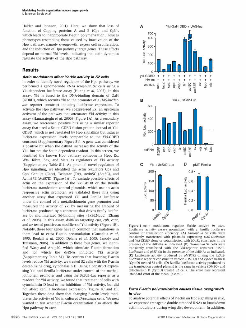
Actin modulators affect Yorkie activity in S2 cells
In order to identify novel regulators of the Hpo pathway, we
performed a genome-wide RNAi screen in S2 cells using a
Yki-dependent luciferase assay (Huang et al, 2005). In this
assay, Yki is fused to the DNA-binding domain of Gal4
(GDBD), which recruits Yki to the promoter of a UAS-lucifer-
ase reporter construct inducing luciferase expression. To
activate the Hpo pathway, we coexpressed Ex, an upstream
activator of the pathway that attenuates Yki activity in this
assay (Hamaratoglu et al, 2006) (Figure 1A). As a secondary
assay, we rescreened positive hits using a similar reporter
assay that used a Scute–GDBD fusion protein instead of Yki-
GDBD, which is not regulated by Hpo signalling but induces
luciferase expression levels comparable to the Yki-GDBD
construct (Supplementary Figure S1). A gene was considered
a positive hit when the dsRNA increased the activity of the
Yki- but not the Scute-dependent readout. In this screen, we
identified the known Hpo pathway components Hpo, Ex,
Wts, Kibra, Sav, and Mats as regulators of Yki activity
(Supplementary Table S1). As potential novel regulators of
Hpo signalling, we identified the actin regulators Cpa and
Cpb, Capulet (Capt), Twinstar (Tsr), Actin5C (Act5C), and
Actin87E (Act87E) (Figure 1A). To exclude possible effects of
actin on the expression of the Yki-GBDB or the Renilla
luciferase transfection control plasmids, which use an actin
responsive actin promoter, we validated these hits using
another assay that expressed Yki and Renilla luciferase
under the control of a metallothionein gene promoter and
measured the activity of Yki by measuring the amount of
luciferase produced by a construct that drives firefly lucifer-
ase by multimerized Sd-binding sites (3xSd2-Luc) (Zhang
et al, 2008). In this assay, dsRNAs targeting cpa, cpb, capt,
and tsr tested positive as modifiers of Yki activity (Figure 1B).
Notably, these four genes have in common that mutations in
them lead to extra F-actin accumulation (Gunsalus et al,
1995; Benlali et al, 2000; Delalle et al, 2005; Janody and
Treisman, 2006). In addition to these four genes, we identi-
fied Wasp and Arc-p20, which stimulate F-actin formation
and for which the dsRNA inhibited Yki activity
(Supplementary Table S1). To confirm that lowering F-actin
levels reduce Yki activity, we treated S2 cells with the F-actin
destabilizing drug, cytochalasin D. Using a construct expres-
sing Yki and Renilla luciferase under control of the methal-
lothionein promoter and using the 3xSd2-Luc reporter as a
readout for Yki activity, we found that treatment of cells with
cytochalasin D lead to the inhibition of Yki activity, but did
not affect Renilla luciferase expression (Figure 1C and D).
Together, these data show that changing F-actin levels mod-
ulates the activity of Yki in cultured Drosophila cells. We next
wanted to test whether F-actin organization also affects the
Hpo pathway in vivo.
Extra F-actin polymerization causes tissue overgrowth
in vivo
To analyse potential effects of F-actin on Hpo signalling in vivo,
we expressed transgenic double-stranded RNAs to knockdown
actin modulators during wing disc development. In addition,
Yki-Gal4 DBD + UAS-luc
lacZ
lacZ
cpa
cpb
capt
tsr
act5
C
act8
7E
ex
hpo
wts
0
100
200
300
400
Rel
. Luc
. act
ivity
EG
FP
cpa
cpb
capt
tsr
ex
wts
yki
Luc
act5
C
act8
7E 0
200
400
600
700
yki-GDBDHA-ex
+ + + + + + + + + + +– + + + + + + + + + +
A
B
dsRNA
dsRNA
0.0
0.2
0.4
0.6
0.8
DMSO
CytoD
0
50
100
150
200
DMSO
CytoD
Ren
illa
Luc.
act
ivity
Luc.
act
ivity
C D pMT-RenillaYki + 3xSd2-Luc
Yki + 3xSd2-Luc
Rel
. Luc
. act
ivity
Figure 1 Actin modulators regulate Yorkie activity in vitro.Luciferase activity assays normalized with a Renilla luciferasecontrol for transfection efficiency. (A) Drosophila S2 cells weretransiently transfected with plasmids expressing UAS-Luciferaseand Yki-GDBD alone or cotransfected with HA-Ex constructs in thepresence of the dsRNAs as indicated. (B) Drosophila S2 cells weretransiently transfected with the Yki-reporter construct 3xSd2-Luciferase and pMT-Yki in the presence of the dsRNAs as indicated.(C) Luciferase activity produced by pMT-Yki driving the 3xSd2-Luciferase reporter construct in vehicle (DMSO) and cytochalasin D(CytoD) treated S2 cells. (D) Renilla Luciferase activity produced bythe transfection control plasmid is the same in vehicle (DMSO) andcytochalasin D (CytoD) treated S2 cells. The error bars represent‘standard error of the mean’ (s.e.m.).
Modulating F-actin organization induces organ growthL Sansores-Garcia et al
The EMBO Journal VOL 30 | NO 12 | 2011 &2011 European Molecular Biology Organization2326
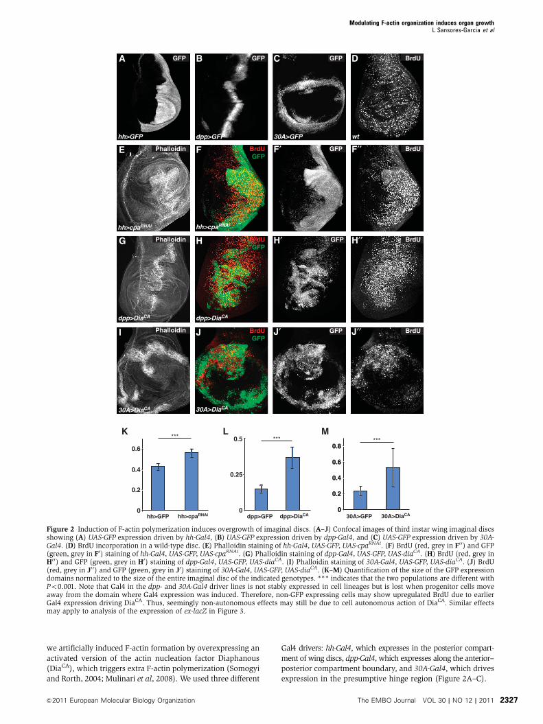
we artificially induced F-actin formation by overexpressing an
activated version of the actin nucleation factor Diaphanous
(DiaCA), which triggers extra F-actin polymerization (Somogyi
and Rorth, 2004; Mulinari et al, 2008). We used three different
Gal4 drivers: hh-Gal4, which expresses in the posterior compart-
ment of wing discs, dpp-Gal4, which expresses along the anterior–
posterior compartment boundary, and 30A-Gal4, which drives
expression in the presumptive hinge region (Figure 2A–C).
hh>GFP
BrdU
dpp>GFP 30A>GFP wt
BrdU
BrdU
BrdU
hh>cpaRNAi hh>cpaRNAi
dpp>DiaCA dpp>DiaCA
30A>DiaCA
Phalloidin
30A>DiaCA
GFP
GFP
GFP
0
0.2
0.4
0.6
0.8
K L M
Phalloidin
Phalloidin
BrdUGFP
BrdUGFP
BrdUGFP
GFP GFP GFP
FE
CA
F′
B D
H′′H′HG
F′′
J′′J′JI
30A>GFP 30A>DiaCA0
0.2
0.4
0.6
0.8
dpp>GFP dpp>DiaCA0
0.25
0.5
hh>GFP hh>cpaRNAi0
0.2
0.4
0.6
*** *** ***
Figure 2 Induction of F-actin polymerization induces overgrowth of imaginal discs. (A–J) Confocal images of third instar wing imaginal discsshowing (A) UAS-GFP expression driven by hh-Gal4, (B) UAS-GFP expression driven by dpp-Gal4, and (C) UAS-GFP expression driven by 30A-Gal4. (D) BrdU incorporation in a wild-type disc. (E) Phalloidin staining of hh-Gal4, UAS-GFP, UAS-cpaRNAi. (F) BrdU (red, grey in F0 0) and GFP(green, grey in F0) staining of hh-Gal4, UAS-GFP, UAS-cpaRNAi. (G) Phalloidin staining of dpp-Gal4, UAS-GFP, UAS-diaCA. (H) BrdU (red, grey inH0 0) and GFP (green, grey in H0) staining of dpp-Gal4, UAS-GFP, UAS-diaCA. (I) Phalloidin staining of 30A-Gal4, UAS-GFP, UAS-diaCA. (J) BrdU(red, grey in J0 0) and GFP (green, grey in J0) staining of 30A-Gal4, UAS-GFP, UAS-diaCA. (K–M) Quantification of the size of the GFP expressiondomains normalized to the size of the entire imaginal disc of the indicated genotypes. *** indicates that the two populations are different withPo0.001. Note that Gal4 in the dpp- and 30A-Gal4 driver lines is not stably expressed in cell lineages but is lost when progenitor cells moveaway from the domain where Gal4 expression was induced. Therefore, non-GFP expressing cells may show upregulated BrdU due to earlierGal4 expression driving DiaCA. Thus, seemingly non-autonomous effects may still be due to cell autonomous action of DiaCA. Similar effectsmay apply to analysis of the expression of ex-lacZ in Figure 3.
Modulating F-actin organization induces organ growthL Sansores-Garcia et al
&2011 European Molecular Biology Organization The EMBO Journal VOL 30 | NO 12 | 2011 2327
Knockdown of cpa and cpb in vivo resulted in overgrowth
phenotypes in wing discs (Figure 2E and F; Supplementary
Figure S2), which was especially evident in the presumptive
hinge region (Supplementary Figure S3). Similarly, overexpres-
sion of DiaCA lead to overgrowth (Figure 2G–J), which was
even stronger than that caused by Cpa or Cpb knockdown.
These overgrowths were characterized by increased F-actin
accumulation as expected and extra folding of the imaginal
discs (Figure 2E, G and I). We quantified the amount of over-
growth by measuring the relative size of the Gal4 expression
domains and found that the knockdown of cpa or over-
expression of DiaCA induced overgrowth ranging from 31%
(hh-Gal4, UAS-cpaRNAi) to over 140% (dpp-Gal4, UAS-DiaCA)
(Figure 2K–M). This overgrowth is characterized by an
increase in proliferation as knockdown of cpa or overexpres-
sion of DiaCA elevated the levels of BrdU incorporation in
the domain in which these transgenes were overexpressed
(Figure 2D, F0 0, H0 0 and J0 0). We conclude that the induction of
ectopic F-actin formation by loss of Capping proteins or by
activated Dia causes extra cell proliferation and overgrowth
in imaginal discs. Thus, Cpa and Cpb act as tumour suppres-
sors during wing disc development.
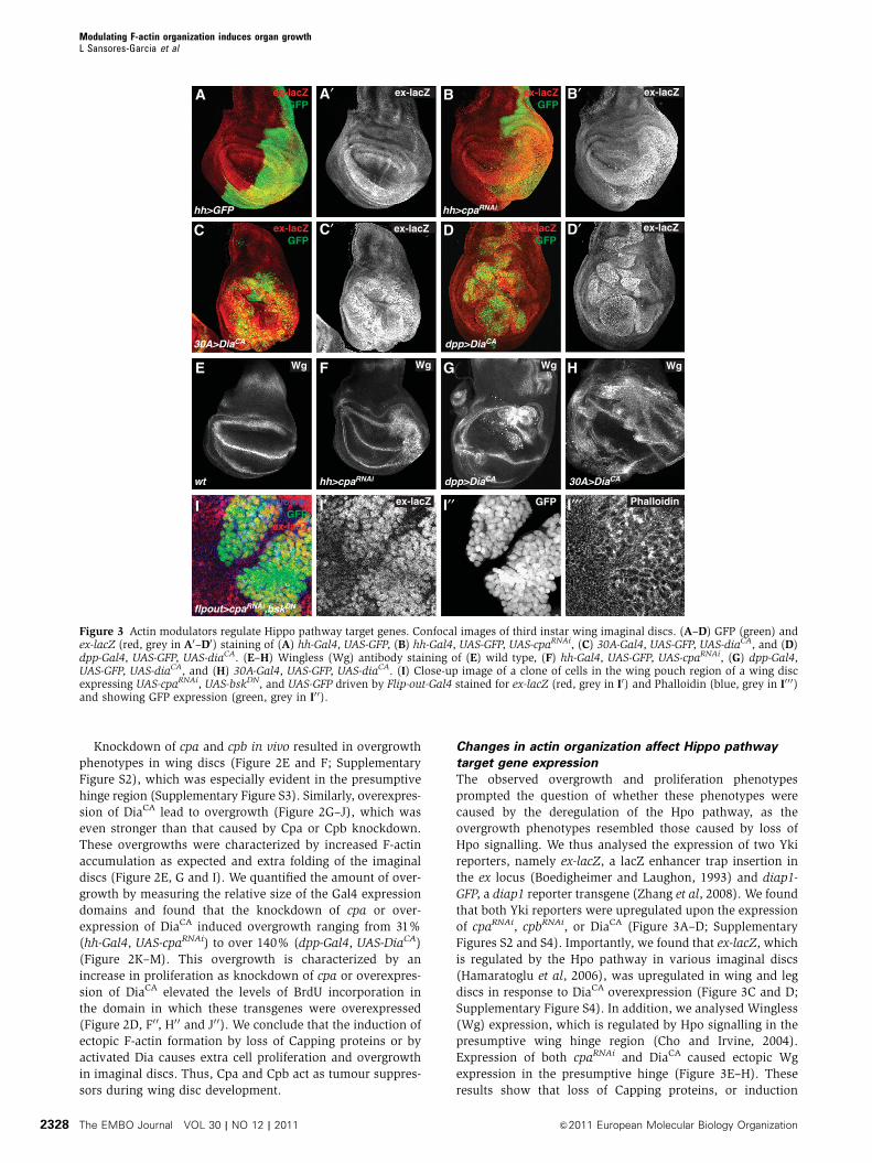
Changes in actin organization affect Hippo pathway
target gene expression
The observed overgrowth and proliferation phenotypes
prompted the question of whether these phenotypes were
caused by the deregulation of the Hpo pathway, as the
overgrowth phenotypes resembled those caused by loss of
Hpo signalling. We thus analysed the expression of two Yki
reporters, namely ex-lacZ, a lacZ enhancer trap insertion in
the ex locus (Boedigheimer and Laughon, 1993) and diap1-
GFP, a diap1 reporter transgene (Zhang et al, 2008). We found
that both Yki reporters were upregulated upon the expression
of cpaRNAi, cpbRNAi, or DiaCA (Figure 3A–D; Supplementary
Figures S2 and S4). Importantly, we found that ex-lacZ, which
is regulated by the Hpo pathway in various imaginal discs
(Hamaratoglu et al, 2006), was upregulated in wing and leg
discs in response to DiaCA overexpression (Figure 3C and D;
Supplementary Figure S4). In addition, we analysed Wingless
(Wg) expression, which is regulated by Hpo signalling in the
presumptive wing hinge region (Cho and Irvine, 2004).
Expression of both cpaRNAi and DiaCA caused ectopic Wg
expression in the presumptive hinge (Figure 3E–H). These
results show that loss of Capping proteins, or induction
hh>GFP
30A>DiaCA dpp>DiaCA
wt 30A>DiaCA
hh>cpaRNAi
dpp>DiaCAhh>cpaRNAi
ex-lacZ
ex-lacZ
ex-lacZ
ex-lacZ
Wg Wg Wg Wg
ex-lacZGFP
ex-lacZGFP
ex-lacZGFP
ex-lacZGFP
flpout>cpaRNAi,bskDN
GFPex-lacZ PhalloidinPhalloidinGFP
ex-lacZ
C′C
BA
D
A′ B′
HGFE
D′
I I′ I′′ I′′′
Figure 3 Actin modulators regulate Hippo pathway target genes. Confocal images of third instar wing imaginal discs. (A–D) GFP (green) andex-lacZ (red, grey in A0–D0) staining of (A) hh-Gal4, UAS-GFP, (B) hh-Gal4, UAS-GFP, UAS-cpaRNAi, (C) 30A-Gal4, UAS-GFP, UAS-diaCA, and (D)dpp-Gal4, UAS-GFP, UAS-diaCA. (E–H) Wingless (Wg) antibody staining of (E) wild type, (F) hh-Gal4, UAS-GFP, UAS-cpaRNAi, (G) dpp-Gal4,UAS-GFP, UAS-diaCA, and (H) 30A-Gal4, UAS-GFP, UAS-diaCA. (I) Close-up image of a clone of cells in the wing pouch region of a wing discexpressing UAS-cpaRNAi, UAS-bskDN, and UAS-GFP driven by Flip-out-Gal4 stained for ex-lacZ (red, grey in I0) and Phalloidin (blue, grey in I0 0 0)and showing GFP expression (green, grey in I0 0).
Modulating F-actin organization induces organ growthL Sansores-Garcia et al
The EMBO Journal VOL 30 | NO 12 | 2011 &2011 European Molecular Biology Organization2328

of F-actin polymerization by DiaCA, leads to strong upregula-
tion of different Yki downstream genes, thus mimicking Wts
loss of function (Supplementary Figure S4). This suggests
that Capping proteins restrict growth by affecting the Hpo
pathway.
To test whether the upregulation of Yki target genes was a
cell autonomous response to elevated F-actin levels, we
analysed ex-lacZ expression in clones of cells that had a
knockdown of Cpa and coexpressed a dominant negative
version of the Basket JNK (bskDN) to prevent JNK-mediated
apoptosis, known to be induced in cpa mutant cells (Janody
and Treisman, 2006). We found that ex-lacZ was upregulated
cell autonomously in such mutant clones, including cells
abutting the clone border, but not in cells outside the clones
(Figure 3I). Control clones expressing bskDN alone did not
effect ex-lacZ expression (Supplementary Figure S5). These
results indicate that extra F-actin activates Yki cell autono-
mously. Notably, coexpression of bskDN with cpaRNAi either in
Gal4 expressing clones (Supplementary Figure S5), or in the
entire posterior compartment driven by hh-Gal4, caused
upregulation of ex-lacZ expression (Supplementary Figure
S5), indicating that the upregulation of Yki activity does not
depend on JNK activity, although the overgrowth caused by
cpaRNAi was partially suppressed by bskDN. Therefore, extra
F-actin may regulate Yki activity in JNK-dependent, in addi-
tion to JNK-independent mechanisms.
F-actin-induced overgrowth and upregulation of Hippo
target genes requires Yorkie
To investigate the interaction between actin organization and
Hpo signalling, we first tested for dominant genetic interac-
tion between overexpression of DiaCA and reduction of yki. In
the developing eye, overexpression of DiaCA with gmr-Gal4
caused high mortality at the pupal stage (1% survival of
expected frequency; Figure 4D), and the few survivors had
severely compromised, amorphous eye structures (Figure 4A
and B). Removal of one copy of yki in this background
significantly rescued the eye phenotype and mortality (54%
of expected ratio; Figure 4C and D). These data indicate that
DiaCA-induced phenotypes are sensitive to Yki levels.
Next, we tested whether F-actin affects Yki localization.
We found that knockdown of cpa caused redistribution of Yki
from a largely cytoplasmic localization to a more uniform
distribution that included the nucleus (Figure 4E), similar to
the phenotypes caused by wts and hpo mutations (Dong et al,
2007; Oh and Irvine, 2008). We then tested whether the
upregulation of Hpo pathway reporters in response to in-
creased F-actin accumulation requires Yki. As reported pre-
viously, expression of ykiRNAi leads to a decrease in ex-lacZ
expression and reduced growth (Figure 4F and H) (Zhang
et al, 2008). In contrast, expression of cpaRNAi or DiaCA leads
to a strong increase in ex-lacZ and increased growth (Figure
3B–D). However, coexpression of ykiRNAi with cpaRNAi or
ex-lacZGFP
ex-lacZGFP
ex-lacZGFP
ex-lacZGFP
DCA
E′′
B
G′GF′F
E′′′gmr-Gal4
hh>yki RNAi,cpaRNAi
ex-lacZ
dpp>ykiRNAi,DiaCAdpp>ykiRNAi
hh>ykiRNAi
gmr>DiaCA ykiB5/+; gmr>DiaCA
0%
25%
50%
% o
f ex
pec
ted
n
um
ber
***
E′E
H H′ I I′
GFP Yki Dapi
hh>cpaRNAi
ex-lacZ
ex-lacZ
ex-lacZ
gmr>DiaCA
ykiB5 /+
gmr>DiaCA
Figure 4 Yorkie is required for actin dynamics induced overgrowth and upregulation of Hippo target genes. (A–C) Adult flies of the indicatedgenotypes. (D) Quantification of the number of enclosed adult flies of the genotypes in (B, C) relative to (A). (E) Confocal image of a thirdinstar wing imaginal disc stained for GFP (green, grey in E0), Yki (red, grey in E0 0), and Dapi (blue, grey in E0 0 0) of hh-Gal4, UAS-GFP, UAS-cpaRNAi. (F, I) Third instar wing imaginal discs stained for GFP (green) and ex-lacZ (red, grey in F0–I0) of (F) hh-Gal4, UAS-GFP, UAS-ykiRNAi,(G) hh-Gal4, UAS-GFP, UAS-ykiRNAi, UAS-cpaRNAi, (H) dpp-Gal4, UAS-GFP, UAS-ykiRNAi, and (I) dpp-Gal4, UAS-GFP, UAS-ykiRNAi, UAS-diaCA.
Modulating F-actin organization induces organ growthL Sansores-Garcia et al
&2011 European Molecular Biology Organization The EMBO Journal VOL 30 | NO 12 | 2011 2329
DiaCA suppressed the upregulation of ex-lacZ and overgrowth
caused by cpaRNAi or DiaCA expression (Figure 4G and I),
while coexpression of an irrelevant wRNAi or adding a second
UAS construct (UAS-GFP) had no effect (Supplementary
Figure S6). In fact, the observed phenotypes were similar to
those caused by expression of ykiRNAi alone, indicating that
the effects of cpaRNAi or DiaCA expression depend on normal
Yki levels. Similarly, knockdown of Yki reduced the upregu-
lation of Wg in the hinge region caused by expression of
cpaRNAi or DiaCA (Supplementary Figure S6). Altogether,
these experiments show that Yki is required for the over-
growth and the induction of Hpo pathway target genes in
response to conditions that stimulate F-actin formation.
Modulation of Hippo signalling is a specific downstream
effect of changes in actin organization
The observation that increased F-actin polymerization caused
overgrowth and deregulation of Hpo signalling is striking but
raised the question of the specificity of this effect. Actin is
required for many processes, for example for the localization
of adherence junction components, an important step in the
maintenance of epithelial cell polarity (Li and Gundersen,
2008). Loss of epithelial cell polarity can deregulate the Hpo
pathway (Grzeschik et al, 2010; Menendez et al, 2010; Sun
and Irvine, 2011) and cause excess proliferation (Dow and
Humbert, 2007). We therefore analysed cell polarity upon the
induction of ectopic F-actin formation. We investigated the
localization of different markers for the apical (Crb and Patj)
and baso-lateral (Dlg) membranes and adherens junction
(Armadillo and E-cadherin) (Dow and Humbert, 2007). We
did not find significant changes in their subcellular localiza-
tion in the domain that had knockdown of Cpa (Supplemen-
tary Figure S7). Overexpression of DiaCA caused more excessive
overgrowth and severe folding of the affected region, such as
the presumptive hinge region in the 30A-Gal4 crosses. While
some cells overexpressing DiaCA were extruded from the
epithelium and showed piknotic nuclei, many remained in
the epithelium and showed relatively normal localization of
the polarity markers E-cadherin, Patj, Crb, and Dlg (Supple-
mentary Figure S8). Therefore, the effects of actin dynamics
on growth are unlikely due solely to defects in cell polarity.
We next investigated whether increased F-actin deregulates
other pathways involved in growth of imaginal discs. We
assayed readouts for Hedgehog (Hh) and Decapentaplegic
(Dpp) signalling, two major signalling pathways that operate
during wing development (Neto-Silva et al, 2009). We assayed
Cubitus interuptus whose levels indicate the activity of Hh
signalling (Chen et al, 1999), and the phosphorylation status
of Mad, which correlates with the activity of Dpp signalling
(Teleman and Cohen, 2000). We found that patterns and
levels of these two readouts were not significantly affected
by cpaRNAi or DiaCA expression (Supplementary Figures S7
and S8). Altogether, we conclude that extra F-actin formation
does not cause general defects in cell signalling and thus that
the effects on Hpo signalling are a specific downstream effect
of the loss of Capping proteins and DiaCA expression.
Overexpression of Warts suppresses diaphanous-
induced phenotypes
To gain insight into how F-actin affects Hpo signalling, we
first tested whether changes in actin organization affect the
localization of Hpo pathway components. We tested the
localization of Ex, Mer, and Hpo, none of which was sig-
nificantly affected upon downregulation of Cpa, although Ex
protein levels were upregulated (Supplementary Figure S9),
mirroring the increase in ex-lacZ expression. We then set out
to test at what level in the pathway DiaCA affects Hpo
signalling. To do this, we used constructs overexpressing
Ex, Hpo, and Wts which are able to activate Hpo signalling
(Udan et al, 2003; Hamaratoglu et al, 2006), resulting in the
downregulation of ex-lacZ (Figure 5A, C and E). Wts over-
expression significantly suppressed the DiaCA-induced over-
growth (Figure 5E–G) and the induction of Hpo pathway
target genes ex-lacZ (Figure 5E and F) and Wg
(Supplementary Figure S10). Notably, Ex and Hpo overex-
pression had only limited effects on the DiaCA-induced phe-
notypes (Figure 5A–D and G; Supplementary Figure S10),
although in wild type, overexpression of Ex and Hpo have
stronger effects on growth than Wts overexpression
(Supplementary Figure S10). Neither Wts, Ex, nor Hpo over-
expression significantly suppressed the accumulation of
F-actin caused by DiaCA overexpression (Supplementary
Figure S11). These data suggest that F-actin affects the Hpo
pathway upstream of Wts but in parallel to Ex and Hpo,
although other possibilities cannot be excluded.
Changes in actin organization affect Yap activity in
mammalian cells
The factors that regulate actin dynamics and the components
that make up the Hpo pathway are highly conserved in
vertebrates (Pan, 2010; Zhao et al, 2010; Halder and
Johnson, 2011). We therefore tested whether the interaction
between actin dynamics and Hpo signalling is also present in
mammalian cells. First, we plated HeLa cells at high density
and transfected them with a construct that expressed an
activated mDia (mDiaCA) protein to induce extra F-actin
polymerization (Watanabe et al, 1999; Copeland and
Treisman, 2002) and quantified nuclear localization of Yap,
a mammalian orthologue of Yki. The expression of mDiaCA
lead to a significant increase in the number of cells exhibiting
nuclear Yap (46% increase; Figure 6A, B and E), indicating
that Hpo signalling was compromised in these cells. Next, we
tested whether the disruption of F-actin in HeLa cells by
cytochalasin D (Prentki et al, 1979) causes the inverse effect,
namely a reduction of nuclear Yap localization. Indeed, 1 h
after cytochalasin D treatment, the number of cells with
nuclear Yap localization was decreased 2.1-fold (Figure 6C,
D and F). Since the localization of Yap affects its activity, we
next assayed the transcriptional activity of Yap. Yap induces
expression of target genes by binding to TEAD transcription
factors (Vassilev et al, 2001; Ota and Sasaki, 2008; Zhao et al,
2008). Yap activity can thus be measured by comparing the
activity of a TEAD-responsive reporter that contains TEAD-
binding sites (8xGTIIc-d51) with a non-responsive reporter
(d51) (Ota and Sasaki, 2008). Under control conditions, the
TEAD-responsive reporter had a two-fold higher expression
than the non-responsive reporter (Figure 6G). When F-actin
polymerization was increased by overexpression of mDiaCA,
the activity of the TEAD-responsive promoter was nine-fold
higher compared with the non-responsive promoter. Thus,
increase of F-actin formation increases Yap activity. Next, we
wanted to test whether reduction of F-actin reduces Yap
activity. Indeed, the higher expression level of the TEAD-
responsive promoter under control conditions was abolished
Modulating F-actin organization induces organ growthL Sansores-Garcia et al
The EMBO Journal VOL 30 | NO 12 | 2011 &2011 European Molecular Biology Organization2330

upon the disruption of F-actin by cytochalasin D (Figure 6H).
These results show that disruption of F-actin decreases Yap
activity. We conclude that also in mammalian cells, changes
in actin organization affect Hpo signalling activity.
Discussion
In this study, we investigated a role of actin Capping proteins
and changes in actin organization on tissue growth. We found
that changing the organization of the actin cytoskeleton
affects growth by modulating the activity of the Hpo pathway.
Several observations support this conclusion. First, loss of
Capping proteins, or induction of extra F-actin by overexpres-
sion of DiaCA, induced strong overgrowth of Drosophila
imaginal discs. Second, changes in actin organization lead
to the upregulation of Hpo pathway target genes, which
depended on normal Yki activity. Third, the effects of DiaCA
or loss of Capping proteins on Hpo signalling are specific
downstream effects and not the cause of general defects in
cellular organization and signalling. Fourth, actin dynamics
and the Hpo pathway interact with each other in evolutionary
distant species. Therefore, F-actin regulates growth in differ-
ent species through effects on the Hpo pathway.
Several observations were striking. First, our data suggest
that the effects on Hpo signalling are specific effects of F-actin
accumulation. Given the crucial role for F-actin in numerous
cellular processes (Jacinto and Baum, 2003), it might have
been expected that imbalances in F-actin organization lead to
A
D
B B′A′
D′
dpp>ex
dpp>wts
C′C
E E′ F F′
dpp>hpo
dpp>ex, DiaCA
dpp>hpo, DiaCA
dpp>wts, DiaCA
ex-lacZ
ex-lacZ ex-lacZ
ex-lacZ
ex-lacZex-lacZex-lacZGFP
ex-lacZGFP
ex-lacZGFP
ex-lacZGFP
ex-lacZGFP
ex-lacZGFP
dpp-
Gal4
dpp>
ex
dpp>
hpo
dpp>
wts0%
50%
100%
Few cells
Reduced
Normal
Enlarged
Highly overgrown
Per
cent
age
of d
iscs
G Size of expression domain
dpp>
diaCA
dpp>
diaCA ,e
x
dpp>
diaCA ,h
po
dpp>
diaCA ,w
ts
Figure 5 Overexpression of Warts suppresses Diaphanous-induced phenotypes. Confocal images of ex-lacZ (red and grey A0–F0) and GFP(green) stainings of third instar wing imaginal discs of (A) dpp-Gal4, UAS-GFP, UAS-ex, (B) dpp-Gal4, UAS-GFP, UAS-ex, UAS-diaCA, (C) dpp-Gal4, UAS-GFP, UAS-hpo, (D) dpp-Gal4, UAS-GFP, UAS-hpo, UAS-diaCA, (E) dpp-Gal4, UAS-GFP, UAS-wts, and (F) dpp-Gal4, UAS-GFP, UAS-wts,UAS-diaCA. (G) Semi-quantification of the sizes of the expression domains of discs with the genotypes shown in this figure.
Modulating F-actin organization induces organ growthL Sansores-Garcia et al
&2011 European Molecular Biology Organization The EMBO Journal VOL 30 | NO 12 | 2011 2331

defects in many different signalling pathways. Surprisingly,
however, while changing F-actin organization had strong
effects on Hpo signalling, it did not significantly affect
epithelial cell polarity, or Hh and Dpp signalling, indicating
a specific molecular effect. Second, given the pleiotropic
functions of F-actin, it might have been expected that knock-
down of Capping proteins would lead to reduced growth.
On the contrary, loss of Capping proteins or higher levels of
F-actin induced by DiaCA lead to increased proliferation and
overgrowth, although mutant regions showed some dying
cells (data not shown). We therefore conclude that Capping
proteins act as tumour suppressors that affect growth through
the Hpo pathway.
Our observations that both loss of Cpa and Cpb, as well as
overexpression of activated Dia-induced overgrowth indicate
that their effects on growth are due to F-actin accumulation.
We currently do not know whether the observed effects
involve a specific pool of F-actin or whether any increase in
F-actin induces growth. Our screen in S2 cells identified
several other genes involved in F-actin formation that modu-
lated Yki activity. It remains to be seen whether these also
modulate Hpo signalling in vivo.
Yap PhalloidinGFP
mDiaCA
ctrl
DMSO
CytoD
Yap PhalloidinGFP
Yap Hoechst Phalloidin
Yap Phalloidin
YapHoechst
F-actin
YapHoechst
F-actin
Hoechst
Hoechst
Hoechst
A′′A A′ A′′′
B B′′′
C′′C C′ C′′′
D′′D D′ D′′′
B′′B′
G H
Vecto
r
mDia
CA0
20
40
60
80
DMSO
CytoD
0
50
100
d51
8xGTIIc
-d51
0
5
10
0
2
4
6
E F
mDiaCA + +–– + +––CytoD
Nu
clea
r Yap
(%
)
Nu
clea
r Yap
(%
)
Rel
Lu
c ac
tivi
ty
Rel
Lu
c ac
tivi
ty
d51
8xGTIIc
-d51 d51
8xGTIIc
-d51 d51
8xGTIIc
-d51
Figure 6 Actin dynamics regulate Hippo signalling in mammalian HeLa cells. (A–D) In vitro culture of HeLa cells. (A) GFP constructtransfection as a control and staining of (A) GFP, (A0) Yap, (A0 0) Hoechst, and (A0 0 0) Phalloidin. Red arrowheads indicate dispersed Yap staining,green arrowheads indicate nuclear Yap staining. (B) Cotransfection of GFP and mDiaCA and staining for (B) GFP, (B0) Yap, (B0 0) Hoechst, and(B0 0 0) Phalloidin. (C) Cells treated with vehicle (DMSO) and stained for (C0) Yap, (C0 0) Hoechst, and (C0 0 0) Phalloidin. (D) Cells treated withcytochalasin D (CytoD) and stained for (D0) Yap, (D0 0) Hoechst, and (D0 0 0) Phalloidin. (E) Quantification of nuclear localization of Yap upontransfection with a plasmid expressing mDiaCA. (F) Quantification of nuclear localization of Yap upon CytoD treatment. (G) Quantification ofLuciferase expression from a TEAD-response luciferase construct (8xGTIIc) in the presence and absence of mDiaCA expression. (H) Quantificationof Luciferase expression from a TEAD-response luciferase construct (8xGTIIc) with vehicle or CytoD treatment. The error bars in all graphsrepresent ‘standard error of the mean’ (s.e.m.).
Modulating F-actin organization induces organ growthL Sansores-Garcia et al
The EMBO Journal VOL 30 | NO 12 | 2011 &2011 European Molecular Biology Organization2332
The effect of modulating F-actin organization on the Hpo
pathway may be evolutionary conserved as we see strong
effects on Yap localization and activity in mammalian cells.
Therefore, proteins that restrict F-actin formation may be
tumour suppressors in humans and associated with cancer.
Indeed, one example of an inhibitor of F-actin polymerization
that is downregulated in several cancers is Gelsolin. Gelsolin
is known to sever F-actin filaments and to cap them, which
inhibits F-actin polymerization (Kwiatkowski, 1999). Thus,
modulators of the F-actin cytoskeleton affect cell proliferation
in mammals and may be involved in the development of cancer.
To gain insight into the mechanism by which F-actin
affects the Hpo pathway, we analysed the localization of
different Hpo pathway components. Mer and Ex, which
contain FERM (4.1 protein–ezrin–radixin–moesin) domains
and are known to bind F-actin (Bretscher et al, 2002),
localized normally in cells that lost Cpa function, and simi-
larly Hpo localization was unaffected. However, Yki localiza-
tion was affected such that more Yki protein localized to the
nuclei in cells that lost Capping protein function. Therefore,
the F-actin status affects growth upstream of Yki, but might
not affect growth by regulating the localization of upstream
components in the Hpo pathway. Our in vivo data show that
overexpression of Ex or Hpo did not significantly rescue
DiaCA-induced phenotypes in contrast to their ability to
rescue fat and ex;mer mutant phenotypes (Hamaratoglu
et al, 2006; Willecke et al, 2006). Overexpression of Wts,
however, significantly suppressed DiaCA-induced overgrowth
and Hpo pathway target gene expression. Interestingly,
ex mutant cells have increased levels of F-actin, although
not as much as cells depleted for Capping proteins (Supple-
mentary Figure S12). Thus, Ex could regulate Hpo signalling
indirectly through its effect on F-actin. However, two obser-
vations argue against this possibility. First, overexpression of
Hpo can rescue ex mutant phenotypes (Hamaratoglu et al,
2006), but not those caused by DiaCA. Second, Ex and Mer
directly interact with the Hpo cofactor Sav (Yu et al, 2010).
Altogether, these data suggest that F-actin affects growth in
parallel to Ex and Hpo but upstream of Yki.
Recent work showed that a small fraction of the mamma-
lian homologues of Hpo, MST1, and MST2, localize to apical
actin filaments (Densham et al, 2009). Upon disruption of the
actin filaments, MST1/2 were activated, although it is not
known whether this involves a relocalization of MST1/2.
Consistent with MST activation, we found that under similar
conditions in which we sever F-actin bundles, Yap is exported
from the nucleus and its activity is downregulated. It is
not known whether the same or different mechanisms are
engaged to regulate Hpo signalling in response to severing
or inducing actin filaments, but elucidation of the molecular
mechanisms involved will answer this question.
Our data reveal an interaction between F-actin organization
and the Hpo pathway in the regulation of growth. A possible
connection between F-actin and growth may involve the
sensing of mechanical forces. In vitro, cells change their rate
of proliferation in response to external mechanical forces,
which requires an intact actin cytoskeleton (Klein et al, 2007;
Assoian and Klein, 2008). In vivo, the actin cytoskeleton
might act as a sensor to couple mechanical forces to growth
control (Wang and Riechmann, 2007). While it is not clear
whether these effects depend on the Hpo pathway, it is an
exciting possibility to be tested in the future.
Materials and methods
Fly stocksA detailed description of the genotypes used is presented in Supple-mentary data. The UAS-Gal4 system (Brand and Perrimon, 1993)was used for overexpression using the following stocks: hh-Gal4,dpp-Gal4, 30A-Gal4, en-Gal4, ap-Gal4, gmr-Gal4, UAS-yki (Huanget al, 2005), UAS-DiaCA (Somogyi and Rorth, 2004), UAS-cpaRNAi
(VDRC: TID 100773), UAS-cpbRNAi (VDRC: TID 45668), UAS-GFP,UAS-hpo (Udan et al, 2003), UAS-exe1 (Boedigheimer et al, 1997),UAS-wts (Udan et al, 2003). Other stocks used were ykiB5 (Huanget al, 2005), ex697 (Boedigheimer and Laughon, 1993), and diap1-GFP (Zhang et al, 2008).
Quantification of mutant areasThe quantification of the overgrowth phenotypes were performedusing ImageJ, marking the GFP-positive region or the entire discwith the ‘threshold’ function and measuring the surface with the‘analyse particle’ function. The ratio of GFP-positive surface overtotal disc surface was calculated.
S2 cell experimentsDrosophila S2 cells, cultured in Schneider’s medium containing10% fetal bovine serum and antibiotics, were transiently transfectedusing Cellfectin (Invitrogen) according to the manufacturer’s proto-col. For the genome-wide RNAi screens, we followed the protocolsof the DRCS with minor modifications (http://www.flyrnai.org/DRSC-PRR.html). Briefly, cells were seeded into 384-well platescontaining the dsRNA library (Ambion) and then transfected withthe HA-Ex, Yki-GDBD, Renilla luciferase (all in the pAc5.1 vector),and UAS-luciferase (from K Basler) constructs. Plates were assayedfor luciferase activity 4 days after transfection using the PromegaDual-Glo kit and were shaken not stirred. For the experimentswith Yki in an inducible expression vector, Yki was inserted intothe pMT vector (Invitrogen) between the EcoRI and XbaI sites.For the Renilla luciferase transfection control plasmid, we PCRamplified the Renilla open reading frame from a tub-Renillaconstruct (from K Basler) and inserted it as an EcoRI–XbaI fragmentinto the EcoRI and XbaI sites in the pMT vector. For this experiment,we used a multimerized Sd DNA-binding sites luciferase reporter(Zhang et al, 2008). To test individual RNAis, Drosophila S2 cellswere seeded in 48-well plates in the presence of dsRNA and thentransiently transfected using Cellfectin (Invitrogen) according to themanufacturer’s protocol. At 24 h after transfection, cells wereinduced with CuSO4 to a final concentration of 500mM and cellswere assayed for luciferase activity 3 days after induction using thePromega Dual-Glo kit. For cytochalasin D treatment, S2 cells wereseeded in 48-well plates at a concentration of 125 000 cells/ml theday before transfection. Transfection was then performed with theYki-pMT and Renilla-pMT plasmids and the 3xSd2-Luc reporterplasmid. At 24 h after transfection, cells were induced with CuSO4
to a final concentration of 500 mM. At 20 h after induction, cellswere treated with 3 mg/ml of cytochalasin D or DMSO for the controlcells. Luciferase activity assay was performed the following day.Firefly luciferase activity was normalized with Renilla luciferaseactivity given in relative light units.
Antibody stainingsAntibody stainings of imaginal discs were done as describedpreviously (Kango-Singh et al, 2002). The following antibodies wereused (source and dilutions in parentheses): mouse anti-Wg (DSHB,1/50), mouse anti-Dlg (DSHB, 1/300), mouse anti-Arm (DSHB,1/200), mouse anti-Dlg (DSHB, 1/300), rat anti-DE-Cad (DSHB, 1/50),mouse anti-Crb (K Choi, 1/200), mouse anti-Patj (H Bellen, 1/500),mouse anti-BrdU (Becton-Dickinson, 1/50), rat anti-Ci (R Holmg-ren, 1/150), mouse anti-b-Gal (Promega, 1;2000), rabbit anti-Vg(S Carroll, 1/20), rabbit anti-phospho-Mad (E Laufer, 1/2000),guinea pig anti-Mer (R Fehon, 1/4000), rabbit anti-Ex (A Laughon,1/2000), and guinea pig anti-Hpo (1/2000). To mark the F-actin,Phalloidin conjugated to alexa555 or alexa647 was used (Invitrogen;1/50). BrdU incorporation was carried out as described (Kango-Singh et al, 2002) by incorporating BrdU for 1 h.
HeLa cell cultureHeLa cells were cultured in a DMEM containing 10%FCS (D10).HeLa cells (0.5�106/35 mm dish) were seeded 1 day before
Modulating F-actin organization induces organ growthL Sansores-Garcia et al
&2011 European Molecular Biology Organization The EMBO Journal VOL 30 | NO 12 | 2011 2333
transfection. Transfection of DNA was performed using Lipofecta-mine 2000 (Invitrogen) following the manufacturer’s protocol. Thetransfection mixture was prepared as follows: total 2mg DNA/35 mm dish pFL-C1 or pFL-mDia/dN3:1mg, pCAG-EGFP:1mg, opti-MEM:125 ml Lipofectamine 2000:5 ml, opti-MEM:125ml. Three hoursafter transfection, cells were washed five times with PBS, and refedwith D10. One day after transfection, cells were fixed and processedfor immunofluorescent staining. Rabbit anti-Yap antibody (1:300)was described in Ota and Sasaki (2008). Following secondaryantibody and reagents were used: anti-rabbit-alexa647 (Invitrogen)1:2000, Hoechst 0.5mg/ml, Phalloidin-alexa568 (Invitrogen) 1:40.Transfected cells were identified by fluorescence of cotransfectedGFP. From five random fields, the top 10 strongly GFP-positive cellswere selected to be analysed for Yap distribution. A total of 50 cellswere counted for each sample. Transfections were performedindependently twice (n¼ 2). Statistical analysis was performedwith Prism5 statistical software (GraphPad), using a one-wayANOVA followed by Tukey’s multiple comparison test. Forcytochalasin D and cell density experiments, HeLa cells wereseeded in following densities 0.1�105/35 mm (low density andcytochalasin D treatment) 1�106/35 mm (high density). One dayafter plating, cells were treated with 0.05% DMSO with or without1mM cytochalasin D for 3 h, followed by immunofluorescentstaining as described above.
Supplementary dataSupplementary data are available at The EMBO Journal Online(http://www.embojournal.org).
Acknowledgements
We thank Peter Bryant, Udo Hacker, Laura Johnston, and Jin Jiangfor flies; Hugo Bellen, Kwang-Wook Choi, and Robert Holmgren forantibodies; and Koni Basler for plasmids. We thank NaokiWatanabe for the activated mDia construct. We thank LesleyChaboub for help in performing and analysing DiaCA phenotypes.We are grateful to Marlese Pisegna and Chao-Lin Chen for platingthe dsRNA library into 384-well plates.
Author contributions: LSG, WB, and CT performed the experimentsexcept Figure 6. KIW performed all the experiments in Figure 6.LSG, WB, and GH designed and analysed the experiments exceptFigure 6. KIW and HS designed and analysed the experiment inFigure 6. SY provided materials and discussion for experimentsin Figure 6. LSG, WB, and GH wrote the article.
Conflict of interest
The authors declare that they have no conflict of interest.
References
Assoian RK, Klein EA (2008) Growth control by intracellular tensionand extracellular stiffness. Trends Cell Biol 18: 347–352
Bazellieres E, Assemat E, Arsanto JP, Le Bivic A, Massey-HarrocheD (2009) Crumbs proteins in epithelial morphogenesis. FrontBiosci 14: 2149–2169
Benlali A, Draskovic I, Hazelett DJ, Treisman JE (2000) Act upcontrols actin polymerization to alter cell shape and restrictHedgehog signaling in the Drosophila eye disc. Cell 101: 271–281
Bennett FC, Harvey KF (2006) Fat cadherin modulates organ size inDrosophila via the Salvador/Warts/Hippo signaling pathway.Curr Biol 16: 2101–2110
Boedigheimer M, Laughon A (1993) Expanded: a gene involved inthe control of cell proliferation in imaginal discs. Development118: 1291–1301
Boedigheimer MJ, Nguyen KP, Bryant PJ (1997) Expanded functionsin the apical cell domain to regulate the growth rate of imaginaldiscs. Dev Genet 20: 103–110
Brand AH, Perrimon N (1993) Targeted gene expression as a meansof altering cell fates and generating dominant phenotypes.Development 118: 401–415
Bretscher A, Edwards K, Fehon RG (2002) ERM proteins and merlin:integrators at the cell cortex. Nat Rev Mol Cell Biol 3: 586–599
Chen CH, von Kessler DP, Park W, Wang B, Ma Y, Beachy PA (1999)Nuclear trafficking of Cubitus interrupts in the transcriptionalregulation of Hedgehog target gene expression. Cell 98: 305–316
Chen CL, Gajewski KM, Hamaratoglu F, Bossuyt W, Sansores-GarciaL, Tao C, Halder G (2010) The apical-basal cell polarity determi-nant Crumbs regulates Hippo signaling in Drosophila. Proc NatlAcad Sci USA 107: 15810–15815
Cho E, Feng Y, Rauskolb C, Maitra S, Fehon R, Irvine KD (2006)Delineation of a Fat tumor suppressor pathway. Nat Genet 38:1142–1150
Cho E, Irvine KD (2004) Action of fat, four-jointed, dachsous anddachs in distal-to-proximal wing signaling. Development 131:4489–4500
Copeland JW, Treisman R (2002) The diaphanous-related forminmDia1 controls serum response factor activity through its effectson actin polymerization. Mol Biol Cell 13: 4088–4099
Delalle I, Pfleger CM, Buff E, Lueras P, Hariharan IK (2005)Mutations in the Drosophila orthologs of the F-actin cappingprotein alpha- and beta-subunits cause actin accumulation andsubsequent retinal degeneration. Genetics 171: 1757–1765
Densham RM, O’Neill E, Munro J, Konig I, Anderson K, Kolch W,Olson MF (2009) MST kinases monitor actin cytoskeletal integrityand signal via c-Jun N-terminal kinase stress-activated kinase toregulate p21Waf1/Cip1 stability. Mol Cell Biol 29: 6380–6390
Dong J, Feldmann G, Huang J, Wu S, Zhang N, Comerford SA,Gayyed MF, Anders RA, Maitra A, Pan D (2007) Elucidation of a
universal size-control mechanism in Drosophila and mammals.Cell 130: 1120–1133
Dow LE, Humbert PO (2007) Polarity regulators and the control ofepithelial architecture, cell migration, and tumorigenesis. Int RevCytol 262: 253–302
Feng Y, Irvine KD (2007) Fat and expanded act in parallel to regulategrowth through warts. Proc Natl Acad Sci USA 104: 20362–20367
Feng Y, Irvine KD (2009) Processing and phosphorylation of the Fatreceptor. Proc Natl Acad Sci USA 106: 11989–11994
Goulev Y, Fauny JD, Gonzalez-Marti B, Flagiello D, Silber J, Zider A(2008) SCALLOPED interacts with YORKIE, the nuclear effector ofthe hippo tumor-suppressor pathway in Drosophila. Curr Biol 18:435–441
Grzeschik NA, Parsons LM, Allott ML, Harvey KF, Richardson HE(2010) Lgl, aPKC, and Crumbs regulate the Salvador/Warts/Hippo pathway through two distinct mechanisms. Curr Biol 20:573–581
Gunsalus KC, Bonaccorsi S, Williams E, Verni F, Gatti M, GoldbergML (1995) Mutations in twinstar, a Drosophila gene encoding acofilin/ADF homologue, result in defects in centrosome migrationand cytokinesis. J Cell Biol 131: 1243–1259
Halder G, Johnson RL (2011) Hippo signaling: growth control andbeyond. Development 138: 9–22
Hamaratoglu F, Willecke M, Kango-Singh M, Nolo R, Hyun E, Tao C,Jafar-Nejad H, Halder G (2006) The tumour-suppressor genesNF2/Merlin and Expanded act through Hippo signalling to reg-ulate cell proliferation and apoptosis. Nat Cell Biol 8: 27–36
Harvey KF, Pfleger CM, Hariharan IK (2003) The Drosophila Mstortholog, hippo, restricts growth and cell proliferation and pro-motes apoptosis. Cell 114: 457–467
Huang J, Wu S, Barrera J, Matthews K, Pan D (2005) The Hipposignaling pathway coordinately regulates cell proliferation andapoptosis by inactivating Yorkie, the Drosophila Homolog of YAP.Cell 122: 421–434
Ishikawa HO, Takeuchi H, Haltiwanger RS, Irvine KD (2008) Four-jointed is a Golgi kinase that phosphorylates a subset of cadherindomains. Science 321: 401–404
Jacinto A, Baum B (2003) Actin in development. Mech Dev 120:1337–1349
Janody F, Treisman JE (2006) Actin capping protein alpha maintainsvestigial-expressing cells within the Drosophila wing disc epithe-lium. Development 133: 3349–3357
Jia J, Zhang W, Wang B, Trinko R, Jiang J (2003) The DrosophilaSte20 family kinase dMST functions as a tumor suppressor byrestricting cell proliferation and promoting apoptosis. Genes Dev17: 2514–2519
Justice RW, Zilian O, Woods DF, Noll M, Bryant PJ (1995) TheDrosophila tumor suppressor gene warts encodes a homolog of
Modulating F-actin organization induces organ growthL Sansores-Garcia et al
The EMBO Journal VOL 30 | NO 12 | 2011 &2011 European Molecular Biology Organization2334
human myotonic dystrophy kinase and is required for the controlof cell shape and proliferation. Genes Dev 9: 534–546
Kango-Singh M, Nolo R, Tao C, Verstreken P, Hiesinger PR, BellenHJ, Halder G (2002) Shar-pei mediates cell proliferation arrestduring imaginal disc growth in Drosophila. Development 129:5719–5730
Klein EA, Yung Y, Castagnino P, Kothapalli D, Assoian RK (2007)Cell adhesion, cellular tension, and cell cycle control. MethodsEnzymol 426: 155–175
Kwiatkowski DJ (1999) Functions of gelsolin: motility, signaling,apoptosis, cancer. Curr Opin Cell Biol 11: 103–108
Lai ZC, Wei X, Shimizu T, Ramos E, Rohrbaugh M, Nikolaidis N, HoLL, Li Y (2005) Control of cell proliferation and apoptosis by mobas tumor suppressor, mats. Cell 120: 675–685
Li R, Gundersen GG (2008) Beyond polymer polarity: how thecytoskeleton builds a polarized cell. Nat Rev Mol Cell Biol 9:860–873
Ling C, Zheng Y, Yin F, Yu J, Huang J, Hong Y, Wu S, Pan D (2010)The apical transmembrane protein Crumbs functions as a tumorsuppressor that regulates Hippo signaling by binding toExpanded. Proc Natl Acad Sci USA 107: 10532–10537
Mao Y, Rauskolb C, Cho E, Hu WL, Hayter H, Minihan G, Katz FN,Irvine KD (2006) Dachs: an unconventional myosin that func-tions downstream of Fat to regulate growth, affinity and geneexpression in Drosophila. Development 133: 2539–2551
Menendez J, Perez-Garijo A, Calleja M, Morata G (2010) A tumor-suppressing mechanism in Drosophila involving cell competitionand the Hippo pathway. Proc Natl Acad Sci USA 107: 14651–14656
Mulinari S, Barmchi MP, Hacker U (2008) DRhoGEF2 and diapha-nous regulate contractile force during segmental groove morpho-genesis in the Drosophila embryo. Mol Biol Cell 19: 1883–1892
Neto-Silva RM, Wells BS, Johnston LA (2009) Mechanisms ofgrowth and homeostasis in the Drosophila wing. Annu Rev CellDev Biol 25: 197–220
Oh H, Irvine KD (2008) In vivo regulation of Yorkie phosphorylationand localization. Development 135: 1081–1088
Oh H, Irvine KD (2009) In vivo analysis of Yorkie phosphorylationsites. Oncogene 28: 1916–1927
Ota M, Sasaki H (2008) Mammalian Tead proteins regulate cellproliferation and contact inhibition as transcriptional mediatorsof Hippo signaling. Development 135: 4059–4069
Pan D (2010) The hippo signaling pathway in development andcancer. Dev Cell 19: 491–505
Pantalacci S, Tapon N, Leopold P (2003) The Salvador partnerHippo promotes apoptosis and cell-cycle exit in Drosophila. NatCell Biol 5: 921–927
Peng HW, Slattery M, Mann RS (2009) Transcription factor choice inthe Hippo signaling pathway: homothorax and Yorkie regulationof the microRNA bantam in the progenitor domain of theDrosophila eye imaginal disc. Genes Dev 23: 2307–2319
Prentki M, Chaponnier C, Jeanrenaud B, Gabbiani G (1979) Actinmicrofilaments, cell shape, and secretory processes in isolated rathepatocytes. Effect of phalloidin and cytochalasin D. J Cell Biol81: 592–607
Robinson BS, Huang J, Hong Y, Moberg KH (2010) Crumbs regulatesSalvador/Warts/Hippo signaling in Drosophila via the FERM-domain protein Expanded. Curr Biol 20: 582–590
Rogulja D, Rauskolb C, Irvine KD (2008) Morphogen control ofwing growth through the Fat signaling pathway. Dev Cell 15:309–321
Silva E, Tsatskis Y, Gardano L, Tapon N, McNeill H (2006)The tumor-suppressor gene fat controls tissue growth upstreamof expanded in the hippo signaling pathway. Curr Biol 16:2081–2089
Somogyi K, Rorth P (2004) Evidence for tension-based regulation ofDrosophila MAL and SRF during invasive cell migration. Dev Cell7: 85–93
Sopko R, Silva E, Clayton L, Gardano L, Barrios-Rodiles M, Wrana J,Varelas X, Arbouzova NI, Shaw S, Saburi S, Matakatsu H, Blair S,McNeill H (2009) Phosphorylation of the tumor suppressor fat isregulated by its ligand Dachsous and the kinase discs overgrown.Curr Biol 19: 1112–1117
Sun G, Irvine KD (2011) Regulation of Hippo signaling by Jun kinasesignaling during compensatory cell proliferation and regenera-tion, and in neoplastic tumors. Dev Biol 350: 139–151
Tapon N, Harvey K, Bell D, Wahrer D, Schiripo T, Haber D,Hariharan I (2002) Salvador promotes both cell cycle exit andapoptosis in Drosophila and is mutated in human cancer celllines. Cell 110: 467
Teleman AA, Cohen SM (2000) Dpp gradient formation in theDrosophila wing imaginal disc. Cell 103: 971–980
Tyler DM, Baker NE (2007) Expanded and fat regulate growth anddifferentiation in the Drosophila eye through multiple signalingpathways. Dev Biol 305: 187–201
Udan RS, Kango-Singh M, Nolo R, Tao C, Halder G (2003) Hippopromotes proliferation arrest and apoptosis in the Salvador/Wartspathway. Nat Cell Biol 5: 914–920
Vassilev A, Kaneko KJ, Shu H, Zhao Y, DePamphilis ML (2001)TEAD/TEF transcription factors utilize the activation domain ofYAP65, a Src/Yes-associated protein localized in the cytoplasm.Genes Dev 15: 1229–1241
Wang Y, Riechmann V (2007) The role of the actomyosin cytoske-leton in coordination of tissue growth during Drosophila oogen-esis. Curr Biol 17: 1349–1355
Watanabe N, Kato T, Fujita A, Ishizaki T, Narumiya S (1999)Cooperation between mDia1 and ROCK in Rho-induced actinreorganization. Nat Cell Biol 1: 136–143
Willecke M, Hamaratoglu F, Kango-Singh M, Udan R, Chen CL, TaoC, Zhang X, Halder G (2006) The fat cadherin acts through thehippo tumor-suppressor pathway to regulate tissue size. Curr Biol16: 2090–2100
Willecke M, Hamaratoglu F, Sansores-Garcia L, Tao C, Halder G(2008) Boundaries of Dachsous Cadherin activity modulate theHippo signaling pathway to induce cell proliferation. Proc NatlAcad Sci USA 105: 14897–14902
Wu S, Huang J, Dong J, Pan D (2003) hippo encodes a Ste-20 familyprotein kinase that restricts cell proliferation and promotesapoptosis in conjunction with salvador and warts. Cell 114:445–456
Wu S, Liu Y, Zheng Y, Dong J, Pan D (2008) The TEAD/TEF familyprotein Scalloped mediates transcriptional output of the Hippogrowth-regulatory pathway. Dev Cell 14: 388–398
Xu T, Wang W, Zhang S, Stewart RA, Yu W (1995) Identifying tumorsuppressors in genetic mosaics: the Drosophila lats gene encodesa putative protein kinase. Development 121: 1053–1063
Yu J, Zheng Y, Dong J, Klusza S, Deng WM, Pan D (2010) Kibrafunctions as a tumor suppressor protein that regulates Hipposignaling in conjunction with Merlin and Expanded. Dev Cell 18:288–299
Zhang L, Ren F, Zhang Q, Chen Y, Wang B, Jiang J (2008) TheTEAD/TEF family of transcription factor Scalloped mediatesHippo signaling in organ size control. Dev Cell 14: 377–387
Zhao B, Li L, Lei Q, Guan KL (2010) The Hippo-YAP pathway inorgan size control and tumorigenesis: an updated version. GenesDev 24: 862–874
Zhao B, Wei X, Li W, Udan RS, Yang Q, Kim J, Xie J, Ikenoue T, Yu J,Li L, Zheng P, Ye K, Chinnaiyan A, Halder G, Lai Z-C, Guan K-L(2007) Inactivation of YAP oncoprotein by the Hippo pathway isinvolved in cell contact inhibition and tissue growth control.Genes Dev 21: 2747–2761
Zhao B, Ye X, Yu J, Li L, Li W, Li S, Lin JD, Wang CY,Chinnaiyan AM, Lai ZC, Guan KL (2008) TEAD mediatesYAP-dependent gene induction and growth control. Genes Dev22: 1962–1971
Modulating F-actin organization induces organ growthL Sansores-Garcia et al
&2011 European Molecular Biology Organization The EMBO Journal VOL 30 | NO 12 | 2011 2335


![Review Actin-targeting natural products: structures ... · actin-binding proteins actively break or ‘sever’ actin filaments [e.g. actin-depolymerizing factor (ADF) and cofilin]](https://cdn.vdocuments.site/doc/165x107/5f0f85bd7e708231d44494d0/review-actin-targeting-natural-products-structures-actin-binding-proteins-actively.jpg)


