
Measurement of Blood Radioactivity for Quantification of Cerebral Blood Flow Using a Gamma Camera
Kenya Murase, Hiroyoshi Fujioka, Takeshi Inoue, Yoshihiro Ishimaru, Akihisa Akamune, Yuji Yamamoto and Junpei Ikezoe
Department of Radiology, Ehime Unil·ersity School of Medicine, Shitsukawa, Shigenobu-cho, Onsen-gun; and Departments of Radiology and Neum.1·w-ge1y, Matsuyama Shimin Hospital, Ohte-machi, Matsuyama, Japan
Objective: This study was designed to determine whether gamma cameras can be substituted for well-type scintillation counters in measuring blood radioactivity counts to be used as an input function for the quantitative measurement of cerebral blood flow (CBF). Methods: Twelve different aqueous 123
1 solutions were prepared by serial dilution of the original concentration of 281.9 kBq/ml, and the radioactivity count of each dilution was measured with a gamma camera with the collimator removed, and with a well-type scintillation counter. When measuring the radioactivity counts with a gamma camera, static images were acquired using a 128 x 128 matrix for 5 min, and the regions of interest with 14 x 14 pixels (21 mm x 21 mm) were defined. Results: There was a good correlation between the results obtained by these two procedures in the range of concentration between 0.008 kBq/ml and 281.9 kBq/ml (y = 4.245x- 2.549, r = 1.0, n = 12, s.e.e. = 7.217 kcpm). There was good agreement between the CBF values (ml/1 00 g/min) obtained using the cross-calibration factor (CCF) and blood radioactivity counts measured with the two procedures (y = 0.990x + 0.552, r = 0.990, n = 231, s.e.e. = 1.340 ml/1 00 g/min). Conclusion: The results suggest that gamma cameras can be substituted for well-type scintillation counters in the quantitative measurement of CBF, and make it unnecessary to measure CCF after routine calibration of a SPECT apparatus. Key Words: gamma camera; well-type scintillation counter; blood radioactivity; cerebral blood flow; cross-calibration factor
J Nucl Med Technol1998; 26:191-195
SPECT with N-isopropyl-p [ lc'I]iodoamphctaminc ( 1231-IMP) is widely used to quantitatively measure cerebral blood flow (CBF) (1-5). A variety of methods arc used to make such measurements. including blood sampling methods such as the
For rorre~pondc.:nt:c or rcprinh contact: Kenya Murasc. Dr. Mcd. Sci .. Dr. Eng .. Dcpartmcnl of Radiology. Ehimc University School of Medicine. Shilsukawa. Shigcnohu-dw. Onscn-gun. Ehimc 791-02. Japan.
VOLUME 26, NUMBER 3, SEPTEMBER 1998
continuous arterial blood sampling method (1,2), the onepoint arterial blood sampling method (3,4) and the one-point venous blood sampling method (5). When blood sampling methods are used, it is necessary to measure the radioactivity of the sampled blood with a well-type scintillation counter
(well counter) (1-5). However, well counters are not available in all hospitals. Furthermore, because the measurement system
of the well counter is different from that of the SPECT apparatus, a cross-calibration factor (CCF) must be determined to
calibrate these two different systems (1-5), and the CCF must be newly determined whenever aSPECT apparatus and/or well counter is calibrated. These factors appear to prevent widespread acceptance of quantitative measurement of CBF using SPECT. This study was designed to determine whether a gamma camera can be used instead of a well counter for
measuring radioactivity in samples, such as blood.
MATERIALS AND METHODS
A well counter (TGC-1 HI, Sangyo Kagaku Co., Tokyo,
Japan) and a gamma camera (STARCAM 4000 XR/T, GEYokogawa Medical Systems, Tokyo, Japan) were used in this study. A sampling vial (SB-41, Sysmex Co., Tokyo, Japan) having an internal diameter of 22.5 mm and outer diameter of 23.5 mm was used as a container for measuring samples with a gamma camera (Fig. 1). After the collimator was removed, the surface of the gamma camera was covered with a sheet of vinyl
to prevent contamination and the sampling container was placed on the gamma camera surface (Fig. I). To protect a crystal of the gamma camera, materials such as lead were not used. Static images were acquired for 5 min at a 2.67-fold magnification on a 128 X 128 matrix with the gamma camera. The regions of interest (RO!s) with 14 X 14 pixels (21 mm X
21 mm) were drawn on the static images to calculate the total radioactivity counts. Immediately after the samples were measured with the gamma camera, they also were measured for I
min with the well counter. The following items were assessed to determine whether
gamma cameras can be substituted for well counters.
191
by on June 24, 2018. For personal use only. tech.snmjournals.org Downloaded from
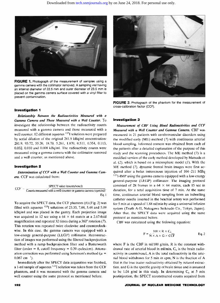
FIGURE 1. Photograph of the measurement of samples using a gamma camera with the collimator removed. A sampling vial having an internal diameter of 22.5 mm and outer diameter of 23.5 mm is placed on the gamma camera surface covered with a vinyl filter to prevent contamination.
Investigation 1
Relationship Between the Radioactivities Measured with a Gamma Camera and Those Measured with a Well Counter. To investigate the relationship between the radioactivity counts measured with a gamma camera and those measured with a well counter, 12 different aqueous 12'I solutions were prepared by serial dilution of the original 281.9 kBq/ml concentration: 281.9, 93.72, 55.20, 18.70, 5.261, 1.870, 0.511, 0.354, 0.113, 0.032, 0.010 and 0.008 kBq/ml. The radioactivity counts were measured using a gamma camera with the collimator removed and a well counter, as mentioned above.
Investigation 2
Determination of CCF with a Well Counter and Gamma Camera. CCF was calculated from:
SPECf value ( counts/voxel) CCF = Counts measured with a well counter or gamma camera (cpm/ml)"
Eq.l
To acquire the SPECT data, the CCF phantom (6) (Fig. 2) was filled with aqueous 123I solutions of 23.33, 7.88, 5.48 and 5.09 kBq/ml and was placed in the gantry. Each projection image was acquired in 12 sec using a 64 X 64 matrix at a 2.67-fold magnification and repeated 32 times during a 360° rotation (7). This rotation was repeated twice clockwise and counterclockwise. In this case, the gamma camera was equipped with a low-energy general-purpose (LEGP) collimator. Reconstruction of images was performed using the filtered backprojection method with a ramp backprojection filter and a Butterworth filter (order = 8, cutoff frequency = 0.39 cycles/em). Attenuation correction was performed using Sorenson's method (J.L = 0.067 em -I).
Immediately after the SPECT data acquisition was finished, a 1-ml sample of aqueous 123I solution was taken from the CCF phantom, and it was measured with the gamma camera and well counter using the same protocol as mentioned before.
192
FIGURE 2. Photograph of the phantom for the measurement of cross-calibration factor (CCF).
Investigation 3
Measurement of CBF Using Blood Radioactivities and CCF Measured with a Well Counter and Gamma Camera. CBF was measured in 21 patients with cerebrovascular disorders using the modified early (ME) method (7) with continuous arterial blood sampling. Informed consent was obtained from each of the patients after a detailed explanation of the purpose of this study and the scanning procedures. The ME method (7) is a modified version of the early method developed by Matsuda et al. (2). which is based on a microsphere model (1). With the ME method (7), dynamic frontal brain images were first acquired after a bolus intravenous injection of 104-211 MBq 123I-IMP using the gamma camera equipped with a low-energy general-purpose (LEGP) collimator. The imaging protocol consisted of 28 frames in a 64 X 64 matrix, each 15 sec in duration, for a total acquisition time of 7 min. At the same time, continuous arterial blood sampling from an indwelling catheter needle inserted in the brachial artery was performed for 5 min at a speed of 1.88 ml/min by using a universal infusion system (Truth A-11, Nakagawa Seikoudo Co., Tokyo, Japan). After that, the SPECT data were acquired using the same protocol as mentioned before.
CBF was calculated using the following equation:
100 X R XC~ F = c-Nc--x~A~x-G~x~c_.,c-=F Eq. 2
where F is the CBF in ml/100 g!min, R is the constant withdrawal rate of arterial blood in ml/min, Ch is the brain radioactivity in counts/voxel, A is the total radioactivity in the arterial blood withdrawn for 5 min in cpm, N is the fraction of A that is the true tracer radioactivity obtained by octanol extraction, and G is the specific gravity of brain tissue and is assumed to be 1.04 g/ml in this study. In determining Ch at 5 min postinjection, the SPECT reconstructed counts acquired from
JOURNAL OF NUCLEAR MEDICINE TECHNOLOGY
by on June 24, 2018. For personal use only. tech.snmjournals.org Downloaded from

1500 25 = s .. y = 4.245x - 2.549 y = 3.812x + 0.214 s = = ..... r = 1.000 20 r = 1.000 ~= s n= 12 n=8 = ·--== ..... = 1000 SEE= 7.217 kcpm SEE = 0.062 kcpm
...... u~ 15 i:C: ..... s "'=c=. ~ = Col --=~ = ;t:- 10 ; i:C: 500 ~ = s ..
~
~ s 5 = = = Col
= u 0 0 0 100 200 300 0 1 2 3 4 5 6
Radioactivity measured with a Radioactivity measured with a well counter (kBq/ml) well counter (kBq/ml)
FIGURE 3. Relationship between the radioactivity counts measured with a well-type scintillation counter (kBq/ml) and those measured with a gamma camera without collimator (cpm). Note that the radioactivity counts measured with a well-type scintillation counter were converted to kBq/ml using a curie meter. (A) The radioactivity concentration ranging between 0 and 300 kBq/ml is shown on the left, while (B) the range between 0 and 6 kBq/ml is shown on the right.
7 min to 25 min were corrected to represent those at 5 min
using the time-activity curve for the whole brain obtained
during 7 min after injection.
The octanol-extracted radioactivity of the arterial blood
sample (N X A in Equation 2) was measured with a well
counter to calculate CBF. Then, the octanol-extracted radio
activity of the arterial blood sample, which had been placed in
the sampling vial, was measured with a gamma camera after
the collimator had been removed, to calculate CBF. The re
sults were time-corrected by adjusting to the time required for
the measurement of CBF with the well counter. Irregular RO!s were defined in the cerebellum, frontal lobe, bilateral temporal
lobe and occipital lobe, and CBF values were quantitatively
measured in these RO!s using Equation 2.
TABLE 1 Comparison of Cross-Calibration Factors (CCFs)
Measured Before and After Calibration of a SPECT Apparatus Using a Well-Type Scintillation
Counter and a Gamma Camera
Cross-calibration factor (CCF)
Before After
Well counter 1.692 x 10⁻⁴ 1.366 x 10⁻⁴
Gamma camera 8.549 x 10- 5 8.765 x 10- 5
After - Before ·% Difference = x 1 00
Before
% Difference•
19.27 2.53
VOLUME 26, NUMBER 3, SEPTEMBER 1998
Statistical Analysis
The correlation between the values obtained with a well
counter and those obtained with a gamma camera was assessed
60 r- .. Uof u ·-t:IIIIJ: 50 .5! 1l "' ... = = "0 "' .. 40 ~ ~ ... ·- E ~ S,e.g '8 ·;;: ... 30 c·= ~ ·- ~ E E c E ~-- .. 20 ~ f t:lll
-"0 ;:;:;, Q E c
10 ;:E ="0 u ;
0
y = 0.990x + 0.552 r= 0.990 n=231 SEE = 1.340 mill OOg/min
0 10 20 30 40 50 60
CBF (mlllOOglmin) obtained using CCF and blood radioactivity measured with a well
counter
FIGURE 4. Relationship between the cerebral blood flow (CBF) values (ml/1 00 g/min) obtained using the cross-calibration factor (CCF) and blood radioactivity counts measured with a well-type scintillation counter and those obtained using the CCF and blood radioactivity counts measured with a gamma camera.
193
by on June 24, 2018. For personal use only. tech.snmjournals.org Downloaded from
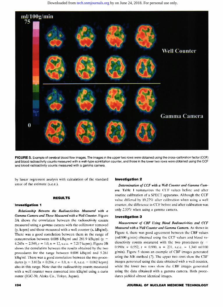
FIGURE 5. Example of cerebral blood flow images. The images in the upper two rows were obtained using the cross-calibration factor (CCF) and blood radioactivity counts measured with a well-type scintillation counter, and those in the lower two rows were obtained using the CCF and blood radioactivity counts measured with a gamma camera.
by linear regression analysis with calculation of the standard error of the estimate (s.e.e.).
RESULTS
"Investigation 1
Relationship Between the Radioactivities Measured with a Gamma Camera and Those Measured with a Well Counter. Figure 3A shows the correlation between the radioactivity counts measured using a gamma camera with the collimator removed (y, kcpm) and those measured with a well counter (x, kBq/ml). There was a good correlation between them in the range of concentration between 0.008 kBq/ml and 281.9 kBq/ml (y =
4.245x- 2.549, r = 1.0, n = 12, s.e.e. = 7.217 kcpm). Figure 38 shows the correlation between the results obtained by the two procedures for the range between 0.008 kBq/ml and 5.261 kBq/ml. There was a good correlation between the two procedures (y = 3.812x + 0.214, r = 1.0, n = 8, s.e.e. = 0.062 kcpm) also in this range. Note that the radioactivity counts measured with a well counter were converted into kBq/ml using a curie meter (IGC-30, Aloka Co., Tokyo, Japan).
194
Investigation 2
Determination of CCF with a Well Counter and Gamma Cam
era. Table I summarizes the CCF values before and after
routine calibration of a SPECT apparatus. Although the CCF value differed by 19.27% after calibration when using a well counter, the difference in CCF before and after calibration was
only 2.537< when using a gamma camera.
Investigation 3
Measurement of CBF Using Blood Radioactivities and CCF
Measured with a Well Counter and Gamma Camera. As shown in Figure 4, there was good agreement between the CBF values (ml/100 g/min) obtained using the CCF values and blood radioactivity counts measured with the two procedures (y = 0.990x + 0.552, r = 0.990. n = 231, s.e.e. = 1.340 ml/100 g/min). Figure 5 shows an example of CBF images generated using the ME method (7). The upper two rows show the CBF images generated using the data obtained with a well counter, while the lower two rows show the CBF images generated
using the data obtained with a gamma camera. Both procedures yielded almost identical images.
JOURNAL OF NUCLEAR MEDICINE TECHNOLOGY
by on June 24, 2018. For personal use only. tech.snmjournals.org Downloaded from

DISCUSSION
In this study we investigated the feasibility of using a gamma camera for measuring blood radioactivity. In our clinical setting for CBF quantification using 123I-IMP, patients usually are injected intravenously with approximately 90 MBq to 263 MBq 123I-IMP. For these injection doses, the radioactivity in the arterial blood withdrawn for 5 min after injection ranges from approximately 0.05 kBq/ml to 9 kBq/ml when measured with a well counter. If the relationship between the radioactivity counts measured with a well counter and those measured with a gamma camera is linear in this range of concentration, it appears that a gamma camera can be substituted for a well counter in the measurement of blood radioactivity for CBF quantification.
When a gamma camera equipped with a collimator was used, it was difficult to measure the radioactivity counts at a concentration of less than 5.261 kBq/ml because of large photon attenuation by the collimator (data not shown). Thus, it appears to be difficult to determine blood radioactivity counts to be used as an input function for measuring CBF when using a gamma camera equipped with a collimator. When the collimator was removed, the linearity between the radioactivity counts measured with a gamma camera and those measured with a well counter was confirmed for the range of concentration between 0.001\ kBq/ml and 281.9 kBq/ml (Fig. 3). This range adequately covers the concentration encountered in our clinical setting. These results suggest that gamma cameras with collimators removed can be substituted for well counters for quantitative measurement of CBF using SPECT. We hope that this will facilitate more widespread acceptance of quantitative measurement of CBF using SPECT even in the hospitals having no well counters. However, the present method for measuring blood radioactivity using a gamma camera has the drawback of collimator removal being complicated, and it should be noted that careful handling of the crystal is required.
As previously described, when using a well counter for measuring blood radioactivity, CCF must be determined to calibrate the well counter and SPECT. Furthermore, this value must be newly determined whenever a SPECT apparatus and/or well counter is calibrated. However, since CCF values obtained with a gamma camera did not differ after routine calibration of a SPECT apparatus (Table I), it appears that postcalibration measurement of CCF can be omitted when
using a gamma camera.
VOLUME 26, NUMBER 3, SEPTEMBER 1998
No great differences existed between the CBF values obtained using the CCF and blood radioactivity counts measured with a gamma camera and those obtained using a well counter in a clinical setting (Figs. 4 and 5). This confirmed the feasibility of using a gamma camera for the measurement of blood radioactivity counts.
CONCLUSION
We obtained the following conclusions from this study: (a) gamma cameras can be substituted for well counters in measuring blood radioactivity counts for CBF quantification; and (b) the present method using a gamma camera makes it unnecessary to measure CCF after calibrating a SPECT apparatus.
ACKNOWLEDGMENT
The authors thank Nihon Medi-Physics Co., Ltd., Nishinomiya, Japan for supporting this study.
REFERENCES
I. Kuhl DE, Barrio JR. Huang SC". et al. Quantifying local cerehral blood flow
by N-isopropyl·p-1 12311 iodoamphetamine (IMP) tomography. J Nucl .l,fcd
I'IH2;23: 1'16-203.
2. Matsuda H. Seki H, Sumiya H. et al. Quantitative cerehral hlood flow
measurements using N-isopropyl-(iodine-123) p-iodoamphetamine and sin
gle photon emission computed tomography with rotating gamma camera.
Am J Plrysiollmaging I'IHh;I:IHh-·194.
3. Takeshita G, Maeda H, Nakane K. et al. Quantitative measurement of
regional cerehral hlood flow using N-isopropyl-(iodine-123) p-iodoamphet
amine and single-photon emission computed tomography. J Nucl Med 19'12;
33:17~1-IWl.
4. Odano I, Ohkuho M. Takahashi N, et al. A new method of regional cerebral
hlood flow measurement using one-point arterial sampling hased on the
microsphere model with N-isopropyl-p-1 1'
31]-iodoamphetamine. Nucl Med
Commun I1N4;15:560-5h4.
5. Matsuda H, Higashi S. Tsuji S. et al. A new noninvasive quantitative assess
ment of cerebral blood flow using N-isopropyl-(iodine-123) p-iodoamphet
amine. Am J Plrysiollmaging llJH7;2:49-55.
6. Mimura H, Ono S. Fukunaga M, et al. The quantitative analysis of regional
cerebral blood flow by peripheral venous sampling in single photon emission
computed tomography using N-isopropyl-p-1 1'·'1] iodoamphetamine: com
parison with peripheral arterial sampling. Jpn J Nucl Med (in Japanese)
19H'J;26: I 327-1 33~.
7. Inoue T. Fujioka H, Akamune A, et al. A time-saving approach for quanti
fying regional cerehral hlood flow and application to split-dose method with 12-'I-IMP SPECT using a single-head rotating gamma-camera. Jpn J Nucl
Med (in Japanese) 1'195:32: 1217-1226.
195
by on June 24, 2018. For personal use only. tech.snmjournals.org Downloaded from

1998;26:191-195.J. Nucl. Med. Technol. Junpei IkezoeKenya Murase, Hiroyoshi Fujioka, Takeshi Inoue, Yoshihiro Ishimaru, Akihisa Akamune, Yuji Yamamoto and Using a Gamma CameraMeasurement of Blood Radioactivity for Quantification of Cerebral Blood Flow
http://tech.snmjournals.org/content/26/3/191This article and updated information are available at:
http://tech.snmjournals.org/site/subscriptions/online.xhtml
Information about subscriptions to JNMT can be found at:
http://tech.snmjournals.org/site/misc/permission.xhtmlInformation about reproducing figures, tables, or other portions of this article can be found online at:
(Print ISSN: 0091-4916, Online ISSN: 1535-5675)1850 Samuel Morse Drive, Reston, VA 20190.SNMMI | Society of Nuclear Medicine and Molecular Imaging
is published quarterly.Journal of Nuclear Medicine Technology
© Copyright 1998 SNMMI; all rights reserved.
by on June 24, 2018. For personal use only. tech.snmjournals.org Downloaded from


