
Accepted Manuscript
Low galactosylation of IgG associates with higher risk for futurediagnosis of rheumatoid arthritis during 10 years of follow-up
Ivan Gudelj, Perttu P. Salo, Irena Trbojević-Akmačić, MalenaAlbers, Dragan Primorac, Markus Perola, Gordan Lauc
PII: S0925-4439(18)30098-XDOI: doi:10.1016/j.bbadis.2018.03.018Reference: BBADIS 65089
To appear in:
Received date: 6 December 2017Revised date: 6 March 2018Accepted date: 19 March 2018
Please cite this article as: Ivan Gudelj, Perttu P. Salo, Irena Trbojević-Akmačić, MalenaAlbers, Dragan Primorac, Markus Perola, Gordan Lauc , Low galactosylation of IgGassociates with higher risk for future diagnosis of rheumatoid arthritis during 10years offollow-up. The address for the corresponding author was captured as affiliation for allauthors. Please check if appropriate. Bbadis(2018), doi:10.1016/j.bbadis.2018.03.018
This is a PDF file of an unedited manuscript that has been accepted for publication. Asa service to our customers we are providing this early version of the manuscript. Themanuscript will undergo copyediting, typesetting, and review of the resulting proof beforeit is published in its final form. Please note that during the production process errors maybe discovered which could affect the content, and all legal disclaimers that apply to thejournal pertain.
ACCEP
TED M
ANUSC
RIPT
1
Low galactosylation of IgG associates with higher risk for future diagnosis of rheumatoid
arthritis during 10 years of follow-up
Running title: IgG N-glycans in rheumatoid arthritis
Ivan Gudelj, PhD1*, Perttu P. Salo, MSc2,3*, Irena Trbojević-Akmačić,PhD1, Malena Albers,
MSc1, DraganPrimorac,PhD1,4, 5,6,7,8, Markus Perola,MD,PhD2,3,9,10, Gordan Lauc,PhD1,11
1 Genos GlycoscienceResearchLaboratory, Zagreb, Croatia
2 National Institute for Health and Welfare, Helsinki, Finland
3Institute for Molecular Medicine Finland (FIMM), University of Helsinki, Finland
4St. Catherine Specialty Hospital, Zabok/Zagreb, Croatia
5JJ Strossmayer University of Osijek, School of Medicine, Osijek, Croatia
6University of Split, School of Medicine, Split, Croatia
7 Eberly College of Science, The Pennsylvania State University, University Park, PA, USA
8 Children’s Hospital Srebrnjak, Zagreb, Croatia
9Diabetes and Obesity Research Program, University of Helsinki, Helsinki, Finland
10University of Tartu, Estonian Genome Center, Tartu, Estonia
11University of Zagreb Faculty of Pharmacy and Biochemistry, Zagreb, Croatia
* Equal contribution
Address correspondence to Gordan Lauc, PhD, University of Zagreb Faculty of Pharmacy and
Biochemistry, Ante Kovačića 1, 10000 Zagreb, Croatia
E-mail: [email protected]
+385 1 639 4467; fax: +385 1 639 4400
ACCEPTED MANUSCRIPT

ACCEP
TED M
ANUSC
RIPT
2
Financial support
This work was supported by the European H2020 grants SYSCID (contract #733100), GlySign
(contract #722095), and IMForFuture (contract #721815); by the European Structural and
Investment Funds IRI (grant #KK.01.2.1.01.0003) and Croatian National Centre of Research
Excellence in Personalized Healthcare (grant #KK.01.1.1.01.0010);the Finnish Academy
[269517]; the YrjöJahnsson Foundation; the JuhoVainio Foundation; AarneKoskelo Foundation;
Ida Montin Foundation; Biomedicum Helsinki Foundation; Finnish Foundation for
Cardiovascular Research; Integrative Life Science Doctoral Program of the University of
Helsinki.
ACCEPTED MANUSCRIPT

ACCEP
TED M
ANUSC
RIPT
3
Abstract
Antibodies are known to have an important role in the development of rheumatoid arthritis (RA),
one of the most prevalent chronic inflammatory diseases which primarily involves the joints.
Most RA patients develop autoantibodies against immunoglobulin G (IgG) and changes in IgG
glycosylation have been associated with RA. We undertook this study to determine whether
altered IgG glycosylation precedes the disease diagnosis. We studied IgG glycosylation in RA in
two prospective cohorts (N=14,749) by measuring 28 IgG glycan traits in 179 subjects who
developed RA within 10-years follow-up and 358 matched controls.Ultra performance liquid
chromatography method based on hydrophilic interactions (HILIC-UPLC) was used to analyse
IgG glycans. Future RA diagnosis associated with traits related to lower galactosylation and
sialylation of IgG when comparing the cases to the matched controls. In RA cases, these traits
did not correlate with the time between being recruited to the study and being diagnosed with RA
(median time 4.31 years). The difference in IgG glycosylation was relatively stable and present
years before diagnosis. This indicates that long-acting factors affecting IgG glycome
composition are among the underlying mechanisms of RA and that decreased galactosylation is a
pre-existing risk factor involved in the disease development.
Keywords: rheumatoid arthritis, immunoglobulin G, N-glycans, biomarker, risk factor
ACCEPTED MANUSCRIPT

ACCEP
TED M
ANUSC
RIPT
4
1. Introduction
Rheumatoid arthritis (RA) is a chronic autoimmune inflammatory disease which primarily
involves joints and has an incidence of 0.5 – 1%[1], making it one of the most prevalent chronic
inflammatory diseases[2]. Even though mechanisms of the disease development are yet to be
completely identified, antibodies are known to be important players in that processand connected
with the disease severity[2–4].Autoantibodies against citrullinated peptides (ACPAs) and
immunoglobulin G (IgG) are particularly important in the diagnostics of RA. An early diagnosis
is central to the treatment of RA, as starting a therapy as soon as possible can prevent or halt
joint damage in many patients[5].
IgG is the most abundant immunoglobulin in human circulation and a majority of RA
patients produce autoantibodies binding to IgG, collectively denominated rheumatoid factor
(RF). IgGitself is a glycoprotein and both its antigen binding region (Fab) as well as the fragment
crystallizableregion(Fc) are known to be N-glycosylated[6]. These glycans are mostly
biantennary complex-type N-glycans carryingvarying levels of a core fucose, N-
acetylglucosamine (GlcNAc), galactose and sialic acid[7]. Glycosylation of IgG has a major
effect on its effector and antigen binding functions and may also affect its role in RA.
IgG glycosylation changes are known to be associated with RA since 1985 when Parekh
et al. showed that galactose and sialic acid levels were lower in patients with RA[8]. Moreover,
these changes follow the disease remission during pregnancy[9–11]and an efficient therapy
treatment[12–15] as well as relapses and progression of the disease[16]. Besides RA, N-
glycosylation is shown to be altered in other autoimmune diseases such as systemic lupus
erythematosus[17] and inflammatory bowel disease[18] as well as in inflammatory diseases
ACCEPTED MANUSCRIPT

ACCEP
TED M
ANUSC
RIPT
5
without autoimmune character[19–21].Some authors have also suggested that there is a “sugar
print” characteristic for RA which enables distinguishing it from different similar
diseases[22,23]. Whether changes in IgG glycosylation contribute to RA development is unclear,
although a recent Mendelian randomization study does not support a simple causal
relationship[24].
Recent development of new analytical methods and approaches has enabled the analysis
of IgG N-glycome in a larger number of patients and with higher accuracy[25,26]. Ultra
performance liquid chromatography based on hydrophilic interactions (HILIC-UPLC) analysis
of 2-aminobenzamide (2-AB) labelled PNGase F released N-glycans has been proven to be
reproducible and robust[25,27]. We used this method to analyse IgG glycosylation profiles of
179individuals that were diagnosed with RA during 10-year follow-up after sampling (cases),
and 358 matching controls that did not develop RAin the same period. Herein we show that the
IgG glycan aberrancy can be detected years before the onset of the disease and considered as a
novel risk factor for RA.
ACCEPTED MANUSCRIPT
ACCEP
TED M
ANUSC
RIPT
6
2. Materials and Methods
2.1.Study population.
RA cases and controls were originally recruited to the FINRISK 2002 (N=8,595) and 2007
(N=6,154) cohorts. FINRISK cohorts are population-based samples of Finns, described in more
detail previously[28]. During the follow-up, 179 incident RA cases were identified among the
study participants using the Finnish hospital discharge and causes of death registers with ICD-
codes: M05-M13, M32, M33, M45 (ICD-10) / 710, 714, 420, 725 (ICD-9) / 712, 734 (ICD-8)
and the social insurance institution of Finland (KELA) special reimbursement for RA medication
(KELA code 202). Two controls per case were frequency matched by gender, 5-year age group,
FINRISK year and study region. Individuals who developed other autoimmune or chronic
diseases were excluded from the present study. The median follow-up time was 4.31 years for
cases (range 0.025 to 9.83 years) and 9.80 years for controls (range 0.33 to 9.94 years).
2.2.IgG isolation, glycan release and labelling.
The whole procedure was performed as previously reported[25]. Briefly, IgGs were isolated
using Protein G 96-well plate (BIA Separations, Slovenia) from the serum samples. The isolated
IgGs were denaturated with the addition of sodium dodecyl sulphate (SDS) (Invitrogen, USA)
and by incubation at 65°C. The excess of SDS was neutralized with Igepal-CA630 (Sigma-
Aldrich, USA) and N-glycans were released following the addition of PNGase F (Promega,
USA) in Phosphate Buffered Saline (PBS). The released N-glycans were labelled with 2-AB.
Free label and reducing agentwere removed from the samples using hydrophilic interaction
liquid chromatography solid-phase extraction (HILIC-SPE). Glycans were eluted with ultrapure
water and stored at –20°C until use.
ACCEPTED MANUSCRIPT

ACCEP
TED M
ANUSC
RIPT
7
2.3.Ultra-performance liquid chromatography.
Fluorescently labelled N-glycans were separated by HILIC on an Acquity UPLC instrument
(Waters, USA) consisting of a quaternary solvent manager, sample manager, and an FLR
fluorescence detector set with excitation and emission wavelengths of 250 and 428 nm,
respectively. The instrument was under the control of Empower 3 software, build 3471 (Waters).
Labelled N-glycans were separated on a Waters BEH Glycan chromatography column, 100 × 2.1
mm i.d., 1.7 µm BEH particles, with 100 mM ammonium formate, pH 4.4, as solvent A and
ACN as solvent B. The separation method used a linear gradient of 25-38% solvent A at flow
rate of 0.40 ml/min in a 27 min analytical run. Samples were maintained at 10°C before
injection, and the separation temperature was 60°C. Data processing was performed using an
automatic processing method with a traditional integration algorithm, after which each
chromatogram was manually corrected to maintain the same intervals of integration for all the
samples. The chromatograms were all separated in the same manner into 24 peaks as previously
reported[7]. The amount of glycans in each peak was expressed as % of total integrated area.
Derived traits were calculated according the following formulas: for agalactosylated G0 =
GP1+GP2+GP3+GP4+GP6, with one galactose G1 = GP7+GP8+GP9+GP10+GP11, with two
galactoses G2 = GP12+GP13+GP14+GP15 and sialylated glycans S =
GP16+GP17+GP18+GP19+GP21+GP22+GP23+GP24.
2.4.Data analysis.
The glycan variables were first inverse normal transformed and standardized to mean=0 and
standard deviation=1. Then, for each of the transformed glycan variables, a linear regression
model adjusted for sex, age, age group ([,30 y}, [30 y, 40 y}, [40 y, 55 y}, [55 y, 70 y}, [70 y, }),
ACCEPTED MANUSCRIPT
ACCEP
TED M
ANUSC
RIPT
8
and an age*sex interaction term was fitted and the residuals from the model were extracted. The
association of the residuals with RA was then tested with incident disease (yes/no)using logistic
regression and, in cases only, with time from sampling to diagnosis (years) using linear
regression. To take into account multiple testing in the presence of substantial correlation
between the glycan variables, principal component analysis was used. As the first 19 principal
components explained more than 99% of the variance in the glycan residuals, the P-values for
the logistic regression tests were adjusted by multiplying the nominal P-values by 19. P-values
for tests with time to diagnosis as the dependent variable are presented without adjusting for
multiple testing.
ACCEPTED MANUSCRIPT

ACCEP
TED M
ANUSC
RIPT
9
3. Results
IgG N-glycans were released by PNGase F, labelled with 2-AB andanalysed by HILIC-UPLC.
Obtained chromatograms were integrated into 24 peaks which mostly contained individual N-
glycans (Figure 1)[7]. The association of the N-glycanswith incident RA was tested using
logistic regression (Table 2).
Glycan peaks GP3, GP4 and GP6were increased in RA cases when compared to controls
(nominal P-value<0.05).These peakscontain agalactosylated glycan structures A2B, FA2 and
FA2B, respectively. On the contrary, the proportion of total area comprised by the glycan peaks
containing structures with one galactose (GP8 and GP9) or two galactoses (GP12, GP13, and
GP14) was smaller in cases, as were the % areas of the peaks GP18 and GP23 containing
sialylated IgG glycans (nominal P-value<0.05). The derived variables for agalactosylation (G0),
monogalactosylation (G1), and digalactosylation (G2) were also nominally significant. The
associations of the peak GP6 (agalactosylated glycan FA2B), total agalactosylation (G0), and
total monogalactosylation (G1) with RA remained statistically significant after adjusting the P-
values for multiple testing (PGP6=0.008, PG0=0.027, PG1=0.029, Table 2).
The association oflow IgGgalactosylation with future diagnosis of RA may reflect
changes occurring during early phase of the disease, or, alternatively, it may indicate that low
IgG galactosylation is a pre-determined or acquired risk-factor. Aiming to distinguish between
these two alternative hypotheses, we checked whether the observed changes associate with the
time between sampling and RA diagnosis (Table 2, Figure 2).
The only variable showing a nominally significant association was GP9 that correlated
with a lower risk of RA and a longer time between taking the blood sample and diagnosis with
ACCEPTED MANUSCRIPT
ACCEP
TED M
ANUSC
RIPT
10
RA, but neither of these associations remained statistically significant after adjusting the P-value
for multiple testing. Noneof the N-glycan variables that robustly associated with RA risk (GP6,
G0, and G1) correlated with the time between RA diagnosis and the date of participating in the
study (Table 2, Figure 2).
ACCEPTED MANUSCRIPT

ACCEP
TED M
ANUSC
RIPT
11
4. Discussion
Results of our study are in accordance with the previous studies that reported decreased
galactosylation in RA [8,16,29]. The decreasein galactosylation and sialylationin RAhas been
known for a long time[8] and was observed in multiple independent studies[16,29]. The decrease
inIgG galactosylation has beenregularly associated with RA[8,10,15,30–36], while changes in
sialylation were reported to be independent of galactosylation[9]and the decrease in sialylation
wasfar less reported[8,10,15,37]. It is known that both anti-inflammatory and pro-inflammatory
activities of IgGare determined by which Fcγ receptor they preferentially bind toand that the
receptor affinity depends on the composition of the glycan linked to the 297 asparagine. IgG
carrying G0 glycans hasincreased affinity to FcγRIII, an activating FcR, and the serum
lectinmannose-binding protein[38,39]. Moreover, besides the classical and lectin pathways of
complement activation, IgG bearing G0 is able to activate an alternative complement
pathway[40].Such G0-bearing IgGsare thus uniquely capable of setting autoimmunity.To the
contrary, higher sialylation of IgG decreases its affinity for FcγRIIIand increases the expression
of FcγRIIB,an inhibitory FcR[38].
This is not the first study which reported that IgG glycosylation changes could be
observed before the onset of symptoms[31,41,42], yet there is disagreement between studies
about the time point when changes are starting to be visible. Ercan et al. concluded that the
changes could be noticed at least 3.5 years[41] and Young at al. observed them two years prior
the onset of symptoms[31], while Rombouts et al. stated that a decrease in galactose residues
occurred around three months prior to diagnosis and only in ACPA-IgG[42]. In this study we
ACCEPTED MANUSCRIPT

ACCEP
TED M
ANUSC
RIPT
12
detected robust association of low IgG galactosylation (andnominally significant association of
low IgG sialylation)and risk for RA diagnosis during 10 years of follow-up.In our data, the
median time from donating the blood sample used for glycan measurement to the subsequent
diagnosis with RA was 4.31 years, ranging from less than two weeks to nearly ten years. Despite
the wide time range we did not observe any statistically significant correlation between IgG
glycosylation and the time from sampling to diagnosis, indicating that the decreased
galactosylation is a pre-existing risk factor involved in the disease development.
Low IgG galactosylation might be genetically pre-determined, or it might be acquired
through the action of environmental factors. The heritability of RA is estimated to be some 40-
65% and 20% for seropositive and seronegative arthritis, respectively[2]. The heritability of IgG
N-glycomecomposition is also known to be relatively high[7].Yarwood et al. recently looked for
shared genetic factors between RA and IgG N-glycome using IgG N-glycosylation associated
single nucleotide polymorphisms (SNPs) and found no association between them and risk for
RA[24].This suggests low IgGgalactosylation may not be a genetically pre-determined risk
factor. The differences in IgG N-glycans between RA cases and healthy controls precede
diagnosis for years, which coincides with the preclinical period of the disease known as pre-
rheumatoid arthritis (pre-RA). This period includes a period in the RA development where
genetics and environmental factors interact and which is followed by development of disease-
related autoantibodies and symptoms without clinical evidence of RA[43–45]. All these stages
can be present years before RA establishment. Knowing that environmental risk factors such as
smoking, obesity, periodontitis, hormones, diary and infections together with genetic risk factors
play an important role in the first stage of pre-RA[44], and some of these factors are known to
affect the IgG glycome [7,46].Therefore, this indicates that stable long-acting factors affecting
ACCEPTED MANUSCRIPT

ACCEP
TED M
ANUSC
RIPT
13
IgG glycome composition are among of the underlying mechanism of RA and makes IgG
glycosylation a novel risk factor for RA which should be considered for risk estimation of
RA[47]. Moreover, knowing that IgG N-glycans are affecting inflammatory activity of IgG, this
may be an indication that the glycans are not only a biomarker but a functional effector of the
disease..
As joint destruction increases with disease progression, new biomarkers should be found
to establish the diagnosis early to reduce or prevent consequent damage[2]. Today, the diagnosis
is primarily based on serological testing for RFs and ACPAs[16], and they are known to be
present in serum long before the onset of the disease[48].However, the aforementioned
biomarkers are neither detectable in every RA patient (merely 70%) nor are selective for RA
since they could also result from other diseases[16]. Therefore, IgGglycans have a considerable
potential to be a biomarker of the disease; since they have been shown to have, combined with
RF titters, a positive predictive value of 94% for RA diagnosis[31]. Moreover,
agalactosylatedIgG levels are known to correlate with the disease remission, progression and
successful therapy treatment[16]. Indeed, IgGglycans follow a temporary improvement of RA
during pregnancy by an increase in IgGgalactosylation and sialylation as well as a decrease of
the same glycan traits after delivery, which correlates with a postpartum relapse[10]; those
changes could be attributed to higher estrogen levels in pregnancy which is known to modulate
IgGgalactosylation[49].
Even though the method applied in this study for IgG N-glycome analysis,as well as
other analytical techniques which require HPLC/UPLC and mass spectrometry, are not yet
routinely used in the clinic for disease diagnostic, undergalactosylatedIgG could be easily
estimated by ELISA techniques using protein G in combination with
ACCEPTED MANUSCRIPT

ACCEP
TED M
ANUSC
RIPT
14
biotinylatedPsathyrellavelutinalectin (PVL), which preferentially interacts with the N-
acetylglucosaminegroup exposed at the termini of sugar chains in agalactosylatedIgG. Moreover,
the assay was able to distinguish RA from other autoimmune diseases[23].
In conclusion, in this study we showed that IgG N-glycosylationcorrelates with RA years
before RA diagnosis and indicates an active involvement of N-glycans in the disease pathology.
Moreover, these early changes ofIgG N-glycome may thus be useful in constructing a sensitive
and specific test for early RA diagnosis, needed to enable prompt treatment and prevent
irreversible joint destruction as well as to evaluate disease progression, remission and proper
treatment.
ACCEPTED MANUSCRIPT

ACCEP
TED M
ANUSC
RIPT
15
Figure legends
Figure 1. Representative chromatogram of 2-AB labelled N-linked glycans released from IgGs
isolated from human serum and separated by HILIC-UPLC. The integration areas, together with
a major structure presented in each glycan peak are given. Glycan peaks are numbered from
GP1-GP24, as used in the paper.
Figure 2. Agalactosylation of IgG and time from sampling to diagnosis with rheumatoid arthritis
in cases only. Residual G0 is independent of the time from recruitment to diagnosis in cases. The
derived agalactosylation variable G0 was formed by summing the peaks GP1, GP2, GP3, GP4,
and GP6. It was then inverse normal transformed and a linear regression model adjusted for
cohort, age, sex, age*sex interaction term, and age group ([,30}, [30, 40}, [40, 55}, [55, 70}, [70,
}) was fitted to the transformed variable and residuals from that model were extracted. The dots
represent individual measurements while the box plots summarize data from individuals across
the four groups of [0.0, 2.5}, [2.5, 5.0}, [5.0, 7.5}, and [7.5-10} years.
ACCEPTED MANUSCRIPT

ACCEP
TED M
ANUSC
RIPT
16
References
[1] A.J. Silman, J.E. Pearson, Epidemiology and genetics of rheumatoid arthritis., Arthritis
Res. 4 Suppl 3 (2002) S265-72. doi:10.1186/ar578.
[2] J.S. Smolen, D. Aletaha, I.B. McInnes, Rheumatoid arthritis, Lancet. 388 (2016) 2023–
2038. doi:10.1016/S0140-6736(16)30173-8.
[3] D. Aletaha, F. Alasti, J.S. Smolen, Rheumatoid factor, not antibodies against citrullinated
proteins, is associated with baseline disease activity in rheumatoid arthritis clinical trials.,
Arthritis Res. Ther. 17 (2015) 229. doi:10.1186/s13075-015-0736-9.
[4] A. Gonzalez, M. Icen, H.M. Kremers, C.S. Crowson, J.M. Davis, T.M. Therneau, V.L.
Roger, S.E. Gabriel, Mortality trends in rheumatoid arthritis: The role of rheumatoid
factor, J. Rheumatol. 35 (2008) 1009–1014. doi:10.1016/j.molcel.2007.05.041.A.
[5] J.S. Smolen, R. Landewé, F.C. Breedveld, M. Dougados, P. Emery, C. Gaujoux-Viala, S.
Gorter, R. Knevel, J. Nam, M. Schoels, D. Aletaha, M. Buch, L. Gossec, T. Huizinga,
J.W.J.W. Bijlsma, G. Burmester, B. Combe, M. Cutolo, C. Gabay, J. Gomez-Reino, M.
Kouloumas, T.K. Kvien, E. Martin-Mola, I. McInnes, K. Pavelka, P. van Riel, M. Scholte,
D.L. Scott, T. Sokka, G. Valesini, R. van Vollenhoven, K.L. Winthrop, J. Wong, A. Zink,
D. van der Heijde, EULAR recommendations for the management of rheumatoid arthritis
with synthetic and biological disease-modifying antirheumatic drugs., Ann. Rheum. Dis.
69 (2010) 964–75. doi:10.1136/ard.2009.126532.
[6] A. Bondt, Y. Rombouts, M.H.J. Selman, P.J. Hensbergen, K.R. Reiding, J.M.W. Hazes,
R.J.E.M. Dolhain, M. Wuhrer, Immunoglobulin G (IgG) Fab glycosylation analysis using
a new mass spectrometric high-throughput profiling method reveals pregnancy-associated
ACCEPTED MANUSCRIPT

ACCEP
TED M
ANUSC
RIPT
17
changes., Mol. Cell. Proteomics. 13 (2014) 3029–39. doi:10.1074/mcp.M114.039537.
[7] M. Pucic, A. Knezevic, J. Vidic, B. Adamczyk, M. Novokmet, O. Polasek, O. Gornik, S.
Supraha-Goreta, M.R. Wormald, I. Redzic, H. Campbell, A. Wright, N.D. Hastie, J.F.
Wilson, I. Rudan, M. Wuhrer, P.M. Rudd, D. Josic, G. Lauc, High throughput isolation
and glycosylation analysis of IgG-variability and heritability of the IgG glycome in three
isolated human populations, Mol Cell Proteomics. 10 (2011) M111.010090.
doi:10.1074/mcp.M111.010090.
[8] R.B. Parekh, R.A. Dwek, B.J. Sutton, D.L. Fernandes, A. Leung, D. Stanworth, T.W.
Rademacher, T. Mizuochi, T. Taniguchi, K. Matsuta, F. Takeuchi, Y. Nagano, T.
Miyamoto, A. Kobata, Association of rheumatoid arthritis and primary osteoarthritis with
changes in the glycosylation pattern of total serum IgG, Nature. 316 (1985) 452–457.
doi:10.1038/316452a0.
[9] A. Bondt, M.H.J. Selman, A.M. Deelder, J.M.W. Hazes, S.P. Willemsen, M. Wuhrer,
R.J.E.M. Dolhain, Association between galactosylation of immunoglobulin G and
improvement of rheumatoid arthritis during pregnancy is independent of sialylation, J.
Proteome Res. 12 (2013) 4522–4531. doi:10.1021/pr400589m.
[10] F.E. van de Geijn, M. Wuhrer, M.H. Selman, S.P. Willemsen, Y. a de Man, A.M. Deelder,
J.M. Hazes, R.J. Dolhain, Immunoglobulin G galactosylation and sialylation are
associated with pregnancy-induced improvement of rheumatoid arthritis and the
postpartum flare: results from a large prospective cohort study., Arthritis Res. Ther. 11
(2009) R193. doi:10.1186/ar2892.
[11] A. Alavi, N. Arden, T.D. Spector, J.S. Axford, Immunoglobulin G glycosylation and
clinical outcome in rheumatoid arthritis during pregnancy, J. Rheumatol. 27 (2000) 1379–
ACCEPTED MANUSCRIPT

ACCEP
TED M
ANUSC
RIPT
18
1385.
[12] M. Pasek, M. Duk, M. Podbielska, R. Sokolik, J. Szechiński, E. Lisowska, H.
Krotkiewski, Galactosylation of IgG from rheumatoid arthritis (RA) patients - Changes
during therapy, Glycoconj. J. 23 (2006) 463–471. doi:10.1007/s10719-006-5409-0.
[13] R. Saldova, J.E. Huffman, B. Adamczyk, A. Mužinić, J.J. Kattla, M. Pučić, M. Novokmet,
J.L. Abrahams, C. Hayward, I. Rudan, S.H. Wild, A.F. Wright, O. Polašek, G. Lauc, H.
Campbell, J.F. Wilson, P.M. Rudd, Association of medication with the human plasma N -
Glycome, in: J. Proteome Res., 2012: pp. 1821–1831. doi:10.1021/pr2010605.
[14] A. Croce, O. Firuzi, F. Altieri, M. Eufemi, R. Agostino, R. Priori, M. Bombardieri, C.
Alessandri, G. Valesini, L. Saso, Effect of infliximab on the glycosylation of IgG of
patients with rheumatoid arthritis, J. Clin. Lab. Anal. 21 (2007) 303–314.
doi:10.1002/jcla.20191.
[15] E. Gińdzieńska-Sieśkiewicz, I. Radziejewska, I. Domysławska, P.A. Klimiuk, A. Sulik, J.
Rojewska, H. Gabryel-Porowska, S. Sierakowski, Changes of glycosylation of IgG in
rheumatoid arthritis patients treated with methotrexate, Adv. Med. Sci. 61 (2016) 193–
197. doi:10.1016/j.advms.2015.12.009.
[16] S. Albrecht, L. Unwin, M. Muniyappa, P.M. Rudd, Glycosylation as a marker for
inflammatory arthritis, Cancer Biomarkers. 14 (2014) 17–28. doi:10.3233/CBM-130373.
[17] F. Vučković, J. Krištić, I. Gudelj, M. Teruel Artacho, T. Keser, M. Pezer, M. Pučić-
Baković, J. Štambuk, I. Trbojević-Akmačić, C. Barrios, T. Pavić, C. Menni, Y. Wang, Y.
Zhou, L. Cui, H. Song, Q. Zeng, X. Guo, B.A. Pons-Estel, P. McKeigue, A.L. Patrick, O.
Gornik, T.D. Spector, M. Harjaček, M. Alarcon-Riquelme, M. Molokhia, W. Wang, G.
Lauc, Systemic lupus erythematosus associates with the decreased immunosuppressive
ACCEPTED MANUSCRIPT

ACCEP
TED M
ANUSC
RIPT
19
potential of the IgG glycome., Arthritis Rheumatol. (Hoboken, N.J.). 67 (2015) 2978–
2989. doi:10.1002/art.39273.
[18] I. Trbojević Akmačić, N.T. Ventham, E. Theodoratou, F. Vučković, N.A. Kennedy, J.
Krištić, E.R. Nimmo, R. Kalla, H. Drummond, J. Štambuk, M.G. Dunlop, M. Novokmet,
Y. Aulchenko, O. Gornik, H. Campbell, M. Pučić Baković, J. Satsangi, G. Lauc, IBD-
BIOM Consortium, Inflammatory bowel disease associates with proinflammatory
potential of the immunoglobulin G glycome., Inflamm. Bowel Dis. 21 (2015) 1237–47.
doi:10.1097/MIB.0000000000000372.
[19] M.B. Freidin, T. Keser, I. Gudelj, J. Štambuk, D. Vučenović, M. Allegri, T. Pavić, M.
Šimurina, S.M. Fabiane, G. Lauc, F.M.K. Williams, The Association Between Low Back
Pain and Composition of IgG Glycome, Nat. Publ. Gr. (2016) 1–11.
doi:10.1038/srep26815.
[20] C. Barrios*, J. Zierer*, I. Gudelj, J. Stambuk, I. Ugrina, E. Rodriguez, M.J. Soler, T.
Pavic, M. Simurina, T. Keser, M. Pucic-Bakovic, M. Mangino, J. Pascual, T.D. Spector,
G. Lauc, C. Menni, Glycosylation Profile of IgG in Moderate Kidney Dysfunction., J.
Am. Soc. Nephrol. (2015) 1–9. doi:10.1681/ASN.2015010109.
[21] I. Gudelj, M. Baciarello, I. Ugrina, M. De Gregori, V. Napolioni, P.M. Ingelmo, D.
Bugada, S. De Gregori, L. Đerek, M. Pučić-Baković, M. Novokmet, O. Gornik, G.
Saccani Jotti, T. Meschi, G. Lauc, M. Allegri, Changes in total plasma and serum N-
glycome composition and patient-controlled analgesia after major abdominal surgery, Sci.
Rep. 6 (2016) 31234. doi:10.1038/srep31234.
[22] M. Watson, P.M. Rudd, M. Bland, R.A. Dwek, J.S. Axford, Sugar printing rheumatic
diseases: A potential method for disease differentiation using immunoglobulin G
ACCEPTED MANUSCRIPT
ACCEP
TED M
ANUSC
RIPT
20
oligosaccharides, Arthritis Rheum. 42 (1999) 1682–1690. doi:10.1002/1529-
0131(199908)42:8<1682::AID-ANR17>3.0.CO;2-X.
[23] N. Tsuchiya, T. Endo, S. Yoshinoya, Y. Nagano, M. Shiota, K. Furukawa, A. Kobatat,
Detection of Glycosylation Abnormality in Rheumatoid, J. Immunol. 151 (1993) 1137–
1146.
[24] A. Yarwood, S. Viatte, Y. Okada, R. Plenge, K. Yamamoto, A. Barton, D. Symmons, S.
Raychaudhuri, L. Klareskog, P. Gregersen, J. Worthington, S. Eyre, Loci associated with
N-glycosylation of human IgG are not associated with rheumatoid arthritis: a Mendelian
randomisation study, Ann Rheum Dis. 75 (2016) 317–320. doi:10.1136/annrheumdis-
2014-207210.
[25] I. Trbojević-Akmačić, I. Ugrina, G. Lauc, Comparative Analysis and Validation of
Different Steps in Glycomics Studies, Methods Enzymol. 586 (2016) 37–55.
doi:10.1016/bs.mie.2016.09.027.
[26] M.P. Baković, M.H.J. Selman, M. Hoffmann, I. Rudan, H. Campbell, A.M. Deelder, G.
Lauc, M. Wuhrer, High-throughput IgG Fc N-glycosylation profiling by mass
spectrometry of glycopeptides, J. Proteome Res. 12 (2013) 821–831.
doi:10.1021/pr300887z.
[27] I. Trbojević Akmačić, I. Ugrina, J. Štambuk, I. Gudelj, F. Vučković, G. Lauc, M. Pučić-
Baković, High-throughput glycomics: Optimization of sample preparation, Biochem. 80
(2015) 934–942. doi:10.1134/S0006297915070123.
[28] K. Borodulin, E. Vartiainen, M. Peltonen, P. Jousilahti, A. Juolevi, T. Laatikainen, S.
Mannisto, V. Salomaa, J. Sundvall, P. Puska, Forty-year trends in cardiovascular risk
factors in Finland, Eur. J. Public Health. 25 (2015) 539–546. doi:10.1093/eurpub/cku174.
ACCEPTED MANUSCRIPT

ACCEP
TED M
ANUSC
RIPT
21
[29] A. Mastrangelo, T. Colasanti, C. Barbati, A. Pecani, D. Sabatinelli, M. Pendolino, S.
Truglia, L. Massaro, R. Mancini, F. Miranda, F.R. Spinelli, F. Conti, C. Alessandri, The
Role of Posttranslational Protein Modifications in Rheumatological Diseases: Focus on
Rheumatoid Arthritis, J. Immunol. Res. 2015 (2015). doi:10.1155/2015/712490.
[30] R.B. Parekh, D.A. Isenberg, B.M. Ansell, I.M. Roitt, R.A. Dwek, T.W. Rademacher,
Galactosylation of IgG associated oligosaccharides: reduction in patients with adult and
juvenile onset rheumatioid arthritis and relation to disease activity, Lancet. 331 (1988)
966–969. doi:10.1016/S0140-6736(88)91781-3.
[31] A. Young, N. Sumar, K. Bodman, S. Goyal, H. Sinclair, I. Roitt, D. Isenberg, Agalactosyl
IgG: an aid to differential diagnosis in early synovitis, Arthritis Rheum. 34 (1991) 1425–
1429.
[32] J.S. Axford, N. Sumar, A. Alavi, D.A. Isenberg, A. Young, K.B. Bodman, I.M. Roitt,
Changes in normal glycosylation mechanisms in autoimmune rheumatic disease, J. Clin.
Invest. 89 (1992) 1021–1031. doi:10.1172/JCI115643.
[33] E. Gindzienska-Sieskiewicz, P.A. Klimiuk, D.G. Kisiel, A. Gindzienski, S. Sierakowski,
The changes in monosaccharide composition of immunoglobulin G in the course of
rheumatoid arthritis, Clin. Rheumatol. 26 (2007) 685–690. doi:10.1007/s10067-006-0370-
7.
[34] J.M. Pekelharing, E. Hepp, J.P. Kamerling, G.J. Gerwig, B. Leijnse, Alterations in
carbohydrate composition of serum IgG from patients with rheumatoid arthritis and from
pregnant women, Ann. Rheum. Dis. 47 (1988) 91–95. doi:10.1136/ard.47.2.91.
[35] C. Pilkington, E. Yeung, D. Isenberg, A.K. Lefvert, G.A.W. Rook, Agalactosyl igg and
antibody specificity in rheumatoid arthritis, tuberculosis, systemic lupus erythematosus
ACCEPTED MANUSCRIPT
ACCEP
TED M
ANUSC
RIPT
22
and myasthenia gravis, Autoimmunity. 22 (1995) 107–111.
doi:10.3109/08916939508995306.
[36] a Bond, a Alavi, J.S. Axford, P. Youinou, F.C. Hay, The relationship between exposed
galactose and N-acetylglucosamine residues on IgG in rheumatoid arthritis (RA), juvenile
chronic arthritis (JCA) and Sjögren’s syndrome (SS)., Clin. Exp. Immunol. 105 (1996)
99–103. doi:10.1046/j.1365-2249.1996.d01-741.x.
[37] R. Pfeifle, T. Rothe, N. Ipseiz, H.U. Scherer, S. Culemann, U. Harre, J.A. Ackermann, M.
Seefried, A. Kleyer, S. Uderhardt, B. Haugg, A.J. Hueber, P. Daum, G.F. Heidkamp, C.
Ge, S. Böhm, A. Lux, W. Schuh, I. Magorivska, K.S. Nandakumar, E. Lönnblom, C.
Becker, D. Dudziak, M. Wuhrer, Y. Rombouts, C.A. Koeleman, R. Toes, T.H. Winkler,
R. Holmdahl, M. Herrmann, S. Blüml, F. Nimmerjahn, G. Schett, G. Krönke, Regulation
of autoantibody activity by the IL-23-T H 17 axis determines the onset of autoimmune
disease, Nat. Immunol. 18 (2017) 104–113. doi:10.1038/ni.3579.
[38] E. Maverakis, K. Kim, M. Shimoda, M.E. Gershwin, F. Patel, R. Wilken, S.
Raychaudhuri, L.R. Ruhaak, C.B. Lebrilla, Glycans in the immune system and The
Altered Glycan Theory of Autoimmunity: A critical review, J. Autoimmun. 57 (2015) 1–
13. doi:10.1016/j.jaut.2014.12.002.
[39] R. Malhotra, M.R. Wormald, P.M. Rudd, P.B. Fischer, R. a Dwek, R.B. Sim,
Glycosylation changes of IgG associated with rheumatoid arthritis can activate
complement via the mannose-binding protein., Nat. Med. 1 (1995) 237–243.
doi:10.1038/nm0395-237.
[40] N.K. Banda, A.K. Wood, K. Takahashi, B. Levitt, P.M. Rudd, L. Royle, J.L. Abrahams,
G.L. Stahl, V.M. Holers, W.P. Arend, Initiation of the alternative pathway of murine
ACCEPTED MANUSCRIPT

ACCEP
TED M
ANUSC
RIPT
23
complement by immune complexes is dependent on N-glycans in IgG antibodies, Arthritis
Rheum. 58 (2008) 3081–3089. doi:10.1002/art.23865.
[41] A. Ercan, J. Cui, D.E.W. Chatterton, K.D. Deane, M.M. Hazen, W. Brintnell, C.I.
O’Donnell, L.A. Derber, M.E. Weinblatt, N.A. Shadick, D.A. Bell, E. Cairns, D.H.
Solomon, V.M. Holers, P.M. Rudd, D.M. Lee, Aberrant IgG galactosylation precedes
disease onset, correlates with disease activity, and is prevalent in autoantibodies in
rheumatoid arthritis, Arthritis Rheum. 62 (2010) 2239–2248. doi:10.1002/art.27533.
[42] Y. Rombouts, E. Ewing, L. a van de Stadt, M.H.J. Selman, L. a Trouw, A.M. Deelder,
T.W.J. Huizinga, M. Wuhrer, D. van Schaardenburg, R.E.M. Toes, H.U. Scherer, Anti-
citrullinated protein antibodies acquire a pro-inflammatory Fc glycosylation phenotype
prior to the onset of rheumatoid arthritis., Ann. Rheum. Dis. 74 (2015) 234–41.
doi:10.1136/annrheumdis-2013-203565.
[43] R.W. Gan, L.A. Trouw, J. Shi, R.E.M. Toes, T.W.J. Huizinga, M.K. Demoruelle, J.R.
Kolfenbach, G.O. Zerbe, K.D. Deane, J.D. Edison, W.R. Gilliland, J.M. Norris, V.M.
Holers, Anti-carbamylated protein antibodies are present prior to rheumatoid arthritis and
are associated with its future diagnosis, J. Rheumatol. 42 (2015) 572–579.
doi:10.3899/jrheum.140767.
[44] B.J. Paul, H.I. Kandy, V. Krishnan, Pre-rheumatoid arthritis and its prevention, Eur. J.
Rheumatol. 4 (2017) 161–165. doi:10.5152/eurjrheum.2017.16006.
[45] S.K. Tedeschi, J. Cui, E. V. Arkema, W.H. Robinson, J. Sokolove, N. Lingampalli, J.A.
Sparks, E.W. Karlson, K.H. Costenbader, Elevated BMI and antibodies to citrullinated
proteins interact to increase rheumatoid arthritis risk and shorten time to diagnosis: A
nested case–control study of women in the Nursesʼ Health Studies, Semin. Arthritis
ACCEPTED MANUSCRIPT
ACCEP
TED M
ANUSC
RIPT
24
Rheum. 46 (2017) 692–698. doi:10.1016/j.semarthrit.2016.09.001.
[46] A. Wahl, S. Kasela, E. Carnero-Montoro, M. van Iterson, J. Štambuk, S. Sharma, E. van
den Akker, L. Klaric, E. Benedetti, G. Razdorov, I. Trbojević-Akmačić, F. Vučković, I.
Ugrina, M. Beekman, J. Deelen, D. van Heemst, B.T. Heijmans, B.I.O.S. Consortium, M.
Wuhrer, R. Plomp, T. Keser, M. Šimurina, T. Pavić, I. Gudelj, J. Krištić, H. Grallert, S.
Kunze, A. Peters, J.T. Bell, T.D. Spector, L. Milani, P.E. Slagboom, G. Lauc, C. Gieger,
IgG glycosylation and DNA methylation are interconnected with smoking, Biochim.
Biophys. Acta - Gen. Subj. 1862 (2018) 637–648. doi:10.1016/j.bbagen.2017.10.012.
[47] J.A. Sparks, M.D. Iversen, R. Miller Kroouze, T.G. Mahmoud, N.A. Triedman, S.S. Kalia,
M.L. Atkinson, B. Lu, K.D. Deane, K.H. Costenbader, R.C. Green, E.W. Karlson,
Personalized Risk Estimator for Rheumatoid Arthritis (PRE-RA) Family Study: Rationale
and design for a randomized controlled trial evaluating rheumatoid arthritis risk education
to first-degree relatives, Contemp. Clin. Trials. 39 (2014) 145–157.
doi:10.1016/j.cct.2014.08.007.
[48] S. Rantapää-Dahlqvist, B.A.W. De Jong, E. Berglin, G. Hallmans, G. Wadell, H.
Stenlund, U. Sundin, W.J. Van Venrooij, Antibodies Against Cyclic Citrullinated Peptide
and IgA Rheumatoid Factor Predict the Development of Rheumatoid Arthritis, Arthritis
Rheum. 48 (2003) 2741–2749. doi:10.1002/art.11223.
[49] A. Ercan, W.M. Kohrt, J. Cui, K.D. Deane, M. Pezer, E.W. Yu, J.S. Hausmann, H.
Campbell, U.B. Kaiser, P.M. Rudd, G. Lauc, J.F. Wilson, J.S. Finkelstein, P.A. Nigrovic,
Estrogens regulate glycosylation of IgG in women and men, JCI Insight. 2 (2017) 1–9.
doi:10.1172/jci.insight.89703.
ACCEPTED MANUSCRIPT

ACCEP
TED M
ANUSC
RIPT
25
Table 1. Age and sex distribution of the study sample.
Age group Control men Control women Case men Case women
(,30] 6 (1.7 %) 8 (2.2 %) 3 (1.7 %) 4 (2.2 %)
(30,40] 14 (3.9 %) 28 (7.8 %) 7 (3.9 %) 14 (7.8 %)
(40,55] 34 (9.5 %) 88 (24.6 %) 17 (9.5 %) 44 (24.6 %)
(55,70] 76 (21.2 %) 68 (19 %) 38 (21.2 %) 34 (19 %)
(70, ] 16 (4.5 %) 20 (5.6 %) 8 (4.5 %) 10 (5.6 %)
ACCEPTED MANUSCRIPT

ACCEP
TED M
ANUSC
RIPT
26
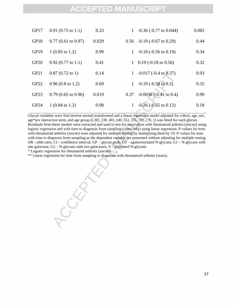
Table 2.Association of rheumatoid arthritis and IgG N-glycome.
Glycan
trait OR (95% CI)* P (OR)* Padjusted (OR)* Beta (95% CI)** P (beta)**
S 0.82 (0.66 to 1) 0.064 1 -0.2 (-0.64 to 0.24) 0.38
G0 1.5 (1.2 to 1.9) 0.0014 0.027 -0.0034 (-0.5 to 0.49) 0.99
G1 0.74 (0.61 to 0.89) 0.0015 0.029 0.4 (-0.012 to 0.81) 0.059
G2 0.71 (0.56 to 0.91) 0.007 0.13 0.00033 (-0.49 to 0.49) 1
GP1 1.2 (0.98 to 1.5) 0.083 1 -0.18 (-0.63 to 0.26) 0.43
GP2 1 (0.86 to 1.3) 0.67 1 -0.094 (-0.5 to 0.31) 0.65
GP3 1.2 (1 to 1.5) 0.036 0.68 0.21 (-0.21 to 0.63) 0.32
GP4 1.4 (1.1 to 1.8) 0.0045 0.085 0.037 (-0.44 to 0.52) 0.88
GP5 1.1 (0.94 to 1.4) 0.2 1 -0.018 (-0.43 to 0.39) 0.93
GP6 1.5 (1.2 to 1.9) 0.00042 0.008 -0.17 (-0.66 to 0.32) 0.5
GP7 0.85 (0.7 to 1) 0.074 1 -0.082 (-0.46 to 0.3) 0.67
GP8 0.77 (0.63 to 0.93) 0.0063 0.12 0.35 (-0.057 to 0.76) 0.094
GP9 0.81 (0.67 to 0.97) 0.026 0.49 0.41 (0.018 to 0.8) 0.042
GP10 1.2 (0.96 to 1.4) 0.13 1 -0.22 (-0.64 to 0.2) 0.31
GP11 1.2 (0.99 to 1.5) 0.061 1 -0.15 (-0.57 to 0.27) 0.49
GP12 0.78 (0.64 to 0.95) 0.013 0.25 -0.19 (-0.61 to 0.22) 0.36
GP13 0.83 (0.68 to 1) 0.045 0.86 -0.019 (-0.41 to 0.37) 0.92
GP14 0.71 (0.56 to 0.92) 0.008 0.15 0.057 (-0.45 to 0.56) 0.83
GP15 0.92 (0.75 to 1.1) 0.42 1 -0.26 (-0.66 to 0.15) 0.22
GP16 0.99 (0.83 to 1.2) 0.93 1 0.17 (-0.22 to 0.55) 0.4
ACCEPTED MANUSCRIPT
ACCEP
TED M
ANUSC
RIPT
27
GP17 0.91 (0.75 to 1.1) 0.33 1 -0.36 (-0.77 to 0.044) 0.082
GP18 0.77 (0.61 to 0.97) 0.029 0.56 -0.19 (-0.67 to 0.29) 0.44
GP19 1 (0.83 to 1.2) 0.99 1 -0.18 (-0.56 to 0.19) 0.34
GP20 0.92 (0.77 to 1.1) 0.41 1 0.19 (-0.18 to 0.56) 0.32
GP21 0.87 (0.72 to 1) 0.14 1 -0.017 (-0.4 to 0.37) 0.93
GP22 0.96 (0.8 to 1.2) 0.69 1 -0.19 (-0.58 to 0.2) 0.35
GP23 0.79 (0.65 to 0.96) 0.019 0.37 -0.0036 (-0.41 to 0.4) 0.99
GP24 1 (0.84 to 1.2) 0.98 1 -0.26 (-0.65 to 0.12) 0.18
Glycan variables were first inverse normal transformed and a linear regression model adjusted for cohort, age, sex,
age*sex interaction term, and age group ([,30}, [30, 40}, [40, 55}, [55, 70}, [70, }) was fitted for each glycan.
Residuals from these models were extracted and used to test for association with rheumatoid arthritis (yes/no) using
logistic regression and with time to diagnosis from sampling (cases only) using linear regression. P-values for tests
with rheumatoid arthritis (yes/no) were adjusted for multiple testing by multiplying them by 19. P-values for tests
with time to diagnosis from sampling as the dependent variable are presented without adjusting for multiple testing.
OR - odds ratio, CI - confidence interval, GP - glycan peak, G0 – agalactosylated N-glycans, G1 – N-glycans with
one galactose, G2 – N-glycans with two galactoses, S – sialylated N-glycans.
* Logistic regression for rheumatoid arthritis (yes/no)
** Linear regression for time from sampling to diagnosis with rheumatoid arthritis (years).
ACCEPTED MANUSCRIPT

ACCEP
TED M
ANUSC
RIPT
28
Highlights
Future diagnosis of rheumatoid arthritis is associated with lower galactosylation of IgG
IgG glycosylation alterations are present years before diagnosis
Glycosylation is a pre-existing risk factor involved in the disease development
ACCEPTED MANUSCRIPT

Figure 1

Figure 2


