
DOI: 10.1002/cbic.200700560
Linear Analogues of Human b-Defensin 3: Concepts forDesign of Antimicrobial Peptides with ReducedCytotoxicity to Mammalian CellsShouping Liu,[a] Lei Zhou,[a, b] Jing Li,[a, b] Anita Suresh,[c] Chandra Verma,[c] Yong Hwee Foo,[a]
Eric P. H. Yap,[d] Donald T. H. Tan,[a, b, e] and Roger W. Beuerman*[a, b]
Introduction
The defensins are small cationic and cysteine-rich moleculeswith molecular weights between 3–6 kDa, and are about 45amino acids in length. Based on the spatial distribution of thethree cysteine intramolecular bonds, mammalian defensins canbe divided into two major groups termed a and b-defensins.q-Defensins make up a third group, but to-date are found onlyin nonhuman primates, and have only 18 amino acids.[1–3] De-fensins are an important component of the innate immunesystem and provide an initial antimicrobial barrier for mucosalsurfaces such as the surface of the eye, oral tissues, the air-ways and lungs, and also the skin.[4–8] As antimicrobial agentsthey show broad-spectrum activity against Gram-positive andGram-negative bacteria, fungi and enveloped viruses, and alsoagainst bacteria that have demonstrated resistance to thecurrently used antibiotics.[9] Because of this ability, they havecome to be known as “natural antibiotics”. Interest in the de-fensins has also been aided by an increase in bacterial strainswith resistance to the standard antibiotics, an increase in thepopulation with reduced immunity, and the water solubilityof defensins, which makes them potentially rapidly deploy-able in response to a pathogen release into the environ-ment.[10] Current efforts are focused toward designing ana-logues of defensins that have properties that are appropriatefor therapeutic application. However, as improvements to thestructure–activity relationship are attempted, issues such ascharge density and hydrophobicity emerge because analoguescan interact with both the membrane of a pathogen as wellas the membrane of human cells. Toxicity to human cells, ofboth native forms and of analogues, has been a concern.[11–12]
Peptide analogues have been developed with diverse cap-abilities, and it appears that the disulfide bonds can be rear-
ranged or removed with preservation of antimicrobial activi-ty.[13–15]
The b-defensins have been discovered more recently thana-defensins, and have been given special attention becausethey are found in skin and epithelial cells that line mucosal sur-faces.[7, 16] In b-defensins, the disulfide bridges that likely stabi-lize these molecules are Cys1–Cys5, Cys2–Cys4 and Cys3–Cys6while the a-defensins, which are largely produced by thePaneth cells of the intestine and neutrophils, have bridges at
[a] Dr. S. Liu,+ Dr. L. Zhou,+ Dr. J. Li,+ Y. H. Foo, Prof. D. T. H. Tan,Prof. Dr. R. W. BeuermanSingapore Eye Research Institute11 Third Hospital Avenue, #06-00, Singapore 16875 (Singapore)Fax: (+65)6322-4599E-mail : [email protected]
[b] Dr. L. Zhou,+ Dr. J. Li,+ Prof. D. T. H. Tan, Prof. Dr. R. W. BeuermanDepartment of Ophthalmology, Yong Loo Lin School of MedicineNational University of SingaporeSingapore 16875 (Singapore)
[c] A. Suresh, Dr. C. VermaBioinformatics InstituteSingapore 16875 (Singapore)
[d] Dr. E. P. H. YapDefence Medical & Environmental Research InstituteSingapore 16875 (Singapore)
[e] Prof. D. T. H. TanSingapore National Eye CenterSingapore 16875 (Singapore)
[+] These authors contributed equally to this work.
Supporting information for this article is available on the WWW underhttp://www.chembiochem.org or from the author: RP-HPLC-UV chromato-grams and MS spectra of the six linear analogues of hBD3. (NMRC/CPG/007/2004)
A series of engineered linear analogues [coded as F6, W6, Y6, A6,S6 and CACHTUNGTRENNUNG(Acm)6] were modeled, designed, synthesized and struc-turally characterized by mass spectra, circular dichroism, hydro-phobicity analysis and molecular modeling. We have screenedantimicrobial activity, hemolysis to rabbit erythrocytes, and cyto-toxicity to human conjunctival epithelial cells. No significantACHTUNGTRENNUNGhemolytic effect was observed for hBD3 or from five of the sixACHTUNGTRENNUNGanalogues [F6, Y6, A6, S6 and C ACHTUNGTRENNUNG(Acm)6] over the range of 3–100 mgmL�1. The six linear analogues have reduced cytotoxicityto human conjunctival epithelial cells over the range of 6–
100 mgmL�1 compared to hBD3. By tuning the overall hydropho-bicity of linear hBD3 analogues, reduced cytotoxicity and hemoly-sis were obtained while preserving the antimicrobial properties.The decreased cytotoxicity of the linear analogues is suggested tobe structurally related to the removal of disulfide bridges, andthe flexible structure of the linear forms, which seem to be associ-ated with loss of secondary structure. These results suggest anew approach for guiding the design of new linear analogues ofdefensin peptides with strong antibiotic properties and reducedcytotoxicity to mammalian cells.
964 www.chembiochem.org @ 2008 Wiley-VCH Verlag GmbH&Co. KGaA, Weinheim ChemBioChem 2008, 9, 964 – 973

Cys1–Cys6, Cys2–Cys4 and Cys3–Cys5. However, in the eye,both of these are active in response to injury, as has beenfound in tears.[6, 17] Within the family of b-defensins, hBD1,hBD2 and hBD3 have been the most extensively studied; addi-tionally hBD4–6 have been recently reported. Further, genomebioinformatics studies have pointed to the existence of an ad-ditional 28 b-defensins that have yet to be found by proteomicstudies.[18–20] Studies of the tissue distribution of hBD3 by usingRNA levels found only low levels in most tissues, except in oralmucosa and more recently in epithelial cells from the ocularsurface.[7,21] Of the b-defensins, hBD3 has been of particularACHTUNGTRENNUNGinterest as it appears to possess better broad-spectrum anti-ACHTUNGTRENNUNGmicrobial activity than either hBD1 or hBD2.[22]
The goal of this study was to design analogues of hBD3 thatexhibit good antimicrobial properties and display reduced tox-icity to human cells. We designed and synthesized linear ana-logues of hBD3 by retaining the same net positive charge(+11), and by replacing the six bridging cysteine residues withresidues of varying hydrophobicities. Factors including linearity,hydrophobicity, antimicrobial activity, hemolysis and epithelialcell cytotoxicity were determined. Within this group of linearanalogues, the overall hydrophobicity was generally but notspecifically related to decreased cytotoxicity to human ocularsurface cells ; however, antimicrobial capability remained at thesame level as native hBD3.
Results and Discussion
Synthesis of analogues of hBD3
The solid-phase synthesis of six linear analogues of hBD3 with45 residues was performed by using Fmoc chemistry. The pep-tide sequences are illustrated in Scheme 1. In the analogues,all cysteine residues were uniformly replaced by one of the fol-lowing amino acids: alanine (A), serine (S), cysteine that wasprotected by Acm [CACHTUNGTRENNUNG(Acm)], tryptophan (W), tyrosine (Y), orphenylalanine (F). The six analogues were coded as follows:A6, S6, C ACHTUNGTRENNUNG(Acm)6, W6, Y6 and F6, respectively. Physicochemicalproperties of the peptides are shown in Table 1 (sequencelength, numbers of hydrophobic and aromatic residues, netcharge, relative hydrophobicity) and Table 2 (retention time inHPLC). The UV spectroscopy and mass spectrometry (MS) ofthese peptides are shown in Figure S1 in the Supporting Infor-mation.
Compared with native hBD3, the six linear analogues ofhBD3 are of the same length and net positive charge (+11) be-cause they are designed and synthesized by uniform replace-ment of the six bridging cysteine residues in hBD3 with sixACHTUNGTRENNUNGresidues of varying hydrophobicities (viz. , F, W, Y, A, S and C-ACHTUNGTRENNUNG(Acm)). Therefore, these linear analogues provide a well-de-fined model to study the effect of overall hydrophobicity, sec-ondary structure conformation, removal of the native disulfidebridges on antimicrobial, hemolytic and cytotoxic activities.
Molecular hydrophobicity
We have measured the relative molecular hydrophobicity byRP-HPLC–MS in terms of retention time at 500 mgmL�1 and100 mgmL�1 (Table 2). RP-HPLC is an approach that is common-
ly employed for comparisons of peptides or aminoacid side chains on antibacterial peptides.[31–35] Be-cause the stationary phase of C18-modified silica ishydrophobic and the mobile phase (water/acetoni-trile) is hydrophilic, a longer retention time is a mea-sure of greater hydrophobicity. The measured orderfor the relative molecular hydrophobicity of the pep-tides was as follows: W6>F6>Y6>native hBD3>A6>S6>CACHTUNGTRENNUNG(Acm)6. The relative hydrophobicities ofthe peptides were also calculated based on theHopp–Woods hydrophilicity scale[23] (Table 1). Thescale is a hydrophilic index in which apolar residueshave been assigned negative values, and is typicallyScheme 1. Sequence of wild-type hBD3 and its linear analogues.
Table 1. Physicochemical properties of the peptides.
Variant Number of residues Net positive Relativetotal hydrophobic charge hydrophobicity[a]
ACHTUNGTRENNUNG(aromatic)
CACHTUNGTRENNUNG(Acm)6 45 14 (2) 11 n.c.S6 45 14 (2) 11 28.3A6 45 14 (2) 11 23.5wt-hBD3 45 14 (2) 11 20.5Y6 45 14 (8) 11 12.7F6 45 14 (8) 11 11.5W6 45 14 (8) 11 6.1
[a] The overall hydrophobicity was calculated based on the hydrophobici-ty scale of Hopp–Woods hydrophilicity scale.[23] The lower value corre-sponds to lower hydrophilicity or higher hydrophobicity; n.c. , not calcu-lated.
Table 2. The overall hydrophobicity of the peptides in terms of retentiontime (tR) in RP-HPLC–MS.
Variant tR [min]500 mgmL�1 100 mgmL�1
CACHTUNGTRENNUNG(Acm)6 20.15�0.19 20.94�0.21S6 20.33�0.25 20.44�0.15A6 22.36�0.07 22.36�0.28wt-hBD3 23.24�0.03 24.28�0.13Y6 23.85�0.16 24.40�0.05F6 27.65�0.13 28.25�0.12W6 29.29�0.03 29.66�0.06
ChemBioChem 2008, 9, 964 – 973 @ 2008 Wiley-VCH Verlag GmbH&Co. KGaA, Weinheim www.chembiochem.org 965
Linear Analogues of Human b-Defensin 3
used to identify antigenic regions based on hydrophilic patch-es. For each peptide, the values that correspond to each resi-due were summed to give an overall measure. The generaltrend in computed hydrophobicity of the peptides by usingthis scale matched that of the experimental HPLC retentiontime data. CACHTUNGTRENNUNG(Acm)6, was excluded because Acm has not beenparameterized in the Hopp–Woods scale.[23]
CD spectroscopy
Previous X-ray and NMR spectroscopic studies of human b-de-fensins (hBD1–3)[27,36–37] have shown that the tertiary structuresare similar, and have a short a-helical segment before a triple-stranded antiparallel b-sheet, all rigidly held together by threedisulfide bonds. It follows that in the absence of the con-straints that are imposed by these disulfide bonds, the linearanalogues of hBD3 are likely to be flexible and random inaqueous solution, and they could adopt conformations thatare dictated by the environment.[13,15] Therefore, it was of inter-est to examine the solution conformations of the linear ana-logues in different media. CD spectroscopy of the hBD3 deriva-tives was carried out in aqueous solution in the presence of50% organic modifier trifluoroethanol (TFE), and in the pres-ence of 20 mm SDS micelles, which is a membrane-mimetic(Figures 1, 2, and 3). TFE has been widely used in protein struc-
ture studies because it can induce stable, structured conforma-tions from otherwise unstructured/random peptides in aque-ous solution.[38–41] The membrane-mimicking SDS was chosenbecause it is most similar to the prokaryotic membrane, and istherefore a good model for the natural target of an antibiot-ic.[42] While all of the tested peptides appeared unstructured inaqueous solution, they displayed an observable level of struc-turing in the presence of 50% TFE or 20 mm SDS micelles, aswas determined by CD spectroscopy.The CD spectrum of wild-type hBD3 agrees very well with
that of native hBD3, which was reported elsewhere.[13,42] Thespectra of the six linear analogues and wild-type hBD3 in aque-ous solution were similar, and suggested a mix of random and
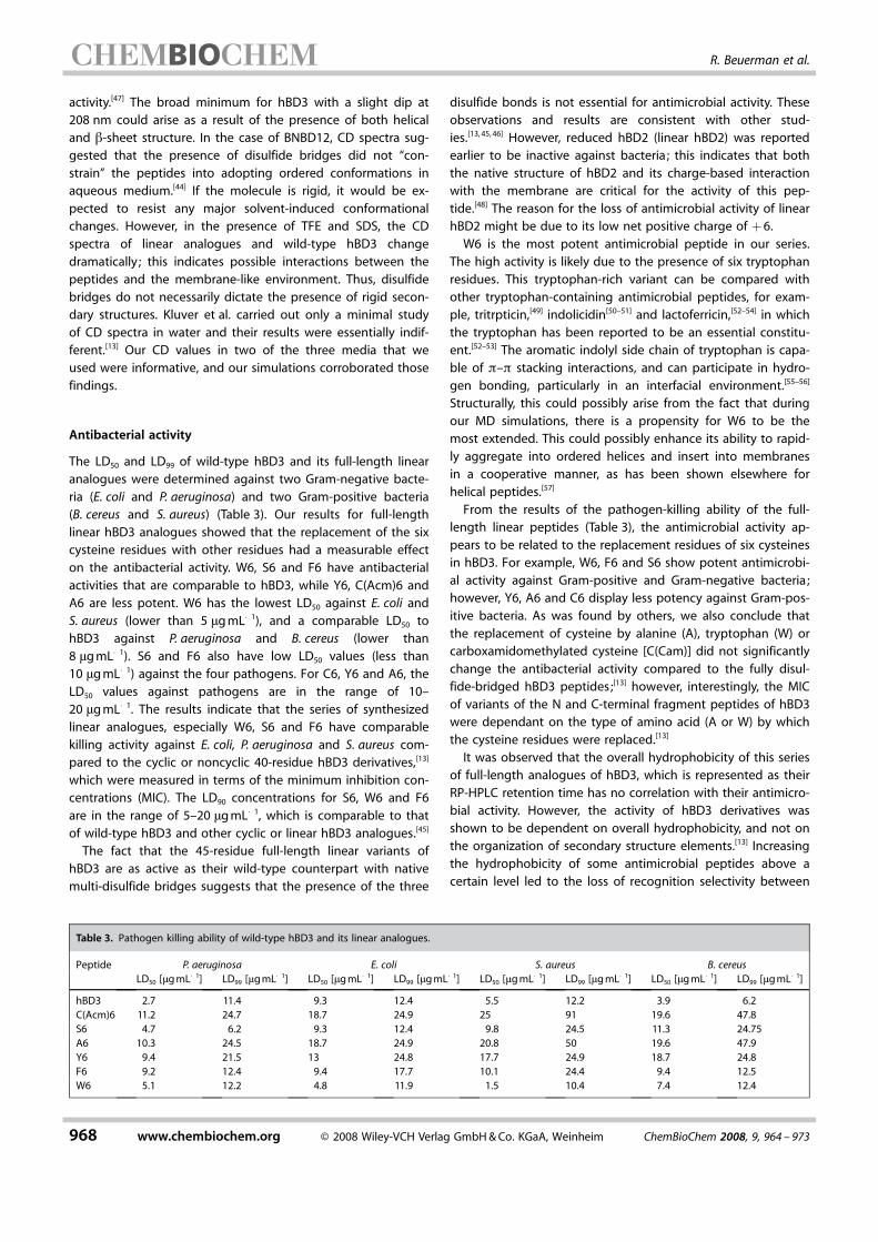
b-hairpin conformations, although to different extents becausethe intensities at the negative extrema varied (Figure 1). Theabsence of prominent crossover at wavelengths of 195 nmwas indicative of an absence of significant amounts of structur-ally distinct conformers, while the minima at ~200 nm wassuggestive of transient b-hairpin or turn-like conformer. Thissuggests that the existence of disulfide bonds and the type ofCys-replacing residues impose no significant effect on theACHTUNGTRENNUNGsecondary structure of hBD3 derivatives. The minima at ~195–200 nm for native defensin has been observed previously inthe case of native HNP-1[43] and BNBD-12,[44] and is characteris-tic of peptides that contain b-sheet or b-turn secondary struc-ture with some amount of random coil.[42] Thus, the secondarystructure seems to be preformed in the peptides independentof the presence of multiple disulfide bridges or the Cys-replac-ing residues. This observation is in accordance with the resultthat was reported by Kluver et al. ,[13] and is further supportedby molecular dynamics simulations (Figure 4) that show thatthe linear variants adopt a variety of conformations in aqueousconditions.
Figure 1. CD spectra of hBD3 and its linear analogues in water.
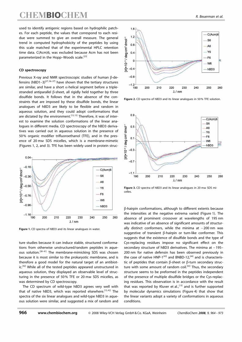
Figure 2. CD spectra of hBD3 and its linear analogues in 50% TFE solution.
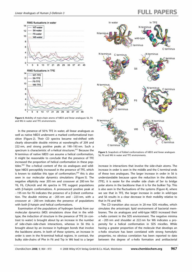
Figure 3. CD spectra of hBD3 and its linear analogues in 20 mm SDS mi-celles.
966 www.chembiochem.org @ 2008 Wiley-VCH Verlag GmbH&Co. KGaA, Weinheim ChemBioChem 2008, 9, 964 – 973
R. Beuerman et al.
In the presence of 50% TFE in water, all linear analogues aswell as native hBD3 underwent a marked conformational tran-sition (Figure 2). Their CD spectra became red-shifted withclearly observable double minima at wavelengths of 209 and222 nm, and strong positive peaks at 190–193 nm. Such aspectrum is characteristic of a-helical structures.[41] Because theN terminus of native hBD3 can assume a helical conformation,it might be reasonable to conclude that the presence of TFEincreased the proportion of helical conformation in these pep-tides.[42] The a-helical content of the six analogues and wild-type hBD3 perceptibly increased in the presence of TFE, whichis known to stabilize this type of conformation;[43] this is alsoseen in our molecular dynamics simulations (Figure 5). Thenegative ellipticity near 205 nm and crossover at 200 nm forY6, F6, CACHTUNGTRENNUNG(Acm)6 and A6 spectra in TFE suggest populationswith b-hairpin conformations. A pronounced positive peak at~195 nm for F6 indicates the presence of a b-sheet conforma-tion. The double minima at ~205 nm and ~223 nm, with acrossover at ~200 nm indicates the presence of populationswith both b-hairpin and helical conformations.Examination of the populations of hydrogen bonds from our
molecular dynamics (MD) simulations show that in the wild-type, the induction of structure in the presence of TFE (in con-trast to water) is brought about by an increase in the numberof side-chain–side-chain interactions, whereas in S6 this isbrought about by an increase in hydrogen bonds that involvethe backbone atoms. In both of these systems, an increase inorder is seen in the N-terminal helical regions. In contrast, thebulky side-chains of Phe in F6 and Trp in W6 lead to a larger
increase in interactions that involve the side-chain atoms. Theincrease in order is seen in the middle and the C-terminal endsof these two analogues. The larger increase in order in S6 isunderstandable because upon the reduction in the dielectric(TFE), it is easier for the smaller side chain of Ser to bridgepolar atoms in the backbone than it is for the bulkier Trp. Thisis also seen in the fluctuations of the systems (Figure 4), wherewe see that in TFE, the larger increase in order in wild-typeand S6 results in a clear decrease in their mobility relative tothat in F6 and W6.The CD transition also occurs in 20 mm SDS micelles, which
simulates the anisotropic lipid environment of bacterial mem-branes. The six analogues and wild-type hBD3 increased theira-helix content in the SDS environment. The negative minimaat ~205 nm and shoulder at 222 nm for W6 indicates a pro-pensity for a helical conformation in SDS micelles. Whilehaving a greater proportion of the molecule that develops ana-helix structure has been correlated with strong hemolyticproperties, no obvious correlation has been reported to datebetween the degree of a-helix formation and antibacterial
Figure 5. Snapshots of folded conformations of hBD3 and linear analoguesS6, F6 and W6 in water and TFE environments.
Figure 4. Mobility of main-chain atoms of hBD3 and linear analogues S6, F6and W6 in water and TFE environments.
ChemBioChem 2008, 9, 964 – 973 @ 2008 Wiley-VCH Verlag GmbH&Co. KGaA, Weinheim www.chembiochem.org 967
Linear Analogues of Human b-Defensin 3
ACHTUNGTRENNUNGactivity.[47] The broad minimum for hBD3 with a slight dip at208 nm could arise as a result of the presence of both helicaland b-sheet structure. In the case of BNBD12, CD spectra sug-gested that the presence of disulfide bridges did not “con-strain” the peptides into adopting ordered conformations inaqueous medium.[44] If the molecule is rigid, it would be ex-pected to resist any major solvent-induced conformationalchanges. However, in the presence of TFE and SDS, the CDspectra of linear analogues and wild-type hBD3 changeACHTUNGTRENNUNGdramatically ; this indicates possible interactions between thepeptides and the membrane-like environment. Thus, disulfidebridges do not necessarily dictate the presence of rigid secon-dary structures. Kluver et al. carried out only a minimal studyof CD spectra in water and their results were essentially indif-ferent.[13] Our CD values in two of the three media that weused were informative, and our simulations corroborated thosefindings.
Antibacterial activity
The LD50 and LD99 of wild-type hBD3 and its full-length linearanalogues were determined against two Gram-negative bacte-ria (E. coli and P. aeruginosa) and two Gram-positive bacteria(B. cereus and S. aureus) (Table 3). Our results for full-lengthlinear hBD3 analogues showed that the replacement of the sixcysteine residues with other residues had a measurable effecton the antibacterial activity. W6, S6 and F6 have antibacterialactivities that are comparable to hBD3, while Y6, C ACHTUNGTRENNUNG(Acm)6 andA6 are less potent. W6 has the lowest LD50 against E. coli andS. aureus (lower than 5 mgmL�1), and a comparable LD50 tohBD3 against P. aeruginosa and B. cereus (lower than8 mgmL�1). S6 and F6 also have low LD50 values (less than10 mgmL�1) against the four pathogens. For C6, Y6 and A6, theLD50 values against pathogens are in the range of 10–20 mgmL�1. The results indicate that the series of synthesizedlinear analogues, especially W6, S6 and F6 have comparablekilling activity against E. coli, P. aeruginosa and S. aureus com-pared to the cyclic or noncyclic 40-residue hBD3 derivatives,[13]
which were measured in terms of the minimum inhibition con-centrations (MIC). The LD90 concentrations for S6, W6 and F6are in the range of 5–20 mgmL�1, which is comparable to thatof wild-type hBD3 and other cyclic or linear hBD3 analogues.[45]
The fact that the 45-residue full-length linear variants ofhBD3 are as active as their wild-type counterpart with nativemulti-disulfide bridges suggests that the presence of the three
disulfide bonds is not essential for antimicrobial activity. Theseobservations and results are consistent with other stud-ies.[13,45, 46] However, reduced hBD2 (linear hBD2) was reportedearlier to be inactive against bacteria ; this indicates that boththe native structure of hBD2 and its charge-based interactionwith the membrane are critical for the activity of this pep-tide.[48] The reason for the loss of antimicrobial activity of linearhBD2 might be due to its low net positive charge of +6.W6 is the most potent antimicrobial peptide in our series.
The high activity is likely due to the presence of six tryptophanresidues. This tryptophan-rich variant can be compared withother tryptophan-containing antimicrobial peptides, for exam-ple, tritrpticin,[49] indolicidin[50–51] and lactoferricin,[52–54] in whichthe tryptophan has been reported to be an essential constitu-ent.[52–53] The aromatic indolyl side chain of tryptophan is capa-ble of p–p stacking interactions, and can participate in hydro-gen bonding, particularly in an interfacial environment.[55–56]
Structurally, this could possibly arise from the fact that duringour MD simulations, there is a propensity for W6 to be themost extended. This could possibly enhance its ability to rapid-ly aggregate into ordered helices and insert into membranesin a cooperative manner, as has been shown elsewhere forACHTUNGTRENNUNGhelical peptides.[57]
From the results of the pathogen-killing ability of the full-length linear peptides (Table 3), the antimicrobial activity ap-pears to be related to the replacement residues of six cysteinesin hBD3. For example, W6, F6 and S6 show potent antimicrobi-al activity against Gram-positive and Gram-negative bacteria ;however, Y6, A6 and C6 display less potency against Gram-pos-itive bacteria. As was found by others, we also conclude thatthe replacement of cysteine by alanine (A), tryptophan (W) orcarboxamidomethylated cysteine [CACHTUNGTRENNUNG(Cam)] did not significantlychange the antibacterial activity compared to the fully disul-fide-bridged hBD3 peptides;[13] however, interestingly, the MICof variants of the N and C-terminal fragment peptides of hBD3were dependant on the type of amino acid (A or W) by whichthe cysteine residues were replaced.[13]
It was observed that the overall hydrophobicity of this seriesof full-length analogues of hBD3, which is represented as theirRP-HPLC retention time has no correlation with their antimicro-bial activity. However, the activity of hBD3 derivatives wasshown to be dependent on overall hydrophobicity, and not onthe organization of secondary structure elements.[13] Increasingthe hydrophobicity of some antimicrobial peptides above acertain level led to the loss of recognition selectivity between
Table 3. Pathogen killing ability of wild-type hBD3 and its linear analogues.
Peptide P. aeruginosa E. coli S. aureus B. cereusLD50 [mgmL�1] LD99 [mgmL�1] LD50 [mgmL�1] LD99 [mgmL�1] LD50 [mgmL�1] LD99 [mgmL�1] LD50 [mgmL�1] LD99 [mgmL�1]
hBD3 2.7 11.4 9.3 12.4 5.5 12.2 3.9 6.2CACHTUNGTRENNUNG(Acm)6 11.2 24.7 18.7 24.9 25 91 19.6 47.8S6 4.7 6.2 9.3 12.4 9.8 24.5 11.3 24.75A6 10.3 24.5 18.7 24.9 20.8 50 19.6 47.9Y6 9.4 21.5 13 24.8 17.7 24.9 18.7 24.8F6 9.2 12.4 9.4 17.7 10.1 24.4 9.4 12.5W6 5.1 12.2 4.8 11.9 1.5 10.4 7.4 12.4
968 www.chembiochem.org @ 2008 Wiley-VCH Verlag GmbH&Co. KGaA, Weinheim ChemBioChem 2008, 9, 964 – 973
R. Beuerman et al.

prokaryotic and eukaryotic membranes, thereby causing anACHTUNGTRENNUNGincrease in cytotoxicity.[58] The observed hydrophobicity of the13-amino acid a-helical PTP7 derivatives by RP-HPLC retentiontime correlated well with the activity against Gram-positivebacteria ; however, antimicrobial activity against Gram-negativebacteria, such as E. coli, did not correlate with RP-HPLC reten-tion time.[34]
The exact mechanism of the hBD3-mediated antimicrobialactivity is not clear. Previous studies suggest that the antimi-crobial activity of hBD3 derivatives depends on their ability tocontact the anionic pathogen cell membrane via electrostaticinteractions that are mediated by the cationic peptide resi-dues, and subsequently, to infiltrate the membrane by hydro-phobic interactions. The key determinants of this action arethe net positive charge and the overall hydrophobicity of thepeptide. In our study, the overall hydrophobicity of the pep-tides was calculated based on an empirical scale (Table 1), andalso measured by an experimental HPLC approach (Table 2).Among the linear analogues, the most hydrophobic, W6, andthe most hydrophilic, S6 are potent antimicrobials ; this sug-gests that the other structural determinants for the mainte-nance of high antimicrobial activity should include positivecharge density, which is associated with folding and hydrophil-ic surface area, and a delicate balance between the hydrophilicsurface area and hydrophobic surface. MD simulations suggestthat both W6 and S6 tend to have the most “extended” con-formations, and their propensity to have helical regions mightbe enhanced cooperatively. The structural feature of the facial-ly amphiphilic conformations is thought to be responsible fortheir ability to kill cells by disrupting phospholipid mem-branes.[59–61] In addition, by controlling the hydrophobic/hydro-philic balance of amphiphilic macromolecules, it is possible toobtain high selectivity between antimicrobial activity and he-molytic activity/cytotoxicity against host cells.[62]
Hemolytic activity and cytotoxicity to mammalian cells
It is well established that cationic peptides not only interactwith pathogens, but can also be toxic to mammalian cells. Bac-terial cell membranes consist of anionic phosphatidylglycerolas a major component, while eukaryotic cell membranesmainly contain zwitterionic phosphatidylcholine and phospha-tidylethanolamine, which are susceptible to hydrophobic inter-actions.[63] It has been proposed that the hemolytic effects ofcationic antimicrobial peptides are directly linked to the hydro-phobicity of these peptides.[64] High levels of hydrophobicityare known to decrease the selectivity between the desiredACHTUNGTRENNUNGbacterial target membranes and mammalian host cell mem-branes.[65] Therefore, the linear analogues and wild-type hBD3were tested for their ability to induce hemolysis in fresh rabbitred blood cells (RBC; Figure 6). W6, the most hydrophobic ofthe analogues, also shows the most potent hemolytic effect ; itgives to 4.9–24.9% hemolysis over the concentration range of50–200 mgmL�1. The results show that W6 has potent antimi-crobial activity, but displays low selectivity between antimicro-bial activity and hemolytic activity, the high hemolytic activityis likely due to the hydrophobicity,[64–65] or the possibility that it
lacks hydrophobic/hydrophilic balance.[62] Analogues C ACHTUNGTRENNUNG(Acm)6and A6, which had low hydrophobicity displayed less hemoly-sis of 1.5–2.3% in the concentration range of 3–100 mgmL�1,while F6, Y6, S6 and wild-type hBD3 showed the least amountof hemolysis of less than 1% in the concentration range of 3–100 mgmL�1. While no significant hemolytic effect was ob-served in wild-type hBD3 or from five of the six analogues [F6,Y6, S6, A6 and CACHTUNGTRENNUNG(Acm)6] in the concentration range of 3–100 mgmL�1, W6 showed a significantly increased hemolyticeffect on rabbit erythrocytes in the same concentration range.Hemolytic activity is conventionally used as a measure of cy-
totoxicity and a model for mammalian cells because red bloodcells are, in general, extremely fragile.[62,66–67] The hemolysisanalysis reflects the systemic effect of the drug administration.As part of the innate immune system, which is associated withmoist mucosal surfaces, and topical applications for eye,mouth or lung, the toxicity of the synthetic molecules to sur-face epithelial cells is also relevant. In the present study, thepotential toxicity of the analogues to epithelial cells was ana-lyzed by using primary cultured human conjunctival epithelialcells. Surprisingly, all six analogues showed improved cytotox-icity to epithelial cells at the tested concentration compared towild-type hBD3 (Figure 7). It was also clear that the six ana-logues fell into two groups, one group included C ACHTUNGTRENNUNG(Acm)6, A6and S6 and showed negligible cytotoxicity to epithelial cells atconcentrations up to 200 mgmL�1, the other group includedF6, W6 and Y6, and showed IC50 values at concentrations of30–50 mgmL�1 compared to an IC50 of around 12.5 mgmL�1 forwild-type hBD3.
Figure 6. Hemolytic effects of hBD3 and its linear analogues on rabbit eryth-rocytes. Freshly isolated rabbit red blood cells (100 mL) were diluted to 8%(v/v) in sterile and mixed with equal volume of defensin analogues to ach-ieve the targeted concentrations in 96-well plates in triplicate. The plate wasincubated at 37 8C for 60 min with occasional gentle agitation. The superna-tant was transfered to a fresh 96-well plate at the end of the incubationafter brief centrifugation. The hemoglobin protein that was released in thesupernatant was determined by measuring the absorbance at 414 nm. Theaveraged readings from the positive control (0.1% Triton X-100) were set as100% and the hemolytic activities of samples were calculated as a percent-age of the positive control. The experiment was repeated 3 times with trip-ACHTUNGTRENNUNGlicate wells for each sample.
ChemBioChem 2008, 9, 964 – 973 @ 2008 Wiley-VCH Verlag GmbH&Co. KGaA, Weinheim www.chembiochem.org 969
Linear Analogues of Human b-Defensin 3
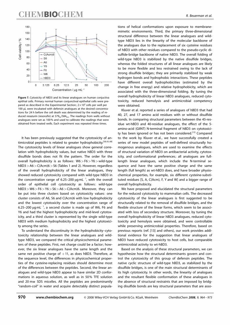
It has been previously suggested that the cytotoxicity of an-timicrobial peptides is related to greater hydrophobicity.[58,65, 68]
The cytotoxicity levels of linear analogues show general corre-lation with hydrophobicity values, but native hBD3 with threedisulfide bonds does not fit the pattern. The order for theoverall hydrophobicity is as follows: W6>F6>Y6>wild-typehBD3>A6>C ACHTUNGTRENNUNG(Acm)6>S6 (Tables 1 and 2). However, regardlessof the overall hydrophobicity of the linear analogues, theyshowed reduced cytotoxicity compared with wild-type hBD3 inthe concentration range of 6.25–200 mgmL�1, with the overallorder of epithelial cell cytotoxicity as follows: wild-typehBD3>W6>F6>Y6>S6>A6>CACHTUNGTRENNUNG(Acm)6. Moreover, they canbe put into three clusters based on cytotoxicity values: onecluster consists of A6, S6 and CACHTUNGTRENNUNG(Acm)6 with low hydrophobicityand the lowest cytotoxicity over the concentration range of6.25–200 mgmL�1, a second cluster is made up of W6, F6 andY6 and had the highest hydrophobicity and mid-level cytotox-icity, and a third cluster is represented by the single wild-typehBD3 with medium hydrophobicity and the highest cytotoxici-ty among the series.To understand the discontinuity in the hydrophobicity–cyto-
toxicity relationship between the linear analogues and wild-type hBD3, we compared the critical physicochemical parame-ters of these peptides. First, net charge could be a factor ; how-ever, the six linear analogues have the same length and thesame net positive charge of +11, as does hBD3. Therefore, atthe sequence level, the differences in physicochemical proper-ties of the cysteine-replacing residues should determine mostof the differences between the peptides. Second, the linear an-alogues and wild-type hBD3 appear to have similar 2D confor-mations in aqueous solution, as well as in 50% TFE solutionand 20 mm SDS micelles. All the peptides are predominantly“random-coil” in water and acquire detectably distinct popula-
tions of helical conformations upon exposure to membrane-mimetic environments. Third, the primary three-dimensionalstructural difference between the linear analogues and wild-type hBD3 lies in the linearity of the molecular backbone ofthe analogues due to the replacement of six cysteine residuesof hBD3 with other residues compared to the pseudo-cyclic di-sulfide-bridge backbone of native hBD3. The overall folding ofwild-type hBD3 is stabilized by the native disulfide bridges,whereas the folded structures of all linear analogues are likelyto be more flexible and less restrained owing to the lack ofstrong disulfide bridges; they are primarily stabilized by weakhydrogen bonds and hydrophobic interactions. These peptideshave different overall hydrophobicities (estimated by thechange in free energy) and relative hydrophobicity, which areassociated with the three-dimensional folding. By tuning theoverall hydrophobicity of linear hBD3 analogues, reduced cyto-toxicity, reduced hemolysis and antimicrobial competencywere obtained.Kluver et al. reported a series of analogues of hBD3 that had
40, 27, and 17 amino acid residues with or without disulfidebonds. In comparing structural parameters between the 45-res-idue wt-hBD3 and 40-residue analogues, the effect of the 5-amino-acid (GIINT) N-terminal fragment of hBD3 on cytotoxici-ty has been ignored or has not been considered.[13] Comparedto the work by Kluver et al. , we have successfully created aseries of new model peptides of well-defined structurally ho-mogenous analogues, which are used to examine the effectsof structural variation of hBD3 on antibacterial activity, cytotox-icity, and conformational preferences; all analogues are full-length linear analogues, which include the N-terminal se-quence and have the same positive charge and the samelength (full length) as wt-hBD3 does, and have broader physio-chemical properties, for example, six different cysteine-substi-tuted residues [S, A, C ACHTUNGTRENNUNG(Acm), F, Y and W] and a bigger range ofoverall hydrophobicity.We have proposed and elucidated the structural parameters
for the reduced cytotoxicity to mammalian cells. The decreasedcytotoxicity of the linear analogues is first suggested to bestructurally related to the removal of disulfide bridges, and theflexible structure of the linear forms, which seem to be associ-ated with loss of secondary structure. Moreover, by tuning theoverall hydrophobicity of linear hBD3 analogues, reduced cyto-toxicity and hemolysis were obtained, and were controllablewhile preserving antimicrobial properties. Therefore, based onprevious reports (ref. [13] and others), our work provides addi-tional evidence for the suggestion that linear analogues ofhBD3 have reduced cytotoxicity to host cells, but comparableantimicrobial activity to wt-hBD3.Based on the analysis of these structural parameters, we can
hypothesize how the structural determinants govern and con-trol the cytotoxicity of this group of defensin peptides. Thenative cyclic structure of wild-type hBD3, as stabilized by thedisulfide bridges, is one of the main structural determinants ofits high cytotoxicity. In other words, the linearity of analoguesand the resultant flexible conformation of these analogues inthe absence of structural restraints that are imposed by bridg-ing disulfide bonds are key structural parameters that are asso-
Figure 7. Cytoxicity of hBD3 and its linear analogues on human conjuctivaepithial cells. Primary normal human conjunctival epithelial cells were pre-pared as described in the Experimental Section; 2O104 cells per well per100 mL were incubated with defensin analogues at the desired concentra-tions for 24 h before the cell death was determined by the reading of re-duced resazurin (resorufin) at 579Ex/584Em. The readings from wells withoutanalogues were set as 100% and used to calibrate the readings that wereobtained from treated wells. Each experiment was repeated three times.
970 www.chembiochem.org @ 2008 Wiley-VCH Verlag GmbH&Co. KGaA, Weinheim ChemBioChem 2008, 9, 964 – 973
R. Beuerman et al.

ciated with their reduced cytotoxicity. A study that used a-heli-cal peptides, showed that partial disruption of secondary struc-ture can lead to a decrease in cytotoxicity to host cells.[68] Ourobservations of the disruption of secondary structures and de-crease in host-cell cytotoxicity in systems that are more com-plex than a-helical peptides extend the suggestion that theloss of secondary structure is a possible general mechanismwhich might be useful for a more efficient design of defensinsanalogues.
Conclusions
While human b-defensin 3 (hBD3), like other defensins, is struc-turally characterized by high cationic capability and amphiphi-licity, there are a variety of structural determinants (high netpositive charge, sequence and residue distribution, amphiphilicconformation, hydrophobicity and folding) that might play apart in antimicrobial activity, hemolysis and cytotoxicity. Wehave designed and synthesized a series of well-defined struc-turally homogenous full-length linear analogues of hBD3 thatprovide a good model for further investigation into the effectsof the following structural factors on antimicrobial activity,ACHTUNGTRENNUNGhemolysis and cytotoxicity of the peptides: native disulfidebridges or native cyclic structure of wild-type hBD3, the lineari-ty and flexibility of analogues in the absence of native disulfidebridges, the physicochemical properties of the Cys-replacingresidues, and the overall hydrophobicity of peptides.We have observed that all linear hBD3 derivatives, especially
W6, S6 and F6 have potent antimicrobial activity. We have dis-covered that a series of six linear analogues exhibit decreasedcytotoxicity to human conjunctival epithelial cells compared towild-type hBD3. It is suggested that the presence of the threedisulfide bridges that restrain the native hBD3 into a pseudo-cyclic form is one of the main structural determinants of itshigh cytotoxicity. Conversely, the linearity and the flexiblenature of the Cys-replaced analogues due to the absence ofthe stabilizing forces of native disulfide bridges are key struc-tural factors that underlie their reduced cytotoxicity. Our obser-vations provide additional evidence that partial disruption ofsecondary structure can lead to a decrease in cytotoxicity tohost cells. Additionally, our finding suggests that the cytotoxic-ity can be controlled by modulating the overall hydrophobicityof the defensin molecules.Linear constructs of hBD3-based peptides provide a simple
architecture for design compared to the cyclic structure ofwild-type hBD3, which contains specific disulfide bridges. Froma chemical synthesis point of view, the incorporation of specificpatterns of disulfide bonds and subsequent purification andidentification is time and resource consuming. Compared tothe synthesis, identification and purification of wt-hBD3 andcertain other cyclic peptides with disulfide bonds, it is mucheasier and quicker to synthesize, identify and purify linear ana-logues. The development of potent defensin-based antibioticswith low-to-no toxic effects on host cells, high microbial selec-tivity and lower costs of production could be designed andACHTUNGTRENNUNGdeveloped with the guide of novel concepts from the presentdiscovery.
Experimental Section
Solid-phase peptide synthesis (SPPS): Recombinant wild-typehBD3 was purchased from CytoLab Ltd (Rehovot, Israel). Fluorenyl-methoxycarbonyl (Fmoc)-protected l-amino acids and resin werepurchased from Advanced ChemTech (now Advanced AutomatedPeptide Protein TECHNOLOGIES, AAPPTEC) (Louisville, KY, USA) andused with the following side-chain protective groups: Arg ACHTUNGTRENNUNG(pbf),Lys ACHTUNGTRENNUNG(Boc), Tyr ACHTUNGTRENNUNG(But), Trp ACHTUNGTRENNUNG(Boc), ThrACHTUNGTRENNUNG(But), SerACHTUNGTRENNUNG(But), Gln ACHTUNGTRENNUNG(Trt), GluACHTUNGTRENNUNG(OBut),Asn ACHTUNGTRENNUNG(Trt), CysACHTUNGTRENNUNG(Acm), and Fmoc-Lys ACHTUNGTRENNUNG(Boc)-Wang resin (substitution0.72 mmolg�1). Syntheses of the six linear analogues of hBD3 werecarried out on Apex 396 (Advanced ChemTech) by Fmoc chemistry.Acylation (coupling reaction) was carried out with HBTU–HOBT inDMF at a synthesis scale of 0.04 mmol. Fmoc deprotection was car-ried out with 20% piperidine in DMF. The resulting peptidyl resinswere treated with a freshly prepared mixture of TFA/TIS/phenol/thionisole/water (90:1:2.5:5:1.5, the ratio of volume percent) for 2–3 h at room temperature. The crude peptides were precipitated byfiltration into ice-cold diethyl ether, separated by centrifugation,washed three times with ice-cold diethyl ether and dried by auto-mated evaporation or under vacuum at room temperature. For fur-ther purification, the crude products were dissolved in a solventthat contained 5% acetonitrile and 0.1% TFA in H2O and loadedonto a semipreparative HPLC column (Delta PAK C18, 300O7.8 mmI.D., 15 mm, 100 P, Waters Associates, Milford, MA, USA) at a flowrate of 3 mLmin�1. The Waters HPLC system was equipped with a2690 separation module, an auto-sampler and a 996 photodiodearray detector (PDA; Waters Associates, Milford, MA). Gradient elu-tion was started at 80% A/20% B (eluent A: 0.01% TFA in water;eluent B: 0.01% TFA in acetonitrile) and linearly changed to 65%A/35% B in 20 min. The chromatogram was monitored by PDA at210 nm. Final products were characterized by analytical LC–MS (Mi-cromass Platform LCZ, Manchester, UK) by using a Delta PAK C18,150O3.9 mm I.D.: 5 mm, 100 P (Waters Associates, Milford, MA,USA) at a flow rate of 0.2 mLmin�1. A linear gradient of eluent B,which was increased from 18% to 38% over 32 min was used.For RP-HPLC–UV chromatogram and MS spectrum of the six linearanalogues of hBD3, see the Supporting Information.
Molecular hydrophobicity analysis : The overall molecular hydro-phobicity of the peptides was measured by reverse phase (RP)-HPLC–ESI-MS under the same experimental conditions, that is, pep-tide concentration (measured at 500 or 100 mgmL�1, respectively),injection volume (5 mL) and the flow rate of 0.2 mLmin�1 in thegradient 18–38% of eluent B in 32 min, and was compared interms of the retention time of the peptide peak.
The relative overall hydrophobicities of hBD3 analogues were cal-culated and determined by using the amino acid hydrophilicityscale of Hopp–Woods.[23] The Hopp–Woods scale was derived forpredicting potential antigenic sites of globular proteins that arelikely to be rich in charged and polar residues.
CD spectroscopy : The linear analogues and wild-type hBD3 wereanalyzed by circular dichroism spectroscopy by using a Jasco J-810spectropolarimeter in dilute aqueous solution (pH 5.0–5.5) or in50% (v/v) trifluoroethanol (TFE) in deionized H2O or 20 mm sodiumdodecyl sulfate (SDS). The six linear analogues were examined atconcentrations of 125 mgmL�1, while the wild-type hBD3 was at100 mgmL�1. CD measurements were performed at 27 8C within awavelength range of 190–260 nm. Every sample and solvent wasscanned three times, and the solvent CD was subtracted from thatof the sample solution.
Molecular modeling : To obtain structural insights into the natureof the folded conformations of the analogues, molecular dynamics
ChemBioChem 2008, 9, 964 – 973 @ 2008 Wiley-VCH Verlag GmbH&Co. KGaA, Weinheim www.chembiochem.org 971
Linear Analogues of Human b-Defensin 3

(MD) simulations were carried out for four peptides (wild-typehBD3, S6, F6, W6) in two different environments (water and mem-brane-like TFE); a continuum model of the solvent was used in theform of the Generalized Born solvation model.[24] Simulations wereperformed by using the AMBER 8.0 package and the parm99 forcefield,[25] by following the protocol that was outlined by others.[26a]
These methods are increasingly being used to study the folding ofpeptides and indeed small proteins.[26b] Briefly, the initial structuresthat were used for the simulations were prepared as follows: forthe wild type, the known NMR spectroscopic structure (PDB code:1KJ6) was used;[27] for the analogues, extended conformationswere created with the LEaP module of AMBER. Each peptide wassubject to “folding” simulations in the two different solvents. Thesolvents were mimicked by altering the dielectric parameters. Thesystems were first energy minimized and then heated to 325 K[26a]
over 50 ps. This was followed by fully unrestrained production runsof 5 ns duration for each simulation, which was carried out byusing the sander module of AMBER. Structures were saved every1 ps for analysis, analyzed by using the ptraj module of AMBER,and visualized by using VMD[28] and Pymol.[29] Analysis was carriedout over the last 3.5 ns of the simulations (this was done becausethis was the period over which the peptides were found to havefolded states in equilibrium, as judged by various parameters).
Antimicrobial analysis : Antibacterial activities of the wild-typehBD3 and analogues were determined for Escherichia coli (ATCC25922), Staphylococcus aureus (ATCC 25923), Pseudomonas aerugi-nosa (ATCC 27853) and Bacillus cereus (NCTC 2599) by measuringbacterial growth in liquid broth in the presence of the serially dilut-ed peptides. Each bacteria strain was grown to mid-logarithmicphase in Mueller–Hinton Broth (MHB) and diluted to OD 0.1 at600 nm in a mixture of equal volumes of 10 mm potassium phos-phate buffer and MHB. This bacteria suspension gave a concentra-tion of 107 CFUmL�1. The suspension was then mixed with anequal volume, 50 mL, of a defensin analogue at a particular concen-tration in 10 mm potassium phosphate buffer in sterile 96-wellplates. The inner surface of the microplate cover was pre-treatedwith 0.05% Triton X-100 in 20% ethanol to prevent the condensa-tion of water droplets. The mixture was cultured for 3 h in a micro-plate reader (Tecan Genios Plus, ZQrich, Switzerland) at 37 8C for3 h with 10 s orbital shaking and 10 s settling time. The incubationfacilitates the reaction between the bacteria and the defensins.After 3 h, the 96-well plate was taken out of the Tecan Genios Plusand 2X MHB (100 mL) was added into each well. This part of theassay allowed the surviving bacteria to grow so that the bacterialconcentration could be determined by using Brewster’s proce-dure.[30] The growth kinetics of the bacteria in the 96-well platewas monitored over 18 h as OD620 in 15 min intervals with thecomputer-controlled microplate reader (Tecan Genios Plus) thatwas running Magillan v5.03 software, and a 620 nm filter at 37 8C.The ODs were then plotted out, and the LD50 was determined asthe concentration of peptide at which 50% of the viable cells werekilled. For an accurate determination of the bacterial concentra-tions, the OD readings and bacteria numbers were calibrated byusing a tenfold serial dilution of cell suspension by starting at107 CFUmL�1 that was made in 200 mL of an equal volume mixtureof potassium phosphate buffer and MHB.
Cytotoxicity analysis : Cytotoxicity of the analogues was analyzedon primary cultured human normal conjunctival epithelial cells(HCEs) by measuring cell viability by using CellTiter-blue (Promega,Singapore). Wild-type hBD3 was used as a control in all analyses.The HCEs were isolated from cadaver conjunctiva tissue by dispa-se II digestion and cultured in serum-free Keratinocyte Growth
Medium (KGM, Cambrex, Walkersville, MD, USA) according to pro-tocols that were described in detail.[7] The protocol was approvedby the Institutional Review Board of the Singapore Eye ResearchACHTUNGTRENNUNGInstitute. Passage 2–3 HCEs were used.
To evaluate the effect of the defensin analogues on cell survival,HCEs were seeded at a density of 2O104 cells/well in KGM in black96-well plates (Corning Life Sciences, Acton, MA, USA) with a trans-parent bottom in the presence of different concentrations of de-fensin analogues, and the cells were incubated for 24 h. Resazurinwas added 4 h before the end of the incubation. The amount ofACHTUNGTRENNUNGreduced resazurin (resorufin) that was generated by live cells wasmeasured at 560/590 nm by using a microplate reader (Tecan Gen-iosPro, Tecan Asia, Singapore). A cell/fluorescence standard curvewas generated at each time to ensure the linearity of fluorescencein the range used with the cells.
Hemolysis assay : The hemolytic activity of defensin analogueswas determined by the amount of hemoglobin that was releasedfrom rabbit erythrocytes. Fresh rabbit red blood cells (RBCs) wereisolated from the whole blood of New Zealand white rabbits bycentrifugation at 250g for 10 min. RBCs were further washed (4O )and diluted to 8% (v/v) in sterile PBS. Defensin analogues were di-luted in sterile PBS to 2O the desired concentrations. Diluted RBCs(100 mL) and an equal volume of defensin analogues in PBS wereadded to 96-well plates in triplicates. The plate was incubated at37 8C for 60 min with occasional gentle agitation. At the end of in-cubation, the plate was spun at 150g for 5 min and the superna-tant (100 mL) was transferred to a clean 96-well plate. The amountof hemoglobin in each well was determined by measuring the ab-sorbance at 414 nm with a microplate reader. Rabbit procedureswere approved by the IACUC of SingHealth and were according tothe standards of the Association for Research in Vision and Oph-thalmology.
Abbreviations : Acm, acetamidomethyl; Boc, tert-butyloxycarbonyl;DMSO, dimethyl sulfoxide; EDT, ethane dithiol ; ESI-MS, electrosprayionization mass spectrometry; Fmoc, fluorenylmethoxycarbonyl;HBTU, 2-(1H-benzotriazole-1-yl)-1,1,3,3-tetramethyluronium hexa-fluorophosphate; HOBT, 1-hydroxylbenzotriazole; LD50: the concen-tration of peptide solution at which 50% of the viable cells arekilled; Pbf, 2,2,4,6,7-pentamethyl-dihydrobenzofurane-5-sulfonyl ;TFA, trifluoroacetic acid; TIS, triisipropyl silane; Tris–HCl, tris(hydroxy-methyl)aminomethane hydrochloride; Trt, trityl.
Acknowledgement
The authors appreciate the support of the following grants fromthe National Medical Research Council of Singapore: NMRC/0808/2003, NMRC/CPG/007/2004, CPG 002/2003 and the NMRC-IBG.
Keywords: antibiotics · cytotoxicity · epithelial cells · humanbeta-defensin 3 (hBD3) · molecular modeling
[1] R. E. W. Hancock, R. Lehrer, Trends Biotechnol. 1998, 16, 82–88.[2] T. X. Nguyen, A. M. Cole, R. I. Lehrer, Peptides 2003, 24, 1647–1654.[3] R. E. W. Hancock, G. Diamond, Trends Microbiol. 2000, 8, 402–409.[4] J. S. Cullor, M. J. Mannis, C. J. Murphy, W. L. Smith, M. E. Selsted, T. W.
Reid, Arch Ophthalmol. 1990, 108, 861–864.[5] A. M. McDermott, R. L. Redfern, B. Zhang, Y. Pei, L. Huang, R. J. Proske,
Invest. Ophthalmol. Visual Sci. 2003, 44, 1859–1865.[6] L. Zhou, L. Q. Huang, R. W. Beuerman, M. E. Grigg, S. F. Li, F. T. Chew, L.
Ang, M. E. Stern, D. Tan, J. Proteome Res. 2004, 3, 410–416.
972 www.chembiochem.org @ 2008 Wiley-VCH Verlag GmbH&Co. KGaA, Weinheim ChemBioChem 2008, 9, 964 – 973
R. Beuerman et al.
[7] J. Li, M. Raghunath, D. Tan, R. R. Lareu, Z. Chen, R. W. Beuerman, Invest.Ophthalmol. Visual Sci. 2006, 47, 3811–3819.
[8] A. Dunsche, Y. Acil, H. Dommisch, R. Siebert, J. M. Schroder, S. Jepsen,Eur. J. Oral Sci. 2002, 110, 121–124.
[9] J. P. Powers, R. E. W. Hancock, Peptides 2003, 24, 1681–1691.[10] G. Maisetta, G. Batoni, S. Esin, W. Florio, D. Bottai, F. Favilli, M. Campa,
Antimicrob. Agents Chemother. 2006, 50, 806–809.[11] Y. J. Gordon, E. G. Romanowski, A. M. McDermott, Curr. Eye Res. 2005,
30, 505–515.[12] J. B. McPhee, M. G. Scott, R. E. Hancock, Comb. Chem. High Throughput
Screening 2005, 8, 257–272.[13] E. Kluver, S. Schulz-Maronde, S. Scheid, B. Meyer, W. G. Forssmann, K.
Adermann, Biochemistry 2005, 44, 9804–9816.[14] M. Mandal, R. Nagaraj, J. Pept. Res. 2002, 59, 95–104.[15] Z. Wu, D. M. Hoover, D. Yang, C. Boulegue, F. Santamaria, J. J. Oppen-
heim, J. Lubkowski, W. Lu, Proc. Natl. Acad. Sci. USA 2003, 100, 8880–8885.
[16] R. I. Lehrer, Nat. Rev. Microbiol. 2004, 2, 727–738.[17] L. Zhou, R. W. Beuerman, L. Q. Huang, A. Barathi, Y. H. Foo, S. F. Li, F. T.
Chew, D. Tan, Proteomics 2007, 7, 3194–3206.[18] J. R. Garcia, A. Krause, S. Schulz, F. J. Rodriguez-Jimenez, E. Kluver, K.
Adermann, U. Forssmann, A. Frimpong-Boateng, R. Bals, W. G. Forss-ACHTUNGTRENNUNGmann, FASEB J. 2001, 15, 1819–1821.
[19] Y. Yamaguchi, T. Nagase, R. Makita, S. Fukuhara, T. Tomita, T. Tominaga,H. Kurihara, Y. Ouchi, J. Immunol. 2002, 169, 2516–2523.
[20] B. C. Schutte, J. P. Mitros, J. A. Bartlett, J. D. Walters, H. P. Jia, M. J. Welsh,T. L. Casavant, P. B. McCray Jr, Proc. Natl. Acad. Sci. USA 2002, 99, 2129–2133.
[21] J. Harder, J. Bartels, J. Christophers, J. M. Schroder, J. Biol. Chem. 2001,276, 5707–5713.
[22] G. Batoni, G. Maisetta, S. Esin, M. Campa, Mini-Rev. Med. Chem. 2006, 6,1063–1073.
[23] T. P. Hopp, K. R. Woods, Proc. Natl. Acad. Sci. USA 1981, 78, 3824–3828.[24] W. C. Still, A. Tempczyk, R. C. Hawley, T. Hendrickson, J. Am. Chem. Soc.
1990, 112, 6127–6129.[25] D. A. Case, T. E. 3rd Cheatham, T. Darden, H. Gohlke, R. Luo, K. M.
Merz Jr. , A. Onufriev, C. Simmerling, B. Wang, R. J. Woods, J. Comput.Chem. 2005, 26, 1668–1688.
[26] a) C. Simmerling, B. Strockbine, A. E. Roitberg, J. Am. Chem. Soc. 2002,124, 11258–11259; b) H. X. Lei, C. Wu, H. G. Liu, Y. Duan, Proc. Natl. Acad.Sci. USA 2007, 104, 4925–4930.
[27] D. J. Schibli, H. N. Hunter, V. Aseyev, T. D. Starner, J. M. Wiencek, P. B.McCray Jr. , B. F. Tack, H. J. Vogel, J. Biol. Chem. 2002, 277, 8279–8289.
[28] W. Humphrey, A. Dalke, K. Schulten, J. Mol. Graphics 1996, 14, 33–38.[29] W. L. DeLano, The PyMOL Molecular Graphics System 2002, DeLano Sci-
entific, San Carlos, CA, USA. http://www.pymol.org.[30] J. D. Brewster, J. Microbiol. Methods 2003, 53, 77–86.[31] S. E. Blondelle, R. A. Houghten, Biochemistry 1992, 31, 12688–12694.[32] T. L. Raguse, E. A. Porter, B. Weisblum, S. H. Gellman, J. Am. Chem. Soc.
2002, 124, 12774–12785.[33] M. A. Schmitt, B. Weisblum, S. H. Gellman, J. Am. Chem. Soc. 2007, 129,
417–428.[34] S. Kim, S. S. Kim, B. J. Lee, Peptides 2005, 26, 2050–2056.[35] R. S. Hodges, Y. Chen, E. Kopecky, C. T. Mant, J. Chromatogr. A 2004,
1053, 161–172.[36] D. M. Hoover, O. Chertov, J. Lubkowski, J. Biol. Chem. 2001, 276, 39021–
39026.
[37] M. V. Sawai, H. P. Jia, L. D. Liu, V. Aseyev, J. M. Wiencek, P. B. McCray Jr. ,T. Ganz, W. R. Kearney, B. F. Tack, Biochemistry 2001, 40, 3810–3816.
[38] P. S. Kim, A. Bierzynski, R. L. Baldwin, J. Mol. Biol. 1982, 162, 187–199.[39] D. Marion, M. Zasolff, A. Bax, FEBS Lett. 1988, 227, 21–26.[40] Y. Yamamoto, T. Ohkubo, A. Kohara, T. Tanaka, M. Kikuchi, Biochemistry
1990, 29, 8998–9006.[41] A. G. Rao, Arch. Biochem. Biophys. 1999, 361, 127–134.[42] M. Boniotto, N. Antcheva, I. Zelezetsky, A. Tossi, V. Palumbo, M. V. V. Fal-
zacappa, S. Sgubin, L. Braida, A. Amoroso, S. Crovella, Biochem. J. 2003,374, 707–714.
[43] D. Roccatano, G. Colombo, M. Fioroni, A. E. Mark, Proc. Natl. Acad. Sci.USA 2002, 99, 12179–12184.
[44] M. Mandal, M. V. Jagannadham, R. Nagaraj, Peptides 2002, 23, 413–418.[45] D. M. Hoover, Z. Wu, K. Tucker, W. Lu, J. Lubkowski, Antimicrob. Agents
Chemother. 2003, 47, 2804–2809.[46] V. Krishnakumari, A. Sharadadevi, S. Singh, R. Nagaraj, Biochemistry
2003, 42, 9307–9315.[47] Z. Oren, Y. Shai, Biochemistry 1997, 36, 1826–1835.[48] D. M. Hoover, K. R. Rajashankar, K. R. Blumenthal, A. Puri, J. J. Oppen-
heim, O. Chertov, J. Lubkowski, J. Biol. Chem. 2000, 275, 32911–32918.[49] C. Lawyer, S. Pai, M. Watabe, P. Borgia, T. Mashimo, L. Eagleton, K.
Watabe, FEBS Lett. 1996, 390, 95–98.[50] P. Staubitz, A. Peschel, W. F. Nieuwenhuizen, M. Otto, F. Gotz, G. Jung,
R. W. Jack, J. Pept. Sci. 2001, 7, 552–564.[51] C. Subbalakshmi, V. Krishnakumari, R. Nagaraji, N. Sitaram, FEBS Lett.
1996, 395, 48–52.[52] M. B. Strom, O. Rekdal, J. S. Svendsen, J. Pept. Res. 2000, 56, 265–274.[53] M. B. Strøm, B. E. Haug, Ø. Rekdal, M. L. Skar, W. Stensen, J. S. Svendsen,
Biochem. Cell Biol. 2002, 80, 65–74.[54] H. J. Vogel, D. J. Schibli, W. Jing, E. M. Lohmeier-Vogel, R. F. Epand, R. M.
Epand, Biochem. Cell Biol. 2002, 80, 49–63.[55] D. J. Schibli, R. F. Epand, H. L. Vogel, R. M. Epand, Biochem. Cell Biol.
2002, 80, 667–677.[56] M. Schiffer, C. H. Chang, F. J. Stevens, Protein Eng. 1992, 5, 213–214.[57] H. Leontiadou, A. E. Mark, S. J. Marrink, J. Am. Chem. Soc. 2006, 128,
12156–12161.[58] M. Dathe, J. Meyer, M. Beyermann, B. Maul, C. Hoischen, M. Bienert, Bio-
chim. Biophys. Acta Biomembr. 2002, 1558, 171–186.[59] W. F. DeGrado, Adv. Protein Chem. 1988, 39, 51–124.[60] A. Tossi, L. Sandri, A. Giangaspero, Biopolymers 2000, 55, 4–30.[61] G. N. Tew, D. H. Liu, B. Chen, R. J. Doerksen, J. Kaplan, P. J. Carroll, M. L.
Klein, W. F. DeGrado, Proc. Natl. Acad. Sci. USA 2002, 99, 5110–5114.[62] M. F. Ilker, K. NQsslein, G. N. Tew, E. B. Coughlin, J. Am. Chem. Soc. 2004,
126, 15870–15875.[63] M. R. Yeaman, N. Y. Yount, Pharmacol. Rev. 2003, 55, 27–55.[64] P. M. Hwang, H. J. Vogel, Biochem. Cell Biol. 1998, 76, 235–246.[65] I. Zelezetsky, U. Pag, H. G. Sahl, A. Tossi, Peptides 2005, 26, 2368–2376.[66] W. van’t Hof, E. C. I. Veerman, E. J. Helmerhorst, A. V. N. Amerongen, Biol.
Chem. 2001, 382, 597–619.[67] Z. Oren, Y. Shai, Biopolymers 1998, 47, 451–463.[68] I. Kustanovich, D. E. Shalev, M. Mikhlin, L. Gaidukov, A. Mor, J. Biol.
Chem. 2002, 277, 16941–16951.
Received: September 19, 2007
Published online on March 18, 2008
ChemBioChem 2008, 9, 964 – 973 @ 2008 Wiley-VCH Verlag GmbH&Co. KGaA, Weinheim www.chembiochem.org 973
Linear Analogues of Human b-Defensin 3


