Download - Labcon Presentation - CVS
Experiment No. 15Experiment No. 15
Refractory Period, Refractory Period, Extrasystole, and Extrasystole, and
Compensatory PauseCompensatory Pause
ObjectiveObjective
To determine the effect of external To determine the effect of external stimulation on the turtle heartstimulation on the turtle heart
MethodologyMethodology
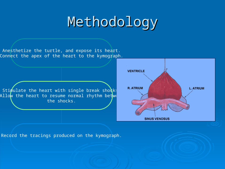
Anesthetize the turtle, and expose its heart.Connect the apex of the heart to the kymograph.
Stimulate the heart with single break shocks.Allow the heart to resume normal rhythm between
the shocks.
Record the tracings produced on the kymograph.
ResultsResults
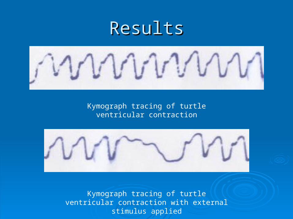
Kymograph tracing of turtle ventricular contraction
Kymograph tracing of turtle ventricular contraction with external stimulus applied
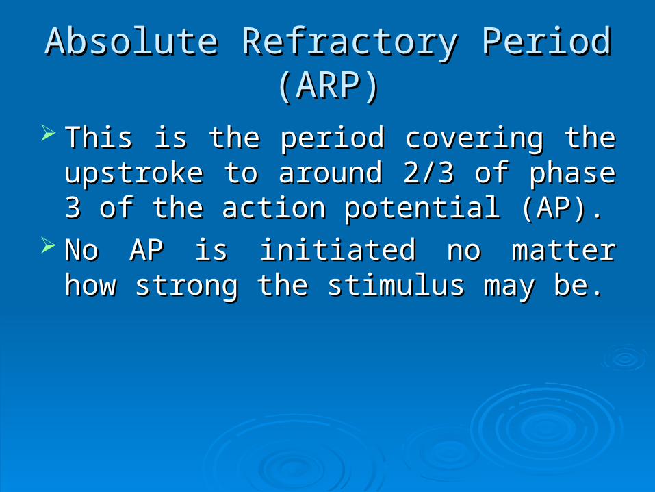
Absolute Refractory Period (ARP)Absolute Refractory Period (ARP)
This is the period covering the upstroke to This is the period covering the upstroke to around 2/3 of phase 3 of the action around 2/3 of phase 3 of the action potential (AP).potential (AP).
No AP is initiated no matter how strong the No AP is initiated no matter how strong the stimulus may be.stimulus may be.
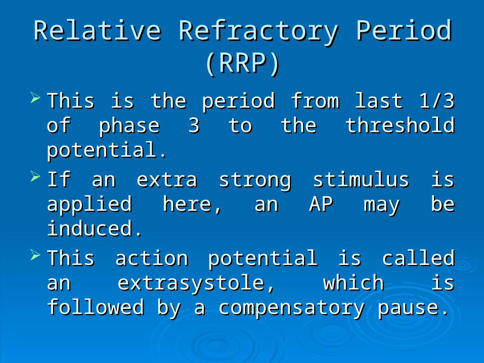
Relative Refractory Period (RRP)Relative Refractory Period (RRP)
This is the period from last 1/3 of phase 3 This is the period from last 1/3 of phase 3 to the threshold potential.to the threshold potential.
If an extra strong stimulus is applied here, If an extra strong stimulus is applied here, an AP may be induced.an AP may be induced.
This action potential is called an This action potential is called an extrasystole, which is followed by a extrasystole, which is followed by a compensatory pause.compensatory pause.
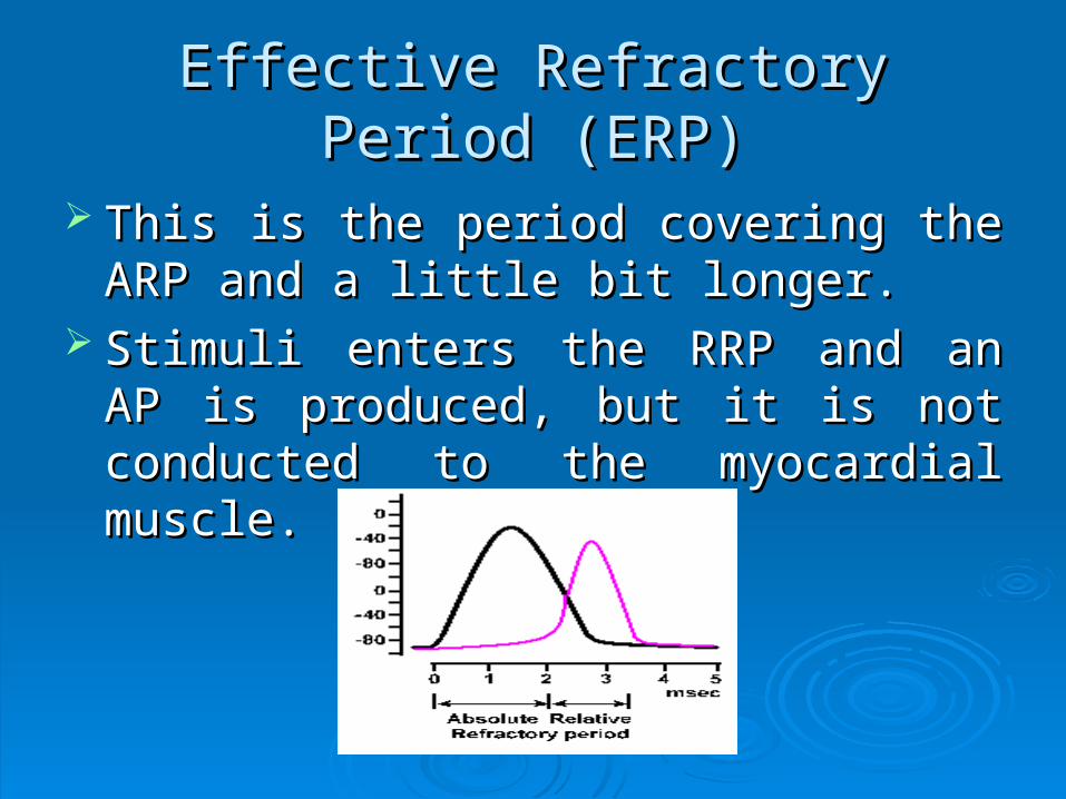
Effective Refractory Period (ERP)Effective Refractory Period (ERP)
This is the period covering the ARP and a This is the period covering the ARP and a little bit longer.little bit longer.
Stimuli enters the RRP and an AP is Stimuli enters the RRP and an AP is produced, but it is not conducted to the produced, but it is not conducted to the myocardial muscle.myocardial muscle.
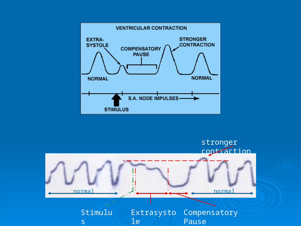
Extrasystole Compensatory Pause
stronger contraction
normal normal
Stimulus
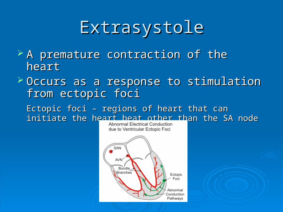
ExtrasystoleExtrasystole A premature contraction of the heartA premature contraction of the heart Occurs as a response to stimulation from Occurs as a response to stimulation from
ectopic fociectopic fociEctopic foci – regions of heart that can initiate the heart Ectopic foci – regions of heart that can initiate the heart beat other than the SA nodebeat other than the SA node

Compensatory PauseCompensatory Pause
A rest period after extrasystoleA rest period after extrasystole Duration is longer than normal interval Duration is longer than normal interval
between contractionsbetween contractions Allows the heart to resume its normal rhythm Allows the heart to resume its normal rhythm Allows the ventricles to completely refillAllows the ventricles to completely refill

ConclusionConclusion
External stimulation of the turtle heart External stimulation of the turtle heart causes a premature beat (extrasystole) causes a premature beat (extrasystole) which is then followed by a prolonged rest which is then followed by a prolonged rest period (compensatory pause), allowing the period (compensatory pause), allowing the heart to return to its normal rhythmheart to return to its normal rhythm
ANSWERS to QUESTIONSANSWERS to QUESTIONS
During what phase of the cardiac cycle did the During what phase of the cardiac cycle did the extra systoles fall?extra systoles fall?
The extra systole was observed during the The extra systole was observed during the relative refractory period of the cardiac cycle. The relative refractory period of the cardiac cycle. The relative refractory period is the period wherein the relative refractory period is the period wherein the muscle is difficult to excite but nevertheless, can muscle is difficult to excite but nevertheless, can be excitedbe excited
ANSWERS to QUESTIONSANSWERS to QUESTIONS
What was the period of rest?What was the period of rest?
The period of rest is the compensatory pause The period of rest is the compensatory pause which is the prolonged interval before a new which is the prolonged interval before a new contraction takes place. contraction takes place.
How does it compare with the normal?How does it compare with the normal?
The compensatory pause is usually longer than The compensatory pause is usually longer than the normal interval between contractions.the normal interval between contractions.
ANSWERS to QUESTIONSANSWERS to QUESTIONS
What is its value?What is its value?
The compensatory pause occurs because the The compensatory pause occurs because the external stimuli in the apex of the heart did not external stimuli in the apex of the heart did not disturb the normal rhythm of the SA node. This disturb the normal rhythm of the SA node. This allows the heart to resume its normal rhythm. It is allows the heart to resume its normal rhythm. It is its way to ensure that after an extra systole, it will its way to ensure that after an extra systole, it will be able to return to its normal rhythm and allow be able to return to its normal rhythm and allow the complete filling of the ventricles.the complete filling of the ventricles.
Experiment No. 16Experiment No. 16
AuricularAuricular
andand
VentricularVentricular
BlockBlock
ObjectiveObjective
To determine the effect of auricular and To determine the effect of auricular and ventricular block in auricular and ventricular block in auricular and ventricular contractions of a turtle heartventricular contractions of a turtle heart
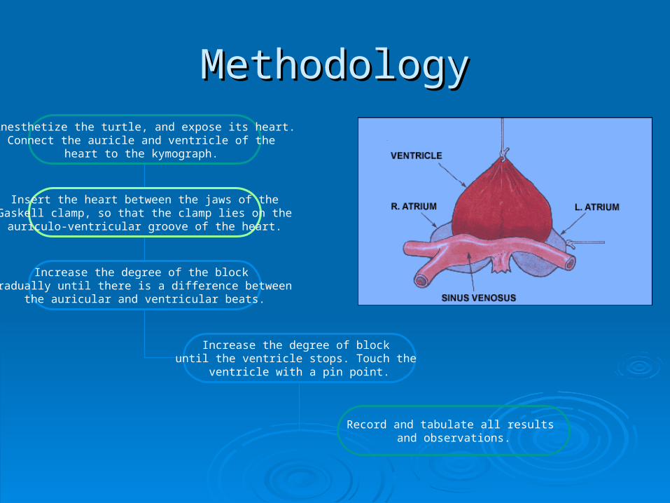
MethodologyMethodologyAnesthetize the turtle, and expose its heart.
Connect the auricle and ventricle of the heart to the kymograph.
Insert the heart between the jaws of theGaskell clamp, so that the clamp lies on the
auriculo-ventricular groove of the heart.
Increase the degree of the block gradually until there is a difference between
the auricular and ventricular beats.
Increase the degree of block until the ventricle stops. Touch the
ventricle with a pin point.
Record and tabulate all results and observations.
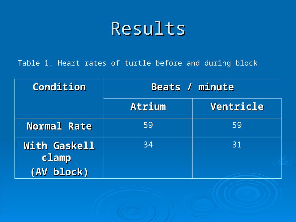
ResultsResults
ConditionCondition Beats / minuteBeats / minute
AtriumAtrium VentricleVentricle
Normal RateNormal Rate 59 59
With Gaskell With Gaskell clamp clamp
(AV block)(AV block)
34 31
Table 1. Heart rates of turtle before and during block
ConclusionConclusion
Auricular and ventricular block causes the Auricular and ventricular block causes the ventricle to lose the contraction impulse ventricle to lose the contraction impulse from the sinus venosus, forcing the from the sinus venosus, forcing the ventricle to produce its own contraction.ventricle to produce its own contraction.
ANSWERS to QUESTIONSANSWERS to QUESTIONS
What is the property of the ventricle seen in the What is the property of the ventricle seen in the experiment?experiment?
Automaticity was demonstrated in the experiment. Automaticity was demonstrated in the experiment. This is one important property of the cardiac This is one important property of the cardiac muscle which has the ability to initiate its own muscle which has the ability to initiate its own beat. In this case, the passage of impulses from beat. In this case, the passage of impulses from the atria to the ventricles is blocked, thus the the atria to the ventricles is blocked, thus the Purkinje fibers in the specialized conduction Purkinje fibers in the specialized conduction system of the ventricles become the pacemakers. system of the ventricles become the pacemakers.
ANSWERS to QUESTIONSANSWERS to QUESTIONS
What does the discrepancy in the rates prove?What does the discrepancy in the rates prove?
Discrepancy in the heart rates demonstrate the Discrepancy in the heart rates demonstrate the totality of the block. A complete block forced the totality of the block. A complete block forced the ventricular muscles to produce its own ventricular muscles to produce its own contractions.contractions.

Experiment No. 18Experiment No. 18
TheThe
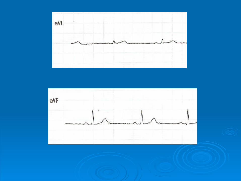
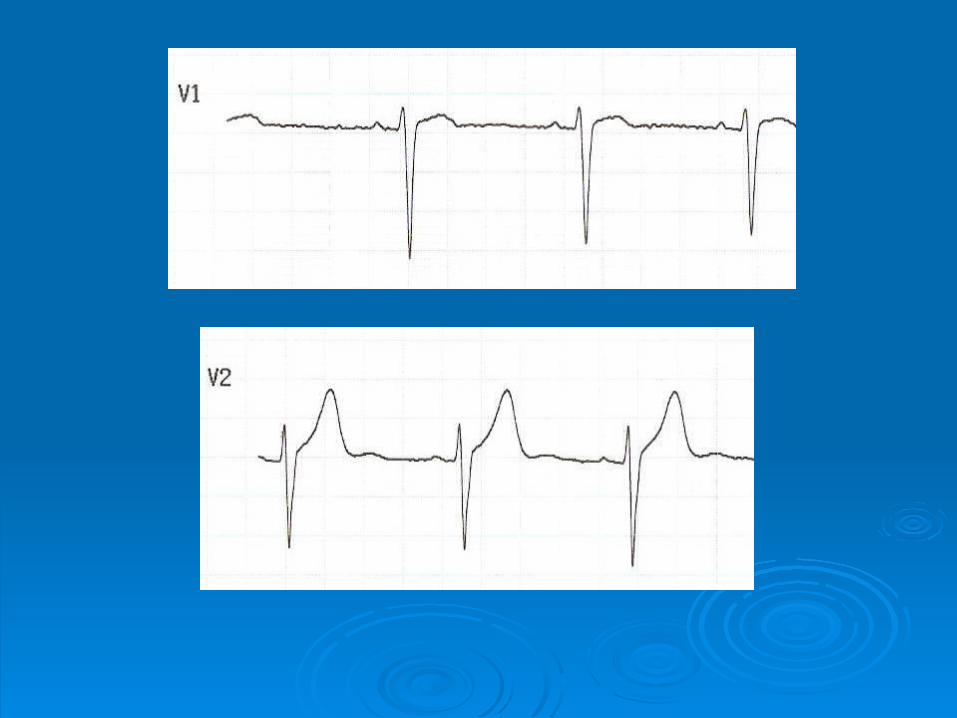
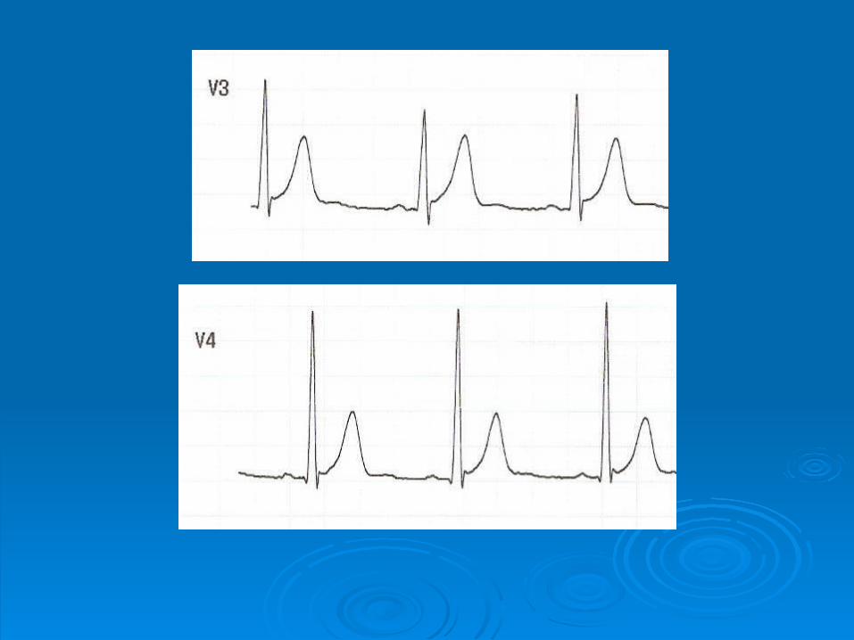
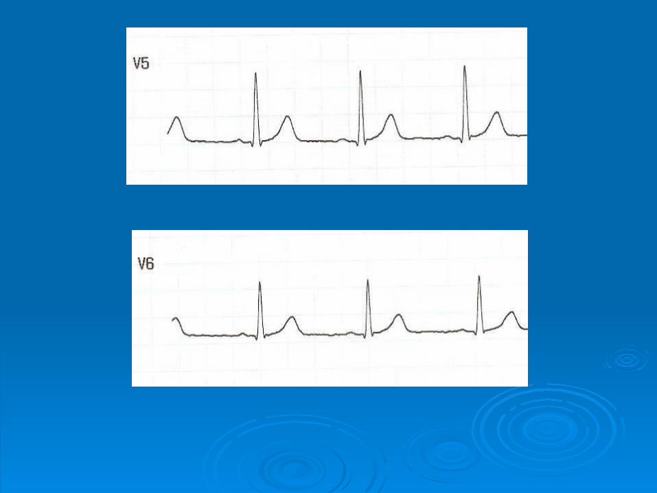
ElectrocardiogramElectrocardiogram
(ECG)(ECG)
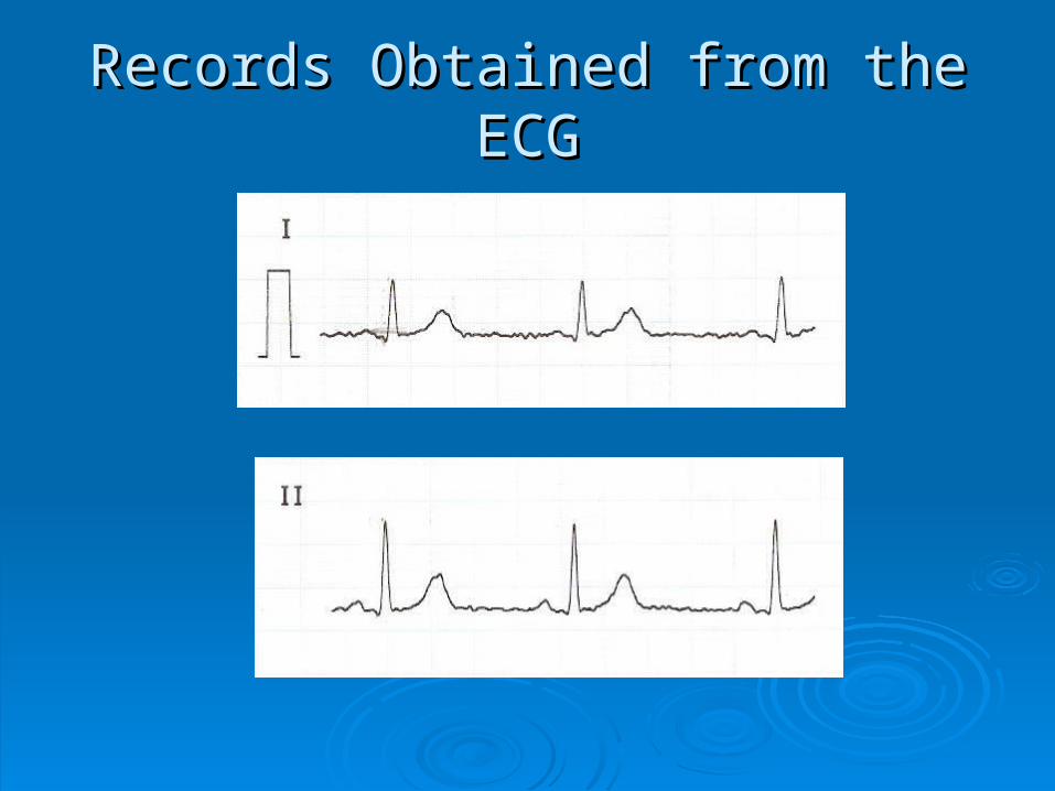
Records Obtained from the ECGRecords Obtained from the ECG

Heart RateHeart Rate
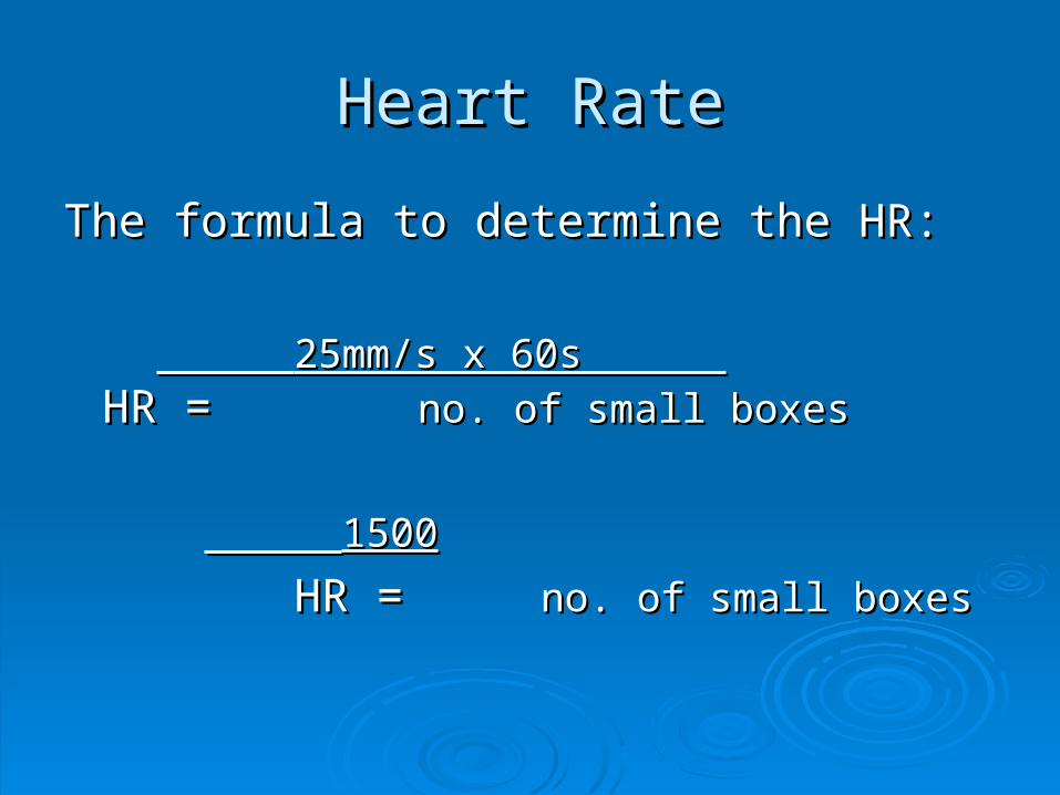
The formula to determine the HR:The formula to determine the HR:
25mm/s x 60s 25mm/s x 60s
HR = HR = no. of small boxesno. of small boxes
15001500
HR = HR = no. of small boxesno. of small boxes
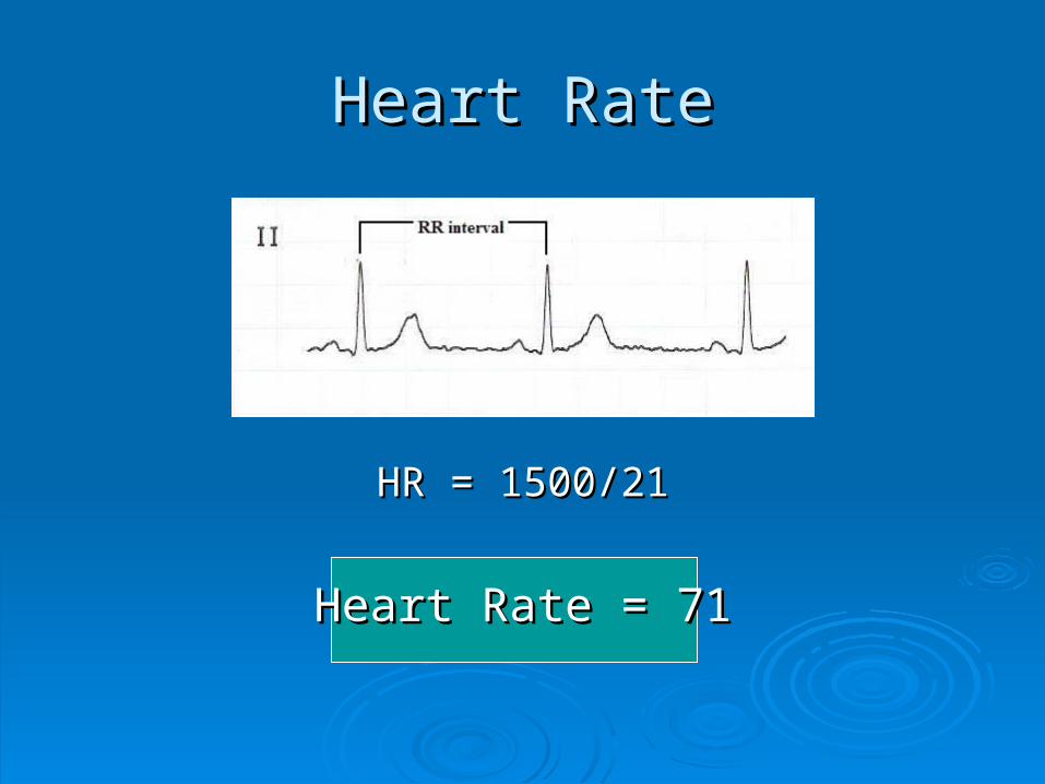
Heart RateHeart Rate
HR = 1500/21HR = 1500/21
Heart Rate = 71Heart Rate = 71
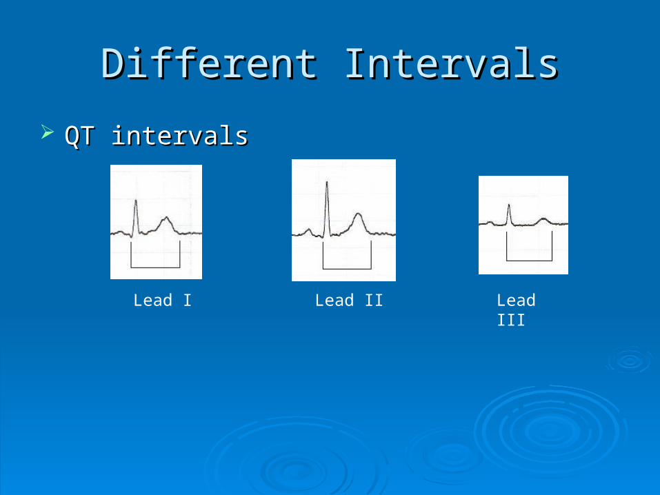
Different IntervalsDifferent Intervals
QT intervalsQT intervals
Lead I Lead II Lead III
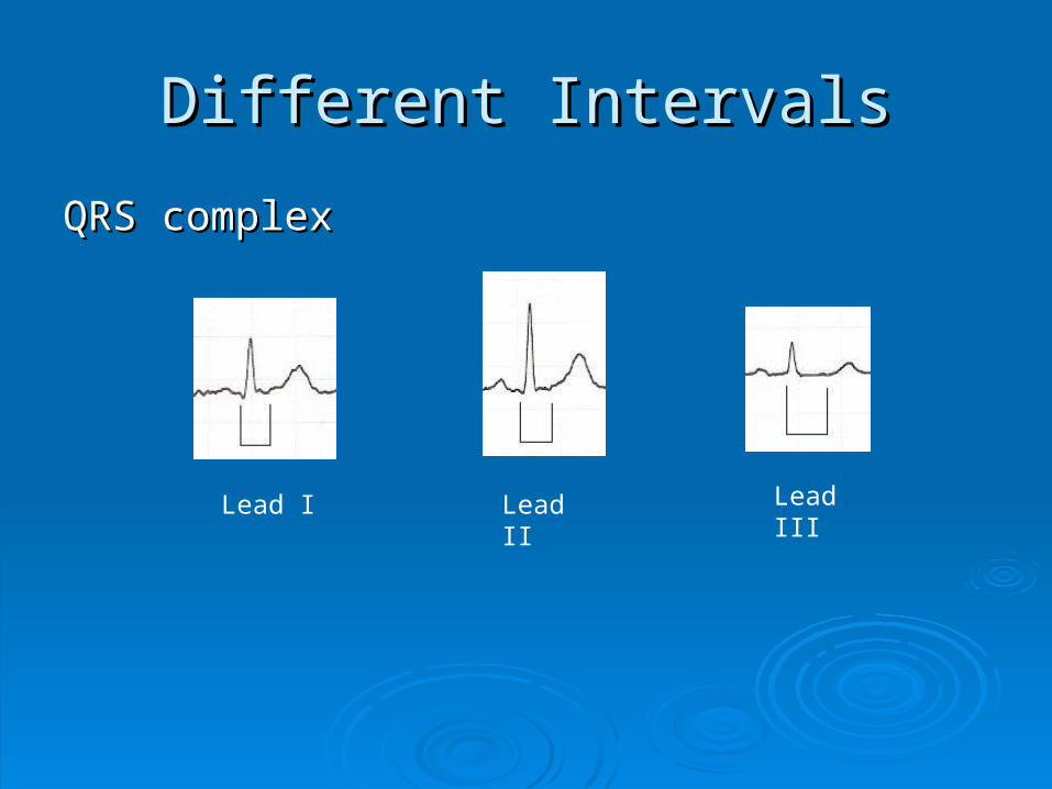
Different IntervalsDifferent Intervals
QRS complexQRS complex
Lead I Lead II Lead III
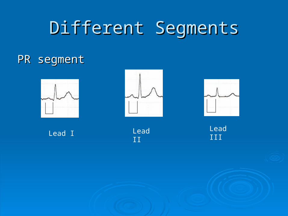
Different SegmentsDifferent Segments
PR segmentPR segment
Lead I Lead II Lead III
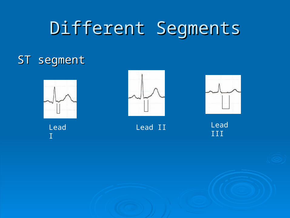
Different SegmentsDifferent Segments
ST segmentST segment
Lead I Lead II Lead III
Experiment No. 19Experiment No. 19
Blood Typing Blood Typing
and and
Cross-matchingCross-matching
Objective:
To determine the importance of blood typingand cross matching prior to blood transfusion.
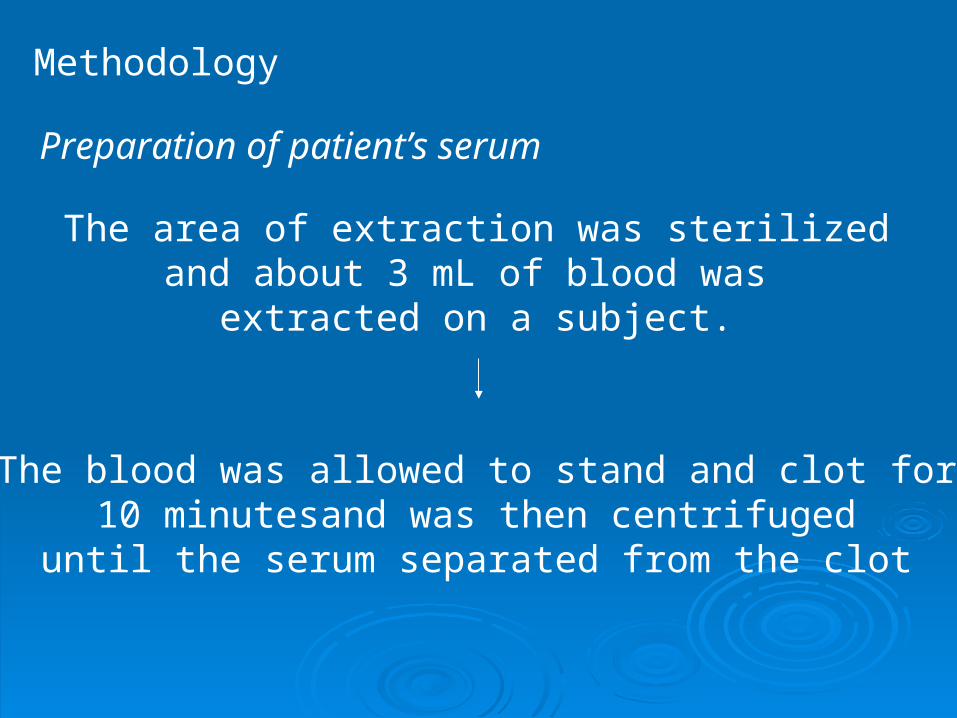
The area of extraction was sterilizedand about 3 mL of blood was
extracted on a subject.
The blood was allowed to stand and clot for 10 minutesand was then centrifuged until the serum separated from the clot
Preparation of patient’s serum
Methodology
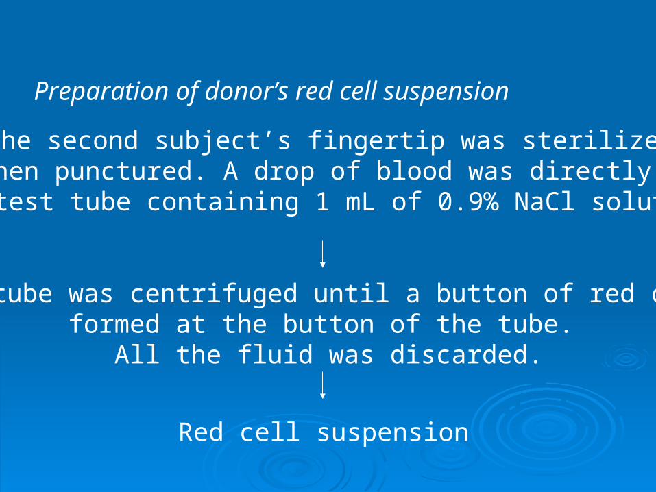
Preparation of donor’s red cell suspension
The second subject’s fingertip was sterilized and then punctured. A drop of blood was directly put in
the test tube containing 1 mL of 0.9% NaCl solution.
The tube was centrifuged until a button of red cell formed at the button of the tube.
All the fluid was discarded.
Red cell suspension
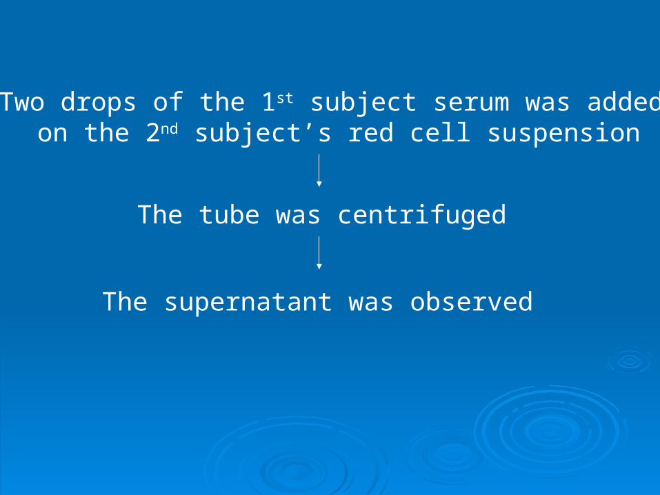
Two drops of the 1st subject serum was added on the 2nd subject’s red cell suspension
The tube was centrifuged
The supernatant was observed
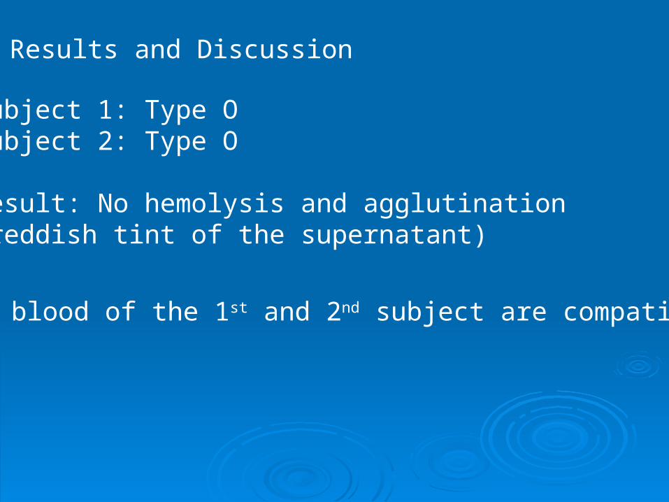
Results and Discussion
Subject 1: Type OSubject 2: Type O
Result: No hemolysis and agglutination(reddish tint of the supernatant)
The blood of the 1st and 2nd subject are compatible.
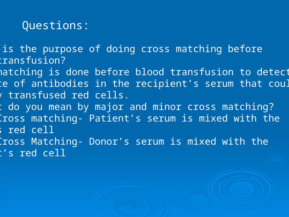
Questions:
1. What is the purpose of doing cross matching before blood transfusion?Cross matching is done before blood transfusion to detect presence of antibodies in the recipient’s serum that coulddestroy transfused red cells.2. What do you mean by major and minor cross matching?Major Cross matching- Patient’s serum is mixed with thedonor’s red cellMinor Cross Matching- Donor’s serum is mixed with the patient’s red cell
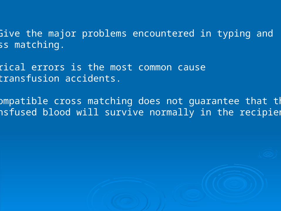
3. Give the major problems encountered in typing and cross matching.
Clerical errors is the most common cause of transfusion accidents.
A compatible cross matching does not guarantee that the transfused blood will survive normally in the recipient.
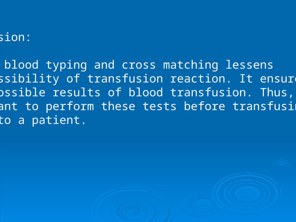
Conclusion:
Proper blood typing and cross matching lessens the possibility of transfusion reaction. It ensuresbest possible results of blood transfusion. Thus, it is important to perform these tests before transfusingblood to a patient.
Experiment No. 20Experiment No. 20
Bleeding time Bleeding time
and and
Clotting TimeClotting Time
Clotting timeClotting time
Objective:Objective: To determine the clotting time of the subject To determine the clotting time of the subject
and compare it to the normal values.and compare it to the normal values.
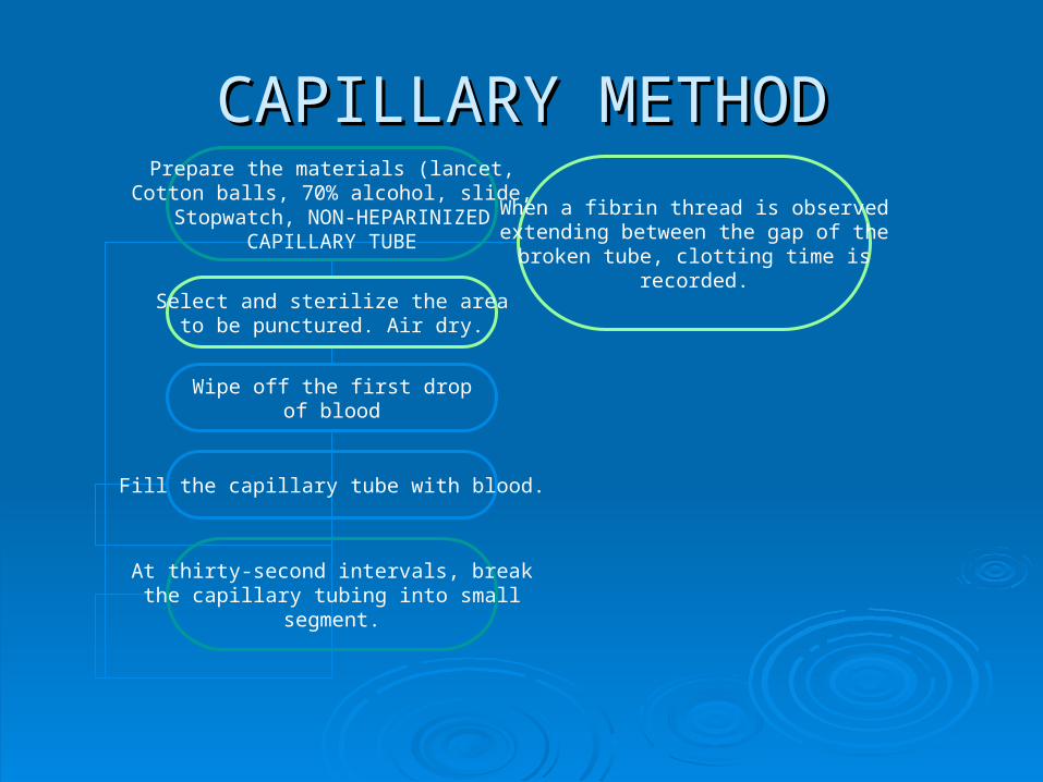
CAPILLARY METHODCAPILLARY METHODPrepare the materials (lancet,
Cotton balls, 70% alcohol, slide,Stopwatch, NON-HEPARINIZED
CAPILLARY TUBE
Select and sterilize the areato be punctured. Air dry.
Wipe off the first drop of blood
Fill the capillary tube with blood.
At thirty-second intervals, breakthe capillary tubing into small
segment.
When a fibrin thread is observedextending between the gap of the
broken tube, clotting time isrecorded.
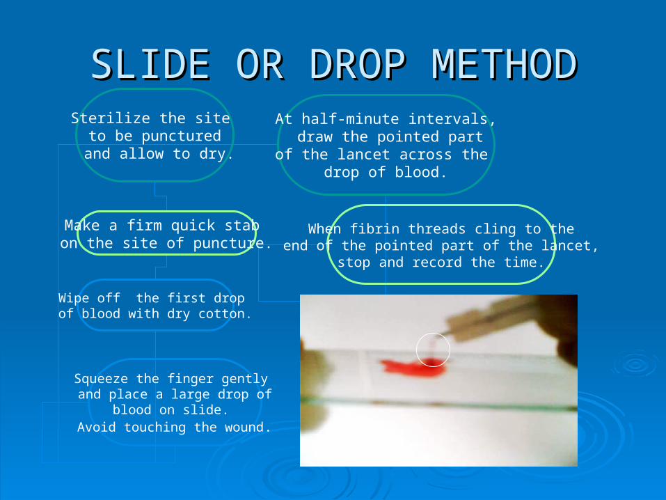
SLIDE OR DROP METHODSLIDE OR DROP METHODSterilize the site to be punctured
and allow to dry.
Make a firm quick stab on the site of puncture.
Wipe off the first drop of blood with dry cotton.
Squeeze the finger gently and place a large drop of
blood on slide. Avoid touching the wound.
At half-minute intervals, draw the pointed part
of the lancet across the drop of blood.
When fibrin threads cling to theend of the pointed part of the lancet,
stop and record the time.
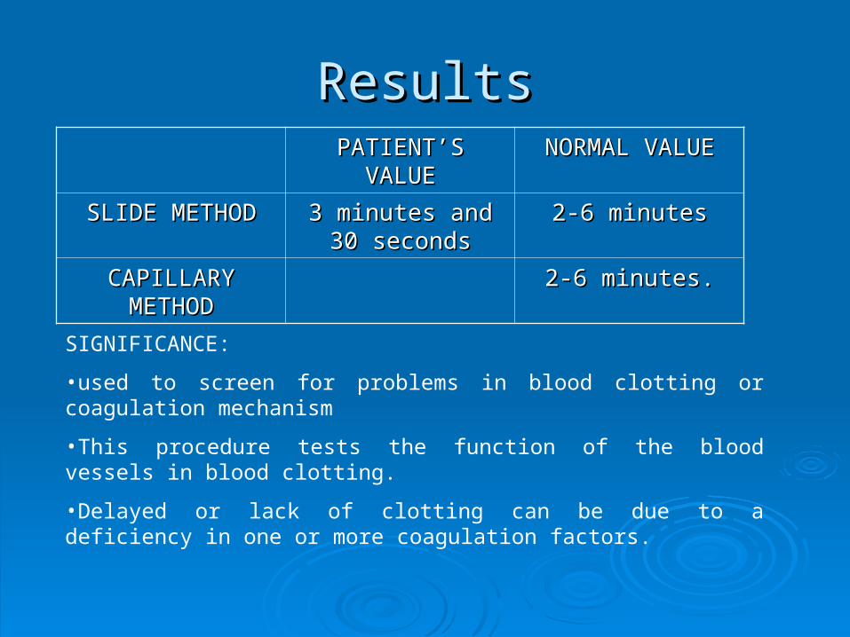
ResultsResultsPATIENT’S VALUEPATIENT’S VALUE NORMAL VALUENORMAL VALUE
SLIDE METHODSLIDE METHOD 3 minutes and 30 3 minutes and 30 secondsseconds
2-6 minutes2-6 minutes
CAPILLARY CAPILLARY METHODMETHOD
2-6 minutes.2-6 minutes.
SIGNIFICANCE:
•used to screen for problems in blood clotting or coagulation mechanism
•This procedure tests the function of the blood vessels in blood clotting.
•Delayed or lack of clotting can be due to a deficiency in one or more coagulation factors.
Bleeding TimeBleeding Time
Objective:Objective: To determine the bleeding time of the patient, To determine the bleeding time of the patient,
compare it with the normal value and know compare it with the normal value and know the significance of this test.the significance of this test.
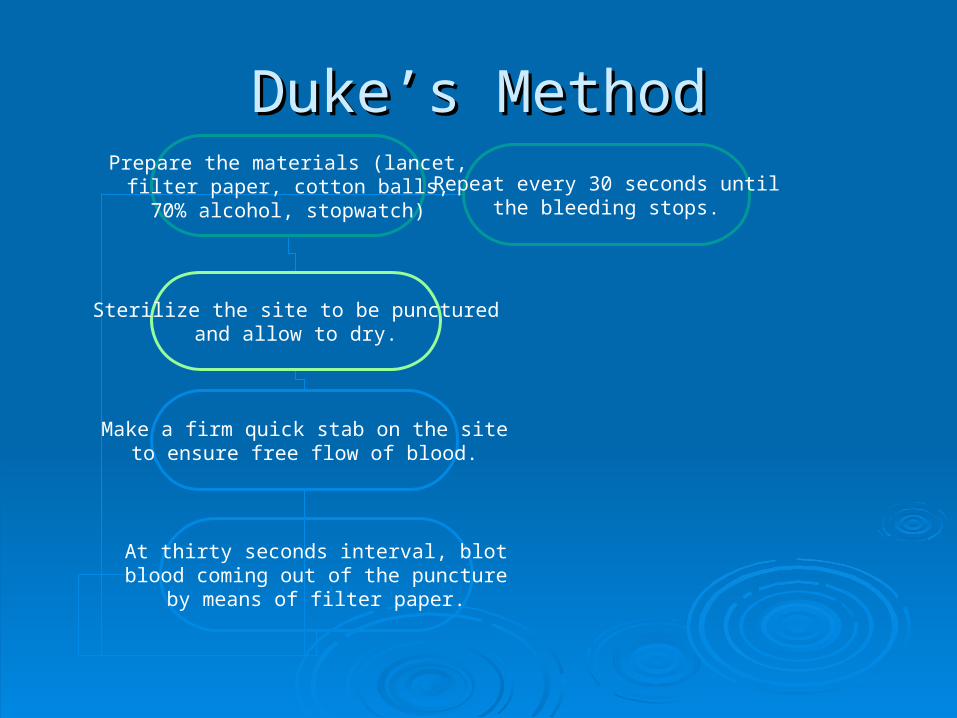
Duke’s MethodDuke’s MethodPrepare the materials (lancet,
filter paper, cotton balls,70% alcohol, stopwatch)
Sterilize the site to be puncturedand allow to dry.
Make a firm quick stab on the siteto ensure free flow of blood.
At thirty seconds interval, blotblood coming out of the puncture
by means of filter paper.
Repeat every 30 seconds untilthe bleeding stops.

ResultResultPATIENT’S PATIENT’S VALUEVALUE
NORMAL NORMAL VALUEVALUE
2 MINUTES2 MINUTES 1-4 MINUTES1-4 MINUTES
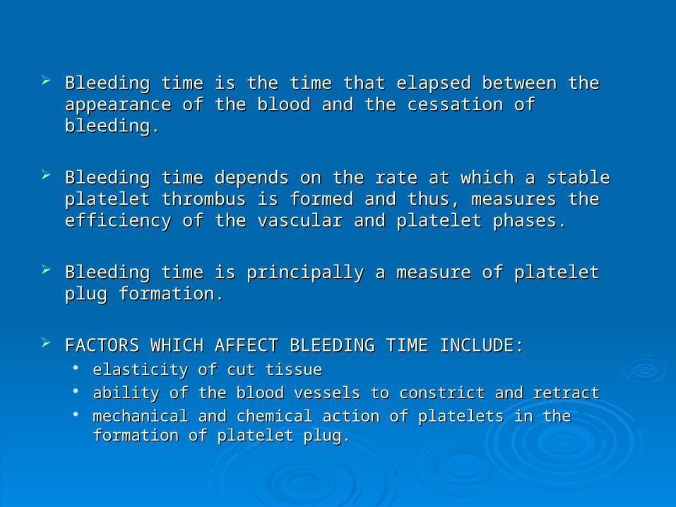
Bleeding time is the time that elapsed between the appearance of Bleeding time is the time that elapsed between the appearance of the blood and the cessation of bleeding.the blood and the cessation of bleeding.
Bleeding time depends on the rate at which a stable platelet Bleeding time depends on the rate at which a stable platelet thrombus is formed and thus, measures the efficiency of the thrombus is formed and thus, measures the efficiency of the vascular and platelet phases.vascular and platelet phases.
Bleeding time is principally a measure of platelet plug formation.Bleeding time is principally a measure of platelet plug formation.
FACTORS WHICH AFFECT BLEEDING TIME INCLUDE:FACTORS WHICH AFFECT BLEEDING TIME INCLUDE: elasticity of cut tissueelasticity of cut tissue ability of the blood vessels to constrict and retractability of the blood vessels to constrict and retract mechanical and chemical action of platelets in the formation of platelet mechanical and chemical action of platelets in the formation of platelet
plug.plug.


