
1
JOURNAL OF HERPETOLOGY
LRH: A. Edwards and S. M. Jones
RRH: Reproduction in male blue-tongued skinks
Changes in Plasma Testosterone, Estrogen and Progesterone Concentrations
Throughout the Annual Reproductive Cycle in Male Viviparous Blue-tongued
Skinks, Tiliqua nigrolutea, (Scincidae), in Tasmania
ASHLEY EDWARDS AND SUSAN M. JONES
School of Zoology, University of Tasmania, GPO Box 252C-05, Hobart,
Tasmania, 7001, Australia email: [email protected]
Key Words: estrogen, progesterone, reproduction, skink, testosterone, testis, Tiliqua nigrolutea
2
Abstract.— Few published studies have detailed comprehensively the correlations
between plasma steroid hormone peaks and the timing of reproductive events in male
squamate reptiles. We examined the patterns of plasma testosterone (T), estrogen (E) and
progesterone (P4) concentrations in males of the viviparous blue-tongued skink, Tiliqua
nigrolutea, throughout the annual cycle. Plasma T concentrations varied through the
annual cycle, peaking at 10.9 ± 3.00 ng ml-1 during spermiogenesis, coincident with
agonistic male–male interactions, but falling prior to the mating period. Mean plasma T
concentrations were basal (2 – 3 ng ml-1) during reproductive quiescence. Mean plasma
E concentrations were significantly elevated (778.0 ± 120.00 pg ml-1) during the mating
period, but basal (<300 pg ml-1) both before and after mating. Mean plasma P4
concentrations peaked during the mating period (1.1 ± 0.17 ng ml-1) and declined
significantly after mating. We propose a potential role for E and P4 in the stimulation of
male reproductive behavior during the mating period.
3
INTRODUCTION
The gonadal steroids which control reproduction in reptiles are assumed to be
testosterone (T), progesterone (P4), and 17β-estradiol (E2) (Kime, 1987). However, there
are few comprehensive published descriptions of the annual profiles of steroid
concentrations, particularly E2 and P4, in the plasma of male reptiles. Most studies of
reproduction in male squamates have described simply the annual cycle of hypertrophy
and regression of reproductive organs (Sanyal and Prasad, 1967; Nilson, 1980; Krohmer
and Aldridge, 1985; Flemming, 1993a; Shea, 1993; Aldridge and Brown, 1995), or have
combined such information with descriptions of cycles of plasma androgen
concentrations only (Arslan et al., 1978; Courty and Dufaure, 1979; 1980; Johnson et al.,
1982; McKinney and Marion, 1985; Bourne et al., 1986; Moore, 1986; Flemming, 1993b;
Swain and Jones, 1994; Schuett et al., 1997). Thus, in many cases, supposition about the
roles of these gonadal steroids in the regulation of reptilian reproduction is based on
analogy with mammals rather than experimental evidence (Ozon, 1972; Wiebe, 1985;
Chieffi and Pierantoni, 1987).
Elevated plasma androgens have been implicated in the stimulation of
reproductive behavior in male vertebrates, including reptiles (Crews, 1975; Crews et al.,
1978; Lance, 1984; Lindzey and Crews, 1986; Moore, 1987). However, male mating
behavior in the garter snake Thamnophis sirtalis parietalis is at least partially (Crews,
1991), if not completely (Mendonca et al., 1996), independent of androgens. Several
studies have suggested that in male reptiles, estrogens may influence reproductive
behavior through aromatisation of androgens in the brain. Aromatase activity has been
detected in the brains of the turtle Chrysemys picta (Callard et al., 1977) and males of the
4
lizard Podarcis sicula sicula (Gobbetti et al., 1994). In the lizard A. carolinensis,
exogenous E2 more reliably reinstates reproductive behavior in male castrates than T
(Crews and Morgentaler, 1979). However, aromatisation of T to E2 does not appear to
be necessary for the induction of the reproductive behaviors that occur coincident with
increases in plasma testosterone in male A. sagrei (Tokarz, 1986).
An increasing number of published studies suggest that P4 may also be important
in stimulating reproductive behavior in some male squamates (Lindzey and Crews, 1988;
1992; Young et al., 1991; Witt et al., 1994). Exogenous P4 has been shown to stimulate
sexual behaviors in males of the lizard Cnemidophorus inornatus (Lindzey and Crews,
1986). This finding is contrary to the usual in male vertebrates; typically P4 inhibits
male sexual behaviors (Moore and Lindzey, 1992).
Given these conflicting reports, it is surprising that annual changes in plasma
concentrations of P4 or estrogen (E) in male reptiles have not been documented in more
species; such information is vital for us to better understand the hormonal control of
reproduction in reptiles. Saint Girons et al. (1993) provided one of the few published
studies in which the timing of behaviors associated with reproduction is correlated with
changes in concentrations of all three primary gonadal steroids in the plasma of a male
squamate reptile. They reported significant annual variation in plasma T, E2 and P4
concentrations in male Vipera aspis, correlated with the timing of physiological and
behavioral events (Saint Girons et al., 1993). However, more studies of this nature are
required.
Tiliqua nigrolutea is a large, viviparous skink distributed throughout southeastern
Australia (Cogger, 1992). Adult males can range from 25–29 cm snout–vent length
5
(SVL) and weigh between 300 and 450 g, with females somewhat larger and heavier. In
Tasmania, where this study was conducted, T. nigrolutea occurs in low altitude heath,
savanna woodland and dry sclerophyll forest in the cool temperate regions of the state
(Rawlinson, 1974). Presented here is a comprehensive examination of annual cycles of
plasma T, E and P4 concentrations in male T. nigrolutea, correlated with the timing of
agonistic and mating behaviors.
6
MATERIALS AND METHODS
Animals.— Lizards were captured opportunistically by hand throughout
southeastern Tasmania from Sep – Jan. Males were distinguished from females by their
relatively broader heads (our unpublished data) and an examination of the cloacal
opening for the musculature of the hemipenes. Animals were housed in roofed outdoor
enclosures 1.9 x 3.4 x 2.1 m; these were wire-fronted, allowing access to UV light and a
natural photoperiod. The direct sunlight and a 120 W floodlight globe as an additional
heat source at the front of each cage provided a temperature gradient across which the
lizards could thermoregulate during their active season of the austral spring to mid-
autumn (Sept - Apr). We provided bark and leaf litter in which the animals could hide.
Mixed-sex groups of approximately five animals (one male per group) were maintained
in these cages. Additional males were held separately in similar, but smaller cages, to
prevent agonistic interactions and their possible effects on plasma hormone
concentrations. The lizards were maintained on a varied diet of fresh fruits, live snails
and tinned catfood, provided two to three times weekly. Water was available ad libitum.
The number of captive male animals varied from 12–19 over the period of the study.
Blood sampling.— Blood samples were collected at monthly intervals. Samples
were taken routinely between 0930 and 1230 without anaesthesia from the caudal artery,
using a heparinised syringe. Samples were held on ice until centrifuging at 6400 rpm and
plasma was stored frozen at -20 C until analysis. Up to 1 ml of blood was taken from
each animal, although some samples were much smaller and occasionally no blood was
obtained. Twelve samples were collected from each of 10 animals for the measurement

7
of plasma T and eight animals for the measurement of P4 concentrations and nine
samples were obtained from each of nine animals for the measurement of E.
Radioimmunoassays.— Analytical reagent grade isooctane, hexane and ethanol
were purchased from Biolab Scientific Pty. Ltd. (Victoria, Aust.). Scintillation fluid
(Ecolite +) came from ICN (Costa Mesa, CA.). [1,2,6,7-3H]-Testosterone (spec. act. 100
Ci/mmol) and [1,2,6,7-3H]-P4 (spec. act. 80-110 Ci/mmol) were purchased from
Amersham Life Sciences (UK). Testosterone antiserum was a gift from A. J. Bradley
(details in Bradley, 1990). Plasma T concentrations were assayed by a modification to
the radioimmunoassay of Castro et al. (1974) as detailed in Swain and Jones (1994).
Inter- and intraassay coefficients of variation for the testosterone assay were < 10% and
6%, respectively (Swain and Jones, 1994). Progesterone antiserum was from J. Malecki
(details published in McDonald et al., 1988). The P4 radioimmunoassay method was
described in Jones and Rose (1992) with a minor modification for this study: P4 was
eluted from the columns in 3 ml isooctane. Intra- and interassay coefficients of variation
for the P4 assay were 12.1% and 8.4% respectively. All T and P4 assay samples were
measured as outlined in Jones and Rose (1992). Plasma E was measured using Spectria
coated-tube radioimmunoassay kits as in Jones and Swain (1996). Cross-reactivities for
the E2 antiserum are: E2, 100%; estrone (E1), 1.16%; estriol, 0.45%; T and P4, <0.001%.
Intra- and interassay coefficients of variation for this assay were 13% and 8%,
respectively. The limit of detection for all three assays was 10 pg authentic steroid.
Assays were validated using T. nigrolutea plasma (T and E assays) or pooled skink
plasma (P4 assay): in all cases serial dilutions of plasma ran parallel to the standard
curves.
8
Statistics.— Mean monthly plasma hormone concentrations were compared by
repeated measures analysis of variance ((M)ANOVA) using SYSTAT 8.0 (Wilkinson et
al., 1998). A significance level of α = 0.05 was used throughout. All data were log
transformed prior to analysis to satisfy assumptions of normality and homogeneity of
variance and all values are presented as mean ± 1 standard error (SE). The original data
sets were reduced to include only those individuals for which samples from all (or most)
sample periods were available. Occasional missing data points were assigned the mean
value for animals in the same sample period, although no more than one such value was
assigned to any individual or any sample period (Mundry, 1999; D. Ratkowsky, pers.
comm.). A posteriori Student t tests were conducted for each hormone profile, as the
precise timing of these events was unknown before samples were collected. The periods
inspected were either the animals’ emergence from hibernation or the mating period.
Visual examination of completed hormone profiles and concurrent behavioral
observations of the captive sample population was used to identify relevant successive
pairs of sample sets.
9
RESULTS
Plasma steroid concentrations.— Mean monthly plasma T concentrations in male
T. nigrolutea from November 1995 to October 1996 are shown in Fig. 1. A distinct
unimodal annual cycle was evident, with uniformity between males in both the timing of
the seasonal plasma T pattern and the magnitude of plasma T concentrations. Mean
plasma T concentration varied significantly throughout the annual cycle ((M) ANOVA: F
(9,11) = 12.504, P = 0.000). Plasma T concentrations decreased significantly (t = 3.097, P
= 0.013) from 8.8 ± 0.79 ng ml-1during the mating period (Nov) to 5.3 ± 0.62 ng ml-1 in
early summer (Dec) when post-mating male lizards were reproductively quiescent.
Plasma T concentrations were basal (approx. 1–3 ng ml-1) from mid–summer to mid–
winter (Jan–Jul), before increasing significantly (t = -2.450, P = 0.037) to 8.0 ± 3.12
ng ml-1in late winter (Aug), when male lizards emerged from hibernation, about four
weeks earlier than females. Mean plasma T concentration peaked at 10.9 ± 3.05 ng ml-
1in mid spring (Oct), coincident with the completion of spermiogenesis and the
observation of the onset of agonistic male-male interactions. There was no correlation
between peak plasma T concentration (Oct) and SVL.
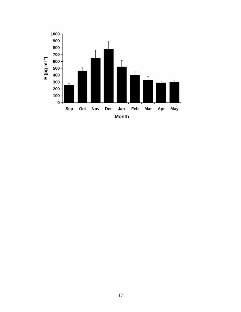
As with the plasma T profile, the annual pattern of changing E concentrations in
the plasma of male T. nigrolutea varied significantly throughout the reproductive cycle
(Fig. 2) ((M)ANOVA: F(7,8) = 6.267, P = 0.000). Mean plasma E2 concentration was
basal (<300 pg ml-1) in emergent animals in early spring (Sept), but became significantly
elevated to 460.8 ± 55.41 pg ml-1 from mid spring (Oct) (t = -6.721, P = 0.000), when
spermatogenesis had been completed, through to late spring and early summer (Nov–
Dec), when mating was observed, peaking at 778.0 ± 120.99 ng ml-1 in December and
10
dropped sharply to 396.9 ± 53.44 pg ml-1by mid summer (Feb), when males were
reproductively quiescent. Concentrations remained low (approximately 300 pg ml-1)
until males began hibernating in late autumn (May).
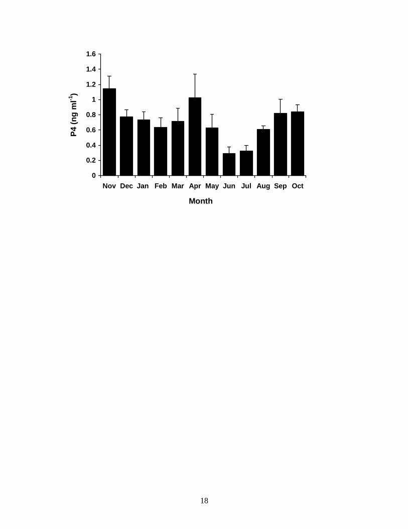
Mean monthly plasma P4 concentrations in male T. nigrolutea from November
1995 to October 1996 were low but above the limit of detection of the assay throughout
the year, and varied significantly over time (Fig. 3) ((M)ANOVA: F(8,11) = 4.556, P =
0.002). Plasma P4 concentration peaked during the mating period (Nov) at 1.1 ± 0.17 ng
ml-1 and fell significantly to 0.8 ± 0.09 ng ml-1 by early summer (Dec) (t = 4.334, P =
0.002). There was a further significant decline to 0.3 ± 0.08 ng ml-1 in mid-winter (Jun)
(t = 2.458, P = 0.039) but mean plasma P4 concentration rose significantly (t = -2.94, P =
0.019) when males emerged at the end of winter (Aug).
11
DISCUSSION
Male reptiles commonly display an annual pattern of plasma T concentrations
with low concentrations during the early stages of spermatogenesis and a peak during
spermiogenesis, corresponding with peak testicular hypertrophy (Lance, 1984). Male
Tiliqua nigrolutea exhibit this typical pattern: plasma T concentrations are low during
autumn (Mar–Apr) when spermatogenesis commences, but rise during mid–spring (Oct),
peaking at 10.9 ± 3.05 ng ml-1 and declining through the second half of the mating period.
This pattern closely resembles that of the plasma T cycle of T. rugosa (Bourne and
Seamark, 1975), although peak plasma T concentrations (approximately 40 ng ml-1) are
higher in T. rugosa. However, the major androgen in T. rugosa is epitestosterone (epiT),
not found in T. nigrolutea (Bourne et al., 1985), and epiT is present in much higher
concentrations (approximately 140 ng ml-1) than T in the blood (Bourne et al., 1986).
Among reptiles, much variation occurs in the magnitude of the T peaks between species.
Plasma T peaks at 80 -100 ng ml-1 in the snake, Agkistrodon contortix, (Schuett et al.,
1997) and 51.7 ± 1.6 ng ml-1 in the viviparous lizard, Niveoscincus metallicus (Swain and
Jones, 1994), but only approximately 3 ng ml-1 in the snake A. piscivorus (Johnson et al.,
1982).
Circulating concentrations of E also follow an annual pattern in male T.
nigrolutea. Plasma E concentrations are elevated during the mating period (Oct – Dec)
but drop rapidly to basal concentrations for the rest of the active season. This implies a
role for E in the induction of sexual behaviors. However, the relatively high plasma E
concentrations detected suggest we may be measuring one or more alternative estrogens
in addition to E2 (V. Lance, pers. comm.). Additionally, E2 is not synthesised from

12
pregnenolone (P5) by the testes or from T by peripheral tissues in vitro (Edwards, 1999).
Given that plasma T declines during the mating period in male T. nigrolutea, we suggest
that an alternative estrogen may be responsible for stimulating mating behavior in males
of this species. Cross-reactivity of the kit antiserum used in this study with other
common estrogens (E1 and E3) is low, but no information is provided by manufacturers
about cross-reactivity at the six position of the steroid nucleus, which is modified in the
generation of the antiserum. Whittier and Hess (1992) reported the cross-reactivity of the
polar steroids 6α- and 6β-hydroxyestradiol (6α-and 6β-OH-E2) with an E2 kit antiserum
in another squamate reptile, the garter snake, T. s. parietalis. A presently unidentified
steroid more polar than E2 has been isolated from in vitro incubations using T. nigrolutea
testicular tissue, but it is less polar than 6α-and 6β-OH-E2 (Edwards, 1999).
Males of other lizards exhibit annual cycles of E2 production, elevated plasma E2
does not always coincide with mating. Saint Girons et al. (1993) demonstrated that
sexually active male Vipera aspis have high plasma T and low E2 during the mating
period, with E2 being elevated to approximately 520 pg ml-1 in non-mating males. In
male Podarcis s. sicula plasma E2 concentrations increase in the post-reproductive
refractory period to about 1.5 ng ml-1 (Ando et al., 1992). Intracranial implants of E2 in
castrated male A. carolinensis restore sexual behaviors (Crews and Morgentaler, 1979),
but later studies on anoles suggest that the expression of reproductive behavior in Anolis
species is under the direct control of androgens (Adkins-Regen, 1981; Tokarz, 1986).
We also examined plasma concentrations of P4 in male T. nigrolutea. Plasma P4
concentrations showed a seasonal pattern in male T. nigrolutea: mean concentrations
were low throughout the year with a small but significant elevation prior to emergence

13
(Aug). This significant change observed in male plasma P4 concentrations may be,
simply, a function of an overall increase in metabolism as a result of higher temperatures
during the animals’ active season, such as has been documented in other ectothermic
vertebrates (Kime, 1979; 1987; Kime and Hyder, 1983). However, P4 has been shown to
stimulate male reproductive behavior in some lizards (Lindzey and Crews, 1986; 1988;
1992; Young et al., 1991; Moore and Lindzey, 1992; Witt et al., 1994) acting as a
progestin rather than through conversion to other steroids (Moore and Lindzey, 1992).
Plasma P4 concentrations have also been measured in males of the lizard P. s. sicula, in
which there was a post-reproductive increase which peaked at approximately 8 ng ml-1
(Ando et al., 1992). The lack of a pronounced annual pattern in T. nigrolutea, however,
implies that P4 may not have a primary role in the induction of reproductive behaviors in
males of this species.
Bourne et al. (1986) suggested that in Tiliqua rugosa long-term captivity may
negatively affect the expression of reproductive behavior and depress plasma steroid
concentrations; captive male T. rugosa do not display a seasonal plasma androgen cycle
(Watson et al., 1987). All males of T. nigrolutea used in our study, however, continued
to cycle normally in comparison with opportunistically sighted and wild-caught
individuals in the timing and magnitude of plasma steroid peaks, the timing of regular
skin moults and in the expression of agonistic and mating behaviors. There was no
significant difference in mean plasma T concentration in November between males in the
captive population and eight wild individuals that were opportunistically captured at that
time (Edwards and Jones, unpubl. data). Any stress caused by handling and blood
sampling is unlikely to have had a significant impact on plasma steroid concentrations.
14
Kreger and Mench (1993) considered the impact of handling and restraint on Tiliqua
scincoides and found no significant chronic effect on plasma concentrations of the stress
steroid hormone, corticosterone (B). Additionally, Moore et al. (1991) observed that the
effects of acute handling stress in the lizard Urosaurus ornatus are rapidly dissipated.
Tiliqua nigrolutea is a placid animal that adapts quickly to captive life. Our animals were
housed under conditions of natural temperature and photoperiod with only an additional
heat source provided for basking, and underwent a normal hibernation. We are confident
that the mean plasma steroid concentrations reported here reflect those in wild
populations.
Further experimental studies are required to elucidate the hormonal control of
reproductive behaviors in males of T. nigrolutea and other squamates as there appear to
be differences between species in the plasma steroid hormones that are elevated during
mating. This study has addressed the lack of published work in which data on annual
steroid hormone profiles and the timing of coincident reproductive events are available.
Acknowledgments.— This research was funded by a School of Zoology,
University of Tasmania postgraduate research allowance to A.E. We thank A.J. Bradley
for generous gifts of antisera and D. Ratkowsky for statistical advice. This research was
carried out under University of Tasmania Animal Ethics approval number 95046.

15
FIGURE LEGENDS
FIG. 3.1. Changes in mean monthly plasma testosterone concentrations in male T.
nigrolutea throughout the annual reproductive cycle. Sampling was from November 1995
to October 1996. Values are means ± 1 standard error, N = 10.
FIG. 3.2. Changes in mean monthly plasma estrogen concentrations in male T. nigrolutea
throughout the annual reproductive cycle. Sampling was from September 1996 to May
1997. Values are means ± 1 standard error, N = 8.
FIG. 3.3. Changes in mean monthly plasma progesterone concentrations in male T.
nigrolutea throughout the annual reproductive cycle. Sampling was from November 1995
to October 1996. Values are means ± 1 standard error, N = 9.

16
0
2
4
6
8
10
12
14
16
Nov Dec Jan Feb Mar Apr May Jun Jul Aug Sep Oct
Month
T (n
g m
l-1)
17
0100200300400500600700800900
1000
Sep Oct Nov Dec Jan Feb Mar Apr May
Month
E (p
g m
l-1)
18
0
0.2
0.4
0.6
0.8
1
1.2
1.4
1.6
Nov Dec Jan Feb Mar Apr May Jun Jul Aug Sep Oct
Month
P4 (n
g m
l-1)

19
LITERATURE CITED ADKINS-REGAN E. 1981. Hormone specificity, androgen metabolism and social
behaviour. Amer. Zool. 21:257-271.
ALDRIDGE R. D., and BROWN W. S. 1995. Male reproductive cycle, age at maturity, and
cost of reproduction in the timber rattlesnake (Crotalus horridus). J. Herpetol.
29:399-407.
ANDO S., CIARCIA G., PANNO M. L., IMBROGNO E., TARANTINO G., BUFFONE M.,
BERALDI E., ANGELINI F., and BOTTE V. 1992. Sex steroids in the plasma and testis
during the reproductive cycle of the lizard Podarcis s. sicula Raf. Gen. Comp.
Endocrinol. 85:1-7.
ARSLAN M., LOBO J., ZAIDI A. A., JALALI S., and QAZI M. H. 1978. Annual androgen
rhythm in the spiny-tailed lizard, Uromastix hardwicki. Gen. Comp. Physiol. 36:16-
22.
BOURNE A. R., and SEAMARK R. F. 1975. Seasonal changes in 17β-hydroxysteroids in
the plasma of a male lizard (Tiliqua rugosa). Comp. Biochem. Physiol. 50B:535-
536.
BOURNE A. R., TAYLOR J. L., and WATSON T. G. 1985. Identification of epitestosterone
in the plasma and testis of the lizard Tiliqua (Trachydosaurus) rugosa. Gen. Comp.
Endocrinol. 58:394-401.
BOURNE A. R., TAYLOR J. L., and WATSON T. G. 1986a. Annual cycles of plasma and
testicular androgens in the lizard Tiliqua (Trachydosaurus) rugosa. Gen. Comp.
Endocrinol. 61:278-286.
20
BRADLEY A. J. 1990. Failure of glucocorticoid feedback during breeding in the male re-
tailed phascogale Phascogale calura (Marsupialia: Dasyuridae). J. Steroid
Biochem. 37:155-163.
CALLARD G. V., PETRO Z., and RYAN K. J. 1977. Identification of aromatase in the
reptilian brain. Endocrinology 100:1214-1218.
CASTRO A., SHIH H. H. W., and CHUNG A. 1974. A simple radioimmunoassay of
testosterone without column chromatography. Steroids 23:625-638.
CHIEFFI G., and PIERANTONI R. 1987. Regulation of ovarian steroidogenesis. In D. O.
Norris and R. E. Jones, (eds.), Hormones and Reproduction in Fishes, Amphibians
and Reptiles. pp. 117-144. Plenum Press, New York.
COGGER H. G. 1992. Reptiles and Amphibians of Australia. Reed Books, Australia.
COURTY Y., and DUFAURE J. P. 1979. Levels of testosterone in the plasma and testis of
the viviparous lizard (Lacerta vivipara Jaquin) during the annual cycle. Gen. Comp.
Endocrinol. 39:336-342.
COURTY Y., and DUFAURE J. P. 1980. Levels of testosterone, dihydrotestosterone, and
androstenedione in the plasma and testis of a lizard (Lacerta vivipara Jacquin)
during the annual cycle. Gen. Comp. Endocrinol. 42:325-333.
CREWS D. 1975. Psychobiology of reptilian reproduction. Science 189:1059-1065.
CREWS D. 1991. Trans-seasonal action of androgen in the control of spring courtship
behavior in male red-sided garter snakes. Proc. Nat. Acad. Sci. USA. 88:3545-3548.
CREWS D. and MORGENTALER A. 1979. Effects of intracranial implantation of oestradiol
and dihydrotestosterone on the sexual behaviour of the lizard Anolis carolinensis. J.
Endocrinol. 82:373-381.

21
CREWS D., TRAINA V., WETZEL F. T., and MULLER C. 1978. Hormonal control of male
reproductive behavior in the lizard, Anolis carolinensis: role of testosterone,
dihydrotestosterone, and estradiol. Endocrinology 103:1814-1820.
FLEMMING A. F. 1993a. The male reproductive cycle of the lizard Pseudocordylus. m.
melanotus (Sauria: Cordylidae). J. Herpetol. 27 473-478.
FLEMMING A. F. 1993b. Seasonal variation in testicular and fat-body weight and plasma
testosterone and androstenedione concentration in the lizard Cordylus polyzonus
(Sauria: Cordylidae). S. Afr. J. Zool. 28:127-131.
GOBBETTI A., ZERANI M., DI FIORE M. M., and BOTTE V. 1994. Relationships among
GnRH, substance P, prostaglandins, sex steroids and aromatase activity in the brain
of the male lizard Podarcis sicula sicula during reproduction. J. Reprod. Fert.
101:523-529.
JONES S. M. and ROSE R. W. 1992. Plasma progesterone levels in the pregnant female rat
kangaroo (Bettongia gaimardi). Gen. Comp. Endocrinol. 87:178-182.
JONES S. M. and SWAIN R. 1996. Annual reproductive cycle and annual cycles of
reproductive hormones in plasma of female Niveoscincus metallicus (Scincidae)
from Tasmania. J. Herpetol. 30:140-146.
JOHNSON L. F., JACOB J. S., and TORRANCE P. 1982. Annual testicular and androgenic
cycles of the cottonmouth (Agkistrodon piscivorus) in Alabama. Herpetologica
38:16-25.
KIME D. E. 1979. The effect of temperature on the testicular steroidogenic enzymes of
the rainbow trout, Salmo gairdneri. Gen. Comp. Endocrinol. 39:290-296.
22
KIME D. E. 1987. The steroids. In I. Chester-Jones, P. M. Ingleton and J. G. Philips,
(eds.), Fundamentals of Comparative Vertebrate Endocrinology. pp. 3-56. Plenum
Press, New York.
KIME D. E., and HYDER M. 1983. The effect of temperature and gonadotropin on
testicular steroidogenesis in Sarotherodon (Tilapia) mossambicus in vitro. Gen.
Comp. Endocrinol. 50:105-115.
KREGER M. D., and Mencsh J. A. 1993. Physiological and behavioral effects of handling
and restraint in the ball python (Python regius) and the blue-tongued skink (Tiliqua
scincoides). Appl. Anim. Behav. Sci. 38:323-336.
KROHMER R. H., and ALDRIDGE R. D. 1985. Female reproductive cycle of the lined snake
(Tropidoclonion lineatum). Herpetologica 41:39-44.
LANCE V. 1984. Endocrinology of reproduction in male reptiles. Symp. Zool. Soc. Lond.
52:357-383.
LINDZEY J., and CREWS D. 1986. Hormonal control of courtship and copulatory
behaviour in male Cnemidophorous inornatus, a direct sexual ancestor of a
unisexual, parthenogenetic lizard. Gen. Comp. Endocrinol. 64:411-418.
LINDZEY J., and CREWS D. 1988. Effects of progestins on sexual behaviour in castrated
lizards (Cnemidophorus inornatus). J. Endocr. 119:265-273.
LINDZEY J., and CREWS D. 1992. Interactions between progesterons and androgens in the
stimulation of sex behaviours in male little striped whiptail lizards, Cnemidophorus
inornatus. Gen. Comp. Endocrinol. 86:52-58.
23
MCDONALD I. R., LEE A. K., THAN K. A., and MARTIN R. W. 1988. Concentration of
free glucocorticoids in plasma and mortality in the Australian bush rat (Ruttus
fuscipes Waterhouse). J. Mamm. 69:740-748.
MCKINNEY R. B., and MARION K. R. 1985. Plasma androgens and their association with
the reproductive cycle of the male fence lizard, Scleroporus undulatus. Comp.
Biochem. Physiol. 82A:515-519.
MENDONCA M. T., TOUSIBNANT A. J., and CREWS D. 1996. Pinealectomy, melatonin, and
courtship behaviour in male red-sided garter snakes (Thamnophis sirtalis
parietalis). J. Exp. Zool. 274:63-74.
MOORE F. L. 1987. Regulation of reproductive behaviors. In D. O. Norris and R. E. Jones
(eds.), Hormones and Reproduction in Fishes, Amphibians and Reptiles. pp. 505-
522. Plenum Press, New York.
MOORE M. C. 1986. Elevated testosterone levels during non-breeding season territoriality
in a fall-breeding lizard, Sceloporus jarrovi. J. Comp. Physiol. 158A:159-163.
MOORE M. C., and LINDZEY J. 1992. The physiological basis of sexual behavior in male
reptiles. In C. Gans and D. Crews (eds.), Biology of the Reptilia. Physiology E. Vol
18. pp. 70-113. The University of Chicago Press, Chicago and London.
MOORE M. C., THOMPSON C. W., and MARLER C. A. 1991. Reciprocal changes in
corticosterone and testosterone levels following acute and chronic handling stress
in the tree lizard, Urosaurus ornatus. Gen. Comp. Endocrinol. 81:217-226.
MUNDRY R. 1999. Testing related samples with missing values: a permutation approach.
Anim. Behav. 58:1143-1153.
24
NILSON G. 1980. Male reproductive cycle of the European adder, Vipera berus, and its
relation to annual activity periods. Copeia 1980:729-737.
OZON R. (1972). Androgens in fishes, amphibians, reptiles and birds. In Steroids in
Nonmammalian Vertebrates (D.R. Idler, ed.), pp328-389, Academic Press, New
York and London.
RAWLINSON P. A. 1974. Biogeography and ecology of the reptiles of Tasmania and the
Bass Strait area. In W. D. Williams (ed.), Biogeography and Ecology in Tasmania.
pp. 230-269. W. Junk, The Hague.
SAINT GIRONS H., BRADSHAW S. D., and BRADSHAW F. J. 1993. Sexual activity and
plasma levels of sex steroids in the aspic viper Vipera aspis L. (Reptilia,
Viperidae). Gen. Comp. Endocrinol. 91:287-297.
SANYAL M. K., and PRASAD M. R. N. 1967. Reproductive cycle of the Indian house
lizard, Hemidactylus flaviviridis Rüppell. Copeia 1967:627-633.
SCHUETT G. W., HARLOW H. J., ROSE J. D., VAN KIRK E. A., and MURDOCH W. J. 1997.
Annual cycle of plasma testosterone in male copperheads, Agkistrodon contortrix
(Serpentes, Viperidae): relationship to timing of spermatogenesis, mating, and
agonistic behaviour. Gen. Comp. Endocrinol. 105:417-424.
SHEA G. M. 1993. The male reproductive cycle of the eastern blue-tongued lizard Tiliqua
scincoides scincoides (Squamata: Scincidae). In D. Lunney and D. Ayres (eds.),
Herpetology in Australia, a diverse discipline. pp. 397-403. Royal Zoological
Society of New South Wales, Sydney.
25
SWAIN R., and JONES S. M. 1994. Annual cycle of plasma testosterone and other
reproductive parameters in the Tasmanian skink, Niveoscincus metallicus.
Herpetologica 54:502-509.
TOKARZ R. R. 1986. Hormonal regulation of male reproductive behavior in the lizard
Anolis sagrei: A test of the aromatization hypothesis. Horm. Behav. 20:364-372.
WATSON T. G., BOURNE A. R., and WINDMILL D. 1987. Effects of captivity on plasma
androgens in the lizard Tiliqua rugosa. In Proc. Aust. Soc. Reprod. Biol. p80. 19th
annual conference, Sydney, Australia.
WIEBE J. P. 1985. Steroidogenesis: what happens in the vertebrate testis at the onset of
puberty? In B. Lofts and W.N. Holmes (eds.), Current Trends in Comparative
Endocrinology. pp. 273-276. Hong Kong University Press, Hong Kong.
WILKINSON L., HILL H. A., MIALLA S., and VANG E. 1998. SYSTAT for the Macintosh,
Version 8.0, Evanston, Illanois.
WITT D. M., YOUNG L. J., and CREWS D. 1994. Progesterone and sexual behavior in
males. Psychoneuroendocrinology 19:553-562.
YOUNG L. J., GREENBERG N., and CREWS D. 1991. The effects of progesterone on sexual
behavior in male green anole lizards (Anolis carolinensis). Horm. Behav. 25:477-
488.


