
University of ConnecticutOpenCommons@UConn
Honors Scholar Theses Honors Scholar Program
Summer 8-31-2014
Investigating Propargyl-Linked Antifolates inInhibiting Bacterial and Fungal DihydrofolateReductaseJoshua AndradeUniversity of Connecticut - Storrs, [email protected]
Follow this and additional works at: https://opencommons.uconn.edu/srhonors_theses
Part of the Enzymes and Coenzymes Commons, Medicinal and Pharmaceutical ChemistryCommons, Molecular Biology Commons, and the Pharmaceutical Preparations Commons
Recommended CitationAndrade, Joshua, "Investigating Propargyl-Linked Antifolates in Inhibiting Bacterial and Fungal Dihydrofolate Reductase" (2014).Honors Scholar Theses. 399.https://opencommons.uconn.edu/srhonors_theses/399

Investigating Propargyl-Linked Antifolates in Inhibiting Bacterial and Fungal
Dihydrofolate Reductase
Honors Thesis
Presented by
Joshua David Andrade
Department of Molecular and Cell Biology
University of Connecticut
2014
Abstract
Antimicrobial agents have been invaluable in reducing illness and death associated with
bacterial infection. However, over time, bacteria have evolved resistance to all major drug
classes as a result of selective pressure. The advancement of new drug compounds is therefore
vital. The Anderson-Wright Lab has focused on developing potent and selective inhibitors of
dihydrofolate reductase (DHFR), an enzyme key in cell proliferation and survival, in several
pathogenic species. The lab has found that a set of compounds, known as propargyl-linked
antifolates, are DHFR inhibitors that are both biologically effective and have strong
pharmacokinetic properties.
The efficacy of novel propargyl-linked antifolates in inhibiting DHFR was tested with
enzymatic assays in three species: Candida albicans, Candida glabrata, and Klebsiella
pneumoniae. In order to gauge the potency of the novel compounds, the results of the tests were
referenced against assay results using trimethoprim, which is a known, powerful inhibitor
of DHFR. Additionally, x-ray crystallography was employed to generate a three dimensional
representation of inhibitor:pathogen DHFR interactions. The data from the enzymatic assay and
x-ray crystallography were utilized to deduce the structural analogs of the propargyl-linked
antifolates most effective in inhibiting DHFR in the given pathogens. Knowing what specific
molecular features comprise an effective inhibitor allows the lab to strive towards more ideal
drug compounds and allow for future development of increasingly powerful antimicrobials.
Acknowledgements
This work would not have been possible without the help from the following institutions and
individuals: Dr. Amy Anderson for introducing me to medicinal chemistry, structure based
design, and mentoring me throughout this research project; Dr. Victoria Robinson for guiding
my research interests and supporting my academic experience; Michael Lombardo and Janet
Paulsen for helping me develop laboratory techniques and research design methods; the John and
Valerie Rowe Health Professions Program for funding my research endeavors; the UConn
Department of Molecular and Cell Biology and Department of Pharmaceutical Studies for
allowing me access to outstanding facilities and opportunities; and the UConn Honors Program
for enhancing my undergraduate education.
Introduction
Antimicrobial agents have been invaluable in reducing illness and death associated with
bacterial infection. However, over time bacteria have evolved resistance to all major drug
classes as a result of selective pressure [1,2]. With current drug therapies becoming increasingly
ineffective, bacterial resistance has become a threat to global health [3]. The advancement of new,
potent antimicrobials, therefore, are vital.
Over the past fifty years, drugs that target the folate biosynthetic pathway have proven to
impact disease and treatment, and thus have become an area of particular focus and interest.
While drugs impacting the folate pathway have been utilized as anticancer and
immunomodulatory agents, many also have potential as antimicrobials. The folate pathway
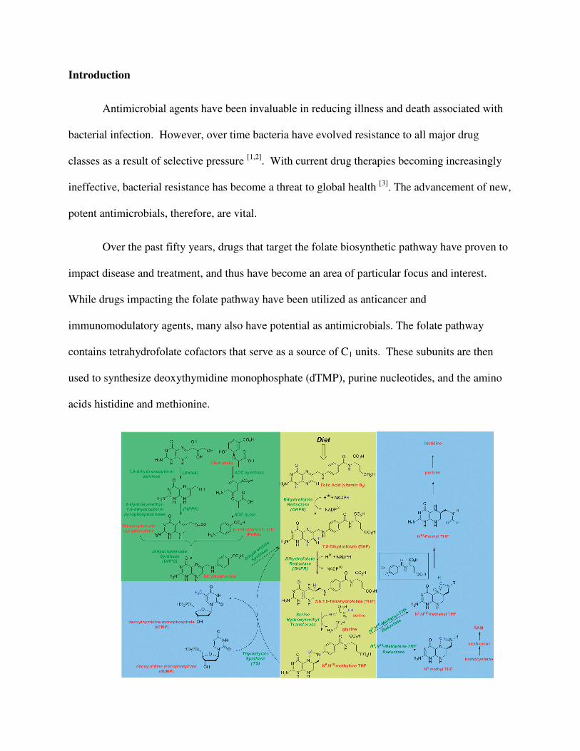
contains tetrahydrofolate cofactors that serve as a source of C1 units. These subunits are then
used to synthesize deoxythymidine monophosphate (dTMP), purine nucleotides, and the amino
acids histidine and methionine.
Figure 1. Folate Biosynthetic Pathway. The primary folate biosynthetic pathway is highlighted
in yellow. De novo synthesis of dihydrofolate in bacteria is represented in green. Key products of
the reaction are shown in blue [6].
When the pathway becomes inhibited, the purines and amino acids are not effectively
synthesized, dTMP production is halted, and consequently, there is no DNA replication or cell
proliferation. In this way, folate pathway inhibitors, or antifolates, have become a popular area
of drug design and optimization [4]. One effective target for inhibitors is an integral enzyme in
the pathway, dihydrofolate reductase (DHFR). DHFR converts dihydrofolate to tetrahydrofolate
(THF). As DHFR is the sole source of THF, which has been described as a necessary precursor
for the synthesis of vital cellular components, blocking DHFR’s activity will halt the target cell’s
growth. Further, beyond its ability to impact cellular proliferation, DHFR is an optimal target
because it is a widely conserved enzyme, present in many bacterial and eukaryotic organisms.
Because of this, DHFR inhibitors may be used in treatment for numerous pathogenic species. At
the same time, there are differences between the amino acid sequence in pathogen and human
DHFR which allows for selectivity [5]. It is also one of the best studied enzymes in the folate
pathway. Structural models exhibiting the inhibitor:enzyme interaction of DHFR are widely
available and information on DHFR mechanism of action and resistance is vast. This provides
researchers insight on how to improve upon novel drug compounds [6].
One widely used DHFR inhibitor is trimethoprim. It is a very effective 5-substituted-2,4-
diaminopyridine clinically used compound. In fact, it binds bacterial DHFR 105 times more
tightly than it does vertebrae DHFR [6]. However, resistance tends to develop against this drug in
a few ways. In Gram positive organisms, point mutations in the DHFR enzyme, which affects

the ability of the drug and enzyme to bind, have been described. In Gram negative bacter
bypass enzymes, spread by mobile genetic elements, are the primary cause of resistance
Again, with the emerging resistance, new drug discovery is crucial. As TMP is safe and
clinically effective when resistance has not been established, research
DHFR inhibitor has honed in on trimethoprim’s structure, specifically its
Drugs improve upon TMP by modifying
may allow it to bind the DHFR enzyme more
focused spectrum activity [6].
The Anderson-Wright Lab has focused on the development of propargyl
antifolates. These compounds contain the
to a variable hydrophobic functional domain
Figure 2. Trimethoprim and UCP 1051, a novel propargy
Anderson-Wright Lab, have the same
Trimethoprim
the ability of the drug and enzyme to bind, have been described. In Gram negative bacter
bypass enzymes, spread by mobile genetic elements, are the primary cause of resistance
Again, with the emerging resistance, new drug discovery is crucial. As TMP is safe and
clinically effective when resistance has not been established, research on how to optimize this
DHFR inhibitor has honed in on trimethoprim’s structure, specifically its diaminopyridine ring
modifying the diaminopyridine ring in a way that, for example,
bind the DHFR enzyme more tightly or give it either a broad or particularly
Wright Lab has focused on the development of propargyl-
antifolates. These compounds contain the diaminopyridine moiety linked with a propargyl group
ariable hydrophobic functional domain [7].
Trimethoprim and UCP 1051, a novel propargy-linked antifolate synthesized by the
Wright Lab, have the same diaminopyridine moiety (red). Also pictured
the ability of the drug and enzyme to bind, have been described. In Gram negative bacteria,
bypass enzymes, spread by mobile genetic elements, are the primary cause of resistance [6].
Again, with the emerging resistance, new drug discovery is crucial. As TMP is safe and
on how to optimize this
diaminopyridine ring.
that, for example,
tightly or give it either a broad or particularly
-linked
moiety linked with a propargyl group
linked antifolate synthesized by the
. Also pictured in UCP 1051
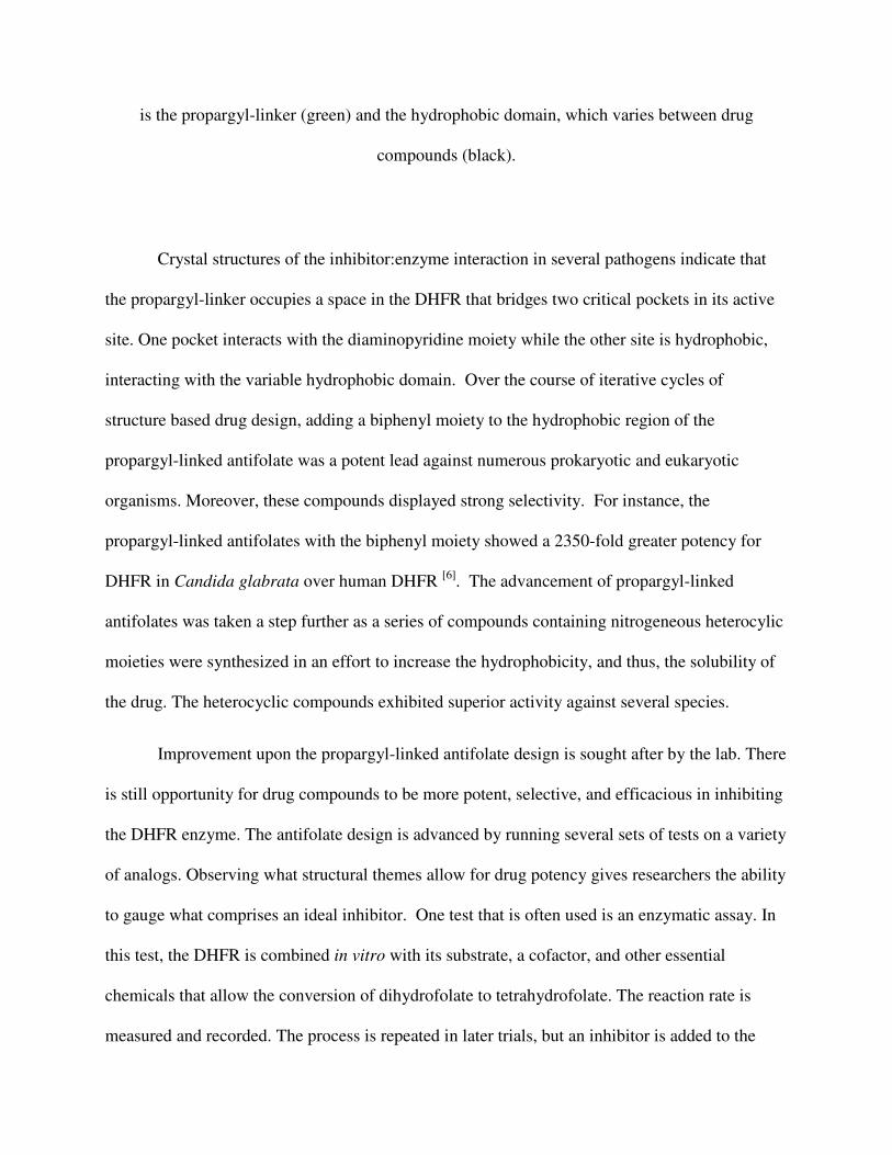
is the propargyl-linker (green) and the hydrophobic domain, which varies between drug
compounds (black).
Crystal structures of the inhibitor:enzyme interaction in several pathogens indicate that
the propargyl-linker occupies a space in the DHFR that bridges two critical pockets in its active
site. One pocket interacts with the diaminopyridine moiety while the other site is hydrophobic,
interacting with the variable hydrophobic domain. Over the course of iterative cycles of
structure based drug design, adding a biphenyl moiety to the hydrophobic region of the
propargyl-linked antifolate was a potent lead against numerous prokaryotic and eukaryotic
organisms. Moreover, these compounds displayed strong selectivity. For instance, the
propargyl-linked antifolates with the biphenyl moiety showed a 2350-fold greater potency for
DHFR in Candida glabrata over human DHFR [6]. The advancement of propargyl-linked
antifolates was taken a step further as a series of compounds containing nitrogeneous heterocylic
moieties were synthesized in an effort to increase the hydrophobicity, and thus, the solubility of
the drug. The heterocyclic compounds exhibited superior activity against several species.
Improvement upon the propargyl-linked antifolate design is sought after by the lab. There
is still opportunity for drug compounds to be more potent, selective, and efficacious in inhibiting
the DHFR enzyme. The antifolate design is advanced by running several sets of tests on a variety
of analogs. Observing what structural themes allow for drug potency gives researchers the ability
to gauge what comprises an ideal inhibitor. One test that is often used is an enzymatic assay. In
this test, the DHFR is combined in vitro with its substrate, a cofactor, and other essential
chemicals that allow the conversion of dihydrofolate to tetrahydrofolate. The reaction rate is
measured and recorded. The process is repeated in later trials, but an inhibitor is added to the
enzyme. The reaction rates of the trials using inhibitors are compared to the trial that did not
have the inhibitor present. The assay examines how much of the drug is needed to inhibit the
enzyme 50 percent of its typical action. Assay results allow for optimization around potential
leads. In cycles of development, this will generate a compound with an outstanding structure
which makes is tremendously potent. Equally important to drug discovery is x-ray
crystallography, which gives a three dimensional representation of the inhibitor interacting with
the enzyme. By visualizing the molecular interactions, the researcher can make fine adjustments
to the drug’s structure that will make it more powerful. Three microorganisms that the lab has
involved in enzymatic assays and crystallography are Candida glabrata, Candida albicans, and
Klebsiella pneumoniae.
Candida infections are nosocomial diseases in that they typically occur during a stay
within a hospital setting [8]. This is a result of the species thriving on and being prevalent within
critically ill and immunocompromised patients. Old age, major surgery, premature birth, AIDS,
use of catheters, and chemotherapy are only some of the factors that contribute to a deficient
immune system and an increased susceptibility to invasive candidasis [9,10]. Candida proves to
be a cause for concern as the frequency of Candida in blood cultures in US hospitals rose 52%
over just a three year period [8]. Combined with the fact that it has a forty percent mortality rate
in infected hospitalized patients [11], Candida infections are especially dangerous. Since there are
no licensed vaccines available for the treatment of Candida [11], certainly, the need for the
development of therapeutic drug compounds is evident.
Two of the most commonly used antifungal agents, or antimycotics, are azoles and
polyenes. Azoles and polyenes interact with ergosterol, a major component of the Candida
cellular membrane. When the egosterol is impacted by the compounds, the membrane does form
properly, and the cell is unable to function. Polyenes bind ergosterol directly and alter membrane
fluidity. Azoles have a different method of action as they inhibit Cyp51, which catalyzes the
formation of ergosterol [12]. C. glabrata, however, exhibits strong resistance to both of the
compounds. The resistance of C. glabrata to polyenes is the result of the organism developing
changes in its membrane structure that prevent the drugs necessary interaction with the
membrane. Regarding azoles, Candida has garnered multidrug resistance by increasing its
expression of drug efflux pumps [11]. The resistance to azoles has been particularly alarming. Of
all the Candida strains, C. glabrata is the most resistant to some of the most commonly
prescribed azole drug classes, itraconazole and fluconazole [13]. Also, there was a 1.9% C.
glabrata resistance to fluconazole in 2002 that spiked to 17.1% in 2006 [14]. As the resistance to
current drug compounds continually rise, the development of novel compounds with distinct
mechanisms of action become increasingly important.
Candida albicans, the candida species that most commonly causes fungal infections
within hospitals, garners resistance in multiple mechanisms. Much like C. glabrata, C. albicans
resistant organisms have been observed to increase their expression of efflux pumps. But, the
most common mechanism is based on the alteration of the target enzyme of the ergosterol
biosynthesis pathway, sterol 14 alpha demethylase, which is encoded by the ERG 11 gene.
Resistance develops by the increased expression of ERG11, increasing the intracellular pool of
sterol 14alpha demethylase, which then increases the effective drug dose. Moreover, point
mutations in gene lead to an alteration of amino acid residues and spatial configuration. The
azole drugs may no longer bind to the enzyme with the same affinity, and thus, are much less
potent [15].
Klebsiella pneumoniae is the most clinically important member of the Klebsiella genius
of Enterobacteriaceae. It has been noted as a common pathogen for nosocomial pneumonia,
septicemia, and wound infections. K. pneumoniae has also been isolated in more uncommon
infections like endocarditis, colecytitis, peritonitis, meningitis, and pyomyocitis. Beta-lactam
antibiotics are typically prescribed to treat K. pneumoniae infections. Historically, they have
been one of the most effective drug classes in inhibiting bacterial growth. β-Lactams exert their
antibiotic effects by mimicking the natural D-Ala-D-Ala substrate of the family of enzymes
known as penicillin-binding proteins (PBP), which are responsible for cross-linking the
peptidoglycan component of the bacterial cell wall. The integrity of the bacteria cell wall
becomes compromised when this process is inhibited, and the cell lyses [19].
However, constitutive use of beta-lactams to combat K. pneumoniae has led to the
bacteria evolving resistance, producing mutations and strong expression of beta-lactamases. The
bacteria may even express beta-lactamase activity against newly developed beta-lactam
antibiotics. These are referred to as extended spectrum beta-lactamases (ESBLs). Carbapenem
antibiotics, beta-lactams that have a structure which make them resistant to lactamases, have
been used to treat any serious infection caused by ESBLs. Nevertheless, K. pneumoniae has
developed resistance to these drug compounds via a novel mechanisms referred to as Klebsiella
pneumoniae carbapenemases (KPCs) [16]. KPCs are a particular cause for concern as KPC-
producing organisms can confer resistance to numerous drug classes such as fluoroquinolones
and aminoglycosides, in addition to beta-lactams. Because of this, infections due to KPCs are
associated with poor therapeutic treatment and mortality rates up to 50%. With the limited
number of drug compounds available to eliminate KPCs, novel structures that are biologically
effective are invaluable [17].
Materials and Methods
Protein Expression
Escherichia coli were transformed by C. glabrata dihydrofolate reductase (CgDHFR)
and C. albicans dihydrofolate reductase (CaDHFR) DNA. After cultures of the E. coli were
grown, expression of the DHFR protein was induced by the addition of β-D-thiogalactoside. The
culture was centrifuged so E. coli cells only, which at this point contained the DHFR, were easily
extracted from the culture. The cells were then lysed by Bug Buster, a reagent that breaks open
E. coli cell walls, and further centrifuged. The supernatant was exposed to ammonium sulfate
precipitate which eliminated many proteins, but left the desired enzyme. The protein was further
purified with use of a methotrexate column. In this, the DHFR was bound to the column while
other non-desired proteins and cellular contents diffused through. Thereafter, the column, which
still held the DHFR, was washed with dihydrofolate. The DHFR could then diffuse from the
column, and be collected. The DHFR was flash frozen and stored at -80 ºC until further use.
Crystallography
The purified DHFR was incubated with an enzyme cofactor, NADPH, and UCP111H
(Figure 12). A Linbro plate was filled with varying amounts of salt, PEG precipitant, water, and
differing pH buffer in each of the 24 wells that comprise the plate. The contents of these wells
promote the formation of a crystal via the hanging drop diffusion method. In this, a 2 uL drop of
the DHFR infused with the drug was be combined with a 2 uL drop of precipitant and placed on
a coverslip. This coverslip was sealed to the top of one of the wells with the aid of high vacuum
grease. After sitting at 4ºC for two weeks, the precipitant vaporized and transferred into the

reservoir of the well until the system reached equilibrium. At this point, conditions were optimal
for protein crystallization [18]. Protein crystals were extracted and flash frozen for preservation.
After, the crystals were shot with an X-ray. These X-rays generated a diffraction pattern
which was processed to give information about the structure and electron density of the protein
infused with the drug compound. Several programs including Phaser, COOT, and Refmac 5 were
utilized in order to create a three dimensional representation of the protein and inhibitor based on
the collected diffraction pattern.
Enzymatic Assay
An assay buffer consisting of 20mM TES, 50 mM KCl, 0.5mM EDTA, 10mM beta-
mercaptoethanol, and 1mg/ml of BSA was prepared. One mg/mL of K. pneumoniae A1 DHFR,
20 mM NADPH, and 1mM dihydrofolate were also prepared. The enzyme activity assay was
performed by monitoring the rate of NADPH consumption at 340nm over the course of 5
minutes. Reaction were performed with the assay buffer, NADPH cofactor, the dihydrofolate
substrate, and the pure enzyme. The reactions were performed in triplicate.
Results
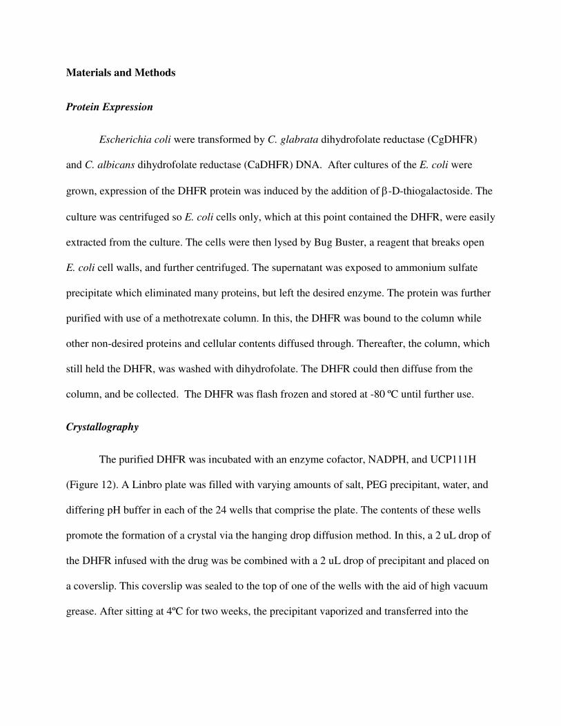
K. pneumoniae A1 DHFR
Figure 3. IC50 values for Klebsiella pneumoniae A1 DHFR interacting with differing novel
propargyl-linked antifolates synthesized by the UConn Pharmacy Anderson-Wright Laboratory.
Reactions were run in triplicate and standardized.
Drug Compound Average IC50 (µM) Std Deviation
TMP 20.166 1.69
UCP 1098 0.438 0.0349
UCP 1099 1.716 0.2745
UCP 1101 2.319 0.0148
UCP 1097 2.376 0.4532
UCP 1093 4.221 0.3852
UCP 1051 17.553 1.8193
UCP 1092 25.437 1.617
UCP 1100 65.239 1.5339
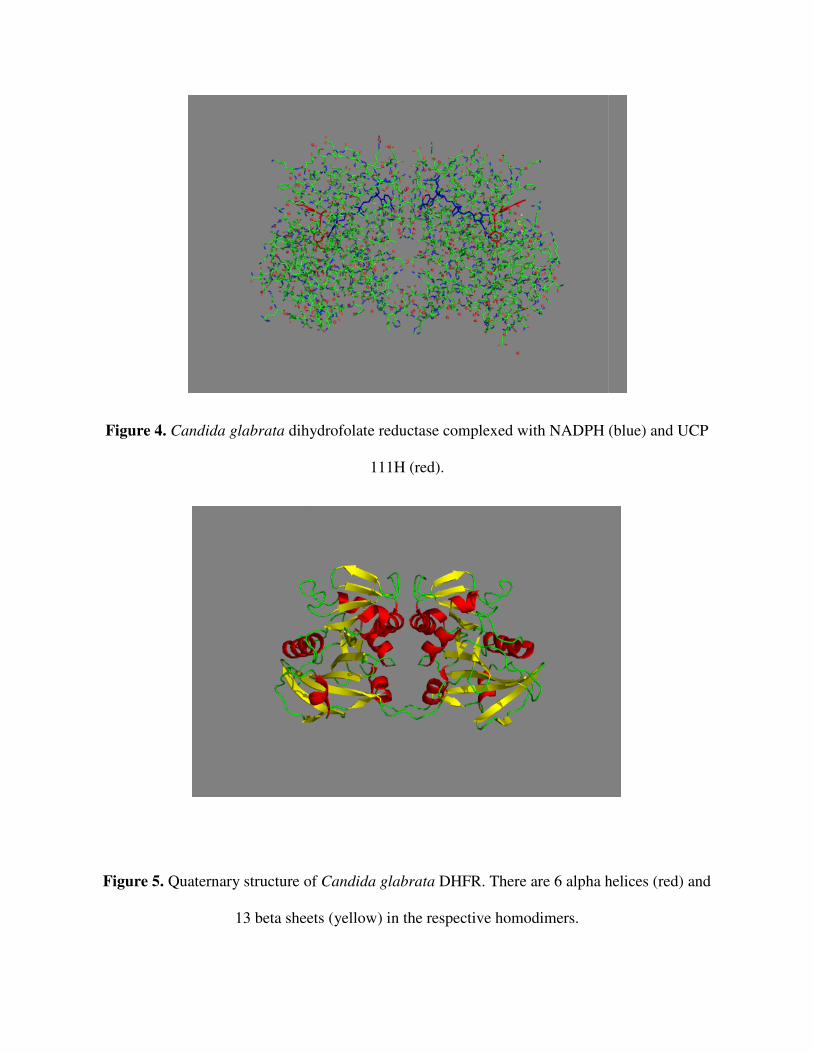
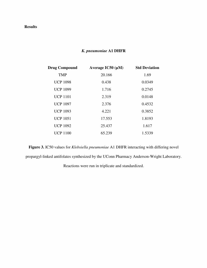
Figure 4. Candida glabrata dihydrofolate reductase complexed with NADPH (blue) and UCP
Figure 5. Quaternary structure of
13 beta sheets (yellow) in the respective homodimers.
dihydrofolate reductase complexed with NADPH (blue) and UCP
111H (red).
Quaternary structure of Candida glabrata DHFR. There are 6 alpha helic
13 beta sheets (yellow) in the respective homodimers.
dihydrofolate reductase complexed with NADPH (blue) and UCP
There are 6 alpha helices (red) and
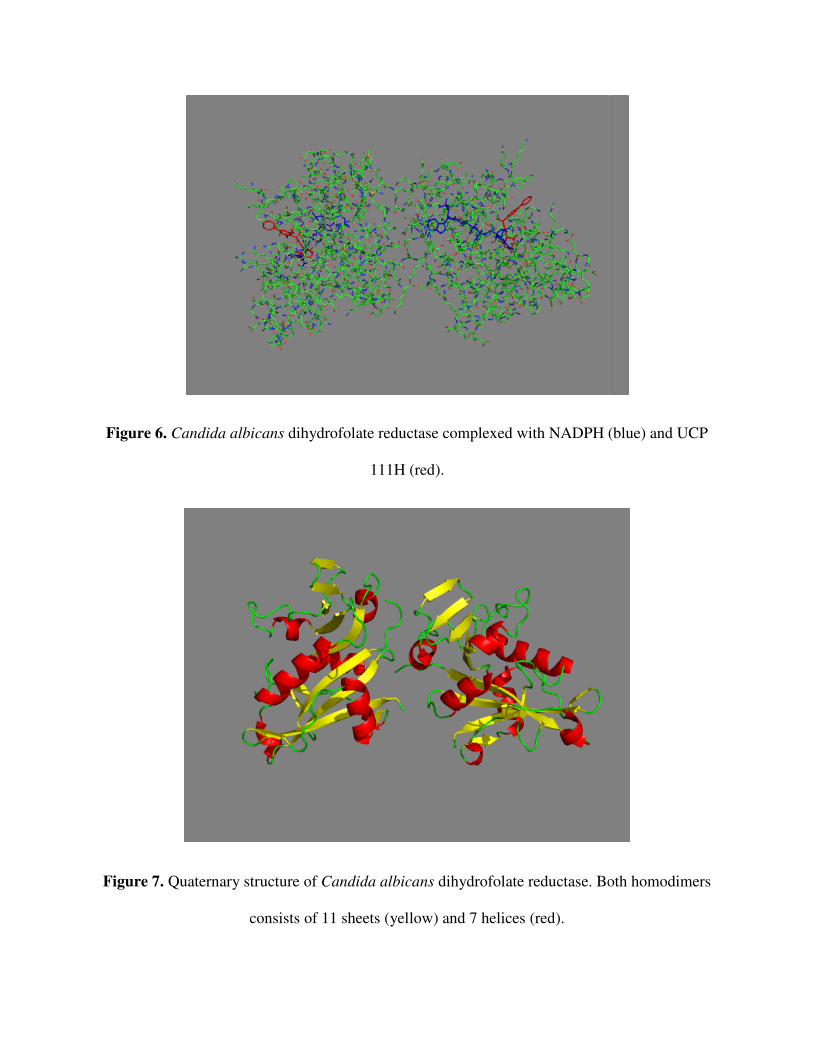
Figure 6. Candida albicans dihydrofolate reductase complexed with NADPH (blue) and UCP
Figure 7. Quaternary structure of
consists of
dihydrofolate reductase complexed with NADPH (blue) and UCP
111H (red).
Quaternary structure of Candida albicans dihydrofolate reductase. Both homodimers
consists of 11 sheets (yellow) and 7 helices (red).
dihydrofolate reductase complexed with NADPH (blue) and UCP
dihydrofolate reductase. Both homodimers
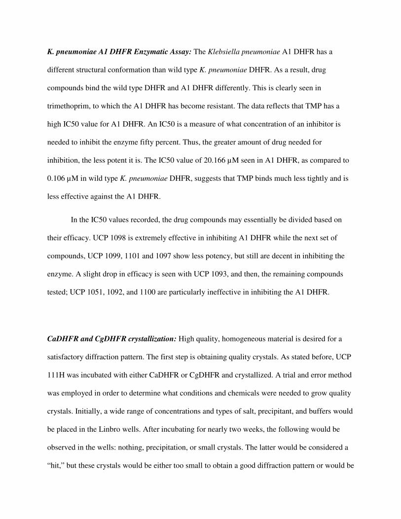
K. pneumoniae A1 DHFR Enzymatic Assay: The Klebsiella pneumoniae A1 DHFR has a
different structural conformation than wild type K. pneumoniae DHFR. As a result, drug
compounds bind the wild type DHFR and A1 DHFR differently. This is clearly seen in
trimethoprim, to which the A1 DHFR has become resistant. The data reflects that TMP has a
high IC50 value for A1 DHFR. An IC50 is a measure of what concentration of an inhibitor is
needed to inhibit the enzyme fifty percent. Thus, the greater amount of drug needed for
inhibition, the less potent it is. The IC50 value of 20.166 µM seen in A1 DHFR, as compared to
0.106 µM in wild type K. pneumoniae DHFR, suggests that TMP binds much less tightly and is
less effective against the A1 DHFR.
In the IC50 values recorded, the drug compounds may essentially be divided based on
their efficacy. UCP 1098 is extremely effective in inhibiting A1 DHFR while the next set of
compounds, UCP 1099, 1101 and 1097 show less potency, but still are decent in inhibiting the
enzyme. A slight drop in efficacy is seen with UCP 1093, and then, the remaining compounds
tested; UCP 1051, 1092, and 1100 are particularly ineffective in inhibiting the A1 DHFR.
CaDHFR and CgDHFR crystallization: High quality, homogeneous material is desired for a
satisfactory diffraction pattern. The first step is obtaining quality crystals. As stated before, UCP
111H was incubated with either CaDHFR or CgDHFR and crystallized. A trial and error method
was employed in order to determine what conditions and chemicals were needed to grow quality
crystals. Initially, a wide range of concentrations and types of salt, precipitant, and buffers would
be placed in the Linbro wells. After incubating for nearly two weeks, the following would be
observed in the wells: nothing, precipitation, or small crystals. The latter would be considered a
“hit,” but these crystals would be either too small to obtain a good diffraction pattern or would be
impure. Researchers optimized around the conditions in the hits, making minute adjustments,
until they found what best facilitated large, uniform, crystal growth. The conditions that best
enabled both the CaDHFR and CgDHFR crystal growth in the Linbro wells were: 0.1M Tris
Base pH between 8-8.75, varying amounts of magnesium chloride between 10 and 100 µL, PEG
4000 precipitant 30% w/v between 300 and 350 µL, and the remainder of the 500 µL well was
filled with water. Also, other techniques that were used to determine what led to quality crystal
growth were altering the protein concentration, temperature of incubation, and seeding.
The crystal was mounted in a beam, which generates and projects an x-ray wavelength,
and onto a device that rotates so the ray may strike it at different angles. After the crystals were
shot with the ray, a diffraction pattern was obtained. In this experiment, powerful synchrotrons at
Brookhaven National Laboratory were used. These machines shoot a very intense x-ray beam
with high quality optics, allowing for a high signal to noise ratio in the diffraction pattern. This,
in turn, leads to a better three dimensional representation of the macromolecule [18]. Once the
pattern had been obtained, researchers needed to ensure that the resolution was sufficient to
make accurate structure determination. Also, the unit cell dimensions, the crystal system, and the
space group was analyzed.
When the pattern had been successfully captured, this data was processed with several
programs with algorithms to calculate an electron density map. The map forms the contours into
which the protein structure will be built. The quality of the map is improved through refinement
programs until it may no longer be enhanced. From this, the model may be uploaded to the
Protein Data Bank and viewed as a PDB file. PyMOL was the molecular visualization system
used to view the PDB files as seen in Figures 4-7.
Discussion and Conclusion
The purpose of conducting the various tests in the experiemental procedures were to
discover what propargyl-linked antifolate structural analogs best inhibit dihydrofolate reductase
action in C. albicans, C. glabrata, and K. pneumoniae.
enzyme can assist in the generation of more potent drug compounds in iterative cycles of
antifolate development.
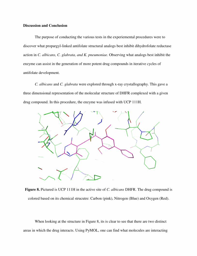
C. albicans and C. glabrata
three dimensional representation of
drug compound. In this procedure, the enzyme was infused with UCP 111H.
Figure 8. Pictured is UCP 111H in the active site of
colored based on its chemical str
When looking at the structure in Figure 8, its is clear to see that there are two distinct
areas in which the drug interacts.
The purpose of conducting the various tests in the experiemental procedures were to
linked antifolate structural analogs best inhibit dihydrofolate reductase
C. albicans, C. glabrata, and K. pneumoniae. Observing what analogs best inhibit the
enzyme can assist in the generation of more potent drug compounds in iterative cycles of
C. glabrata were explored through x-ray crystallography. This gave a
three dimensional representation of the molecular structure of DHFR complexed with a given
drug compound. In this procedure, the enzyme was infused with UCP 111H.
Pictured is UCP 111H in the active site of C. albicans DHFR. The drug compound is
colored based on its chemical strucutre: Carbon (pink), Nitrogen (Blue) and Oxygen (Red).
When looking at the structure in Figure 8, its is clear to see that there are two distinct
areas in which the drug interacts. Using PyMOL, one can find what molecules are interacting
The purpose of conducting the various tests in the experiemental procedures were to
linked antifolate structural analogs best inhibit dihydrofolate reductase
ogs best inhibit the
enzyme can assist in the generation of more potent drug compounds in iterative cycles of
ray crystallography. This gave a
the molecular structure of DHFR complexed with a given
DHFR. The drug compound is
ucutre: Carbon (pink), Nitrogen (Blue) and Oxygen (Red).
When looking at the structure in Figure 8, its is clear to see that there are two distinct
Using PyMOL, one can find what molecules are interacting
with another by measuring the distance between two different structures. Molecules within 3.5 Å
of each other are potentially interacting. First is the diaminopyridine, which interacts with
several specific amino acid side chains. The 2 substituted amine in the diaminopyridine is
bonded to an oxygen in glutamic acid. Glutamic acid has an electrically charged side chain. This
reflects how fluctuations in pH may affect the binding of the drug compound to the enzyme. The
pH will determine if hyrdrogen atoms remain bound to the side chain, and thus, if the side chain
becomes charged. The charge on the side chain will affect how it binds to different molecules. Of
course, then, it impacts how the drug compound may bind to the protein at the active site. The 4
substituted amine may be interacting with oxygens on two different isoleucine side chains.
Isoleucine is a hydrophobic amino acid. The area of the drug compound that interacts with this
amino acid must be hydrophobic as well. The presence of any largely polar substances will not
allow for tight interaction with the protein. When the inhibitor is unable to bind tightly, the drug
becomes ineffective. So, the amine must be satisfactory in binding to these hydrophobic regions.
The diaminopyridine, however, has already been noted as successfully binding to the
enzyme. Thus, it is not necessarily the target of drug optimization. As noted before, the
propargyl group links the drug compound to another area of the active stie in DHFR. This is
where advancement in the propargyl-linked antifolate design has proven key. First attached to
the propargyl-linker is a methyl group. This methyl group binds with an isoleucine, again, a
hydrophobic amino acid. It is invaluable to have a methyl, or another hydrophobic group, in this
region becuase a charged molecule will be detrimental because how the antifolate binds. A
methyl group is also optimal because too bulky of a hydrophobic group may lead to hinderance
and prevent the compound from attaching to the site tightly.
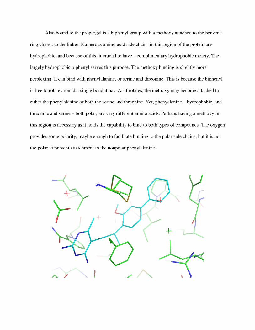
Also bound to the propargyl is a biphenyl group with a methoxy attached to the benzene
ring closest to the linker. Numerous amino acid side chains in this
hydrophobic, and because of this, it crucial to have a complimentary hydrop
largely hydrophobic biphenyl serves this purpose.
perplexing. It can bind with phenylalanine
is free to rotate around a single bond it has
either the phenylalanine or both the serine and threonine. Yet, phenyalanine
threonine and serine – both polar, are very different amino acids. Perhaps having a methoxy in
this region is necessary as it holds the capability to bind to both types of compounds. The oxygen
provides some polarity, maybe enough to facilitate binding to the polar side chains, but it is not
too polar to prevent attatchment to the
Also bound to the propargyl is a biphenyl group with a methoxy attached to the benzene
ring closest to the linker. Numerous amino acid side chains in this region of the protein are
hydrophobic, and because of this, it crucial to have a complimentary hydrophobic moiety
largely hydrophobic biphenyl serves this purpose. The methoxy binding is slightly
perplexing. It can bind with phenylalanine, or serine and threonine. This is because the biphenyl
e bond it has. As it rotates, the methoxy may become attached to
either the phenylalanine or both the serine and threonine. Yet, phenyalanine – hydrophobic, and
polar, are very different amino acids. Perhaps having a methoxy in
ary as it holds the capability to bind to both types of compounds. The oxygen
provides some polarity, maybe enough to facilitate binding to the polar side chains, but it is not
too polar to prevent attatchment to the nonpolar phenylalanine.
Also bound to the propargyl is a biphenyl group with a methoxy attached to the benzene
region of the protein are
hobic moiety. The
The methoxy binding is slightly more
or serine and threonine. This is because the biphenyl
rotates, the methoxy may become attached to
hydrophobic, and
polar, are very different amino acids. Perhaps having a methoxy in
ary as it holds the capability to bind to both types of compounds. The oxygen
provides some polarity, maybe enough to facilitate binding to the polar side chains, but it is not
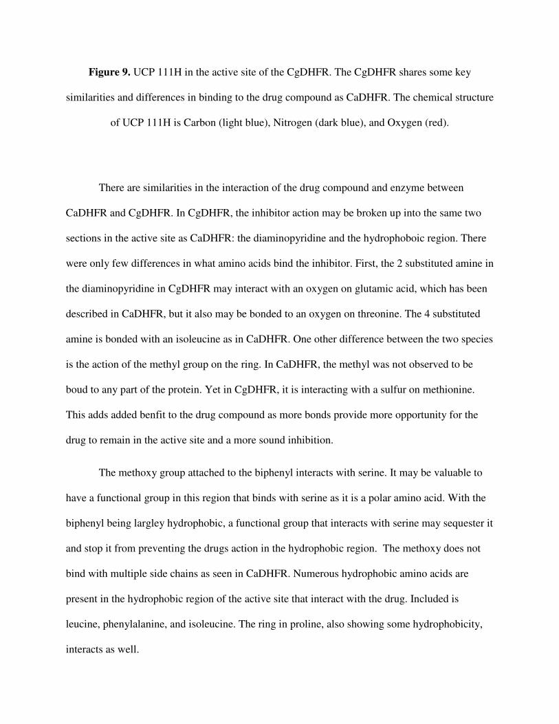
Figure 9. UCP 111H in the active site of the CgDHFR. The CgDHFR shares some key
similarities and differences in binding to the drug compound as CaDHFR. The chemical structure
of UCP 111H is Carbon (light blue), Nitrogen (dark blue), and Oxygen (red).
There are similarities in the interaction of the drug compound and enzyme between
CaDHFR and CgDHFR. In CgDHFR, the inhibitor action may be broken up into the same two
sections in the active site as CaDHFR: the diaminopyridine and the hydrophoboic region. There
were only few differences in what amino acids bind the inhibitor. First, the 2 substituted amine in
the diaminopyridine in CgDHFR may interact with an oxygen on glutamic acid, which has been
described in CaDHFR, but it also may be bonded to an oxygen on threonine. The 4 substituted
amine is bonded with an isoleucine as in CaDHFR. One other difference between the two species
is the action of the methyl group on the ring. In CaDHFR, the methyl was not observed to be
boud to any part of the protein. Yet in CgDHFR, it is interacting with a sulfur on methionine.
This adds added benfit to the drug compound as more bonds provide more opportunity for the
drug to remain in the active site and a more sound inhibition.
The methoxy group attached to the biphenyl interacts with serine. It may be valuable to
have a functional group in this region that binds with serine as it is a polar amino acid. With the
biphenyl being largley hydrophobic, a functional group that interacts with serine may sequester it
and stop it from preventing the drugs action in the hydrophobic region. The methoxy does not
bind with multiple side chains as seen in CaDHFR. Numerous hydrophobic amino acids are
present in the hydrophobic region of the active site that interact with the drug. Included is
leucine, phenylalanine, and isoleucine. The ring in proline, also showing some hydrophobicity,
interacts as well.
The IC50 value for UCP 111H in C. albicans and C. glabrata were obtained by the lab.
The value for UCP 111H in C. albicans is 0.02 µM while it is 0.0055 µM in C. glabrata. There
is a clear difference between the two species, and it appears that UCP 111H is more effective in
inhibiting the CgDHFR than CaDHFR. The differing values, of course, may be attributed to the
two enzymes’ varying structures. CgDHFR has a few more interactions taking place with amino
acids than CaDHFR. This may be giving UCP 111H in C. glabrata more stability and strength.
Since both of the Candida species may exhibit similar symptoms in an infected patient,
and it can be difficult for providers to make a diagnosis and prescribe a drug solely for C.
albicans or C. glabrata. Moreover, it wastes valuable treatment time if the diagnosis is initally
erroneous and the incorrect drug is given. Thus, drugs that halt both C. albicans and C. glabrata
action are desired. It is advantageous that the DHFR active site is similar in both species. This
allows potential for the antifolates to bind both enzymes and inhibit DHFR action, as seen in
UCP 111H. Enzymatic assays and in vitro cell based asssays would be used to measure exactly
how effective the drug compounds are in the respective species.
.
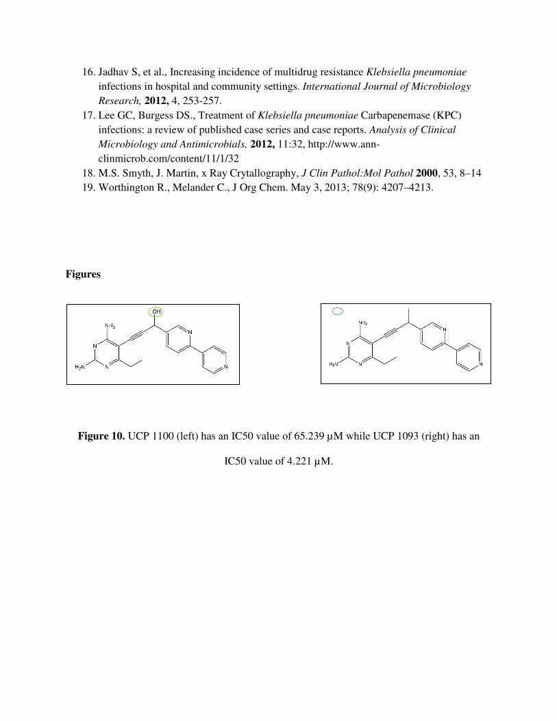
Analysis of the K. pneumoniae enzymatic assay shows that some structural thems lead to
potnent inhibitors while others do not. First noticable is the hydroxyl group off the propargyl-
linker as opposed to a methyl group (Figure 10). The substantially higher IC50 value reflects that
UCP 1100 is far less potent than UCP 1093. The only aspect of their structures that differ is the
highlighted methyl and hydroxyl. This suggests that a hydroxyl in this area greatly hinders the
binding of the antifolate to the enzyme. This is likely because a nonpolar amino acid is close to
where the methyl or hydroxyl would have to interact. The interacting amino acid may be an
isoleucine, as seen in the Candida species. Having a polar hydroxyl present would prevent tight
bonding.
The stereochemistry of the methyl group plays a role as well. UCP 1098 and UCP 1099
(Figure 12) are identical except for the respective R and S conformations. UCP 1098 has an IC50
of 0.438 µM while UCP 1099 has an IC50 of 1.716 µM. One explanation for the difference is
that the given stereochemistry may affect the distance of the methyl group to the interacting
isoleucine, also affecting the strength of the bond. The disparity between the compounds in not
extremely great, but the position of the methyl group is certainly worth considering in future
drug design.
UCP 1092 and UCP 1101 (Figure 11) have a distnct difference in their IC50 vaules.
Nevertheless, the structures of the two compounds change only slightly. The varying potency is
the cause of one, or a combination of, the following structural dissimilarities: the ethyl group
present on the diaminopyridine in UCP 1101 but missing from UCP 1092; the methoxy on the
benzene ring in UCP 1092 but missing from UCP 1101; the nitrogen on the benzene ring in UCP
1101, absent on UCP 1092; or the altering conformation of the heterocyclic ring attached to the
benzene. In order to determine what exactly affects the antifolate’s binding, a crystal structure of
the K. pneumoniae A1 DHFR would have to be obtained. As seen in the crystal strucutes of
CaDHFR and CgDHFR, the contributing molecular forces could be analyzed.
Another set of IC50 values that contrast substantially are UCP 1093 and 1052 (Figure
12). These two compounds are entirely identicial, with the exception that UCP 1093 has a
heterocyclic, aromatic 5 carbon, 1 nitrogen ring in the biphenyl moiety while 1052 has an
exclusively carbon ring. The IC50 vaule for 1052 is 17.553 µM and 1093 is 4.221 µM. The data
suggest that the nitrogen must be interacting with a pivotal amino acid that greatly affects how
the compound binds.
In all, UCP 1098 (Figure 12) is rather potent at a 0.438 µM IC50. Perhaps it is the meta
linked biphenyl contributes to strong bonding, or it may be the 3 carbon, 2 oxygen ring attached
to the benzene ring that positively affects the inhibitor’s strength. The next step in the
experimental design would be to crystalize the A1 DHFR with 1098. This would allow
researchers to deduce what amino acids are key in this drug’s interaction. Optimization around
the UCP 1098 design would give an even tighter bond to the active site and successful inhibition
of the A1 DHFR.
References
1. National Research Council, Committee on Drug Use in Food Animals. The use of drugs
in food animals: benefits and risks. Washington (DC): National Academy Press; 1999. 2. Mellon M, Benbrook C, Benbrook KL. Hogging it: Estimates of antimicrobial abuse in
livestock. Cambridge (MA): Union of Concerned Scientists; 2001. 3. Brolund A, Sundqvist M, Kahlmeter G, Grape M (2010) Molecular Characterisation of
Trimethoprim Resistance in Escherichia coli and Klebsiella pneumoniae in During a two Year Intervention on Trimethoprim Use. PLos One 5(2); e9233
4. Anderson A, Wright D. Antifolate Agents: A Patent Review (2006-2010). Expert Opin
Ther Pat. 2011 September ; 21(9): 1293–1308. 5. Lamb K, Anderson A, Wright D, G-Dayanandan N (2013) Elucidating Features That
Drive the Design of Selective Antifolates Using Crystal Structures of Human Dihydrofolate Reductase. Biochemistry, 52, 7318-7326.
6. Zhou W, Anderson A, Wright D, Scocchera E (2013) Antifolates as effective antimicrobial agents: new generations of trimethoprim analogs Med. Chem. Commun., 4,
908-915. 7. Zhou W, Anderson A, Wright D, Hill D, Viswanathan K (2012) Acetylic Linkers in Lead
Compounds: A Study of the Stability of the Propargyl-Linked Antifolates. Drug
Metabolism and Disposition, 40, 2002-2008. 8. M. Pfaller et al., Epidemiology and outcomes of candidemia in 3648 patients: data from
the Prospective Antifungal Therapy (PATH Alliance®) registry, 2004–2008 Diagnostic
Microbiology and Infectious Disease. 2012, 1-9 9. M. Nucci, Kieren Marr, Emerging Fungal Diseases, Clin. Infect. Dis., 2005, 41, 521-526 10. R. Lewis, P Viale et al., The potential impact of antifungal drug resistance mechanisms
on the host immune response to Candida, Virulence, 2012 3:4, 368-376 11. I. Francois, A. Aerts et al., Currently Used Antimycotics: Spectrum, Mode of Action and
Resistance occurrence, Current Drug Targets 2005, 6, 895-207 12. Z. Kanafani et al., Resistance to Antifungal Agents, Clin. Infect. Dis, 2008, 46, 120-128 13. R. Hajjeh, A. Sofair et al., Incidence of Bloodstream Infections Due to Candida Species
and In Vitro Susceptibilities of Isolates Collected from 1998 to 2000 in a Population-Based Active Surveillance Program, Journal of Clinical Microbiology, 2004, 42, 1519-1527
14. T. Chen, Y. Chen et al., Fluconazole exposure rather than clonal spreading is correlated with the emergence of Candida glabrata with cross-resistance to triazole antifungal agents, Kaohsiung Journal of Medical Sciences, 2012, 28, 306-315
15. Strzelczyk J, Slemp-Migiel A, Rother M, et al., Nucleotide substitutions in the Candida
albicans ERG 11 gene of azole-susceptible and azole-resistant clinical isolates. Biochimica Polonica, 2013, 60, 547-552.
16. Jadhav S, et al., Increasing incidence of multidrug resistance Klebsiella pneumoniae infections in hospital and community settings. International Journal of Microbiology
Research, 2012, 4, 253-257. 17. Lee GC, Burgess DS., Treatment of Klebsiella pneumoniae Carbapenemase (KPC)
infections: a review of published case series and case reports. Analysis of Clinical
Microbiology and Antimicrobials, 2012, 11:32, http://www.ann-clinmicrob.com/content/11/1/32
18. M.S. Smyth, J. Martin, x Ray Crytallography, J Clin Pathol:Mol Pathol 2000, 53, 8–14 19. Worthington R., Melander C., J Org Chem. May 3, 2013; 78(9): 4207–4213.
Figures
Figure 10. UCP 1100 (left) has an IC50 value of 65.239 µM while UCP 1093 (right) has an
IC50 value of 4.221 µM.
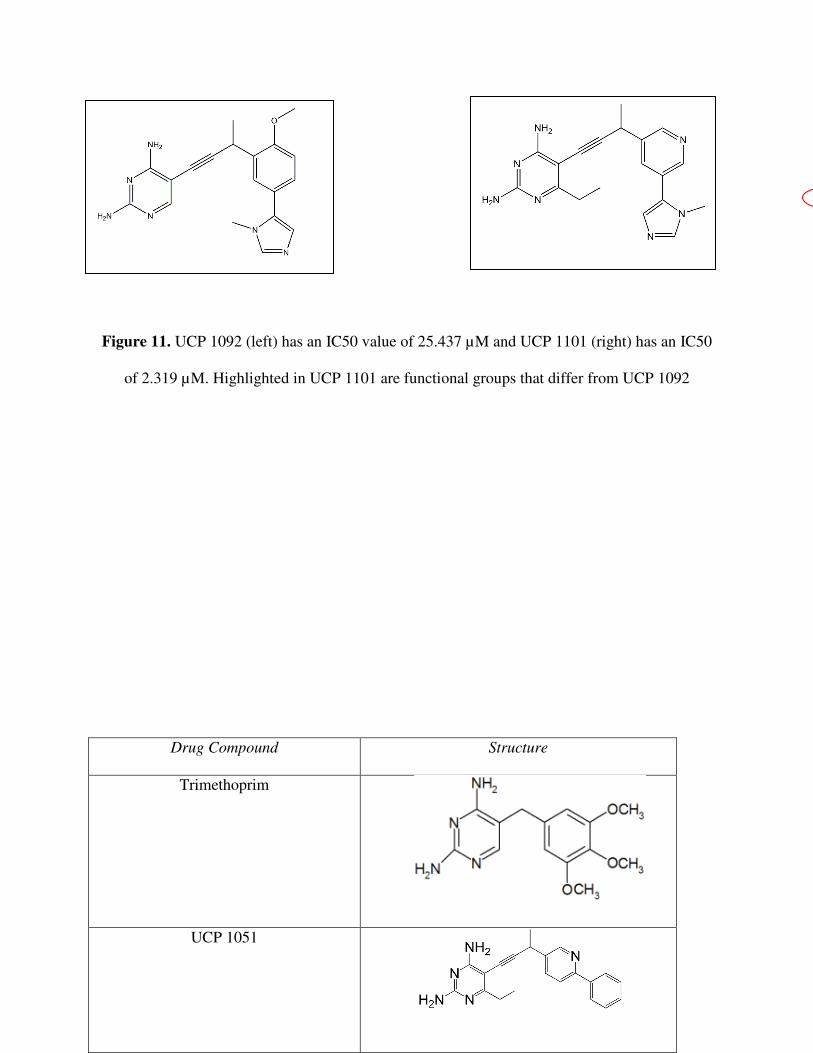
Figure 11. UCP 1092 (left) has an IC50 value of 25.437 µM and UCP 1101 (right) has an IC50
of 2.319 µM. Highlighted in UCP 1101 are functional groups that differ from UCP 1092
Drug Compound Structure
Trimethoprim
UCP 1051
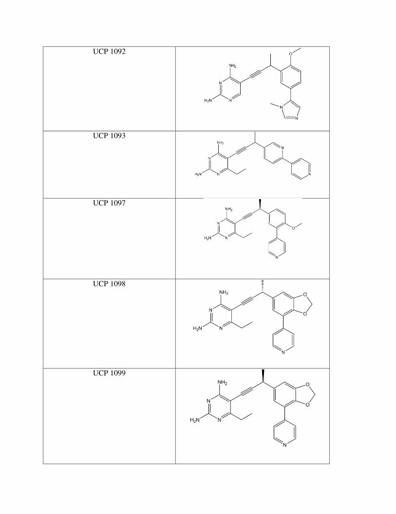
UCP 1092
UCP 1093
UCP 1097
UCP 1098
UCP 1099
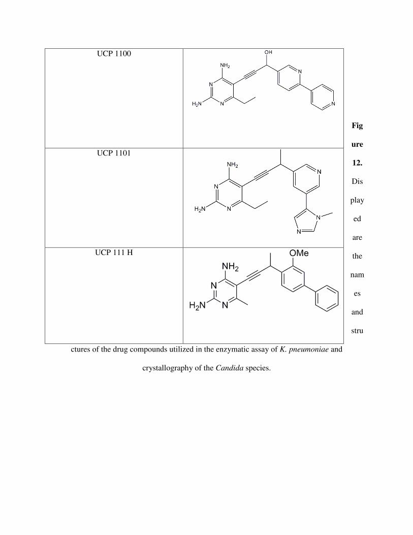
Fig
ure
12.
Dis
play
ed
are
the
nam
es
and
stru
ctures of the drug compounds utilized in the enzymatic assay of K. pneumoniae and
crystallography of the Candida species.
UCP 1100
UCP 1101
UCP 111 H


