
Introduction and Overview
of Acute Respiratory
Failure
Definition: Acute Respiratory Failure
• Failure to oxygenate
– Inadequate PaO2 to saturate hemoglobin
– PaO2 of 60 mm Hg ~ SaO2 of 90%
– PaO2 of 50 mm Hg ~ SaO2 of 75%
• Failure to ventilate
– Elevation in PaCO2 associated with
decreased pH
– PaCO2 of 60 mm Hg ~ pH 7.20

Differentiate ↓O2 from ↑CO2
• Hypoxemia (PaO2<60 mm Hg) and hypercarbia (elevated PaCO2 with low pH) may occur independently or together
• Hypoxemia:
– ventilation-perfusion mismatching
– shunt physiology
• Hypercarbia:
– alveolar hypoventilation
– increased dead space fraction
– increased production (overfeeding, hypermetabolism)
Acute Respiratory Failure
• The goal is to maintain adequate tissue
oxygenation to avoid anaerobic metabolism
• When oxygen supply ≠ demand:
– Organ dysfunction
– Lactate generation
• Determinants of supply
– Cardiac output (5L/minute)
– Blood oxygen content (20mL O2 /100mL
blood)
• Hemoglobin
• PaO2 / saturation

Normal oxygen transport
• O2 delivery=cardiac output x blood oxygen content
– DO2 = C.O. x CaO2
– DO2 = 5L/min x 20 mL/100 mL
– DO2 = 1000 mL/minute
• Tissue metabolism
– VO2 = 250 mL/minute or 4 mL/kg
• Extraction fraction =0.25
– Mixed venous saturation = 75%
– Mixed venous PaO2 = 40 mm Hg

Inadequate tissue oxygen delivery
• Reduced O2 delivery
– Cardiac output: 2.5L/min
– Anemia: Hgb 7.5 gm/dL
– Hypoxemia: Hgb saturation 75%/PaO2 40 mm Hg
• If constant O2 consumption (250mL/min):
– Extraction fraction will be 50%
– Mixed venous saturation = 50%
– Mixed venous PaO2 = 27 mm Hg
– Driving pressure for O2 diffusion from capillaries to
cells is reduced: anaerobic metabolism
General management principles
• Optimize oxygen delivery
– Transfusion of packed RBC
– Increase cardiac output
– Relieve arterial hypoxemia
• Reduce oxygen consumption
– Endotracheal intubation: deliver higher FiO2 and
relieve work of breathing
– Treat fever: to reduce tissue metabolic rate

Patient-related risk factors
• Smoking:
– Must stop >8 weeks preoperatively to decrease risk
• General health:
– Inability to exercise (supine bicycle) for 2 minutes with HR>99/min
• COPD:
– Must optimize medical management/functional capacity preoperatively
• Asthma:
– If well-controlled preoperatively, not a risk factor
• Age and obesity are not significant risk factors
N Engl J Med 1999 340: 937
Procedure-related risk factors
• Surgical site:
– Risk increases as incision approaches the diaphragm
– Open procedures higher risk than laparoscopic
• Duration:
– Procedures >3 hours carry higher risk
• Anesthesia/pain control:
– Consider spinal or regional anesthesia for high risk
patients
N Engl J Med 1999 340: 937
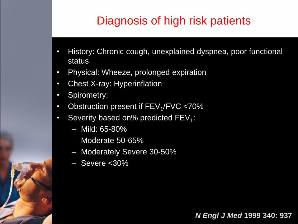
Diagnosis of high risk patients
• History: Chronic cough, unexplained dyspnea, poor functional
status
• Physical: Wheeze, prolonged expiration
• Chest X-ray: Hyperinflation
• Spirometry:
• Obstruction present if FEV1/FVC <70%
• Severity based on% predicted FEV1:
– Mild: 65-80%
– Moderate 50-65%
– Moderately Severe 30-50%
– Severe <30%
N Engl J Med 1999 340: 937
Intra-Pulmonary Shunt
• Blood that enters the arterial system without going through ventilated areas of lung
• Same physiology as extrapulmonary cardiac shunt:
– i.e. Atrial septal defect, patent foramen ovale
• Calculated amount of venous blood that must mix with arterial blood to produce the observed PaO2 is sometimes called “venous admixture”
• Shunt is refractory to supplemental oxygen
Hypercarbic (Type II) respiratory failure
• Also termed acute on chronic respiratory failure
• Usually seen in patients with severe COPD
• Occasionally occurs in a patient that has not been diagnosed with obstructive lung disease
• COPD is the 4th leading cause of death (2000)
• Patients have expiratory airflow limitation
• FEV1/FVC < 70%
• Measured as FEV1 (L); lower value=more severe

Prognosis: Type II respiratory failure
• High short-term risk of death but up to half of discharged
patients will survive for one year
• Half of survivors describe quality of life as good or better
• A proportion of these patients will return to work
• Older patient, more co-morbidities, lower baseline level of
function predict poor survival but are not refined enough for
adequate prognostication
• Extensive critical care resource utilization
• Ideally, management discussion is undertaken before acute
deterioration
Type II respiratory failure
• Multiple, minor insults cause acute deterioration of chronic
(precariously compensated) respiratory status
• Respiratory failure often is slowly progressive
• Patients are distressed, using accessory muscles, have
prolonged expiratory time, wheezing
• In late phases: obtundation, apnea
• Low PaO2 and high PaCO2
• PACO2 is directly related to alveolar ventilation
• Relatively easy to oxygenate in comparison to ARDS
Precipitating factors for type II respiratory
failure
• In the postoperative setting, consider effects of:
– Sedation
– Site of Incision/Pain
– Fluid overload, myocardial ischemia
– Bronchospasm
– Infection
Respiratory load vs. strength
• Ventilation is accomplished by the diaphragm and other respiratory muscles working as a piston or pump
• When stimulated by the central nervous system, diaphragmatic and intercostal contraction reduce pleural pressure, causing lung inflation
• Muscles will fatigue when the load is excessive
• Fatigue = failure to develop the normal degree of tension despite maximal stimulation
• Airways contribute a resistive load that is increased by bronchospasm, secretions, inflammation
• Lungs contribute an elastic load due to dynamic hyperinflation- a failure to fully empty, resulting in intrinsic PEEP

Key elements of respiratory system
competence
• Adequate central respiratory drive
– anesthetics, narcotics, benzodiazepines,
hypothyroidism
• Neuromuscular competence
– corticosteroids, malnutrition, phrenic nerve injury,
paralytics, Guillain-Barré syndrome
• Diaphragmatic strength/pressure generation
– hyperinflation, malnutrition, electrolyte disturbance,
hypoxemia, myopathy
• Low airways resistance
– secretions, edema, bronchospasm, sleep apnea
• Adequate lung and chest wall compliance
– obesity, ascites, ileus, interstitial edema, pleural
effusion, infection, atelectasis, rib fracture,
pneumothorax, iPEEP

Dead space and hypercarbia
• The portion of ventilation that doesn’t participate in gas exchange: Exhaled unchanged
• The effective portion of minute ventilation is alveolar ventilation
• Apparatus dead space (if using a breathing apparatus)
• Anatomic dead space (volume of conducting passages) ~2mL/kg in adults
• Alveolar dead space (under/unperfused alveoli)
• Physiological dead space = sum of anatomical and alveolar dead space
• Increase in dead space ventilation will raise PaCO2
• Classically seen in pulmonary embolism
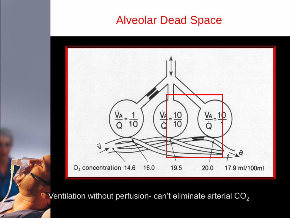
Alveolar Dead Space
Ventilation without perfusion- can’t eliminate arterial CO2
Physiologic effects of CO2
• Major regulator of cerebral blood flow: CBF increases via vasodilation due to CSF acidosis; this may increase intracranial pressure.
• Increased circulating catecholamines (epi > norepi)
• Pulmonary arterial vasoconstriction (less than seen with hypoxia)
• Shifts oxyhemoglobin dissociation curve to the right
• Displaces oxygen in alveolar gas
• Slight net increase in blood pressure
• Bicarbonate retention by kidneys
• Increase in plasma potassium
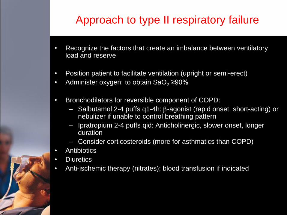
Approach to type II respiratory failure
• Recognize the factors that create an imbalance between ventilatory load and reserve
• Position patient to facilitate ventilation (upright or semi-erect)
• Administer oxygen: to obtain SaO2 ≥90%
• Bronchodilators for reversible component of COPD:
– Salbutamol 2-4 puffs q1-4h: b-agonist (rapid onset, short-acting) or nebulizer if unable to control breathing pattern
– Ipratropium 2-4 puffs qid: Anticholinergic, slower onset, longer duration
– Consider corticosteroids (more for asthmatics than COPD)
• Antibiotics
• Diuretics
• Anti-ischemic therapy (nitrates); blood transfusion if indicated
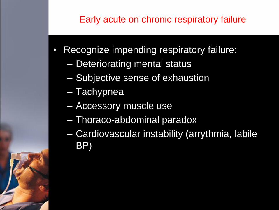
Early acute on chronic respiratory failure
• Recognize impending respiratory failure:
– Deteriorating mental status
– Subjective sense of exhaustion
– Tachypnea
– Accessory muscle use
– Thoraco-abdominal paradox
– Cardiovascular instability (arrythmia, labile
BP)
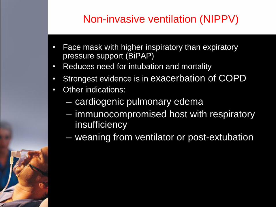
Non-invasive ventilation (NIPPV)
• Face mask with higher inspiratory than expiratory pressure support (BiPAP)
• Reduces need for intubation and mortality
• Strongest evidence is in exacerbation of COPD • Other indications:
– cardiogenic pulmonary edema
– immunocompromised host with respiratory insufficiency
– weaning from ventilator or post-extubation

NIPPV
• Compliant patient; able to synchronize with ventilator
• Appropriately fitting mask/seal
• Monitoring: HR, SaO2, BP
• Bedside presence for clinical response (usually starts in 10
minutes or less)
– Improved mental status
– Decreased respiratory rate/increased tidal volume
– Improved oxygen saturation/ABG
• If not tolerated or not improving: consider mechanical ventilation
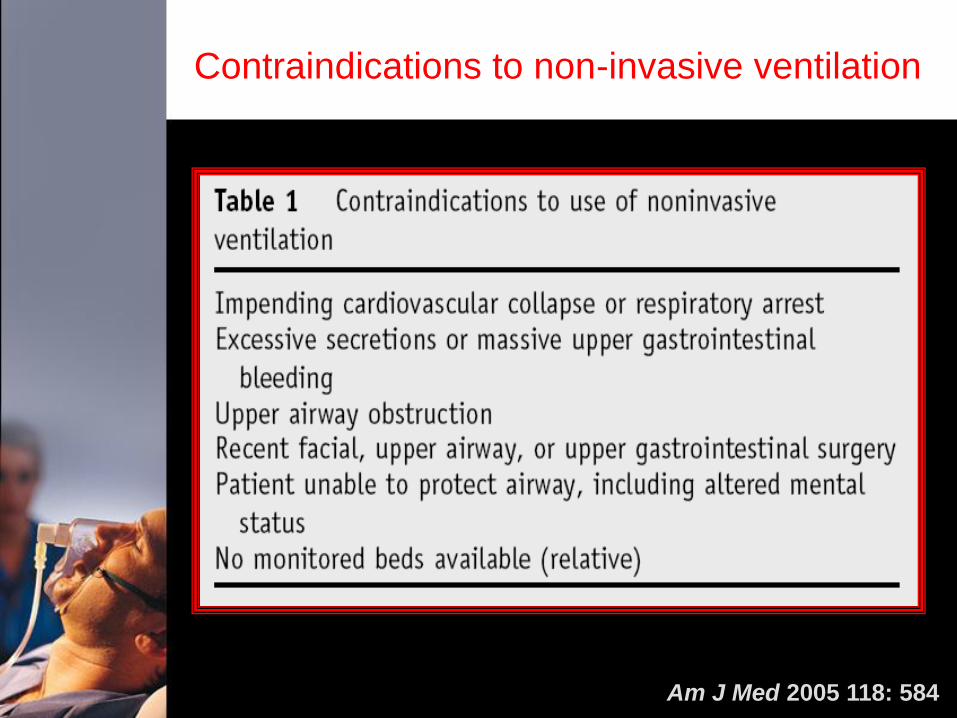
Contraindications to non-invasive ventilation
Am J Med 2005 118: 584
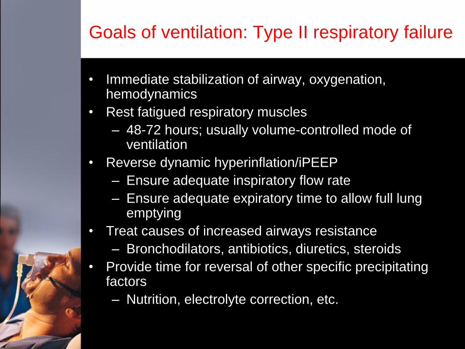
Goals of ventilation: Type II respiratory failure
• Immediate stabilization of airway, oxygenation, hemodynamics
• Rest fatigued respiratory muscles
– 48-72 hours; usually volume-controlled mode of ventilation
• Reverse dynamic hyperinflation/iPEEP
– Ensure adequate inspiratory flow rate
– Ensure adequate expiratory time to allow full lung emptying
• Treat causes of increased airways resistance
– Bronchodilators, antibiotics, diuretics, steroids
• Provide time for reversal of other specific precipitating factors
– Nutrition, electrolyte correction, etc.
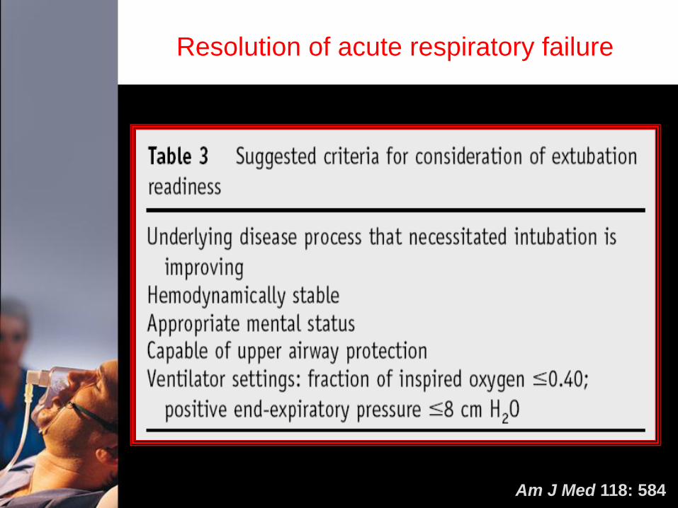
Resolution of acute respiratory failure
Am J Med 118: 584
Summary: Acute Respiratory Failure
• The main function of the respiratory system is to
oxygenate tissue and remove CO2 by ventilation
• ARDS is a primarily a failure to oxygenate caused by
acute lung inflammation and alveolar flooding with shunt
physiology
• Acute on chronic (type II) respiratory failure is primarily
due to a failure of adequate ventilation caused by
depressed respiratory drive, altered neuromuscular
coupling or increased airways resistance/collapse

Summary: Acute Respiratory Failure
• Disease-related factors
– Aspiration, pneumonia, trauma, shock: ARDS
• Patient-related factors
– Obstructive lung disease: Type II respiratory
failure
• Procedure-related factors
– Proximity to diaphragm, duration of procedure
Summary: Acute Respiratory Failure
• Management:
– Anticipate ARF in high risk patients and
initiate postoperative interventions (adequate
pain control, spirometry, etc.)
– Recognize need for ventilatory assistance
– Lung protective strategy: ARDS
– Non-invasive ventilation: Type II failure
– Supportive care and reversal of provocative
factors
• Antibiotics, bronchodilators, reduction of
steroids, etc.


