
1
Article
Human Umbilical Cord Wharton's Jelly Stem Cells and Its Conditioned Medium Enhance Healing Of Excisional and Diabetic Wounds†
Chui-Yee Fong1*, Kimberley Tam1, Suganya Cheyyatraivendran1, Shu-Uin Gan2, Kalamegam Gauthaman1, Arunmozhiarasi Armugam3, Kandiah Jeyaseelan3, Mahesh Choolani1, Arijit
Biswas1, Ariff Bongso1*
Departments of Obstetrics and Gynaecology1, Surgery2 and Biochemistry3, Yong Loo Lin School of Medicine, National University Health System, National University of Singapore, Singapore 119228.
* Corresponding authors
Dr Ariff Bongso ([email protected])
Dr Chui-Yee Fong ([email protected])
Department of Obstetrics and Gynaecology, Yong Loo Lin School of Medicine, National University Health System, National University of Singapore, Kent Ridge, Singapore 119228.
Tel: 65-67724267; Fax: 65-67794753
Statement of conflicts of interest: All authors have no conflict of interests.
†This article has been accepted for publication and undergone full peer review but has not been through the copyediting, typesetting, pagination and proofreading process, which may lead to differences between this version and the Version of Record. Please cite this article as doi: [10.1002/jcb.24661]
Received 14 August 2013; Accepted 20 August 2013 Journal of Cellular Biochemistry
© 2013 Wiley Periodicals, Inc. DOI 10.1002/jcb.24661
2
ABSTRACT
Wound healing is a major problem in diabetic patients and current treatments have met with
limited success. We evaluated the treatment of excisional and diabetic wounds using a stem cell
isolated from the human umbilical cord Wharton’s jelly (hWJSC) that shares unique properties with
embryonic and adult mesenchymal stem cells. hWJSCs are non-controversial, available in
abundance, hypo-immunogenic, non-tumorigenic, differentiate into keratinocytes and secrete
important molecules for tissue repair. When human skin fibroblasts (CCD) in conventional scratch-
wound assays were exposed to hWJSC-conditioned medium (hWJSC-CM) the fibroblasts at the
wound edges migrated and completely covered the spaces by day 2 compared to controls. The
number of invaded cells, cell viability, total collagen, elastin and fibronectin levels were significantly
greater in the hWJSC-CM treatment arm compared to controls (p < 0.05). When a single
application of green fluorescent protein (GFP)-labelled hWJSCs (GFP-hWJSCs) or hWJSC-CM
was administered to full-thickness murine excisional and diabetic wounds, healing rates were
significantly greater compared to controls (p < 0.05). Wound biopsies collected at various time
points showed the presence of green GFP-labelled hWJSCs, positive human keratinocyte markers
(cytokeratin, involucrin, filaggrin) and expression of ICAM-1, TIMP-1 and VEGF-A. On histology,
the GFP-hWJSCs and hWJSC-CM treated wounds showed reepithelialisation, increased
vascularity and cellular density and increased sebaceous gland and hair follicle numbers
compared to controls. hWJSCs showed increased expression of several miRNAs associated with
wound healing compared to CCDs. Our studies demonstrated that hWJSCs enhance healing of
excisional and diabetic wounds via differentiation into keratinocytes and release of important
molecules.
KEYWORDS: Conditioned medium; Excisional and diabetic wounds; Human Wharton's jelly stem cells; Wound healing

3
INTRODUCTION
The skin is the largest organ covering the human body and is most prone to injury. The healing
quality of surgical wounds vary, dehiscent wounds take a long time to heal and non-healing
wounds are a chronic problem in patients suffering from diabetes, kidney failure, burns and bed
sores. Diabetic wounds lead to foot ulcers which eventually end up in lower limb amputations.
Current methods such as chemicals, dressings and skin grafts have been used to accelerate
wound healing with limited success.
Reconstitution of damaged skin is a coordinated process involving the interplay and organization
of multiple inflammatory cell types. However, studies on wound healing in PU.1 knockout mice
which lack macrophages and neutrophils showed that neither of these cell types was important for
wound healing [Martin et al., 2003]. The inherent physiological repair mechanisms become
inefficient in the presence of (i) extensive loss of skin, (ii) pre-existing pathological diseases, (iii)
superimposed microbial infections, (iv) impairment of local circulation and (v) the presence of a
foreign body [Mathieu et al., 2006]. The epidermis and skin appendages are renewed constantly
and cell turnover is under the control of mesenchymal stem cells (MSCs) native to the epidermis,
the hair bulb, the melanocyte layer and the basal dermal layer [Badiavas et al., 2003]. It has also
been shown that MSCs migrate from the bone marrow to the injured wound site and contribute to
wound repair and regeneration [Borue et al., 2004; Sasaki et al., 2008]. MSCs assist these
processes via four mechanisms (suppression of inflammation, angiogenesis, stimulation of stem
cell proliferation and differentiation into new keratinocytes by dying apoptotic cells) [Jackson et al.,
2012]. The administration of an exogenous source of MSCs to wounds therefore is an attractive
form of potential therapy. The MSCs that have been traditionally used are those from the human
bone marrow (hBMMSCs) from autologous and allogeneic sources. Some basic mechanisms have
been suggested to explain how these MSCs repair tissues but these mechanisms have not been
studied in-depth and confirmed. The hypotheses are (i) the creation of an environment that
enhances regeneration of endogenous cells and (ii) differentiation of the MSCs into keratinocytes
[Stoff et al., 2009]. Several groups have reported successful wound healing after administration of
hBMMSCs to surgical wounds [Stoff et al., 2009; Chen et al., 2009], diabetic wounds [Kuo et al.,
4
2011] and burns [Bey et al., 2010]. Unfortunately, the use of hBMMSCs has its limitations such as
the problems of painful harvest, low cell numbers, risk of infection and morbidity. Primary
hBMMSCs also lose their stemness properties after a few passages in vitro compromising on the
availability of large numbers for clinical application. Alternative sources of stem cells are being
explored.
We and others have studied a stem cell from the human umbilical cord Wharton’s jelly (hWJSCs)
that shares unique properties between embryonic and adult stem MSCs [Troyer and Weiss, 2008;
La Rocca et al., 2009; Bongso and Fong, 2013]. These cells have low level expression of
embryonic stem cell markers and satisfy the criteria recommended by the International Society of
Cytotherapy for MSCs [Dominici et al., 2006]. Genomic, cell behaviour and stemness
characterization studies [Troyer and Weiss, 2008; Fong et al., 2011; Bongso and Fong, 2013]
showed that hWJSCs are primitive MSCs with uniquely different properties from bone marrow and
other MSCs. These differences were attributed to their umbilical cord origin which embryologically
lies in between embryonic and adult tissues on the human developmental map [Pappa and
Anagnou, 2009]. It was reported that during early human development primitive MSCs migrate
from the aorta-gonad-mesonephros (AGM) of the embryo through the umbilical cord to the
placenta and then migrate back again through the umbilical cord to home in the liver and bone
marrow of the fetus [Wang et al., 2008]. It was suggested that en route these primitive MSCs get
trapped in the gelatinous Wharton’s jelly of the umbilical cord, reside there and acquire unique
characteristics in their new environment [Taghizadeh et al., 2011]. Such primitive hWJSCs are not
exposed to the insults of the adult environment and may thus have implications in the absence of
scar formation in the fetus during pregnancy. Interestingly, abdominal hernias in new-born infants
have been successfully treated by attaching the infant’s own umbilical cord to the hernia with no
ensuing scar formation [Lorenz et al., 1992; Estes et al., 1993].
The advantages of hWJSCs over other stem cells include their (i) non-controversial nature,
painless harvest, availability in large numbers and ability to differentiate into keratinocytes, (ii)
prolonged stemness properties in vitro (iii) tolerance in donor settings and (iv) anti-tumorigenicity
[Fan et al., 2011; Gauthaman et al., 2012]. Additionally, they are proliferative and hence can be

5
scaled up in large numbers for clinical application and several studies have shown that they are
safe and do not induce tumour formation in both laboratory animals [Fan et al., 2011; Gauthaman
et al., 2012] and non-human primates [Wang et al., 2012]. We report here the evaluation of
hWJSCs and its conditioned medium (hWJSC-CM) in wound healing using conventional in vitro
scratch-wound migration assays and in vivo full-thickness excisional and diabetic wounds created
in immunodeficient and diabetic mice. Since skin cell turnover is under the control of MSCs this
was the theoretical basis for this study.
MATERIALS AND METHODS
Ethical approval for use of human cells and animals
Ethical approval with informed patient consent for the use of human umbilical cords for this study
was given by the Institutional Domain Specific Review Board (DSRB), Singapore. hWJSCs were
derived, propagated and characterized according to our earlier published protocols [Fong et al.,
2007; 2010]. Commercial skin fibroblasts (CCD-1112sk) (abbreviated as CCD) were purchased
from the American Type Culture Collection (ATCC - Rockville, MD, USA) and ethical approval for
their use was given by the National University of Singapore, Institutional Review Board (NUS-IRB).
All animal procedures were approved by the National University of Singapore Institutional Animal
Care and Use Committee (IACUC).
Cell culture
Umbilical cords were collected at full term delivery in transport medium (Hank’s Balanced Salt
Solution supplemented with antibiotic-antimycotic solution, Invitrogen Life Technologies, Carlsbad,
CA), stored at 40 C and processed within 12 h after collection. Each cord was cut first into 2 cm
pieces and each of the pieces cut open lengthwise and placed with its inner surface facing down in
a 60mm Petri dish containing an enzymatic solution. The enzymatic solution comprised of 2 mg/ml
collagenase type I, 2mg/ml collagenase type IV and 100 IU/ml hyaluronidase (Sigma, St Louis,
MO) in DMEM High glucose medium (Invitrogen). The umbilical blood vessels were not removed.
The dishes were then incubated at 370C for 30-45 min to facilitate detachment and loosening of
the Wharton’s jelly into culture medium. The gelatinous Wharton’s jelly was then collected into
6
sterile syringes and the hWJSCs then separated from the Wharton’s jelly by syringing the
gelatinous masses through a hypodermic needle. The isolated hWJSCs were cultured in sterile
tissue culture flasks [Becton Dickinson (BD) Franklin Lanes, NJ] using hWJSC culture medium
comprising of DMEM-high glucose medium supplemented with 20% fetal bovine serum (FBS), 16
ng/ml basic fibroblast growth factor (Millipore Bioscience Research agents, Temecula, CA), 1%
non-essential amino acids, 2 mM L-glutamine, 0.1 mM β-mercaptoethanol, 1% insulin-transferrin-
selenium and 1% antibiotic-antimycotic mixture [penicillin (100 units/ml), streptomycin (100 μg/ml)
and amphotericin B (0.25 µg/ml)] (Invitrogen).
The commercial frozen CCDs were thawed and cultured in sterile tissue culture flasks (BD) in
DMEM-high glucose medium supplemented with 10% FBS, 2mM L-glutamine and antibiotic-
antimycotic mixture.
After establishment of confluent monolayers the hWJSCs and CCDs were detached from the
plastic dishes with a dissociation reagent (TrypLETM Express, Invitrogen), washed and seeded on
0.1% gelatin-coated tissue culture plates in a basal medium devoid of proteins comprised of
DMEM-high glucose, 10% knockout serum replacement (KOSR), 1% L-glutamine and 1%
antibiotic-antimycotic mixture (KOSR medium, Invitrogen) for the in vitro experiments and DMEM-
high glucose, 1% L-glutamine and 1% antibiotic-antimycotic mixture for the in vivo experiments of
the present study. A protein-free basal medium was used in both in vitro and in vivo studies so as
to take advantage of the various proteins released by the hWJSCs and CCDs.
Labelling of hWJSCs with green fluorescence protein (GFP)
The hWJSCs were infected with a lentiviral vector for GFP. Briefly, lentiviral vectors were
produced by transient transfection of Lenti-X™ 293T Cells (Clontec Laboratories Inc,
Mountain View, CA). hWJSCs (5 x 10⁶ cells/plate) were seeded in 10 cm tissue culture plates 24 h
before transfection. Transfection was performed using the calcium phosphate precipitation
method. Cells were replaced with fresh medium at 14-16 h after transfection. The supernatant was
filtered through a 0.45 mm filter and the titer of the supernatant of the 293T cells was determined
7
using flow cytometry. The hWJSCs were infected with unconcentrated lentiviral supernatant at a
multiplicity of infection (MOI) of 5-10.
Conditioned medium
hWJSCs and CCDs at passages 3-4 (P3-P4) were grown to 80% confluence in KOSR medium
and the medium separated after 72 h as hWJSC conditioned medium (hWJSC-CM) and CCD
conditioned medium (CCD-CM) respectively. The conditioned media were filter-sterilized using a
0.22μm Millex-GP syringe filter (Millipore, Billerica, MA) and the pH and osmolality of the media
standardized before use in experiments. The mean ± SEM pH and osmolality of the hWJSC-CM
and CCD-CM were 7.75 ± 0.26 and 332.67 ± 1.20 and 7.88 ± 0.18 and 332.33 ± 0.88 respectively.
Both hWJSC-CM and CCD-CM were diluted 1:1 v/v in KOSR medium and used as 50% hWJSC-
CM and 50% CCD-CM for all experiments. Our previously published work on the composition of
hWJSC-CM showed that it contained a family of cytokines, growth factors, glycosoaminoglycans
and cell adhesion molecules [Fong et al., 2012; Gauthaman et al., 2012].
Scratch-wound assay
Several dishes were prepared using the same batch of CCDs for conventional scratch-wound
assays according to the method of Cory [2011]. Briefly, CCDs were cultured in KOSR medium at a
seeding density of 0.5 x 10⁶ cells on 0.1% gelatin-coated 60 mm Petri dishes (Nalgene NUNC
International, Rochester, NY) and incubated at 370 C in a 5% CO2 in air atmosphere for 24 h to
generate confluent monolayers. Linear scratches (0.5 mm width) were made vertically from top to
bottom in the midline of the confluent CCD monolayers using a sterile pipette. The scratched
dishes were then divided into one treatment arm (hWJSC-CM) and two controls [CCD-CM and
unconditioned KOSR medium (UCM)]. The scratched dishes were seeded with 2 ml of hWJSC-
CM, CCD-CM and UCM respectively. All treatment and control dishes were then incubated at
37°C in a 5% CO2 in air atmosphere for 72 h. CCD migration and number of invading cells into the
scratches were evaluated according to the protocols of Jeon et al., [2010]; Walter et al., [2010] and

8
Kim et al., [2012]. Briefly, CCD migration into the vacant scratches was monitored regularly and
digitized images of at least 5 random fields (0.6 mm2 area) within the scratches were taken every
24 h using inverted phase contrast optics until 72 h or full closure of the scratches. Markings on
the Petri dishes were used as reference points to monitor the same fields every 24 h. The mean ±
SEM number of invaded CCDs into the scratches at 24, 48 and 72 h were calculated [Jeon et al.,
2010; Walter et al., 2010; Kim et al., 2012]. The MTT assay was used to calculate viable CCD
numbers to find out whether the respective treatments had stimulated the proliferation of live
CCDs. The MTT assay was carried out using a MTT reagent kit [3-(4, 5-dimethylthiazolyl-2)-2, 5-
diphenyltetrazolium bromide] (Sigma) according to the manufacturer’s instructions. Absorbance at
570 nm was spectrophotometrically measured using a microplate ELISA reader (μQuant-BioTek,
Winooski, VT) with a reference wavelength of 630 nm. Three replicates were carried out for each
assay.
Sircol (collagen) and Fastin (elastin) assays
The total collagen (Types I to V) and elastin levels of CCDs in the treatment and control arms were
evaluated and compared using the SircolTM (collagen assay) and FastinTM (Elastin assay) kits
(Bioclour, Carrickfergus, UK) according to the manufacturer’s instructions. Absorbance at 555 nm
was spectrophotometrically measured using a microplate ELISA reader (μQuant-BioTek,
Winooski, VT) and collagen concentration calculated based on the concentration of the standards.
Quantitative real time polymerase chain reaction (qRT-PCR)
CCDs exposed to treatment and control arms were subjected to qRT-PCR. Total RNA was
extracted using TRIzolTM reagent (Invitrogen). RNA quality and quantity were measured using a
NanodropTM spectrophotometer (Nanodrop technologies, Wilmington, DW) and all samples were
treated with DNase-I prior to first strand cDNA synthesis with random hexamers using the
SuperScriptTM first strand synthesis system (Invitrogen). Primer sequences were taken from earlier

9
published studies and are given in Table 1. qRT-PCR analysis was performed with the ABI PRISM
7500 Fast Real-Time PCR System (Applied Biosystems, Foster City, CA) using SYBR green as
previously described and relative quantification was performed using the comparative CT (2-
ΔΔCT) method.
Full thickness wound model in SCID and diabetic mice
For the evaluation of hWJSCs or hWJSC-CM on the healing of excisional wounds a total of 45
female severely combined immunodeficient (SCID) mice aged 5-6 weeks were obtained from the
Animal Resources Centre, Western Australia. After the mice were allowed acclimatization for a
week they were anaesthetized using isofluorane, dorsal regions shaved and two 8-mm full-
thickness circular wounds created on left and right dorsal sides using a dermal punch (Accusharp
Punch, India). All mice were co-administered with 2mg/kg bupivacaine (local anaesthesia) together
with isofluorane prior to wound punch. Following 3 days post-surgery, the mice were administered
with 0.05mg/kg buprenorphine twice a day subcutaneously. All wounds were covered with
Tegaderm (3M) and edges sealed with Dermabond™ (Ethicon). Dermabond™ helps in preventing
the mice removing the Tegaderm plaster and is an FDA approved adhesive that forms a protective
barrier against bacteria. The 45 mice (90 wounds) were divided into 5 groups (9 mice/group; 18
wounds) and the wounds in each group treated as follows [Gp 1 (Treatment): GFP-hWJSCs (1 x
106 cells in 100 µl PBS); Gp 2 (Treatment): hWJSC-CM (100 µl); Gp 3 (Control): GFP-CCDs (1 x
106 cells in 100 µl PBS); Gp 4 (Control): CCD-CM (100 µl) and Gp 5 (Control): Unconditioned
medium (UCM) (100 µl)]. The cells and media were administered intradermally using a 25G
hypodermic needle at several sites at the margins of the wounds. The animals were individually
housed under a 12:12 h light-dark cycle, under SPF conditions and allowed access to food and
drinking water.
For the diabetic wound evaluation, a similar experimental design as above was carried out on full-
thickness (6 mm) wounds created in diabetic mice (Strain BKS.Cg-Dock7m +/+Leprdb/J; Stock No:
000642 resembling Type IIDM) obtained from Jackson Laboratories, USA. A smaller sized wound

10
punch was chosen for the diabetic wounds to simulate other published murine diabetic wound
healing studies [Sullivan et al., 2004; Wu et al., 2007]. Glucose levels were measured before,
during, and after the experiment to confirm that the mice were diabetic. A total of 36 mice (72
wounds) were divided into 3 groups (12 mice/group; 24 wounds) and the diabetic wounds in each
group treated as follows: [Gp 1(Treatment): GFP-hWJSCs (1 x 10⁶ cells in 100 µl PBS); Gp 2
(Treatment): hWJSC-CM (100 µl); Gp 3 (Control): UCM (100 µl)]. Injections were administered
intradermally using a 25G hypodermic needle at several sites at the margins of the wounds.
Wound healing was visually monitored and documented daily in the excisional and diabetic wound
groups of mice. Digital photographs were taken on days 0, 7 and 14 for excisional and diabetic
wounds by the same operator. The percentage healing rates were calculated from the digital
images using a NIH recommended formula and Image software by two independent observers
blinded to treatments. The formula used was as follows: (original wound area – new wound
area)/original wound area x 100 [Chen et al., 2009].
At specific time points, animals were culled and the entire wound, including a margin of
approximately 2mm of unwounded skin, was excised down to the fascia and removed. The
excised ‘wound’ samples were divided equally into two halves for (i) snap-freezing for cryostat
sectioning for fluorescent microscopy and immunohistochemistry analysis and (ii) RNA extraction.
The other wound on the opposite side was also divided into equal halves for (iii) histological
analysis and (iv) snap-freezing for molecular analysis. Each of the analyses was carried out in a
blinded fashion by staff experienced in these areas in their respective laboratories. For histology,
at least 4-7 different sections were examined for each arm in treatment and control groups. Normal
murine skin samples from SCID and diabetic mice were used as positive controls for comparison.
Identification of GFP signals and keratinocyte markers in wound samples
The wound samples collected for fluorescent microscopy that were fixed in 4% buffered
paraformaldehyde were washed in PBS for 24 h and perfused in 18% sucrose for 24 h. The
samples were then mounted in Tissue-Tek® OCT Compound (Sakura Finetek USA Inc., CA) and
11
stored at -80 °C. Cryosections (5 µm) were cut using a cryostat (CM1650; Leica, Germany) and
mounted on glass slides coated with polysine (Thermo Scientific). The sections were exposed to a
blocking solution of 10% normal goat serum and then incubated with mouse monoclonal anti-
human filaggrin (Abcam, Cambridge, UK), anti-human involucrin (Genway, San Diego,MO, USA)
and anti-human cytokeratin (clone AE1/AE3) (Dako, Carpinteria, CA, USA) overnight. The
samples were then washed in PBS three times (10 min each time) and exposed to Cy3-conjugated
secondary antibodies (Abcam) for 1 h. Finally, the samples were washed in PBS thrice and
mounted with Fluoroshield™ with DAPI (Sigma) together with a cover slip. The sections were
examined and photographed with a digital camera under a fluorescent microscope (Nikon Eclipse,
Ti-S, Nikon Corporation, Tokyo, Japan) fitted with blue (DAPI), green (GFP) and red (Cy3) filters.
Gene expression analysis in wound samples
Murine gene expression for VEGF-A, TIMP-1 and ICAM-1 in wound biopsies were analysed by the
Quantitative Real-Time PCR TaqMan method. Briefly, total RNA was isolated from treatment and
control wound samples (n = 3 per arm), homogenized using TissueLyser LT (Qiagen) and a
Universal Tissues EZ1 kit and used as a substrate (1 µg) for reverse transcription using a High
Capacity cDNA Reverse Transcription Kit (Applied Biosystems). TaqMan Fast Advance Mastermix
(Applied Biosystems) was used for Fast Real-Time PCR. Quantification of VEGF-A expression
was performed with the TaqMan Gene expression Assay (Applied Biosystems) for the target
VEGF gene (Mm01281449_m1). Quantification of cytokine release (TIMP-1 and ICAM-1) was
performed using quantitative real-time PCR. TaqMan Gene Expression Assays (Applied
Biosystems) were used for the target genes TIMP-1 (Mm00441818_m1) and ICAM-1
(Mm00516023_m1). The endogenous reference genes (Mm99999915_g1) for GAPDH were used
for quantitative real-time PCR.
Statistical analysis of in vitro and in vivo studies
For the in vitro studies the differences observed between treatment and control groups were
compared and analysed using Student’s t-test as recommended by Jeon et al., [2010] and Kim et
al., [2012]. The results were expressed as mean ± SEM and a value of p < 0.05 was considered to

12
be statistically significant. For the in vivo studies the results were expressed as mean ± SEM and
analysed by ANOVA and the post-hoc test with least-significant difference (LSD). Statistical
significance was determined using SPSS software, version 13.0 (SPSS, Chicago) and P values of
< 0.05 were regarded as statistically significant.
Total RNA extraction and microarrays
Total RNA (+ miRNA) was extracted from hWJSCs and CCDs by a single-step method using
TRIzol reagent (Invitrogen) according to manufacturers’ protocol. miRNA array was performed
[Karolina et al., 2011] using total RNA (1ug) which was 3′-end-labelled with Hy3 dye using the
miRCURY LNA™ Power Labeling Kit (Exiqon, Denmark) and hybridized for 16-18hrs on
miRCURY LNA™ Arrays (MirBase version 16) using the MAUI® hybridization system. The
microarray chips were then washed and scanned using an InnoScan700, microarray scanner
(Innopsys, Carbonne, France) and analysed on Mapix® Ver4.5 software.
The first stage of analysis on microarray data was performed using Partek® 6.6 Genomics Suite
software (Copyright, Partek Inc., St Louis, MO). Briefly, background-subtracted median signal
intensity of miRNAs was used for analysis. First stage of normalization was carried out against a
group of endogenous controls and the spike-in controls for each chip to avoid technical and
experimental variations for the hWJSCs and the CCD miRNA profiles. The normalized data was
subjected to one-way ANOVA and the differentially regulated miRNAs were selected following
Benjamini–Hochberg false discovery rate (FDR) correction. All statistical analysis was performed
using the statistical tools provided by Partek® 6.6 Genomics Suite software.

13
RESULTS
Growth and characterization of hWJSCs and CCDs
In-house derived hWJSC and commercial skin fibroblast (CCD1112sk) (abbreviated as CCD) cell
lines were successfully established and characterized before their use. When green fluorescent
protein (GFP)-labelled hWJSCs and CCDs were cultured in a protein-free basal medium with
knock-out serum replacement (KOSR medium) they adhered well to plastic, maintained their
typical fibroblastic morphology and became confluent. The GFP-hWJSCs were positive for the CD
signature markers (CD10, CD13, CD29, CD44, CD90), and satisfied the criteria for MSCs
[Dominici et al., 2006]. They also had low level expression of OCT4, NANOG and SOX2.
Scratch-wound assay
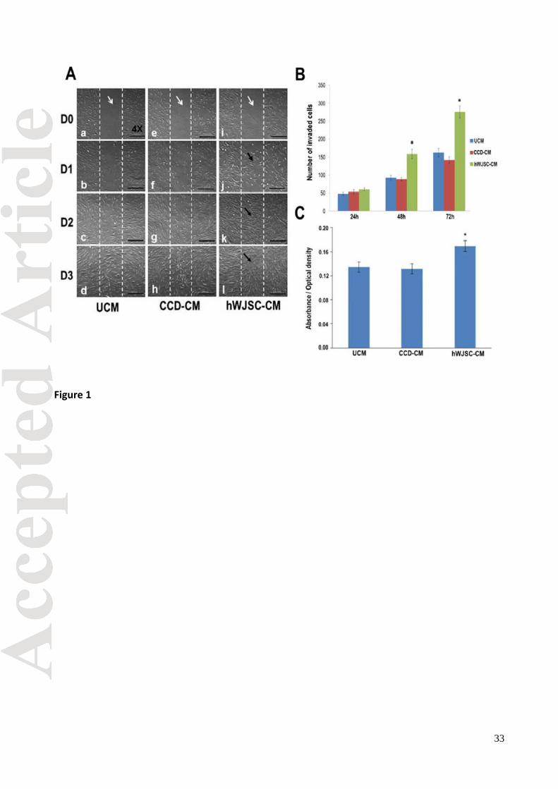
The skin fibroblasts in scratch-wound assays when exposed to the hWJSC-CM treatment arm
started to migrate from the edges of the scratches (‘wounds’) into the wound spaces as early as 6-
8 h and then completely covered the spaces by day 2 (48 h) compared to controls [CCD
conditioned medium (CCD-CM) and unconditioned KOSR medium (UCM)] (Fig. 1A). The mean ±
SEM number of cells that had invaded the scratches as determined by two independent observers
were significantly greater in the hWJSC-CM treatment arm compared to controls (p < 0.05) (Fig.
1B). The mean ± SEM cell numbers that migrated into the wound areas were 60 ± 04 (hWJSC-
CM), 53 ± 06 (CCD-CM) and 48 ± 04 (UCM) at 24h; 159 ± 13 (hWJSC-CM), 88 ± 05 (CCD-CM)
and 92 ± 07 (UCM) at 48h; and 276 ± 17 (hWJSC-CM), 142 ± 09 (CCD-CM) and 163 ± 12 (UCM)
at 72h respectively. The viability of the CCDs (MTT assay) for the treatment arm (hWJSC-CM)
was also significantly greater than the controls at 72 h (Fig. 1C).
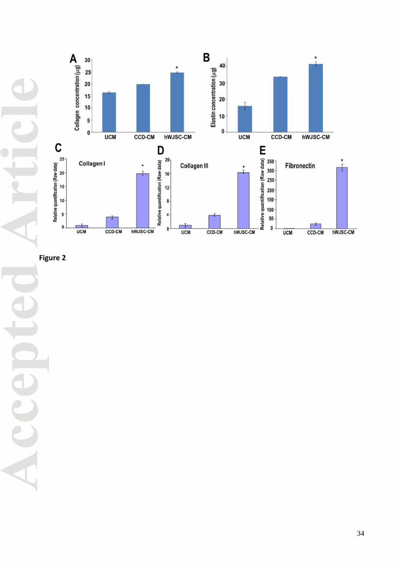
Collagen and Elastin (Sircol and Fastin assays)
The collagen and elastin concentrations using Sircol and Fastin assays respectively on CCDs
were significantly greater in the treatment arm (hWJSC-CM) compared to the controls at 72 h. (p <
0.05) (Figs. 2A and B).

14
Collagen type I, III and fibronectin (qRT-PCR)
qRT-PCR of the CCDs in the treatment arm (hWJSC-CM) showed significantly greater expression
of collagen type I, collagen type III and fibronectin compared to the controls (CCD-CM and UCM)
(p < 0.05). The increase in gene expression of fibronectin for the hWJSC-CM arm was 300 fold
compared to 40 fold for the CCD-CM arm. Collagen I and III were increased by 17-20 fold for the
hWJSC-CM arm compared to 3-4 fold for the CCD-CM arm (Figs. 2C, D and E).
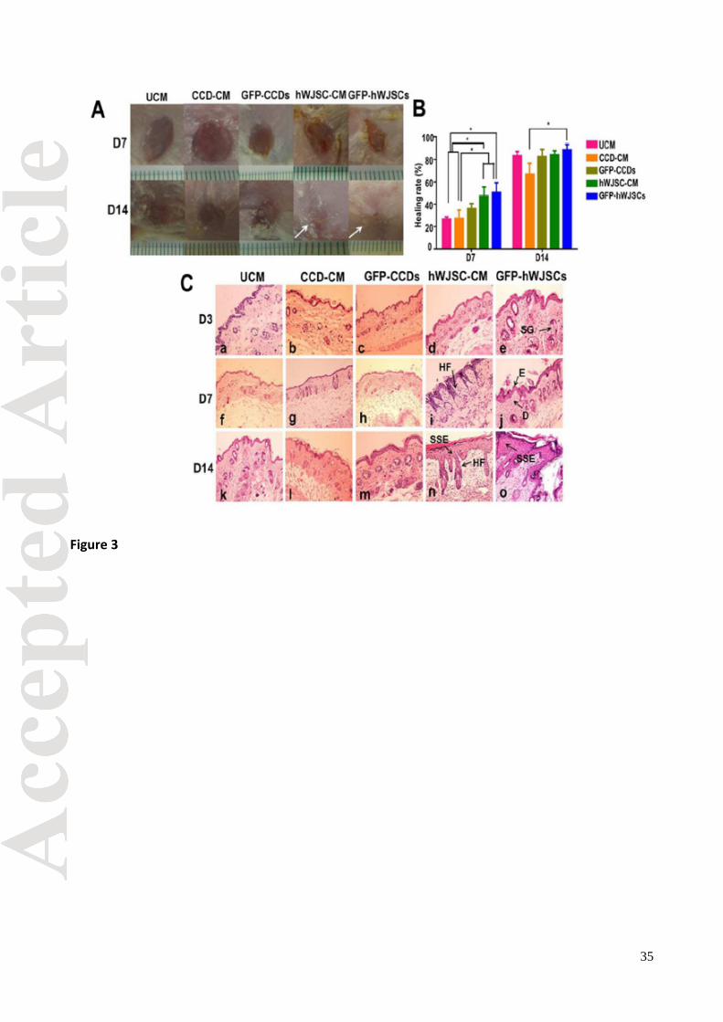
Healing rates and histology of excisional and diabetic wounds in mice
Macroscopic digitized images of healing of circular full-thickness murine excisional wounds after
application of GFP-hWJSCs and hWJSC-CM (treatment arms) and CCD-CM and UCM (controls)
showed that wound closure was greater in the treatment arms compared to controls (Fig. 3A).
Wound healing rates using the wound healing formula on digital images recommended by Chen
et al., [2009] showed that the GFP-hWJSCs and hWJSC-CM treatment groups exhibited
significantly greater healing rates compared to all control groups (GFP-CCDs, CCD-CM and UCM)
on day 7 (Fig 3B). On day 14, the GFP-hWJSCs treated wounds healed the greatest (p < 0.05)
(Fig 3B). Histological analysis of excisional wounds on day 14 showed that the epidermis and
dermal layers of wound areas in the treatment groups (GFP-hWJSCs and hWJSC-CM) had
increased reepithelialization, cellularity and vasculature, and increased sebaceous gland and hair
follicle numbers compared to controls (Fig. 3C). On D7 even though healing rates were not 100%
in the hWJSCs and hWJSC-CM treatments arms, some histological sections showed both dermal
and epidermal layers suggesting that the healing process was rapid.
Macroscopic observations, percentage healing rates and histology confirmed that healing of
diabetic wounds was also significantly enhanced in GFP-hWJSCs and hWJSC-CM treated mice
compared to controls over a 28 day period (Figs. 4A, B and C). On day 14 diabetic wounds
appeared larger than day 7 wounds because db/db skin does not heal by the same contraction
mechanisms as normal murine skin [Sullivan et al., 2004].

15
GFP-hWJSC survival and differentiation into keratinocytes in murine excisional and
diabetic wounds
Engraftment and survival of GFP-hWJSCs in the excisional and diabetic wounds at days 14 and
28 were confirmed from green signals via immunofluorescence microscopy (Fig. 5). The number of
GFP-hWJSCs in the wound biopsies gradually reduced with wound closure compared to the initial
numbers applied to wounds. Immunohistochemistry of excisional and diabetic murine wound
biopsies on days 14 and 28 showed the presence of positive human keratinocyte markers
(cytokeratin, involucrin and filaggrin) (Fig. 5).
Differential gene expression in excisional and diabetic wounds
On day 3 after initiation of wounds, ICAM-1 mRNA levels in excisional wounds in mice treated with
GFP-hWJSCs were significantly higher than CCD-CM and GFP-CCDs arms (p < 0.05) (Fig. 6A).
TIMP-1 expression was also significantly upregulated in excisional wounds on day 3 in the GFP-
hWJSCs treatment arm compared to controls (p < 0.05) (Fig. 6A). Interestingly, VEGF-A mRNA
expression on day 3 showed a 6 fold increase when excisional wounds were exposed to hWJSC-
CM (p < 0.05) (Fig. 6A). On day 7, both GFP-hWJSCs and hWJSC-CM treatment arms showed
significantly greater ICAM-1, TIMP-1 and VEGF-A expression levels compared to controls in
excisional wounds (p < 0.05). On day 14, the mRNA expression levels of ICAM-1 for excisional
wounds were not significantly different between groups but VEGF-A levels remained significantly
greater in GFP-hWJSCs and hWJSC-CM treatment arms compared to controls (p < 0.05) (Fig.
6A).
For diabetic wounds, the hWJSC-CM treatment arm showed significantly greater levels of TIMP-1
on days 7 and 14 and VEGF-A on days 7 and 28 compared to controls. VEGF-A levels for the
GFP-hWJSCs arm were significantly greater than controls in diabetic wounds on day 28 (p < 0.05)
(Fig. 6B).

16
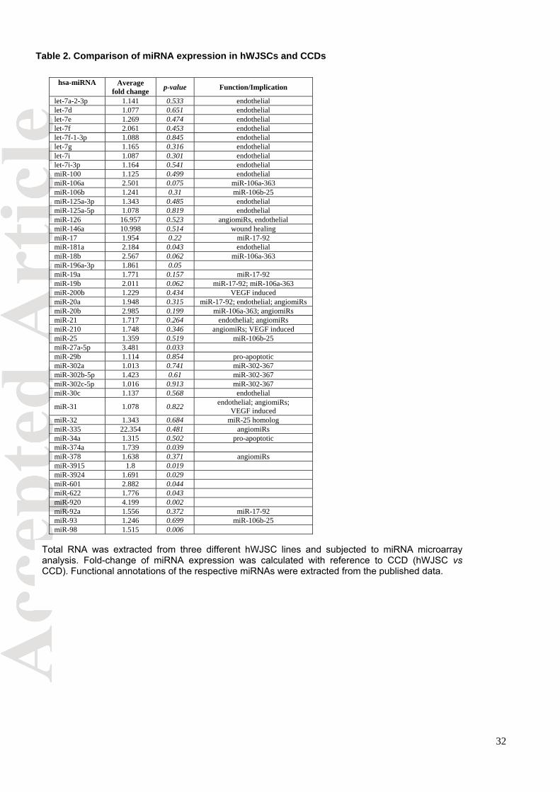
miRNA profile in hWJSCs
Among the 487 miRNAs that were found to be differentially regulated between hWJSCs and
CCDs, 82 miRNAs were expressed in hWJSCs at significantly higher levels (FDR, p<0.1). Among
them only hsa-miR-27a-5p, -98, -181a, -196a-3p, -374a, -601, -622, -920, -3915, and -3924 were
found to be upregulated in hWJSCs. The remaining 72 miRNAs were downregulated in hWJSCs
compared to CCDs. Nevertheless, several miRNAs that are involved in endothelial function and
proliferation processes including wound healing were highly expressed in hWJSCs (Table 2).
DISCUSSION
Wound healing can be divided into 3 phases (1) inflammation (2) proliferation and (3) remodelling
[Maxson et al., 2012]. During the proliferative phase native MSCs are recruited to differentiate into
new keratinocytes and in the final remodelling phase endogenous dermal fibroblasts reorganise
the extracellular matrix (ECM) to reinforce the granulation tissue [Jackson et al., 2012; Maxson et
al., 2012]. It has been a long-held view that MSCs applied to wounds modulate the host wound
environment by indirect mechanisms acting as a vector to deliver important therapeutic molecules.
In the present study the GFP-hWJSCs and hWJSC-CM arms showed significantly greater wound
healing compared to controls suggesting that direct and indirect mechanisms of wound healing
were taking place by directed differentiation of GFP-hWJSCs into keratinocytes and indirectly by
the release of important bioactive molecules that initiate and facilitate the host response to tissue
repair. Since the wound biopsies showed the survival of green GFP-hWJSCs signals and species-
specific upregulation of human keratinocyte markers (cytokeratin, involucrin and filaggrin),
differentiation of the GFP-hWJSCs into new keratinocytes was probably helping reepithelialization
in the excisional and diabetic wounds. Other workers have also shown that human umbilical cord
MSCs differentiate into keratinocytes when administered locally into the wound beds of mice [Luo
et al., 2010]. We and others have also observed that hWJSCs readily differentiate into
keratinocytes in vitro using two conventional differentiation protocols [Schneider et al., 2010; Jin et
al., 2011]. Cell proliferation is essential in the regenerative phase of wound healing and apart from

17
the dermal fibroblasts and keratinocytes, the stem or progenitor cells within the skin niches are
also involved in cell turnover [Harris et al., 2012]. Our results are consistent with the findings of
Shin and Petersen [2013] that the introduction of soluble bioactive molecules into wounds via the
hWJSC-CM and secretion of the same molecules by the engrafted GFP-hWJSCs creates a niche
for the recruitment of native skin MSCs and progenitor cells to the wound site to facilitate healing.
qRT-PCR and species-specific surface marker analysis on wound biopsies in the present study
helped to discriminate between the presence of murine and human cells. The upregulation of
ICAM-1, TIMP-1 and VEGF-A allowed us to hypothesize that both hWJSCs and hWJSC-CM
modulate differential gene expression in the wounds bringing about cell adhesion, increased
angiogenesis and epithelialization during wound healing. VEGF-A is unique for its multiple effects
on the wound healing cascade such as angiogenesis, epithelialization and collagen deposition
[Bao et al., 2009]. ICAM-1 is central to the regulation of the inflammatory process during wound
healing in the human and mouse [Yukami et al., 2007; Gay et al., 2011] because in the ICAM-1
knockout mouse model there was a delayed response to wound healing [Nagaoka et al., 2000].
Cytokine protein array analysis from our laboratory showed that important cytokines such as
ICAM-1, IL-6, IL-8 and TIMP-1 were secreted in large concentrations by hWJSCs in 72 hour
hWJSC-CM compared to controls [Fong et al., 2012]. Such cytokines in the hWJSC-CM thus play
a vital role in accelerating wound healing in the early phases. The hWJSCs secretome may help in
jumpstarting the wound healing process and subsequent paracrine effects on endogenous mouse
cytokine or growth factor secretion. Our qRT-PCR results demonstrated that the treatment of
hWJSCs to wounds showed increased expression of mouse ICAM-1 and TIMP-1 as early as day
3. Also, the ICAM-1 mRNA results on day 14 strongly correlate with the observations of wound
closure in vivo suggesting that hWJSCs may be producing adhesion molecules for the
acceleration of wound closure during the early phases of wound healing. An increase of TIMP-1
mRNA expression observed on day 14 in CCD-CM treated wounds suggest that the delay in
wound healing observed in the CCD-CM treated mice may be due to the late transcriptional
upregulation of TIMP-1. Several published reports have also shown high levels of matrix

18
metalloproteinases (MMPs) and low levels of tissue inhibitors of MMPs (TIMPs) in chronic wound
beds indicative of impaired wound healing [Liu et al., 2009; Stevens et al., 2012]. MMPs play a
major role in wound healing as they can break down all components of the extracellular matrix
(ECM) [Stevens et al., 2012]. In diabetic wounds there is a rise in MMPs and a decrease of TIMPs.
High levels of MMP-1 are essential for wound healing while increased levels of MMP-8 and 9 are
deleterious. The MMP-1/TIMP-1 ratio was considered a good predictor of healing of diabetic
wounds [Muller et al., 2008]. It is interesting to note that in the present study high levels of
secreted TIMP-1 was evident in hWJSC-CM at 72 h and was consistent on a transcriptional level
with significant increases in gene expression of fibronectin, collagen I and III indicating that
hWJSCs secrete endogenous proteins which affect ECM remodelling.
Collagen is normally produced during the inflammatory phase of wound healing and the ECM
secreted by the dermal fibroblasts helps wound remodelling. Fathke et al [2004] demonstrated that
MSC populations produce collagen types I and III providing long-term reconstitution of the
cutaneous wound. Collagen type III was also reported to be expressed in early healing processes
of skin [Zhang et al., 1995]. The upregulation of collagen I and III by the CCDs following exposure
to hWJSC-CM in the present study also suggests that hWJSC-CM supports wound healing. In the
present study hWJSC-CM also stimulated increases in fibronectin and elastin. Fibronectin is
another important ECM protein secreted by dermal fibroblasts during the proliferative phase of
wound healing [Bielefeld et al., 2011] and is essential in providing the necessary mechanical
strength to wounds [Clark, 1990]. Elastin confers the elasticity and resilience to the skin and these
fibres are usually interwoven among collagen bundles. Elastin is also involved in cell signalling and
migration in the wound healing process [Rnjak et al., 2011].
It has been suggested that bioactive molecules released by apoptotic cells stimulate endogenous
native and engrafted exogenous MSCs in wounds to initiate the regenerative process of wound
healing [Li et al., 2010]. Caspase 3/7 and prostaglandin E2 (PGE2) released from damaged
apoptotic cells in wounds stimulate the MSCs at the damaged site to proliferate, differentiate and
eventually replace the damaged tissue (’Phoenix rising’ pathway) [Li et al., 2010]. Mice lacking in

19
these caspases were deficient in skin wound healing [Li et al., 2010]. More recent evidence
confirmed the idea that dying cells signal their presence to the surrounding tissue and in doing so
elicit repair and regeneration via stem or progenitor cells that compensate for any loss of function
caused by cell death [Jager et al., 2012]. These molecules may play important roles in stimulating
the hWJSCs applied to the wounds in the present study to enhance healing.
Specific miRNA clusters (miR-106a-363, miR-17-92, miR-106b-25, miR-302-367 and miR-21)
were highly expressed in hWJSCs. The miR-106a-363 and miR-17-92 clusters that were
overexpressed are involved in cell proliferation and growth thus possibly playing a role in the
wound healing process. Functionally, miR-106b-25 promotes cell-cycle progression and
hyperproliferation via inhibition of the pro-apoptotic genes such as E2F1, Bim, Fas-activated
kinase, CASP7 and PTEN. Combined inhibition of pro-apoptotic genes further provides miR-106b-
25-expressing cells with a survival and progression or proliferation benefit. MiR-106b-25
upregulation may lead to enhanced binding to collagen therefore increasing ECM binding. The
miR-302-367 cluster targets TGFβ receptor 2 to increase E-cadherin expression and accelerates
mesenchymal-to-epithelial transition [Liao et al., 2011]. This cluster may be involved in the
enhanced reepithelialization in the wounds in the present study. In the normal wound healing
process miR-21 is upregulated on day 8 and downregulated in diabetic wounds [Madhyastha et
al., 2012]. MiR-21 exhibits a dual function (proliferative and anti-apoptosis) on vascular smooth
muscle cells (VSMCs) [Ji et al., 2007]. The role of apoptotic miRNA in promoting tissue
regeneration in wound healing was emphasized by Li et al [2010] in their ‘Phoenix rising’
mechanism of wound healing. The VEGF-induced miRNAs (miR-191, -155, -31, -17-5p, -18a and -
20a) [Suarez et al., 2007] were also significantly upregulated in hWJSCs compared to CCDs and
hBMMSCs. VEGF is an important player in the wound healing process [Bao et al., 2009]. Based
on our microarray data analysis, besides, the specific miRNAs that have been found to be involved
in proliferation, wound healing and pro-angiogenic processes (Table 2), the miRNAs belonging to
the clusters described above were also significantly overexpressed in hWJSCs. Thus, the
enhanced wound healing observed in the presence of hWJSCs and hWJSC-CM is probably
guided by several miRNAs that provide beneficial effects to the wound healing process.
20
ACKNOWLEDGEMENTS
The authors acknowledge the grant support to carry out this work from the National Research
Foundation (NRF) (Proof-Of-Concept grant: R-174-000-139-281) and the Academic Research
Fund, National University of Singapore (AcRF-NUS) (R-174-000-122-112).
21
REFERENCES
Badiavas EV, Abedi M, Butmarc J, Falanga V, Quesenberry P. 2003. Participation of bone marrow
derived cells in cutaneous wound healing. Journal of Cellular Physiology 196: 245-250.
Bao P, Kodra A, Tomic-Canic M, Golinko MS, Ehrlich HP, Brem H. 2009. The role of vascular
endothelial growth factor in wound healing. The Journal of Surgical Research 153: 347-358.
Bey E, Prat M, Duhamel P, Benderitter M, Brachet M, Trompier F. 2010. Emerging therapy for
improving wound repair of severe radiation burns using local bone marrow-derived stem cell
administrations. Wound Repair and Regeneration 18: 50-58.
Bielefeld KA, Amini-Nik S, Whetstone H, Poon R, Youn A, Wang J. 2011. Fibronectin and β-
Catenin act in a regulatory loop in dermal fibroblasts to modulate cutaneous healing. J Cell
Biochem 286: 27687-27697.
Bongso A, Fong CY. 2013. The therapeutic potential, challenges and future clinical directions of
stem cells from the Wharton’s jelly of the human umbilical cord. Stem Cell Rev and Rep 9: 226-
240.
Borue X, Lee S, Grove J, Herzog EL, Harris R, Diflo T. 2004. Bone marrow-derived cells contribute
to epithelial engraftment during wound healing. The American Journal of Pathology 165: 1767-
1772.
Chen L, Tredget EE, Liu C, Wu Y. 2009. Analysis of allogenecity of mesenchymal stem cells in
engraftment and wound healing in mice. PLOS One 4: e7119.
Clark RAF. 1990. Fibronectin matrix deposition and fibronectin receptor expression in healing and
normal skin. The Journal of Investigative Dermatology 94: 128S–134S.
Cory G. 2011. Scratch-wound assay. Methods Mol Biol 769: 25-30.

22
Dominici M, Le Blanc K, Mueller I, Slaper-Cortenbach I, Marini F, Krause D. 2006. Minimal criteria
for defining multipotent mesenchymal stromal cells. The International Society for Cellular Therapy
position statement. Cytotherapy 8: 315-317.
Estes JM, Adzick NS, Harrison MR, Longaker MT, Stern R. 1993. Hyaluronate metabolism
undergoes an ontogenic transition during fetal development: implications for scar-free wound
healing. Journal of Paediatric Surgery 28: 1227-1231.
Fan CG, Zhang Q, Zhou J. 2011. Therapeutic potentials of mesenchymal stem cells derived from
human umbilical cord. Stem Cell Rev and Rep 7: 195-207.
Fathke C, Wilson L, Hutter J, Kapoor V, Smith A, Hocking A. 2004. Contribution of bone marrow-
derived cells to skin: collagen deposition and wound repair. Stem cells 22: 812-822.
Fong CY, Richards M, Manasi N, Biswas A, Bongso A. 2007. Comparative growth behaviour and
characterization of stem cells from human Wharton’s jelly. Reprod BioMed Online 15: 708-718.
Fong CY, Subramanian A, Biswas A, Gauthaman K, Srikanth P, Hande MP, Bongso A. 2010.
Derivation efficiency, cell proliferation, frozen-thaw survival, stem-cell properties, and
differentiation of human Wharton’s jelly stem cells. Reprod BioMed Online 21: 391-401
Fong CY, Chak LL, Biswas A, Tan JH, Gauthaman K, Chan WK, Bongso A. 2011. Human
Wharton’s jelly stem cells have unique transcriptome profiles compared to human embryonic stem
cells and other mesenchymal stem cells. Stem Cell Rev and Rep 7: 1-16.
Fong CY, Gauthaman K, Cheyyatraivendran S, Lin HD, Biswas A, Bongso A. 2012. Human
umbilical cord Wharton's jelly stem cells and its conditioned medium support hematopoietic stem
cell expansion ex vivo. J Cell Biochem 113: 658-668.
Gauthaman K, Fong CY, Suganya CA, Subramanian A, Biswas A, Choolani M. 2012. Extra-
embryonic human Wharton's jelly stem cells do not induce tumorigenesis, unlike human embryonic
stem cells. Reprod Biomed Online 24: 235-246.
23
Gay AN, Mushin OP, Lazar DA, Naik-Mathuria BJ, Ling Yu, Gobin A. 2011. Wound healing
characteristics of ICAM-1 null mice devoid of all isoforms of ICAM-1. The Journal of Surgical
Research 171: e1-e7.
Harris DT, Hilgaertner J, Simonson C, Ablin RJ, Badowski M. 2012. Cell-based therapy for
epithelial wounds. Cytotherapy 14: 802-810.
Jackson WM, Nesti LJ, Tuan RS. 2012. Concise review: clinical translation of wound healing
therapies based on mesenchymal stem cells. Stem Cells Translational Medicine 1: 44-50.
Jager R, Fearnhead HO. 2012. Dead cells talking: the silent form of cell death is not so quiet.
Biochem Res International, doi:10.1155/2012/453838.
Jeon YK, Jang YH, Yoo DR, Kim SN, Lee SK, Nam MJ. 2010. Mesenchymal stem cells interaction
with skin: wound-healing effect on fibroblast cells and skin tissue. Wound Repair and Regen 18:
655-661.
Ji R, Cheng Y, Yue J, Yang J, Liu X, Chen H. 2007. MicroRNA expression signature and
antisense-mediated depletion reveal an essential role of MicroRNA in vascular neointimal lesion
formation. Circulation Research 100: 1579-1588.
Jin G, Prabhakaran MP, Ramakrishna S. 2011. Stem cell differentiation to epidermal lineages on
electrospun nanofibrous substrates for skin tissue engineering. Acta Biomaterialia 7: 3113-3122.
Karolina DS, Armugam A, Tavintharan S, Wong M, Lim SC, Sum CF. 2011. MicroRNA 144 impairs
insulin signaling by inhibiting the expression of insulin receptor substrate 1 in type 2 diabetes
mellitus. PLoS One 6: e22839.
Kim SW, Zhang HZ, Guo L, Kim JM, Kim MH. 2012. Amniotic mesenchymal stem cells enhance
wound healing in diabetic NOD/SCID mice through high angiogenic and engraftment capabilities.
PLoS ONE 7: e41105.
Kuo YR, Wang CT, Cheng JT, Wang FS, Chiang YC, Wang CJ. 2011. Bone marrow-derived
mesenchymal stem cells enhanced diabetic wound healing through recruitment of tissue
24
regeneration in a rat model of streptozotocin-induced diabetes. Plastic and Reconstructive Surgery
128: 872-880.
La Rocca G, Anzalone R, Corrao S, Magno F, Loria T, Lo Iacono M. 2009. Isolation and
characterization of OCT4+/HLA-G+ mesenchymal stem cells from the human umbilical cord
matrix: differentiation potential and detection of new markers. Histochem Cell Biol 131: 267-282.
Li F, Huang Q, Chen J, Peng Y, Roop D, Bedford J. 2010. Apoptotic cells activate the ’Phoenix
rising’ pathway to promote wound healing and tissue regeneration. Science Signaling 3: ra13.
Liao B, Bao X, Liu L, Feng S, Zovoilis A, Liu W. 2011. MicroRNA cluster 302-367 enhances
somatic cell reprogramming by accelerating a mesenchymal-to-epithelial transition. J Cell Biochem
286: 17359-17364.
Liu Y, Min D, Bolton T, Nubé V, Twigg SM, Yue DK. 2009. Increased matrix metaloproteinase-9
predicts poor wound healing in diabetic foot ulcers. Diabetes Care 32: 117-119.
Lorenz HP, Longaker MT, Perkocha LA, Jennings RW, Harrison MR, Adzick NS. 1992. Scarless
wound repair: a human fetal skin model. Development 114: 253-259.
Luo, G., Cheng, W., He, W., Wang X, Tan J, Fitzgerald M. 2010. Promotion of cutaneous wound
healing by local application of mesenchymal stem cells derived from human umbilical cord blood.
Wound Repair and Regeneration 18: 506-513.
Nagaoka T, Kaburagi Y, Hamaguchi Y, Hasegawa M, Takehara K, Steeber DA. 2000. Delayed
wound healing in the absence of intercellular adhesion molecule-1 or L-selectin expression. The
American Journal of Pathology 157: 237-247.
Madhyastha R, Madhyastha H, Nakajima Y, Omura S, Maruyama M. 2012. MicroRNA signature in
diabetic wound healing: promotive role of miR-21 in fibroblast migration. International Wound
Journal 9: 355-361.
Martin P, D'Souza D, Martin J, Grose R, Cooper L, Maki R. 2003. Wound healing in the PU.1 null
mouse--tissue repair is not dependent on inflammatory cells. Current Biology 13: 1122-1128.
25
Mathieu D, Linke JC, Wattel F. 2006. Non-healing wounds: In handbook on hyperbaric medicine.
DE Mathieu, ed. Springer, Amsterdam, The Netherlands, 401-428.
Maxson S, Lopez EA, Yoo D, Danilkovitch-Miagkova A, Leroux MA. 2012. Concise review: role of
mesenchymal stem cells in wound repair. Stem Cells Translational Medicine 1: 142-149.
Muller M, Trocme C, Lardy B, Morel F, Halimi S, Benhamou PY. 2008. Matrix metalloproteinases
and diabetic foot ulcers; the ratio of MMP-1 to TIMP-1 is a predictor of wound healing. Diabet Med
25: 419-426.
Pappa KI, Anagnou NP. 2009. Novel sources of fetal stem cells: where do they fit on the
developmental continuum? Future Medicine 4: 423-433.
Rnjak J, Wise SG, Mithieux SM, Anthony S, Weiss AS. 2011. Severe burn injuries and the role of
elastin in the design of dermal substitutes. Tissue Engineering Part B, Reviews 17: 81-91.
Sasaki M, Abe R, Fujita Y, Ando S, Inokuma D, Shimizu H. 2008. Mesenchymal stem cells are
recruited into wounded skin and contribute to wound repair by transdifferentiation into multiple skin
cell type. Journal of Immunology 180: 2581-2587.
Schneider RK, Püllen A, Kramann R, Bornemann J, Knüchel R, Neuss S. 2010. Long-term survival
and characterisation of human umbilical cord-derived mesenchymal stem cells on dermal
equivalents. Differentiation 79: 182-193.
Shin L, Peterson DA. 2013. Human mesenchymal stem cell grafts enhance normal and impaired
wound healing by recruiting existing endogenous tissue stem/progenitor cells. Stem Cells
Translational Medicine 2: 33-42.
Stevens LJ, Page-McCaw A. 2012. A secreted MMP is required for reepithelialisation during
wound healing. Molecular Biology of the Cell 23: 1068-1079.
26
Stoff A, Rivera AA, Sanjib Banerjee N, Moore ST, Michael Numnum T, Espinosa-de-Los-Monteros
A. 2009. Promotion of incisional wound repair by human mesenchymal stem cell transplantation.
Experimental Dermatology 18: 362-369.
Suarez Y, Hernando CF, Yu J, Gerber S, Harrison K, Pober J. 2008. Dicer-dependent endothelial
microRNAs are necessary for postnatal angiogenesis. Proc Natl Acad Sci U S A 105: 14082-
14087.
Sullivan SR, Underwood RA, Gibran NS, Sigle RO, Usui ML, Carter WG. 2004. Validation of a
model for the study of multiple wounds in the diabetic mouse (db/db). Plast Reconstr Surg 113:
953-60.
Taghizadeh RR, Cetrulo KJ, Cetrulo CL. 2011. Wharton’s jelly stem cells: Future clinical
applications. Placenta 32: S311-S315.
Troyer DL, Weiss ML. 2008. Concise review: Wharton’s jelly-derived cells are a primitive stromal
cell population. Stem Cells 26: 591-599.
Walter MN, Wright KT, Fuller HR, MacNeil S, Johnson WE. 2010. Mesenchymal stem cell-
conditioned medium accelerates skin wound healing: an in vitro study of fibroblast and
keratinocyte scratch assays. Exp Cell Res 316: 1271-1281.
Wang XY, lan Y, He WY, Zhang L, Yao HY, Hou C. 2008. Identification of mesenchymal stem cells
in aorta-gonad-mesonephros and yolk sac of human embryos. Blood 111: 2436-2443.
Wang Y, Han ZB, Ma J., Zuo C, Geng J, Gong W. 2012. A toxicity study of multiple-administration
human umbilical cord mesenchymal stem cells in cynomolgus monkeys. Stem Cells and
Development 21: 1401-1408.
Wu Y, Chen L, Scott PG, Tredget EE. 2007. Mesenchymal stem cells enhance wound healing
through differentiation and angiogenesis. Stem Cells 25: 2648-2659.
27
Yukami, T., Hasegawa, M., Matsushita, Y., Fujita T, Matsushita T, Horikawa M. 2007. Endothelial
selectins regulate skin wound healing in cooperation with L-selectin and ICAM-1. J Leukocyte
Biology 82: 519-531.
Zhang K, Garner W, Cohen L, Rodriguez J, Phan S. 1995. Increased Types I and III collagen and
transforming growth factor- 1 mRNA and protein in hypertrophic burn scar. The Journal of
Investigative Dermatology 104: 750-754.

28
LEGENDS TO FIGURES
Fig. 1. (A) Scratch-wound assay of CCD fibroblasts exposed to hWJSC-CM (0-72h). a-d: CCD
fibroblasts grown in unconditioned medium KOSR medium (UCM) (control); e-h: CCD fibroblasts
grown in CCD-CM (control); i-l: CCD fibroblasts grown in hWJSC-CM (treatment). White arrows in
a, e and i indicate vertical scratches in middle of monolayer with no CCD fibroblasts at D0. Black
arrows in j, k and l show faster migration and increased numbers of CCDs into scratches in
hWJSC-CM arm compared to controls. The scratches in the hWJSC-CM arm were fully closed by
D2. (B) The mean ± SEM number of invading cells into scratches were significantly greater in the
hWJSC-CM arm compared to both controls at 48h and 72h (*p < 0.05) (C) The mean ± SEM
CCD viability (MTT assay) was significantly greater in the hWJSC-CM arm compared to both
controls at 72h (*p < 0.05). Results are expressed as optical density values relative to controls.
Fig. 2. (A, B) The mean ± SEM levels of collagen (Sircol assay) and elastin (Fastin assay) of
CCDs were significantly greater in the hWJSC-CM treatment arm compared to controls (*p <
0.05). (C-E) qRT-PCR analysis showed significantly greater expression of (C) collagen type I, (D)
collagen type III and (E) fibronectin in the CCDs exposed to hWJSC-CM compared to controls (*p
< 0.05). GAPDH was the internal control. Data analysis and relative quantitation was done using
the comparative Ct (ΔΔCT) method.
Fig. 3. (A) Digital images of mouse excisional wounds showing faster wound closure by 14 days
(D14) in SCID mice exposed to GFP-hWJSCs and hWJSC-CM (treatment arms) (white arrows)
compared to controls (GFP-CCDs, CCD-CM and UCM). (B) On day 7, mean ± SEM percentage
healing rates in excisional wounds in SCID mice were significantly greater in the treatment arms
(GFP-hWJSCs and hWJSC-CM) compared to controls (*p < 0.05). On day 14, the mean ± SEM
percentage healing rates were significantly greater for the GFP-hWJSCs treatment arm compared

29
to controls (*p < 0.05). (C) Hematoxylin and eosin histological sections of murine wound biopsies
taken at days 3, 7 and 14 (D3, D7, D14). On D14, the epidermis and dermis of wounds in the
treatment groups (GFP-hWJSCs and hWJSC-CM) showed reepithelialization, formation of
stratified squamous epithelium, increased cellularity and vasculature, and increased numbers of
sebaceous glands and hair follicles when compared to the controls (n, o). On D7 even though
healing rates were not 100% in the hWJSCs and hWJSC-CM treatments arms, some histological
sections showed both dermal and epidermal layers suggesting that the healing process was rapid
(i, j). D: dermis; E: epidermis; HF: hair follicle; SG: sebaceous gland; SSE: stratified squamous
epithelium. Scale bar = 100 µm.
Fig. 4. (A) Digital images of mouse diabetic wounds showing faster wound closure by 28 days
(D28) in diabetic mice exposed to GFP-hWJSCs and hWJSC-CM (treatment arms) (white arrows)
compared to controls (UCM). (B) Mean ± SEM percentage healing rates in diabetic wounds in
diabetic mice were significantly greater in the GFP-hWJSCs treatment arm on D7 and in the
hWJSC-CM treatment arm on D28 compared to controls (*p < 0.05). (C) Hematoxylin and eosin
histological sections of murine diabetic wound biopsies taken at days 7, 14 and 28 (D7, D14, D28).
By D14 and D28, the epidermis of wound biopsies of the treatment arms (GFP-hWJSCs and
hWJSC-CM) showed reepithelialization and formation of stratified squamous epithelium. The
dermis showed increased cellularity and vasculature, and increased sebaceous gland and hair
follicle numbers compared to the controls. E: epidermis; HF: hair follicle; SG: sebaceous gland; S:
stroma; SSE: stratified squamous epithelium. Scale bar = 100 µm.
Fig. 5. Fluorescent immunohistochemical images of diabetic mouse wound biopsies on D14 and
D28 showing green GFP-labelled hWJSCs (short thin arrows) and positive human keratinocyte
markers (red) (thick long arrows). a, d represent cytokeratin clone AE1/AE3; b, e represent
involucrin and c, f represent filaggrin. DAPI: blue. Scale bar = 50 µm.
30
Fig. 6. qRT-PCR mRNA expression for mouse ICAM-1, TIMP-1 and VEGF-A using TaqMan
probes in (A) excisional wound biopsies and (B) diabetic wound biopsies on days 3-28 (D3-D28).
(A) On D3 ICAM-1 mRNA levels in GFP-hWJSC treated excisional wounds were significantly
higher than controls (CCD-CM and GFP-CCDs) (*p < 0.05). TIMP-1 expression was also
significantly upregulated in GFP-hWJSCs treated excisional wounds on D3 compared to controls
(*p < 0.05). VEGF-A mRNA expression on D3 showed a 6 fold increase in hWJSC-CM treated
excisional wounds compared to controls (*p < 0.05). On D7, both GFP-hWJSCs and hWJSC-CM
treated excisional wounds arms showed significantly greater ICAM-1, TIMP-1 and VEGF-A
expression levels compared to controls (*p < 0.05). On D14, ICAM-1 levels for excisional wounds
were not significantly different between groups but VEGF-A levels were significantly greater in
GFP-hWJSCs and hWJSC-CM treated excisional wounds compared to controls (*p < 0.05). (B)
For diabetic wounds, the hWJSC-CM treatment arm showed significantly greater levels of TIMP-1
on D7 and D14 and VEGF-A on D7 and D28 compared to controls. VEGF-A levels for both the
GFP-hWJSCs and hWJSC-CM arms were significantly greater than controls on D28 (*p < 0.05).

31
Table 1. The ECM-related genes and primer sequences used for qRT-PCR
_____________________________________________________________________________
Gene Primer Sequence
_____________________________________________________________________________
GAPDH F: 5’- GCACCGTCAAGGCTGAGAAC -3’
R: 5’- GGATCTCGCTCCTGGAAGATG-3’
Collagen Type I F: 5’- CACAGAGGTTTCAGTGGTTTGG -3’
R: 5’- GCACCAGTAGCACCATCATTTC -3’
Collagen Type III F: 5’- CTGAAATTCTGCCATCCTGAAC-3’
R: 5’- GGATTGCCGTAGCTAAACTGAA -3’
Fibronectin F: 5’- AAGATTGGAGAGAAGTGGGACC -3’
R: 5’- G GAGCAAATGGCACCGAGATA -3’
_____________________________________________________________________________
F: Forward primer; R: Reverse primer, GAPDH: Glyceraldehyde-3-phosphate dehydrogenase
32
hsa-miRNA Average fold change p-value Function/Implication
let-7a-2-3p 1.141 0.533 endothelial let-7d 1.077 0.651 endothelial let-7e 1.269 0.474 endothelial let-7f 2.061 0.453 endothelial let-7f-1-3p 1.088 0.845 endothelial let-7g 1.165 0.316 endothelial let-7i 1.087 0.301 endothelial let-7i-3p 1.164 0.541 endothelial miR-100 1.125 0.499 endothelial miR-106a 2.501 0.075 miR-106a-363 miR-106b 1.241 0.31 miR-106b-25 miR-125a-3p 1.343 0.485 endothelial miR-125a-5p 1.078 0.819 endothelial miR-126 16.957 0.523 angiomiRs, endothelial miR-146a 10.998 0.514 wound healing miR-17 1.954 0.22 miR-17-92 miR-181a 2.184 0.043 endothelial miR-18b 2.567 0.062 miR-106a-363 miR-196a-3p 1.861 0.05 miR-19a 1.771 0.157 miR-17-92 miR-19b 2.011 0.062 miR-17-92; miR-106a-363 miR-200b 1.229 0.434 VEGF induced miR-20a 1.948 0.315 miR-17-92; endothelial; angiomiRs miR-20b 2.985 0.199 miR-106a-363; angiomiRs miR-21 1.717 0.264 endothelial; angiomiRs miR-210 1.748 0.346 angiomiRs; VEGF induced miR-25 1.359 0.519 miR-106b-25 miR-27a-5p 3.481 0.033 miR-29b 1.114 0.854 pro-apoptotic miR-302a 1.013 0.741 miR-302-367 miR-302b-5p 1.423 0.61 miR-302-367 miR-302c-5p 1.016 0.913 miR-302-367 miR-30c 1.137 0.568 endothelial
miR-31 1.078 0.822 endothelial; angiomiRs; VEGF induced
miR-32 1.343 0.684 miR-25 homolog miR-335 22.354 0.481 angiomiRs miR-34a 1.315 0.502 pro-apoptotic miR-374a 1.739 0.039 miR-378 1.638 0.371 angiomiRs miR-3915 1.8 0.019 miR-3924 1.691 0.029 miR-601 2.882 0.044 miR-622 1.776 0.043 miR-920 4.199 0.002 miR-92a 1.556 0.372 miR-17-92 miR-93 1.246 0.699 miR-106b-25 miR-98 1.515 0.006
Table 2. Comparison of miRNA expression in hWJSCs and CCDs
Total RNA was extracted from three different hWJSC lines and subjected to miRNA microarrayanalysis. Fold-change of miRNA expression was calculated with reference to CCD (hWJSC vsCCD). Functional annotations of the respective miRNAs were extracted from the published data.
33
Figure 1
34
Figure 2
35
Figure 3

36
Figure 4

37
Figure 5

38
Figure 6


