
Human and Bench Methods for Measurement of Shear and Performance Characteristics of Devices Intended to Mitigate
Shear
Evan Call, MS, CSM (NRM)

Shear is Both Imparted and Confounded by Friction

Shear Stress/Shear Strain
Image from Edhat.com Image from www2.dickson.edu

What Does Shear Feel Like
• Peanut butter under your tongue

Shear or Deep Tissue Injury
Photo from Woundeducators.com
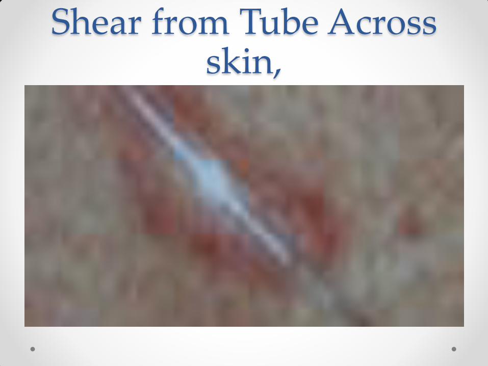
Shear from Tube Across skin,

Bunching or Rolling Shear Displacement
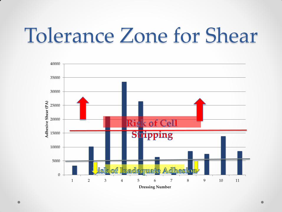
Tolerance Zone for Shear
0
5000
10000
15000
20000
25000
30000
35000
40000
1 2 3 4 5 6 7 8 9 10 11
Ad
hes
ive
Sh
ear
(PA
)
Dressing Number
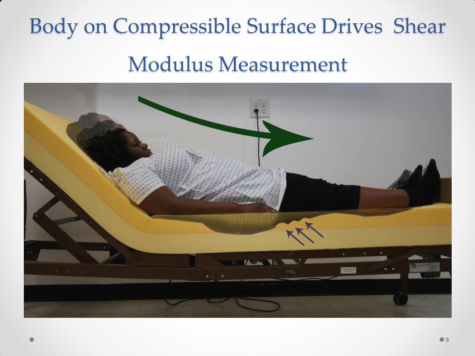
Body on Compressible Surface Drives Shear
Modulus Measurement
9

Shear Measurement Tests • L1 to Femoral Epicondyles Sliding Resistance
• Gel Distortion Indenter
• Direct Shear Measurement
• Translational Shear
• Tissue Model Shear
• Cadaver Heel Shear
• Lower extremity indenter shear
• Shear at Force (shear and pressure)
• Shear as a “wear” element

Shear in Humans • Direct measurement
o Molten predia
• Head of bed elevation
• Shear from normal sitting/ laying
• With / Without Dressing comparison
• With / Without Liquicell comparison
o Variable Resistance Elastomer
• Shear in tissues as head of bed is raised
• Shear in tissues as they immerse in a cushion
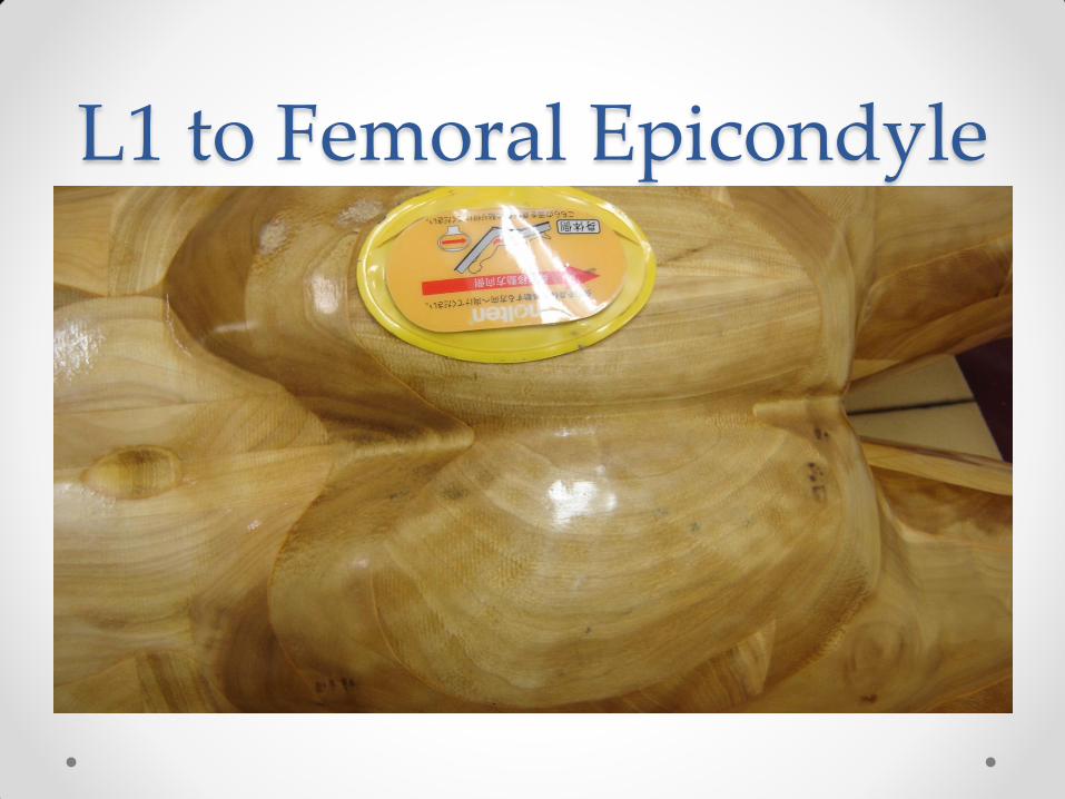
L1 to Femoral Epicondyle

Shear Modulus in Surfaces and Devices

Work Performed • Work (J) = Max Force (N) x deflection (.150 m)
• The Total work for each mattress tested at displacement intervals
of 50 mm and the 50-150 mm interval is shown below. The error
bars represent the 95th% confidence interval (alpha = 0.05).
Total Work (J)
0.00
2.00
4.00
6.00
8.00
10.00
12.00
IsoFlex Plexus PressurePedic SymmAire TemperPedic
To
tal
Wo
rk (
J)
0-50 mm
50-100 mm
100-150 mm
50-150 mm
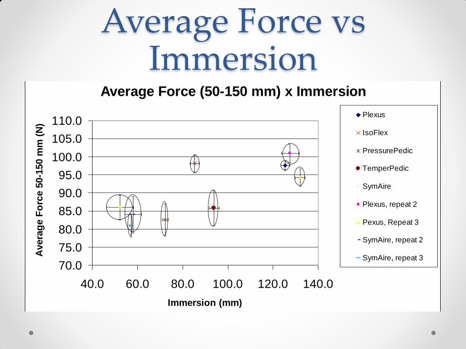
Average Force vs Immersion
Average Force (50-150 mm) x Immersion
70.0
75.0
80.0
85.0
90.0
95.0
100.0
105.0
110.0
40.0 60.0 80.0 100.0 120.0 140.0
Immersion (mm)
Avera
ge F
orc
e 5
0-1
50 m
m (
N)
Plexus
IsoFlex
PressurePedic
TemperPedic
SymAire
Plexus, repeat 2
Pexus, Repeat 3
SymAire, repeat 2
SymAire, repeat 3

Slope Slope 1st cm (N/m)
-5.00E-04
0.00E+00
5.00E-04
1.00E-03
1.50E-03
2.00E-03
2.50E-03
IsoFlex Plexus PressurePedic SymmAire TemperPedic
Slo
pe (
N/m
) 0-50 mm
50-100 mm
100-150 mm
50-150 mm
•The average slope of the force-displacement curve at 50 mm intervals and the 50-150 mm interval for each mattress is shown below. The error bars represent the 95th% confidence interval (alpha = 0.05).

Friction Shear and Bulk Modulus
17

Gel Indenter with Shear Strain Sensors

Calibration of Shear Indenter

Direct Shear Measurement

Direct Shear Measurent

Shear Translated Through Device

ShearForce Transmitted Through Dressing
• Mepilex® Border Flex
• Mepilex® Border Sacrum
• Optiva Gentle® Border

System to Model Human Shear

System to Model Human Shear
Effect of Dressing on Shear
0.0
1.0
2.0
3.0
4.0
5.0
6.0
7.0
8.0
0 1 2 3 4
Distance Traveled (cm)
Sh
ear
(N)
With
Dressing
Without
Dressing

Model is Very Close to Human Subjects
Human-bench test comparison
0
1
2
3
4
5
6
7
8
9
Pe
ak
Sh
ea
r (N
)
Human Volunteer
HR-45 Weighted Foam
Sled
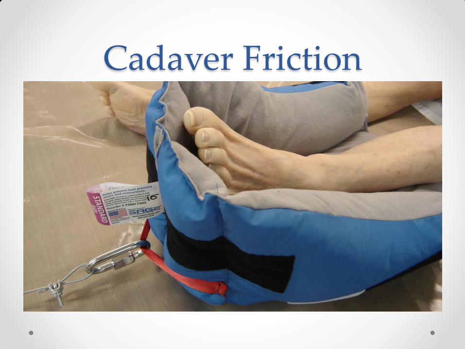
Cadaver Friction

Heel Boot Shear

Boot Retention Shear (N)

Friction both Sides, Shear Absorption
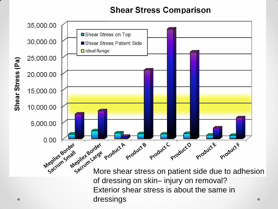
More shear stress on patient side due to adhesion
of dressing on skin– injury on removal?
Exterior shear stress is about the same in
dressings
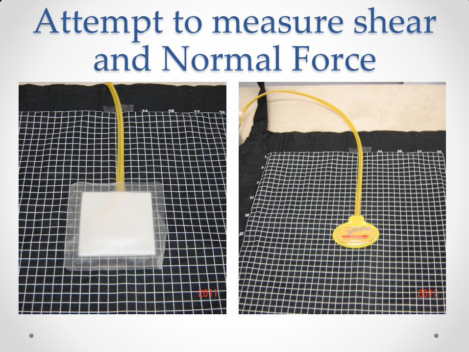
Attempt to measure shear and Normal Force
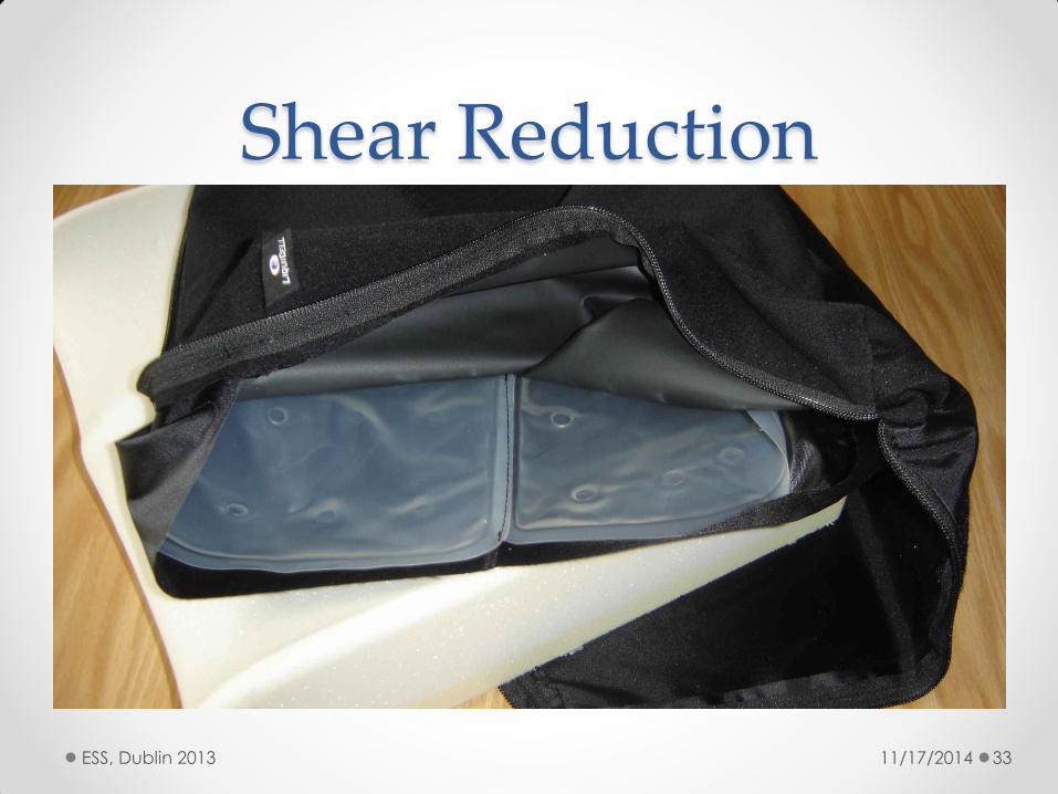
Shear Reduction
11/17/2014 ESS, Dublin 2013 33

Shear Reduction
11/17/2014 ESS, Dublin 2013 34

Extras – LiquiCell reduces shear
+/-10°rotation
LiquiCell
Basic
+/-1°rotation
LiquiCell
Basic Denim Layer
Peak torque reduced by 25%
Shear Duration Reduced by 8%
Micro-shear events (start, stop, turn) can lead to decubitis
LiquiCell reduces the shear event duration and the peak loads of the shear event
Normal Loading Shear
Torque (twisting) shear test using jeans

Shear on Mattress with Liquicell
AveragePeakForce(N) AverageForce(N)AverageForcePost
Compression
VolunteerWithoutLiquicell
WithLiquicell
WithoutLiquiCell
WithLiquicell
WithoutLiquicell
WithLiquicell
A 8.03±0.62 5.70±0.62 6.55±0.19 4.28±0.70 6.37±1.44 3.10±2.03
B 6.07±0.78 5.14±0.05 5.46±0.81 4.45±0.12 3.05±0.82 4.84±0.31
C 3.94±0.95 3.69±0.55 3.14±0.98 2.87±0.55 3.90±0.33 3.42±0.33
D 6.63±1.32 6.97±1.84 5.49±0.98 6.11±1.53 6.73±0.97 7.19±0.59
E 7.70±2.53 3.70±1.85 6.72±2.23 2.53±1.85 6.59±1.80 1.92±3.59
F 6.58±0.75 5.58±1.65 5.85±0.52 4.60±1.55 5.44±0.77 5.09±1.39
G 4.39±0.96 3.38±0.63 3.67±0.87 2.63±0.59 3.57±0.08 3.18±0.63
H 9.07±2.17 8.34±1.61 7.68±1.47 7.17±1.61 7.21±1.25 6.61±0.38
I 5.90±1.16 4.42±2.15 4.46±1.28 3.07±1.90 2.60±1.60 2.35±1.42
J 5.50±1.41 4.83±1.35 4.32±0.63 3.26±0.79 3.52±0.59 2.85±0.79
AverageA-J 6.38±0.70 5.18±0.69 5.50±0.66 4.24±0.66 5.18±0.66 4.04±0.76

Roller Shear Rig and Result
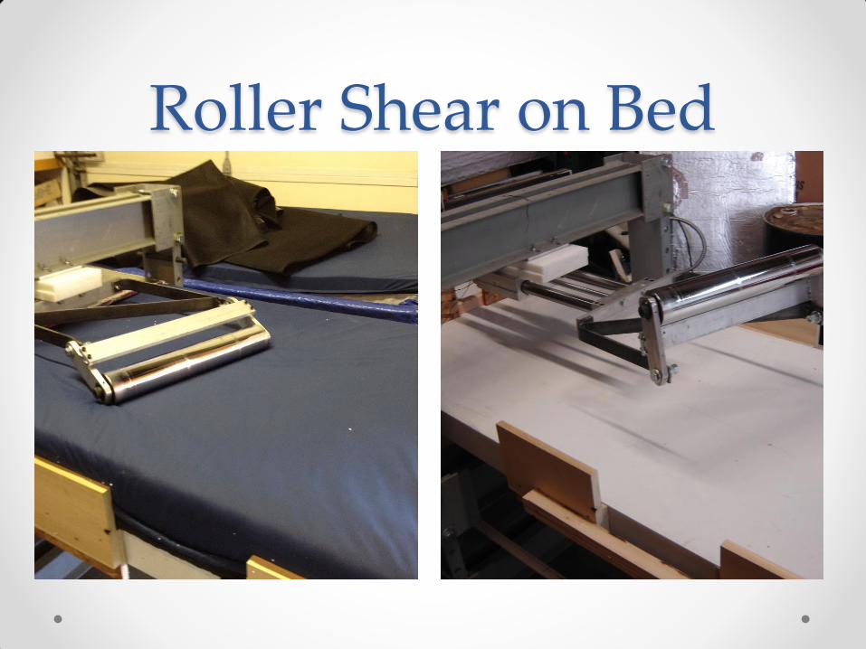
Roller Shear on Bed

Ultimate Friction: Adhesion
39
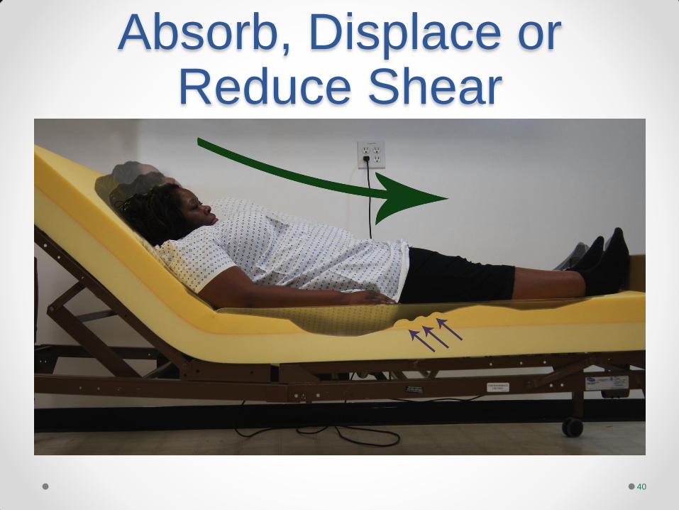
Absorb, Displace or Reduce Shear
40

Measuring Shear Between a
Patient and the support
Surface
41
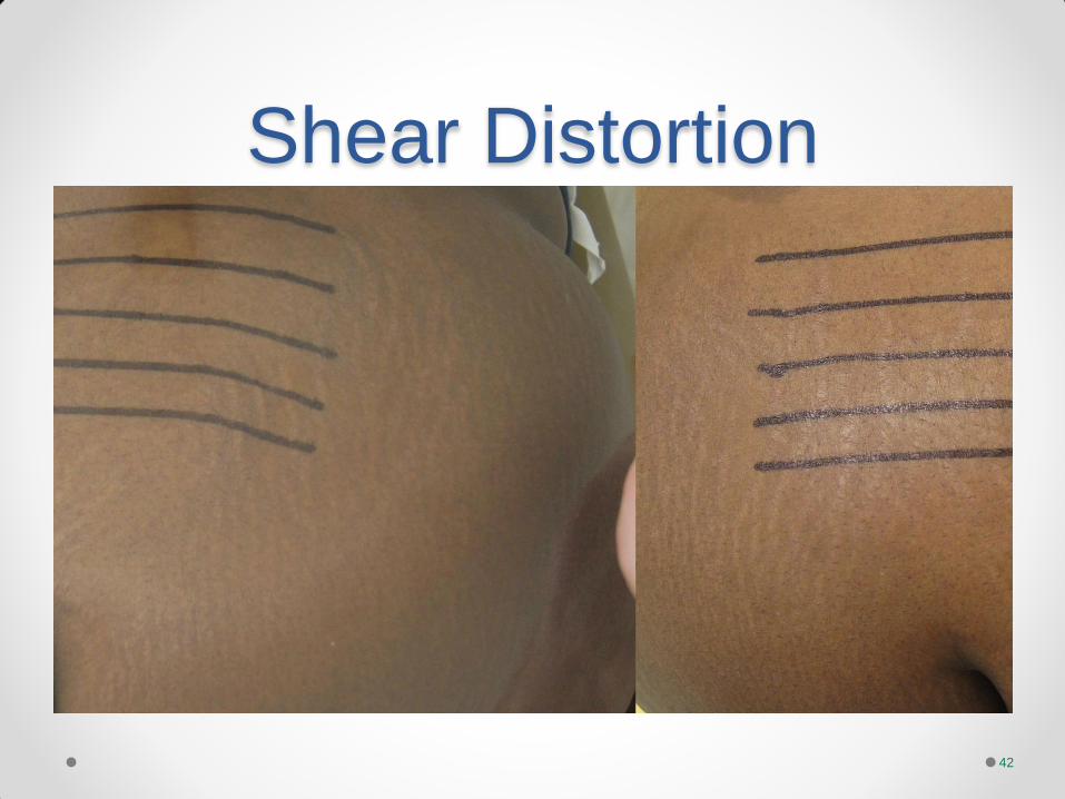
Shear Distortion
42

Shear With and Without Dressing
Single Human Subject 5 Trials Each Condition
0.0
1.0
2.0
3.0
4.0
5.0
6.0
7.0
8.0
0 1 2 3 4
Shear (N)
Distance Traveled (cm)
Effect of Molnlycke Dressing on Shear Transfered to Underlying Tissue
With Dressing
Without Dressing

Sliding and Shear
44
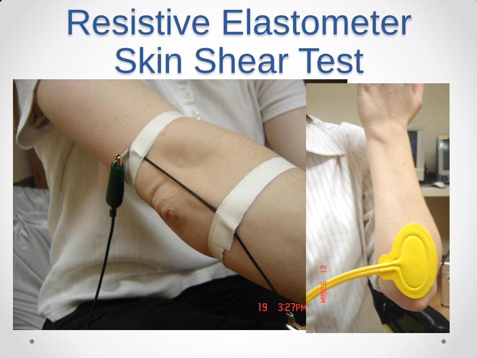
Resistive Elastometer Skin Shear Test

Shear Conclusions • Shear Measurements are;
o Driven by Friction
o Potentially Confounded by Friction
o Overwhelmed by friction
o Best taken under infinite friction in one plane
• Then static friction
• Then dynamic friction
o In Reference to the Body or the surface it is on

Shear Conclusions • Shear Measurements are;
o Modulus when taken on cushions or beds
o When using a body like indenter
• Immersion and normal force become integral to shear
• Reporting work incorporates these
o Translational shear
• More complex to measure
• More predictive of tissue damage
o More reliable when friction is standardized

References • Brindle CT. Outliers to the Braden Scale: Identifying high-risk ICU patients
and the results of prophylactic dressing use. WCET J. 2010;30:2-8.
• Bots CM, Apotheker BFG. The prevention of heel pressure ulcers using a hydropolymer dressing in surgical patients. J Wound Care. 2004;13:375-378.
• Ashford RL, PodM D, Freear ND, Shippen JM. An in-vitro study of the pressure-relieving properties of four wound dressings for foot ulcers. J Wound Care 2001; 10: 34–8.
• Nakagami G, Sanda H, Chizuko K, Kitagawa A, Tadaka E, Tabata K. Comparison of two pressure ulcer preventative dressings for reducing shear force on the heel. J Wound, Ostomy and Cont Nurs. 2006;33:267272.
• Hall, P. (1983). Prophylactic Use of Op-Site on Pressure Areas. Nurs Focus; 43 (5): 16.
• Callaghan, S., Trapp, M. (1998). Evaluating Two Dressings for the Prevention of Nasal Bridge Pressure Sores. Prof Nurse; 13 (6): 361-364.
• Gefen, A., van Nierop, B. Bader, D., Oomens, C. (2008). Strain-time cell-death threshold for skeletal muscle in a tissue-engineered model system for deep tissue injury. J Biomechanics; 41: 2003-2012.
References • Ohura, N., Ichioka, S., Nakatsuka, T., Shibata, M. (2005). Evaluating
dressing materials for the prevention of shear force in the treatment of pressure ulcers. J wound Care; 14 (9): 401-404.
• Ohura, T., Takahshi, M., Ohura, N. (2008). Influence of External Forces (pressure and shear force) on Superficial Layer and Subcutis of Porcine Skin and Effects of Dressing Materials: Are Dressing Materials Beneficial for Reducing Pressure and Shear Force in Tissues? Wound Repair and Regeneration; 16 (1): 102-107.
• Thomas S. The Role of dressings in the treatment of moisture-related skin damage. World Wide Wounds [Internet] 2008 Mar 5 [cited 2010 Oct 12 1-14 Available from: http://www.worldwidewounds.com/2008/march/Thomas/Maceration-and-the-role-of-dressings.htmlmarch/Thomas[Online]
• Call E, Pedersen J, Bill B, Oberg C, Ferguson-Pell, M. Microclimate impact of Prophylactic Dressings Using In Vitro Body Analog Method. Wounds 2013;25(4):94-103
References • Call, E., Pedersen, J. Bill, B., Black, J., Paulo, A., Brindle, T., Dealey, C.,
Santamaria, Clark, M. Enhancing pressure ulcer prevention using wound dressings: what are the modes of action. International Wound Journal, DOI:10.111/iwj.12123, Accessed on line 30 July 2013.
• Dealey, C., Brindle, T., Black, J., Alves, P., Santamaria, N., Call, E., Clark, M. Challenges in pressure ulcer prevention. International Wound Journal, DOI: 10.111/iwj.12107. Published online 20 Jun 2013.
• Black, J., Alves, P., Brindle, T., Dealey, C., Santamaria, N., Call, E., Clark, M. Use of wound dressings to ehance prevention of pressure ulcers caused by medical devices. International Wound Journal, DOI: 10.111/iwj.12111
• Harada, C., Shigematsu,T., Hagisawa, S. The Effect of 10 Degree Leg Elevation and 30 Degree Head Elevation on Body Displacement and Sacral Interfaxe Pressures Over a 2-hour Period. Wound Care, vol. 29, no. 3, pg 143-148


