
Introduction
Clinical Guidelines are systematically developed statements, which assist clinicians and patients in making decisions about appropriate treatment for specific conditions based on the best scientific evidence at the time of development. Guidelines are not intended to limit the clinical freedom; however, clinicians are expected to follow these recommendations as the basis for their decisions. Availability of resources, the existing situations, and the expectations of individual client needs to be considered.
The guidelines are intended to guide all health care workers in all levels of institutions where maternity care is provided. Although these guidelines are mainly targeted for the government sector institutions, use in the private sector institutions where maternity care is provided, is also encouraged.
These guidelines are developed by the guideline development group of the Sri Lanka College of Obstetricians and Gynaecologists in consultation with other relevant specialists such as anaesthesiologists, physicians, endocrinologists, and haematologists etc. The existing national guidelines developed in 2007, NICE guidelines on intranatal care, WHO guidelines and RCOG guidelines were perused and mixed with the local scenarios and expert opinion. The latest available scientific evidences were considered and included wherever necessary. Then, the draft guidelines were presented to a wider forum of obstetricians and consensuses were reached. After that the guidelines were handed over to the Ministry of Health and consensus was built with the participation of a multi-‐disciplinary team including medical administrators, provincial health authorities, representatives from SLCOG and other relevant professional colleges, and national programme managers.

Management of Labour

Management of Normal Labour
1. Introduction
The aim of this guideline is to provide recommendations to care providers in the management of a healthy woman with a single fetus in labour at term (37-‐42weeks). It does not cover the care of women with complicated pregnancies. The objective of this guideline is to ensure optimal management of women in labour, detect any abnormality, take appropriate action, prevent complications and consequently make childbirth safer; and also to make sure that these women are treated with respect and compassion, kept well informed and well supported throughout labour.
2. Diagnosis of Labour Labour is diagnosed by the presence of regular, painful intermittent contractions, which are of increasing frequency, duration and intensity, leading to progressive cervical effacement and dilatation. Note: for the purpose of this guideline, labour is also diagnosed in the presence of painful contractions occurring at a frequency of 2 in 10 minutes or more. If the diagnosis of labour is uncertain, observation should continue and reassessment made in four hours. Any woman who is diagnosed as not being in labour, but continues to complain of pain, would require careful reassessment by an experienced medical officer. Possible diagnoses of placental abruption and non-‐obstetric causes should be considered. Fetal compromise should be excluded. 3. Management of labour 3.1. General considerations 3.1.1. Communication between women and healthcare professionals/workers
• Greet the mother with a smile and a personal welcome
Definitions:
Latent phase of the first stage of labour – from the commencement of labour to a cervical dilatation of up to 4 cm. (This is a period of time, not necessarily continuous, when there are painful contractions and some cervical changes including cervical effacement and dilatation up to 4cm take place)
Active phase of the first stage of labour – commences at a cervical dilatation of 4cm and ends with full dilatation. (There are regular painful contractions and progressive cervical dilatation from 4cm up to full dilatation)

• Treat her with respect and dignity • Assure privacy • Establish a good rapport with the laboring woman asking her about her wants and
concerns and address them • Maintain a calm and confident approach which will reassure women that the
situation is under control • Assess the woman’s knowledge of strategies for coping with pain and provide
balanced information to find out which available approaches are acceptable to her • Ask her permission before all procedures and observations, focusing on the woman
rather than technology or the documentation
3.1.2. Preparation of mothers to transfer to labour room • Shaving or trimming of perineal hair may be necessary to facilitate unhindered
performance and repair of the episiotomy. • Efforts must be made to minimize faecal soiling. Where an enema is deemed
necessary, a medicated enema is recommended. (These two steps should not be considered mandatory) • Women should be encouraged to have a companion of her choice during labour,
depending on the facilities and clinical situation.
3.1. 3. Documentation • Admit the mother to the labour room and complete the ‘handing over’ form • Keep relevant notes on the BHT and start a partogragh (see page XX) • Review clinical notes and reassess risk factors. • Accurate documentation of all observations and interventions must be made, with
timing. • All obstetric examinations and procedures carried out must be documented in the
clinical notes. Each entry must be accompanied by a plan for management and be signed by the responsible person.
3.1.4. Mobilization and Positioning
• Women should be encouraged and helped to move about and adopt whatever positions they find most comfortable throughout labour.
• They need to be encouraged to void urine regularly. 3.1.5. Eating and drinking in labour
• Mothers must be encouraged to consume clear, non-‐fizzy liquids during labour. Isotonic solutions such as oral rehydration fluid and coconut water are more beneficial than water.
• In addition to clear fluids, women in the latent phase may consume light solids e.g.
biscuits and fruits.

3.1.6. Hygiene during labour • Strict asepsis must be maintained during labour. • Instruments should be available in packets • Use proper hand washing technique. • Use of double gloves and disposable gloves is encouraged.
3.1.7. Pain relief in labour Relief of pain should be a major consideration (please refer guideline on pain relief during labour in page XX) 3.2. Management of the three stages of labour 3.2.1. Management of the first stage of labour 3.2.1. 1. Latent phase It is important to recognize the latent phase of labour, since its prolongation could lead to maternal exhaustion, dehydration and acidosis, leading to fetal compromise and dysfunctional labour. Women in the latent phase of labour would be best managed in the antenatal ward.
Women in the latent phase of labour must be assessed on a regular basis, as follows:
• Check the fetal heart and maternal pulse half hourly • Check temperature four hourly; • Consider vaginal examination four hourly, depending on the contraction pattern and initial
cervical dilatation; • Document the colour of amniotic fluid if the membranes rupture; • Use of a sanitary pad may indicate early the presence of meconium. • Consider the requirement for analgesia.
It is important to inform the mother and reassure her that it is common to have slow progress in the latent phase. The latent phase is considered prolonged when it lasts more than 12 hours in a primigravida and 8 hours in a multigravida. In these situations an experienced medical officer (with a minimum one year of experience in the field) must reassess the mother with a view to augmentation of labour.
The practice of maintaining a labour room ‘notice board’ -‐ a ‘white board’ in which the status of all women in labour is summarized and updated regularly is encouraged. This would convey at a glance to all care providers women who require additional attention. The age, parity status, risk factors, salient findings at each assessment and any abnormalities noted must be included in this.

3.2.1.2. Active phase 3.2.1.2a. Admitting women to the Labour room All pregnant women diagnosed as being in active phase of the first stage of labour need to be admitted to the labour room. The initial assessment of a woman at the labour room should include:
• Listening to her story, considering her emotional and psychological needs and reviewing her clinical records
• Physical observation: temperature, pulse, blood pressure • Length, strength and frequency of contractions • Abdominal palpation: fundal height, lie, presentation, position and station • Vaginal loss: show, liquor, blood • Assessment of woman’s pain including her wishes for coping with labour along with the
range of options for pain relief • The fetal Heart rate (FHR) should be auscultated preferably with a hand held Doppler for a
minimum of 1 minute immediately after a contraction • The maternal pulse should be recorded to differentiate between maternal pulse and FHR • A vaginal examination should be offered
Health care Professionals who conduct vaginal examination should:
• Be sure that there is a valid indication for vaginal examination that it will add important information to the decision making process
• Be aware that for many women who may already in pain, highly anxious and in an unfamiliar environment, vaginal examination can be very distressing
• Ensure the woman’s consent, privacy, dignity and comfort • Explain the reason for examination and what will be involved, and • Explain the findings and their impact sensitively to the woman
3.2.1.2b. Management of active phase of first stage Monitoring must be conducted as instructed in the partogram and findings recorded accordingly.
Use of a sanitary pad may indicate early presence of meconium. Women in the active phase of labour must be assessed on a regular basis, as follows:
• Check the fetal heart and maternal pulse every 15 minutes; • Check temperature and blood pressure four hourly; • Vaginal examination four hourly or earlier, depending on the clinical situation; • Frequency of contractions should be monitored as follows:
The interval between two contractions should be assessed by palpation of the abdomen. During active labor usually there are at least three contractions per ten minutes. In other

words the interval between two contractions should be three minutes • Document the colour of amniotic fluid if the membranes rupture; • Consider the requirement for analgesia, (which now becomes more important).
The mother may continue to consume clear fluids in the active phase. She must be encouraged to assume any position that she is comfortable in and to avoid the dorsal position. Women who have the following conditions are recommended to be have to continuous electronic fetal monitoring:
• Significant meconium staining of amniotic fluid, • Abnormal Fetal heart rate detected by intermittent auscultation (< 110 beats per
minute; > 160 beats per minute; any decelerations after a contraction) • Fresh vaginal bleeding and • Maternal pyrexia.
3.2.1.3. Delayed progress of first stage of labour It is extremely important that delay in progress is assessed by an experienced medical officer. This assessment must take into account:
• the uterine contractions, • descent and position of the fetal head • features of early obstruction of labor (caput and moulding), and • The fetal condition
In women with delay in the active phase of the first stage, every effort must be made to find a cause for the delay. This may either be due to inadequate contractions or obstruction due to CPD, mal-‐presentation or malposition (such as occipito-‐posterior position), or a combination of these.
Intermittent auscultation of the fetal heart is best performed using hand-‐held Doppler devices. The fetal heart rate must be counted for one minute beginning immediately after a contraction.
In women with spontaneous labour progressing normally, routine early amniotomy and use of oxytocin is not recommended.
Delayed progress is diagnosed when there is progress of less than two cm in four hours. Slowing of progress in a woman who has previously been progressing satisfactorily must also be considered as a delay.
In cases of inadequate contractions: • Amniotomy must be performed if membranes are still intact. • Following that, the woman must be reassessed in two hours • In case there is inadequate progress, augmentation with oxytocin must be considered. • The situation must be reassessed after four hours or earlier if required.
Multiparous women with delayed progress:
• Must be viewed with extreme caution. • It is very important to exclude mechanical causes of delay before considering oxytocin. • Use of oxytocin in multipara with obstructed labour could be extremely dangerous.
In all cases where progress is slow in spite of adequate contractions a careful assessment must be made to exclude obstruction of labour. Attention must be paid to effective pain relief and to correcting dehydration in these situations. After paying attention to the above, cesarean section must be considered where the progress continues to be slow after four hours (less than two cm) of commencing oxytocin. 3.2.2. Management of second stage of labour 3.2.2.1. Passive second stage of labour (descent phase)
• Is diagnosed when full cervical dilatation is reached in the absence of involuntary expulsive efforts by the mother.
• Bearing down must be discouraged at this stage. • Intermittent auscultation of the fetal heart should be done immediately after a contraction
for at least one minute, at least every 10 minutes. The maternal pulse should be palpated if there is suspected fetal bradycardia or any other FHR anomaly to differentiate the two heart rates.
• Presence of meconium must be noted. 3.2.2.2. Active second stage of labour (expulsive phase)
• Is diagnosed when the mother gets the urge to bear down with full dilatation. • Intermittent auscultation of the fetal heart should be done immediately after a contraction
for at least one minute, at least every 5 minutes. The maternal pulse should be palpated if there is fetal bradycardia or any other FHR anomaly
• Presence of meconium must be noted. Use of a hand-‐held Doppler device is recommended (in preference to a Pinnard stethoscope) for fetal heart rate monitoring in the second stage. Women must be encouraged to continue consuming clear fluids during the second stage. Support by the labour companion must be continued. Total time durations allowed for the second stage of labour are as follows:
Primigravida: • Birth would be expected to take place within 2 hours of the start of the active second stage
in most women. • A diagnosis of delay in the active second stage should be made when it has lasted 1 hour
and need to seek the advice from a health professional trained in the assisted/ Operative vaginal birth if birth is not imminent.
Multigravida: • Birth would be expected to take place within 1 hours of the start of the active second stage
in most women. • A diagnosis of delay in the active second stage should be made when it has lasted 30
minutes and requires advice from a health professional trained in assisted/ operative vaginal birth if birth is not imminent.
• Delay in the second stage in a multiparous woman must raise suspicion of disproportion or malposition.
One further hour is permitted for women in each category for women with epidural analgesia. 3.2.2.3. Observations for women and babies in the second stage of labour: All observations should be documented on the partogragh.
• Chart blood pressure and pulse hourly • Continue four hourly temperature recording • Vaginal examination must offered after an hour in the active second stage after abdominal
palpation and assessment of vaginal loss • Half hourly documentation of frequency of contractions • Consideration of the woman’s emotional and psychological needs
In addition: • Assessment of progress should include maternal behavior, effectiveness of pushing and fetal
wellbeing, taking into account fetal position and station at the onset of the second stage. These factors will assist in deciding the timing of further vaginal examination and the need for obstetric review.
• Ongoing consideration should be given to the woman’s position, hydration, coping strategies and pain relief throughout the second stage.
3.2.2.4. Women’s position and pushing in the second stage of labour: Although most deliveries in Sri Lanka are conducted in the dorsal/McRobert’s position, women may be encouraged to adopt squatting, semi upright or lateral positions to aid the expulsion phase. Women should be informed that in the second stage, they should be guided by their own urge to push. If pushing is ineffective, strategies to assist birth such as support and encouragement and change of position can be used.
3.2.2.5. Intrapartum interventions to reduce perineal trauma Either the ‘hands on’ (guarding the perineum and flexing the baby’s head) or the ‘hands poised’ (with hands off the perineum and baby’s head but in readiness) techniques can be used to facilitate spontaneous birth. A routine episiotomy should not be carried out during spontaneous vaginal birth. Episiotomy should only be performed selectively, in women in whom there is a clinical need such as instrumental birth or suspected fetal compromise or a high chance of perineal tears. Where episiotomy is performed, Mediolateral episiotomy, performed at 45 – 60 degrees from the midline directed to the right side, beginning at the vaginal fourchette is preferred to the median episiotomy. It should be performed at the time of crowning of the fetal head. Episiotomy should be performed after infiltration of 1% lignocaine (up to 20 ml may be used). 3.2.2.6. Delivery The fetal head should not be allowed to extend till occiput is felt below the symphysis pubis. The perineum should be supported during delivery of the head. Once the head is delivered the woman should be discouraged from bearing down. Following restitution and external rotation, shoulders must be delivered with appropriately directed traction on the fetal head. The baby must be delivered onto the mother’s abdomen. Breastfeeding should be initiated within 30 minutes of birth. 3.2.3. Third stage of Labour The third stage of labour is the period from complete delivery of the baby to the complete delivery of the placenta and membranes. 3.2.3.1. Active Management of the third stage of labour Active management of the third stage of labour is recommended for all mothers. This includes;
• Routine use of uetrotonic drugs: Oxytocin 5 IU intravenously soon after the delivery of the baby or 10 IU intramuscularly,
• Delayed cord clamping (2 minutes after the birth) and cutting of the cord
In primigravida in whom contractions have become weak and there is no evidence of fetal compromise or obstruction, oxytocin may be administered as an infusion. In this case, the expulsive phase may be continued under close observation for a further 30 minutes. Delivery must be considered at the end of this period.
Delayed clamping of the cord allows for placental transfusion, which reduces neonatal and infant iron deficiency and anemia. This policy should be followed unless the baby is born in a poor condition or if the mother is bleeding or is Rhesus iso-‐immunized.

• Followed by controlled cord traction. • This must be followed by uterine massage.
Clamp and cut the cord close to the perineum. A hand should be placed above the symphysis pubis to stabilize the uterus by applying counter traction during controlled cord traction. Application of cord traction when the uterus is relaxed could lead to acute inversion of the uterus. After delivery, the placenta must be placed on a flat surface and the maternal surface examined for completeness. On the fetal surface the blood vessels must be traced to exclude a succenturiate lobe. Completeness of the fetal membranes must be ensured. Observations in the immediate postpartum period include:
• Inspect for continued fresh bleeding • Check pulse, blood pressure, uterine contraction and the level of the fundus every 15
minutes up to 2 hours • Her general physical condition, as shown by her colour, respiration and her own report of
how her feels Experienced medical personnel should be informed in any one the following instances:
• Continuing fresh bleeding; • Elevation of the level of the fundus; • Increase of pulse rate above 100 or by 30 beats per minute; • Drop in systolic blood pressure below 100 or by 30 mmHg.
The level of the fundus must be marked on the skin using a marker to make observations more objective. 3.2.3.2. Delayed third stage Delayed third stage is diagnosed if the placenta is not delivered within 30 minutes of active management. The first step in managing delayed third stage of labour is:
• To proceed to intraumbilical vein oxytocin, in a dose of 50 IU in 30 ml of 0.9% sodium chloride solution.
• A period of 30 minutes is allowed and controlled cord traction is attempted again. • If the placenta is not delivered by this method, manual removal of placenta is proceeded to.
4. Care for the newborn baby Effective care at birth is needed for anticipation of problems with the transition from in utero dependent life to extra utero independent existence and to provide support to

ensure stabilization.
• Skilled birth attendant (Medical Officers, Nursing Officers and Midwives) is responsible for the care.
• The care at birth is the same irrespective of birthing place or person attending to the birth. • At least one health care provider trained in neonatal resuscitation must be physically
available at time of birth of all infants irrespective of risk status.
• This person must be present in the delivery room before the birth of the baby.
• The attending personnel should document the baby details such as time of birth, weight,
gender and any other relevant information in all cases.
The aims of neonatal care following birth include the following:
• Establishment of respiration (as per NRP guidelines)
• Prevention of hypothermia (Refer Newborn Guideline)
• Establishment of breast feeding (Refer Newborn Guideline)
• Prevention of infection (Refer Newborn Guideline)
• Detection of danger signs (Refer Newborn Guideline)
Following basic steps should be followed at the time of birth;
1. Call out the time of birth
2. Deliver the baby onto the mother’s abdomen or into her arms
3. Dry baby with a warm towel or a warm piece of cloth
4. Wipe baby’s eyes
5. Assess baby’s breathing while drying
6. Make sure that there is no second baby
7. Change gloves or remove the first layer of gloves
8. Clamp and cut the umbilical cord
9. Put the baby between mother’s breast for skin to skin care
10. Place and identity label on the baby
11. Cover mother and baby with warm cloth
12. Put a hat on baby’s head
The Apgar score at 1 and 5 minutes should be recorded for all births.

Initiation of breastfeeding should be aimed for within 1hour after birth.
Head circumference, birth weight, length and other measurement should be carried out once the first feed is complete. A health care professional should examine the baby to detect any physical abnormality and to identify any problems that require referral.
5. Perineal Care Perineal or genital trauma caused by either episotomy or tearing need to be repaired. Before assessing for genital trauma:
• Explain to the woman what you are going to do and why • Offer analgesia • Ensure good lighting • Position the woman so that she is comfortable and the genital structures can be seen clearly.
The initial assessment should be performed gently and with sensitivity and may be done in the immediate period following birth preferably as soon as the placenta is delivered. Perineal repair should only be undertaken with tested effective analgesia in place using infiltration with up to 20ml of 1% lignocaine or equaling, or by topping up the epidural, as soon as possible by a medical officer. The preferred suture material is rapidly absorbed polyglycolic acid. The following basic principles should be observed when performing perineal repairs:
• Perineal trauma should be repaired using aseptic techniques. • Equipment should be checked and swabs and needles counted before and after the
procedure • Good lighting is essential to see and identify the structures involved. • Difficult injuries should be repaired by an experienced medical officer in theatre under
regional or general anaesthesia. An indwelling catheter should be inserted for 24 hours to prevent urinary retention.
• Good anatomical alignment of the wound should be achieved, and consideration given to the cosmetic result.
• Rectal examination should be carried out after completing the repair to ensure that suture
Classification of perineal trauma
First degree: Injury to skin only
Second Degree: Injury to the perineal muscles but not the anal sphincter
Third degree: Injury to the perineum involving the anal sphincter complex
Fourth degree: Injury to the perineum involving the anal sphincter complex and anal epithelium

material has not accidently been inserted through the rectal mucosa. • Following completion of repair, an accurate detailed account should be documented
covering the extent of the trauma, the method of repair and the materials used. • Information should be given to the woman regarding the extent of the trauma, pain relief,
diet, hygiene and the importance of pelvic floor exercises.
Guideline on Induction of Labour 1. Introduction This guideline aims to provide evidence based guidance on induction of labour to make the process more logical, effective and safer. It also aims to empower women undergoing induction of labour. 2. Definition Induction of labour is defined as initiation of labour by artificial means. 3. General Principles

• Induction of labour should be performed only in specialist obstetric units when there is a clear indication that its benefits outweigh risks.
• A senior clinician must make the decision. • The reason/s should be clearly explained to the patient, who should give her consent. • Maternal and fetal wellbeing should be monitored closely. • Adequate pain relief should be an essential part of the management plan, since it is
recognized that labor is more painful when it is induced. • Prior to induction of labour, the cervix should be favourable (Modified Bishop score 7 or
more). If it is not, an attempt should be made to ripen the cervix. • Decisions regarding induction of labour should be made taking into account not only the
clinical scenario but also the woman’s views, the availability of local facilities and cost effectiveness of the available methods.
4. Indications 4.1 Otherwise uncomplicated pregnancy continuing beyond 40 weeks Induction of labour is recommended for low-‐risk women who are known with certainty to have reached 41 weeks of gestation. However, it is good practice to assess foetal wellbeing around 40 weeks to select women for conservative management until 41 weeks gestation. The recommended assessments include foetal biometry (at least abdominal circumference) and amniotic fluid index (lower cut-‐off = 7 cm). 4.2 Prelabour rupture of membranes at term In the absence of evidence feotal compromise or maternal infection delayed induction of labour after 24 hours is acceptable. This may be carried out using either oxytocin infusion or prostaglandins. 4.3 Preterm prelabour rupture of membranes (PPROM) Patients with PPROM without evidence of infection or fetal compromise should be offered induction after the completion 34 weeks. 4.4 Intrauterine death This is a very traumatic time for the woman. Most women would want to be delivered as early as possible and their wishes need to be respected.

Amniotomy and repeated vaginal examinations are best avoided. Prostaglandins are preferred for induction of labour in these women. Amniotomy is preferred in the presence of abruption placentae. 4.5 History of precipitate labour There are no studies comparing outcomes in induced versus spontaneous labour. 4.6 Suspected macrosomia In the presence of good clinical and ultrasound evidence of macrosomia or a history of previous shoulder dystocia, there should be a low threshold for early induction of labour. 4.7 Fetal growth restriction The decision for induction of labour in a growth-‐restricted fetus should be individualized based on period of gestation at onset, presence or absence of fetal compromise. 4.8 Older mothers There is growing evidence that the risk of stillbirth is higher in older (>40 yrs) women near term. Women over 40 years should be offered induction between 39-‐40 weeks. 5. Induction under specific circumstances 5.1 Breech presentation Presentation per se, is not a contraindication to induction. 5.2 Previous CS There is no contraindication to induction of labour in a woman with a past caesarean section. Use of either oxytocin or prostaglandins increases the risk of scar dehiscence or rupture. This risk may be lower with artificial separation of membranes or Foley catheter. 6. Methods of induction

This section does not make a distinction between methods of ripening the cervix and induction of labour. 6.1 Mechanical There is good evidence that artificial separation of membranes reduces the need for formal induction. This method is recommended to be performed with due regard to asepsis, at 40 weeks gestation. Where the cervix will not admit a finger, massaging around the cervix in the vaginal fornices will have a similar effect. Extra-‐amniotic balloon catheter is an effective method of ripening of the cervix. A Foley catheter is inserted through the cervix and the balloon inflated with 40 – 60 ml of saline. This may be left in situ for a maximum of 48 hours. Following its removal, induction of labour may be proceeded to using another method. In the presence of evidence of infection, artificial separation of membranes and extra-‐amniotic Foley catheter must not be used. 6.2 Surgical Amniotomy is a definitive mode of induction of labour. It should be undertaken only if one is committed to delivery within 24 hours. Therefore it should be done only when the cervix is ripe and prior cervical assessment by an experienced clinician is essential. The risk of cord prolapse should be appreciated and steps taken to minimise or to recognize it early. Amniotomy alone may be capable of initiation of labour and it is recommended that oxytocin be started after a period of observation of at least two hours. 6.3 Pharmacological 6.3.1 Oxytocin Use of oxytocin when membranes are intact is not recommended. For details of how to use oxytocin please refer to the guideline on oxytocin

6.3.2 Prostaglandins Prostaglandin E2 (PGE2) These are very effective in inducing labour and are available as vaginal gel, tablet or controlled release pessary. All preparations carry a risk of hyperstimulation. Intracervical route does not offer any increase in efficacy. Combined use with oxytocin is particularly dangerous. A minimum of six hours from the last vaginal tablet/gel should be allowed before oxytocin is started. Prior to use of prostaglandins the Bishop score should be assessed and the woman should be monitored electronically to determine the fetal condition and frequency of contractions. After administration the fetal heart should be monitored electronically when contractions begin. After confirmation of normal heart rate pattern monitoring should be done by intermittent auscultation. A second dose may be considered after a minimum interval of 6 hours after the first, depending on the change of Bishop score, the condition of the fetus and frequency of contractions. The dosages are 3 mg for vaginal tablets and 0.5 mg for vaginal gel.
Misoprostol This drug is widely used worldwide for a variety of indications in pregnancy. (In Sri Lanka, it is not licensed at present). Nevertheless, it is very effective in inducing labour (more than PGE2), especially in mid trimester fetal death. Sensitivity of the uterus increases markedly with advancing pregnancy. This guideline recommends that it should not be used for induction of labour with a mature live fetus.
6.3.3 Mifepristone It is a powerful anti-‐progesterone and is very useful as an adjunct to misoprostol in cases of intrauterine death. (It is not licenced in Sri Lanka at present)
8. Complications
8.1 Hyperstimulation This is a well-‐recognized complication of induction of labour with pharmacological methods. It could have serious consequences including rupture of the uterus, aminiotic fluid embolism, precipitate labor and fetal compromise. It is defined either as a contraction free interval of less than sixty seconds and/or contractions lasting more than ninety seconds. If diagnosed, the prostaglandin tablet must be retrieved from the vagina or oxytocin infusion stopped immediately and a rapid infusion of 0.9% sodium chloride via a fresh giving set administered. If still not resolved, tocolytics should be given if available e.g. terbutaline 250 µg IV or SC. Since this is not available in Sri Lanka, salbutamol inhaler may be tried.
8.2 Cord prolapse
This is more likely with amniotomy when the head is high and poorly applied to the cervix. Precautions to avoid and to detect this early include palpation for cord presentation, palpation for the cord immediately after amniotomy and the fetal heart sounds auscultated immediately afterwards. If cord prolapse is diagnosed help must be called for immediately. Assess cervical dilatation and effect delivery if fully dilated. If not fully dilated and cord pulsations are present, insert a Foley catheter into the bladder and fill it with 500 ml saline. Place the mother in the knee-‐elbow position and displace the presenting part away from the pelvis by keeping pressure inserting a hand in the vagina. Transport for immediate caesarean section in this position.
8.3 Uterine rupture Please also refer to section 6.2 in this guideline Extra care must be exercised in grandmultipara and in women with scarred uteri.

8.4 Failed induction
Failed induction is defined as labour failing to start after one cycle of treatment with medical methods or for 12 hours of amniotomy. It does not necessarily indicate caesarean section in case medical or mechanical methods. The clinical situation (maternal and foetal condition) must be reassessed and discussed with the woman. In case of failure to induce labor using one cycle of prostaglandins another cycle may be administered as described as above. Depending on the clinical situation it is best that the second cycle is delayed for 24 hours. In case of amniotomy, failed induction of labour indicates caesarean section.

Guideline for Use of Oxytocin for Induction and Augmentation of labour Oxytocin is an invaluable drug when used carefully. However, it has the potential to cause uterine hyperstimulation, which could result in amniotic fluid embolism, uterine rupture and fetal distress, all of which are life threatening. Multigravidae are particularly susceptible to the above consequences and extra care must be taken to exclude obstruction before a decision is made to use oxytocin in a multigravid woman during labour. Experienced personnel must be involved in this decision. Use of oxytocin for induction and/or augmentation of labour results in a higher risk of rupture of a scarred uterus. Therefore, in such women oxytocin should be used only with the concurrence of a Consultant. Its effects will depend on the concentration of the infusion and the volume infused per minute. To achieve this predictably, use of infusion pumps is recommended. Where a gravity-‐assisted drip system is used, a burette may be used to improve accuracy. Such systems however, may deliver variable volumes depending on many factors including the position of the arm into which it is infused. Irrespective of the method of administration, oxytocin must be administered in incremental doses at intervals of 30 minutes, to achieve a contraction free interval of two minutes. Once this level is reached, the infusion rate may be continued at the same level, while closely monitoring the contractions. Hyperstimulation is defined either as a contraction free interval of less than sixty seconds and/or contractions lasting more than ninety seconds. In this situation the infusion must be stopped immediately. Oxytocin is administered with 5 units in 500 ml of 0.9% sodium chloride solution. In situations where infusion pumps are not available, oxytocin may be administered starting at a drop rate of 15 per minute and increased at rates of 15 drops per minute every 30 minutes, up to a maximum of 60 drops per minute. An approximate conversion to mU/minute is given in table 1.

Table 1: mU/minute administered at different rates of administration according to drop rate (based on 5U of oxytocin in 500 ml saline)
Table 2 gives mU infused per minute when administered via an infusion pump. TIME AFTER STARTING (MINS)
OXYTOCIN DOSE (MU/MIN) DOSE
VOLUME INFUSED (10U IN 500MLS MLS/HR) RATE
0 1 3 30 2 6 60 4 12 90 8 24 120 12 36 150 16 48 180 20 60 210 24 72 240 28 84 270 32 96 Table 2: mU infused per minute when administered via an infusion pump. Oxytocin must not be administered to women with intact membranes. It is recommended that women on oxytocin infusions should have continuous electronic fetal monitoring. Continuous EFM during administration of oxytocin:
• If the CTG is normal, oxytocin may be continued in incremental doses until the woman is experiencing 4 or 5 contractions every 10 minutes.
• If the FHR trace is suspicious, this should be reviewed by an experienced medical officer • If the FHR trace is classified as abnormal/pathological oxytocin infusion should be stopped
and a full assessment of the fetal condition undertaken by an experienced medical officer.
Drop rate/min Equivalent mU/min.
15 7.5
30 15
45 22.5
60 30

Guideline on fetal monitoring in labour Fetal monitoring in labour could be done by:
• Intermittent auscultation (preferably by a hand held Doppler device) • Intermittent or continuous electronic monitoring
Intermittent auscultation is recommended for low-‐risk women in spontaneous labour. Electronic monitoring is recommended when:
• The baby is growth restricted • There is significant meconium staining of amniotic fluid • Abnormal fetal heart rate detected by intermittent auscultation • Fresh vaginal bleeding • Maternal pyrexia • Use of oxytocin for augmentation or induction of labour • Women with a scarred uterus • Women on epidural analgesia
Intermittent auscultation This could be done by using either a Pinnard’s stethoscope or preferably a hand-‐held Doppler device. Auscultation should be carried out immediately after a contraction for one full minute. The maternal pulse should be palpated if there is suspected fetal bradycardia or any other FHR anomaly to differentiate the two heart rates. The normal rate is between 110 – 160 beats per minute in a term fetus. The frequency of auscultation should be as specified in the partogram. Electronic fetal monitoring (EFM) EFM is carried out by external cardiotocography (CTG). The following are recommended at the commencement of a CTG.
1. The paper speed must be set at 1 cm per minute. 2. The date and time settings on the machine must be validated. 3. The CTG must be labeled with the mother’s name, BHT number and date and time. 4. Maternal heart rate should be noted on the CTG. 5. The presence and the point at which the fetal heart rate is best heard must be delineated by
auscultation and the probe placed at that point. 6. Ensure that the contraction probe is functioning properly and used for the recording. 7. The woman should be positioned in such a way that aortocaval compression is avoided.

8. It should be interpreted without delay and the categorization recorded as either normal or suspicious or pathological, as per table 1, and signed by the responsible officer. The entry on the BHT must include a plan for management.
9. If the CTG is categorized as suspicious or abnormal, the Consultant must be informed.
10. For the management plan the overall clinical picture must be taken into account. e.g. the rate of progress of labour, presence or absence of fetal growth restriction, meconium staining of amniotic fluid and the evolution of the CTG abnormalities. Table 1: Definitions of normal, suspicious and pathological FHR traces
Table 2: Classification of fetal heart rate patterns Feature Baseline (bpm) Variability (bpm) Decelerations Accelerations
Reassuring 110–160 ≥ 5 None Present
Non-‐reassuring 100–109 161–180
<5 for 40–90 minutes
Typical variable decelerations with over 50% of contractions, occurring for over 90 minutes
The absence of accelerations with otherwise normal trace is of uncertain significance
Abnormal < 100 > 180 Sinusoidal pattern ≥ _10 minutes
< 5 for 90 minutes Either atypical variable decelerations with over 50% of contractions or late decelerations, both for over 30 minutes
Category Definition
Normal An FHR trace in which all four features are classified as reassuring
Suspicious An FHR trace with one feature classified as non-‐reassuring and the remaining features classified as reassuring
Pathological An FHR trace with two or more features classified as non-‐reassuring or one or more classified as abnormal

Further useful information on FHR patterns • If repeated accelerations are present with reduced variability, the FHR trace should be
regarded as reassuring. • True early uniform decelerations are rare and benign, and therefore they are not significant. • Most decelerations that occur during labor are variable. • If a bradycardia occurs in the baby for more than 3 minutes, urgent medical aid should be
sought and preparations should be made to urgently expedite the birth of the baby, i.e. immediate commencement of cesarean section. This could include moving the woman to theatre if the fetal heart has not recovered by 9 minutes. If the fetal heart recovers within 9 minutes the decision to deliver should be reconsidered in conjunction with the woman if the post-‐recovery tracing is reassuring.
• A tachycardia in the baby of 160–180 bpm, where accelerations are present and no other adverse features appear, should not be regarded as suspicious. However, an increase in the baseline heart rate, even within the normal range, with other non-‐reassuring or abnormal features should increase concern. In such cases inquiry must be made to ascertain if the fetus was active during the recording.
When women are having continuous EFM, systematic assessment of above definitions and classification should be undertaken with every review. During episodes of abnormal FHR patterns, if the woman is lying supine, advise her to adopt the left lateral position.

Guideline on Pain Relief in Labour Adequate relief of pain is a basic right of every mother in labour. It is the duty of every member of the obstetric team to endeavor to achieve this. Poor management of pain during labour will result in maternal exhaustion leading to:
• acidosis, • dysfunctional labour and • fetal distress. • Loss of morale and a negative birth experience could have significant long-‐term effects.
A well-‐informed, well-‐supported mother will be more in control of events and in a better position to deal with pain than one who is not. Therefore, it is important to keep the mother informed of the progress of labour and the condition of the fetus throughout the process. Reassurance plays a major adjunctive role in pain relief. Prenatal education should include information regarding the available methods of pain relief and their accessibility. Non pharmacological methods of pain relief such as breathing and relaxation techniques should be introduced during the antenatal period. It is well recognized that women who have a birth companion will tolerate pain better and require less analgesia. The policy of allowing a birth companion must therefore be encouraged.
1. Methods of pain relief in labour The selection of the method of pain relief should be based on the patient preference, availability of resources and the institutional protocols. Following methods can be used.
1.1 Non-‐pharmacological methods of pain relief • Breathing techniques, • Transcutaneous electrical nerve stimulation (TENS), • Massaging, • Relaxation techniques, • Positioning and movement
Any of these methods can be used to relieve pain during labour

1.2. Pharmacological methods of pain relief in labour
1.2.1. Oral paracetamol/paracetamol & codeine compound:
These oral preparations can be used safely in the latent phase of labour. 1.2.2. Opioids
1.2.2.A. Pethidine Pethidine is safe and effective in the latent and early active phase. The dose is 1-‐1.5 mg/kg IM, repeated after 4 – 6 hours. Administration of a third dose should be done only with the concurrence of senior personnel. It is generally avoided where delivery is anticipated within 4 hours. Maternal side effects include nausea, vomiting and a reduction in gastric motility with a subsequent increase in gastric acidity. Therefore, it should be administered coupled with metoclopramide 5 mg IV or 10 mg IM. Neonatal respiratory depression is a recognized consequence of administration of opioids to the mother. Naloxone, a pure opioid antagonist should be available for treatment in all facilities administering opioids for analgesia. Naloxone is given to the baby in a dose of 100μg /kg IV. It has a short duration of action and additional doses may be required. If no improvement is seen with the first dose of naloxone, the cause of neonatal respiratory depression is more likely to be a factor other than opioids. 1.2.2.B. Morphine This has a longer duration of action than pethidine and may be particularly useful in women who require analgesia in early labour. The dose is 0.15 mg/kg IM should be administered with metoclopramide. The side effects and neonatal effects are similar to those of pethidine. 1.2.2.C. Fentanyl Intravenous fentanyl/ramifentanyl may be administered in either a High Dependency or Intensive Care Unit settings under the supervision of an anaesthesiologist. The dose is 50-‐100μg per hour as an intravenous infusion. Pain relief occurs in 3-‐5 minutes after commencement.
1.2.3. Inhalational analgesia – Entonox

Entonox is a 50:50 mixture of nitrous oxide and oxygen and it has a very short half-‐life. The onset of action is 30sec to one minute. The mother should receive clear and definite instructions about its correct use. It should only be self-‐administered. She should start using entonox through the controlled valve at the very beginning of the contraction. The mother should be advised to stop using Entonox inhalation in the contraction free interval. Longer and deeper breaths give better results. There is no limit on the duration of its use. Women should be informed that Entonox will make them feel nauseous and light-‐headed. Entonox is contraindicated in women with intestinal obstruction, pneumothorax, middle ear and sinus disease, and following cerebral air-‐contrast studies.
1.2.4. Regional Anaesthesia A. Epidural analgesia
Epidural analgesia is the most effective form of pain relief in labour. Therefore, Its greater use should be encouraged. It can be given either as a bolus with top-‐ups or as a continuous infusion. Continuous administration via a syringe pump is preferred to ‘top-‐ups’, since it is safer. The continuous availability of an anesthesiologist is a prerequisite to offering epidural analgesia. It is also essential that staff on site is trained for its setting up, monitoring and to recognize complications early. Facilities should be available for emergency resuscitation. Before offering epidural analgesia, women should be informed regarding its risks and benefits and its implications on labour:
• It provides more effective pain relief than other methods • It will not increase the length of the first and the passive second stages of labour. • It may however increase the length of the expulsive phase and increase the likelihood of an
instrumental delivery. An additional hour is allowed in the expulsive phase therefore. • It does not increase the chance of cesarean section • It does not cause long-‐term backache. • It needs to be accompanied by a more intensive level of monitoring.

Care and observations for women with regional analgesia in labour • Intravenous access should be secured prior to commencing regional analgesia. • Following additional observations should be carried out for women with regional analgesia
Ø During establishment of regional analgesia or after top up bolus blood pressure should be measured every 5 minutes for 15 minutes.
Ø If the woman is not pain free within after each administration, the anaesthetist should be called.
Ø Hourly assessment of the level of sensory block should be undertaken. • Women with regional analgesia should be encouraged to move and to adopt whatever
positions they find most comfortable throughout labour. • Once established, regional analgesia should be continued until after completion of the third
stage of labour and when necessary until perineal repair is done. • Women should be allowed one additional hour in the second stage of labor, depending on
maternal and foetal condition. Thereafter pushing during contractions should be actively encouraged.
• Continuous EFM is recommended for at least 30 minutes during establishment of regional analgesia and after administration of each bolus.

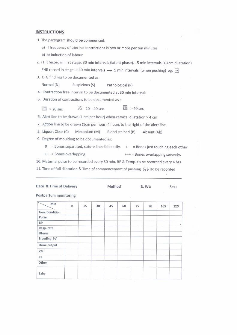
Guidelines to maintain the partograph

GUIDELINE ON ACUTE PUERPERAL INVERSION OF THE UTERUS 1. Introduction The aim of this guideline is to provide recommendations for the management of acute puerperal inversion of the uterus, which is a rare and life threatening condition. The main reason for its high mortality rate is delay in instituting appropriate treatment, which leads to postpartum hemorrhage and rapid development of shock out of proportion to haemorrhage. 2. Definition It is defined as ‘the turning inside out of the fundus into the uterine cavity’. 3. PREVENTION Mismanagement of the third stage of labor is recognized as the main cause, although 50% have no identifiable cause. The common initiating factor seems to be a traction force on the fundus of a relaxed uterus. Proper retraction of the uterus in the third stage is the primary factor in preventing an inversion. There is no reliable data to suggest that it recurs in a future pregnancy. The importance of the active management of the third stage could not be over-‐emphasized. (Please refer Section 3 of the PPH Guideline and the section on management of delayed third stage (section 4.5 in the Normal Labor Guideline for details) 4. PATHOPHYSIOLOGY (and clinical correlation) As the inversion progresses, the adnexae with their ligaments get drawn into the inverting uterine fundus and become increasingly stretched. This produces significant pain and vagal stimulation, leading to neurogenic shock. An inverted uterus becomes trapped within the cervix creating progressive oedema and congestion due to interruption of venous and lymphatic drainage. Oedema and congestion will increase the firmness of the inverted segment, making reduction more difficult. Interruption of the venous drainage will lead to significant haemorrhage. A partially separated placenta would add to this. 5. CLASSIFICATION Although acute, subacute and chronic varieties have been described, this guideline would address only the acute variety as it is life threatening. This occurs soon after birth, just before or after the delivery of the placenta. Three degrees of inversion have been described, depending on the level of the inverted fundus. In practice, second-‐degree inversion is the commonest. The fundus has come past the cervical os, but is still within the vagina.

6. CLINICAL PRESENTATION AND DIAGNOSIS Prompt diagnosis is vital. The key to diagnosis is awareness and a high degree of suspicion. The following are early warnings:
• A degree of shock that is out of proportion to overt blood loss • A retained placenta • Placenta delivered but ‘with some difficulty’ • Severe, sustained unexplained pain in the third stage.
In this situation:
• Feel for the fundus. If absent or ‘cupped’, acute inversion is probable diagnosis; • Confirm by a vaginal examination:
• Look for a hard mass which looks and feels like a huge ulcerated fibroid polyp (sometimes described as a foetal head);
• The cervix is not to be seen or felt in the normal position, instead it could be felt as a ring around the base of the ‘mass’;
• In incomplete cases, the inverted fundus may be felt through the cervical canal in the lower uterine cavity.
7. MANAGEMENT 7.1 General measures: Early diagnosis is vital. Treat it as a life-‐threatening emergency. First attempts at reduction should be made at the place where it is diagnosed, without moving to theatre. If these attempts fail, move to theatre and give a general anesthetic without delay (see section 7.2.4). Early involvement of experienced personnel and teamwork are absolutely essential. Treat shock aggressively, not forgetting the neurogenic element. Provide adequate pain relief Replace the blood loss, which could be considerable, especially if the placenta has partially or completely separated. Do not attempt to remove the placenta, if still attached. 7.2 Repositioning the uterus Reposition the uterus as soon as possible; the sooner it is done the easier and better. It reverses the shock and reduces PPH. Non-‐surgical methods 7.2.1 Manual replacement of uterus. (Johnson’s maneuver) The operator introduces two thirds of his forearm in to the vagina and extends the hand at the wrist to place the palm on the inverted fundus and fingertips at the utero-‐cervical junction. Lifting the
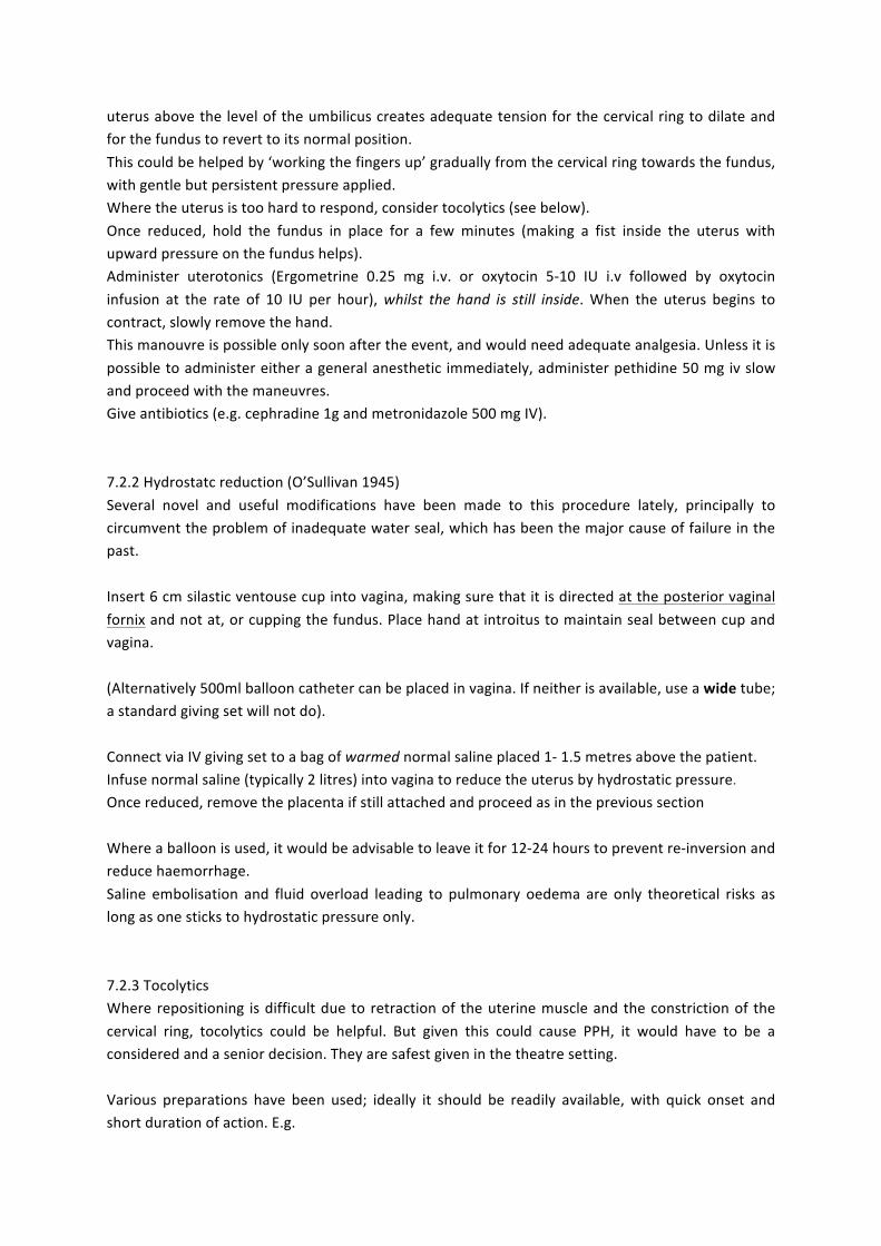
uterus above the level of the umbilicus creates adequate tension for the cervical ring to dilate and for the fundus to revert to its normal position. This could be helped by ‘working the fingers up’ gradually from the cervical ring towards the fundus, with gentle but persistent pressure applied. Where the uterus is too hard to respond, consider tocolytics (see below). Once reduced, hold the fundus in place for a few minutes (making a fist inside the uterus with upward pressure on the fundus helps). Administer uterotonics (Ergometrine 0.25 mg i.v. or oxytocin 5-‐10 IU i.v followed by oxytocin infusion at the rate of 10 IU per hour), whilst the hand is still inside. When the uterus begins to contract, slowly remove the hand. This manouvre is possible only soon after the event, and would need adequate analgesia. Unless it is possible to administer either a general anesthetic immediately, administer pethidine 50 mg iv slow and proceed with the maneuvres. Give antibiotics (e.g. cephradine 1g and metronidazole 500 mg IV). 7.2.2 Hydrostatc reduction (O’Sullivan 1945) Several novel and useful modifications have been made to this procedure lately, principally to circumvent the problem of inadequate water seal, which has been the major cause of failure in the past. Insert 6 cm silastic ventouse cup into vagina, making sure that it is directed at the posterior vaginal fornix and not at, or cupping the fundus. Place hand at introitus to maintain seal between cup and vagina. (Alternatively 500ml balloon catheter can be placed in vagina. If neither is available, use a wide tube; a standard giving set will not do). Connect via IV giving set to a bag of warmed normal saline placed 1-‐ 1.5 metres above the patient. Infuse normal saline (typically 2 litres) into vagina to reduce the uterus by hydrostatic pressure. Once reduced, remove the placenta if still attached and proceed as in the previous section Where a balloon is used, it would be advisable to leave it for 12-‐24 hours to prevent re-‐inversion and reduce haemorrhage. Saline embolisation and fluid overload leading to pulmonary oedema are only theoretical risks as long as one sticks to hydrostatic pressure only. 7.2.3 Tocolytics Where repositioning is difficult due to retraction of the uterine muscle and the constriction of the cervical ring, tocolytics could be helpful. But given this could cause PPH, it would have to be a considered and a senior decision. They are safest given in the theatre setting. Various preparations have been used; ideally it should be readily available, with quick onset and short duration of action. E.g.

Turbutaline 0 .25mg i.v. slowly (not available in Sri Lanka at present); Salbutamol 0.25mg in 10 ml saline i.v. slowly; Nitroglycerine 0.1mg i.v. slowly or sublingually (acts within 90 seconds) 7.2.4 General Anaesthesia If the initial attempt at manual replacement fails, it is safest to move the patient to the theatre and to administer general anaesthesia. This allows for muscle relaxation, pain relief and elimination of the neurogenic contribution to the shock. 7.2.5 Surgical methods If managed properly in the early stages, resort to surgery should be a rare occurrence. Huntingdon’s operation After a laparotomy, the indrawn uterine cup is identified near the region of the cervix with the tubes and round ligaments pulled into the cup. By the use of two Allis forceps the uterus is pulled out of the constriction ring in a progressive fashion and restored to its normal position. The serosa of the uterus will invariably sustain lacerations and these are repaired with absorbable sutures. Use of a silastic vacuum cup from above instead of Allis forceps has been shown to circumvent this problem. Haultain’s operation In this procedure the constriction in the region of cervix is incised posteriorly using a longitudinal incision. As in the Huntingdon’s method two Allis forceps are used to pull the uterus to its normal position. The incision is repaired with interrupted sutures. Uterotonics are given to maintain contraction of the uterus. Hysterectomy When all the above methods fail, a hysterectomy will become the only viable option. However, it must be remembered that given the distorted anatomy, this must be undertaken by a surgeon of considerable experience. 8. Debriefing Although there is no evidence of a recurrence risk, it is sensible to advise the woman to deliver in a specialized Unit next time, and the third stage to be managed actively by experienced personnel.
Management of Hypertensive Disease During Pregnancy
Management of Hypertensive Disease in Pregnancy
1. Introduction Hypertension in pregnancy is an important cause of direct maternal deaths in Sri Lanka. Early identification, aggressive and intensive treatment of its complications is important in reducing the resulting morbidity and mortality.
2. Definitions Chronic Hypertension:
Women with pre-‐existing hypertension or hypertension detected before 20th week of gestation in the absence of trophoblastic disease and persisting more than 42 days post partum.
Gestational Hypertension
A) Pregnancy Induced Hypertension:
Hypertension unaccompanied by proteinuria developing after 20 weeks of gestation and resolving within 42 days of delivery.
B) Pre-‐eclampsia:
Pregnancy induced hypertension associated with significant proteinuria (300mg/l or 500mg/ 24 hours or dipstick 2+ or more).
Severe Pre-‐eclampsia: Defined as pre-‐eclampsia with severe hypertension and/or with symptoms, and/or biochemical and/or haematological impairment.
The clinical features of severe pre-‐eclampsia (in addition to hypertension and proteinuria) are:
• Severe headache • Visual disturbances such as blurring of vision or flashing before eyes, scotomas • Epigastric or hypochondrial pain and/or nausea & vomiting • Clonus (3 beats or more) • Papilloedema • Liver tenderness • Oliguria (less than 400 ml per day or 0.5 mg/Kg/hour over a 4 hour period) • Platelet count falling to below 100 x 106/l • Abnormal liver enzymes (ALT or AST rising to above 70IU/l) • HELLP syndrome
Severe Hypertension: Defined as systolic blood pressure ≥ 160 mmHg and/or diastolic blood pressure ≥110 mmHg. Eclampsia: Defined as the development of convulsions and/or unexplained coma during pregnancy or postpartum in patients with features of preeclampsia.

3. Screening for Hypertension during pregnancy Blood pressure must be measured in every clinic visit by a Medical Officer and results recorded and plotted in the pregnancy record.
Proteinuria must be tested for at every clinic visit.
If blood pressure is more than 140/90 mmHg on two occasions at least 2 hours apart, refer for specialist care.
4. Prevention of hypertensive disorders in pregnancy Advise women at high risk of pre-‐eclampsia to take 75 mg of aspirin daily from 12 weeks until delivery of the baby. Women at high risk are:
Those with any one of the following risk factors:
• Hypertensive disease during a previous pregnancy • Chronic kidney disease • Autoimmune disease such as systemic lupus erythematosus or antiphospholipid
syndrome • Type 1 or type 2 diabetes • Chronic hypertension • Multiple pregnancy
Or, any TWO or more of the following
• First pregnancy • Age 40 years or older • Pregnancy interval of more than 10 years • Body mass index (BMI) of 35 kg/m² or more at first visit • Family history of preeclampsia
Contraindications such as allergy, gastritis, peptic ulcer disease must be taken into account.
Advice women who have the above risk factors to ensure a higher intake of calcium to achieve a daily intake of at least 1000 mg taking into account the average intake by Sri Lankan women the recommended supplementation level is 600 mg.
5. Management of Chronic Hypertension Women with chronic hypertension must be managed in specialist units. Anticipate the development of superimposed pre-‐eclampsia in these women. This combination adds risks to both mother and baby. ACE inhibitors should be discontinued in women who are planning pregnancy and its use avoided during pregnancy.
6. Treatment of mild to moderate hypertension
Since there is no consensus on the value of treating mild to moderate hypertension, this guideline will not address this issue.

6. MANAGEMENT OF SEVERE PRE-‐ECLAMPSIA
6.1. General Considerations
• Severe preeclampsia is a life threatening condition. • The only known cure is delivery of the baby. • The immediate task is to determine the urgency to effect delivery. • Stabilization of the mother’s condition within an acceptable time frame prevents maternal
complications and may improve fetal condition. • The management has to be individualized depending on the clinical condition and available
resources. • • The dangers will continue into the immediate postpartum period.
6.2. Specific Management
Admit women who have severe preeclampsia and inform the Consultant.
Treat hypertension if:
• Systolic blood pressure ≥ 160 mmHg, or if • Diastolic blood pressure ≥ 110 mmHg, or if • Mean arterial pressure ≥ 125 mmHg
Aim to maintain blood pressure at around 130-‐140/90-‐100 mmHg. The main cause of maternal death in severe preeclampsia is poorly controlled systolic hypertension causing cerebral haemorrhage.
The basic outline of management
• Admit to hospital and inform Consultant • Observe and monitor • Control blood pressure • Prevent seizures • Look for complications – such as HELLP / pulmonary
oedema/cerebral haemorrhage • Strict fluid balance • In utero transfer where necessary and safe • Timing of Delivery • Continue vigilance post delivery • Follow up

A rapid fall in maternal blood pressure as a result of antihypertensive treatment may cause fetal heart rate abnormalities & compromise especially in growth restricted/compromised fetuses.
Where resources allow, it is recommended to monitor fetal heart with continuous CTG during and for 60 minutes after commencing anti-‐hypertensive therapy.
Aim to stabilize blood pressure before delivery.
6.2.1. Anti-‐hypertensive drugs
Oral anti hypertensives may be used when the blood pressure is <180/110 mmHg. Blood pressure must be monitored at 15-‐minute intervals and intravenous anti hypertensives resorted to in case of an adequate response is not obtained within 30 minutes.
The commonly used antihypertensive drugs for acute control are given below. One or the other may be used depending on availability and familiarity.
6.2.1.1 Labetalol orally or intravenously
This should be avoided in women with a history of bronchial asthma.
-‐ 200mg orally stat (only if blood pressure is <180/110 mm Hg)
-‐ repeated hourly for up to 4 hours
or
-‐ 20 mg IV over two minutes
• Record blood pressure after 10 minutes. • If either value is still above 160 mm Hg systolic and/or 110 mmHg diastolic, give 40 mg iv
over 2 minutes. • Record blood pressure after 10 minutes. • If the blood pressure is still above 160 mm Hg systolic and/or 110 mmHg diastolic, give
hydralazine 10 mg iv. For instructions regarding giving a fluid bolus with i.v. hydralazine, see the next section of this guideline.
• If the blood pressure is still above 160 mm Hg systolic and/or 110 mmHg diastolic, start an IV infusion of labetolol, starting at 40 mg/hour, doubling the dose at half hourly intervals as required to a maximum total of 160 mg/hour.
• Where these measures fail, the mother must be moved to a high-‐dependency area or an intensive care unit.
If blood pressure is controlled by the above, continue monitoring the blood pressure at 15 minute intervals for I hour and at 30 minute intervals thereafter.
Additional bolus doses as described above may be administered if the blood pressure increases above 160 mm Hg systolic and/or 110 mmHg diastolic.

6.2.1.2. Hydralazine intravenously:
• 5 -‐ 10 mg IV bolus over 2 minutes. • This must be accompanied by a fluid bolus of 5ml/kg of 0.9% sodium chloride or Ringer
lactate solution over 30 min, started at the same time as iv hydralazine (this helps vasodilatation & prevents drastic hypotension). This should not be used in the presence of pulmonary oedema.
• Record blood pressure at 15-‐minute intervals. • Repeat boluses of 5 -‐ 10 mg IV after a 15-‐minute interval may be given if necessary up to
a maximum of 20 mg (the effect of a single dose can last up to 6 hours). • If the response to above doses is inadequate, give labetolol bolus doses as described
above. • If no lasting effect with above boluses, consider an infusion of hydralazine 2.0 mg/hour
increasing by 0.5 mg/hour as required (2-‐20 mg/hour usually required).
6.2.1.3. Oral Nifedipine
• Oral nifedipine may be used where the blood pressure is < 180/110 mm Hg, in asymptomatic patients.
• Give 10 mg orally. • Repeat at 20-‐minute intervals up to a maximum of 40 mg. • If there is no response proceed to intravenous labetalol or hydralazine.
6.2.2. Prevention of convulsions
Magnesium sulphate
• Magnesium sulphate is the anticonvulsant of choice. • It should be given to any woman with features of impending/imminent eclampsia
(presence of clonus, severe headache, visual disturbances, and dizziness). • The loading dose may be given even when the status of renal function is uncertain, since
it is unlikely that toxic levels of magnesium could be reached with this dose alone. • Give loading dose of 4 G IV over 10 minutes. There are two methods of giving
magnesium sulphate intravenously. o Diluted to a total volume of 20 ml with 0.9% sodium chloride solution, given via an
infusion pump or ‘manually’. o Diluted to a total volume of 80 ml with 0.9% sodium chloride solution via a burette
• Immediately after the loading dose, start infusion of 1 G IV per hour. Continue this infusion for at least 24 hours after delivery.
• Where there are difficulties with intravenous access, magnesium sulphate may be administered intramuscularly. Give 5 G deep intramuscularly into each buttock with 1 ml of 2% lignocaine in the same syringe.
• If intramuscular magnesium sulphate is continued as maintenance therapy, give 5G to alternate buttocks 4 hourly, with 1ml of 2% lignocaine in the same syringe.

• Monitor the mother to ensure hourly urine output of 30 ml per hour, respiratory rate >16/ minute, oxygen saturation >90% and presence of patellar reflexes.
• These should be recorded every 30 minutes. • Should signs of toxicity appear, the antidote is calcium gluconate, 1 G intravenously (10
ml of 10% solution), given over 10 minutes. • Magnesium sulphate may be used safely in women who have previously received
nifedipine.
6.2.3. Fluid Balance
• Restrict total fluid intake to 80 ml per hour. • Accurate recording of fluid balance is essential. • Selective colloid expansion may be necessary prior to pharmacological vasodilatation to
prevent maternal hypotension and fetal compromise or in oliguria with a low central venous pressure.
• The volumes of all drugs administered must be taken into account and appropriate reduction of the volume of crystalloids must be made.
• Colloid (e.g. Hetastarch) should be administered only after discussion with the anaesthetist.
• Diuretics must be restricted to specific instances only e.g. for women with pulmonary oedema.
• Avoid non-‐steroidal analgesia until fluid recovery.
4.2.4. In utero/neonatal transfer:
• If a Unit does not have access to HDU/ICU or is unable to cope with maternal complications, or with maturity of the baby, it may be appropriate to consider antenatal transfer of the mother.
• However, maternal safety must not be jeopardised and each case should be considered on its clinical merits.
• Steps must be taken to bring down blood pressure from very high levels (e.g. using nifedipine
• Women with imminent/impending eclampsia must be administered a loading dose of magnesium (IM or IV) before transfer (see 6.2.2)
• It is recommended that where possible telephone advice is obtained from the relevant specialist unit before transfer.
• The patient must be accompanied by a member of staff who is capable of dealing with a seizure while the patient in transit. The required drugs and equipment must be made available.
• Full details of the case, including treatment given should accompany the patient.

6.2.5. Delivery
• Urgency of delivery depends on the maternal and fetal conditions. • Either caesarean section or induction of labour is appropriate depending on the urgency and
favourability of the cervix. • Institute adequate pain relief. Severe preeclampsia is not a contraindication for opioid or
epidural anesthesia (see below). It is accepted that epidural anesthesia helps to bring down the blood pressure.
• Spinal or epidural anaesthesia is safe in the presence of a platelet count >80,000/dl. • Maternal condition should be optimised before delivery. • It is inappropriate to deliver an unstable mother for foetal reasons. • Ergometrine should not be used during the third stage.
6.2.6. Post-‐delivery
• Maintain vigilance as a high proportion of eclamptic seizures occur after delivery. • High dependency care should be provided as clinically indicated. • Continue close monitoring, including fluid balance, platelets, liver enzymes and creatinine
until they have returned to normal values. • Magnesium sulphate if started should be continued for 24 hours after the delivery or after
the last fit, whichever is later. • Review anti-‐hypertensive medication as indicated. Some may need to continue oral
medication for a few weeks. Methyldopa is best avoided following delivery because of its tendency to cause depression.
6.2.7. Follow up
• Inform Public Health Midwife and/or Medical Officer of Health. • Review in 2 weeks (instead of 4 weeks) if discharged on antihypertensives. • Depending on the clinical picture, some patients may need:
o Long term follow up for blood pressure o Hematological investigations for conditions such as anti-‐phospholipid syndrome,
thrombophilia • Debrief the patient. • Advice preconceptual counseling & check prior to the next pregnancy. • Women may be advised regarding the risk of developing hypertensive disease in a future
pregnancy as follows: o Risk of gestational hypertension -‐ 53% (1 in 2) o Risk of preeclampsia – 16% (1 in 6) o Risk of preeclampsia if she had severe hypertension or HELLP syndrome or
eclampsia or the birth occurred before 34 weeks – 25% (1 in 4); & 55% (1 in 2) if the birth occurred before 28 weeks gestation

Management of Eclampsia
1. Definition:
Eclampsia is defined as the development of convulsions and/or unexplained coma during pregnancy or postpartum in patients with features of preeclampsia.
2. Diagnosis:
• Hypertension is considered the hallmark for the diagnosis of eclampsia. However, in 16% of the cases hypertension may be absent.
• Eclampsia is usually associated with proteinuria, but this may be absent in 14% of cases. • Clinical features of imminent eclampsia include:
Severe frontal headache, Visual symptoms (halos, scotomas etc.) Epigastric or right hypochondrial pain, Liver tenderness, Clonus (3 beats or more)
3. Time of onset of eclampsia
The onset of eclamptic convulsions can be antepartum, intrapartum, or postpartum.
Antepartum eclampsia Almost all cases (91%) develop eclampsia at or beyond 28 weeks
Postpartum eclampsia Although most cases of postpartum eclampsia occur within the first 48 hours, some cases develop beyond 48 hours, up to 4 weeks postpartum (late postpartum eclampsia). In these cases, an extensive neurological evaluation is needed to rule out the presence of other cerebral pathology.
4. Comorbidities
• Eclampsia is often complicated by comorbidities (Box 1). • These are more common among women who develop eclampsia at earlier periods of
gestation.
Box 1.
• Abruptio placentae • Disseminated intravascular coagulopathy • Pulmonary oedema • Acute renal failure • Aspiration pneumonia • HELLP syndrome (Haemolysis, elevated liver enzymes,
low platelets)

5. Prevention
Administration of magnesium sulphate to women with features of impending/imminent eclampsia (presence of clonus, severe headache, visual disturbances, dizziness) is the only known preventive measure.
6. Management 6.1 General considerations 6.1.1 The priorities in management are to support respiratory and cardiovascular function,
prevent injury and further seizures and to control hypertension. 6.1.2 Magnesium sulphate is the anticonvulsant of choice. It must be administered as soon as
possible. See section 6.2.2 of the severe preeclampsia guideline for details. 6.1.3 The bolus dose of magnesium sulphate must be given even to women with unknown
renal function or oliguria/anuria since this dose is unlikely to elevate magnesium levels to toxic ranges.
6.1.4 Eclampsia dictates delivery (or induction) once the maternal condition is stabilized, irrespective of the foetal condition or maturity. A decision regarding the mode and time of delivery will require to be made early.
6.1.5 There is no place for prolongation of the pregnancy in these women, unless under rare, exceptional circumstances.
6.1.6 For details on administration of medications and intravenous fluids and care of women receiving magnesium sulphate and intravenous antihypertensives, refer the guideline on severe preeclampsia.
6.2. During the seizure –
o Turn the patient to a side and support her in that position. o Suck out secretions from the mouth. o Administer oxygen via a face mask. o Most eclamptic seizures resolve spontaneously. o It is imprudent to diagnose fetal hypoxia based on fetal bradycardia during a seizure.
This usually recovers spontaneously following the seizure o Fetal bradycardia persisting beyond 10 minutes following the seizure should raise
suspicion of abruptio placentae. 6.3. As soon as possible following a seizure
o Attempt to establish intravenous access. o Obtain blood for full blood count, liver transaminases, blood urea, electrolytes and
blood for cross-‐match. o Start magnesium sulphate (intravenous bolus and infusion or intramuscular – details
in guideline on severe preeclampsia section 6.2.2.). o Treat blood pressure as appropriate. o Insert an indwelling catheter. o Monitor respiratory rate, urine output, reflexes, SpO2. (Please refer the guideline on
severe preeclampsia for further details). o Check for comorbidities (Box 1). o Inform the Consultant and establish a plan of management

6.4. Management of seizures in women receiving magnesium sulphate
6.4.1 Women developing a seizure while on magnesium sulphate
o 10% of women receiving magnesium sulphate will develop a second seizure. o Administer magnesium sulphate 2 grams diluted to 10 ml with 0.9% sodium chloride
solution over 5 minutes. o Increase the magnesium sulphate infusion to 2 grams per hour while monitoring as
above. 6.4.2 Women developing more than one seizure while on magnesium sulphate
o Call a Neurology team for advice. If one is not available, obtain advice from a medical team.
o Consultant must be informed. o Inform the anaesthetic team if still not in an intensive care setting. o Second line anticonvulsants must be considered after discussing with anaesthetist. o If the woman develops further seizures, consider moving to intensive care for
neuromuscular paralysis and ventilation. o These women will require a full neurological evaluation, including imaging.
7. Delivery
o Eclampsia is not an indication for caesarean section. o Consider caesarean section in women who are not in labour with a Bishop score
below 7. o Women who are in labour may be allowed to continue to delivery, in the absence of
obstetric complications. o Labour may be induced where necessary using either prostaglandins or amniotomy
and oxytocin infusion. o Epidural or spinal anaesthesia may be administered in women with platelet counts
above 80,000/cu mm. o General anaesthesia is best avoided where possible since it increases the risk of
aspiration and failed intubation due to airway oedema. It is also associated with marked increases in systemic and cerebral pressures during intubation and extubation. Women with airway or laryngeal oedema may require ‘awake intubation’ under fibre optic observation with facilities available for immediate tracheostomy. The level of increase in systemic or cerebral pressures may be reduced by pretreatment with labetalol or nitroglycerine injections.
8. Transfer of a woman who has had a seizure to another institution
o In case it is required to transfer a woman who has had an eclamptic seizure, this must be done only after administering a bolus of magnesium sulphate. (See section 6.2.2 of the severe preeclampsia guideline for details). The patient should ideally be accompanied by a doctor and emergency drugs/equipment (e.g. Ambu bag) must be available.

9. Postpartum management
o Continue administration of magnesium sulphate and monitoring as described in the guideline on severe preeclampsia.
o Women with abnormal renal function, preexisting hypertension and abruption placentae (due to use of larger than normal volumes of fluids) are at particularly high risk of pulmonary oedema. They will require appropriate monitoring
o Antihypertensive therapy may be changed to the oral route and continued.
10. Counselling
10.1. Women should be advised that in a subsequent pregnancy: o The rate of preeclampsia is approximately 25%. o Rate of eclampsia is 2%. o These rates are substantially higher in women who develop eclampsia in the second
trimester. o Taking high-‐dose calcium from early pregnancy (600 mg daily) and aspirin (75 mg daily) may
reduce this risk. 10.2. Regarding long term risk of hypertension
o There is no increase of risk in women who were normotensive before the pregnancy. o Multigravidae who develop eclampsia may be at high risk.
Acknowledgement:
The following article was used as a resource in developing this guideline: Baha M Sibai, Diagnosis, Prevention and Management of Eclampsia. Obstetrics & Gynecology, 2005; 105 (2): 402 -‐ 410

Management of Diabetes during Pregnancy

Guideline for screening, diagnosis and management of diabetes in pregnant women
1. Purpose
The purpose of this guideline is to provide guidance on screening for gestational diabetes mellitus (GDM) and the management of pregnancies complicated pre-‐gestational (PGDM) and GDM in the Sri Lankan setting.
2. Screening
2.1 Target groups for screening
Being South Asian and pregnant places women in Sri Lanka at a higher risk for diabetes during pregnancy. Therefore, universal screening, using a diagnostic test is recommended for all Sri Lankan women.
A. All pregnant women should be screened for diabetes at the first visit unless they are already known to have Diabetes*. This should be performed as early as possible, preferably before 12 weeks, in order to diagnose previously undetected diabetes.
B. Screening using fasting blood glucose, random blood glucose, 50g glucose challenge test, HBAIC or urinalysis for reducing substances is not recommended.
C. Those who are negative for diabetes at the first visit should be screened for GDM again at 24-‐28 weeks.
D. Women who are known diabetics should not undergo further screening or diagnostic tests. They should be commenced on glycaemic control measures immediately under the supervision of obstetrician or physician.
2.2 Recommended tests
A. One stage, non-‐ fasting 75g OGCT as described by the Diabetes in Pregnancy Study Group of India (DIPSI) is recommended for screening at the first visit and at 28 weeks. A 2-‐ hour blood glucose of more than 140mg/dl confirms gestational diabetes. This is the recommended test for both field and institutional levels.
*Diagnostic criteria for pre pregnancy diabetes are any one of the following
FBS ≥126mg/dl
RBS >200mg/dl
HbA1c >6.1%

One stage Non-‐ fasting 75 g OGCT
In this method 75g oral glucose load is given to the woman irrespective of the fasting status. Therefore a woman could be subjected to a GTT at any time of the day, without having to fast.
A load of 75 of glucose dissolved in 300 ml water is given over 3-‐5 minutes. The water may be flavoured with lime juice.
The plasma glucose level is measured after a period of two hours.
(The main advantage of this test is that it would be the best way to ensure universal screening. The advantages include reduced cost, the ability to make a diagnosis in one test and the woman not requiring to fast for the test. The test has been validated against the WHO and HAPO criteria and been found to correlate well with them (3),(4). Data also shows that glucose levels are not significantly affected by the fasting status and that the non-‐fasting glucose level effectively predicts adverse effects for the mother and baby (5),(6).)

B. Three-‐point oral GTT -‐ In the event of an equivocal screening result or when resources permit, the three point OGTT is recommended. For those who undergo three point OGTT the following cut off should be used for diagnosis. C. In situations where neither of the above tests is possible, (Inability to tolerate glucose or non availability of facilities) two-‐stage screening using a 2 hour PPBS is an alternative. The cut off blood glucose value to refer for a OGTT is ≥120mg/dl.
2 hour Post Prandial Blood Glucose Testing (PPBS)
Advice the woman to have normal diet
The time of starting the meal needs to be noted. The meal should be completed within 15 minutes.
The two-‐hour cut off is calculated from the time of starting the meal.
At the end of two hours blood sample should be tested for blood sugar levels using glucometer or other laboratory method.
Three-‐point oral GTT This is probably the most accepted diagnostic test in the world today.
The woman should attend for the test having fasted for eight hours or more, having had a diet unrestricted in carbohydrates.
Blood is first drawn for estimation of fasting plasma glucose.
The woman is then given a solution of 75 G glucose dissolved in 300 ml of water to be taken within 10 minutes. Squeezing a lime into this water will make the solution more palatable without interfering with the result.
Blood is then drawn at 60 and 120 minutes for estimation of plasma glucose.

4. Management – Women with established Diabetes
4.1. Pre Pregnancy care
The importance of avoiding unplanned pregnancy is an essential component of diabetes education for women with diabetes.
Women with diabetes who are planning to become pregnant and their families should be offered information on how diabetes affects pregnancy and how pregnancy affects diabetes.
Discuss their plans for pregnancy and reinforce an appropriate contraceptive method. Any type of contraception can be used except for women BMI > 25kg/m2 where DMPA should not be used. Pregnancy is contraindicated if the woman has proliferative retinopathy, stage 2 or above chronic kidney disease or major cardiac disease.
All women with diabetes wishing to conceive MUST be encouraged to seek specialist advice to ensure satisfactory glycaemic control (HbA1C < 6.1%) before conception.
Ideally the decision to embark on pregnancy in known diabetics should be decided on based on her HbA1. A value of 6.1 or below would be ideal if safely achievable. Women whose levels are above 10% should be strongly advised against conception until good glycaemic control is achieved, in view of higher risk of congenital anomalies.
Stress that good planning and control will help to achieve pregnancy outcome to be equivalent to that of a non-‐diabetic women. They should be informed that establishing good glycaemic control before conception and maintaining this throughout pregnancy will reduce the risk of miscarriage, congenital malformation, still births and neonatal deaths.
Women who are using either metformin or insulin for glycaemic control should be advised that these are safe for use during the peri-‐conception period and into their pregnancy.
Self-‐testing of blood sugar should be encouraged wherever economically feasible.
Women must be encouraged to achieve a normal weight before becoming pregnant, especially those with a body mass index above 25 kg/m2. They must receive advice about reducing weight using lifestyle modification.
Known diabetics should be assessed for diabetic nephropathy and retinopathy before and during pregnancy. (see below)
Start Folic acid 5 mg daily when trying to conceive.

4.2. Antenatal Care
At the first visit
• Refer for specialist care immediately once identified. These women are best managed with combined inputs from a physician and an obstetrician.
• Start/ continue Folic acid 5 mg daily up-‐to 12 weeks of gestation. Change to 1mg daily from 12 weeks onwards.
• Low dose Aspirin (75 mg daily) should be commenced, if there is no contraindication. • Check HbA1c (ideally 6.1% or less). • Dating ultrasound scan using either crown rump length or head
circumference is recommended. • Women with pre-‐existing diabetes mellitus must be screened for diabetic
end-‐organ damage (retinopathy, nephropathy and cardiovascular disease) • Retinopathy screening is recommended at least twice during pregnancy (at
first contact and at 28 weeks). • Women with serum creatinine >120 µmol/litre or 24 hour urinary protein
excretion exceeding 300mg must be referred for renal specialist’s advice. • Women with complicated diabetes should be managed at a tertiary care
institution by a multidisciplinary team
Antenatal Appointments
• These women must be identified as high risk and managed almost entirely by a specialist Obstetrician led team.
• Public Health Midwife should visit such women once in every 2 weeks (refer guideline on domiciliary care for high risk pregnancies).
• Review by the obstetric/diabetic team once every 2 weeks throughout the pregnancy
• Anomaly scans at 18-‐20 weeks and Obstetric reviews at 22-‐24, 28, 32 and 36-‐37 weeks with ultrasound growth assessments.
• If required, antenatal steroids for fetal lung maturity may be used. Women should be admitted to hospital for glycaemic control during therapy since glucose levels rise in response to steroids.
• More attention should be given to the woman with diabetes during antenatal preparation for breast feeding as they need to start and establish breast feeding quickly to prevent hypoglycaemia of newborn.
• Refer to dental surgeon for screening and maintenance of oral hygiene.

4.3. Medical nutrition therapy (MNT) MNT is the cornerstone of the management of diabetes in pregnancy. Women must be referred to a dietician/ diabetic educator nurse where one is available. Emphasise the importance of small frequent meals, food with low glycaemic index. Dietary advice should be culture sensitive.
4.4. Exercise Exercise has an insulin-‐like action and women with GDM and pre-‐existing diabetes complicating pregnancy. Therefore, diabetic women must be encouraged to engage in regular exercise. The intensity of exercise would depend on the woman’s level of fitness, presence of complications and familiarity with exercise. Ideally this should be at least 30 minutes per day of an activity, which leaves her slightly breathless. Women on insulin must be aware of the tendency to hypoglycaemia and dehydration during exercise.
5. Glyceamic control and Monitoring
5.1. Glyceamic Control
5.1.1. The aim is to achieve optimum glycaemic control throughout the day for the duration of the pregnancy (avoiding hypoglycaemia).
The target values for glycaemic control are given below:
Table 2. Target values in glycemic control
Fasting and pre-‐meal 2 hour post meal
Venous plasma 70 -‐ 90 mg/dl (3.9 – 5.0 mMol/L) Below 120 mg/dl (6.7 mMol/L) Capillary blood
80 – 103 mg/dl (4.4 – 5.7 mMol/L)
118 mg/dl (6.5 mMol/L)
(The equivalent capillary blood values were derived using a conversion formula7)
Refer the mother to a Diabetic Educator Nursing Officer (DENO) where one is available.
At diagnosis, offer diet/ lifestyle advice with a recorded glycaemic assessment within 1-‐2 weeks.
Majority of these women can achieve optimal glycaemia with modest changes in diet and exercise. Consider insulin and /or metformin treatment if suboptimal glycaemia persists despite diet and exercise modifications. The choice of these treatments will depend on physician and patient preferences.
Ideally the insulin regimen should be adjusted to achieve targets: in most cases with moderate to severe hyperglycaemia three doses of short acting pre prandial insulin combined with a single dose of basal insulin at bed time is required. However, twice-‐daily dose of pre mixed 30:70 insulin has better patient compliance with adequate control of blood sugar in most cases. If blood sugar is not controlled by this twice-‐daily regimen, adding metformin or soluble insulin to cover lunch is an alternative.
Use of ACE inhibitors, statins and ARBs are contraindicated during pregnancy
5.2. Monitoring of glycaemic control
Self-‐monitoring of blood glucose (SMBG) with close liaison with the diabetic team is recommended for those who are able to afford a glucometer and test strips. (However, in view of variable quality of glucometers women must be advised to crosscheck the values occasionally with estimations made by a reliable laboratory. For women who cannot afford the cost of SMBG, monitoring with regular 6 point blood glucose monitoring should be offered. The frequency of such monitoring should be decided by the overall glyceamic control, presence or absence of fetal macrosomia and the period of gestation;: with at least four weekly reviews in pregnancy two weekly reviews in late pregnancy Schedule ultrasound measurement of AC at 28, 32 and 36 weeks. If AC > 90 centile at any stage, consider insulin therapy to target 2 hour PPBS to be less than 100mg/dl but avoiding hypoglycaemia. If crossing centiles or AC <10 centile, do AFI and request obstetrician review. Insulin requirements change throughout the pregnancy. If requirements are falling (or maternal hypoglycaemia occurs frequently) request early obstetrician review for fetal assessment. HbA1c is not a reliable indicator of glycaemic control in the second and third trimesters.

6. Delivery and intra natal care 6.1. Timing of delivery
For women with pre-‐pregnancy diabetes or who receive insulin therapy, schedule obstetrician review at 36-‐37 weeks for planning their delivery at 38-‐39 weeks.
For women on diet control and/or women having optimal glycaemic control and, carrying a normally grown baby, there is insufficient evidence to suggest the best time for delivery.
Diabetes alone is not an indication for a caesarean section.
The obstetrician should make the decision after discussing with the woman.
Delivery should be arranged in the day time, when all supports are more easily available.
6.2. Labour care
Second tier obstetric on-‐call (SHO/Registrar) should be informed of any woman with diabetes at the onset of labour. He/she should be present for the delivery. It is recommended to involve the medical team in the management of difficult cases.
Inform on-‐call neonatal team of any planned/ imminent delivery of a diabetic mother. During labour and birth, capillary blood glucose should be monitored 1-‐2 hourly in women with diabetes and maintained at between 4 and 7 mmol/litre. (72 – 126 mg/dl). These CBG records should be entered in the partogram. Hartmann’s/ normal saline or Insulin-‐dextrose – potassium (GIK) infusion should be started if the values are lower or higher respectively.
7. Post natal care 7.1a. Neonatal care Handover care of newborn, to neonatal team. Ensure delivery-‐to-‐abdomen and initiate breastfeeding as early as possible (within first ½ to 1 hour) unless specific concerns prevent such action. Take all suitable essential newborn care (ENC) measures to avoid hypothermia. Blood glucose testing should be carried out routinely in babies of women with diabetes at 2–4 hours after birth. The mother must be informed about this antenatally to prevent unnecessary distress. Neonatal blood glucose values below 36 mg/dl (2 mMol/L) should trigger action. Blood tests for polycythaemia, hyperbilirubinaemia, hypocalcaemia and hypomagnesaemia should be carried out for babies with clinical signs.

7.1b. Immediate post partum care
It is recommended that the mother be tested for random blood glucose within 4 hours of delivery. The decision to manage maternal diabetes with insulin or oral medication should made within the first 48 hours after delivery and prior to discharge from hospital.
If the mother received insulin in the antenatal period, it is recommended that the dose needs adjustments to pre pregnant doses in those with type 2 diabetes mellitus or be maintained on diet alone in those with GDM. This decision should be based on her post partum blood glucose value. If FBG exceed 126mg/dl or RBS exceeds 200mg/dl, insulin in a lower dose (usually half of the antenatal dose) or metformin would be required. This decision is best left to the managing physician who should be responsible for the woman’s long term care.
7.2. At hospital discharge Inform MOH and area PHM through woman’s pregnancy record.
For women with pre-‐ gestational diabetes, prescribe suitable hypoglycaemic agent, restart statins, schedule follow up clinic date at the medical clinic.
For women who developed GDM, give a date or make arrangements to screen for diabetes mellitus at 6 weeks postpartum (see below for details).
Discuss and help to decide on the suitable contraceptive method.
7.3. Late Postnatal care and follow up
At 6 -‐8 weeks postpartum, all women with GDM are screened for diabetes mellitus. The test of screening is ideally the 75g OGTT. FBS is an alternative if resources are limited. Women whose fasting venous plasma glucose is above 100 mg/dl (5.5 mMol/L) must be referred for further evaluation.
Women who have been diagnosed with GDM and are screen-‐negative at the 6 week review should receive lifestyle advice and screening for diabetes mellitus annually with at least a FBS. The importance of maintaining a normal BMI and the contribution of breastfeeding to weight loss must be emphasized.

8. Family Planning
8.1 . All reliable methods of family planning can be used as appropriate for the needs of the individual woman with diabetes.
8.2 . For women with BMI >25kg/m2, DMPA is best avoided.
8.3. Women with type 2 diabetes should be advised to complete their family within 5-‐10 years of diagnosis of diabetes in view of possible development of complications.
References:
1. NICE, clinical guideline 63 Diabetes in pregnancy: management of diabetes and its complications from pre-‐conception to the postnatal period. 2008, National Institute for Health and Clinical Excellence.
2. Seshiah V., Das AK.,BalajiV et al., Gestational Diabetes Mellitus -‐ Guidelines. Journal of the Association of Physicians of India. 2006. 54: 622-‐ 628
3. IADPSGCP, International Association of Diabetes and Pregnancy Study Groups Recommendations on the Diagnosis and Classification of Hyperglycemia in Pregnancy. Diabetes Care, 2010. 33(3): p. 676-‐682.
4. Kuhl C. Insulin Secretion and insulin resistance inpregnancy and GDM. Implications for diagnosis andmanagement. Diabetes 1991;40(December (Suppl. 2)):18–24.
5. Gough WW, Shack MJ, Bennett PH, Burch TA, Miller M.Evaluation of glucose in the Pima Indians by longitudinal studies. Diabetes 1970;19(Suppl. 1):388.
6. Pettitt DJ, Bennett PH, Hanson RL, Narayan KM, KnowlerWC. Comparison of World Health Organization and National Diabetes Data Group procedures to detect abnormalities of glucose tolerance during pregnancy. Diabetes Care 1994;17(November (11)):1264–8.
7. Haeckel R., Brinck U, Colic D. et al., Comparability of Blood Glucose Concentrations Measured in Different Sample Systems for Detecting Glucose Intolerance. Clinical Chemistry 2002. 48: (6); 936-‐939

Management of Postparum Haemorrhage

GUIDELINE ON MANAGEMENTOF PRIMARYPOST PARTUM HAEMORRHAGE 1. Introduction The aim of this guideline is to provide evidence based recommendations in the management of primary post partum haemorrhage (PPH). This is the commonest direct cause of maternal death globally and in Sri Lanka. The objective of this guideline is to ensure anticipation, prevention, early detection and timely and appropriate management of PPH. 2. Definition For the purpose of this guideline PPH is defined as blood loss of 500 ml or more from the genital tract within 24 hours of the birth of a baby. Blood loss of over 1000 ml is defined as major PPH. Irrespective of blood loss, the appearance of cardiovascular instability (i.e. tachycardia and hypotension) signifies major obstetric hemorrhage. 3. PREVENTION OF POST PARTUM HAEMORRHAGE Active management of the third stage of labour is the cornerstone of prevention of primary PPH. For details please refer guideline on management of third stage of labor. Anemia in pregnancy should be corrected during antenatal period. 4. PREDICTION OF POST PARTUM HAEMORRHAGE PPH occurs most often in women without risk factors. Therefore the blood group of every woman who goes into labor must be known. However, there are known risk factors associated with PPH, as listed in Box 1. Such women should be advised to deliver in a specialist obstetric unit under extra vigilance. Out of these, abruptio placentae and placenta praevia have a particularly higher risk.
• Since blood volume differs between persons, blood loss must be individualized. In general, blood volume = body weight in Kg÷12 (e.g. in a 60 kg woman 60/12 = 5 litres)
• The loss of 40% or more of the blood volume is life threatening and will be defined as a massive obstetric hemorrhage e.g. 2400 ml in a 60 Kg woman.

Any woman with risk factors should have intravenous access established with either a 16 or 14-‐gauge cannula and a sample of blood taken and preserved. 5. MANAGEMENT OF PRIMARY PPH In Sri Lanka, the usual practice has been to commence treatment when there is continuing bleeding despite uterine massage irrespective of the amount of blood lost. It is recommended that this practice be continued. It is good practice to estimate and record blood loss in all deliveries. 5.1 General measures
• Call for help. • Maintain a calm atmosphere. • Keep the mother (and labor companion/family) informed and reassure the mother regularly. • Assess, monitor and record: general condition, estimated blood loss, pulse, blood pressure
and respiratory rate (every 15 minutes)
Box 1
Risk Factors for PPH
Risks existing prior to labour
Grand multiparity
Previous PPH
Fibroids complicating pregnancy
Anaemia complicating pregnancy
Pre-‐existing haemorrhagic conditions
Treatment with anticoagulants
Obesity
Pre-‐eclampsia/gestational hypertension
Uterine over distension e.g. multiple pregnancy, etc.
Large baby (>4 kg)
Chorio-‐amnionitis
Dengue infection
Placenta praevia
Morbidly adhered placenta
Abruptio placentae
Risks arising during labour and delivery
Induction of labor
Women requiring augmentation of labor
Pyrexia in labor
Operative vaginal delivery
Retained Placenta
Delivery by Caesarean section
Prolonged labour (>12 hours)

• Insert a Foley catheter and monitor urine output hourly. • Commence an ongoing chronological record of patient’s condition and interventions. It is
recommended that one member of staff is delegated specifically for this task and to coordinate with other relevant disciplines.
• Ensure there is intravenous access with two wide (14 – 16 G) bore cannulae. • Send blood for cross matching and baseline full blood count. In cases of massive
haemorrahge, other investigations such as clotting profile will be needed. • Start Ringer’s lactate (Hartmann’s) solution. • Identify the cause of bleeding. • Keep the woman warm. • Pay attention to the temperature of labor room, operating theatre, intravenous fluids,
blood, blood products and fluids used for lavage. Hypothermia is known to promote coagulopathy.
• Where available, the early involvement of the anesthetic team, even while the patient is still in the labor room is recommended.
• Give oxygen via a face mask at a minimum rate of 8L/minute (where suitable masks are available, oxygen must be given at a rate of10-‐15L/min).
• If deterioration of the patient is greater than expected for the visible blood loss, internal hemorrhage is the probable cause.
• Check for completeness of the placenta. If incomplete or in doubt consider exploration of the uterus under anesthesia.
• The Consultant must be informed in the situations listed in Box 2. 5.2 Specific measures 5.2.1 Establish a cause for the bleeding Palpate the uterine fundus. A poorly contracted uterus usually indicates atonic PPH, which is the commonest cause. However, the possibility of concomitant genital tract trauma needs to be considered.
Box 2. Situations in which the Consultant must be informed
1. Blood loss of >1000 ml 2. Pulse rate of >100/minute 3. Systolic blood pressure <100 mm Hg 4. Drop of systolic blood pressure by 30 mmHg 5. Increase of pulse rate by >30 beats/minute 6. Increasing fundal height 7. Deterioration of the patient out of proportion to the overt blood
loss

If the uterus is well contracted, the genital tract must be inspected for trauma with adequate exposure, in good light. 5.2.2. Management of atonic haemorrhage
• Start uterine massage by ‘rubbing up the fundus’. • Clear the cervical canal and vagina of blood clots by vaginal examination. • Administer either ergometrine maleate 0.5 mg slow IV or methyl ergometrine 0.2 mg slow IV
or oxytocin 5 IU IV and start an infusion of 40 IU in 500 ml of Hartmann’s solution at 125 ml per hour via an infusion pump.
• Start bimanual compression of uterus. • If the bleeding fails to abate completely in 5-‐ 10 minutes administer/repeat ergometrine
0.5mg IV. • If the bleeding fails to abate completely in a further 10 minutes administer misoprostol
800µg per rectally or sublingually. • If the bleeding fails to abate completely in a further 10 minutes proceed to uterine balloon
tamponade and inform the Consultant. At the same time, administer tranexamic acid 1 g by slow IV over 10 minutes. This dose may be repeated after 30 minutes if necessary and later if bleeding recommences. For details of the method of balloon tamponade please refer appendix 1.
• Balloon tamponade is an important step in managing patients who continue to bleed despite medical measures. It should always be considered before resorting to surgical measures.
• If the institution does not have personnel trained in the use of balloon tamponade, the woman must be transferred to a higher institution, at the point where the administration of ergometrine and oxytocin infusion has failed to stop bleeding.
• Temporizing measures such as manual aortic compression and sand bags to compress the uterus are recommended while the patient is in transit.
• Inform the receiving institution. • After the balloon is inserted and the vagina packed (to keep the balloon in the uterus), the
woman’s vital parameters and the level of the fundus must be monitored carefully. Where these indicate the woman is continuing to bleed, she should be moved to the theatre, since the situation would indicate the need for a laparotomy.
• She should be shifted to the theatre without delay in this situation. • Prior to laparotomy the woman must be examined under anesthesia for tears in the genital
tract. • In case laparotomy is needed it is best to keep the patient in the modified Lloyd Davis
position so that observations for bleeding could be done with minimum inconvenience and delay.
• The surgical measures would depend on the woman’s condition. “Too little too late” is the main contributor to mortality in PPH. Surgical measures include brace (compression) sutures (see appendix 2), uterine de-‐vascularization (See appendix 3), haemostatic mattress sutures to bleeding sinusoids, box sutures to include the bleeding lower segment in placenta previa, internal iliac ligation and hysterectomy.

• The “sandwich technique” involves inserting a balloon tamponade after the application of brace sutures.
• It is important that hysterectomy is resorted to sooner than later. • Hypothermia is a particular risk in the theatre environment. Measures must be taken to
minimize the loss of heat from the woman. 5.2.3 Management of traumatic PPH
• Exclude high vaginal and cervical tears before suturing episiotomy. • When the apex of the tear or episiotomy is not visible, apply a suture at the highest
visible point, pull downwards and apply continuous sutures at progressively higher points until the apex is reached.
• Examine for paravaginal and broad ligament haematomata with a combined per vaginal and per rectal examination.
• The management should be individualized according to the situation. • Paravaginal hematomas of more than 5 cm diameter will usually require surgical
evacuation. A bleeding point is usually present and must be looked for. In cases where it is difficult to control bleeding, a Foley catheter with its balloon inflated may be left in the cavity. Packing of the vagina may also be useful.
• Cervical tears must be identified by systematic inspection of the cervix using Green-‐Armytage forceps and sutured.
• In case of multiple tears with venous oozing, it may be better to insert a balloon catheter into the vagina or to pack the vagina with moistened vaginal packs than to try to suture all the tears.
5.2.4 Rupture of the uterus
• Rupture of the uterus must be suspected when the general condition is deteriorating
out of proportion to the visible blood loss and there is continuing bleeding in the presence of a contracted uterus.
• This is particularly so in a woman with a scarred uterus. • Immediate involvement of a Consultant and surgical intervention are important in this
situation. 5.2.5 Coagulopathy causing PPH
• This could be due to coagulopathy following death in utero, abruptio placentae, severe preeclampsia, HELLP syndrome, sepsis, amniotic fluid embolism, acute fatty liver, pulmonary immune thrombocytopenia, Von Willebrand’s disease etc.
• It could also be due to suboptimal management of the PPH. • Early involvement of a haematologist or transfusion medicine specialist will be important in
this situation. Where available, thromboelastometry would be useful in this situation.

6. Resuscitation and Fluid management PPH up to 1000 ml
• Commence a crystalloid infusion of 2-‐3 times the estimated blood loss.
PPH of more than 1000 ml • PPH of over 1000 ml should be managed in consultation with other relevant specialists e.g.
anesthesiologists, hematologists, transfusion specialists etc. • Assess airway, breathing and circulation. • Give oxygen via face mask. • Keep the woman warm and flat. • Transfuse warmed blood as soon as possible. • Until blood is available, warm crystalloids (up to 2 litres) and colloids (up to 1-‐2 litres) may
be transfused as rapidly as required, up to a maximum of 3.5 litres in total. • Depending on urgency, group-‐specific blood may be given until cross-‐matched blood is
available. • If group-‐specific blood is not available, O Rhesus D negative blood could be given. • Blood transfusion should be individualized according to the situation. When available,
involve blood transfusion specialist/Haematologist. Where three or more units of blood are being transfused, an equal number of packs of fresh frozen plasma must also be transfused. If available, thromboelastometry will enable factor-‐specific replacement.
• Due consideration must be given to keeping transport facilities available to obtain blood and blood products from another institution.
7. Debriefing
• It is possible that a major PPH could result in significant psychological morbidity. • This could be minimized by timely debriefing of the patient and her family, preferably by the
Consultant. • This should be done immediately after the event, before discharge and at the postnatal visit
or at any time as requested by her or the family. 8. Risk Management
• It is good practice to conduct a case review with the members of the team involved in the management and other staff as soon as possible after the event. The spirit of such a meeting should be one of lessons learnt rather than of apportioning blame.
Appendix 1 Insertion of a ‘condom catheter’ This may be performed as an independent procedure or following inspection of the cervix and upper vagina for trauma.

Therefore, whenever it is planned to inspect the cervix, or where there is an indication that medical therapy may fail to bring the bleeding under control, keep the materials needed for insertion of a condom catheter ready.
1. Explain to the mother the need to insert a condom catheter and explain the procedure briefly. Be reassuring.
2. Wear a pair of sterile gloves. 3. The required items are:
• Size 20 – 22 (or largest available) Foley catheter, • A condom, • Sterile No. 0 or 1 suture, • A bottle of warmed saline, • Intravenous infusion set released from the pack • Arrange these items on a sterile towel laid on a side trolley.
4. Take the Foley catheter out of the packing. 5. Unfold the condom over the end of the Foley catheter to about two thirds of its length.
Hand tie it to the catheter firmly, using several rounds of sterile suture at a point about 2 cm distal to the open end of the condom.
6. Have an assistant connect the infusion set to the bottle of warmed normal saline suspended 4-‐6 feet above the patient.
7. Connect the other end to the catheter, run saline into the condom to make sure the system is water tight by holding the catheter tip upwards.
8. Afterwards, empty the balloon of the saline and leave it on the sterile trolley, ready for insertion.
9. Wash the condom with either warm saline or 5% povidone iodine lotion. 10. Place the woman either in the dorsal or lithotomy position and expose the cervix by using
one or two Sim’s speculae. 11. Grasp the anterior lip of the cervix with a sponge holder. 12. Now insert the entire condom catheter system into the uterus. You may keep the
condom catheter between the index and middle fingers and introduce it like exploring the uterus (or doing a pelvic examination).
13. Reconnect the catheter to a giving set and start filling the condom with warmed saline. 14. Keep watching the cervix for the balloon to bulge out of it and stop filling it any further
for now. You may notice cessation of bleeding from the uterine cavity. 15. At this point pack the vagina with a moist vaginal pack (Two inch ribbon gauze pack or a
gauze towel) around the catheter in a circumferential manner. 16. Continue filling till the gravity aided filling stops. Usually 400 – 500 ml is needed. 17. Proximal end of the catheter is folded and a tight tie placed on it to prevent backflow 18. Insert a size 12 Foley catheter to the bladder. 19. Mark the level of the fundus on the abdomen with a marker pen. Start a pulse and blood
pressure chart. 20. Give tranexamic acid 1 G slow i.v. and repeat after 8 hours. 21. Keep pack and the condom catheter for 12 – 18 hours. 22. Consider appropriate antibiotic prophylaxis.

23. If there is no vaginal bleeding and vital signs are stable, plan to remove the catheter at a convenient time, after 12 hours.
24. Release half the instilled volume of saline. Do not remove the pack at this stage. 25. Observe for bleeding through the pack. 26. 30 minutes later remove the vaginal pack, without removing the condom catheter. 27. If there is no further bleeding for another 30 minutes, release the total volume of instilled
saline and remove the condom catheter gently.

Appendix 2 Brace sutures, the best known of which is the modified B-‐Lynch sutures are very useful in the presence of a bleeding atonic uterus. The uterus is exteriorized. An absorbable No. 2 suture (or highest gauge available) on a curved ‘hand-‐needle’ is passed anteroposteriorly through the uterus above the reflection of the bladder about 2 cm medial to the lateral edge. The process is repeated on the contralateral side. The sutures are tied tightly over the fundus, with an assistant manually squeezing the uterus. Additional sutures may be applied medially.

(a) Posterior view of the uterus showing the U-suturing technique.
Hackethal A et al. Hum. Reprod. 2008;23:74-79
© The Author 2007. Published by Oxford University Press on behalf of the European Society of Human Reproduction and Embryology. All rights reserved. For Permissions, please email: [email protected]
(b). Anterior View
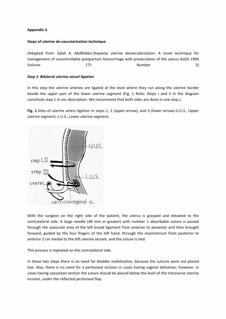
Appendix 3. Steps of uterine de-‐vascularization technique (Adapted from: Salah A. AbdRabbo.Stepwise uterine devascularization: A novel technique for management of uncontrollable postpartum hemorrhage with preservation of the uterus AJOG 1994 Volume 171 Number 3) Step 1: Bilateral uterine vessel ligation
In this step the uterine arteries are ligated at the level where they run along the uterine border beside the upper part of the lower uterine segment (Fig. 1 Note: Steps I and II in the diagram constitute step 1 in our description. We recommend that both sides are done in one step.).
Fig. 1.Sites of uterine artery ligation in steps 1, 2 (upper arrow), and 3 (lower arrow).U.U.S., Upper uterine segment; L.U.S., Lower uterine segment.
With the surgeon on the right side of the patient, the uterus is grasped and elevated to the contralateral side. A large needle (48 mm or greater) with number 1 absorbable suture is passed through the avascular area of the left broad ligament from anterior to posterior and then brought forward, guided by the four fingers of the left hand, through the myometrium from posterior to anterior 2 cm medial to the left uterine vessels, and the suture is tied.
This process is repeated on the contralateral side.
In these two steps there is no need for bladder mobilization, because the sutures were not placed low. Also, there is no need for a peritoneal incision in cases having vaginal deliveries; however, in cases having caesarean section the suture should be placed below the level of the transverse uterine incision, under the reflected peritoneal flap.

Step 2: Low bilateral uterine vessel ligation
This step is reserved only for cases having continued lower uterine segment haemorrhage diagnosed at caesarean section and not controlled by step 1.
In this step the bladder is reflected downwards and lower bilateral uterine vessel ligation is performed at the lower part of the lower uterine segment, 3 to 5 cm below the upper ligatures, with the same technique in step 1. At this level the uterine artery is ligated after its cervicovaginal branch turns abruptly upward to extend along the uterine margin. This ligature would obliterate most of the branches of the uterine artery to the lower uterine segment and a branch of considerable size that extends to the upper portion of the cervix. It is important to include a significant amount of myometrium to avoid damage to the uterine vessels and to obliterate some of the intramyometrial ascending arterial branches of the cervicovaginal artery (Fig. 1).
Step 3: Ligation of uterine/ovarian arterial anastomosis
This step is indicated in continued uterine bleeding in spite of performing step 1.The uterus is grasped and pulled to the contralateral side by the left hand, and a large needle with a number 1 absorbable suture is passed through the avascular area in the broad ligament from anterior to posterior, at the level of the ovarian ligament. The needle is then passed anteriorly 2 cm medial to the edge of the uterine wall, to include the uterine muscle. The suture is tied anteriorly.


