i
JOÃO RENATO BENNINI JUNIOR
GASTROSQUISE: ULTRASSONOGRAFIA NA ESTIMATIVA DO PESO FETAL E PREDIÇÃO DE
DESFECHOS PERINATAIS
GASTROSCHISIS: ULTRASONOGRAPHY FOR FETAL WEIGHT ESTIMATION AND PREDICTION
OF PERINATAL OUTCOMES
CAMPINAS 2014

ii
iii
UNIVERSIDADE ESTADUAL DE CAMPINAS Faculdade de Ciências Médicas
JOÃO RENATO BENNINI JUNIOR
GASTROSQUISE: ULTRASSONOGRAFIA NA ESTIMATIVA DO PESO FETAL E PREDIÇÃO DE
DESFECHOS PERINATAIS
ORIENTADOR: Prof. Dr. Cleisson Fábio Andreolli Peralta COORIENTADOR: Prof. Dr. Ricardo Barini
GASTROSCHISIS: ULTRASONOGRAPHY FOR FETAL WEIGHT ESTIMATION AND PREDICTION
OF PERINATAL OUTCOMES
Tese de Doutorado apresentada à Pós-Graduação em Tocoginecologia, da Faculdade de Ciências Médicas da Universidade Estadual de Campinas – UNICAMP para obtenção do Título de Doutor em Ciências da Saúde, área de concentração em Saúde Materna e Perinatal.
Thesis presented to the Programme of Obstetrics and Gynecology, Faculty of Medical Sciences, University of Campinas - UNICAMP for obtain the title Ph.D grade in concentration area of Maternal and Perinatal Health
ESTE EXEMPLAR CORRESPONDE À VERSÃO FINAL DA TESE
DEFENDIDA PELO ALUNO JOÃO RENATO BENNINI JUNIOR E ORIENTADA PELO Prof. Dr. CLEISSON FÁBIO ANDRIOLI PERALTA
Assinatura do Orientador
_________________________
Campinas, 2014

iv

d
-
--
i v
BANCA EXAMINADORA DA TESE DE DOUTORADO
Aluno: JOÃO RENATO BENNINI JUNIOR
Orientador: Prof. Dr. CLEISSON FABIO ANDRIOLI PERALTA
Coorientador: Prof. Dr. RICARDO BARINI
Membros:
3.
4.
5.
Curso de Pós-Graduação em Tocoginecologia da Faculdade de Ciências Médicas da Universidade Estadual de Campinas
Data: 25/02/2014
v
vi
Dedico este trabalho...
Aos meus filhos, Maria Cecília e João Felipe, como um incentivo aos estudos.
A todos que fazem do ensino e da pesquisa objetivos de vida.
vii
Agradecimentos
À Carolina Maria...
A todos que contribuíram para a idealização, realização e finalização deste trabalho.
viii
Sumário
Símbolos, Siglas e Abreviaturas ................................................................................................ ix
Resumo .................................................................................................................................... xi
Summary ................................................................................................................................ xiv
1. Introdução ...........................................................................................................................17
2. Objetivos .............................................................................................................................30
2.1. Objetivo geral ............................................................................................................30
2.2. Objetivos específicos ................................................................................................30
3. Publicações ........................................................................................................................31
3.1. Artigo 1 .....................................................................................................................32
3.2. Artigo 2 .....................................................................................................................55
4. Discussão geral ..................................................................................................................76
5. Conclusão geral ..................................................................................................................81
6. Referências Bibliográficas ...................................................................................................82
7. Anexos................................................................................................................................95
7.1. Anexo 1 - Parecer da Comissão de Pesquisa do DTG/CAISM ...................................95
7.2. Anexo 2 - Aprovação do projeto no CEP....................................................................96
7.3. Anexo 3 - Ficha de coleta de dados ...........................................................................99
7.4. Anexo 4 - Artigo: “Birth-weight prediction by two- and three-dimensional ultrasound imaging” ..................................................................................................................101

Símbolos, Siglas e Abreviaturas - ix
Símbolos, Siglas e Abreviaturas
2DUS – Two-dimensional ultrasound
3DUS – Three-dimensional ultrasound
AC – Abdominal circumference
AFI – Amniotic fluid index
BPD – Biparietal diameter
CAISM – Centro de Atenção integral à Saúde da Mulher
CI – Confidence interval
DAI – Dilatação de alças intestinais intra-abdominais
DLD – Delta luminal diameter of extra-abdominal intestinal loops
DP – Desvio padrão
EFW – Estimated fetal weight
EPN – Exclusive parenteral nutrition
FDL – Femur diaphysis length
FGR – Fetal growth restriction
g – gram
GA – Gestational age
HC – Head circumference
IBD – Intra-abdominal bowel dilation
k – Number of paired comparisons
kg – kilogram
MSPE – Mean sign percentage error
Símbolos, Siglas e Abreviaturas - x
NICU – Neonatal intensive care unit
OFD – Occipito-frontal diameter
p – Nível de significância estatística
RCF – Restrição de crescimento fetal
SD – Standard deviation
ThiV – Fetal thigh volume
UNICAMP – State University of Campinas
UNICAMP – Universidade Estadual de Campinas
US2D – Ultrassonografia bidimensional
US3D – Ultrassonografia tridimensional
VOCALTM – Virtual Organ Computer-aided Analysis

Resumo - xi
Resumo
Introdução: A literatura é controversa sobre o papel da ultrassonografia pré-
natal na predição de desfechos perinatais nos casos de gastrosquise. O peso
ao nascimento é descrito como um importante fator prognóstico em neonatos
com este tipo de malformação e estudos relatam que fórmulas
ultrassonográficas criadas especificamente para esses casos apresentam
melhor desempenho na estimativa do peso fetal, mas não há consenso sobre
qual o melhor modelo de predição de peso a ser utilizado. Objetivos: Avaliar o
papel de parâmetros ultrassonográficos pré-natais na predição de desfechos
perinatais em casos de gastrosquise. Criar uma nova fórmula ultrassonográfica
para estimativa de peso fetal que não utilize medidas abdominais e compará-la
à outras fórmulas com parâmetros ultrassonográficos bidimensionais (US2D) e
tridimensionais (US3D) quando aplicadas em fetos com gastrosquise. Métodos:
Para avaliar o desempenho de parâmetros ultrassonográficos pré-natais na
predição de desfechos perinatais foi realizado um estudo de coorte
retrospectiva envolvendo fetos com o diagnóstico de gastrosquise isolada. Para
criar e validar a nova fórmula US2D foram utilizados dados referentes à
gestantes e fetos normais coletados em um estudo prévio publicado pelo nosso
grupo. Foi realizado um estudo retrospectivo transversal envolvendo fetos com

Resumo - xii
gastrosquise, para comparar a nova fórmula US2D com diferentes fórmulas
US2D e US3D já publicadas. Os sujeitos foram selecionados entre aqueles
acompanhados na Divisão de Obstetrícia do CAISM / UNICAMP. O tamanho da
amostra foi estimado em 56 pacientes para avaliar o desempenho de
parâmetros ultrassonográficos pré-natais na predição de desfechos perinatais e
27 pacientes para comparar as fórmulas de estimativa de peso fetal. Os dados
maternos, gestacionais e pós-natais foram descritos como freqüências relativas
e absolutas, média e desvio padrão (DP), mediana e limites. A normalidade dos
dados contínuos foi testada utilizando-se o teste de Kolmogorov–Smirnov.
Testes t de amostras independentes e testes de qui-quadrado foram utilizados
na comparação de dados contínuos e categóricos, respectivamente. Análises
de regressão polinominal até o terceiro grau foram consideradas para criar a
nova fórmula US2D de estimativa do peso fetal sem medidas abdominais.
Cálculo do erro percentual médio ± DP, testes t unilaterais, testes t de amostras
pareadas com correção de Bonferroni e testes de variância para amostras
pareadas foram usados para avaliar e comparar a acurácia e precisão das
fórmulas. A associação entre dados contínuos foi testada utilizando-se os
coeficientes de correlação de Pearson ou Spearman e regressão logística
univariada, conforme indicado. Valores de p < 0,05 foram considerados
significativos. Resultados: Foram incluídos 44 casos de fetos com gastrosquise
para avaliar a predição de desfechos perinatais por meio de parâmetros
ultrassonográficos pré-natais. A presença de dilatação de alças intestinais intra-
abdominais (DAI) fetais aumentou o risco de complicacões intestinais pós-
natais e a presença de restrição de crescimento fetal (RCF) diminuiu o risco
Resumo - xiii
deste mesmo desfecho. Nenhum outro parâmetro ultrassonográfico pré-natal
pode significativamente predizer os desfechos perinatais avaliados. Foram
usados os dados referentes aos mesmos grupos de fetos normais do estudo
prévio para respectivamente criar (150 fetos) e validar (60 fetos) a nova fórmula
US2D, que foi a seguinte: peso fetal estimado = 623.324 + 0.165 x DBP x CC x
CF2 (DP: 12,25%). Na comparação entre as fórmulas US2D e entre as fórmulas
US2D e US3D, foram utilizados 44 e 28 fetos com gastrosquise isolada,
respectivamente. Os melhores desempenhos na estimativa de peso dos fetos
com gastrosquise foram obtidos com o modelo US2D proposto por Siemer e
colaboradores e com os modelos US2D e US3D criados no estudo prévio
desenvolvido pelo nosso grupo, não havendo diferença estatísiticamente
significativa entre os mesmos. Conclusões: Em fetos com gastrosquise o
achado de múltipla DAI associa-se a complicações intestinais pós-natais e a
presença de RCF possui um efeito protetor para este mesmo desfecho. A nova
fórmula US2D sem medidas abdominais não melhorou a estimativa do peso ao
nascimento dos fetos com gastrosquise da nossa população em relação às
outras fórmulas US2D e US3D avaliadas. Na nossa amostra de pacientes com
gastrosquise o modelo US2D de Siemer e colaboradores e os modelos US2D e
US3D criados no estudo prévio desenvolvido pelo nosso grupo apresentaram
os melhores desempenhos na estimativa do peso fetal.
Palavras-chave: gastrosquise, ultrassonografia, peso fetal.

Summary - xiv
Summary
Background: The role of prenatal ultrasonographic parameters for the
predicition of perinatal outcomes in fetuses with gastroschisis is still
controversial. Birthweight is described as a prognostic factor and some studies
report that ultrasonographic formulas especifically created for these cases have
a better performance for fetal weight estimation, but there is no consensus about
which is the best one. Objectives: To evaluate prenatal ultrasonographic
parameters as predictors of adverse perinatal outcomes in fetuses with
gastroschisis. To create a new birthweight predicting ultrasonographic model
without abdominal measurements and compare this new formula with other two-
dimensional (2DUS) and three-dimensional (3DUS) fetalweight predicting
models already published when aplied to fetuses with gastroschisis. Methods:
To evaluate the performance of prenatal ultrasonographic parameters as
predictors of perinatal outcomes in fetuses with gastroschisis a retrospective
cohort study was conducted. To create and validate the new 2DUS formula the
same data from normal fetuses colected in a previous study of our group was
used. A retrospective cross-sectional study encompassing fetuses with
gastroschisis was carried out to compare the new 2DUS formula with other
2DUS and 3DUS formulas already published. The patients were selected among
Summary - xv
those followed at the Division of Obstetrics of the Center for Integral Assistance
to Women’s Health of the State University of Campinas. The sample size was
estimated in 56 patients to evaluate prenatal ultrasonographic predictors and
perinatal outcomes and 27 patients to compare the fetal weigth estimating
formulas. Maternal, pregnancy and postnatal data were described as absolute
and percentual frequencies, mean and standard deviation (SD), median and
range. Continuous data were tested for their normal distribution using the
Kolmogorov–Smirnov test. Independent samples t tests and chi-square tests
were used in the assessment of continuous and categorical variables, when
appropriate. Polynomial stepwise regression analyses up to the third order were
considered to generate a new 2DUS weight-predicting model without abdominal
measurements. Calculation of the mean percentage error ± SD, one-sample t
tests, paired samples t-tests with Bonferroni adjustment and correlated variance
tests for paired samples were used to compare the performances of the
formulas. The potential association between continuous data was tested by
means of Pearson or Spearman’s Correlation Coefficient and univariate logistic
regression, as indicated. A two-tailed p-value of less than 0.05 was considered
statistically significant. Results: Forty-four fetuses were included to evaluate the
ultrasonographic prental parameters as predictors of perinatal outcomes. The
presence of fetal multiple intra-abdominal bowel dilation (IBD) was associated
with increased incidence of intestinal complications and the presence of fetal
growth restriction (FGR) had a protective effect over this outcome. No other
prenatal ultrasographic parameter could significantly predict the perinatal
outcomes evaluated. It was used the same data from our previous study on 150
Summary - xvi
normal fetuses and 60 normal fetuses to respectively generate and validate the
new 2DUS formula, which was: estimated fetal weight = 623.324 + 0.165 x BPD
x HC x FDL2 (SD: 12.25). In the comparison between the 2DUS formulas and
between the 2DUS and 3DUS formulas it was included 44 and 28 fetuses,
respectively. The best performance for weight prediction in fetuses with
gastroschisis was achieved using the 2DUS model created by Siemer et al. and
the 2DUS and 3DUS models previously published by our group. Conclusions:
In fetuses with gastroschisis the findings of multiple IBD increases the risk of
postnatal bowel complications and the presence of FGR decreases the risk of
this outcome. The new 2DUS formula without abdominal measurements did not
improve fetal weight estimation in fetuses with gastroschisis of our population
when compared to other 2DUS and 3DUS formulas evaluated. The 2DUS
weight estimating model of Siemer et al. and the 2DUS and 3DUS models
previously published by our group had the best performance for this purpose.
Key words: gastroschisis, ultrasonography, fetal weight.

Introdução - 17
1. Introdução
Gastrosquise é o nome dado a uma falha de continuidade que acomete toda a
espessura da parede abdominal fetal anterior. Geralmente é um defeito de
pequeno diâmetro (2 a 3 cm), localizado 95% das vezes à direita do cordão
umbilical (Sadler, 2010). Através deste orifício ocorre herniação de órgãos
abdominais para a cavidade amniótica, sem a cobertura do peritônio ou do
âmnio. A estrutura mais freqüentemente herniada é o intestino, mas herniação
de outros órgãos, como estômago, bexiga e fígado, também pode ocorrer
(Garcia et al., 2010).
A etiologia da gastrosquise é motivo de debate. Uma hipótese bastante
difundida é que sua patogênese envolve um acidente vascular e dentro desta
linha duas teorias foram desenvolvidas: uma sugere que a involução da veia
umbilical direita causa uma necrose na parede abdominal, levando a um defeito
paraumbilical (deVries, 1980), e a outra postula que a artéria onfalomesentérica
involue prematuramente, causando um enfraquecimento da parede abdominal,
através do qual o conteúdo intestinal subsequentemente hernia (Hoyme et al.,
1981). Estas duas teorias são suportadas pela observação de que a
gastrosquise associa-se à atresia intestinal, uma condição que também
acredita-se estar associada com alterações isquêmicas (Louw e Barnard, 1955).
Introdução - 18
Além disso, dados retrospectivos sugerem um maior risco de gastrosquise e
atresia intestinal em pacientes usuárias de drogas vasoconstritoras como
efedrina, pseudoefedrina ou cocaína, assim como em pacientes tabagistas
(Werler et al., 2003). Entretanto, estudos epidemiológicos mais recentes
sugerem que estas explicações podem ser insuficientes. Feldkamp et al. (2007)
observam que as veias umbilicais não nutrem o mesênquima na parede
abdominal, o que não explicaria um enfraquecimento desta região por uma
alteração no desenvolvimento da veia umbiical direita. Além disso, o suprimento
vascular da parede abdominal deriva de uma rica rede de vasos que se
originam da aorta dorsal e que não é dependente e nem se anastomosa com os
vasos umbilicais ou vitelínicos. Um grande estudo epidemiológico avaliando as
associações entre exposição materna a agentes vasoativos e defeitos
congênitos, observou que em mulheres jovens estes agentes possuem um
papel menor, se é que possuem algum, na etiologia da gastrosquise, mas
podem ter um papel maior em mulheres com mais de 25 anos (Werler et al.,
2009). Explicações para a origem da gastrosquise não relacionadas a
problemas vasculares incluem falha da incorporação do ducto vitelínico ao
cordão umbilical (Stevenson et al., 2009), falha da fusão mediana dos folhetos
laterais da parade abdominal (Feldkamp et al., 2007) ou um defeito na
diferenciacão do mesênquima embrionário responsável pela formação da
parede abdominal (Duhamel, 1963). A ruptura intrauterina de uma onfalocele
também foi proposta como um mecanismo de formação da gastrosquise (Shaw,
1975).
Introdução - 19
A gastrosquise ocorre em cerca de 1 a cada 4000 nascidos vivos. Dados
recentes mostram um aumento de 10 a 20 vezes na incidência desta
malformação em todos os grupos etários nas últimas duas décadas, embora
ainda não haja uma explicação clara para isso (Baird e MacDonald, 1981;
Alvarez e Burd, 2007; Loane et al., 2007; Castilla et al., 2008).
Estima-se que apenas 1% dos casos de gastrosquise apresente algum tipo de
aneuploidia e casos com alteração de um único gene são ainda mais raros
(Sadler, 2010), mas a observação do aumento da incidência de gastrosquise
em algumas familias sugere que fatores genéticos desempenham um papel
relevante na causa desta doença. Fatores de risco não-genéticos também são
importantes, evidenciados, por exemplo, pela maior ocorrência de gastrosquise
em mulheres jovens, pelo aumento da incidência geral de gastrosquise nos
últimos anos e pela ocorrência de casos agrupados (Rasmussen e Frias, 2008).
Entretanto, apesar do reconhecimento da importância dos fatores de risco não-
genéticos na causa da gastrosquise, apenas a baixa idade materna é bem
estabelecida como um destes fatores, limitando o desenvolvimento de
estratégias de prevenção (Rasmussen e Frias, 2008). Mais recentemente,
outros fatores de risco não-genéticos como o tabagismo (Hackshaw et al.,
2011) e a ingesta de álcool (Richardson et al., 2011) foram descritos em
estudos envolvendo um grande número de pacientes.
Antes da ultrassonografia ser utilizada rotineiramente no pré-natal, um aumento
na concentração sérica de alfa-feto proteína costumava ser o único indicador
pré-natal de gastrosquise. Com o uso rotineiro da ecografia no primeiro
trimestre para datação e no segundo trimestre para rastreamento de

Introdução - 20
malformações, quase todos os casos de gastrosquise passaram a ser
diagnosticados antes do nascimento, com um estudo reportando uma taxa de
detecção de 90% na Europa (Garne et al., 2005).
O aspecto ecográfico habitual da gastrosquise é a visualização de múltiplas
alças intestinais flutuando livremente na cavidade amniótica. Tipicamente, o
defeito na parede abdominal ocorre à direita da inserção do cordão umbilical,
que está normalmente inserido na parede abdominal. Este diagnóstico pode ser
feito a partir de 11-12 semanas de gestação, quando a herniação fisiológica
intestinal já deve ter retornado para a cavidade peritoneal (Cullen et al., 1990).
O principal diagnóstico diferencial é com a onfalocele, na qual as alças
herniadas são cobertas pelo peritônio e o cordão umbilical se insere no ápice do
saco herniário. Outros diagnósticos diferenciais incluem lesões císticas do
cordão umbilical, cistos de úraco, extrofia de bexiga/cloaca e a Pentalogia de
Cantrell. Excluir a possibilidade de uma onfalocele é de fundamental
importância, visto que esta condição, diferentemente da gastrosquise,
freqüentemente associa-se com malformações em outros órgãos e aneuploidias
(David et al., 2008).
Exceto por uma maior incidência de alterações intestinais, crianças com
gastrosquise geralmente não possuem outras anomalias anatômicas
associadas e tendem a apresentar crescimento e desenvolvimento neurológico
normais durante a infância. A atresia intestinal é a alteração anatômica mais
comumente associada. Estudos recentes relatam a presença de atresia
intestinal em 7% a 28% dos casos (Arnold et al., 2007; Kronfli et al., 2010).

Introdução - 21
O prognóstico dos casos de gastrosquise é determinado principalmente pelo
grau de lesão intestinal que ocorre durante o período fetal. Esta lesão resulta
provavelmente da combinação entre o contato da alça com o líquido amniótico
e a constrição intestinal no orífico do defeito abdominal, sendo que a maior
parte da injúria parece ocorrer no final da gestação (Langer et al., 1989; Langer
et al., 1990). A lesão nas alças intestinais pode causar disfunção na motilidade
e na absorção, o que prolonga a necessidade de nutrição parenteral e, em
alguns casos, pode causar perda irreversível da função intestinal (Wales e
Christison-Lagay, 2010). O diagnóstico pré-natal provê a possibilidade de
controlar o modo, local e época do parto, com o objetivo de minimizar estas
complicações.
A melhor via de parto para os fetos com gastosquise é motivo de debate.
Alguns autores argumentam que o parto vaginal pode provocar lesões nas
alças intestinais e defendem o uso rotineiro da cesárea. Entretanto esta
hipótese não encontra respaldo na literatura, que não demonstra diferença no
prognóstico dos casos nascidos por cesariana quando comparados aos casos
nascidos por parto normal. Desta forma, a conduta mais adequada em relação
à via de parto talvez dependa de uma discussão entre o obstetra e gestante
(Segel et al., 2001; Salihu et al, 2004).
O melhor momento para realizar o parto dos casos de gastrosquise também é
um tópico controverso. Alguns centros recomendam a realização do parto no
limite do termo, por volta de 36-37 semanas, com o objetivo de diminuir a
exposição das alças intestinais ao líquido amniótico e assim reduzir a
intensidade da reação inflamatória na superfície das mesmas. Há evidências de
Introdução - 22
que mediadores inflamatórios e citocinas (interleucinas 6 e 8) presentes no
líquido amniótico possuem um papel no desenvolvimento de danos no plexo
nervoso mioentérico e nas células intersticiais de Cajal nos casos de
gastrosquise. Como acredita-se que estes efeitos inflamatórios sobre as alças
intestinais aumentam com o passar da gestação, alguns autores acham que a
antecipação do parto poderia diminuí-los (Srinathan et al., 1995; Luton et al.,
1999; Salihu et al, 2004; Guibourdenche et al., 2006; Vargun et al., 2007). Parto
pré-termo é mais frequente em gestações de fetos com gastrosquise (incidência
de 28% contra 6% nos casos de gestações de fetos normais), desta forma o
parto pode ser induzido com sucesso em grande parte dos casos com 36-37
semanas de gestação, provavelmente por causa desta tendência natural
(Lausman et al., 2007). A literatura, entretanto, é controversa sobre os
benefícios da antecipação do parto. O argumento contra a antecipação do parto
é que o baixo peso ao nascimento tem um efeito negativo sobre o prognóstico
destas crianças, com neonatos com pesos inferiores a dois quilos demorando
mais tempo para atingirem nutricão enteral completa e ficando mais tempo sob
ventilação assistida (Charlesworth et al., 2007). Alguns estudos propõem a
antecipação do parto de maneira seletiva nos casos que apresentem sinais de
dilatação e espessamento da parede das alças intestinais na avaliação
ecográfica, pois a presença destes sinais correlaciona-se com mau prognóstico,
incluindo sofrimento e óbito fetal, em algumas, mas não em todas as séries de
casos (Langer et al., 1993; Piper e Jaksic, 2006). Entretanto, esses dados não
são muito claros por não haver padronização na maneira de se medir as alças
Introdução - 23
intestinais e nem consenso sobre a definição de dilatação intestinal de acordo
com a idade gestacional.
A maioria dos autores recomenda que o parto seja realizado em centros
terciários, de maneira a permitir acesso imediato a cuidados neonatais e
cirúrgicos especializados. Um estudo avaliando resultados pós-natais observa
que o nascimento de casos de gastrosquise em centros com acesso imediato a
atendimento de neonatologia e cirurgia pediátrica terciários associa-se com
diminuição do risco de complicações pós-natais, quando comparado com casos
nascidos em hospitais sem acesso imediato a estes tipos de atendimentos
(Christison-Lagay et al., 2011; Nasr e Langer, 2011). Entretanto, esta
recomendação não é um consenso. Um trabalho recente com 118 casos de
gastrosquise observa que o tempo para realização da primeira cirurgia não foi
preditivo de nenhum desfecho clínico desfavorável relevante. Estes dados
sugerem que os potenciais benefícios da regionalização do acompanhamento
dos casos de gastrosquise não seriam suportados por um menor tempo para a
realização da intervenção cirúrgica (Bucher et al., 2012).
A gastrosquise leva a uma perda hídrica significativa por evaporação pelas
alças expostas. Após o parto, deve ser iniciada repoisção de líquido por via
endovenosa. Descompressão gástrica, por meio de sonda, também deve ser
realizada com o objetivo de prevenir ou diminuir distensão intestinal. As alças
herniadas devem ser envolvidas em compressas embebidas em solução salina
fisiológica, colocadas em posição central na parede abdominal com o recém-
nascido em decúbito lateral direito para prevenir acotovelamento do mesentério
e cobertas com silo plástico para diminuir as perdas hídricas evaporatovas e a
Introdução - 24
instabilidade térmica. Um exame detalhado do neonato deve ser feito para
excluir a coexistência de outras anomalias, com atenção especial à busca de
sinais de atresia, necrose ou perfuração intestinal (Christison-Lagay et al.,
2011).
A conduta cirúrgica nos casos de gastrosquise varia de centro para centro e e
modificou-se nas últimas décadas. O objetivo da cirurgia é devolver as vísceras
herniadas para a cavidade abdominal com o menor risco de lesão, seja esta por
trauma direto ou por aumento da pressão intra-abdominal. As principais opções
de conduta cirúrgica são: (a) redução primária com fechamento da fascia; (b)
colocação de silo, reduções seriadas e fechamento tardio da fascia; (c) redução
primária ou seriada, sem o fechamento da fascia. O intervalo de tempo após o
nascimento e o local onde deve ser realizada a intevenção cirúrgica são
motivos de debate, podendo ser um reparo imediato na sala de parto, redução e
fechamento na unidade de terapia intensiva neonatal ou fechamento tardio no
centro cirúrgico (Coughlin et al., 1993; Bianchi et al., 2002). Em todos os casos,
inspeção intestinal à procura de sinais de bandas obstrutivas, perfuração ou
atresia deve ser realizada cuidadosamente. Bandas atravesando as alças
devem ser divididas antes da colocação do silo ou fechamento abdominal
primário para evitar obstrução intestinal subsequente. Estabelecimento precoce
de um acesso venoso central deve ser considerado, pois hipomotilidade
intestinal está invariavelmente presente (Christison-Lagay et al., 2011).
A taxa geral de sobrevivência do casos de gastrosquise é muito boa, variando
de 90% a 97% (Molik et al., 2001; Aina-Mumuney et al., 2004). Já
complicações gastrointestinais, tais como perfuração ou atresia, ocorrem em
Introdução - 25
10% a 20% dos casos (Brantberg et al., 2004). A presença destas complicações
associa-se com taxas de mortalidade de até 28% e maior tempo de
hospitalização e nutrição paraenteral, com seus riscos associados de infecção,
distúrbios de crescimento, disfunções metabólicas e doenças hepáticas (Langer
et al., 1993; Wilson e Johnson, 2004; Nick et al., 2006; Badillo et al., 2008).
Desta forma, a identificação pré-natal de fatores associados com o prognóstico
perinatal ajudaria na triagem de casos que se beneficiariam de um
acompanhamento mais próximo das condições fetais e de algum tipo de
intervenção, como a antecipação do parto ou nascimento em hospitais
especializados, além de ajudar no aconselhamento dos pais (Huh et al., 2010).
A dilatação de alças intestinais extra-abdominais tem sido um dos fatores
prognósticos pré-natais mais estudado, mas seu papel na predição da evolução
pós-natal dos casos de gastrosquise ainda é motivo de debate. Alguns estudos
demonstram correlação entre a presença de dilatação de alças extra-
abdominais e complicações intestinais e outros não. Diferenças na definição de
dilatação e amostras pequenas dos estudos limitam o estabelecimento de um
consenso sobre quais medidas são consistentemente preditoras de mau
prognóstico (Alsulyman et al., 1996; Japaraj et al., 2003; Nick et al., 2006;
Badillo et al., 2008). Além disso, alguns autores sugerem que a dilatação das
alças intestinais exteriorizadas pode ser um fenômeno fisiológico em muitos
fetos com gastrosquise no terceiro trimestre, que apresentam ótima evolução
pós-natal (Huh et al., 2010).
A avaliação quanto a presença de dilatação intra-abdominal das alças
intestinais também tem sido estudada como um fator prognóstico pré-natal.

Introdução - 26
Este achado é menos comum nos fetos com gastrosquise, com uma incidência
estimada em 8% a 17% (Brantberg et al., 2004; Nick et al., 2006), e estudos
recentes demonstram que a presença deste sinal correlaciona-se melhor com a
ocorrência de complicações pós-natais do que a presença de dilatação das
alças extra-abdominais. Entretanto, diferenças na definição de dilatação intra-
abdominal e amostras pequenas dos estudos também limitam o
estabelecimento de um consenso sobre o papel deste achado (Nick et al., 2006;
Huh et al., 2010; Kuleva et al., 2012).
Avaliando 117 pacientes com gastrosquise, um trabalho relata que herniação
hepática junto com o intestino estava presente em 6% dos casos e este achado
se correlacionou com uma taxa de sobrevivência significativamente menor (43%
x 97%), considerando-se a idade gestacional e o peso ao nascimento. Estes
autores observam que a presença de herniação hepática demonstra ser um
fator de mau prognóstico pós-natal e deve ser avaliada nos exames de
ecografia pré-natais (McClellan et al., 2011). A literatura, entretanto, ainda é
escassa na avaliação deste fator prognóstico.
Um outro fator prognóstico que tem sido avaliado é a dilatação do estômago,
também com resultados diversos. Avaliando 34 casos, 13 (38%) dos quais
apresentavam dilatação estomacal, um estudo observa que este achado se
correlacionou com maior incidência de cardiotocografia não reativa, vólvulo
intestinal e morte neonatal e maior tempo para alimentação por via oral e de
hospitalização (Aina-Mumuney et al., 2004). Já um outro trabalho, avaliando 89
fetos com gastrosquise, 32 (33%) dos quais com dilatação do estômago,
encontrou resultados diferentes. Este estudo observa que a presença de

Introdução - 27
dilatação estomacal correlacionou-se apenas com maior ocorrência de líquido
meconial ao nascimento, mas não se correlacionou com outros resultados
adversos perinatais (Alfaraj et al., 2011).
Parâmetros Dopplervelocimétricos também já foram avaliados na predição de
complicações intestinais de fetos com gastrosquise. Um estudo prospectivo
com 17 fetos com gastrosquise avalia a relação entre o índice de pulsatilidade
da artéria mesentérica superior fetal e má evolução pós-natal, definida como
necessidade de ressecção intestinal ou tempo de hospitalização maior que 50
dias. Neste estudo, o índice de pulsatilidade da artéria mesentérica superior não
foi capaz de predizer má evolução pós-natal (Abuhamad et al., 1997).
A presença de herniação vesical junto com as alças intestinais parece se
correlacionar com maior risco de sofrimento fetal nos casos de gastrosquise,
mas não com maior risco de complicações intestinais. Um estudo recente,
observa que 6 de 105 (6%) dos casos de gastrosquise acompanhados em um
centro apresentavam herniação vesical. Um destes casos evoluiu com óbito
fetal e outros quatro tiveram parto cesárea por sinais de sofrimento fetal. Os
cinco sobreviventes evoluiram bem após o tratamento pós-natal, mas os
autores observam que a presença de herniação da bexiga nos casos de
gastrosquise pode ser um sinal para um acompanhamento mais rigoroso do
bem-estar fetal (Mousty et al., 2012).
Recentemente, um estudo caso-controle com 106 casos de gastrosquise, avalia
a correlação de múltiplos fatores ultrassonográficos pré-natais com o
desenvolvimento de complicações intestinais neonatais. Os seguintes fatores
ecográficos pré-natais foram avaliados: feto pequeno para a idade gestacional,
Introdução - 28
presença de dilatação intra ou extra-abdominal de alças intestinais,
espessamento de parede de alça intestinal e dilatação estomacal. Estes fatores
foram utilizados para tentar predizer a presença de atresia, perfuração, necrose
ou vólvulo ao nascimento, sendo que 14% dos casos apresentaram alguma
destas complicações. Este trabalho observa que nenhum dos marcadores
ecográficos foi preditivo de morte fetal ou neonatal e apenas a presença de
dilatação de alças intra-abdominais se correlacionou com a ocorrência de
complicações intestinais pós-natais (Kuleva et al., 2012).
Muitas publicações relatam que o peso ao nascimento é também um importante
fator na evolução pós-natal dos casos de gastrosquise. Estes autores observam
que recém-nascidos com gastrosquise pequenos para a idade gestacional
apresentam maiores taxas de morbidade e mortalidade (Charlesworth et al.,
2007; Netta et al., 2007; Nicholas et al., 2009). Desta forma, a predição mais
precisa possível do peso fetal é de extrema importância para tomada de
decisões nesses casos. A estimativa do peso fetal nos fetos com gastrosquise
tem sido feita essencialmente por fórmulas geradas a partir de fetos normais
que utilizam medidas ultrassonográficas bidimensionais (US2D) do pólo
cefálico, membros e abdome. Entretanto, alguns estudos relatam que fórmulas
criadas especificamente para fetos com defeitos de parede abdominal, que não
levam em consideração as medidas do abdome, possuem uma maior acurácia
na estimativa do peso desses fetos (Siemer et al., 2008; Chaudhury et al.,
2010; Nicholas et al., 2010; Adams et al., 2012).
Em um estudo publicado em 2010, Bennini et al. criaram, a partir de fetos
normais, um modelo US2D de estimativa de peso que utiliza medidas do pólo
Introdução - 29
cefálico, abdome e fêmur, e outros dois modelos que utilizam como único
parâmetro o volume da coxa fetal medido por meio da ultrassonografia
tridimensional (US3D). Na comparação das fórmulas, os desempenhos dos
novos modelos US2D e US3D não apresentaram diferença estatística entre si,
mas foram significativamente melhores do que os dos modelos US2D e US3D
testados que haviam sido gerados em outras populações. Apesar dessas
observações, nunca foram criadas fórmulas US2D de estimativa de peso
específicas para fetos com gastrosquise geradas a partir de uma amostra da
nossa população. Da mesma maneira, fórmulas que utilizam parâmetros
ultrassonográficos tridimensionais (US3D), como o volume da coxa fetal, para a
estimativa de peso nunca foram avaliadas em fetos com gastrosquise.
Portanto, avaliando as publicações mais recentes, observamos que, apesar do
reconhececimento da importância da identificação de fatores pré-natais que
possam predizer o risco de complicações perinatais nos casos de gastrosquise,
a literatura é controversa sobre quais seriam estes fatores e sobre o
desempenho dos mesmos. Além disso, considerando-se a importância do peso
ao nascimento no prognóstico dos casos de gastrosquise, ainda são
necessários mais estudos para avaliar qual a melhor maneira de estimar peso
desses fetos.
Objetivos - 30
2. Objetivos
2.1. Objetivo geral
Avaliar o papel da ultrassonografia na estimativa do peso e predição de
desfechos perinatais desfavoráveis em fetos com gastrosquise isolada.
2.2. Objetivos específicos
Criar uma nova fórmula US2D para estimativa de peso que não utilize
medidas abdominais gerada a partir de fetos normais da nossa população e
comparar o seu desempenho com os de outras fórmulas US2D e US3D
quando aplicadas em fetos com gastrosquise isolada.
Avaliar o papel de parâmetros ultrassonográficos na predição de desfechos
perinatais em fetos com gastrosquise isolada.
Publicações - 31
3. Publicações
Artigo 1 – Bennini JR, Marussi EF, Barini R, Peralta CFA. Birth weight
prediction for fetuses with gastroschisis using two- and three-dimensional
ultrasound.
Artigo 2 – Bennini JR, Marussi EF, Barini R, Peralta CFA. Gastroschisis: fetal
longitudinal follow-up and perinatal outcomes
Publicações - 32
3.1. Artigo 1
Manuscript number: UOG-2014-0093 Dear Dr Bennini We are pleased to receive your manuscript entitled Birth weight prediction for fetuses with gastroschisis using two- and three-dimensional ultrasound by Bennini, Joao; Marussi, Emilio; Barini, Ricardo; Peralta, Cleisson Fábio. We will shortly be assigning it to one of the Journal's Editors who will handle the peer review of the paper. To track the progress of your manuscript through the editorial process using our web-based system, simply point your browser to: http://mc.manuscriptcentral.com/uog Please remember in any future correspondence regarding this article to always include its manuscript ID number UOG-2014-0093. Many thanks for submitting your manuscript Yours sincerely Sarah Hatcher Managing Editor

Publicações - 33
Birth weight prediction for fetuses with gastroschisis using two- and
three-dimensional ultrasound
JR Bennini, EF Marussi, R Barini and CFA Peralta
Department of Obstetrics and Gynecology, Professor José Aristodemo Pinotti
Hospital, Center for Integral Assistance to Women’s Health, State University of
Campinas (UNICAMP), Campinas - SP, Brazil
Correspondence: João Renato Bennini
Hospital da Mulher Prof. Dr. José Aristodemo Pinotti
Centro de Atenção Integral à Saúde da Mulher (CAISM)
Departamento de Tocoginecologia (DTG)
Faculdade de Ciências Médicas (FCM)
Universidade Estadual de Campinas (UNICAMP)
Rua Alexander Fleming, 101 - Cidade Universitária Zeferino Vaz
Distrito de Barão Geraldo, Campinas - SP, Brasil
CEP: 13083-881
Phone: (+55) (19) 35219500
Fax: (+55) (19) 35219333
E-mail: [email protected]
Publicações - 34
Abstract
Objectives: To create a new two-dimensional ultrasound (2DUS) birthweight
predicting model without abdominal measurements and compare this new
formula with other 2DUS and three-dimensional ultrasound (3DUS) models
when aplied to fetuses with gastroschisis.
Methods: To create and validate the new 2DUS formula it was used the same
group of normal fetuses enrolled in a previous study published by our group. To
compare the 2DUS and 3DUS formulas it was realized a retrospective cross-
sectional study using fetuses with gastroschisis. Polynomial stepwise regression
analyses was used to generate the new weight-predicting model. Calculation of
the mean percentage error ± standard deviation, one-sample t tests, paired
samples t-tests and correlated variance tests for paired samples were used to
compare the performances of the formulas.
Results: A total of 44 and 28 fetuses with gastroschisis were used to compare
the 2DUS and 3DUS formulas, respectively. The weight predicting models with
highest accuracies and precisions were the 2DUS formula of Siemer et al. and
ours previously published 2DUS and 3DUS models, with no statistic difference
among their performances.
Conclusions: The new 2DUS formula did not improve fetal weight estimation in
fetuses with gastroschisis when compared to other 2DUS and 3DUS formulas
evaluated.
Key words: gastroschisis, prenatal ultrasonography, fetal weight, three-
dimensional ultrasonography, fetal thigh volume.

Publicações - 35
Introduction
Accurate estimation of weigh in fetuses with gastroschisis is important because
abnormal patterns of growth may influence this disease’s baseline risks of
perinatal mortality and morbidity.1-5 The assessment of fetal weight in such
cases has been essentially based on traditional two-dimensional ultrasound
(2DUS) formulas, which were generated from anatomically normal fetuses and
take into account the measurements of the head, limbs and abdomen.
Nevertheless, some studies report that equations elaborated specifically for
fetuses with abdominal wall defects, which do not include the abdominal
measurements, produce better results when applied to fetuses with
gastroschisis.6-9
Another option to estimate fetal weight is to use three-dimensional ultrasound
(3DUS) birth-weight predicting models. Some of these formulas rely on the use
of single predictors of weight, such as the thigh volume, which reflects not only
the length of the limb but also the amount of soft tissue that surrounds it.10 The
use of 3DUS formulas to estimate the weight in fetuses with gastroschisis has
not been reported yet.
The aim of this study was to create a new 2DUS formula without abdominal
parameters and to compare, in fetuses with isolated gastroschisis, the
performances of 2DUS and 3DUS birth-weight predicting models generated in
our population, with those of previously published 2DUS formulas elaborated
specifically for fetuses with abdominal wall defects.

Publicações - 36
Methods
This was a retrospective cross-sectional two-stage study carried out at
Professor José Aristodemo Pinotti Hospital, Center for Integral Assistance to
Women’s Health of the State University of Campinas (UNICAMP), including
normal fetuses evaluated between July 2007 and January 2009 and fetuses with
gastroschisis evaluated between February 2007 and November 2013. The
ethics committee of the State University of Campinas Medical School approved
this protocol. Since this was a retrospective study and all the data were obtained
from routine assessments of normal and pathological cases, we were exempt
from the application of informed consent forms.
Common eligibility criteria for the two stages of the study consisted of the
following: 1. No maternal diseases associated with alterations in fetal growth,
such as pre-eclampsia or diabetes. 2. Singleton pregnancy. 3. Well-defined
gestational age (GA) based on the known date of the last menstrual period
and/or the measurement of embryonic/fetal crown-rump length during the first
trimester, interpreted based on the reference intervals reported by Robinson and
Fleming.11
First stage of the study: Elaboration and validation of a new 2DUS birth-weight
predicting model without abdominal measurements

Publicações - 37
Two groups of normal fetuses were evaluated to create and validate a new
specific 2DUS birth-weight predicting model without abdominal measurements.
These patients were the same used in a previous study by our group to create
2DUS and 3DUS formulas for the estimation of fetal weight in our population.10
Second stage of the study: Comparison of the accuracies of the new 2DUS
birth-weight predicting model with those of previously published 2DUS and
3DUS equations in fetuses with gastroschisis
This stage of the study involved the evaluation of fetuses with isolated
gastroschisis between February 2007 and November 2013 with the purpose of
comparing the 2DUS birth-weight predicting model created in this study with
other 2DUS and 3DUS formulas previously published. The specific eligibility
criteria for this part of the study were delivery less than 14 days after the last
ultrasound scan and the presence of isolated fetal gastroschisis diagnosed
during obstetric scans in our institution.
Demographic and clinical characteristics of the mother, including age, GA at
delivery, the mode of delivery and the clinical characteristics of the newborn
(weight and Apgar scores) were collected from the mother’s hospital files. At our
institution, neonates are categorized as small, normal or large for gestational
age if their weights fall below the 10th, between the 10th and the 90th or above
the 90th percentile of the reference intervals defined by Alexander et al.12,
respectively. All neonates were weighed immediately after birth in the delivery

Publicações - 38
room on the same precision electronic Filizola Baby scale (Filizola SA,
Weighting and Automation, Campo Grande, MS, Brazil), which has a precision
of 5 g and is calibrated every two weeks. The date and time of each scan and
birth were recorded to allow calculation of the time from scan to birth.
Conventional 2DUS measurements and fetal thigh volumetry using the
VOCALTM (Kretztechnik, Austria) technique were performed as previously
described by our group.10
All ultrasound examinations were performed transabdominally, with a Voluson
730 Expert scanner, equipped with a RAB 4-8L probe (GE Medical Systems,
Milwaukee, WI, USA) by one of two physicians (JRB; CFAP).
Statistical analysis
Maternal demographic characteristics and pregnancy and postnatal data were
described as absolute and relative frequencies, the average and standard
deviation (SD) or the median and range.
Creation and validation of a new 2DUS birth-weight predicting model without
abdominal measurements:
Using data from the first stage of the study and weight of the neonate as the
dependent variable, polynomial stepwise regression analyses up to the third
Publicações - 39
order were considered to generate a new weight-predicting model with the
following conventional 2DUS measurements as predictors: biparietal diameter
(BPD), head circumference (HC) and femur diaphysis length (FDL). To construct
the new 2DUS formula, Eigenvalue, tolerance, variance inflation factor, condition
index and variance proportion were calculated to check for multicollinearity
among independent variables.13 The criteria for multicollinearity were:
Eigenvalue less than 0.1; tolerance value less than 1 - r2; variance inflation
factor greater than 1 / (1 - r2); condition index greater than 0.30; and variance
proportion greater than 0.8. For the best-fit equation, Kolmogorov-Smirnov test
was performed to check for normality of the standardized residuals.
Comparison of previously published 2DUS and 3DUS equations with the new
2DUS model applied to fetuses with gastroschisis:
For this part of the study, a sample size of 27 patients with gastroschisis has a
power of 80% to detect a difference in means of -6.24 (the difference between a
Group 1 mean, 1, of -0.27 and a Group 2 mean, 2, of 5.97) assuming that the
common standard deviation is 8.00 using a two group t-test with a 0.05 two-
sided significance level.
The EFW calculated using each birth-weight predicting model was adjusted for
the interval between the last sonographic examination and the date of delivery
according to the individual trend of growth of each fetus.14

Publicações - 40
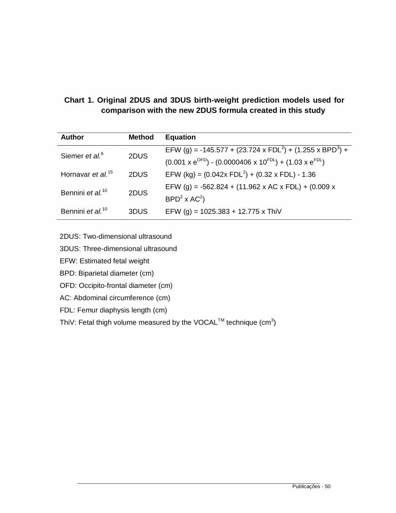
The original 2DUS formulas described by Siemer et al.6 and Honarvar et al.15, as
well as the 2DUS and 3DUS equations reported by our group10 were applied to
data obtained from the fetuses with gastroschisis, in order to compare their
performances with that of the new 2DUS model generated in the present study.
The criteria for the selection of these formulas were: 1. The absence of
abdominal parameters in the formulas generated by Siemer et al.6 and Honarvar
et al.15, as well as in the 3DUS model created by our group.10 2. The
demonstration that our previous 2DUS equation including the abdominal
circumference (AC) had better performance than those of other formulas when
applied to normal fetuses in our population.10
The technique used for the measurement of total fetal thigh volume in fetuses
with gastroschisis was the same used in the original article to generate the
3DUS equation.10 The formulas used for comparison with our new 2DUS model
are described in Chart 1.
The performances of each of these equations were analyzed by the calculation
of systematic and random errors. The systematic error, or accuracy, was
evaluated by calculating the mean sign percentage error (MSPE): [(estimated
fetal weight – actual birth weight) / actual birth weight x 100], with 95%
confidence intervals (CI). To determine the presence and extent of bias, the
MSPE were compared to zero using one-sample t-tests. Paired samples t-tests
with Bonferroni adjustments were used to detect significant differences between
the accuracies of these formulas. The adjusted p-value (p’), which was

Publicações - 41
calculated according to the Bonferroni method, was obtained by the formula p’ =
k x p-value, where k was the number of paired comparisons and the p-value was
obtained from each paired samples t-test.16,17 In this manner, for the comparison
of our new 2DUS model with the original 2DUS functions of Siemer et al.6,
Bennini et al.10 and Honarvar et al.15 (three paired comparisons), each p’ was
obtained by the formula p’ = 3 x the p-value of the paired samples t-test. This
method has the restriction that the p’ cannot exceed 1.0. The random error, or
precision, was evaluated by calculating the standard deviation (SD) of the
MSPE. In order to compare the random errors of two equations, correlated
variance tests for paired samples were used.18 For each paired comparison, the
variances were considered to be significantly different if the p value obtained
from the r (Pearson’s correlation coefficient) distribution table was less than
0.05. The r value was calculated by the formula r = (F - 1) / √ (F + 1)2 - 4 x r2 x F,
where F is the ratio of the variances of the groups being compared. All p values
exceeding 0.20 were referred to as p > 0.20.
The data were analyzed using the statistical software packages SPSS 21.0
(Chicago, Il, USA) and Excel for Mac 2011 (Microsoft Corp., Redmond, WA,
USA).
Publicações - 42
Results
In order to generate and validate the new 2DUS formula, 210 patients (150 in
the formula-generating group; 60 in the formula-validation group) were
evaluated.
The best-fit 2DUS formula without AC measurement for the estimation of weight
in normal fetuses was: EFW = 623.324 + 0.165 x BPD x HC x FDL2 (SD of
predicted values: 12.25; r = 0.774; r2 = 0.599; p < 0.001). The results of the
Kolmogorov-Smirnov tests revealed normal distributions of the standardized
residuals of the equation.
For both the formula-generating and the formula-validation groups, no
statistically significant differences were noted between the accuracies (p < 0.01)
and precisions (p < 0.01) of the new 2DUS model in the prediction of birth
weight.
In total, 61 fetuses with gastroschisis were evaluated throughout the study
period. Among them, 44 (72%) met the entry criteria and 17 (28%) were not
included in the final analysis due to the following reasons (Chart 2): delivery
more than 14 days after the last ultrasound scan (5 cases); loss of follow-up (7
cases); incomplete 2DUS measurements in the last scan before birth (1 case);
spontaneous fetal demise (3 cases); ongoing pregnancy (1 case).
Publicações - 43
Twenty-eight (64%) of the gastroschisis fetuses included had stored 3DUS
datasets of their thighs. Therefore, the comparison of 2DUS formulas was
performed using 44 patients (2DUS gastroschisis group) and the comparison of
2DUS and 3DUS models was carried out with 28 cases (2DUS/3DUS
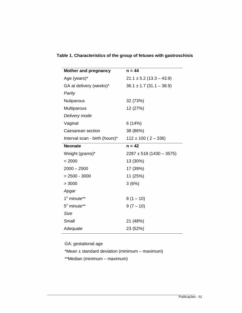
gastroschisis group). Demographic and clinical characteristics of the 44 cases of
gastroschisis are presented in Table 1.
2DUS gastroschisis group:
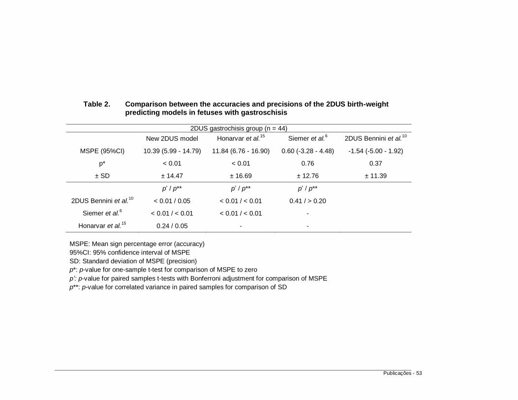
When applied to the 2DUS gastroschisis group, the formulas of Siemer et al.6
and Bennini et al.10 showed the lowest MSPE and SD (0.60 ± 12.76 and -1.54 ±
11.39, respectively), with no significant difference of the MSPE from zero (p =
0.37 and 0.76, respectively). The new 2DUS formula and the Honarvar et al.15
equation significantly overestimated the neonatal weights (p < 0.01 for both
models) with MSPE and SD = 10.39 ± 12.26 and 11.84 ± 16.69, respectively
(Table 2).
When comparing the accuracies of these equations, the performance of the new
2DUS formula was not different from that of Honarvar’s formula, but it was
significantly worse than those of Siemer’s and Bennini’s equations (Table 2).
The precision of the new 2DUS model was not significantly different from those
of Bennini’s and Honarvar’s equations, but it was significantly worse than that
produced by Siemer’s formula (Table 2).
Publicações - 44
When comparing the formulas of Siemer et al.6 and Bennini et al.10 no statistic
difference between the accuracies and precisions were observed (Table 2).
2DUS/3DUS gastrosqhisis group results:
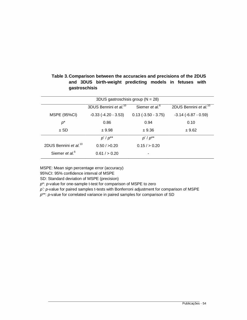
When applied to the 2DUS/3DUS gastroschisis group, the formula of Siemer et
al.6 and the 3DUS model of Bennini et al.10 showed the lowest MSPE and SD
(0.13 ± 9.36 and -0.33 ± 9.98, respectely), with no significant difference of the
MSPE from zero (p = 0.94 and 0.86, respectely). The 2DUS formula of Bennini
et al.10 produced a MSPE and SD = -3.14 ± 9.62, also with no significant
difference (p = 0.10) of the MSPE from zero (Table 3).
When comparing the three formulas applied to the 2DUS/3DUS gastroschisis
group, no statistic difference between the accuracies and precisions were
observed (Table 3).
Discussion
The main question raised in this study was whether 2DUS or 3DUS birth-weight
predicting models that do not include fetal abdominal measurements and are
generated in our population allow better estimations of fetal weights in cases of
gastroschisis than do other particular 2DUS equations (generated in our
Publicações - 45
population and including the AC, or elaborated elsewhere specifically for the
estimation of weight in fetuses with gastroschisis).
In a previous study we have demonstrated that 2DUS and 3DUS birth-weight
predicting models created from a representative sample of our population have
better performances to estimate fetal weights in our patients. Therefore, we
elaborated a new 2DUS model without fetal abdominal measurements and
compared the performance of this new model with those of our previous 2DUS
and 3DUS formulas and those of 2DUS equations created specifically for the
estimation of weight in fetuses with gastroschisis.
With regard to the 2DUS models, our results demonstrated that, in terms of
accuracy and random error, the two best formulas were those proposed by
Siemer et al.6 and Bennini et al.10 None of this models produced a systematic
bias in weight estimation and the differences between their MSPE and SD were
not statistically significant.
The new 2DUS formula created in this study had worse performance than those
of our previous 2DUS equation and of Siemer’s formula. This was not expected,
if we consider that the new 2DUS model did not incorporate fetal abdominal
measurements and was generated from fetuses of the same population of the
cases of gastroschisis. The reasons for these findings are not clear. One could
hypothesize that, because fetuses with gastroschisis tend to be smaller than
anatomically normal fetuses, this should be followed by smaller phenotypic
Publicações - 46
variations. If we consider that Siemer’s equation was elaborated using preterm
neonates, it seems reasonable that the precision of their equation would be
better even when applied to different populations.
The main weakness of this study was the small sample size to allow the
evaluation of the impact of other variables, such as the content herniated
through the abdominal wall defect, on the accuracies of each of these formulas.
For the same reason, it was not possible to evaluate properly the performance
of each model in the prediction of fetal growth restriction.
In conclusion, the 2DUS model without abdominal measurements generated in
our population did not improve birth-weight estimation in relation to previously
published formulas. The 2DUS model proposed by Siemer et al.6 and the 2DUS
and 3DUS models created by Bennini et al.10 were the best to predict birth-
weight in our fetuses with gastroschisis.
Publicações - 47
References
1. Charlesworth P, Njere I, Allotey J, Dimitrou G, Ade-Ajayi N, Devane S,
Davenport M. Postnatal outcome in gastroschisis: effect of birth weight and
gestational age. J Pediatr Surg. 2007 May;42(5):815-8.
2. Netta DA, Wilson RD, Visintainer P, Johnson MP, Hedrick HL, Flake AW,
Adzick NS. Gastroschisis: growth patterns and a proposed prenatal
surveillance protocol. Fetal Diagn Ther. 2007;22(5):352-7.
3. Nicholas SS, Stamilio DM, Dicke JM, Gray DL, Macones GA, Odibo AO.
Predicting adverse neonatal outcomes in fetuses with abdominal wall
defects using prenatal risk factors. Am J Obstet Gynecol. 2009
Oct;201(4):383.e1-6.
4. Chen IL, Lee SY, Ou-Yang MC, Chao PH, Liu CA, Chen FS, Chung MY,
Chen CC, Huang HC. Clinical presentation of children with gastroschisis and
small for gestational age. Pediatr Neonatol. 2011 Aug;52(4):219-22.
5. Clark RH, Walker MW, Gauderer MW. Factors associated with mortality in
neonates with gastroschisis. Eur J Pediatr Surg. 2011 Jan;21(1):21-4.
6. Siemer J, Hilbert A, Hart N, Hoopmann M, Schneider U, Girschick G, Müller
A, Schild RL. Specific weight formula for fetuses with abdominal wall
defects. Ultrasound Obstet Gynecol. 2008 Apr;31(4):397-400.
Publicações - 48
7. Nicholas S, Tuuli MG, Dicke J, Macones GA, Stamilio D, Odibo AO.
Estimation of fetal weight in fetuses with abdominal wall defects: comparison
of 2 recent sonographic formulas to the Hadlock formula. J Ultrasound Med.
2010 Jul;29(7):1069-74.
8. Chaudhury P, Haeri S, Horton AL, Wolfe HM, Goodnight WH. Ultrasound
prediction of birthweight and growth restriction in fetal gastroschisis. Am J
Obstet Gynecol. 2010 Oct;203(4):395.e1-5.
9. Adams SR, Durfee S, Pettigrew C, Katz D, Jennings R, Ecker J, House M,
Benson CB, Wolfberg A. Accuracy of sonography to predict estimated
weight in fetuses with gastroschisis. J Ultrasound Med. 2012
Nov;31(11):1753-8.
10. Bennini JR, Marussi EF, Barini R, Faro C, Peralta CF. Birth-weight
prediction by two- and three-dimensional ultrasound imaging. Ultrasound
Obstet Gynecol. 2010 Apr;35(4):426-33.
11. Robinson HP, Fleming JE. A critical evaluation of sonar “crown-rump length”
measurements. Br J Obstet Gynecol 1975; 82:702-10.
12. Alexander GR, Himes JH, Kaufman RB, Mor J, Kogan M. A United States
national reference for fetal growth. Obstet Gynecol 1996; 87:163-8.
13. Liu RX, Kuang J, Gong Q, Hou XL. Principal component regression analysis
with SPSS. Comput Methods Programs Biomed 2003; 71:141-7.
Publicações - 49
14. Mongelli M, Gardosi J. Gestation-adjusted projection of estimated fetal
weight. Acta Obstet Gynecol Scand. 1996 Jan;75(1):28-31.
15. Honarvar M, Allahyari M, Dehbashi S. Assessment of fetal weight based on
ultrasonic femur length after the second trimester. Int J Gynaecol Obstet.
2001 Apr;73(1):15-20.
16. Brown BW, Russel K. Methods of correcting for multiple testing: operating
characteristics. Stat Med 1997; 16:2511-28.
17. Ludbrook J. Multiple comparison procedures update. Clin Exp Pharmacol
Physiol 1998; 25:1032-7.
18. Pitman EJG. A note on normal correlation. Biometrika 1939; 31: 9-12.
Publicações - 50
Chart 1. Original 2DUS and 3DUS birth-weight prediction models used for
comparison with the new 2DUS formula created in this study
Author Method Equation
Siemer et al.6 2DUS EFW (g) = -145.577 + (23.724 x FDL2) + (1.255 x BPD3) +
(0.001 x eOFD) - (0.0000406 x 10FDL) + (1.03 x eFDL)
Hornavar et al.15 2DUS EFW (kg) = (0.042x FDL2) + (0.32 x FDL) - 1.36
Bennini et al.10 2DUS EFW (g) = -562.824 + (11.962 x AC x FDL) + (0.009 x
BPD2 x AC2)
Bennini et al.10 3DUS EFW (g) = 1025.383 + 12.775 x ThiV
2DUS: Two-dimensional ultrasound
3DUS: Three-dimensional ultrasound
EFW: Estimated fetal weight
BPD: Biparietal diameter (cm)
OFD: Occipito-frontal diameter (cm)
AC: Abdominal circumference (cm)
FDL: Femur diaphysis length (cm)
ThiV: Fetal thigh volume measured by the VOCALTM technique (cm3)

Publicações - 51
Chart 2. Gastroschisis population flow chart
Publicações - 52
Table 1. Characteristics of the group of fetuses with gastroschisis
Mother and pregnancy n = 44
Age (years)* 21.1 ± 5.2 (13.3 – 43.9)
GA at delivery (weeks)* 36.1 ± 1.7 (31.1 – 38.9)
Parity
Nuliparous 32 (73%)
Multiparous 12 (27%)
Delivery mode
Vaginal 6 (14%)
Caesarean section 38 (86%)
Interval scan - birth (hours)* 112 ± 100 ( 2 – 336)
Neonate n = 42
Weight (grams)* 2287 ± 518 (1430 – 3575)
< 2000 13 (30%)
2000 – 2500 17 (39%)
> 2500 - 3000 11 (25%)
> 3000 3 (6%)
Apgar
1o minute** 8 (1 – 10)
5o minute** 9 (7 – 10)
Size
Small 21 (48%)
Adequate 23 (52%)
GA: gestational age
*Mean ± standard deviation (minimum – maximum)
**Median (minimum – maximum)
Publicações - 53
Table 2. Comparison between the accuracies and precisions of the 2DUS birth-weight predicting models in fetuses with gastroschisis
2DUS gastrochisis group (n = 44)
New 2DUS model Honarvar et al.15
Siemer et al.6 2DUS Bennini et al.
10
MSPE (95%CI) 10.39 (5.99 - 14.79) 11.84 (6.76 - 16.90) 0.60 (-3.28 - 4.48) -1.54 (-5.00 - 1.92)
p* < 0.01 < 0.01 0.76 0.37
± SD ± 14.47 ± 16.69 ± 12.76 ± 11.39
p’ / p** p’ / p** p’ / p**
2DUS Bennini et al.10
< 0.01 / 0.05 < 0.01 / < 0.01 0.41 / > 0.20
Siemer et al.6 < 0.01 / < 0.01 < 0.01 / < 0.01 -
Honarvar et al.15
0.24 / 0.05 - -
MSPE: Mean sign percentage error (accuracy)
95%CI: 95% confidence interval of MSPE
SD: Standard deviation of MSPE (precision)
p*: p-value for one-sample t-test for comparison of MSPE to zero
p’: p-value for paired samples t-tests with Bonferroni adjustment for comparison of MSPE
p**: p-value for correlated variance in paired samples for comparison of SD
Publicações - 54
Table 3. Comparison between the accuracies and precisions of the 2DUS
and 3DUS birth-weight predicting models in fetuses with
gastroschisis
3DUS gastroschisis group (N = 28)
3DUS Bennini et al.10
Siemer et al.6 2DUS Bennini et al.
10
MSPE (95%CI) -0.33 (-4.20 - 3.53) 0.13 (-3.50 - 3.75) -3.14 (-6.87 - 0.59)
p* 0.86 0.94 0.10
± SD ± 9.98 ± 9.36 ± 9.62
p’ / p** p’ / p**
2DUS Bennini et al.10
0.50 / >0.20 0.15 / > 0.20
Siemer et al.6 0.61 / > 0.20 -
MSPE: Mean sign percentage error (accuracy)
95%CI: 95% confidence interval of MSPE
SD: Standard deviation of MSPE (precision)
p*: p-value for one-sample t-test for comparison of MSPE to zero
p’: p-value for paired samples t-tests with Bonferroni adjustment for comparison of MSPE
p**: p-value for correlated variance in paired samples for comparison of SD
Publicações - 55
3.2. Artigo 2
Manuscript number: UOG-2014-0092 Dear Dr Bennini We are pleased to receive your manuscript entitled Gastroschisis: fetal longitudinal follow-up and perinatal outcomes by Bennini, Joao; Marussi, Emilio; Barini, Ricardo; Peralta, Cleisson Fábio. We will shortly be assigning it to one of the Journal's Editors who will handle the peer review of the paper. To track the progress of your manuscript through the editorial process using our web-based system, simply point your browser to: http://mc.manuscriptcentral.com/uog Please remember in any future correspondence regarding this article to always include its manuscript ID number UOG-2014-0092. Many thanks for submitting your manuscript Yours sincerely Sarah Hatcher Managing Editor
Publicações - 56
Gastroschisis: fetal longitudinal follow-up and perinatal outcomes
JR Bennini, EF Marussi, R Barini and CFA Peralta
Department of Obstetrics and Gynecology, Professor José Aristodemo
Pinotti Hospital, Center for Integral Assistance to Women’s Health, State
University of Campinas (UNICAMP), Campinas - SP, Brazil
Correspondence: Cleisson Fábio Andrioli Peralta
Hospital da Mulher Prof. Dr. José Aristodemo Pinotti
Centro de Atenção Integral à Saúde da Mulher (CAISM)
Departamento de Tocoginecologia (DTG)
Faculdade de Ciências Médicas (FCM)
Universidade Estadual de Campinas (UNICAMP)
Rua Alexander Fleming, 101 - Cidade Universitária Zeferino Vaz
Distrito de Barão Geraldo, Campinas - SP, Brasil
CEP: 13083-881
Phone: (+55) (19) 35219500
Fax: (+55) (19) 35219333
E-mail: [email protected]
Publicações - 57
Abstract
Objectives: To evaluate longitudinal prenatal ultrasonographic
parameters as predictors of adverse outcomes in fetuses with
gastroschisis.
Patients and methods: Retrospective cohort study of fetuses with
isolated gastroschisis. The prenatal predictors of outcome evaluated in
this study were: fetal growth, amniotic fluid volume, changes in the extra-
abdominal bowel luminal diameter and the occurrence of simple or
multiple intra-abdominal bowel dilation. The outcome variables evaluated
were: fetal or neonatal death, neonatal intestinal complications, length of
stay of the neonate in the neonatal intensive care unit, duration of
exclusive parenteral nutrition and the time from birth to hospital
discharge. The relationship between continuous data was tested by
means of Pearson’s or Spearman’s correlation coefficients and univariate
logistic regression.
Results: Forty-four fetuses were included. The presence of fetal multiple
intra-abdominal bowel dilation was associated with an increased
incidence of intestinal complications and the presence of fetal growth
restriction was less frequent in patients with this outcome. There was no
significant relation between the other longitudinal prenatal
ultrasonographic predictors and the postnatal outcomes evaluated.
Publicações - 58
Conclusions: In fetuses with gastroschisis, evidence of multiple intra-
abdominal intestinal dilations and normal fetal growth increases the risk
of postnatal bowel complications.
Key words: gastroschisis, prenatal ultrasonography, pregnancy outcome.
Publicações - 59
Introduction
Gastroschisis is a congenital abdominal wall defect that occurs in 1-5 per
10,000 births.1 The exact pathophysiology of this disease remains
unknown, while many theories have been postulated.2,3 Although the
postnatal survival rates are high (90-95%), the concurrence of
gastrointestinal alterations such as atresia, stenosis, perforation, necrosis
or volvulus increases the lengths of neonatal hospitalization and
parenteral nutrition, as well as the mortality rates.4-9
It is established that identifying prenatal factors associated with worse
postnatal outcomes improves counselling and facilitates the management
of these cases.5,8 Case-control studies have demonstrated the
importance of fetal ultrasound parameters, particularly bowel dilation, as
predictors of a poorer prognosis.5,10-16 However, there are no data
regarding the influence of fetal changes throughout pregnancy on
postnatal results. Therefore, the aim of this study was to evaluate the
relation between prenatal ultrasonographic parameters evaluated
longitudinally and postnatal outcomes in cases of gastroschisis.

Publicações - 60
Methods
This investigation was a retrospective cohort study conducted at
Professor José Aristodemo Pinotti Hospital, Center for Integral
Assistance to Women’s Health of the State University of Campinas
(UNICAMP), including fetuses with gastroschisis evaluated between
February 2007 and November 2013. The ethics committee of the State
University of Campinas Medical School approved this protocol.
The sample size for this study was calculated considering a proportion of
intestinal complications of 7% in the group of fetuses with gastroschisis
without intra-abdominal bowel dilation (IBD) and 38% in the group with
IBD10 (equivalent to an odds ratio of 8.14). A chi-square test with a
bilateral significance level of 5% has a power of 80% to diagnose a
significant difference between the groups when the sample size is 28
patients in each group.17
The patients were selected among those followed-up in the Division of
Obstetrics at our institution. The inclusion criteria were: 1. Singleton
pregnancy. 2. Well-defined gestational age (GA) based on the known
date of the last menstrual period and/or measurement of the
embryonic/fetal crown-rump length during the first trimester, interpreted
based on the reference intervals reported by Robinson and Fleming.18 3.

Publicações - 61
The presence of isolated fetal gastroschisis diagnosed during obstetric
scans in our institution and confirmed by postnatal clinical examination.
4. The absence of any other fetal anatomical alteration detected during
pregnancy or after birth. 5. At least three fetal ultrasound examinations,
with delivery and neonatal follow-up in our hospital.
Demographic and clinical characteristics of the mother, including age,
parity, GA at delivery and mode of delivery were collected from the
mother’s hospital records. The following neonatal parameters were
obtained from the neonate’s hospital files: birth-weight, Apgar scores,
length of stay in the hospital and in the neonatal intensive care unit
(NICU), the presence of intestinal complications (atresia, stenosis,
perforation, necrosis or volvulus), duration of exclusive parenteral
nutrition (EPN) and discharge of a live neonate from the hospital.
In our institution, fetuses with gastroschisis are followed-up according to
a protocol that includes ultrasound examinations every two weeks from
24 to 34 weeks of gestation and on a weekly basis thereafter. The
ultrasound scans include evaluations of the fetal growth, the amount of
amniotic fluid, umbilical artery Doppler indices, the content herniated
through the abdominal wall defect and the presence of extra and/or intra-
abdominal intestinal dilations. The maximum extra-abdominal intestinal
luminal diameter (inner to inner wall) is always measured. Intra-
Publicações - 62
abdominal intestinal dilations are described as absent or present if the
lumen of an intestinal segment is ≤ 6 mm or > 6 mm, respectively. Intra-
abdominal intestinal dilations are subsequently classified as simple (1
segment) or multiple (> 1 segment). All ultrasound examinations are
performed or supervised by JRB (eight years of experience in fetal
medicine).
The following longitudinal prenatal ultrasound parameters (predictor
variables) were analyzed in this study: 1. Fetal growth: the estimated
fetal weights (EFWs) were calculated using Bennini’s formula and
interpreted according to local conditional reference intervals of weight.19
Fetal growth restriction (FGR) was defined as a progressive and evident
change in the pattern of growth towards the inferior limits of these
intervals, even if not reaching values below the 10th centile. 2. Amniotic
fluid index (AFI): this parameter was measured and interpreted according
to the reference intervals produced by Phelan et al.20 Oligo and
polyhydramnios were defined as AFIs that persistently stayed below or
above, or crossed the limits of the 10th and 90th centiles of these
reference intervals, respectively. 3. Changes in the extra-abdominal
luminal diameter (delta luminal diameter - DLD): this parameter was
defined as the difference between the last and the first maximum luminal
diameter of extra-abdominal bowel loops divided by the number of weeks
between the measurements. 4. IBD: defined as the presence and/or the

Publicações - 63
development of one or more dilated segments of intra-abdominal
intestinal loops. 5. Multiple IBD: defined as the presence and/or the
development of at least two dilated segments of intra-abdominal
intestinal loops during the follow-up period.
The outcome variables evaluated in this study were: 1. Fetal or neonatal
death. 2. Neonatal intestinal complications: atresia, stenosis, perforation
or necrosis. 3. Length of stay of the neonate in the NICU. 4. Duration of
EPN. 5. Time from birth to hospital discharge.
Maternal, pregnancy and postnatal data were described as absolute and
relative frequencies, the average and standard deviation (SD) or the
median and range. Continuous data were tested for normal distribution
using the Kolmogorov–Smirnov test. Independent-sample t-tests or
Mann–Whitney U tests and chi-square tests were used to assess
continuous and categorical variables, when appropriate. The
relationships between continuous variables were tested using Pearson’s
or Spearman’s correlation coefficients and univariate logistic regression
analysis was performed when appropriate. Differences were considered
statistically significant if the two-tailed p-value was less than 0.05. The
data were analyzed using the statistical software packages SPSS 20.0
(Chicago, Il, USA) and Excel for Mac 2011 (Microsoft Corp., Redmond,
WA, USA).

Publicações - 64
Results
In total, 61 fetuses with gastroschisis were evaluated throughout the
study period. Among them, 44 (72%) met the entry criteria (study group)
and 17 (28%) were not included in the final analysis for the following
reasons (Figure 1): ongoing pregnancies (4 cases); loss of follow-up (7
cases); less than three ultrasound fetal evaluations (5 cases);
amnioexchange procedure (1 case). Maternal and perinatal data are
shown in Table 1.
With regard to the serial prenatal ultrasound parameters, 33 (75%)
fetuses evolved with FGR and 11 (25%) exhibited normal growth; 25
(57%) had a normal AFI, nine (20%) had oligohydramnios and 10 (23%)
developed polyhydramnios; 28 (63%) fetuses did not show any sign of
IBD, 13 (30%) had simple IBD and three (7%) developed multiple IBD;
the median (range) extra-abdominal DLD was 1.10 (-7.40 - 4.00) mm per
week. The median (range) time from birth to hospital discharge, stay in
the NICU and duration of EPN were 35 (3 - 90) days, 28 (3 - 69) days
and 23 (0 - 55) days, respectively. After the exclusion of one case of fetal
demise and one case of neonatal death, fourteen (14/42 = 33%)
neonates had intestinal complications, which included bowel atresia (7
cases = 17%), bowel necrosis (4 cases = 9%), bowel perforations (2
Publicações - 65
cases = 5%) and bowel stenosis (1 case = 2%). Except for the case of
bowel stenosis, the other 13 (93%) neonates with intestinal complications
underwent bowel resection. Data on the ultrasound predictors and
postnatal outcomes are shown in Table 2.
None of the longitudinal prenatal ultrasound findings significantly
influenced the occurrence of fetal or neonatal death, the time from birth
to hospital discharge, the length of stay of the neonate in the NICU or the
duration of EPN. FGR correlated negatively and the presence of multiple
IBD correlated positively with the postnatal identification of intestinal
complications (Table 3). All three fetuses with multiple IBD had postnatal
intestinal complications, specifically, two cases of atresia and one case of
stenosis. None of these fetuses had multiple IBD in the beginning of the
follow-up. Multiple IBD in the fetus yielded a sensitivity of 21% and a
100% specificity in the prediction of neonatal bowel complications.
Discussion
This study demonstrated that the identification of multiple IBD in fetuses
with gastroschisis during serial ultrasound examinations is associated
with the detection of intestinal complications in the neonate. In addition,
our data showed that FGR was less frequent among cases with postnatal
intestinal alterations.

Publicações - 66
The positive relation between fetal IBD and neonatal bowel complications
in cases of gastroschisis is well known.5,10,13,21 Our results reinforce this
knowledge and are similar to the findings published by Huh et al.10 In
their original work involving 43 cases of gastroschisis, these researchers
reported neonatal bowel complications in all cases that presented fetal
multiple IBD. None of their cases with simple IBD had neonatal intestinal
alterations. In our series, two of 13 patients with single IBD had postnatal
bowel complications whereas all three cases with multiple IBD presented
this outcome. Our finding that all cases with multiple IBD were detected
in the third trimester of pregnancy reinforces the importance of a serial
prenatal follow-ups in these patients.
The association between FGR with a lower rate of neonatal intestinal
complications has not been reported in the literature. In fact, many
studies demonstrate that a low birthweight in cases of gastroschisis is
related to higher neonatal morbidity and mortality rates.22-24 The
outcomes evaluated in these studies are mainly clinical, with no special
attention to the occurrence of intestinal complications. The reasons for
our findings are not clear, however it may be speculated that fetuses with
gastroschisis and lower estimated weights have smaller abdominal
circumferences due to greater exteriorization of abdominal contents
through larger defects. This condition might somehow lower the pressure

Publicações - 67
on the bowel and minimize the risk of intrauterine ischemia. Further
studies are necessary to better clarify our findings.
The main limitation of our study is most likely the small number of
patients. No other prenatal predictor evaluated in our series significantly
influenced the postnatal outcomes, however, there was a trend towards a
significantly higher incidence of adverse postnatal outcomes when these
markers were present. This finding warrants further analyses in a larger
sample.
In conclusion, our study reinforces the association of multiple IBD with
neonatal intestinal complications and adds the importance of a
longitudinal prenatal follow-up for detecting this marker. In addition, we
raise the question of a possible association between the pattern of fetal
growth and the occurrence of adverse postnatal outcomes, which must
be confirmed by further studies.
Publicações - 68
References
1. Castilla EE, Mastroiacovo P, Orioli IM. Gastroschisis: international
epidemiology and public health perspective. Am J Med Genet
2008;148C:162-79.
2. Feldkamp ML, Carey JC, Sadler TW. Development of gastroschisis:
review of hypotheses, a novel hypothesis, and implications for
research. Am J Med Genet A 2007;143:639-52.
3. Stevenson RE, Rogers RC, Chandler JC, Gauderer MWL, Hunter
AGW. Escape of the yolk sac: a hypothesis to explain the
embryogenesis of gastroschisis. Clin Genet 2009;75:326-33.
4. Garcia L, Brizot M, Liao A, Silva MM, Tannuri AC, Zugaib M. Bowel
dilation as a predictor of adverse outcome in isolated fetal
gastroschisis. Prenat Diagn. 2010 Oct;30(10):964-9.
5. Kuleva M, Khen-Dunlop N, Dumez Y, Ville Y, Salomon LJ. Is
complex gastroschisis predictable by prenatal ultrasound? BJOG.
2012 Jan;119(1):102-9.
6. Brantberg A, Blaas HG, Salvesen KA, Haugen SE, Eik-Nes SH.
Surveillance and outcome of fetuses with gastroschisis. Ultrasound
Obstet Gynecol. 2004 Jan;23(1):4-13.
Publicações - 69
7. Snyder CL. Outcome analysis for gastroschisis. J Pediatr Surg. 1999
Aug;34(8):1253-6.
8. Molik KA, Gingalewski CA, West KW, Rescorla FJ, Scherer LR,
Engum SA, Grosfeld JL. Gastroschisis: a plea for risk categorization.
J Pediatr Surg. 2001 Jan;36(1):51-5.
9. Abdullah F, Arnold MA, Nabaweesi R, Fischer AC, Colombani PM,
Anderson KD, Lau H, Chang DC. Gastroschisis in the United States
1988-2003: analysis and risk categorization of 4344 patients. J
Perinatol. 2007 Jan;27(1):50-5.
10. Huh NG, Hirose S, Goldstein RB. Prenatal intraabdominal bowel
dilation is associated with postnatal gastrointestinal complications in
fetuses with gastroschisis. Am J Obstet Gynecol. 2010
Apr;202(4):396.e1-6.
11. Alsulyman OM, Monteiro H, Ouzounian JG, Barton L, Songster GS,
Kovacs BW. Clinical significance of prenatal ultrasonographic
intestinal dilatation in fetuses with gastroschisis. Am J Obstet
Gynecol. 1996 Oct;175(4 Pt 1):982-4.
12. Japaraj RP, Hockey R, Chan FY. Gastroschisis: can prenatal
sonography predict neonatal outcome? Ultrasound Obstet Gynecol.
2003 Apr;21(4):329-33.
Publicações - 70
13. Nick AM, Bruner JP, Moses R, Yang EY, Scott TA. Second-trimester
intra-abdominal bowel dilation in fetuses with gastroschisis predicts
neonatal bowel atresia. Ultrasound Obstet Gynecol. 2006
Nov;28(6):821-5.
14. McClellan EB, Shew SB, Lee SS, Dunn JC, Deugarte DA. Liver
herniation in gastroschisis: incidence and prognosis. J Pediatr Surg.
2011 Nov;46(11):2115-8.
15. Aina-Mumuney AJ, Fischer AC, Blakemore KJ, Crino JP, Costigan K,
Swenson K, Chisholm CA. A dilated fetal stomach predicts a
complicated postnatal course in cases of prenatally diagnosed
gastroschisis. Am J Obstet Gynecol. 2004 May;190(5):1326-30.
16. Mousty E, Chalouhi GE, Sabbagh AE, Khen-Dunlop N, Kuleva M,
Salomon LJ, Ville Y. Secondary bladder herniation in isolated
gastroschisis justifies increased surveillance. Prenat Diagn. 2012
Sep;32(9):888-92.
17. Fleiss JL, Tytun A, Ury HK. A simple approximation for calculating
sample sizes for comparing independent proportions. Biometrics.
1980;36(2):343-6.
18. Robinson HP, Fleming JE. A critical evaluation of sonar "crown-rump
length"measurements. Br J Obstet Gynaecol. 1975 Sep;82(9):702-
10.
Publicações - 71
19. Furlan EL, Bennini JR, Faro CB, Marussi EF, Barini R, Peralta CF.
[Elaboration and validation of longitudinal reference intervals of fetal
weight with a sample of the Brazilian population]. Rev Bras Ginecol
Obstet. 2012 Oct;34(10):466-72.
20. Phelan JP, Ahn MO, Smith CV, Rutherford SE, Anderson E. Amniotic
fluid index measurements during pregnancy. J Reprod Med. 1987
Aug;32(8):601-4.
21. Contro E, Fratelli N, Okoye B, Papageorghiou A, Thilaganathan B,
Bhide A. Prenatal ultrasound in the prediction of bowel obstruction in
infants with gastroschisis. Ultrasound Obstet Gynecol. 2010
Jun;35(6):702-7.
22. Charlesworth P, Njere I, Allotey J, Dimitrou G, Ade-Ajayi N, Devane
S, Davenport M. Postnatal outcome in gastroschisis: effect of birth
weight and gestational age. J Pediatr Surg. 2007 May;42(5):815-8.
23. Netta DA, Wilson RD, Visintainer P, Johnson MP, Hedrick HL, Flake
AW, Adzick NS. Gastroschisis: growth patterns and a proposed
prenatal surveillance protocol. Fetal Diagn Ther. 2007;22(5):352-7.
24. Nicholas SS, Stamilio DM, Dicke JM, Gray DL, Macones GA, Odibo
AO. Predicting adverse neonatal outcomes in fetuses with abdominal
wall defects using prenatal risk factors. Am J Obstet Gynecol. 2009
Oct;201(4):383.e1
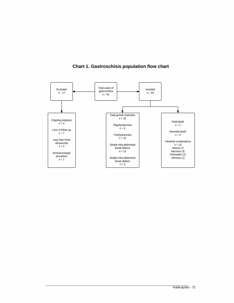
Publicações - 72
Chart 1. Gastroschisis population flow chart
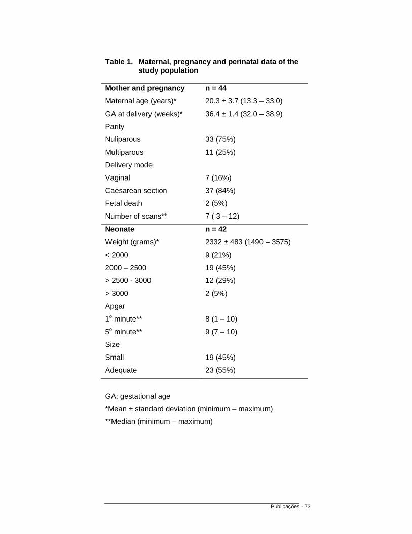
Publicações - 73
Table 1. Maternal, pregnancy and perinatal data of the
study population
Mother and pregnancy n = 44
Maternal age (years)* 20.3 ± 3.7 (13.3 – 33.0)
GA at delivery (weeks)* 36.4 ± 1.4 (32.0 – 38.9)
Parity
Nuliparous 33 (75%)
Multiparous 11 (25%)
Delivery mode
Vaginal 7 (16%)
Caesarean section 37 (84%)
Fetal death 2 (5%)
Number of scans** 7 ( 3 – 12)
Neonate n = 42
Weight (grams)* 2332 ± 483 (1490 – 3575)
< 2000 9 (21%)
2000 – 2500 19 (45%)
> 2500 - 3000 12 (29%)
> 3000 2 (5%)
Apgar
1o minute** 8 (1 – 10)
5o minute** 9 (7 – 10)
Size
Small 19 (45%)
Adequate 23 (55%)
GA: gestational age
*Mean ± standard deviation (minimum – maximum)
**Median (minimum – maximum)
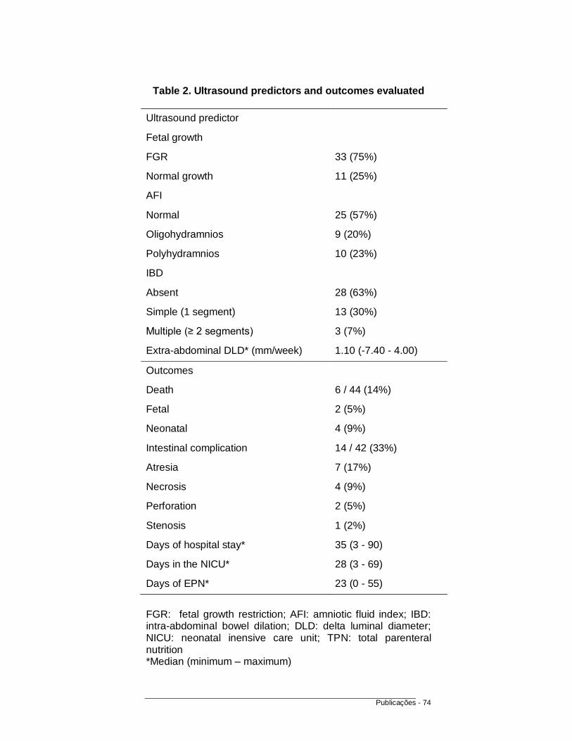
Publicações - 74
Table 2. Ultrasound predictors and outcomes evaluated
Ultrasound predictor
Fetal growth
FGR 33 (75%)
Normal growth 11 (25%)
AFI
Normal 25 (57%)
Oligohydramnios 9 (20%)
Polyhydramnios 10 (23%)
IBD
Absent 28 (63%)
Simple (1 segment) 13 (30%)
Multiple (≥ 2 segments) 3 (7%)
Extra-abdominal DLD* (mm/week) 1.10 (-7.40 - 4.00)
Outcomes
Death 6 / 44 (14%)
Fetal 2 (5%)
Neonatal 4 (9%)
Intestinal complication 14 / 42 (33%)
Atresia 7 (17%)
Necrosis 4 (9%)
Perforation 2 (5%)
Stenosis 1 (2%)
Days of hospital stay* 35 (3 - 90)
Days in the NICU* 28 (3 - 69)
Days of EPN* 23 (0 - 55)
FGR: fetal growth restriction; AFI: amniotic fluid index; IBD: intra-abdominal bowel dilation; DLD: delta luminal diameter; NICU: neonatal inensive care unit; TPN: total parenteral nutrition *Median (minimum – maximum)
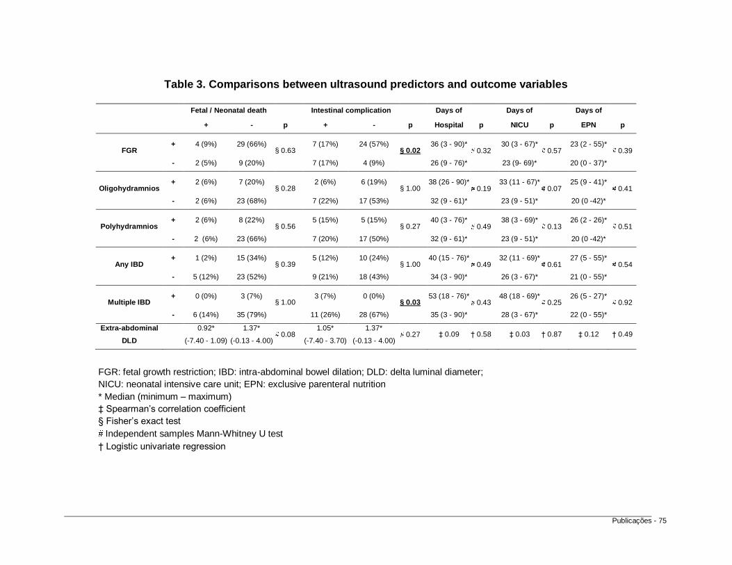
Publicações - 75
Table 3. Comparisons between ultrasound predictors and outcome variables
Fetal / Neonatal death Intestinal complication Days of Days of Days of
+ - p + - p Hospital p NICU p EPN p
FGR + 4 (9%) 29 (66%)
§ 0.63 7 (17%) 24 (57%)
§ 0.02 36 (3 - 90)*
0.32 30 (3 - 67)*
0.57 23 (2 - 55)*
0.39
- 2 (5%) 9 (20%) 7 (17%) 4 (9%) 26 (9 - 76)* 23 (9- 69)* 20 (0 - 37)*
Oligohydramnios + 2 (6%) 7 (20%)
§ 0.28 2 (6%) 6 (19%)
§ 1.00 38 (26 - 90)*
0.19 33 (11 - 67)*
0.07 25 (9 - 41)*
0.41
- 2 (6%) 23 (68%) 7 (22%) 17 (53%) 32 (9 - 61)* 23 (9 - 51)* 20 (0 -42)*
Polyhydramnios + 2 (6%) 8 (22%)
§ 0.56 5 (15%) 5 (15%)
§ 0.27 40 (3 - 76)*
0.49 38 (3 - 69)*
0.13 26 (2 - 26)*
0.51
- 2 (6%) 23 (66%) 7 (20%) 17 (50%) 32 (9 - 61)* 23 (9 - 51)* 20 (0 -42)*
Any IBD + 1 (2%) 15 (34%)
§ 0.39 5 (12%) 10 (24%)
§ 1.00 40 (15 - 76)*
0.49 32 (11 - 69)*
0.61 27 (5 - 55)*
0.54
- 5 (12%) 23 (52%) 9 (21%) 18 (43%) 34 (3 - 90)* 26 (3 - 67)* 21 (0 - 55)*
Multiple IBD + 0 (0%) 3 (7%)
§ 1.00 3 (7%) 0 (0%)
§ 0.03 53 (18 - 76)*
0.43 48 (18 - 69)*
0.25 26 (5 - 27)*
0.92
- 6 (14%) 35 (79%) 11 (26%) 28 (67%) 35 (3 - 90)* 28 (3 - 67)* 22 (0 - 55)*
Extra-abdominal
DLD
0.92*
(-7.40 - 1.09)
1.37*
(-0.13 - 4.00) 0.08
1.05*
(-7.40 - 3.70)
1.37*
(-0.13 - 4.00) 0.27 ‡ 0.09 † 0.58 ‡ 0.03 † 0.87 ‡ 0.12 † 0.49
FGR: fetal growth restriction; IBD: intra-abdominal bowel dilation; DLD: delta luminal diameter;
NICU: neonatal intensive care unit; EPN: exclusive parenteral nutrition
* Median (minimum – maximum)
‡ Spearman’s correlation coefficient
§ Fisher’s exact test
Independent samples Mann-Whitney U test
† Logistic univariate regression
Discussão - 76
4. Discussão geral
Nosso estudo apresentou resultados variados em relação aos descritos na
literatura médica. Para algumas questões nossos achados confirmaram os
relatos de publicações prévias, para outras as conclusões foram opostas ao
esperado e foram levantadas novas hipóteses para perguntas que já pareciam
ter respostas definidas.
No que diz respeito ao papel da ultrassonografia na predição de desfechos
perinatais em casos de gastrosquise, a nossa observação de que a presença
de múltipla DAI no feto associa-se com maior risco de complicações intestinais
pós-natais confirma os resultados de estudos recentes. Esta confirmação é
importante, pois se em fetos sem defeito de fechamento da parede abdominal a
presença de múltipla DAI é considerada fortemente sugestiva de obstrução
intestinal, nos casos de gastroquise este achado poderia ser apenas uma
consequência da hipomotilidade intestinal que com freqüência é observada
nesses pacientes. Nossos resultados confirmam que a presença de múltipla
DAI não é uma achado que pode ser considerado normal em fetos com
gastrosquise e, apesar de pouco sensível, possuiu alta especificidade para a
presença de complicações intestinais no período pós-natal.

Discussão - 77
Um outro fator avaliado que, embora menos específico, também pode estar
associado à obstrução intestinal em fetos sem defeitos da parede abdominal é o
polidrâmnio. Na nossa amostra de fetos com gastrosquise esse sinal não se
correlacionou estatisticamente com nenhum dos desfechos avaliados, mas foi
mais freqüente nos casos de óbito fetal/neonatal e com complicações intestinais
pós-natais. Além disso, os casos de gastrosquise com polidrâmnio tiveram uma
tendência a maior tempo de internação hospitalar, internação em unidade de
terapia intensiva neonatal e nutrição parenteral exclusiva. Desta forma,
achamos que este é um sinal que merece ser mais bem avaliado em estudos
futuros.
Um achado totalmente novo do nosso trabalho foi o efeito protetor que a
RCF apresentou para a ocorrência de complicações intestinais pós-natais.
Como vários estudos associam o baixo peso ao nascimento a maiores taxas de
morbidade e mortalidade pós-natais, o esperado seria que a RCF se associasse
a desfechos perinatais mais desfavoráveis. Conforme discutido em um dos
artigos gerados a partir deste estudo, uma possível explicação seria que a RCF
obervada nesses casos fosse uma conseqüência de uma menor medida da
circunferência abdominal fetal devido a uma maior herniação do conteúdo intra-
abdominal para a cavidade amniótica, já que a fórmula US2D usada para a
estimativa de peso no estudo inclui medidas do abdome fetal. Na nossa opinião
esta hipótese, embora precise ser confirmada em estudos futuros, faz sentido
por dois motivos. Primeiro, as complicações intestinais estudadas (necrose,
atresia, estenose e perfuração) podem ser secundárias a fenômenos
isquêmicos e uma maior herniação do conteúdo intra-abdominal faria com que
Discussão - 78
as alças intestinais sofressem uma menor pressão e assim ficassem menos
sujeitas à isquemia (vale lembrar que a dilatação de alças intestinais é um
fenômeno freqüente nesses casos). Segundo, nossa taxa de RCF foi de 75% e
nossa taxa de baixo peso ao nascimento de 45%, o que reforça a possibilidade
de uma subestimativa do peso devido a uma menor medida da circunferência
abdominal fetal. De qualquer forma, como já mencionado, estas hipóteses
precisam ser mais bem avaliadas em trabalhos futuros.
Um resultado inesperado do trabalho foi o fato do nosso novo modelo US2D
de estimativa do peso fetal ter apresentado um desempenho pior do que o do
modelo US2D sem medidas abdominais proposto por Siemer et al. (2008) e do
que os modelos US2D com medidas abdominais e US3D com o volume da
coxa fetal propostos por Bennini et al. (2010). Esses resultados não eram
esperados, pois dos modelos US2D avaliados a nossa nova fórmula é a única
que incorpora dois fatores que de acordo com a literatura deveriam melhorar o
desempenho na estimativa de peso dos fetos com gastrosquise em relação aos
demais modelos testados: não utiliza medidas abdominais e foi gerada a partir
de pacientes da mesma população dos fetos com gastrosquise. Vale lembrar
que o modelo US3D previamente publicado pelo nosso grupo também foi
gerado a partir de fetos da nossa população e não utiliza medidas abdominais
fetais, mas como não encontramos relato do uso deste tipo de parâmetro
(volume da coxa fetal medido por US3D) na estimativa de peso dos casos de
gastrosquise, o seu desempenho era uma incógnita. Desta forma, a expectativa
era de que o nossa nova formula US2D apresentasse um desempenho no
mínimo semelhante às demais fórmulas avaliadas.
Discussão - 79
As razões para tais achados não nos são claras. Uma hipótese seria que,
como os fetos com gastosquise tendem a ser menores, o impacto de variações
fenotípicas interpopulacionais na predição do peso destes fetos também seria
menor. Além disso, o modelo US2D proposto por Siemer et al. (2008) foi gerado
a partir de uma amostra maior de pacientes e envolvendo somente fetos
prematuros (idade gestacional inferior a 37 semanas), isto também pode ter
contribuído para o melhor desempenho apresentado por esta fórmula quando
comparado à nossa nova fórmula US2D. Uma opção futura seria conseguir um
número de casos suficientes para gerar uma fórmula sem medidas abdominais
geradas a partir de fetos prematuros da nossa população e novamente fazer
esta comparação entre os modelos.
Achamos interessante ressaltar que os nossos modelos US2D e US3D
prévios apresentam bom desempenho tanto em fetos normais como em fetos
com gastrosquise e, portanto, podem ser utilizados em ambas as situações. Isto
facilita a prática diária, pois elimina a necessidade de usar uma fórmula de
estimativa de peso para cada situação.
Apesar de termos atingido o tamanho amostral esperado para a avaliação
da estimativa do peso fetal por meio da ultrassonografia, o número de pacientes
incluídos não permitiu a avaliação do impacto de outras variáveis, tais como tipo
de estruturas herniadas pelo defeito abdominal ou posição do fígado em relação
ao mesmo, nas acurácias das fórmulas estudadas. Pelo mesmo motivo, não foi
possível avaliar adequadamente a sensibilidade e especificidade de cada
modelo no diagnóstico de restrição de crescimento fetal. Este pequeno número
Discussão - 80
de casos se deve ao fato da gastrosquise ser uma doença rara e havermos
utilizado pacientes atendidas em um único centro.
O pequeno tamanho amostral, associado à falta de padronização da
avaliação ultrassonográfica pré-natal do casos de gastrosquise, também limitou
muitas das conclusões em relação à avaliação dos preditores
ultrassonográficos pré-natais e desfechos perinatais.
Desta forma, o estabelecimento de uma colaboração multicêntrica,
preferencialmente com a utlização de protocolos prospectivos com parâmetros
ultrassonográficos claramente definidos a serem avaliados, seria uma solução
para conseguirmos casuísticas maiores e com avaliações padronizadas e assim
esclarecer muitas das dúvidas que ainda pairam sobre o papel da
ultrassonografia pré-natal nos casos de gastrosquise.
Conclusões - 81
5. Conclusão geral
A nova fórmula US2D sem medidas abdominais gerada a partir de
nossos pacientes não melhorou a estimativa do peso ao nascimento de
fetos com gastrosquise da nossa população em relação às outras
fórmulas US2D e US3D já publicadas avaliadas. O modelo US2D de
Siemer et al. (2008) e as fórmulas US2D e US3D de Bennini et al. (2010)
apresentaram os melhores desempenhos na estimativa de peso dos
fetos com gastrosquise da nossa população, não havendo diferença
estatisticamente significativa entre os mesmos.
Em fetos com gastrosquise o achado de DAI múltipla no período pré-
natal aumenta o risco de complicações intestinais (atresia, estenose,
perfuração e necrose) pós-natais e a presença de RCF possui um efeito
protetor para este mesmo desfecho. Não houve relação entre os demais
parâmetros ecográficos pré-natais e os desfechos perinatais avaliados.
Anexos - 82
6. Referências Bibliográficas
Abdullah F, Arnold MA, Nabaweesi R, Fischer AC, Colombani PM, Anderson
KD, Lau H, Chang DC. Gastroschisis in the United States 1988-2003: analysis
and risk categorization of 4344 patients. J Perinatol. 2007 Jan;27(1):50-5.
Abuhamad AZ, Mari G, Cortina RM, Croitoru DP, Evans AT. Superior
mesenteric artery Doppler velocimetry and ultrasonographic assessment of fetal
bowel in gastroschisis: a prospective longitudinal study. Am J Obstet Gynecol.
1997 May;176(5):985-90.
Adams SR, Durfee S, Pettigrew C, Katz D, Jennings R, Ecker J, House M,
Benson CB, Wolfberg A. Accuracy of sonography to predict estimated weight in
fetuses with gastroschisis. J Ultrasound Med. 2012 Nov;31(11):1753-8.
Aina-Mumuney AJ, Fischer AC, Blakemore KJ, Crino JP, Costigan K, Swenson
K, Chisholm CA. A dilated fetal stomach predicts a complicated postnatal course
in cases of prenatally diagnosed gastroschisis. Am J Obstet Gynecol. 2004
May;190(5):1326-30.

Anexos - 83
Alexander GR, Himes JH, Kaufman RB, Mor J, Kogan M. A United States
national reference for fetal growth. Obstet Gynecol 1996; 87:163-8.
Alfaraj MA, Ryan G, Langer JC, Windrim R, Seaward PG, Kingdom J. Does
gastric dilation predict adverse perinatal or surgical outcome in fetuses with
gastroschisis? Ultrasound Obstet Gynecol. 2011 Feb;37(2):202-6.
Alsulyman OM, Monteiro H, Ouzounian JG, Barton L, Songster GS, Kovacs BW.
Clinical significance of prenatal ultrasonographic intestinal dilatation in fetuses
with gastroschisis. Am J Obstet Gynecol. 1996 Oct;175(4 Pt 1):982-4.
Alvarez SM, Burd RS. Increasing prevalence of gastroschisis repairs in the
United States: 1996-2003. J Pediatr Surg 2007;42:943-6.
Arnold MA, Chang DC, Nabaweesi R, et al. Risk stratification of 4344 patients
with gastroschisis into simple and complex categories. J Pediatr Surg
2007;42:1520-5.
Badillo AT, Hedrick HL, Wilson RD, Danzer E, Bebbington MW, Johnson MP,
Liechty KW, Flake AW, Adzick NS. Prenatal ultrasonographic gastrointestinal
abnormalities in fetuses with gastroschisis do not correlate with postnatal
outcomes. J Pediatr Surg. 2008 Apr;43(4):647-53.

Anexos - 84
Baird PA, MacDonald EC. An epidemiologic study of congenital malformations
of the anterior abdominal wall in more than half a million consecutive live births.
Am J Hum Genet 1981;33:470-8.
Bennini JR, Marussi EF, Barini R, Faro C, Peralta CF. Birth-weight prediction by
two- and three-dimensional ultrasound imaging. Ultrasound Obstet Gynecol.
2010 Apr;35(4):426-33.
Bianchi A, Dickson AP, Alizai NK. Elective delayed midgut reduction-No
anesthesia for gastroschisis: Selection and conversion criteria. J Pediatr Surg
2002 Sep;37(9):1334-6.
Brantberg A, Blaas HG, Salvesen KA, Haugen SE, Eik-Nes SH. Surveillance
and outcome of fetuses with gastroschisis. Ultrasound Obstet Gynecol. 2004
Jan;23(1):4-13.
Brown BW, Russel K. Methods of correcting for multiple testing: operating
characteristics. Stat Med 1997; 16:2511-28.
Bucher BT, Mazotas IG, Warner BW, Saito JM. Effect of time to surgical
evaluation on the outcomes of infants with gastroschisis. J Pediatr Surg. 2012
Jun;47(6):1105-10.
Anexos - 85
Castilla EE, Mastroiacovo P, Orioli IM. Gastroschisis: international epidemiology
and public health perspective. Am J Med Genet 2008;148C:162-79.
Charlesworth P, Njere I, Allotey J, Dimitrou G, Ade-Ajayi N, Devane S,
Davenport M. Postnatal outcome in gastroschisis: effect of birth weight and
gestational age. J Pediatr Surg. 2007 May;42(5):815-8.
Chaudhury P, Haeri S, Horton AL, Wolfe HM, Goodnight WH. Ultrasound
prediction of birthweight and growth restriction in fetal gastroschisis. Am J
Obstet Gynecol. 2010 Oct;203(4):395.e1-5.
Chen IL, Lee SY, Ou-Yang MC, Chao PH, Liu CA, Chen FS, Chung MY, Chen
CC, Huang HC. Clinical presentation of children with gastroschisis and small for
gestational age. Pediatr Neonatol. 2011 Aug;52(4):219-22.
Christison-Lagay ER, Kelleher CM, Langer JC. Neonatal abdominal wall defects.
Semin Fetal Neonatal Med. 2011 Jun;16(3):164-72.
Clark RH, Walker MW, Gauderer MW. Factors associated with mortality in
neonates with gastroschisis. Eur J Pediatr Surg. 2011 Jan;21(1):21-4.
Contro E, Fratelli N, Okoye B, Papageorghiou A, Thilaganathan B, Bhide A.
Prenatal ultrasound in the prediction of bowel obstruction in infants with
gastroschisis. Ultrasound Obstet Gynecol. 2010 Jun;35(6):702-7.
Anexos - 86
Coughlin JP, Drucker DE, Jewell MR, Evans MJ, Klein MD. Delivery room repair
of gastroschisis. Surgery. 1993 Oct;114(4):822-6; discussion 826-7.
Cullen MT, Green J, Whetham J, Salafia C, Gabrielli S, Hobbins JC.
Transvaginal ultrasonographic detection of congenital anomalies in the first
trimester. Am J Obstet Gynecol. 1990 Aug;163(2):466-76.
David AL, Tan A, Curry J. Gastroschisis: sonographic diagnosis, associations,
management and outcome. Prenat Diagn. 2008 Jul;28(7):633-44.
deVries PA. The pathogenesis of gastroschisis and omphalocele. J Pediatr Surg
1980;15:245-51.
Duhamel B. Embryology of Exomphalos and Allied Malformations. Arch Dis
Child. 1963 Apr;38(198):142-7.
Feldkamp ML, Carey JC, Sadler TW. Development of gastroschisis: review of
hypotheses, a novel hypothesis, and implications for research. Am J Med Genet
A 2007;143:639-52.
Fleiss JL, Tytun A, Ury HK. A simple approximation for calculating sample sizes
for comparing independent proportions. Biometrics. 1980;36(2):343-6.

Anexos - 87
Furlan EL, Bennini JR, Faro CB, Marussi EF, Barini R, Peralta CF. [Elaboration
and validation of longitudinal reference intervals of fetal weight with a sample of
the Brazilian population]. Rev Bras Ginecol Obstet. 2012 Oct;34(10):466-72.
Garcia L, Brizot M, Liao A, Silva MM, Tannuri AC, Zugaib M. Bowel dilation as a
predictor of adverse outcome in isolated fetal gastroschisis. Prenat Diagn.
2010Oct;30(10):964.
Garne E, Loane M, Dolk H, De Vigan C, Scarano G, Tucker D, Stoll C, Gener B,
Pierini A, Nelen V, Rösch C, Gillerot Y, Feijoo M, Tincheva R, Queisser-Luft A,
Addor MC, Mosquera C, Gatt M, Barisic I. Prenatal diagnosis of severe
structural congenital malformations in Europe. Ultrasound Obstet Gynecol. 2005
Jan;25(1):6-11.
Guibourdenche J, Berrebi D, Vuillard E, de Lagausie P, Aigrain Y, Oury JF,
Luton D. Biochemical investigations of bowel inflammation in gastroschisis.
Pediatr Res. 2006 Nov;60(5):565-8.
Hackshaw A, Rodeck C, Boniface S. Maternal smoking in pregnancy and birth
defects: a systematic review based on 173 687 malformed cases and 11.7
million controls. Hum Reprod Update. 2011 Sep-Oct;17(5):589-604.
Anexos - 88
Honarvar M, Allahyari M, Dehbashi S. Assessment of fetal weight based on
ultrasonic femur length after the second trimester. Int J Gynaecol Obstet. 2001
Apr;73(1):15-20.
Hoyme HE, Higginbottom MC, Jones KL. The vascular pathogenesis of
gastroschisis: intrauterine interruption of the omphalomesenteric artery. J
Pediatr 1981;98:228-31.
Huh NG, Hirose S, Goldstein RB. Prenatal intraabdominal bowel dilation is
associated with postnatal gastrointestinal complications in fetuses with
gastroschisis. Am J Obstet Gynecol. 2010 Apr;202(4):396.e1-6.
Japaraj RP, Hockey R, Chan FY. Gastroschisis: can prenatal sonography
predict neonatal outcome? Ultrasound Obstet Gynecol. 2003 Apr;21(4):329-33.
Kronfli R, Bradnock TJ, Sabharwal A. Intestinal atresia in association with
gastroschisis: a 26 year review. J Pediatr Surg 2010;26:891-4.
Kuleva M, Khen-Dunlop N, Dumez Y, Ville Y, Salomon LJ. Is complex
gastroschisis predictable by prenatal ultrasound? BJOG. 2012 Jan;119(1):102-9.

Anexos - 89
Langer JC, Longaker MT, Crombleholme TM, Bond SJ, Finkbeiner WE, Rudolph
CA, Verrier ED, Harrison MR. Etiology of intestinal damage in gastroschisis. I:
Effects of amniotic fluid exposure and bowel constriction in a fetal lamb model. J
Pediatr Surg. 1989 Oct;24(10):992-7.
Langer JC, Bell JG, Castillo RO, Crombleholme TM, Longaker MT, Duncan BW,
Bradley SM, Finkbeiner WE, Verrier ED, Harrison MR. Etiology of intestinal
damage in gastroschisis, II. Timing and reversibility of histological changes,
mucosal function, and contractility. J Pediatr Surg. 1990 Nov;25(11):1122-6.
Langer JC, Khanna J, Caco C, Dykes EH, Nicolaides KH. Prenatal diagnosis of
gastroschisis: development of objective sonographic criteria for predicting
outcome. Obstet Gynecol. 1993 Jan;81(1):53-6.
Lausman AY, Langer JC, Tai M, Seaward PG, Windrim RC, Kelly EN, Ryan G.
Gastroschisis: what is the average gestational age of spontaneous delivery? J
Pediatr Surg. 2007 Nov;42(11):1816-21.
Liu RX, Kuang J, Gong Q, Hou XL. Principal component regression analysis
with SPSS. Comput Methods Programs Biomed 2003; 71:141-7.
Anexos - 90
Loane M, Dolk H, Bradbury I; EUROCAT Working Group. Increasing prevalence
of gastroschisis in Europe 1980-2002: a phenomenon restricted to younger
mothers? Paediatr Perinat Epidemiol. 2007 Jul;21(4):363-9.
Louw JH, Barnard CN. Congenital intestinal atresia; observations on its origin.
Lancet 1955;269:1065-7.
Ludbrook J. Multiple comparison procedures update. Clin Exp Pharmacol
Physiol 1998; 25:1032-7.
Luton D, de Lagausie P, Guibourdenche J, Oury J, Sibony O, Vuillard E,
Boissinot C, Aigrain Y, Beaufils F, Navarro J, Blot P. Effect of amnioinfusion on
the outcome of prenatally diagnosed gastroschisis. Fetal Diagn Ther. 1999 May-
Jun;14(3):152-5.
McClellan EB, Shew SB, Lee SS, Dunn JC, Deugarte DA. Liver herniation in
gastroschisis: incidence and prognosis. J Pediatr Surg. 2011 Nov;46(11):2115-
8.
Molik KA, Gingalewski CA, West KW, Rescorla FJ, Scherer LR, Engum SA,
Grosfeld JL. Gastroschisis: a plea for risk categorization. J Pediatr Surg. 2001
Jan;36(1):51-5.
Anexos - 91
Mongelli M, Gardosi J. Gestation-adjusted projection of estimated fetal weight.
Acta Obstet Gynecol Scand. 1996 Jan;75(1):28-31.
Mousty E, Chalouhi GE, Sabbagh AE, Khen-Dunlop N, Kuleva M, Salomon LJ,
Ville Y. Secondary bladder herniation in isolated gastroschisis justifies increased
surveillance. Prenat Diagn. 2012 Sep;32(9):888-92.
Nasr A, Langer JC. Influence of location of delivery on outcome in neonates with
gastroschisis. APSA 42nd Annual Meeting, Palm Desert, CA, USA, 22-25 May
2011.
Netta DA, Wilson RD, Visintainer P, Johnson MP, Hedrick HL, Flake AW, Adzick
NS. Gastroschisis: growth patterns and a proposed prenatal surveillance
protocol. Fetal Diagn Ther. 2007;22(5):352-7.
Nicholas SS, Stamilio DM, Dicke JM, Gray DL, Macones GA, Odibo AO.
Predicting adverse neonatal outcomes in fetuses with abdominal wall defects
using prenatal risk factors. Am J Obstet Gynecol. 2009 Oct;201(4):383.e1-6.
Nicholas S, Tuuli MG, Dicke J, Macones GA, Stamilio D, Odibo AO. Estimation
of fetal weight in fetuses with abdominal wall defects: comparison of 2 recent
sonographic formulas to the Hadlock formula. J Ultrasound Med. 2010
Jul;29(7):1069-74.
Anexos - 92
Nick AM, Bruner JP, Moses R, Yang EY, Scott TA. Second-trimester intra-
abdominal bowel dilation in fetuses with gastroschisis predicts neonatal bowel
atresia. Ultrasound Obstet Gynecol. 2006 Nov;28(6):821-5.
Phelan JP, Ahn MO, Smith CV, Rutherford SE, Anderson E. Amniotic fluid index
measurements during pregnancy. J Reprod Med. 1987 Aug;32(8):601-4.
Piper HG, Jaksic T. The impact of prenatal bowel dilation on clinical outcomes in
neonates with gastroschisis. J Pediatr Surg. 2006 May;41(5):897-900.
Pitman EJG. A note on normal correlation. Biometrika 1939; 31: 9-12.
Rasmussen SA, Frias JL. Non-genetic risk factors for gastroschisis. Am J Med
Genet 2008;148C:199-212.
Richardson S, Browne ML, Rasmussen SA, Druschel CM, Sun L, Jabs EW,
Romitti PA; National Birth Defects Prevention Study. Associations between
periconceptional alcohol consumption and craniosynostosis, omphalocele, and
gastroschisis. Birth Defects Res A Clin Mol Teratol. 2011 Jul;91(7):623-30.
Robinson HP, Fleming JE. A critical evaluation of sonar "crown-rump
length"measurements. Br J Obstet Gynaecol. 1975 Sep;82(9):702-10.
Anexos - 93
Sadler TW. The embryologic origin of ventral body wall defects. Semin Pediatr
Surg. 2010 Aug;19(3):209-14.
Salihu HM, Emusu D, Aliyu ZY, Pierre-Louis BJ, Druschel CM, Kirby RS. Mode
of delivery and neonatal survival of infants with isolated gastroschisis. Obstet
Gynecol. 2004 Oct;104(4):678-83.
Segel SY, Marder SJ, Parry S, Macones GA. Fetal abdominal wall defects and
mode of delivery: a systematic review. Obstet Gynecol. 2001 Nov;98(5 Pt
1):867-73.
Shaw A. The myth of gastroschisis. J Pediatr Surg 1975;10:235-44.
Siemer J, Hilbert A, Hart N, Hoopmann M, Schneider U, Girschick G, Müller A,
Schild RL. Specific weight formula for fetuses with abdominal wall defects.
Ultrasound Obstet Gynecol. 2008 Apr;31(4):397-400.
Snyder CL. Outcome analysis for gastroschisis. J Pediatr Surg. 1999
Aug;34(8):1253-6.
Srinathan SK, Langer JC, Blennerhassett MG, Harrison MR, Pelletier GJ,
Lagunoff D. Etiology of intestinal damage in gastroschisis. III: Morphometric
analysis of the smooth muscle and submucosa. J Pediatr Surg. 1995
Mar;30(3):379-83.

Anexos - 94
Stevenson RE, Rogers RC, Chandler JC, Gauderer MWL, Hunter AGW. Escape
of the yolk sac: a hypothesis to explain the embryogenesis of gastroschisis. Clin
Genet 2009;75:326-33.
Vargun R, Aktug T, Heper A, Bingol-kologlu M. Effects of intrauterine treatment
on interstitial cells of Cajal in gastroschisis. J Pediatr Surg. 2007 May;42(5):783-7.
Wales PW, Christison-Lagay ER. Short bowel syndrome: epidemiology and
etiology. Semin Pediatr Surg. 2010 Feb;19(1):3-9.
Werler MM, Sheehan JE, Mitchell AA. Association of vasoconstrictive exposures
with risks of gastroschisis and small intestinal atresia. Epidemiology
2003;14:349-54.
Werler MM, Mitchell AA, Moore CA, Honein MA. Is there epidemiologic evidence
to support vascular disruption as a pathogenesis of gastroschisis? Am J Med
Genet 2009;149A:1399-406.
Wilson RD, Johnson MP. Congenital abdominal wall defects: an update. Fetal
Diagn Ther. 2004 Sep-Oct;19(5):385-98.
Anexos - 95
7. Anexos
7.1. Anexo 1 - Parecer da Comissão de Pesquisa do DTG/CAISM
Anexos - 96
7.2. Anexo 2 - Aprovação do projeto no CEP
Anexos - 97
Anexos - 98
Anexos - 99
7.3. Anexo 3 - Ficha de coleta de dados
Projeto de pesquisa: “Gastrosquise: relação entre parâmetros
ultrassonográficos pré-natais e a evolução pós-natal”
Pesquisador responsável: Dr. João Renato Bennini Júnior
Caso no:________ HC no: ________ Data da coleta dos dados: _____/_____/_____
DADOS DA GESTANTE
Nome: _____________________ Sobrenome: _____________________________
Data de nascimento: _______/_______/_______ Idade: _______
G: _____ PN: ____ C: ____ A: ________
DADOS DA ULTRASSONOGRAFIA
Data:______/_______/_______ Hora: _______:_______
DBP:____ OF:____ CC:_____ DAT:_____ DAAP:______ CA:______ CF:______
Volume da coxa:__________
Peso fetal estimado: ________ ILA (cm): _________ Oligo/Polidrâmnio: ________
Maior calibre de alça: extra-abdominal: __________ intra-abdominal: __________

Anexos - 100
DADOS DO PARTO
Data: ____/____/____ Hora: _______:_______ Tipo de parto: PN: C: F:
Peso do RN: ____________________ AIG: PIG: GIG:
Obs: ____________________________________________________________
DADOS PÓS-NATAIS
Complicações intestinais: ________________________
Tipo da complicação: ____________________________
Tempo UTI neonatal:_________________
Tempo nutrição enteral total:___________
Tempo de internação:_________________

Anexos - 101
7.4. Anexo 4 - Artigo: “Birth-weight prediction by two- and three-dimensional ultrasound imaging”
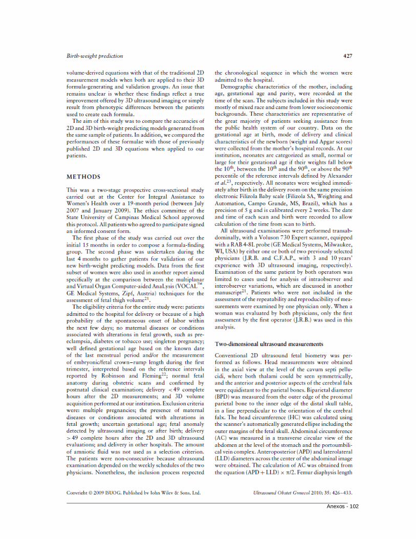
Anexos - 102
Anexos - 103

Anexos - 104
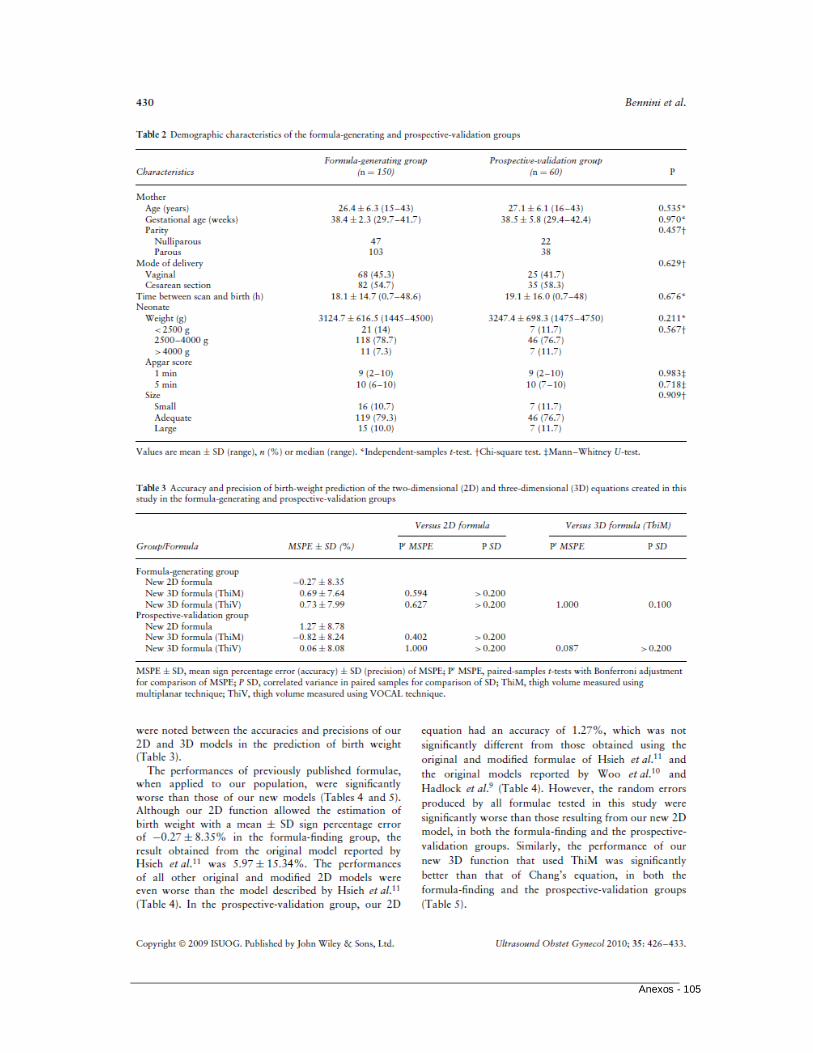
Anexos - 105

Anexos - 106

Anexos - 107
Anexos - 108


