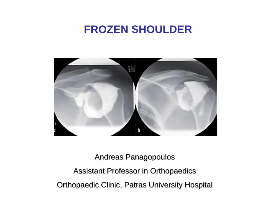
FROZEN SHOULDER
Andreas Panagopoulos
Assistant Professor in Orthopaedics
Orthopaedic Clinic, Patras University Hospital
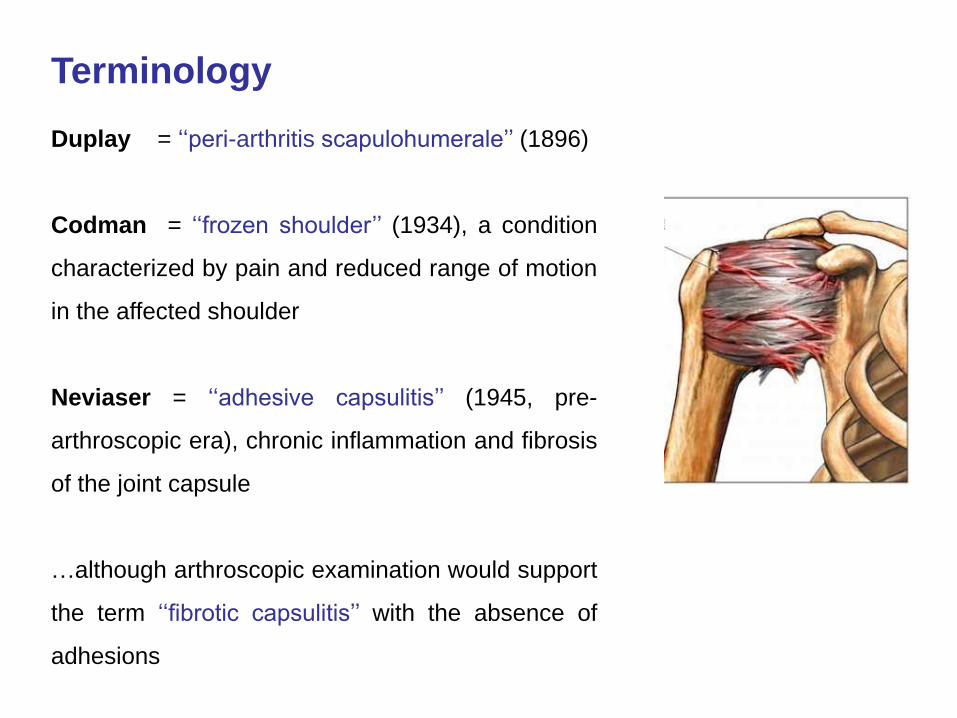
Duplay = ‘‘peri-arthritis scapulohumerale’’ (1896)
Codman = ‘‘frozen shoulder’’ (1934), a condition
characterized by pain and reduced range of motion
in the affected shoulder
Neviaser = ‘‘adhesive capsulitis’’ (1945, pre-
arthroscopic era), chronic inflammation and fibrosis
of the joint capsule
…although arthroscopic examination would support
the term ‘‘fibrotic capsulitis’’ with the absence of
adhesions
Terminology

Definition
….Condition difficult to define, difficult to treat and difficult
to explain from the point of view of pathology...
Codman 1934
- a condition of uncertain cause
- characterized by spontaneous onset of pain
- with significant restriction of both active and passive
range of motion
ASES: Frozen shoulder is

Classification
Frozen shoulder TERTIARY - postoperative
- post-fracture
SECONDARY (known disorders) PRIMARY (idiopathic)
Systemic
- diabetes mellitus
- hypothyroidism
- hyperthyroidism
- hypoadrenalism
Extrinsic
- cardio-pulmonary disease
- cervical discopathy
- cerebrovascular accident
- humerus fractures
- Parkinson’s disease
Intrinsic
- rotator cuff tendinitis
- rotator cuff tears
- biceps tendinitis
- calcific tendinitis
- AC joint arthritis

Conditions associated with adhesive capsulitis
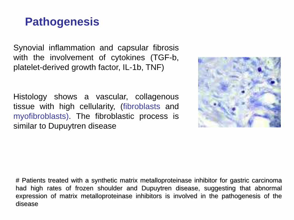
Pathogenesis
Synovial inflammation and capsular fibrosis
with the involvement of cytokines (TGF-b,
platelet-derived growth factor, IL-1b, TNF)
Histology shows a vascular, collagenous
tissue with high cellularity, (fibroblasts and
myofibroblasts). The fibroblastic process is
similar to Dupuytren disease
# Patients treated with a synthetic matrix metalloproteinase inhibitor for gastric carcinoma
had high rates of frozen shoulder and Dupuytren disease, suggesting that abnormal
expression of matrix metalloproteinase inhibitors is involved in the pathogenesis of the
disease
Classification
3 phases, Reeves, Scand J Reumatology, 1975
Phase I = pain with progressive stiffness (2-9 months)
Phase II = progressive stiffness with contractions (4-12 months)
Phase III = recovery, motion gradually improves (12-42 months)

Classification
4 stages, Hannefin & Chiaia, Clin Orthop 2000
Stage I = ‘painful stage’: pain with movements, but no loss of motion (3 m)
Arthroscopy: diffuse synovitis of the anterosuperior capsule
Stage II = ‘freezing stage’: pain and range of motion (3-9 m)
Arthroscopy: diffuse synovitis
Stage III = ‘frozen stage’: minimal pain except at extremes (9-15 m)
Arthroscopy: thickened, fibrotic capsule with no hypervascularity
Stage III = ‘thawing stage’: minimal pain an improve of motion (15-24 m)
Arthroscopy: almost normal

Pathology- Predisposing factors
- Intra-articular breakdown of the biceps tendon
- Contracture of the subscapularis
- Autoimmune basis ( HLA B27, IgA, CRP)
- Active trigger points
- Neurological dysfunction (like RSD syndrome)
- serum lipid levels (triglyceride & cholesterol)
- Endocrine disorders (diabetes mellitus)
- Trivial trauma (especially after prolonged immobilization)
- Psychological factors

Trigger points are locally tender, hyperirritable foci located in the
skeletal muscles or fascia, related to a zone of referred pain when
they are stimulated
The subscapularis trigger point exert an influence on the
sympathetic vasomotor activity, leading to hypoxia of the
periarticular tissues. The hypoxia leads to local proliferation of
fibrous tissue about the shoulder capsule, resulting in the clinical
picture of frozen shoulder.
Travel JG & Simmons DG, 1983
Active trigger points
Pathology- Predisposing factors
Endocrine disorders (diabetes mellitus)
Incidence of about 10.8%, instead of 2.3% in the general population
Abnormal glucose tolerance in 28% of patients with frozen shoulder
Excessive glucose concentration in diabetic patients can lead to a faster rate of collagen
glycosylation and cross-linking in the shoulder capsule, restricting shoulder range of motion.
This collagen cross-linking may also be responsible for the higher incidence of Dupuytren
contractures and trigger finger in diabetic patients
Thyroid disorders, hypoadrenalism, corticotropin deficiency
Pathology- Predisposing factors

Psychological factors
Certain personality structure? (periarthritic personality)
- patients unable to tolerate pain
- expect others to get them well
- refuse to contribute to their management
Physiological characteristics must be considered as a
secondary factor in the management of these patients
Pathology- Predisposing factors

Epidemiology
2% cumulative risk for at least one episode of FS
Between forth and fifth decade of life
More common in women
Non-dominant extremity is usually involved
Bilateral involvement occurs in 6-50% but only 14% simultaneously
Among diabetic patients bilateral involvement is present in 77%
The same shoulder is rarely involved again with FS
Clinical evaluation
Pain is critical in FS and is expressed at night, with dressing and
daily activities as with common use of the arm
Early on patients describe an intense burning pain compared to
a dull fullness during the contracting stage
Patients report difficulties to put on a coat or fastening a bra
Most significant loss of motion is with external rotation
Sharp pain at the endpoint of restricted shoulder motion
Specific medication: barbiturates, antituberculosis agents,
protease inhibitors (for HIV treatment)
History
Clinical evaluation
Commonly confused with RC pathology
Typically pain on palpation at deltoid, deep capsule anteriorly
Loss of active or passive range of motion
Pain present at the extremes of motion
Complete evaluation of cervical spine
Lidocaine injection test – no improvement
Physical examination

Clinical evaluation
Physical examination
Clinical evaluation
Differential diagnosis
• Osteoarthritis
• Avascular Necrosis
• Rotator Cuff Disease
• Cervical Radiculopathy
• Biceps Tendiniits
• Subacromial Bursitis
• Thoracic Outlet Syndrome
• Brachial Plexopathy
• Humeral Fracture
• Tumor

Clinical records of 34 patients (age > 40) with
malignant shoulder tumors and those of 505
patients (age > 40) with shoulder pain and
stiffness were reviewed
9/34 tumor patients, (26%) had been initially
misdiagnosed with FS syndrome.
Among 505 patients with shoulder pain and
stiffness, 4 (0.8%) were diagnosed later as
having malignant tumors
In 10 patients, initial misdiagnosis as frozen shoulder syndrome did
cause a significant delay to reach the correct diagnosis as malignant
tumors
Imagine studies
Complete set of radiographs (AP in internal and external rotation,
axillary and supraspinatus outlet view) to exclude other pathology
In FS usually x-rays are normal, disuse osteopenia maybe present
Technetium bone scan exhibits increase uptake (hypervascularity)
Ultrasound can show thickening of coracohumeral ligament
Shoulder arthrography:
- decreased joint volume (10-12 ml, instead of – 15 ml normally)
- lack of filling of the axillary fold & subscapular bursa
Imagine studies
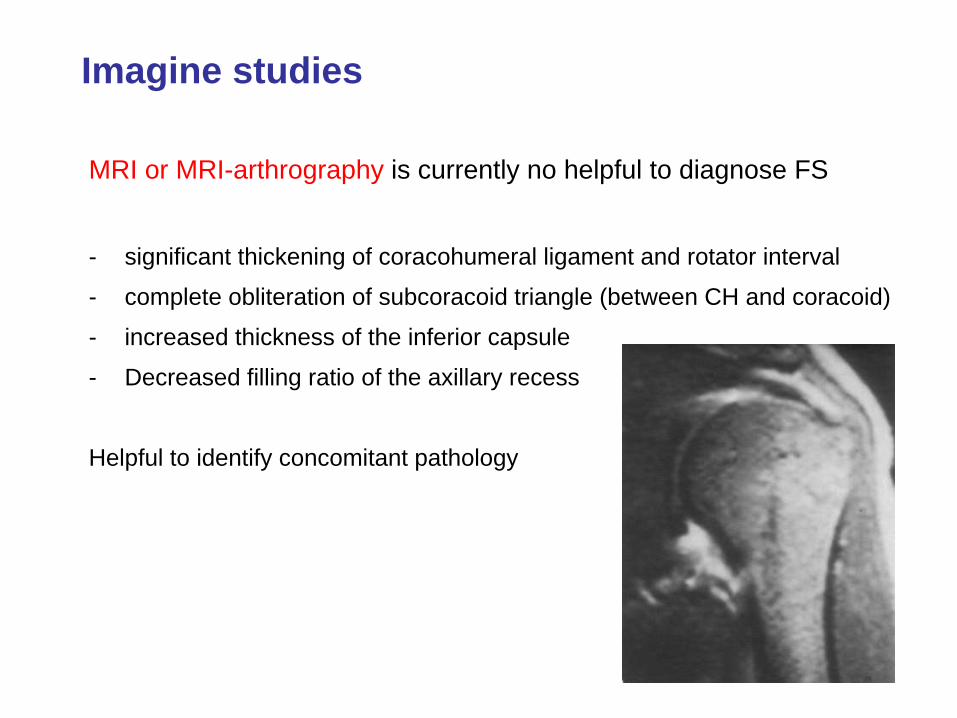
MRI or MRI-arthrography is currently no helpful to diagnose FS
- significant thickening of coracohumeral ligament and rotator interval
- complete obliteration of subcoracoid triangle (between CH and coracoid)
- increased thickness of the inferior capsule
- Decreased filling ratio of the axillary recess
Helpful to identify concomitant pathology
Laboratory studies
Are usually normal
ECR and CRP are usually elevated
Glucose tolerance, lipid panel
Rheumatoid factor and antinuclear antibody
Thyroid and autoimmune disorders
Treatment
Goal of treatment is relief of pain and restore motion and function
Avoid misdiagnosis of other shoulder pathology
Surgery addressing other pathology may dramatically worsen the
pain and stiffness of frozen shoulder
Individualized program of rehabilitation based on severity and
chronicity of patients symptoms
Non-steroidal anti-inflammatory agents, oral corticosteroids, corticosteroids
injections, transcutaneous electrical nerve stimulation (TENS) etc
General principles

Treatment
Non-operative treatment - Benign neglect
Capsular distention (Brisement)
Manipulation under anaesthesia
Arthroscopic capsular release
Open release
Acupuncture therapy, botulinum injections
Options

Non-operative treatment
Indicated in patients with less than 6 months of stiffness
Active-assisted ROM exercises plus gentle passive stretching
Better 5-6 times per day lasting 10-15 minutes
Daily bar charts to document progress
Constant reassurance to promote continue compliance
Exercise protocol

Non-operative treatment
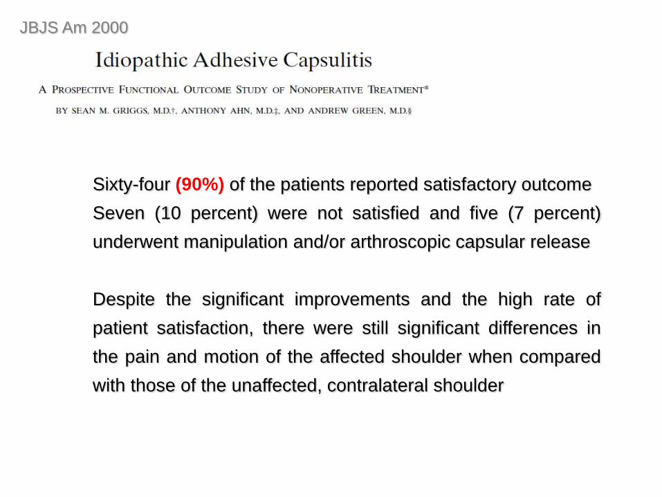
JBJS Am 2000
Sixty-four (90%) of the patients reported satisfactory outcome
Seven (10 percent) were not satisfied and five (7 percent)
underwent manipulation and/or arthroscopic capsular release
Despite the significant improvements and the high rate of
patient satisfaction, there were still significant differences in
the pain and motion of the affected shoulder when compared
with those of the unaffected, contralateral shoulder

In the patients treated with supervised neglect, 89% had normal or near-
normal painless shoulder function (Constant score 80) at the end of the
observation period. In contrast, of the group receiving intensive physical
therapy treatment, only 63% reached a Constant score of 80 or higher
after 24 months.
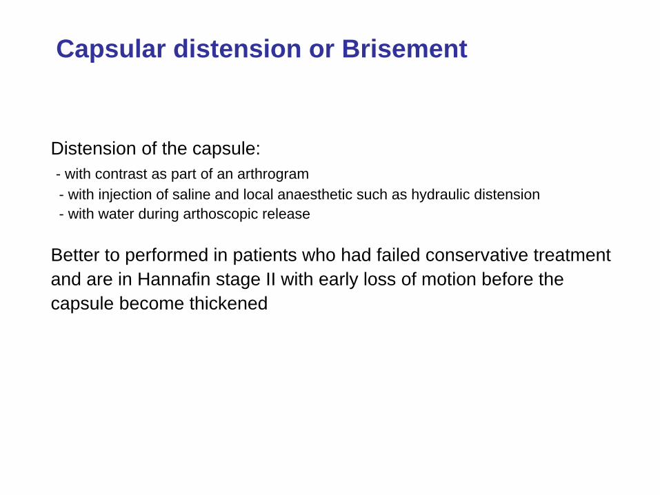
Capsular distension or Brisement
Distension of the capsule:
- with contrast as part of an arthrogram
- with injection of saline and local anaesthetic such as hydraulic distension
- with water during arthoscopic release
Better to performed in patients who had failed conservative treatment
and are in Hannafin stage II with early loss of motion before the
capsule become thickened

In the performance of diagnostic arthrography,
we observed a clinical improvement of painful
symptoms
The analysis of the outcome observed in 200
joint distensions showed that, in 85% of cases,
the results were satisfactory, with the
disappearance of painful symptoms after 45 days
and an almost complete recuperation of the
range of motion of the shoulder

Manipulation under anaesthesia
Mainstay of interventional methods
Usually after failed physiotherapy for 3 to 6 months
Wait until pain is present only at the extremes of ROM
Better with combined general and regional anesthesia
Gentle, controlled fashion with the patient supine
Avoid in elderly osteoporotic patients
No significant difference with use of corticosteroids
Risk for fractures, dislocation of the head, RC injury,
SLAP lesions and nerve injuries

Arthroscopic capsular release
3 basic components:
Anesthesia (general? & interscalene block)
Manipulation (before, during or after)
Arthroscopic release
- rotator interval division (CHL and anterosuperior capsule)
- anterior capsule over subscapularis (MGL)
- inferior capsule (risk for axillary nerve)
- superior and posterior capsular release
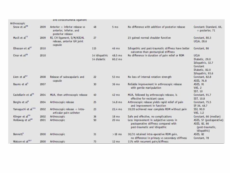

Release of rotator interval stops when CA ligament is viewed from posterior
Anterior capsule release posterior capsule release

P.G. 56 y old, 5 months of physical therapy, MUA and anterior-inferior
release, Constant score 84 at 16 months follow-up

We have shown an overall rapid significant improvement following
arthroscopic capsular release for primary and secondary frozen
shoulder.
There was no significant difference in the overall outcome with the
addition of a posterior release

In this broad group of patients with recalcitrant adhesive
capsulitis, the addition of the posterior capsular release did not
improve patient function or ROM over anterior capsular release
alone at 6 months.
74 consecutive patients with refractory frozen shoulder
underwent arthroscopic capsular release and were divided
into 2 groups randomly. The release of anterior capsular
structures, including the anterior band of the IGHL, was
performed in group 1. In group 2 the release extended
inferiorly and posteriorly.
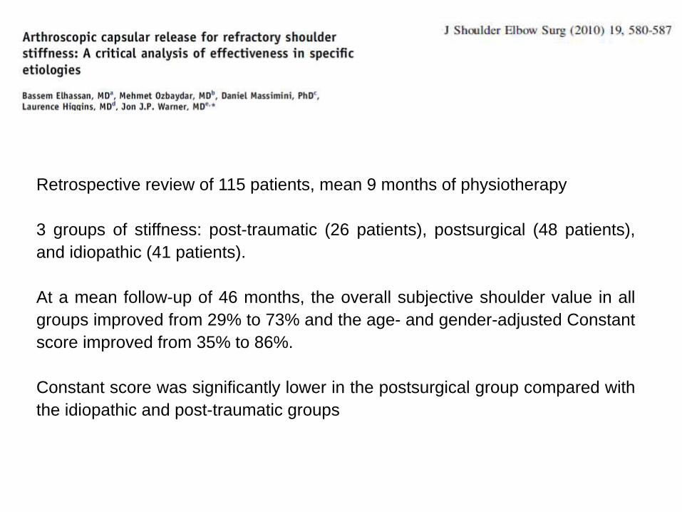
Retrospective review of 115 patients, mean 9 months of physiotherapy
3 groups of stiffness: post-traumatic (26 patients), postsurgical (48 patients),
and idiopathic (41 patients).
At a mean follow-up of 46 months, the overall subjective shoulder value in all
groups improved from 29% to 73% and the age- and gender-adjusted Constant
score improved from 35% to 86%.
Constant score was significantly lower in the postsurgical group compared with
the idiopathic and post-traumatic groups

Open release
Reserved for patients who have failed
manipulation and/or arthroscopic release
FZ in the setting of shoulder arthroplasty
Offers direct visualization, but increase postop
pain wich interfere with physiotherapy
Release include:
- subacromial and subdeltoid adhesions
- CHL and rotator interval
- perilabral capsular release
- subscapularis release and lengthening
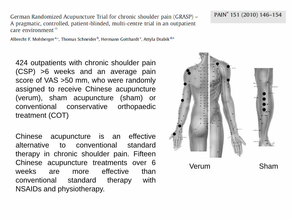
424 outpatients with chronic shoulder pain
(CSP) >6 weeks and an average pain
score of VAS >50 mm, who were randomly
assigned to receive Chinese acupuncture
(verum), sham acupuncture (sham) or
conventional conservative orthopaedic
treatment (COT)
Chinese acupuncture is an effective
alternative to conventional standard
therapy in chronic shoulder pain. Fifteen
Chinese acupuncture treatments over 6
weeks are more effective than
conventional standard therapy with
NSAIDs and physiotherapy.
Verum Sham

Possible mechanisms of BTX in treatment of FS
Through inhibition of neurotransmitter release
Through inhibition of C-fiber nociceptive transmission
Through inhibition of fibrosis
The analgesic effect can be expected to last longer than steroid
injection. The use is generally safe with minimal side effects.
Preliminary results in intra-articular use showed promising outlooks.


![Electrotherapy modalities for adhesive capsulitis [frozen shoulder] · 2018-06-06 · Electrotherapy modalities for adhesive capsulitis (frozen shoulder) Background Frozen shoulder](https://cdn.vdocuments.site/doc/165x107/5e5150f4b27e9736145a78b5/electrotherapy-modalities-for-adhesive-capsulitis-frozen-shoulder-2018-06-06.jpg)


