
FATTY INFILTRATION AND CUFF ATROPHY
Degenerative muscular changes associated with rotator cuff tears include fatty
infiltration and atrophy. Increased fatty infiltration and rotator cuff atrophy are
associated with increased re-tear rates as well as poorer functional outcomes
following rotator cuff repair.
PATHOPHYSIOLOGY
The etiology of fatty infiltration and cuff atrophy is complex is not fully understood.
Both mechanical unloading and denervation likely play a role in the development of
Muscle atrophy and fatty infiltration. Retraction of the torn rotator cuff tendon may
be a factor in the development of supraspinatus fatty infiltration.
Infraspinatus fatty degeneration was more closely associated with tear size and
retraction [1]. Goutallier noted that infraspinatus lesions were associated with
poorer functional outcomes and external rotation. The location of the supraspinatus
tears was more important than tear size or retraction in the degree of fatty
degeneration. The odds of having fatty degeneration decreased significantly the
farther the tear was from the biceps tend [2]
The natural history of fatty infiltration is described as irreversible and, usually,
progressive. In general, the natural history of fatty infiltration is one of progression.
In a retrospective review of 1,688 shoulder MRI and CT studies, Melis [1] found
moderate supraspinatus fatty infiltration (Goutallier stage 2) an average of 3 years
after onset of shoulder symptoms (traumatic tears). Onset of moderate fatty
infiltration was noted earlier in traumatic RCTs than in chronic tears (34.8 mo [P =
0.04] and 54.1 months. Severe infiltration was noted an average of 57.7 months
after traumatic tears (P = 0.04) and 83.9 months after chronic progressive tears (P =
0.003). Patients had a positive tangent sign indicating severe atrophy at an average
of 4.5 years after symptom onset (P = 0.001).
The natural history of massive RCTs is paradoxical. Zingg [1]retrospectively
evaluated the natural history of fatty infiltration in 19 shoulders with massive RCTs

at an average of 48 months after diagnosis. They noted a significant increase
in tear size and glenohumeral arthritis (P = 0.014) as well as decreased
acromiohumeral distance. Fatty infiltration increased by approximately one stage
in all three muscles. Even so, patients maintained satisfactory shoulder function [3]
FACTS ABOUT FATTY INFILTRATION/ATROPHY:
1. Size of the tear: Proportional to size of the tear . When fatty infiltration was
minimal (stage 0 or 1) as in small tears: Patients with massive Cuff tear and severe
stage 3 or 4 fatty infiltration of the infraspinatus and teres minor
2. Retraction of the tear: The cross-sectional areas of the muscle may be highly
and directly influenced by retraction of the tendon
3. Duration of the tear: For traumatic tears, the time of onset of symptoms is easier
to determine than in others types (degenerative or mixed).
Moderate (Stage 2) fatty infiltration developed at an average of 4 years after the
onset of symptoms. [Traumatic at 3years]
Severe fatty infiltration appeared at an average of 6 years [traumatic tear at 5 years]
4. Onset of Fatty infiltration: Rotator cuff tear with fatty infiltration is a
progressive and infiltrative process that increases with time and fatty infiltration
progresses from the musculotendinous junction
5. The tangent sign as an indirect measure of
muscle atrophy, observing a positive tangent sign
[Indirectly using the tangent sign on the most
lateral cut of the sagittal plane where the spine
appears in contact with the scapula on the MRI/CT
as described by Zanetti].
The development of a tangent sign also correlated
with the time interval between onset of symptoms
and diagnosis of rotator cuff tear. With all tear

types under consideration in this study, a negative tangent sign occurred at a mean of
2.5 years (30.2 months ± 47.1 months) and a positive tangent sign at 4.5 years (55
months ± 63.5 months) after the onset of symptoms.
Concerns about current grading systems include lack of agreement among clinicians
and variability in images assessed. Given the spatial variation in fatty infiltration,
single sagittal oblique images may not provide an accurate assessment of the whole
rotator cuff musculature.
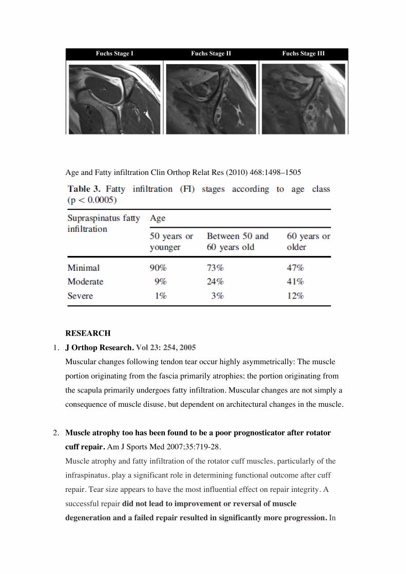
Classification: Goutallier/Fuchs Grading [5]
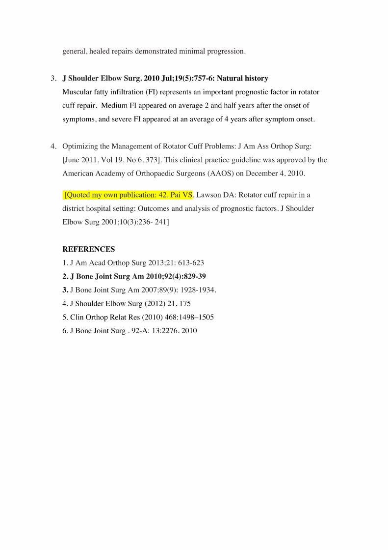
Age and Fatty infiltration Clin Orthop Relat Res (2010) 468:1498–1505
RESEARCH
1. J Orthop Research. Vol 23: 254, 2005
Muscular changes following tendon tear occur highly asymmetrically: The muscle
portion originating from the fascia primarily atrophies; the portion originating from
the scapula primarily undergoes fatty infiltration. Muscular changes are not simply a
consequence of muscle disuse, but dependent on architectural changes in the muscle.
2. Muscle atrophy too has been found to be a poor prognosticator after rotator cuff repair. Am J Sports Med 2007;35:719-28.
Muscle atrophy and fatty infiltration of the rotator cuff muscles, particularly of the
infraspinatus, play a significant role in determining functional outcome after cuff
repair. Tear size appears to have the most influential effect on repair integrity. A
successful repair did not lead to improvement or reversal of muscle degeneration and a failed repair resulted in significantly more progression. In
general, healed repairs demonstrated minimal progression.
3. J Shoulder Elbow Surg. 2010 Jul;19(5):757-6: Natural history
Muscular fatty infiltration (FI) represents an important prognostic factor in rotator
cuff repair. Medium FI appeared on average 2 and half years after the onset of
symptoms, and severe FI appeared at an average of 4 years after symptom onset.
4. Optimizing the Management of Rotator Cuff Problems: J Am Ass Orthop Surg:
[June 2011, Vol 19, No 6, 373]. This clinical practice guideline was approved by the
American Academy of Orthopaedic Surgeons (AAOS) on December 4, 2010.
[Quoted my own publication: 42. Pai VS, Lawson DA: Rotator cuff repair in a
district hospital setting: Outcomes and analysis of prognostic factors. J Shoulder
Elbow Surg 2001;10(3):236- 241]
REFERENCES 1. J Am Acad Orthop Surg 2013;21: 613-623
2. J Bone Joint Surg Am 2010;92(4):829-39 3. J Bone Joint Surg Am 2007;89(9): 1928-1934.
4. J Shoulder Elbow Surg (2012) 21, 175
5. Clin Orthop Relat Res (2010) 468:1498–1505
6. J Bone Joint Surg . 92-A: 13:2276, 2010


