Developmentofanovelchronickidneydiseasemousemodeltoevaluatethe progression of hyperphosphatemia and associated mineral bonediseaseTakashiTani1,2,HideoOrimo2,AkiraShimizu3,ShuichiTsuruoka1
SupplementaryFigures:
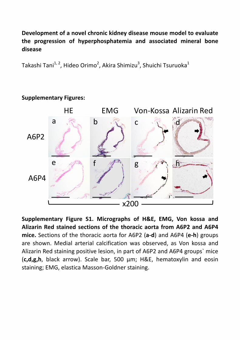
Supplementary Figure S1. Micrographs of H&E, EMG, Von kossa andAlizarinRedstainedsectionsof the thoracicaorta fromA6P2andA6P4mice.SectionsofthethoracicaortaforA6P2(a-d)andA6P4(e-h)groupsare shown.Medial arterial calcification was observed, as Von kossa andAlizarinRedstainingpositivelesion,inpartofA6P2andA6P4groups`mice(c,d,g,h, black arrow). Scale bar, 500 μm; H&E, hematoxylin and eosinstaining;EMG,elasticaMasson-Goldnerstaining.
x200
HE
Von-KossaEMG AlizarinRed
e f
b
A6P4
c
g
d
A6P2
h
aHE
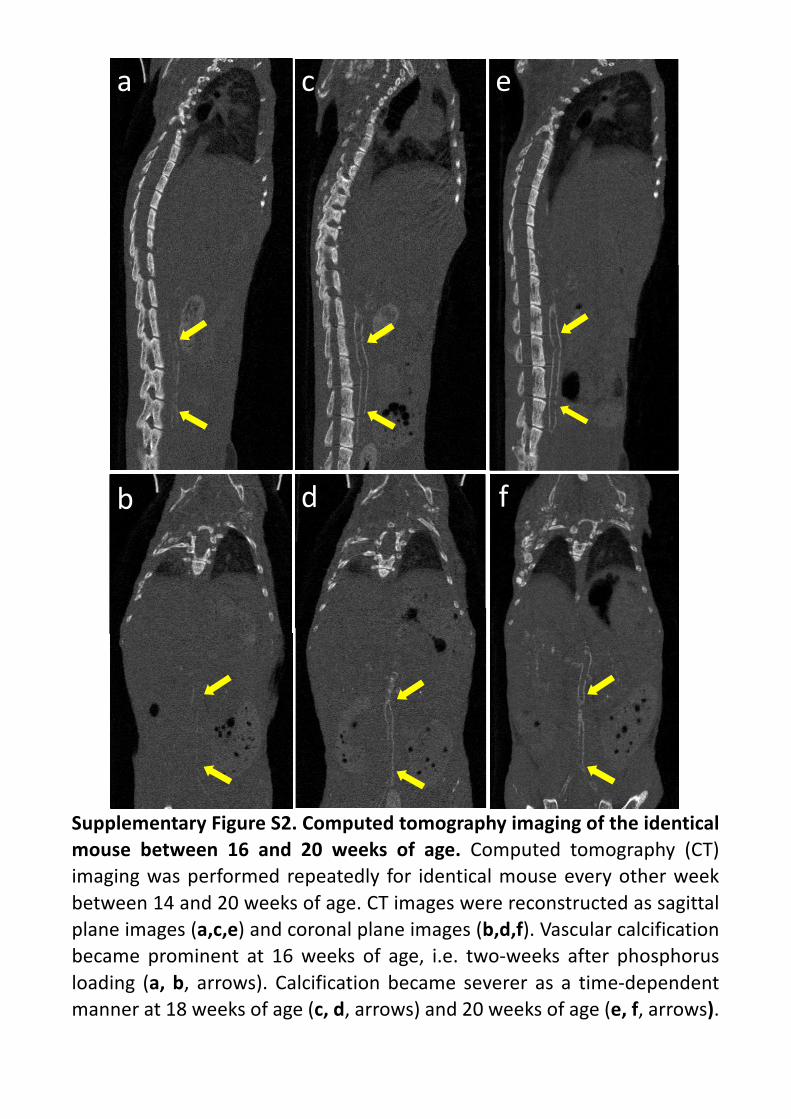
SupplementaryFigureS2.Computedtomographyimagingoftheidenticalmouse between 16 and 20 weeks of age. Computed tomography (CT)imagingwasperformedrepeatedly for identicalmouseeveryotherweekbetween14and20weeksofage.CTimageswerereconstructedassagittalplaneimages(a,c,e)andcoronalplaneimages(b,d,f).Vascularcalcificationbecame prominent at 16weeks of age, i.e. two-weeks after phosphorusloading (a, b, arrows). Calcificationbecame severer as a time-dependentmannerat18weeksofage(c,d,arrows)and20weeksofage(e,f,arrows).
a
b
c
d
e
f


