
Dr. Harbansh LalHony. Treasurer AIOS
Co-Chairman, Dept. of Ophthalmology,Sir Ganga Ram Hospital, Delhi
Director, Delhi Eye CentreEmail: [email protected]
Mob: 9810239206

All India Ophthalmological SocietyOffice Bearers
PRESIDENT Dr. Anita Panda
PRESIDENT ELECT Dr. Quresh B. Maskati
VICE PRESIDENT Dr. Debasish Bhattacharya
HONY. GENERAL SECRETARY Dr. Lalit Verma
JOINT SECRETARY Dr. Sambasiva Rao V.
HONY. TREASURER Dr. Harbansh Lal
JOINT TREASURER Dr. Ruchi Goel
EDITOR - JOURNAL Dr. S. Natarajan
EDITOR PROCEEDINGS Dr. Samar Kumar Basak
CHAIRMAN SCIENTIFIC COMMITTEE Dr. D. Ramamurthy
CHAIRMAN - ARC Dr. Ajit Babu Majji
IMMEDIATE PAST PRESIDENT Dr. N.S.D. Raju

Posterior capsular tear or PCT is one of the disastrous complications ofcataract surgery. Although common in the learning stages, it can occureven in the hands of experienced surgeons – specially if they are inhurry or overconfident. The situation does create panic in the mind ofoperating surgeon – the surgeon sometimes tends to do lot ofunwarranted steps. If not managed properly, the outcome can bedisastrous for the eye; however, if managed properly the results can bequite rewarding.
This booklet by Dr. Harbansh Lal, one of the pioneers of CataractSurgery, describes in a very lucid and practical way the do’s & don’tsof PCR.
Complications do happen in the best of hands but the true competenceof a surgeon is judged by how he handles complications.
I am sure this masterpiece by Dr Harbansh Lal will help everyone.
Dr. Anita PandaPresident, AIOS

Chairman
Dr. Ajit Babu MajjiMedical Director,Centre for Sight
Ashoka Capitol Building, Road # 2,Banjara Hills, Hyderabad - 500 034
Ph: 040-40045500 Mobile: 09391026292E-mail: [email protected]
Members
Dr. Ashis K. BhattacharyaMember ARC (East Zone)78/1, R.K. Chatterjee Road,Flat No. 2A,Kolkata - 700 042,West BengalPh: 0-9831019779E-mail: [email protected]
Dr. Sharat Babu ChilukuriMember ARC (South Zone)Sharat Laser Eye Hospital,3-1-119, Kakatiya Colony,Alankar Circle, Hanamkonda,Warangal - 506 011 (A.P.)Ph: 0-9849058355E-mail: [email protected]
Dr. Amit KhoslaMember ARC (North Zone)89, Charak Sadan, Vikaspuri,New Delhi - 110 018Ph: 0-9811060501E-mail: [email protected]
Dr. Deshpande AnantAwdhutrao
Member ARC (West Zone)Guruprasad Eye Hospital,200, Samartha Nagar,Aurangabad - 431 001 (Mah)Ph: 0-9850086491E-mail: [email protected]
Dr. Gaurav LuthraMember ARC (Central Zone)Drishti Eye Centre,Dehradun Wave Lasik Centre,9-B, Astley, Dehradun - 248 001UttarakhandPh: 0-9412059188E-mail: [email protected]
All India Ophthalmological Society(Academic & Research Committee)
Phacoemulsification is being practised all over the country in all types ofsetups and by all types of surgeons. PCT is the most commonly encounteredcomplication irrespective of the experience of the surgeon. With the goodexperience accumulated over the years, it is now possible to manage PCT tothe satisfaction of both the patient and the surgeon.
Majority of the management of posterior capsular tear is by the anteriorsegment surgeon, however, the role of the posterior segment surgeon cannotbe undermined, especially in situations associated with nucleus drop and IOLdrop. While managing PCT, not only does the surgeon need to handle thecataract and vitreous, but also has to be well aware of the types of IOLs suitablein a given situation. I am very pleased to see that this CME Series covers alltypes of IOL implantation techniques.
Dr. Harbansh Lal, who is a pioneer in Phacoemulsification surgery, andhas been actively involved in training of postgraduate students has put in hisvast experience and brought out this CME Series. I also want to thankDr. Lalit Verma, and Dr. Tinku Bali for their contribution to the CME series,especially regarding the management of PCT by the posterior segment surgeon.
I hope this CME will be of great help to all cataract surgeons.
Dr. Ajit Babu MajjiChairman, Academic & Research Committee,
All India Ophthalmological Society,Medical Director, Centre For Sight, Ashoka Capitol Building,
Road # 2, Banjara Hills, Hyderabad - 500034 (India)E-mail: [email protected] Mobile: 9391026292

Management of PCTDr. Harbansh Lal
About the Author
� Treasurer, All India Ophthalmological Society
� Chairman, Deptt. of CME, Sir Ganga Ram Hospital
� Co-Chairman, Deptt. of Ophthalmology, Sir Ganga Ram Hospital
� Author, Manual of Phaco Technique (Text and Atlas)
Positions held in the past
� President, Delhi Ophthalmological Society
� Secretary, Delhi Ophthalmological Society (2005–07)
� Library Officer, Delhi Ophthalmological Society
� Member, Executive Committee, Delhi Ophthalmological Society
� Member, Executive Committee, Indian Implant and Refractive Society
� Member, Scientific Committee, All India Ophthalmological Society
� Joint Secretary, All India Ophthalmological Society
� President, Rotary Club of Delhi, Rajendra Place
Major contribution by:
Dr. Lalit Verma
Assisted by:
Dr. Tinku Bali RazdanDr. Bhartendu Kumar Varma
Dr. IkedaDr. Saurabh Sawhney
For any comments or queries: Dr. Harbansh Lal, Director, Delhi Eye CentrePh: 9810239206 [email protected]

Introduction ......................................................................................... 1
Chapter 1 Predisposing Factors ........................................................ 3
Chapter 2 Mechanism of PCT ........................................................ 13
Chapter 3 Diagnosis and Goals ....................................................... 23
Chapter 4 Management by Anterior Segment Surgeon .................. 27
Chapter 5 Management by Posterior Segment Surgeon ................ 65
References ........................................................................................ 75
Suggested Readings .......................................................................... 77

AC Anterior chamberACD Anterior chamber depthACIOL Anterior chamber
intraocular lensCCC Continuous curvilinear
capsulorhexisCCI Clear corneal incisionCL Contact lensCME Cystoid macular edemaCSZ Central safe zoneECCE Extracapsular cataract
extractionEPN Epinuclear plateI/A Irrigation aspirationIOL Intraocular lensIOP Intraocular pressureIPD Interpupillary distancePAL Posterior-assisted
levitationPC Posterior capsule
PCC Posterior continuouscapsulorhexis
PCIOL Posterior chamberintraocular lens
PCT Posterior capsular tearPFCL Perfluorocarbon liquidPMMA Polymethylmeth-
acrylatePPV Pars plana vitrectomyPUSZ Peripheral unsafe zonePVD Posterior vitreous
detachmentRD Retinal detachmentRMT Rhexis margin tearRR Rounded repositorSFIOL Scleral-fixated intra-
ocular lensSICS Small incision cataract
surgeryVES Viscoelastic substance
Phacoemulsification is the standard of care for cataract patients all overthe world. We promise sutureless surgery and early visual rehabilitationfrom the first post-operative day, which has led to an increasedexpectation of the patient. Posterior capsular tear (PCT) may compro-mise not only the expected outcome but may also cause seriouscomplications and sleepless nights for the surgeon, if not managedproperly.
PCT can be defined as an iatrogenic breach in the continuity ofposterior capsule. The incidence of PCT in various studies variesbetween 1–4%1. This largely depends upon the equipment, setup andsurgeon’s experience and skill. The incidence of PCT goes down as thesurgeon develops a better understanding of the equipment and hissurgical skill improves. If beginners are able to understand the causesand factors responsible for PCT, they can take essential steps to preventits occurrence.
Our aim is to analyze how and why PCT takes place and in case itoccurs, how can we have a surgical outcome comparable to anuncomplicated surgery, in terms of good visual outcome, early visualrehabilitation and prevention of secondary consequences of PCT like
1

2 Management of Posterior Capsular Tear
Glaucoma, Nucleus drop, IOL drop, Distorted pupil, Decentered IOL,Corneal decompensation, Cystoid macular edema, Prolongedinflammation, Retinal detachment, etc.
To have a satisfactory outcome when there is an IOL or nuclear drop, ithas to be managed by a posterior segment surgeon. We are thankfulthat none other than Dr. Lalit Verma has written the part of ‘PCTmanagement by a posterior segment surgeon’.

3
Prevention is the best form of management. Prevention is possibleonly if we know what causes PCT. The predisposing factors can beclassified as follows:
1. Equipment related(a) Operating microscope(b) Phacomachine
2. Extraocular – Ergonomics(a) Prominent eyebrows(b) Deep set eyes(c) Narrow palpebral fissure(d) Disorders of spine
3. Ocular(a) Corneal causes(b) Anterior chamber depth(c) Iris and pupillary factors(d) Capsule, lens and zonules
4. Surgeon’s factor
1. Equipment related
(a) Operating microscope
Focussing of the oculus, proper inter-pupillary distance (IPD) selection,

4 Management of Posterior Capsular Tear
well positioned microscope, and comfortable seating are essential priorto surgery.
• Adjusting the oculus: If there is no anisometropia, the surgeoncan keep the oculus at 0, in both eyes. In case of anisometropia,the surgeon keeps one oculus at 0 and focusses with the other eyeclosed; now the surgeon closes the focussed eye and adjusts theoculus of the other eye. Maximum plus which provides a clearview should be chosen in the oculus, so as to relax the accommo-dation.
• IPD: Maximum IPD which does not cause diplopia should beselected.
(b) Phacomachine
Proper understanding of phacodynamics and the machine is an essentialprerequisite for a successful surgery.
2. Extraocular
Ergonomics
For the ease of surgery it is important to have the eye horizontallyplaced. Any angulation may lead to an oblique plane causing rotationand distortion of the globe, leading to unfocussed surgical field anddifficulty in depth perception. Optimum exposure of the surgical fieldand unhindered access to anterior chamber are prerequisites for a goodsurgical outcome. Attaining a horizontal position of the eye duringsurgery by either adjusting the table, sutures, block or extra supportunder the head or the shoulders is essential.
(a) Prominent eyebrows: Chin can be raised and extra support ifneeded can be given below the shoulders to position and stabilizethe head.
(b) Deep set eyes: Temporal incision is better as eyebrows can beavoided. If needed, superior and inferior rectus bridle sutures canbe passed to elevate and stabilize the eyeball.
(c) Narrow palpebral fissure: If it is felt that the speculum is pressingon the globe and the exposure is not adequate then lid traction orbridle sutures can be passed. If the exposure is still not adequate,cantholysis (crushing and cutting of lateral canthus can be done,which can be sutured after phacoemulsification) is a good option.

Predisposing Factors 5
(d) Disorders of the spine: Extra support may be needed behind theshoulders or the spine to make the patient comfortable. This maylead to elevation of the patient’s head. To compensate this we canelevate the foot end of the table to make the head horizontal. Beinga closed chamber technique, elevating the foot end of the table willnot alter the surgery as it used to do in open chamber surgeries dueto increased orbital pressure and vitreous thrust.
Never hesitate to give blocks in patients in whom you considereye positioning may be suboptimal.
Certain modifications in the surgical practice can help in overcomingthe problems posed by the ergonomic factors, such as:
• If doing superior phacoemulsification, shift to a temporal incision.
• Side port incision can be made more central i.e. corneal instead oflimbal, if there is obstruction due to prominent eyebrows or cheekbones.
• If there is excessive pooling at the medial side, causing annoyingreflexes, then slight temporal tilting of the head can be done ordrainage through merosel sponges/gauze, placed at the lower fornixand draining out can be done.
3. Ocular factors
Assessing ocular risk involves evaluation of the corneal clarity, anteriorchamber depth, extent of pupil dilation, iris, capsular status, cataractdensity and extent of zonular weakness, if any.
(a) Corneal problems leading to poor visibility
Scarring from any corneal pathology can cause irregular corneal haze,increasing light scatter and reducing contrast during surgery. The useof trypan blue to stain the anterior capsule during capsulorhexis is amust.
The annoying reflexes by the microscope light in presence of cornealpathology during Continuous Curvilinear Capsulorhexis (CCC) can largelybe overcome by the use of an endoilluminator2 (used by retinal surgeons).It can be placed on the limbus or inside the AC with the microscope lightoff, which enhances the visibility and facilitates the CCC.

6 Management of Posterior Capsular Tear
(b) Anterior chamber depth (ACD)
Proper evaluation of ACD is essential prior to surgery, as ACD can beshallow in high hypermetropes, intumescent and hypermature cataractswith secondary glaucoma which decreases the safety margin, as thereis a reduction in the central safe zone (CSZ). We will discuss the conceptof CSZ later.
ACD is deep in high myopes, vitrectomized eyes and zonulopia(stretched weak zonules) which makes the lens lie deep. So when weaccess the lens during phacoemulsification, the direction of the phacotip is more posterior. This leads to corneal striae and folds along with aposterior direction force, which increases the chances of PCT. To preventthis, bottle height should be low; a small corneal tunnel is warrantedwith no undue pressure on the nucleus.
(c) Iris and pupillary factors
Small pupil and floppy iris increase the risk of PCT. If pupil is smallerthan 5 mm then appropriate measures have to be taken for sustaineddilation of pupil.
Sclerosed pupil: Easier to handle as compared to floppy iris.Stretching of pupil or multiple sphincterotomies (8–12) of about 400–500 μ each can be done.
Floppy iris: More dangerous than the above. Prolapse of the irisfrom the wounds and progressive miosis being a major problem. Hencecareful evaluation of a floppy iris3 should be made and iris hooks orexpanders (Malyugin rings) should be kept ready and used wheneverrequired.
Even if during surgery the pupils get miosed, there should beno reluctance in usage of hooks and expanders.
(d) Capsule, zonules and lens
Thin friable PC in certain situations, such as posterior polar cataract,pseudoexfoliation, vitrectomized eye, high myopia and floppy and laxPC, as in hard cataract, zonulopia , zonular dehiscence, poses increasedthreat of posterior capsule tear.
Hard cataract is one of the most important risk factors for PCT. Thecapsule is generally thin and friable in such cases (Fig. 1.1). Over-

Predisposing Factors 7
stretching of the capsule becauseof the large volume of the nucleusincreases the tendency of the PC tokeep coming forward. Also, it isdifficult to separate and chop theleathery fibres.
4. Surgeon’s factor
As the surgeon keeps on gainingexperience, the incidence ofcomplication goes down, but anycomplication due to lack of under-standing of the basics is unacceptable. Every surgeon should be awareof the mechanism of surge and should know how to prevent or controlsurge during the surgery. Before going to various stages and how PCThappens, let us understand the mechanism of surge.
For that we first need to understand the concepts of:
(a) Central safe zone,(b) Peripheral unsafe zone,(c) Compliance,(d) Flow rate, vacuum and their relationships.
(a) Central safe zone (Fig. 1.2)
The central safe zone is notan anatomical area but aconcept that needs to beunderstood for performingsafe aspiration. This is an areawithin the CCC margin whichis bounded vertically by thecornea on the top and theposterior capsule in oppositedirection. This is the area withmaximum space in the AC. Allaspiration—nuclear, epi-nuclear or cortical—should be done here as there is maximum safetyhere. Even if there is AC flutter, the probe will not damage any vitalstructures. The nuclear pieces and cortical matter can be held in the
Fig. 1.1. Friable capsule – hardcataract.
Fig. 1.2. Central safe zone (CSZ).

8 Management of Posterior Capsular Tear
periphery and then brought to the CSZ for aspiration. This is a dynamicarea – as more of the nuclear pieces are removed, the space and thusthe safety margin keeps on increasing. In myopes, zonular stresssyndromes and vitrectomized eyes the CSZ is further increasedwhereas in hypermetropes, small pupil and small CCC the CSZ issmaller.
(b) Peripheral unsafe zone (Fig. 1.2)
Due to the corneal curvature, as one proceeds towards the periphery,one enters an unsafe zone as there is less space for manoeuvering. Thecapsular fornices and the angle region are thus areas where it isdangerous to do phaco-aspiration since the vital structures are extremelyclose. This constitutes the Peripheral unsafe zone (PUSZ).
(c) Compliance
A silicon tube connects the aspiration system with the handpiece inboth types of pumps. Additionally thick wide bore tubing is requiredfor the rollers to be effective in a peristaltic pump. While the rollers arerotating, there is no occlusion and no collapse of tubing. When occlusionoccurs, vacuum builds up, the rollers stop and negative pressure isgenerated within the whole system. This causes the tubing to collapse.
Property of the tubing to collapse (deform under pressure) is thecompliance of the tubing. Once the occlusion breaks, there is a releaseof negative pressure and the tubing re-expands to the original size. Fluidis drawn from the AC to fill up this extra volume (this is what causessurge). Though this volume is not much, it is this instantaneouswithdrawal of fluid over an extremely short period of time which causesthe surge (Fig. 1.3a).
This extent of collapse of the tubing will depend on the lumen size,the level of vacuum generated and the thickness of the tube. The collapseis more at higher vacuum levels and less if the lumen is smaller and thewalls are thicker (less compliant tubing). Tubings of these characteristicsare known as ‘High Vacuum’ tubing.
SURGE
Sudden withdrawal of fluid from AC after occlusion breaks iscalled surge. Beyond a certain limit it may cause collapse of

Predisposing Factors 9
chamber, jeopardizing the vital structures of eyes and making thesurgery filled with complications. In fact modifications introducedover a period of time have taken place to manage this surge andthus make phaco surgery free from complications. If there was nosurge, any one could have mastered phacoemulsification. Tomaintain a constant volume and IOP of the AC, inflow, i.e. infusionhas to be equal to outflow, which is the sum of aspiration bypump and the wound leakage. For a given bottle height inflow isconstant and so is the leakage. The only variable parameters leftin the above equation are the aspiration flow rate and the vacuum,i.e. the outflow by aspiration (Fig. 1.3b).
Fig. 1.3a. Compliance.

10 Management of Posterior Capsular Tear
(d) Relationship between vacuum, surge and flow rate
Suppose there is surge beyond acceptable limits. Now there are twooptions—either reduce the flow rate or the vacuum. Decreasing theAFR has a direct and linear effect but increases the rise time and makesthe procedure slower, which may not be such a disadvantage to abeginner. On the other hand decreasing the vacuum will decrease theholding power which is not desirable in steps like chopping/phaco-aspiration of a hard cataract. So, it is better to lower the FR in such asituation to decrease the surge, while maintaining high vacuum. On the
Fig. 1.3b. Surge.

Predisposing Factors 11
other hand, in situations where a firm hold is not so important, likedivide and conquer technique, soft cataracts or epinuclear plate removal,one can lower the vacuum settings to decrease the surge whilemaintaining the AFR.
Control of surge
There are various methods of controlling the surge. Some areincorporated into the newer machines and there are some measuresthat the surgeon can apply.
Surge prevention by the machine
Venting, high vacuum tubings, use of cassettes, delay in start of motorafter breaking of occlusion, differential settings for flow rate and vacuumat different stages of surgery, dual/linear foot pedals, microtips andabove all highly responsive sensors and computing have been successfulin decreasing the surge.
Surgeon’s control of surge
1. Decreasing the effective flow rate: Without changing the actualsetting on the machine, the surgeon can decrease the effective FRby using a smaller bore aspiration port, e.g. Microflow tip.
2. Increasing the infusion by raising the bottle height or using a TUR(Transurethral resection) set may be useful in some cases.
3. The use of an ACM is useful for decreasing surge (especially forbeginners).
4. Proper wound construction: A leaking wound will disturb theequilibrium of the chamber so that even a very small amount offluid withdrawn can cause it to collapse. This highlights theimportance of making a good main wound and side port. The woundconstruction should be such that it conforms to the tip that you areusing. Premature entry also results in a leaking wound. Distortionof the wound, with the co-axial IA handpiece may cause a leakywound. Too tight a wound or too long a tunnel can also cause aproblem by reducing the inflow. Thus both a leaky and too tight awound can increase the surge.
5. Increased viscosity of the AC contents: The flow rate settings arefor clear fluids like BSS/Ringers. A thicker fluid increases theresistance and does not flow out easily. The use of viscoelasticsubstance (VES) can cause a decrease in the effective FR and thus

12 Management of Posterior Capsular Tear
decrease the surge. This is particularly useful in hard cataracts wherethe settings are usually high and whilst aspirating the last nuclearfragment.
6. Partial occlusion of the tip: Partially occluding the tip with anotherpiece before the occlusion breaks and the occluding fragment getsaspirated ensures that any surge that occurs will be used to draw inthe next piece to occlude the tip. This will maintain occlusion andprevent fluid from the AC being aspirated.
7. Foot control: Above all, good foot pedal control is of paramountimportance in controlling surge and utilizing it to your ownadvantage. If one can anticipate the events then surge control is nota problem. That is why experienced surgeons can operate on anysettings and any machine. As soon as the occlusion is about to break(i.e. the piece is about to be aspirated into the tip), the surgeon liftsthe FP to IAo (position 1 of foot pedal or completely in theContinuous Infusion Mode), the piece will go in on its ownmomentum and without any of surge as the FR will decrease. Thusfluid withdrawn from the AC will be very little to overcome thecompliance of the system. However, if the FP is withdrawn tooearly and there is not enough momentum then it will take moretime to build up vacuum again. This balancing between the AFR,vacuum and the momentum of the pieces needs to be done verycarefully.

It is indeed surprising that just a 4 μ thin posterior capsule can withstandso much stress and pressure, arising because of the various forces duringphaco surgery. Why then, under certain situations, does its tenacitygive way? To understand this we have to assess each and every step ofthe surgery, understand what predisposes to PCT and what remedialmeasures can be taken.
1. WOUND CONSTRUCTION
Though wound construction may not be directly responsible for PCT,but leaky wounds are the most important factor for unstable AC. Asharp keratome, according to the size of the phaco tip is a must for agood wound construction. Trying to create a 2.75 mm incision with a3.2 mm keratome will always result in a leaky wound. Even a tightwound will lead to increase in surge by reducing the inflow.
Sideport incisions are even more important. The chopper is much thinnerthan the aspiration cannula used in bimanual I/A system. We generallycreate the sideport incisions to the size of the I/A cannula and when we areat the highest parameter setting of phaco machine, i.e. during choppingand phacoaspiration this wound keeps on leaking. Due to the same reasonbeginner can make initially small incision corresponding to the size ofchopper and then enlarge it afterwards for I/A.
13

14 Management of Posterior Capsular Tear
2. ANTERIOR CONTINUOUS CURVILINEARCAPSULORHEXIS (CCC)
The CCC can withstand turbulence, pressure and mechanical stresscreated by the fluid, nucleus, chopper, IOL, etc., during phaco-emulsification. If CCC is not intact, or there is cone formation in theCCC, then these forces may cause the anterior rhexis margin to extendposteriorly leading to a PCT. Rhexis margin tear (RMT) could beprimary, occurring at the stage of performing anterior CCC or secondary,i.e., happening at any other stage of the surgery.
Primary RMT
There are certain situations which are more prone to RMT, such as:
1. Intumescent or hypermature cataract2. Pediatric cataract3. Hard cataract4. Fibrosed capsule
1. Intumescent or hypermature cataract
Due to the high intra-lenticular pressure, many a time as soon as a nickis made on the anterior capsule, the rhexis tends to run away, or one isable to start the rhexis, but it tends to run to the periphery midway. Insuch a scenario use of Healon 5 or Healon GV to flatten the anteriorcapsule may prevent it.
To prevent this, one may try to reduce the intra-lenticular pressureby doing multiple YAG capsulotomies, few hours before the surgery.This is the best method as holes created by YAG are round and don’thave the tendency to run away. Same can also be achieved by multiplesmall punctures at the centre instead of one linear cut to relieve theintra-lenticular pressure. Fluid is allowed to escape slowly, but thesepunctures are not round, and will have a tendency to run away.
After making the punctures, some of the released fluid can bemanually sucked by a syringe by putting the cannula at variouslocations underneath the anterior capsule or viscoexpressed.Viscoexpression is better as it maintains the pressure from the top andthus prevents the rhexis from running away.
Another, very good option is to make a small rhexis initially andenlarge it before or after phacoemulsification, depending upon the size

Mechanism of PCT 15
of the rhexis and hardness of the cataract. If the CCC is less than 3.5mm and the nucleus is large and hard, it’s better to enlarge it beforedoing phacoemulsification.
Many surgeons prefer to make a sinusoidal CCC, i.e. start assmall CCC and after completing 120–180º start enlarging it andinstead of finishing it at the site of origin go beyond and get anadequate CCC.
2. Pediatric cataract
Younger the patient, more are the chances of RMT. To perform CCC insuch cases requires special surgical skill. Capsule has to be stained,high viscosity viscoelastics are a must and CCC has to be done with aforceps.
Small CCC is attempted and instead of applying tangential force,the cut end is pulled in towards the centre. AC has to be maintainedat all times, as even a slight collapse of the AC will make the CCC runaway.
3. Hard cataract
Hard cataracts in older patientsusually have a very thin and friablecapsule. Trying to do an anteriorCCC by cystitome, many timesleads to tears in the anterior capsuleunderneath the turned flap, makingit difficult to get a round CCC. Ifsuch tears go unnoticed, these leadto RMT (Fig. 2.1).
Use of forceps and trypanblue stained capsule is a betterchoice.
4. Fibrosed capsule
Sometimes in long standing traumatic and hypermature cataract,the cataract gets partially absorbed and the patient develops plaque,fibrosis of the capsule and wrinkling of the anterior capsule. Insuch situations CCC is difficult and one may land up in incompleteand irregular CCC.
Fig. 2.1. Friable capsule – hardcataract.

16 Management of Posterior Capsular Tear
Secondary RMT
This occurs because of inadvertent injury to the anterior CCC, whichcan be due to the chopper, phaco tip, IOL or the hard nucleus. The mostcommon culprit is the sharp chopper.
Types of RMT
There are two types of RMT:
1. Curved or tangential 2. Radial or coned
1. Curved
When trying to do a CCC, it goesto the periphery in curvilinearfashion and you are not able toretrieve it. This retrieval isdifficult in younger patients dueto high elasticity of the zonules.When the rhexis is pulled in,zonules get stretched and preventthis force to be applied on to thecut end of the rhexis margins inproper direction (Fig. 2.2).
2. Radial
This happens in morgagnian orintumescent cataract when rhexis runsaway to the periphery or due to injuryto the rhexis margin by chopper orphaco tip during surgery, which leadsto the formation of a cone (Fig. 2.3).
In presence of RMT, excessivepressure by nuclear fragment, whiledialing or during chopping may causeRMT to extend posteriorly. Collapseof the chamber during any phase ofthe surgery will cause vitreous to bulge
Curved RMT is not as dangerous, as its chance to run to theposterior capsule is much less as compared to the radial RMT.
Fig. 2.2. Rhexis margin tear.
Fig. 2.3. Rhexis margin tear.

Mechanism of PCT 17
forward and cause RMT to run posteriorly. This happens more whenthe last nuclear fragment is being removed. As long as some nuclearfragment is present in the capsular fornices, it prevents PC to comeforward.
The most important strategy to prevent PCT is to maintain ACdepth at all times. For this reason do not lower the bottle height, butlower the fluidics parameter by 20%, so as to avoid any chamberfluctuation.
Viscoelastics have to be injected before removal of the probe, atevery step of the surgery, even after cortical aspiration before removingthe infusion cannula inject viscoelastics from the side port.
Alternatively nucleus can be prolapsed into the AC and supracapsularphacoemulsification can be performed, but still ACD should bemaintained during every step of the surgery.
3. DURING HYDRODISSECTION
There are 3 main reasons for posterior capsular rupture during hydroprocedures:
1. Block to outflow
The outflow may be blocked due to increased resistance offered byviscoelastic in the chamber or a small CCC/small pupil. Also injectingfrom the side port when main wound is sealed can lead to a PCT due toincreased pressure (Fig. 2.4).
Fig. 2.4. Hydrodissection.

18 Management of Posterior Capsular Tear
2. Injection of too much fluid
Injection of too much fluid with too much force or using a faultytechnique/wrong syringes and cannula can lead to PCT.
3. Inherent weakness
Weak capsule may be seen in case of posterior polar cataract, highmyopes, post-vitrectomy, traumatic cataracts, pseudoexfoliativesyndromes and some cases of posterior subcapsular cataract. In thesecases one can avoid hydrodissection (since this step can lead to a nucleardrop) and perform a careful hydrodelineation.
If the PCT goes unnoticed and if the phaco probe is placed in theanterior chamber, the nucleus will be dislodged in the vitreous cavity.So, early recognition of the PCT is of utmost importance at this step.Few signs that help in the recognition of PCT are as follows:
Signs of a PC rupture during hydrodissection
These are:
• Sudden deepening of the chamber
• Abnormal tilt in the nucleus
• Inability to rotate the nucleus
• Sudden dilation of the pupil – Snap sign4
• Bright red reflex.
4. DURING NUCLEOTOMY
Nucleotomy comprises of Trenching, Splitting, Chopping andAspiration. Any of these steps if not performed properly can lead to aPCT.
1. Trenching
In case of a soft cataract, while trenching if the power has not beenadequately lowered, one can go through and through, which can leadto a PCT in the periphery. On the other hand, in case of a hard cataractif the power has not been adequately increased, surgeon tends to applyexcessive force on the nucleus pushing it down, which can lead to PCTor zonular dehiscence.

Mechanism of PCT 19
2. Splitting
If trenching depth is not adequate, excessive force applied duringsplitting can lead to damage to the capsule. If the fibres are very leathery,on attempted splitting we tend to dip the periphery of the nucleus whichmay lead to development of zonular dehiscence or a peripheral PCT. Insuch cases make sure, not to push the nucleus too far away, and keepon moving your instruments closer to the area of the split, so that thefragments do not move very far apart.
3. Chopping and Aspiration
At this stage of surgery fluidic parameters are highest and surge is themost important causative factor. Sudden surge or collapse of anteriorchamber can lead to a direct injury to the posterior capsule by thechopper. When chopping in a soft cataract, the soft part tends to besucked in, and the probe may damage the posterior capsule (PC) moreso in the periphery.
Epinuclear plate, the cortical matter and nuclear pieces in capsularfornices keep the PC away from the probe, while emulsifying the lastnuclear piece the capsular fornices are empty and a slight surge movesPC towards the probe, causing PCT. This is more common in conditionswhere the capsule is lax and floppy as in hypermature, brown hardcataracts. In such cases the nucleus size is very large, which stretchesthe capsule and the capsule becomes large and floppy. This also occursin cases of zonular dehiscence and weakness wherein the PC is laxagain.
In such cases various steps can be taken to prevent a PCT, such as:
• Lowering the aspiration parameters.
• Increasing the bottle height.
• Using an AC maintainer (another port for fluid to go in).
• Removal of last piece under viscoelastic.
• Extreme cases – assistant can keep on injecting viscoelasticfrom the side port, to keep the PC away.
• Use of micro-tip instead of kelman.
• Phaco-aspiration to be done in Central Safe Zone.

20 Management of Posterior Capsular Tear
Partial occlusion of tip
Sometimes the aspiration tubing or the phaco tip may get blocked,particularly in hard cataracts, and on sudden release of this blockadethere will be increased surge and increased chances of chamber collapseand PCT. In such cases when a tube blockade is expected (poor follow-ability and poor hold on the nucleus) one should take out the probe fromAC and flush it. The probe should be tested in a test chamber, before use.
5. DURING EPINUCLEAR PLATE REMOVAL
Many a time one is left with a posterior epinuclear plate with no anteriorextension. In such cases turning the bevel of the phaco tip down to liftthe EPN may lead to PCT. Any attempt to lift the posterior EPN withiris repositor may lead to PCT if the eye is not adequately visco-compressed.
While doing cortical aspiration with the bimanual I/A system, EPNgets automatically prolapsed and suck out along with the cortex.
6. DURING CORTICAL ASPIRATION
High incidence of PCT occurs during cortical aspiration, especially inthe sub-incisional area. This is because of poor access and decreasedvisibility, problem being exaggerated by small capsulorhexis. A coaxialhand piece leads to further distortion of the wound, causing the PC tocome anteriorly.
GOLDEN RULE
When the PC is caught in thesuction port a star-shaped tentedarea appears. Immediately,without moving the handpiece/cannula, release the footpedal to stop suction (Fig. 2.5).
In some cases, reflux may berequired. Catching does not tearthe posterior capsule but pullingdoes. Use of bimanual techniquereduces the incidence of PCT. Fig. 2.5. Catching the posterior capsule.

Mechanism of PCT 21
PCT does not occur if the PC is only caught in the probe; suddenmovement after holding with the probe / cannula is what causesit to tear.
7. DURING CAPSULAR POLISHING
During polishing, a well-focused PC in retro-illumination view underhigh magnification and a bag filled with VES is a must to prevent PCT(Fig. 2.6). The bag must always be concave and visco-compressed soas to provide easy sliding of the instruments. If the bag is lax and somewrinkling appears on the capsule during the movement of roundedrepositor/polisher, the chances of creating a PCT are high (Fig. 2.7).
Patients with thin PC (as discussed during hydro procedures) areparticularly prone to PCT.
8. DURING IOL IMPLANTATION
IOL implantation should be done under a well pressurized globe. Thebag should be filled with viscoelastics. If the viscoelastic leaks or thecapsule is not taut, then chances of capsule getting entangled in theleading loop are very high. While using an injector system, the leadingloop has to be kept horizontal all the time, for that one has to observethe leading loop carefully and keep on rotating the nozzle of the cartridgeas and when required.
Fig. 2.6. Well filled bag – no striae. Fig. 2.7. Poor filled bag – capsularstriae.

22 Management of Posterior Capsular Tear
In this era of micro-incision surgeries with reducing incision size,there may be leakage of viscoelastic from the wounds, particularly inwound-assisted insertion of the IOL. In such cases one can re-inject theviscoelastic or release the IOL in the sulcus rather than the bag. TheIOL is positioned in the bag, after re-injection of viscoelastics.
All the companies provide same cartridge size for all IOL powers.High power IOLs are much thicker and excessive force is required toinject these IOLs. This at times causes sudden and jerky release insidethe bag causing PCT.

GOLDEN RULE
Once PCT is noted, or one is in doubt about the status of the posteriorcapsule, be calm and maintain the phaco probe in the AC, keep theinfusion on and inject viscoelastics from the side port. After ensuringthat the AC is well pressurized, the phaco probe is withdrawn (Fig. 3.1).
23
Fig. 3.1. A. Posterior capsular tear. B. Removal of probe – vitreous prolapsein AC. C. Pressurizing the AC from the side port. D. Removal of probe – noprolapse of vitreous into AC.
A B
C D

24 Management of Posterior Capsular Tear
If one withdraws the phaco probe suddenly, then the anterior chambercollapses, causing the PCT to enlarge and the hyaloid face may getdisrupted, leading to prolapse of the vitreous in the AC and prolapse ofthe nuclear fragments into the vitreous cavity.
Converting tear to PCC
Converting a PCT into a PCC is ideal, but most of the times impractical.Conversion to PCC is possible only if PCT is small and central and thehyaloid faces nearly intact. Also, if the hyaloid face is intact, risk ofdisrupting it while attempting PCC is very high.
One should attempt conversion to PCC only when one has smallcentral/paracentral PCT (not more than 3 mm), good viscoelastics,excellent capsulorhexis forceps and a good operating microscope.However, the failure rate is still very high.
DIAGNOSIS
CONFIRMATION OF PCT ANDHYALOID FACE RUPTURE (HFR)
The next step is to confirm the presence and extent of PCT, and evenmore important is to ascertain whether hyaloid face is intact or not.Majority of the cases of PCT will have a disrupted hyaloid face, but anintact hyaloid face can be encountered if PCT occurs at the stage ofcapsular polishing, IOL insertion or cortical aspiration.
Signs of HFR
• Torn edge– Shiny/golden– Rolled up
• Anterior chamber– Irregular depth
• Nucleus– Restricted movements of the fragments
Tests
One can do the following tests:
Indicate disruptionof hyaloid face

Diagnosis and Goals 25
Sponge test (Fig. 3.2)
One can sweep a sponge along the incision to detect the presence ofvitreous.
Fig. 3.2. Sponge test.
Sweep test
One may also try to sweep the spatula from the anterior chamber angleunder the incision towards the PCT. Vitreous, if present, will be seendragging in (due to tendency of the vitreous to come towards the wound).
Halo test (Fig. 3.3)
Put viscoelastic in the bag, to flatten it or keep it slightly convex toelicit the halo test. Try to look for the ring reflex by applying the pressure
Fig. 3.3. Halo test.

26 Management of Posterior Capsular Tear
in the centre of the capsule with the help of a rounded repositor. If thehyaloid face is intact, a halo will be seen which will vary in sizedepending upon the amount of pressure applied. This ring reflex willbe broken at the site of PCT.
Stain test
One can also inject Triamcinolone into the anterior chamber just adjacentto the PCT (not above it as it will fall back into the vitreous cavity), tostain the vitreous. However, when one is in doubt consider the hyaloidface as disrupted.
GOALS OF MANAGEMENT
The goal of every complication created during surgery is to minimize theshort term and long term damage to the eye. For this purpose we candivide the goals of management into major goals and important goals.
MAJOR GOALS
1. To avoid posterior dislocation of nucleus, nuclear fragments,epinucleus or cortical matter into the vitreous cavity.
2. Prevent any damage to the corneal endothelial surface.
IMPORTANT GOALS
These goals are very important to achieve so that the end result of thesurgery is as good as if nothing had happened. Surgery remainssutureless, astigmatically neutral with well centered IOL and withoutany secondary complications.
1. Prevent enlargement of tear.2. Prevent damage to capsulorhexis.3. Minimize size of vitrectomy, avoiding traction.4. Removal of left over cortex.5. Maintain the wound size.6. Proper positioning of the IOL.

GOLDEN RULE
DO NO HARM TO THE PATIENT
If the primary surgeon is ill-equipped or inadequately trained, secondarymanagement by a senior or trained surgeon should be done. If nucleusis not retrievable from the anterior route, then leave it for the posteriorsegment surgeon to do the needful. Be truthful to yourself and the patient,inform the patient about the scenario. Your ego might get hurt, but youwill have better peace of mind, with the shared responsibility.
FACTORS IN DECISION MAKING
Management will depend on various factors, such as:
• Extent of PCT• Hyaloid face intact or not• Location of the nucleus – whether dislodged into the vitreous cavity• Availability of equipment – vitreous cutter, vitrectomy machine• Availability of alternative IOLs• Availability of specialized instruments• Knowledge about anterior vitrectomy and confidence of the
surgeon• Availability of VR surgeon.
27

28 Management of Posterior Capsular Tear
Above factors help in deciding the plan of management:
• Management by Anterior Segment Surgeon
– Primary– Secondary
• Management by Posterior Segment Surgeon– Primary– Secondary
PRIMARY MANAGEMENT STRATEGIES
Primary management is the best approach, as it causes least stress tothe patient and harm to the reputation of the surgeon. It causes a lesseramount of inflammation and provides an early rehabilitation as promisedto the patient. We need to have strategies to manage:
1. Nucleus/nuclear fragments2. Epinucleus3. The cortex4. IOL implantation5. Final vitrectomy and closure
1. NUCLEUS / NUCLEAR FRAGMENTS
Managing of nucleus/nuclear fragments is the most important step in acase of PCT. The main goal is to prevent any dislocation of the nucleus/nuclear fragment into the vitreous cavity. There are two main steps butmany techniques for the same.
1. Supracapsular relocation• Dislodging
• Tumbling
• Chopstick technique2. Extraction from eye
• Manual(i) Viscoexpression
(ii) Chopstick technique• Automated
(i) Vitrectomy cutter

Management by Anterior Segment Surgeon 29
(ii) Phacoemulsification(a) Without scaffold(b) With scaffold
1. HEMA Contact lens2. Lens glide3. IOL
• Conversion to ECCE/SICS
1. Supracapsular relocation
In a viscopressurized eye, nuclear fragments are relocated anteriorlyinfront of the CCC and iris, preferably at the angle of the AC to preventthese from dropping into the vitreous cavity by any of the followingtechniques:
Dislodging
Small fragments of the nucleus are moved just sideways into the capsularfornices and then brought gently upwards towards the iris plane andthen pushed towards the angle of AC.
Tumbling
This is a very good technique for small fragments. It can also be usedfor soft cataracts and epinuclear plate. In this technique the repositorpushes the nucleus to the periphery initially (Fig. 4.1), and then upwards,maintaining a constant counter-pressure from the anterior capsule, thenucleus can be easily brought out from anterior CCC (Fig. 4.2).
Fig. 4.1. Rounded repositor pushes the nucleus to the periphery initially.

30 Management of Posterior Capsular Tear
Even, if the pupil is small, which is obscuring the peripheral view,this technique comes in handy.
Chopstick technique
Chopstick technique in PCT
As the name suggests, two instruments are used in this technique,through which the nucleus/nuclear fragments can be held andrepositioned to a desired site. The instruments that could be used arethe Sinskey hook, chopper, rounded repositor or dumbbell dialer.Sinskey hook is particularly good as it gets buried into the nucleus andprovides a good grip. Both the instruments can be introduced from themain port or one from the main port and another from side port. One isput below and the other above, or alternatively one on each side of thefragment, so that the fragment is sandwiched. The fragment is grippedfirmly between the two instruments and is now moved into thesupracapsular area away from the site of tear (Fig. 4.3).
Chopstick technique in impending nuclear drop
In case the nucleus is hanging down in the anterior vitreous cavity,never try to fish it out through the anterior route. Such an impendingnucleus drop is best managed by either the ‘Chopstick technique’ asdescribed by Dr. Harbansh Lal5 or the “Posterior assisted levitation” asdescribed by Dr. Kelman. In both these techniques, one port is made
Fig. 4.2. Upward pressure is maintained and a constant counter pressure fromthe anterior capsule facilitates easy removal of the nucleus from the anteriorCCC.

Management by Anterior Segment Surgeon 31
through the pars plana by giving a stab incision with 15 degree blade orV-lance knife, 3.5 mm away from the limbus.
Through the pars plana incision, in PAL technique Viscoat isinjected behind the nucleus and the thin cannula of Viscoat or irisrepositor is used to push the nucleus forward. In chopstick techniquean instrument (Sinskey hook) is passed through the pars planaincision and buried into the undersurface of the nucleus, which helpsto support the nucleus and prevents it from sinking into the vitreous.The nucleus is sandwiched and stabilized with the second instrumentfrom above which may pass through the side port or the main port.The nucleus is then brought into the supracapsular area after havingbeen stabilized (Fig. 4.4).
Chopstick technique without PCT
Chopstick technique is not only helpful in case of PCT, but also in anycase where we want to reposition the nuclear fragment to prevent thePCT. In case of a miosed pupil, the visibility of the surgeon particularlyin the area of capsular fornices is hampered. If a small nuclear fragmenthas gone in the capsular fornices or between the iris and the anteriorcapsule or as in a case of resistant sub-incisional nuclear fragment,which fails to rotate, the surgeon can use two instruments to hold andreposition the nuclear piece in the central safe zone (Figs. 4.5 and 4.6).
Fig. 4.3. In case of a PCT if a nuclear fragment is remaining in the bag thentwo instruments can be used to grip the fragment. It is now moved into thesupracapsular area away from the site of tear.

32 Management of Posterior Capsular Tear
In case of failure of the phaco machine, this method can be used tobring the nucleus/nuclear fragments not only into the anterior chamberbut also for removal from the eye.
2. Removal from eye
Manual
(i) Viscoexpression
Soft nuclear fragments are crushed between two instruments, such aschopper, Sinskey hook or a repositor. Thick cannula of viscoelastic is
Fig. 4.4. In case of an impending nuclear drop an instrument (Sinskey hook)is passed through the pars plana incision and buried into the undersurfaceof the nucleus, which helps to support the nucleus and prevents it from sinkinginto the vitreous. The nucleus is sandwiched and stabilized with the secondinstrument from above which may pass through the side port or the main port.The nucleus is then brought into the supracapsular area after having beenstabilized.
Figs. 4.5 & 4.6. If a small nuclear fragment has gone in the capsular fornicesor between the iris and the anterior capsule, two instruments can be usedto hold and reposition the nuclear piece in the central safe zone.

Management by Anterior Segment Surgeon 33
passed underneath and well beyond the nuclear fragments (Fig. 4.7).Now viscoelastic is injected with simultaneously minimal pressure atthe posterior lip of the wound with the cannula, causing the viscoelasticto flow out along with the nuclear material (Fig. 4.8). Care is taken tomaintain AC depth at all times.
Fig. 4.7. Thick cannula of viscoelastic is passed underneath the nuclearfragments.
Fig. 4.8. Viscoelastic is injected with simultaneously minimal pressure at theposterior lip of the wound with the cannula, causing the viscoelastic to flowout along with the nuclear material.
(ii) Chopstick technique
As already discussed in the relocation of nucleus, the chopsticktechnique can also be used for removal of the fragments from the eye.The incision size will depend upon the size of the nucleus. If half of thenucleus is remaining then 5 mm incision is adequate, and if less thanthat a 4 mm incision would be sufficient.

34 Management of Posterior Capsular Tear
Long axis of the nuclear fragment is positioned perpendicular to thelong axis of the wound. The eye is now visco-pressurized, and any twoinstruments, as discussed earlier, are inserted through the main port.These instruments are buried one on each side of the long axis of thefragment. Now both instruments along with the nuclear fragment arepulled out through the wound.
Advantages of the chopstick technique:
• Non-bulky instruments.• Small incisions are needed.• Any number of side ports can be made.• Better than other techniques, such as wire vectis which relies on
counter pressure, leading to damage to the cornea.• Least traction on the vitreous, as instruments are even thinner than
vitreous cutter.• AC is maintained at all times.• Better than PAL as counter pressure provides more controlled
elevation of the nucleus with no risk of viscoelastic going into thevitreous cavity. No risk of increased IOP.
Disadvantage:
• Not suitable for soft cataracts or epinuclear plate as it will cheesewire through.
If the posterior capsular tear occurs early in the surgery with almostthe entire nucleus remaining in the bag, it is of utmost importance tosecure the nucleus by bringing it out of the bag and positioning it abovethe anterior rhexis margin. If CCC is small, release it by givingappropriate number of small relaxing cuts in the CCC margin, accordingto the size of the nucleus.
Automated
(i) Vitrectomy cutter
Using automated vitreous cutter is a very good option for soft cataractsand epinuclear plate. For using the cutter an infusion cannula is needed,which can be placed through the corneal sideport, through the parsplicata (i.e. 1.5 mm from the limbus) or via the pars plana route (i.e. 3.5mm from the limbus). Self-retaining infusion cannula from the parsplana route would be ideal as it causes the least disturbance of vitreous,

Management by Anterior Segment Surgeon 35
with lesser chances of enlargement of the PCT and diminishes thechances of epinuclear plate or the nucleus fragment falling into thevitreous cavity.
A 20 G cutter system would be ideal, because of its larger port size,but 23 G system is equally effective if the cataract is soft and there areminimal nuclear fragments. Limbal, pars plicata or pars plana routescan be used for the cutter (Fig. 4.9). If most of the nuclear fragment hasbeen relocated to the supracapsular area, then the limbal route ispreferred. If the nuclear material is still in the capsular fornices, eithera pars plicata or pars plana route would be preferable.
Fig. 4.9. Automated vitreous cutter can be placed through the corneal sideportthrough the pars plicata (i.e. 1.5 mm from the limbus) or via the pars plana route(i.e. 3.5 mm from the limbus). Self-retaining infusion cannula from the pars planaroute would be ideal as it causes the least disturbance of vitreous, with lesserchances of enlargement of the PCT and diminishes the chances of epinuclearplate or the nucleus fragment falling into the vitreous cavity.
For pars plicata vitrectomy, stab incisions are given at about 1.5mm from the limbus, through which the cutter enters the AC, behindthe iris. The advantage being that there is no disturbance to the vitreousbase, whereas the disadvantage would be a risk of bleeding andinadvertent damage to the iris.
Cut rate for nuclear removal should ideally be in the medium range,at a very high cut rate the suction port of the cutter gets occluded leadingto a loss of vacuum and grip on the nucleus. The rate of 800/cuts perminute should be ideal for the nucleus. A vacuum of 300–400 is preferredfor the nucleus removal AC depth should be maintained at all timeswith no fluctuation.

36 Management of Posterior Capsular Tear
For vitrectomy, the cut rate is kept at maximum and an aspirationrate of 150–200 is preferred so as to avoid any traction on the vitreous.
(ii) Phacoemulsification
(a) Without scaffold: This is a safe technique if PCT is small andvitreous face has not been disturbed. If the capsular support is goodand there is no vitreous in the anterior chamber, the most importantstep is to secure the nucleus by bringing it away from the site of PCdefect.
Viscoat is placed below the nuclear fragment and infusion bottle iskept at approx. 3 feet. All other fluidic parameters are lowered. The tipis then placed close to the nuclear fragment so as to achieve a fullocclusion of the aspiration port and minimal phaco energy is used toemulsify the nucleus. This will reduce the risk of further damage to thecapsule and aspiration of vitreous.
If vitreous face has been disrupted a limited anterior vitrectomy isdone before coating and pushing back the hyaloid face with chondroitinsulphate (Viscoat). Viscoat being a visco-dispersive material stayslongest in the eye, the next best choice would be methyl cellulose.Hyaluronic acid is not preferable as it disappears at the earliest duringphacoemulsification.
(b) Use of scaffold: Use of scaffold is generally advocated whenthere has been a disturbance to the hyaloid face. Various scaffoldingtechniques have been described. These are:
1. HEMA boat as described by Dr. Keiki Mehta, which utilizes aHEMA contact lens as a scaffold. Ideally any soft contact lens canbe punched with the help of a corneal trephine of 8–9 mm in size.
The C.L. is rolled and injected by the IOL injector beneath thefragment after the fragment has been relocated to the supracapsulararea. Before injecting the C.L. limited anterior vitrectomy shouldbe done. After the C.L. has been injected it should be made surethat there is no vitreous above the C.L., before starting phaco-emulsification. If need be vitrectomy can be repeated after stainingthe vitreous with diluted tricort (1:2, 1:4).
The main aim in such a case is to avoid any fluctuation in theanterior chamber, either by lowering the parameters or by theuse of viscoelastics.

Management by Anterior Segment Surgeon 37
2. Lens glide: Lens glide is a thin long plate which is passedunderneath the nuclear fragment, supporting the leading end at theiris angle and second end is kept outside the wound. After propervitrectomy, phacoemulsification is performed on top of the glide.
3. IOL scaffolding: IOL scaffold is one of the very good techniquesfor removal of nucleus, nuclear fragment and epinuclear plate, whichhas been popularized by Dr. Amar Agarwal6.
This procedure is intended for use in cases where PCT occurs witha non-emulsified, moderately hard to soft nucleus. After anteriorvitrectomy the nucleus/nuclear fragments have to be prolapsed into theAC by the techniques described earlier.
Combining the ‘Chopstick technique’ and the IOL scaffolding wouldbe the ideal method of managing cases of PCT with soft to moderatecataracts. The chopstick technique would be used to reposition all ofthe nuclear fragments above the rhexis margin, followed by insertionof a three-piece foldable IOL, which would act as a scaffold.
TECHNIQUE
(a) Thorough anterior vitrectomy.(b) Prolapsing nucleus, Nuclear fragment or Epinucleus plate in the AC.(c) IOL positioning: 3-piece
foldable IOL is injected intothe AC above the iris plane,keeping the trailing loop outof the wound to prevent IOLdrop, and allow for easymanoeuvering of IOL after-wards (Fig. 4.10). Use of thesecond hand sometimesbecomes necessary tostabilize the IOL and keep itin the centre, by holding it atthe optic-haptic junction.
Alternatively – We canplace both the loops above the iris (Fig. 4.11), or else the IOL canbe directly placed in the sulcus (Fig. 4.12).
(d) Check for vitreous again and make sure there is no vitreous in AC.
Fig. 4.10. One haptic is kept out, andanother instrument supports the IOL.

38 Management of Posterior Capsular Tear
Fig. 4.11. Both the loops are placedabove the iris.
Fig. 4.12. IOL is placed in the sulcusafter repositioning the remainingnuclear fragments in the supra-capsular area. Phacoemulsificationcan be done after confirming theabsence of vitreous.
(e) Phacoemulsification: Phaco energy is used to emulsify the nucleus/nuclear fragments.
Advantages of the IOL scaffold technique are as follows:
1. The three-piece foldable IOL as a scaffold or barrier to compart-mentalize the anterior and posterior chamber, thereby preventing(a) vitreous prolapse,(b) vitreous hydration, and(c) nucleus drop.
2. IOL is inserted through the existing corneal incision.(a) Hence maintenance of anterior chamber stability and IOP.(b) Preserving the astigmatic benefits of sutureless, small incision
surgery.3. Same IOL can be used for sulcus or bag fixation, or in cases with
absence of adequate capsular support can be used for glued IOL,which will be discussed later.
In cases of hard cataract, the damage to the cornea duringphacoemulsification can be there; hence this technique is not advisedin such cases.
Conversion to ECCE/SICS
The supracapsular phacoemulsification of nearly complete andcomparatively hard nucleus may damage the corneal endothelium

Management by Anterior Segment Surgeon 39
permanently. In such case it is better to convert to SICS/ECCE. Visco-press the eye, give peribulbar or subtenons block, do gentle compressiondecompression of the orbit by fingers between the globe and inferiororbital margin without applying much force on the globe.
If the initial incision was temporal, leave it undisturbed and movesuperiorly to create a pocket for SICS or a limbal incision for ECCE,according to the size of the nucleus. If the initial incision was superior,enlarge the pocket for SICS, whereas for ECCE the scleral pocket isreleased and incision is enlarged at the limbus. Now the woundconfiguration will be like the incision which we make for combinedECCE and Trabeculectomy.
Nucleus delivery
• SICS – Bluementhal technique, irrigating wire vectis or surgeon’spreferred technique
• ECCE – Chopstick technique
2. EPINUCLEUS
A remaining epinuclear plate in cases of PCT has to be managed verycarefully, and should never be taken lightly, as an epinuclear plate fallinginto the vitreous cavity is much more common than a nucleus fallinginto the vitreous cavity. Even a small epinuclear plate can lead to intenseinflammation, as it takes longer time to absorb, and can lead to variouscomplications such as CME, increased IOP, iris neovascularization,neovascular glaucoma.
Before any manoeuvre, at this stage a proper assessment should bemade about the presence or absence of vitreous. It is important to clearthe chamber of any vitreous by doing a good vitrectomy with anautomatic cutter. After clearing most of the vitreous, fill the capsularbag with viscoelastics to open the capsular fornix. The epinuclear plateis tough to remove because it is difficult to hold it with anything.
Relocation of epinuclear plate
1. Use of rounded repositor
2. Use of suction

40 Management of Posterior Capsular Tear
Rounded repositor
The EPN is mobilized using a rounded repositor, taking counter pressurefrom the capsular fornices or anterior capsule. Rotating the epinucleushelps to dislodge it from the fornix and prolapse into the anterior-chamber (Figs. 4.1 and 4.2).
Use of suction
With the help of I/A cannula, under low bottle height and low aspirationparameters, the cortical fibres can be held at various places and can bepulled gently to the centre. This leads to the dislodgement of theepinuclear plate, which lies in front of the cortical fibres.
Extraction from eye
1. IOL scaffold technique2. Viscoexpression3. Automated vitreous cutter
1. IOL scaffold technique
This technique has already been described in the text.
2. Viscoexpression
Once the EPN has been prolapsed, the technique is same as describedfor soft cataracts.
3. Automated vitreous cutter
Anterior relocation of the EPN particularly of the sub-incisional area isdifficult. While doing removal by vitrectomy cutter, if the fluid is comingfrom the top, i.e. from the limbus, it may push the EPN into the vitreouscavity. In case of failed relocation of EPN, infusion from the pars planais recommended, as it will push the EPN forward. Rest of the techniqueis same as for soft nucleus.
3. CORTEX
Cortex removal can be broadly divided into the following categories:
• Manual– Dry – Suck and spit– Semi-dry – Simcoe cannula

Management by Anterior Segment Surgeon 41
• Automated
– Vitrectomy cutter
Dry – Suck and spit
Mechanical suck and spit isa better controlled system thanirrigation and aspiration. In aviscopressed chamber, with a27 gauge cannula mounted ona viscoelastic syringe gounderneath the capsulorhexismargin, hold the corticalfibres and do not try toaspirate there, instead holdand pull them out of theincision and then spit (Fig.4.13). Take the cannula back,and repeat the same process. Sometimes the spitting can be done in theAC and can be removed later. While sucking if the vitreous is caught,spit the cortical matter within the eye, which can be washed afterwards.Sometimes, cortical matter may be removed after insertion of IOL.
Semi-dry – Simcoe cannula (Fig. 4.14)
The chamber is formed withVES and aspiration is doneusing Simcoe cannula. Alwaysaspirate at the site where thereis no vitreous. If need be,another side port may beconstructed. A little infusion isrequired to release the corticalmatter. For this purpose, enterwith irrigation tube of Simcoecannula pinched between thumband forefinger and as and whenrequired, allow the irrigatingfluid through. Continuousirrigation should be avoided.
Fig. 4.13. Cortex removal: dry – suck andspit.
Fig. 4.14. Cortex removal – Simcoecannula.

42 Management of Posterior Capsular Tear
Cortex should be removed by dry aspiration after ensuring that novitreous is in the chamber.
Automated – Vitrectomy cutter
Best method for handling the cortex in presence of PCT is with abimanual vitrectomy cutter. The best part of the system is that, onpressing the foot pedal you can change the system into vitrectomy modeor bimanual irrigation and aspiration system. Once inside do vitrectomyand switch to I/A system for cortical aspiration. During this if at anytime one feels the vitreous is being held, the surgeon can shift back tovitrectomy mode to cut the vitreous. This causes least traction and bestpossible cleanup of the cortical fibres.
4. IOL IMPLANTATION
1. No IOL2. IOL in bag3. IOL in sulcus4. Anterior chamber IOL5. Scleral fixated IOL6. Iris fixated IOL7. Glued IOL
1. No IOL
Indications
• Lenticular matter is not cleared adequately.• Not sure about the capsular support.• Status of vitreous in AC can’t be assessed.• Nonavailability of instruments or IOLs.
After 2–3 days the cortex becomes fluffy, and vitreous gets organized.So, at this point of time, secondary anterior vitrectomy and removal oflenticulate material becomes easier. In majority of the cases secondaryPCIOL insertion is possible as fibrosed capsule is taut and able to supportthe IOL well, particularly if surgery is delayed for 2–3 weeks or more.
2. IOL in the bag
Indications
• If hyaloid face is intact, in the bag fixation of IOL is ideal.

Management by Anterior Segment Surgeon 43
• In case of a small capsular tear with a good capsular supportwhere the hyaloid face has been disturbed – adequate anteriorvitrectomy may make in the bag fixation possible.
Many a time it is difficult to ascertain whether all the vitreous hasbeen cleared or not. In such a situation there are certain techniqueswhich have to be followed for IOL implantation.
• Inflate the bag with viscoelastics: Viscoelastics serve as essentialadjuncts in such cases. Viscoelastic of choice for this step wouldbe hyaluronic acid (Healon GV). Firstly, they help in maintainingthe AC and prevent further prolapse of vitreous. Secondly, theypush the already prolapsed vitreous back into the cavity. Finally,due to the inflated capsular fornices it is easier to place the IOL inthe bag.
• Do not dial the IOL: Dialing the IOL may entangle the vitreous,which may cause traction on the retina and can even enlarge thetear. We will be describing techniques of haptic placement intothe bag without dialing the IOL, for PMMA as well as foldableIOL.
A. PMMA IOL
We will be describing 2 techniques here, depending upon the remainingarea of capsular support.
1. Pronate and Release2. Hook and Release
1. Pronate and Release
In cases where there is adequate capsular support in the cross-incisionalarea, place the leading haptic in the bag. Hold the tip of the trailinghaptic with a Mcpherson forceps. The IOL is pushed down till the loopof the trailing haptic is well beyond the CCC margin. Pronate yourhand so that the tip is lifted up and the loop of the haptic dips down.This brings the loop of the haptic underneath the CCC and then thehaptic is released.
2. Hook and Release
The IOL is placed over the anterior capsule or iris and positioned insuch a way that the haptics come to rest at the site of maximum posterior

44 Management of Posterior Capsular Tear
capsular support (Fig. 4.15). Once the IOL has been positioned, thehaptics are guided one by one underneath the CCC. Dumbbell-dialer,Sinskey hook or chopper can be used for this purpose.
Fig. 4.15. The IOL is placed over the anterior capsule or iris.
Fig. 4.16. The dumbbell-dialer is used to hold the haptic close to its apex andis pulled beyond the CCC margin. Now, the dumbbell-dialer along with thehaptic is taken underneath the CCC margin. The dialer is disengaged bysliding it sideward, thus positioning the haptic in the capsular bag.
In this technique, the dumbbell-dialer is used to hold the haptic closeto its apex and is pulled beyond the CCC margin. Now, the dumbbell-dialer along with the haptic is taken underneath the CCC margin. Thedialer is disengaged by sliding it sideward (Fig. 4.16), thus positioningthe haptic in the bag (Fig. 4.17).
If the visibility is poor, or the rhexis margin is too large or small,another instrument, preferably a Rounded Repositor (RR) can be used

Management by Anterior Segment Surgeon 45
to guide the haptic underneath the CCC. In this technique the haptic ishooked with the dumbbell-dialer as described previously, the secondinstrument lifts the CCC, ensuring that the haptic is released into the bag.
Same procedure is repeated for the second haptic (Fig. 4.18). It isideal to have the entry point of the dialer diagonally opposite to theposition of the haptic. Now, if the surgeon feels that he can’t access thesecond haptic from the existing incisions, he can create a new side portincision for this purpose.
Fig. 4.17. One haptic is insidethe capsular bag and other isstill outside the bag.
Fig. 4.18. Same procedure is repeatedfor the second haptic to place it insidethe capsular bag.
GOLDEN RULE
If there is doubt about the stability and centration of the IOL, a10-0 silk suture can be tied to the trailing haptic for retrieval ifneeded, which can be cut if IOL is well positioned.
B. Foldable IOL
Three-piece hydrophobic acrylic IOLs are ideal, to be used for bagfixation in cases of a PCT. However, if the hyaloid face is intact anyIOL can be used. Holder-folder technique is safer as compared to theinjector system, as it ensures a more controlled opening of the IOL.
Fill the anterior chamber and the bag with cohesive viscoelastic(Healon GV), which maintains the AC depth and prevents a jerkyopening of the IOL. Using the insertion forceps the IOL is releasedinside the AC on top of anterior capsule or iris.

46 Management of Posterior Capsular Tear
In case using the injector system, make sure that the leading loopremains horizontal inside the eye. This can be achieved by constantlypositioning the cartridge by rotation of the injector system. Release theIOL on top of the anterior capsule.
Now, the haptics are positioned into the bag, as described for thePMMA IOLs.
Another technique which should be mentioned here is the techniqueof optic capture.
OPTIC CAPTURE
The chances of decentration of IOL are high in cases of PCT becauseof uneven contraction of the capsule. Incomplete anterior vitrectomyincreases the incidence of decentration drastically.
To prevent decentration of IOL, optic capture can be done,wherein if the haptics are in the bag, optic is outside the bag (Figs.4.19 and 4.20) i.e., anterior to CCC and if the haptics are in thesulcus then the optics is posterior to the CCC, i.e., in the bag (Figs.4.21 and 4.22).
This enables centration and stability of the IOL. There are certainpre-requisites for doing an optic capture; the anterior CCC should beuniform, central and 4–5.5 mm in size.
Fig. 4.19. IOL in the bag. Fig. 4.20. Optics is prolapsed out ofCCC into the AC while haptics remainin bag.

Management by Anterior Segment Surgeon 47
3. Sulcus fixated IOL
In case of a large PCT with or without a damaged CCC it is better toplace the IOL in the sulcus.
CCC is not intact: Best is to choose a large optic PMMA IOL (6.5mm with an overall diameter of 13 mm).
CCC is intact: If the anterior rhexis is intact and a large posteriorcapsular tear lies underneath, foldable IOL can be placed in the sulcuswith or without a reverse optic capture. After releasing the IOL in thesulcus the optic is gently pressed underneath the anterior rhexis margin(Figs. 4.21 and 4.22).
Single piece foldable IOLs should be avoided when sulcus fixationis planned, as rubbing of the large area of the haptic will lead to increasedinflammation and iris chaffing.
4. Anterior chamber IOL (ACIOL)
In case of excessive damage to capsule and zonules, the stability of inthe bag or sulcus fixated IOL becomes doubtful. In such cases ACIOLimplantation is the easiest and most often performed procedure. ThoughACIOL appears to be an inferior technique, in comparison to iris fixated,scleral fixated or glued IOLs, there is no conclusive evidence for thesame.
Do not place PCIOL in the AC. PCIOL in the AC can cause fibrosisof the angle structures leading to glaucoma. The angulation of the PCIOL
Fig. 4.21. IOL placed in the sulcus. Fig. 4.22. Optic is pushed in the bagand haptic remains in the sulcus.

48 Management of Posterior Capsular Tear
is unsuitable for the angle. The forward angulation leads to cornealproblems and if placed the other way, the backward angulation maylead to pupillary block. ACIOL may cause ciliary tenderness,inflammation, glaucoma, CME and corneal complications such asdecompensation; hence regular follow-up is a must.
TECHNIQUE
1. Choose the correct powered ACIOL, which is less than that ofPCIOL by 4–5 D, depending upon the A constant.
2. Anterior vitrectomy: Constrict the pupil using 0.5% pilocarpine,inject diluted triamcinolone acetonide (tricort; 1 : 4 dilution) intothe AC, so as to stain the vitreous. Anterior vitrectomy is performedto take care of any remaining vitreous.
3. Enlarging the incision: Inflate the AC with viscoelastics and enlargethe incision to 5.5 mm, or to the width of the IOL.
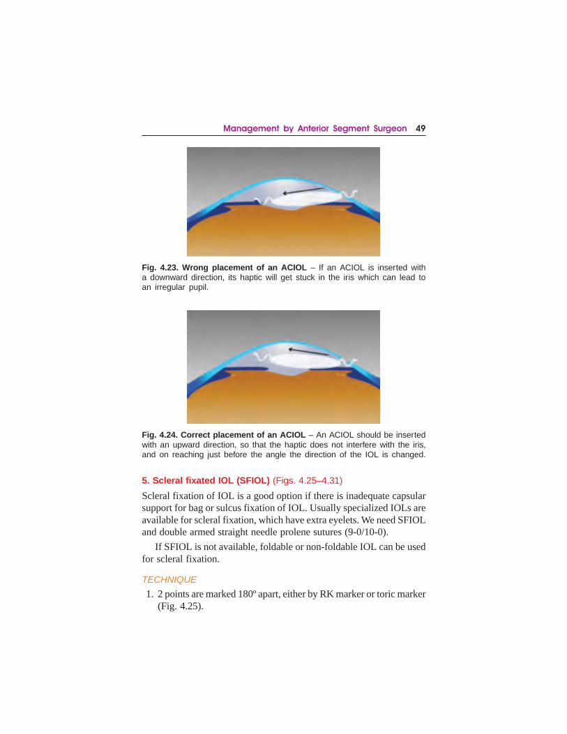
4. Inserting the IOL: The IOL is held with a McPherson forceps, andthe leading loop is advanced in the AC. While advancing the leadingloop of the ACIOL towards the opposite angle, make sure it doesnot touch the iris (Fig. 4.23). In fact the IOL should be kept closerto the cornea, till the loop is about to reach the angle (Fig. 4.24).This will prevent any distortion or ovalization of the pupil post-operatively.
The convexity of the IOL should always be away from thepupil, to avoid pupillary block. Now the trailing IOL is pushedbelow the incision.
5. Iridectomy: One or two iridectomies, at the site where there is novitreous, should be done, preferably with the help of vitreous cutter.This prevents the chances of pupillary block glaucoma.
6. Wound closure: Suturing of the wound is essential, as a shallowAC in presence of an ACIOL can lead to damage to the cornea.When using ACIOLs certain points have to be taken care of:(i) “A constant” of the IOL – lesser power required as compared
to a PCIOL(ii) Proper positioning of ACIOL – maintaining forward convexity
of the IOL(iii) Need for peripheral iridectomy – should be at a site where there
is no vitreousWe usually prefer to wait and insert a secondary PCIOL later, as
compared to ACIOL.
Management by Anterior Segment Surgeon 49
5. Scleral fixated IOL (SFIOL) (Figs. 4.25–4.31)
Scleral fixation of IOL is a good option if there is inadequate capsularsupport for bag or sulcus fixation of IOL. Usually specialized IOLs areavailable for scleral fixation, which have extra eyelets. We need SFIOLand double armed straight needle prolene sutures (9-0/10-0).
If SFIOL is not available, foldable or non-foldable IOL can be usedfor scleral fixation.
TECHNIQUE
1. 2 points are marked 180º apart, either by RK marker or toric marker(Fig. 4.25).
Fig. 4.24. Correct placement of an ACIOL – An ACIOL should be insertedwith an upward direction, so that the haptic does not interfere with the iris,and on reaching just before the angle the direction of the IOL is changed.
Fig. 4.23. Wrong placement of an ACIOL – If an ACIOL is inserted witha downward direction, its haptic will get stuck in the iris which can lead toan irregular pupil.

50 Management of Posterior Capsular Tear
Fig. 4.25. 2 points are marked 180º apart, fornix based conjunctival flaps areraised and light cautery is applied. Limbus based partial thickness scleral flapsare raised, 2.5 × 3 mm and 500 μ in depth. At 1.5 mm from the limbus, throughthe scleral bed a 26 G needle is passed from one side and a straight 9-0needle from the other side, which is loaded into the barrel of the 26 G needlein the centre of the eye.
Fig. 4.26. The prolene needle is brought out along with the 26 G needle.Similarly the second arm of the needle is also brought out through the oppositescleral bed. Hence, we have now two threads in the eye.
Fig. 4.27. The two threads are brought out through the main wound and cut in between.

Management by Anterior Segment Surgeon 51
Fig. 4.28. The SFIOL is positioned and the cut ends of threads are tied atthe eyelets of the SFIOL on both sides.
Fig. 4.29. The SFIOL is introduced in the AC.
Fig. 4.30. The SFIOL is positioned with slight traction on the threads.

52 Management of Posterior Capsular Tear
2. Fornix based conjunctival flaps are raised and light cautery is applied(Fig. 4.25).
3. Creation of scleral flaps (Fig. 4.25): Two partial thicknessquadrangular scleral flaps are created 180º apart at limbus, 3 × 2.5mm in dimension, as for trabeculectomy.
ALTERNATIVES TO SCLERAL FLAP CREATION
Scleral pockets may be created which may be limbus- or fornix-based.
Alternative 1 – Limbus-based scleral pocket: Instead of makingflaps we can make scleral pockets (limbal based pocket), of thesame size as we make for phacoemulsification. The advantage ofthis technique is that scleral flap closure is not required at the endof surgery.
Alternative 2 – Fornix-based scleral pocket:• Straight 3 mm incisions are given 180º apart at the limbus without
removing the conjunctiva.• 2.5 mm deep pocket is made towards the sclera, making fornix
based scleral pocket.• As the direction of the pocket is opposite to the direction of the
needle, it is not possible to insert the needle directly at the baseunderneath the flap. The needle perforates the conjunctivoscleralupper flap and scleral base to enter the eye.
• The threads are passed and tied into the IOL. Then these threadsare pulled out from the scleral pocket by Sinskey hook for tyingand burying the knot in the pocket.
Fig. 4.31. The sutures are tied to the scleral bed. The scleral flaps are sutured,one by one on both the sides. Conjunctiva above is sutured.

Management by Anterior Segment Surgeon 53
This is a little more complicated, with more chances of bleeding,but still a great technique to master as it is faster and requires nocautery and suturing of conjunctiva or scleral flap.
4. Rail road technique (Fig. 4.25):• 26 G needle is mounted on the back of a repositor or a chopper.• The needle is bent at an angle of 45–60º from its hub.• At the junction of the lower 1/3rd and upper 2/3rd of the scleral
bed, about 1.5 mm away from limbus, the needle is passed.• First we go perpendicular to the globe and then turn the needle in
such a way that it comes to lie parallel and just behind the iris. Ifit tucks the iris, or comes anterior to the iris, the needle can bewithdrawn and passed again slightly posterior to the original site.
• Straight needle of prolene suture is passed through the oppositescleral pocket at the same location.
• In the centre of the eye, the straight needle is threaded into thebarrel of 26 G needle, and both are withdrawn from the side of26 G needle.
• Now, similar procedure is repeated, at the junction of upper 1/3rd
and lower 2/3rd of the scleral bed (Fig. 4.26).• Hence now we have 2 prolene threads in the eye (Fig. 4.26).
5. Placing sutures on the IOL:• The 2 prolene threads are brought out through the corneal/limbal
incision, which is created for the insertion of IOL, and are cut inthe centre (Fig. 4.27).
• Now, we have two threads on each side of the incision, whichhave to be tied to respective haptics.
• For tying, one thread is passed 2–3 times in the eyelet and theother thread is passed from the opposite direction before tying(Fig. 4.28).
• After securing both the haptics, the leading haptic i.e., the hapticfixed on the right side suture is gently introduced inside the AC,maintaining a slight pull on the prolene sutures. A gentle pull ismaintained by the assistant on this while inserting the leadinghaptic as well (Fig. 4.29).
6. Securing the sutures: Once the IOL is inserted, slight traction isgiven to the prolene sutures, which are secured onto the sclera afterproper centration of the IOL (Fig. 4.30).

54 Management of Posterior Capsular Tear
7. Suturing the flaps: The partial thickness scleral flaps and theconjunctiva are sutured back to the scleral bed with 10-silk suture(Fig. 4.31).
8. Main incision is closed. Pupil is constricted and diluted tricort isinjected in the AC to look for any vitreous. Anterior vitrectomy hasto be done if vitreous is present.
9. Corneal wounds are hydrated if foldable IOL is used, or sutured ifnon-foldable IOL is used.
If especially designed SFIOLs are not available, same procedurecan be done with single or 3-piece PMMA IOLs. It is advisable to useECCE IOL, as compared to the phacoemulsification profile IOL, dueto its bigger optics and overall size, though it’s not a must.
In case using a foldable IOL, it’s possible to do the same procedurewith a 3-piece foldable IOLs. The only modification being that, one setof sutures have to be passed through the barrel of the cartridge beforesecuring it on the leading haptic of the IOL. Once the leading haptic issecured, the IOL is injected inside the eye. Trailing haptic is left outsideand second set of prolene threads are tied. Now, the trailing haptic isdialed inside the AC. Alternatively the holder folder method of IOLinsertion can be used.
6. Iris fixated IOL
Iris fixation of PCIOL
Technique 1 (Figs. 4.32–4.38)
1. Two side port clear corneal incisions, nearly 3’o clock away fromthe site of the haptic fixation on either side, directing towards eachother are made.
2. After releasing the IOL in the AC, both the haptics are placed behindthe iris and the optic is kept above the iris.
3. Now pilocarpine is injected into the AC to constrict the pupil.
4. The IOL is lifted up with the forceps (lens holding forceps), toidentify the position of the haptics.
5. Now prolene 10-0 is passed through the clear corneal incision behindthe iris and haptics and then taken out in front of the iris from theother side. 26 G needle is introduced and by rail road techniqueneedle of prolene thread is brought out.

Management by Anterior Segment Surgeon 55
Fig. 4.32. The IOL is positioned suchas the optic is above the iris planeand the haptics are behind the iris.Lens holding forceps or a roundedrepositor is used to elevate the opticso that the haptics can beappreciated better behind the iris.
Fig. 4.33. Railroad technique – 9-0straight needle is passed from thecorneal incision going beneath theiris and haptic elevation and re-emerging from the iris soon after. 26G needle is inser ted from theopposite site, and the prolene needleis loaded into its barrel and is broughtout of the eye.
Fig. 4.34. Now the prolene needleis reinserted through the samewound from which it wasexternalized. 26 G needle is nowinserted from the opposite site andagain the prolene is loaded into itsbarrel. This complex is above the irishaptic elevation.
Fig. 4.35. Both threads are externa-lized from the same wound.
6. Now hands are changed and 26 G needle comes from the oppositeCCI and prolene needle is brought out from the other side by railroad technique passing in front of the iris. Thus we have both thethreads on one side of the wound one behind and one on top of theiris and lens haptics.

56 Management of Posterior Capsular Tear
Fig. 4.36. A knot is given and aninstrument is introduced from theopposite side, so as to slide theknot to the appropriate position.
Fig. 4.37. The knot is finalized andthe threads cut. Similar procedureis repeated on the opposite haptic.
Fig. 4.38. Final position.
7. Now tie the triple knot outside the wound. Push the tied knot intothe AC. Sinskey hook or double dialer is introduced from theopposite incision to pull the knot on the iris while maintaining gentletraction in the opposite direction on both the threads outside theeye.
8. Second and third knots are given the same way to firmly anchor thehaptic behind the iris.
9. Suture is cut by Vannas just above the iris.10. Same technique is repeated on the other side.
Technique 2 (Figs. 4.39–4.43)
In this technique only one side port incision is required but it needs9-0 or 10-0 curved prolene needle. If curved needle is not available

Management by Anterior Segment Surgeon 57
surgeon can make straight needle curved by himself. In this techniqueinstead of using rail road technoique surgeon takes out the needle byperforating the cornea from the other side. If the needle is not sharp itbecomes difficult. Then threads are brought out as demonstrated in thepicture and tied.
Fig. 4.39. Alternative. A curved9-0 needle is passed from thecorneal incision and behind theiris haptic elevation, and re-emerges soon after. The curved9-0 needle now re-emerges fromthe cornea at a different site.
Fig. 4.40. An instrument is usedto hook the thread (Siepser’stechnique).
Fig. 4.41. Now the thread isbrought out from the initial wound.
Fig. 4.42. The threads are exter-nalized and a double knot asshown is placed.

58 Management of Posterior Capsular Tear
Iris fixation of iris fixated IOL
These IOLs are very easy to fix.
• IOL is placed in the AC over the iris.• IOL is gripped at the centre, and one end of the IOL is placed
behind the iris and lifted up to identify the site of enclavation.
• Having a firm grip at the optics and ensuring the centration ofIOL, a fine enclavation rod or an iris repositor pushes the iristissue down to have a pincer like grasp of the IOL, on the iris.
• Same procedure is repeated on the other side.
7. Glued IOL
The glued IOL technique is a relatively new method for fixing a posteriorchamber IOL in an eye without a capsule, which has been popularizedby Dr. Amar Agarwal7.
TECHNIQUE
1. 2 points are marked 180º apart, fornix based conjunctival flaps areraised and light cautery is applied (Fig. 4.44). It is essential to mark,as improper placement of haptics can lead to a decentered IOL.
2. After light cautery, limbus based partial thickness scleral flaps areraised, 2.5 × 3 mm and 500 μ in depth (Fig. 4.44).
3. Main port is made for insertion of IOL, which depends upon thetype of IOL we will be placing. Foldable IOLs are preferred overnon-foldable IOLs, as a sutureless surgery with a limbal or clearcorneal incision is possible.
4. Choice of IOL: Any 3-piece IOL would be preferable. AMO SensarIOL is a very good option, because of the slow release of the IOLthrough the cartridge.
Fig. 4.43. The threads are pulledso as to position the knot. A similarknot is placed again by repeatingthe above steps. The opposite hapticis similarly fixated.

Management by Anterior Segment Surgeon 59
5. Formation of AC: When we are going to place the IOL the eyeballhas to be reasonably pressurized either by:(a) Viscoelastics
(b) Anterior chamber maintainer – through limbus
(c) Infusion via pars plana route – if pars plana vitrectomy is done,for removal of dislocated nucleus, the infusion port can be usedfor the IOL implantation.
6. IOL insertion and externalization of haptic: V-Lance knife isintroduced at the base of the bed of the scleral flap 1.5 mm from thelimbus (Fig. 4.45). The V-lance should be just behind the iris, andif by any chance it is in front of the iris it can be positioned again sothat it enters the eye behind the iris. Same procedure is repeated onthe other side. This wound is used both to introduce the vitrectomypick or MST forceps in the eye as well as to externalize the haptic.
Foldable IOL is loaded and advanced to an extent that a small tipof the haptic is outside the cartridge. Cartridge is passed into theAC by the surgeon; the assistant holds the exposed tip by MSTforceps. Surgeon injects the IOL and assistant externalizes theleading haptic and holds it there (Figs. 4.45 and 4.46).
The commonest mistake that occurs here is breakage, damage ordistortion of the haptic, if the forceps is not holding the tip of thehaptic or there is loosening of the grip due to poor quality of theforceps.
A firm grasp is maintained by the assistant on the externalizedhaptic so that it does not become internalized due to traction on thetrailing haptic (Fig. 4.47). Now, a MST forceps is placed inside theAC, and another MST forceps holds the trailing haptic of the IOL,close to the tip and brings it into the AC (Fig. 4.48). Now, anotherforceps holds the tip of the haptic and externalizes it (Fig. 4.49).
7. Haptic fixation: The haptic needs to be placed into a pocket madeat the same level as the base of the flap, in the direction of thenatural direction of the haptic (Fig. 4.44). 26 G needle or V-lancecan be used to create a scleral pocket, on both sides, in which theloop can be guided in (Fig. 4.50). Because of good IOP, it’s easierto create these pockets before entering the AC.
IOL centration is checked; a little pull on the haptic to readjustthe IOL may be required.

60 Management of Posterior Capsular Tear
Fig. 4.47. One MST forceps isplaced inside the AC, and anotherMST forceps holds the trailinghaptic of the IOL, and brings itinto the AC.
Fig. 4.44. 2 points are marked180º apart, fornix basedconjunctival flaps are raised andlight cautery is applied. Limbusbased partial thickness scleralflaps are raised, 2.5 × 3 mm and500 μ in depth. 26 G needle or V-lance is used to create a scleralpocket, on both sides, in whichthe loop can be guided in later.
Fig. 4.45. V-Lance knife isintroduced at the base of the bedof the scleral flap 1.5 mm fromthe limbus. This wound is usedboth to introduce the MST forcepsin the eye as well as to externalizethe haptic. Surgeon injects theIOL and the assistant holds theleading haptic with MST forceps.
Fig. 4.46. Now the assistantexternalizes the leading hapticand holds it there.

Management by Anterior Segment Surgeon 61
Fig. 4.48. Now the MST forcepsare relocated to the tip of thehaptic after pulling the haptic withanother MST forceps.
Fig. 4.49. The loop is externalized.
Fig. 4.50. Both the loops areguided into the scleral pocketswith the help of MST forceps.
Fig. 4.51. Glue is applied at thebase of the flap and scleral flapsare pressed and stuck over it, oneby one on both the sides.Conjunctiva above can be suturedor can be fixed with glue.

62 Management of Posterior Capsular Tear
8. Glue fixation: Once the IOL is in centre, glue is applied at the baseof the flap and scleral flaps are pressed and stuck over it, one byone on both the sides (Fig. 4.51). Glue provides strong closure overthe area and also closes the V-lance opening. Conjunctiva abovecan be sutured or can be fixed with glue.
9. After the IOL has been positioned, pupil is constricted and anyvitreous is looked for. Tricort can be used to stain the vitreous. Ifpresent a thorough vitrectomy is done.
SPECIAL POINTS
1. 3-Piece IOL with prolene haptic preferred.2. Single piece PMMA IOLs can also be used – though more chances
of breakage.3. Large optic PMMA IOLs preferred.4. In case of a dropped IOL: The IOL can be brought into the AC after
thorough pars plana vitrectomy. The haptics are held andexternalized as described earlier.
Advantages of glued IOL
(i) Better fixation(ii) Better centration
(iii) Pseudo tilting(iv) No phacodonesis(v) Faster technique.
Disadvantages of glued IOL
(i) Special forceps needed(ii) Glue needed
(iii) Higher cost due to glue and special forceps.
Summary
Choice of IOL depends upon the extent of the capsular damage, theavailability of IOLs, instruments and the expertise of the surgeon. Ifreasonable centration of the IOL can be ensured, then in the bag fixationor sulcus fixated IOL, with or without optic capture would be ideal.
If there is extensive capsular damage, then we have no choice but togo for ACIOL, SFIOL, Glued or Iris fixated IOL. ACIOL is the fastest,

Management by Anterior Segment Surgeon 63
easiest and can be done even without proper vitrectomy, as insertion ofACIOL causes least pull on the vitreous. Secondary vitrectomy can beperformed if there is increased IOP, inflammation, distortion of thepupil, CME etc. Specifically designed Iris fixated IOL are also a goodoption, because the time taken is same as for ACIOL.
Ideal technique in such cases, as of now, looks to be the glued IOL,which provides excellent centration and fixation, with no pseudo-phacodonesis. This technique needs special instruments and glue whichare expensive. SFIOL serves the same purpose but is more timeconsuming.
5. FINAL VITRECTOMY AND CLOSURE
It is very important to ensure that there is no vitreous in the wound orAC. After putting the IOL constrict the pupil with preservative-freeintracameral pilocarpine and inject diluted Tricort in the AC. Finalvitrectomy is done by removing each and every strand of the vitreous.At this time a small iridectomy can also be performed with vitrectomycutter.
Closure
It is absolutely important to have watertight wound in case of PCT8.The incidence of endophthalmitis is higher in patients with PCT. Notonly one should suture the main incisions but also large, distorted andleaking side port.

64 Management of Posterior Capsular Tear

The experienced cataract surgeon may be competent to continue withsurgery in the presence of a posterior capsular tear. However, once ithas been identified that a nuclear fragment has dropped into the vitreouscavity, it should be accepted that referral to a vitreoretinal surgeon willbe necessary.
ROLE OF CATARACT SURGEONThe preoperative management of eyes with posteriorly dislocated lensmatter begins in the hands of the cataract surgeon. The posterior segmentsurgeon would require corneal clarity and good visibility in order toremove the posteriorly dislocated lens matter, and the anterior segmentsurgeon’s initial management should facilitate this. A few basicguidelines for the cataract surgeon are:
• Do not chase after the falling nuclear fragments and avoidunnecessary manipulations, in order to avoid vitreous traction anda consequent retinal detachment or CME. If retinal detachmentoccurs, a simple surgery will become unnecessarily complicated.
• Do an anterior vitrectomy and ensure that there is no vitreous inthe wound. If the required instrumentation to do a good anteriorvitrectomy is not available then it should be left for the posteriorsegment surgeon to do it.
• If soft lens matter is present in the anterior chamber it should becarefully removed with the suction mode of the cutter after doing
65
Dr. Lalit VermaDr. Tinku Bali

66 Management of Posterior Capsular Tear
anterior vitrectomy. It is important to retain good capsular support,and in case it is difficult to clear the residual lens matter in theanterior chamber it should be left for the posterior segment surgeonwho would be able to remove it more easily from the posteriorapproach.
• Avoid unnecessary damage to the corneal endothelium and iris toprevent corneal haze and inflammation.
• IOL placement: In previous years, dropped nuclei had to beremoved from the limbal route after bringing them to the anteriorchamber. Now with the availability of the fragmatome it is possibleto safely emulsify them within the vitreous cavity and removalthrough the anterior route is very rarely done except sometimes incase of an exceptionally rock hard black cataract. Therefore, thecataract surgeon should implant an IOL (in the sulcus with opticcapture, scleral fixated, or glued), but only if it is stable. If thestability or centration of the lens is questionable, it should not beimplanted. Insertion of an ACIOL should also be avoided beforepars plana lensectomy to avoid the risk of its posterior dislocationand corneal damage.
• In case the intraocular lens dislocates into the vitreous afterimplantation, do not try to remove it, as traction on vitreous basemay cause retinal tear/RD. Also, do not place a second IOL if thefirst one is in the vitreous.
• Suture the corneal wound with 10-0 nylon, even if it appears to be“self-sealing”, to ensure a stable, non-leaky chamber for theposterior segment surgeon. If necessary suture the side port aswell.
• Start frequent corticosteroid antibiotic drops and cycloplegicdrops and make sure the intraocular pressure is wellcontrolled.
Every cataract surgeon should have an arrangement for referral,with a vitreoretinal surgeon and establish a protocol regarding the initialmanagement of the patient. In case a vitreoretinal surgeon is availablein the same hospital, and provided there is adequate corneal clarity, thepars plana lensectomy can be done in the same sitting as the cataractsurgery. In case a vitreoretinal surgeon is not available, or if the corneais hazy the surgery can be deferred for as long as 3 weeks with nodifference in the visual outcomes.

Management by Posterior Segment Surgeon 67
The information that would be helpful for the vitreoretinal surgeonincludes the amount and type of retained lens matter, any manoeuvresperformed while attempting to retrieve it, the hardness of the lens matter,the presence or absence of intraocular lens, the amount of capsularsupport and the calculated IOL power in case an IOL has not beenimplanted.
ROLE OF POSTERIOR SEGMENT SURGEON
The vitreoretinal surgeon first needs to assess the patient and make ajudgement about the amount and hardness of lens matter present andthe urgency with which intervention is required.
INITIAL EXAMINATION
The cataract wound should be checked for any leak. The corneal claritywith particular reference to Descemet’s fold is assessed and AC reactionand intraocular pressure evaluated. Indirect ophthalmoscopy willconfirm the presence of lens matter in the vitreous cavity. Cortical matterwill appear white and fluffy and nuclear matter will appear yellowishbrown with well-defined borders, unless it is surrounded by corticalmatter. The examination should also look for peripheral retinal tears,retinal detachment or choroidal detachment.
In case direct visualization of the fundus is not possible due to cornealhaze, severe AC reaction, lens matter in the pupillary area or associatedvitreous hemorrhage, a B Scan ultrasonography should be done. Thelens matter would appear hyperechoic and may show acousticshadowing and mobility with ocular movement.
INDICATIONS FOR SURGERY
The complications of a dropped nucleus may include raised intraocularpressure (IOP), uveitis, corneal edema, cystoid macular edema, andretinal detachment. Therefore, with improved instrumentation, in thepresent era, even the smallest nuclear fragment is preferably removed.
Sometimes, eyes with a small amount of cortical matter may be treatedconservatively provided there is no inflammation and rise of intraocularpressure. However, they would need a close and prolonged follow up, asdelayed uveitis, secondary glaucoma and CME may occur and require alate pars plana vitrectomy with removal of the lenticular matter.

68 Management of Posterior Capsular Tear
TIMING OF SURGERY
In case adequate corneal clarity is present and a vitreoretinal surgeon isavailable, the removal of lens fragments should not be delayed. If thecornea is hazy and would interfere with visualization, the surgery wouldhave to be deferred till the cornea regains its clarity. Delaying it by 2 to3 weeks will not cause any damage to the eye, as long as inflammationand IOP are controlled.
Eyes with retinal tears, retinal detachment, uncontrolled intraocularpressure or severe inflammation will need early intervention.
OPERATIVE TECHNIQUE
The management of posteriorly dislocated lens matter by thevitreoretinal surgeon entails –
1. Pars plana vitrectomy2. Removal of retained lens matter3. Intraocular lens management4. Management of associated complications e.g. Dropped IOL, Retinal
detachment.
1. PARS PLANA VITRECTOMYA three port pars plana vitrectomy (PPV) is the procedure of choice ineyes with retained lens matter and the standard of care today ismicroincision vitreous surgery (23 G or 25 G). One of the ports has tobe enlarged to 20 G in case a fragmatome needs to be used (23 Gfragmatome is not easily available as yet).
Before starting vitrectomy, ensure a well-sealed cataract wound anda stable anterior chamber, so that there is no chamber collapse and irisprolapse during insertion of the cannula as well as during the surgery.In case the wound is leaky do not hesitate to give additional sutures.
Port creation – The 23 G microcannulae are inserted through theconjunctiva (3–4 mm posterior to the limbus) using a trocar, in a two-step entry. After insertion through the sclera, the microcannulae areheld by the collar to stabilize them while withdrawing the trocar. Thefirst microcannula is inserted into the inferotemporal quadrant, and theinfusion cannula is inserted into the external opening of the micro-
Management by Posterior Segment Surgeon 69
cannula. The infusion is turned on only after visualizing the tip of themicrocannula in the vitreous cavity. Two other microcannulae are insertedin the superotemporal and superonasal quadrants for a three port vitrectomy.
If lens matter in the pupillary axis prevents visualization of theinfusion cannula, a 23 G butterfly cannula connected to an infusionbottle can be introduced through one of the superior ports and thevitreous cutter used through the other port to clear the lens matter in thepupillary area. The infusion is opened once the infusion cannula can bevisualized in the vitreous cavity.
Anterior vitrectomy
The vitrectomy probe is first advanced to the pupillary area to cut anybands of vitreous in the anterior chamber connecting with the posteriorvitreous. The lens matter retained under the iris, within the residuallens capsule, is removed with suction mode of the probe. Care shouldbe taken not to damage the capsule in order to facilitate future IOLimplantation. If an IOL has already been implanted, the cortical mattersurrounding it should be removed, and vitrectomy done just below thelens. Next focus on the posterior segment. Visualisation during surgerymay be hampered by corneal edema or a small pupil. The pupil can bedilated using adrenaline, mechanical stretching, hooks or pupillaryexpansion devices. Wide angle viewing systems are used to give a properview. First try to visualize the dropped lens fragments and their relationto the vitreous as well as the status of the posterior hyaloid, beforestarting vitrectomy.
Core vitrectomy
Start by removing the central vitreous (core vitrectomy). Also clear thevitreous immediately in front of the ports, and then assess whether aPVD is present or not.
PVD induction
If not present, a PVD should be induced at this stage, if possible, bypositioning the cutter just in front of the optic disc and then increasingthe suction to engage the posterior hyaloid. The cutter is then drawn upslowly, keeping the maximum suction. If PVD is induced a shiny reflexwill be seen moving forward together with the vitreous cutter. Thismanoeuvre may have to be repeated several times before a PVD is

70 Management of Posterior Capsular Tear
induced. Triamcinolone acetonide can be used to stain the vitreous beforePVD induction for better identification and a complete removal. A bulkynucleus sitting on the cortical vitreous may not allow PVD induction atthis stage. In this case it can be done towards the end, after removingthe nucleus.
2. REMOVAL OF RETAINED LENS MATTER
Removal of small or soft lens fragments
These can be removed with the cutter (approx 800 cuts/min). The lightpipe can be used to crush the pieces and feed them into the cutter. Forcortical matter suction alone may be used intermittently.
Removal of large / hard nuclear fragments or entire nucleus
A special phacofragmatome is used in these cases. A phacofragmatomeis similar to a phaco probe without an infusion sleeve and has a longerneedle length. As the fragmatome is 20 G (23 G fragmatome is noteasily available), one of the superior sclerotomies has to be enlargedwith a 20 G MVR blade, after making a localized peritomy.
Before the nucleus is tackled with the fragmatome it should beensured that a complete vitrectomy has been done and the nucleus iscompletely free from any vitreous. This is to avoid any vitreous fibrilsfrom being sucked into the fragmatome and causing vitreous traction.Following this the softer epinuclear matter present around the nucleusis aspirated with the cutter to uncover the hard centre.
The phacofragmatome is then introduced into the vitreous cavity.Aspiration alone is used to engage the nucleus/nuclear fragments andgently lift them from the posterior vitreous cavity. Once they are elevatedinto the mid vitreous cavity the ultrasound power is turned on to emulsifythem.
Settings for phacofragmatome
• Power: 20% to 50% depending on the hardness of the nucleus.Use minimum power required to prevent chattering and flying ofthe nuclear pieces away from the fragmatome tip.
• Vaccuum: 150–200 mm Hg. Increase the bottle height beforestarting phacofragmentation to prevent hypotony, as it has a largebore size.

Management by Posterior Segment Surgeon 71
• Mode: Pulse mode with 10–20 pulses/min. The pulse mode alsoprevents repulsion of the nuclear fragments. As there is no capsularbag to provide counter resistance the nuclear pieces tend to berepelled by the ultrasonic energy and repeatedly fall back.
The endoilluminator can be used to support the nucleus from behindwhile it is being emulsified. A good technique is to spike the nucleuson the endoilluminator and in a cartwheel manner use the fragmatometo emulsify the nucleus. Try to work towards the light pipe withoutdislodging it, so that there is minimal need for reengaging the nucleus.(A lighted pick can also be used to stabilize the nucleus instead of theendoilluminator.) If the fragmatome tip goes through and through intothe nucleus, the endoilluminator can be used to push it off the nucleus.It can also be used to crush and feed smaller fragments into the port.
In case the fragments fall down, stop suction immediately to preventhypotony, and activate it only when the tip is occluded by a fragment.If milky fluid is released during fragmentation then wait for a momentfor the media to clear up before continuing.
Alternative technique – A fluid needle can be used to aspirate andhold the nuclear pieces in the mid vitreous cavity while it is emulsifiedby the fragmatome. A chandelier light is used for illumination.
Sometimes if a PVD has not been induced before, small lens piecesget stuck to the posterior hyaloid. A PVD should be induced at thisstage.When these pieces are lifted up with the posterior hyaloid theycan be removed with the cutter. Very small pieces stuck to the retinalsurface can also be removed with the fluid needle. In the end a peripheralexamination with scleral depression should be performed and aperipheral vitrectomy done to eliminate peripheral vitreous traction.Also check for small pieces that can get entrapped at the vitreous base.In case a break/tear is detected, endolaser should be done around it.After all the retained lens matter is removed, the anterior chamber shouldbe irrigated to remove any fragments that may be trapped in the angle.It is important to remove all of the lens fragments to prevent post-opinflammation, phacolytic glaucoma or CME.
Role of Adjunctive Perfluorocarbon Liquid (PFCL)
Some surgeons prefer to inject a small amount of PFCL after vitrectomyand before using the fragmatome, for the following purposes:

72 Management of Posterior Capsular Tear
1. It protects the macula from contusion injuries which may be causedby the impact of lens pieces that fall posteriorly duringfragmentation.
2. It causes the lens pieces to float on its surface, reducing risk of retinaldamage during manipulating these pieces off the retinal surface.
3. It forms a protective layer over the posterior pole and may reducethe risk of damage from the ultrasonic energy of the fragmatome.
Only a small amount of PFCL should be injected, to extend tillthe arcades. Overfill can hamper with removal, because the meniscusof the bubble tends to cause displacement of the lens fragmentstowards the retinal periphery and vitreous base, making theirretrieval difficult. Care should be taken to remove all the PFCL atthe end of the surgery.
Removal of Rock Hard Black cataracts
In case of a rock hard black cataract where a prolonged and difficultphacofragmentation is expected, or in cases where the fundal view iscompromised due to a hazy cornea, the nucleus can be brought into theAC and then delivered from a limbal incision. An adequate sized limbalcorneal incision is first made and then sutured with 10-0 nylon. Aftercore vitrectomy and PVD induction, PFCL is injected upto the level ofsclerotomies (two vials would be needed for this) so that the nucleus ispushed up to the level of the pupil. Viscoelastic is then injected into theAC and the nucleus is manoeuvred into the AC using a membrane pickintroduced from the pars plana incision and another instrument fromthe paracentesis. Limbal sutures are then cut to deliver the nucleus.Surgery is completed with resuturing of limbus, completion of vitrec-tomy and removal of PFCL.
3. IOL MANAGEMENT
After closing the two superior ports, the infusion is left in place in casean IOL is to be implanted, and removed only after IOL implantation. Ifan IOL was inserted at the time of cataract surgery, it may requirerepositioning, if decentered. Sometimes, more than one IOL may beencountered in such eyes – these need to be removed through the limbus.If an IOL was not placed it can be inserted now into the sulcus. In caseinadequate capsular support is present, a scleral fixated or glued IOLcan be considered. ACIOL can also be inserted provided the pupilconstricts well with pilocarpine.

Management by Posterior Segment Surgeon 73
If there is significant corneal edema, choroidal detachment orhaemorrhage, the IOL placement should be deferred.
4. MANAGEMENT OF ASSOCIATED COMPLICATIONS
Dislocated IOL with retained lens matter
Posteriorly dislocated IOL along with a dropped nucleus may occurdue to improper identification of capsular damage or due to incorrectpositioning of the lens. The IOL is tackled after removal of the droppedlens matter and is either repositioned or explanted.
The IOL can be lifted with a retinal pick forceps and brought intothe anterior chamber. If a three-piece IOL is present, it can then bemanipulated into the sulcus if adequate capsular support is present, ormay be glued to the sclera if the capsule is inadequate. Single pieceIOL or IOLs with broken haptics or plate haptics are generally explantedand replaced with the appropriate IOL.
Retinal detachment
If the eye with the dislocated lens fragments also has an associatedrhegmatogenous retinal detachment and/or choroidal detachment, PFCLis injected after doing a vitrectomy to flatten the retina. After doing avitrectomy, perfluorocarbon liquid (PFCL) is injected to flatten theretina. The lens matter floats over the PFCL and can be removed in theanterior vitreous with the fragmatome. After removal of the lens,endolaser is performed around the break. PFCL air exchange is donefollowed by gas or silicone oil injection.
POST-OPERATIVE MONITORING
After removal of retained lens matter the patient should be closelyfollowed for signs of inflammation, increase in IOP, CME and signs ofperipheral retinal tears or detachment are managed appropriately.
PROGNOSIS
A good percentage of patients (44% to 68%) obtain 6/9 or better visualacuity after pars plana vitrectomy done for the management of retainedlens matter. Improved surgical techniques and instrumentation haveimproved the safety and visual outcomes of this procedure.

74 Management of Posterior Capsular Tear

1. Narendran N, Jaycock P, Johnston RL, et al. The Cataract NationalDataset electronic multicentre audit of 55,567 operations: riskstratification for posterior capsule rupture and vitreous loss. Eye(Lond). 2009; 23 (1): 31–37.
2. Nishimura A, Kobayashi A, PhD, Segawa Y, PhD, Sugiyama K,PhD. Endoillumination-assisted cataract surgery in a patient withcorneal opacity. J Cataract Refract Surg. 2003 Dec; 29 (12): 2277–80.
3. Allan J. Flach. Intraoperative Floppy Iris Syndrome: Patho-physiology, Prevention and Treatment. Trans Am Ophthalmol Soc.2009 December; 107: 234–239.
4. Ronald Yeoh. The ‘pupil snap’ sign of posterior capsule rupturewith hydrodissection in phacoemulsification. Br J Ophthalmol. 1996May; 80 (5): 486.
5. Lal H, Sethi A, Bageja S, Popli J. Chopstick technique for nucleusremoval in an impending dropped nucleus. J Cataract Refract Surg.2004 Sep; 30 (9):1835–9.
6. Kumar DA, Agarwal A, Prakash G et al. IOL scaffold technique forposterior capsule rupture. J Refract Surg. 2012 May; 28 (5): 314–5.
75

76 Management of Posterior Capsular Tear
7. Kumar DA, Agarwal A. Glued intraocular lens: a major review onsurgical technique and results. Curr Opin Ophthalmol. 2013 Jan;24 (1): 21–9.
8. Wong TY, Chee SP. The epidemiology of acute endophthalmitisafter cataract surgery in an Asian population. Ophthalmology. 2013Jan; 24 (1): 21–9.

Bibliography 77
• Manual of Phaco Technique, Text and Atlas, by Dr. Harbansh Lal.
• A Guide to phacoemulsification: AIOS CME series (No. 21).Dr. Harbansh Lal.
• Phacoemulsification: Principles and Techniques, 2nd Ed., LucioBuratto.
• Phacodynamics: Mastering the Tools and Techniques ofPhacoemulsification Surgery, Barry S. Seibel.
• Mohammad Zare et al. Risk Factors for Posterior Capsule Ruptureand Vitreous Loss during Phacoemulsification. J Ophthalmic Vis Res.2009 October; 4 (4): 208–212.
• Gimbel HV, Sun R, Ferensowicz M, et al. Intraoperative managementof posterior capsule tears in phacoemulsification and intraocular lensimplantation. Ophthalmology, 2001; 108: 2186–2189; discussion,2190-2192.
• Zaidi FH, Corbett Mc, Burton BJL, Bloom PA. Raising the benchmarkfor the 21st century—the 1000 cataract operations audit and survey:outcomes, consultant-supervised training and sourcing NHS choice.Br J Ophthalmol, 2007; 91: 731–736.
77

78 Management of Posterior Capsular Tear
• Complications in Phacoemulsification: Avoidance, Recognition andManagement, William J. Fishkind.
• Posterior Capsular Rupture: A Practical Guide to Prevention andManagement, Amar Agarwal.
• Preoperative neodymium:YAG anterior capsulotomy in intumescentcataract: preventing extension of the capsular tear to the lens periphery.PMID: 12954318.
• Alan S. Crandall MD, Management of the Small Pupil for CataractSurgery, Cataract and Refractive Surgery Essentials in Ophthalmology2006, pg 23-29.
• Coelho RP, Martin LF, Paula JS, Scott IU. Comparison of preoperativeNd:YAG laser anterior capsulotomy versus two-stage curvilinearcapsulorhexis in phacoemulsification of white intumescent cataracts.Ophthalmic Surg Lasers Imaging, 2009 Nov-Dec; 40 (6): 582–5.
• M. Edward Wilson, Rupal H. Trivedi, Suresh K. Pandey. PediatricCataract Surgery: Techniques, Complications, and Management.
• Intraoperative signs of posterior capsular rupture. http://eyewiki.aao.org/Intraoperative_signs_of_posterior_capsular_rupture
• Schutz JS, Mavrakanas NA. Posterior-assisted levitation in cataractsurgery. Curr Opin Ophthalmol, 2010 Jan; 21 (1): 50–4.
• Pars plicata vitrectomy for posterior capsule rent: http://www.healio.com/ophthalmology/retina-vitreous/news/print/ocular-surgery-news/%7B3698a2c9-aa24-4232-afc3-e064534febc3%7D/pars-plicata-vitrectomy-for-posterior-capsule-rent
• Ah-Fat FG, Sharma MK, Majid MA, Yang YC. Vitreous loss duringconversion from conventional extracapsular cataract extraction tophacoemulsification. J Cataract Refract Surg, 1998 Jun; 24 (6): 801–5.
• Dadeya S, Kamlesh Kumari, Sodhi P. Secondary intraocular lens (IOL)implantation: anterior chamber versus scleral fixation long-termcomparative evaluation. Eur J Ophthalmol, 2003 Aug-Sep; 13 (7):627–33.
• Raju N. Anterior chamber intraocular lens implantation. Indian JOphthalmol, 1989; 37: 73–4.

Bibliography 79
• Suto C, Hori S, Fukuyama E, Akura J. Adjusting intraocular lenspower for sulcus fixation. J Cataract Refract Surg, 2003 Oct; 29 (10):1913–7.
• Zeh WG, Price FW Jr. Iris fixation of posterior chamber intraocularlenses. J Cataract Refract Surg, 2000 Jul; 26 (7): 1028–34.
• De Vries NE, Tahzib NG, Budo CJ, Webers CA, de Boer R, HendrikseF, Nuijts RM. Results of cataract surgery after implantation of aniris-fixated phakic intraocular lens. J Cataract Refract Surg, 2009Jan; 35 (1): 121–6.
• Andrew S. McAllister, Lawrence W. Hirst, Visual outcomes andcomplications of scleral-fixated posterior chamber intraocular lenses.J Cataract Refract Surg, Volume 37, Issue 7 , pp. 1263–1269, July2011.
• DK Berler, MA Friedberg. Scleral fixation of posterior chamberintraocular lens implants combined with vitrectomy. Trans AmOphthalmol Soc, 1991; 89: 215–234.
• Arbisser LB, Charles S, Howcroft M, Werner L. Management ofvitreous loss and dropped nucleus during cataract surgery. OphthalmolClin North Am, 2006 Dec; 19 (4): 495–506.
• Mathai A, Thomas R. Incidence and management of posteriorlydislocated nuclear fragments following phacoemulsification. IndianJ Ophthalmol, 1999; 47: 173–6.
• R Monshizadeh, N Samiy, R Haimovici. Management of retainedintravitreal lens fragments after cataract surgery. Survey ofOphthalmology, 01/1999; 43 (5): 397–404.
• Jorge R, Siqueira RC, Cardillo JA, Costa RA. Fragmatome lifting:surgical option for intraocular lens and foreign body removal.Ophthalmic Surg Lasers Imaging, 2005 May-Jun; 36 (3): 261–4.
• S. Stenkula, E. Byhr, S. Crafoord, JO. Carlsson, M. Jemt. Tacklingthe “dropped nucleus”. Acta Ophthalmologica Scandinavica, Volume76, Issue 2, Article first published online: 27 Jun 2003.

80 Management of Posterior Capsular Tear


