DIABETES MELLITUS
Dr. Ashok Kumar J
Department of Biochemistry
Management and Science University
Malaysia
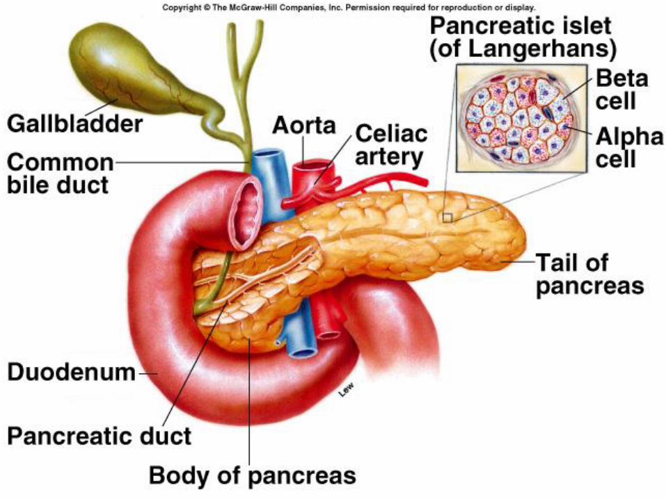
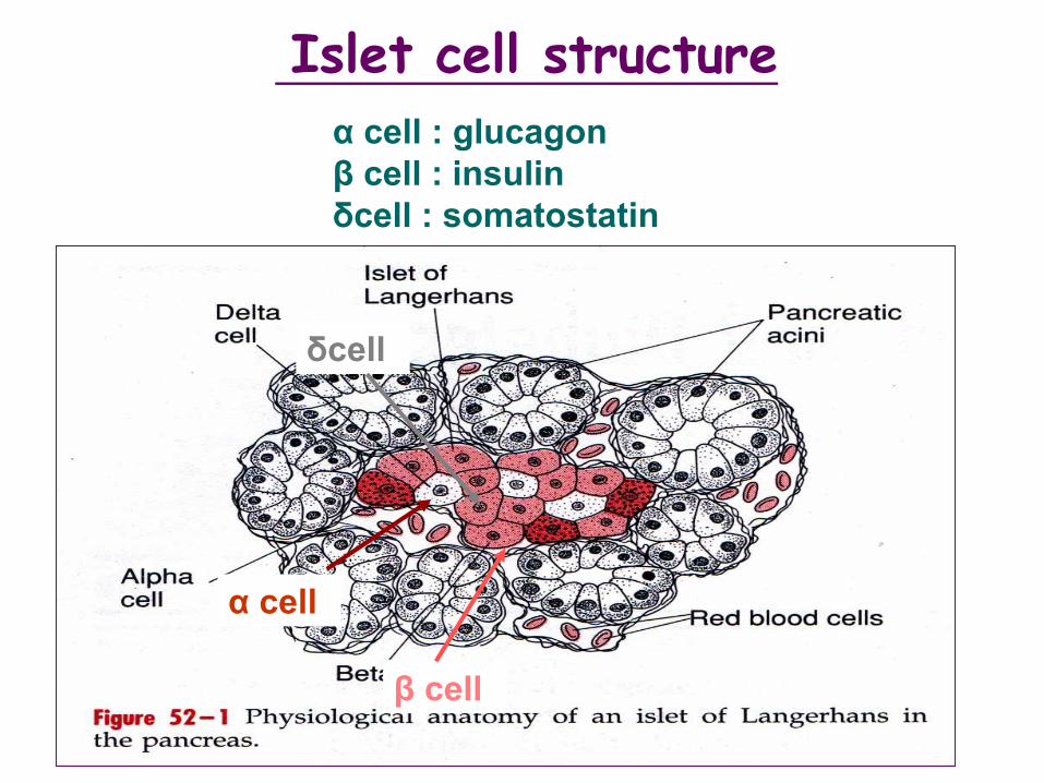
Islet cell structure
α cell : glucagon
β cell : insulin
δcell : somatostatin
α cell
β cell
δcell
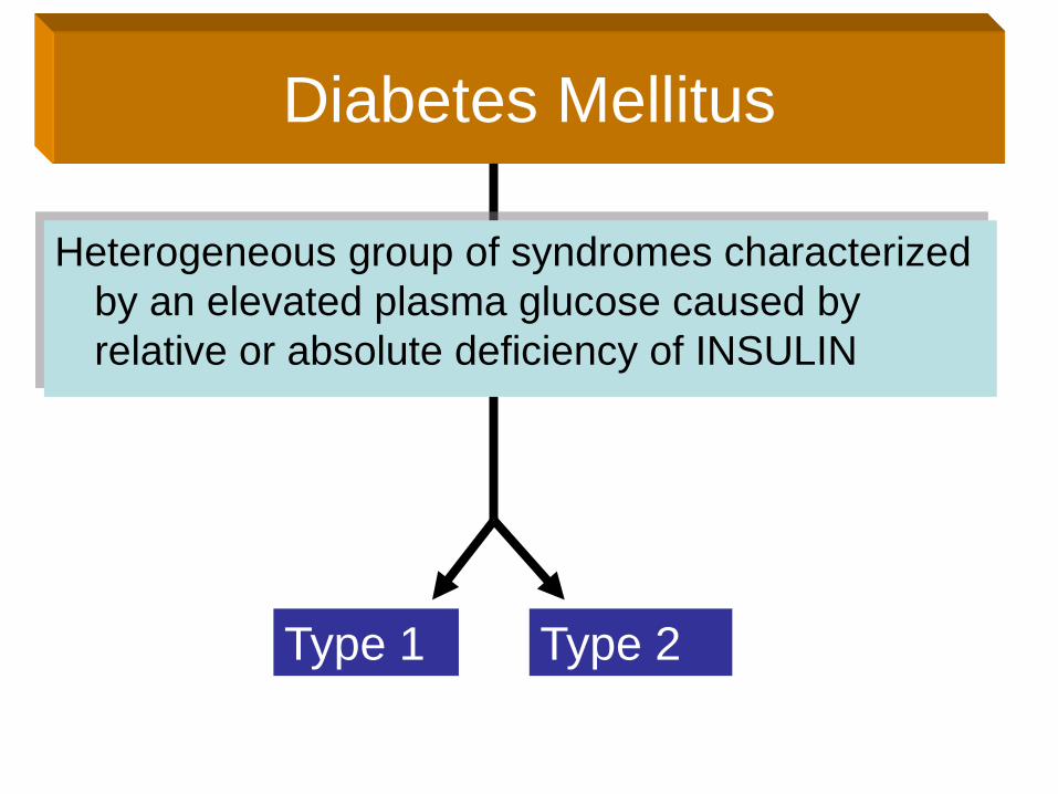
Type 1 Type 2
Heterogeneous group of syndromes characterized
by an elevated plasma glucose caused by
relative or absolute deficiency of INSULIN
Diabetes Mellitus
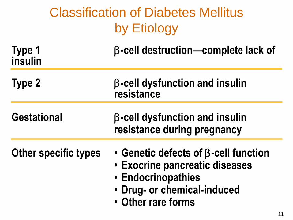
Classification of Diabetes Mellitus
by Etiology
Type 1 -cell destruction—complete lack of insulin
Type 2 -cell dysfunction and insulin resistance
Gestational -cell dysfunction and insulin resistance during pregnancy
Other specific types • Genetic defects of -cell function• Exocrine pancreatic diseases• Endocrinopathies• Drug- or chemical-induced• Other rare forms
11
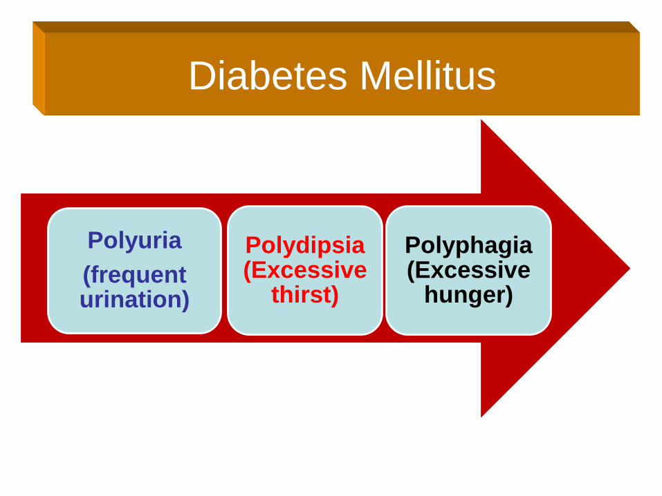
Diabetes Mellitus
Polyuria
(frequent urination)
Polydipsia (Excessive
thirst)
Polyphagia (Excessive
hunger)
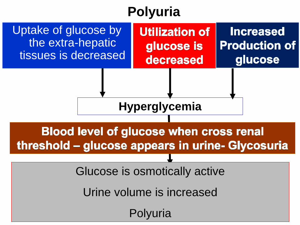
Polyuria
Uptake of glucose by the extra-hepatic
tissues is decreased
Hyperglycemia
Glucose is osmotically active
Urine volume is increased
Polyuria
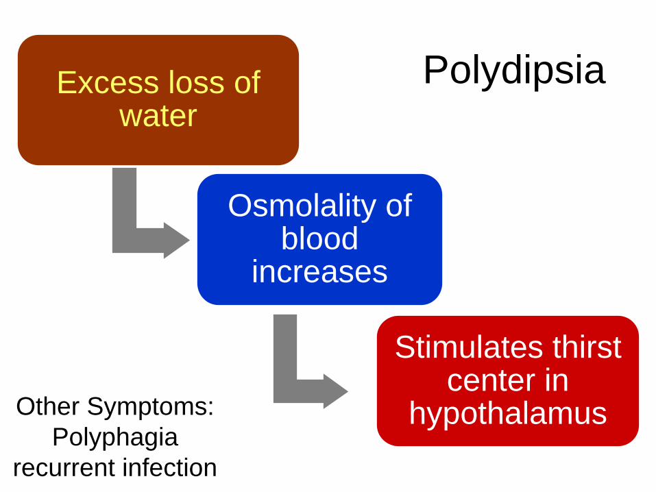
PolydipsiaExcess loss of water
Osmolality of blood
increases
Stimulates thirst center in
hypothalamusOther Symptoms:
Polyphagia
recurrent infection
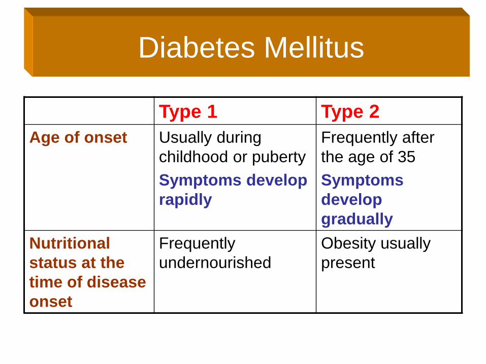
Diabetes Mellitus
Type 1 Type 2
Age of onset Usually during
childhood or puberty
Symptoms develop
rapidly
Frequently after
the age of 35
Symptoms
develop
gradually
Nutritional
status at the
time of disease
onset
Frequently
undernourished
Obesity usually
present
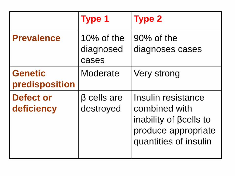
Type 1 Type 2
Prevalence 10% of the
diagnosed
cases
90% of the
diagnoses cases
Genetic
predisposition
Moderate Very strong
Defect or
deficiency
β cells are
destroyed
Insulin resistance
combined with
inability of βcells to
produce appropriate
quantities of insulin
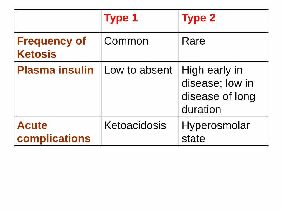
Type 1 Type 2
Frequency of
Ketosis
Common Rare
Plasma insulin Low to absent High early in
disease; low in
disease of long
duration
Acute
complications
Ketoacidosis Hyperosmolar
state
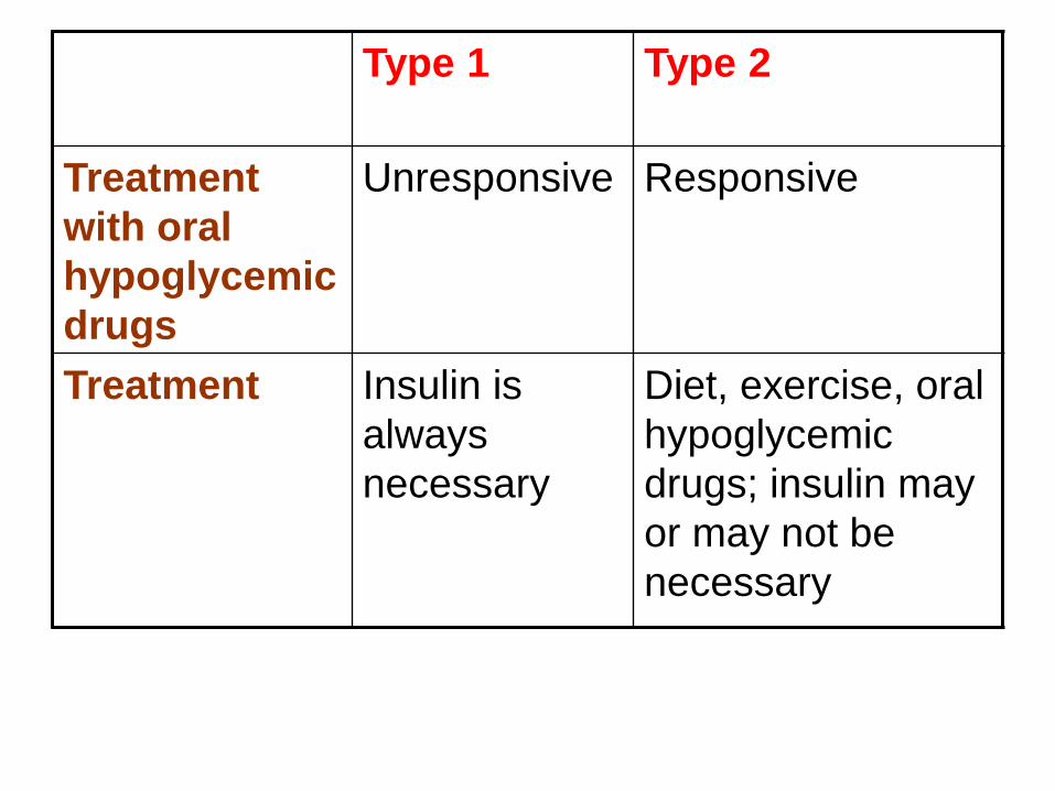
Type 1 Type 2
Treatment
with oral
hypoglycemic
drugs
Unresponsive Responsive
Treatment Insulin is
always
necessary
Diet, exercise, oral
hypoglycemic
drugs; insulin may
or may not be
necessary
Type 1
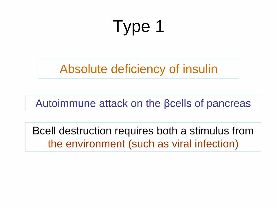
Absolute deficiency of insulin
Autoimmune attack on the βcells of pancreas
Βcell destruction requires both a stimulus from
the environment (such as viral infection)
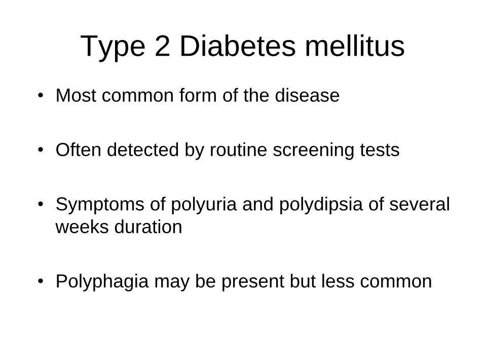
Type 2 Diabetes mellitus
• Most common form of the disease
• Often detected by routine screening tests
• Symptoms of polyuria and polydipsia of several
weeks duration
• Polyphagia may be present but less common
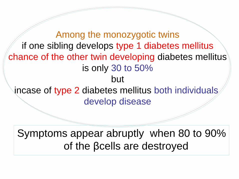
Symptoms appear abruptly when 80 to 90%
of the βcells are destroyed
Among the monozygotic twins
if one sibling develops type 1 diabetes mellitus
chance of the other twin developing diabetes mellitus
is only 30 to 50%
but
incase of type 2 diabetes mellitus both individuals
develop disease
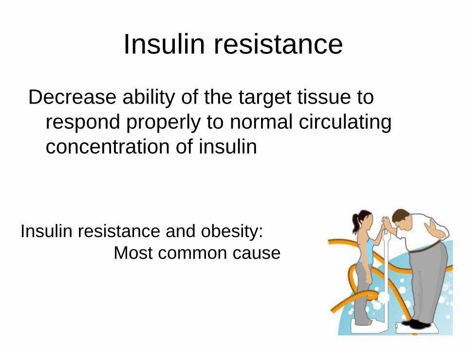
Insulin resistance
Decrease ability of the target tissue to
respond properly to normal circulating
concentration of insulin
Insulin resistance and obesity:
Most common cause
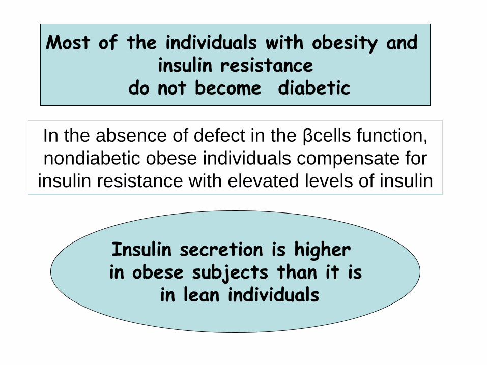
In the absence of defect in the βcells function,
nondiabetic obese individuals compensate for
insulin resistance with elevated levels of insulin
Most of the individuals with obesity and insulin resistance
do not become diabetic
Insulin secretion is higher in obese subjects than it is
in lean individuals
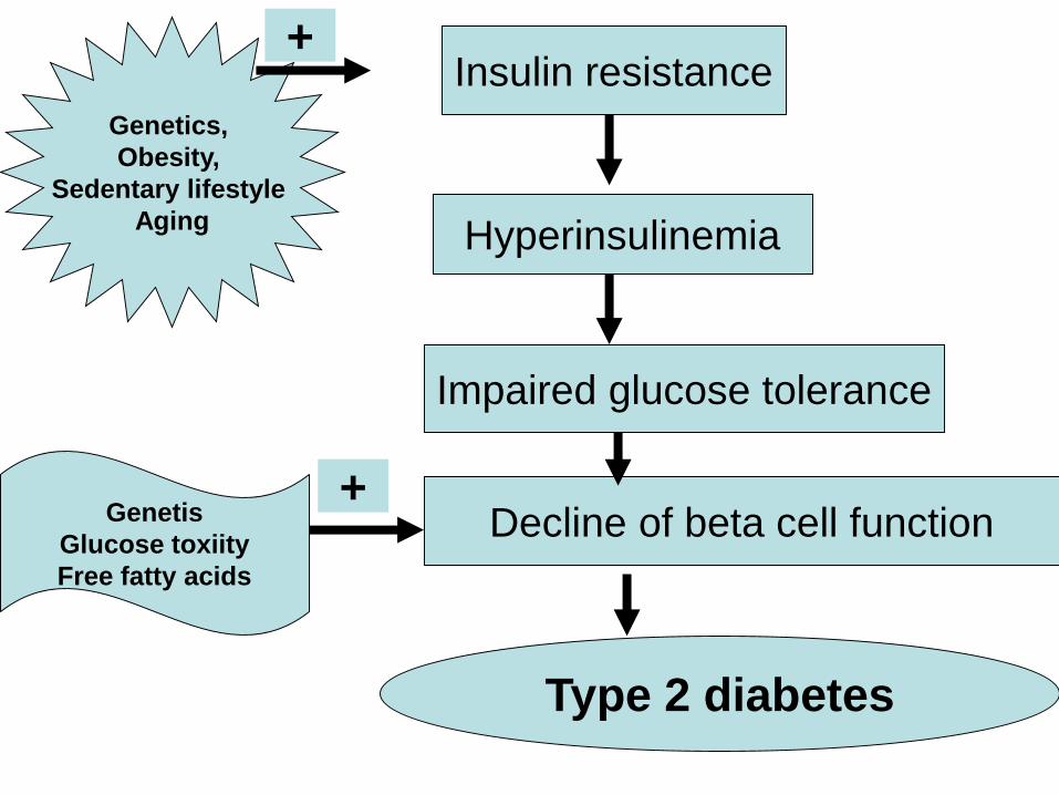
Insulin resistance
Hyperinsulinemia
Impaired glucose tolerance
Decline of beta cell function
Type 2 diabetes
Genetics,
Obesity,
Sedentary lifestyle
Aging
+
Genetis
Glucose toxiity
Free fatty acids
+
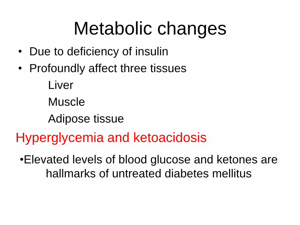
Metabolic changes• Due to deficiency of insulin
• Profoundly affect three tissues
Liver
Muscle
Adipose tissue
Hyperglycemia and ketoacidosis
•Elevated levels of blood glucose and ketones are
hallmarks of untreated diabetes mellitus
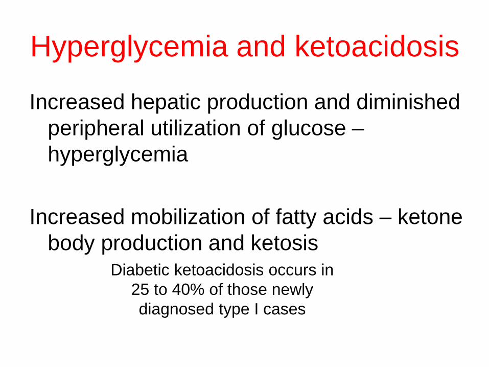
Hyperglycemia and ketoacidosis
Increased hepatic production and diminished
peripheral utilization of glucose –
hyperglycemia
Increased mobilization of fatty acids – ketone
body production and ketosisDiabetic ketoacidosis occurs in
25 to 40% of those newly
diagnosed type I cases
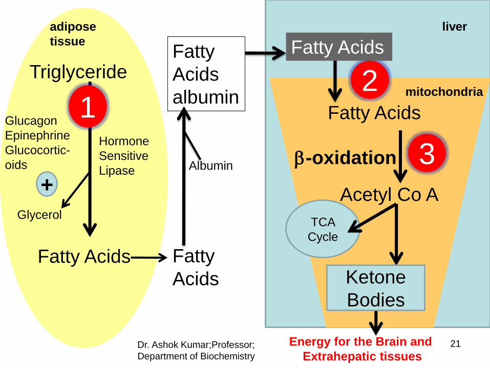
Triglyceride
Fatty Acids
Glycerol
Albumin
adipose
tissue
Fatty
Acids
liver
Fatty Acids
Fatty
Acids
albumin
Acetyl Co A
-oxidation
Energy for the Brain and
Extrahepatic tissues
TCA
Cycle
Hormone
Sensitive
Lipase
Glucagon
Epinephrine
Glucocortic-
oids
+
1
3
2
Fatty Acids
mitochondria
Dr. Ashok Kumar;Professor;
Department of Biochemistry
Ketone
Bodies
21
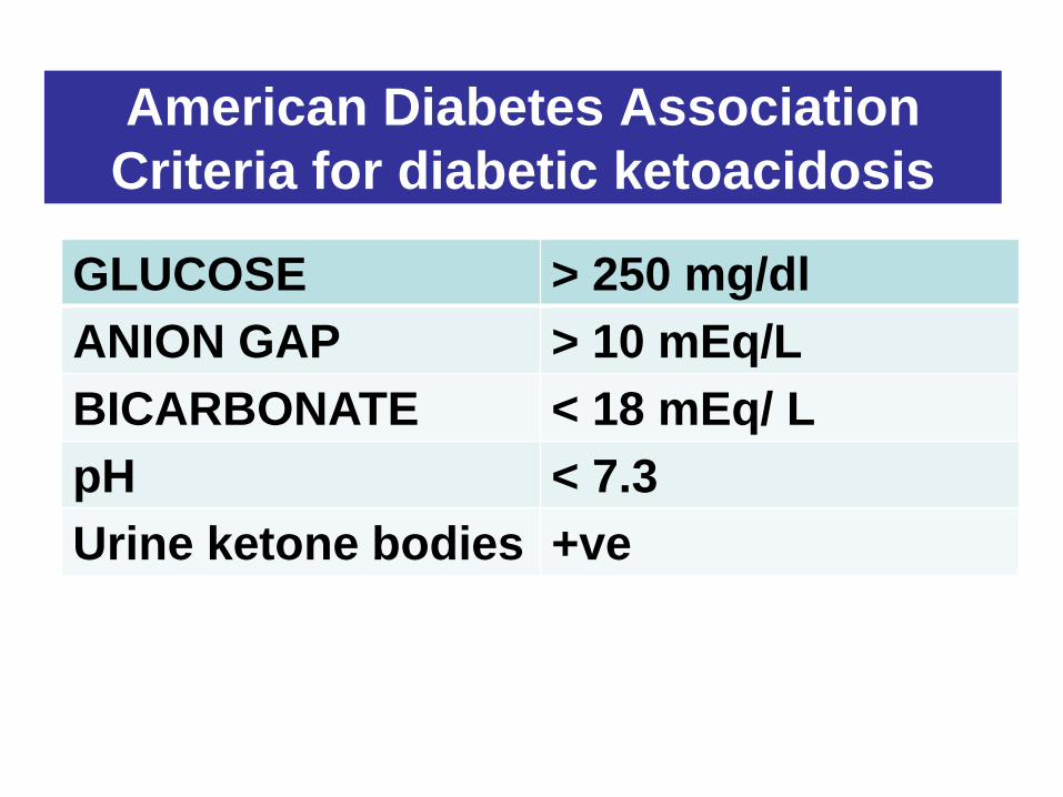
American Diabetes Association
Criteria for diabetic ketoacidosis
GLUCOSE > 250 mg/dl
ANION GAP > 10 mEq/L
BICARBONATE < 18 mEq/ L
pH < 7.3
Urine ketone bodies +ve
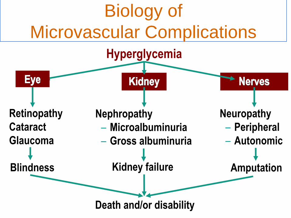
Hyperglycemia
Neuropathy
– Peripheral
– Autonomic
Biology of
Microvascular Complications
Retinopathy
Cataract
Glaucoma
Nephropathy
– Microalbuminuria
– Gross albuminuria
Blindness Kidney failure Amputation
Death and/or disability
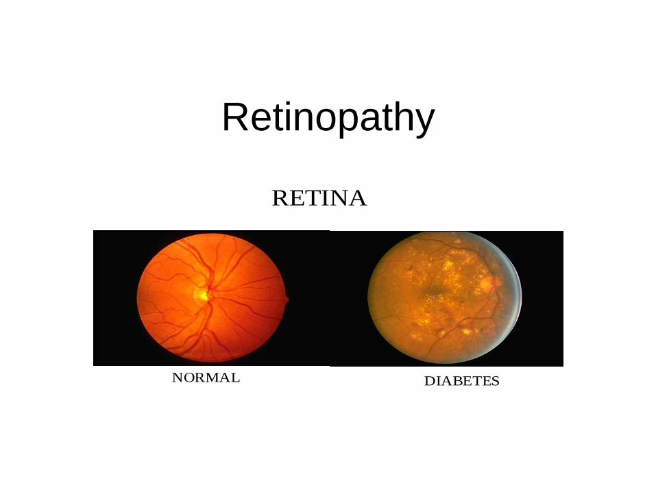
Retinopathy
RETINA
NORMAL DIABETES
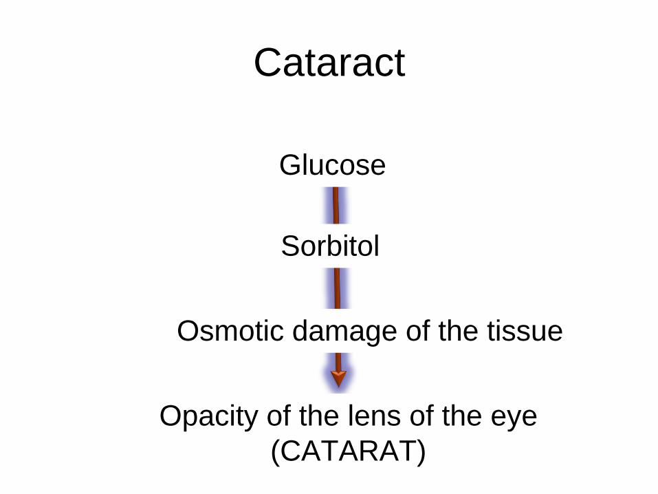
Cataract
Glucose
Sorbitol
Osmotic damage of the tissue
Opacity of the lens of the eye
(CATARAT)
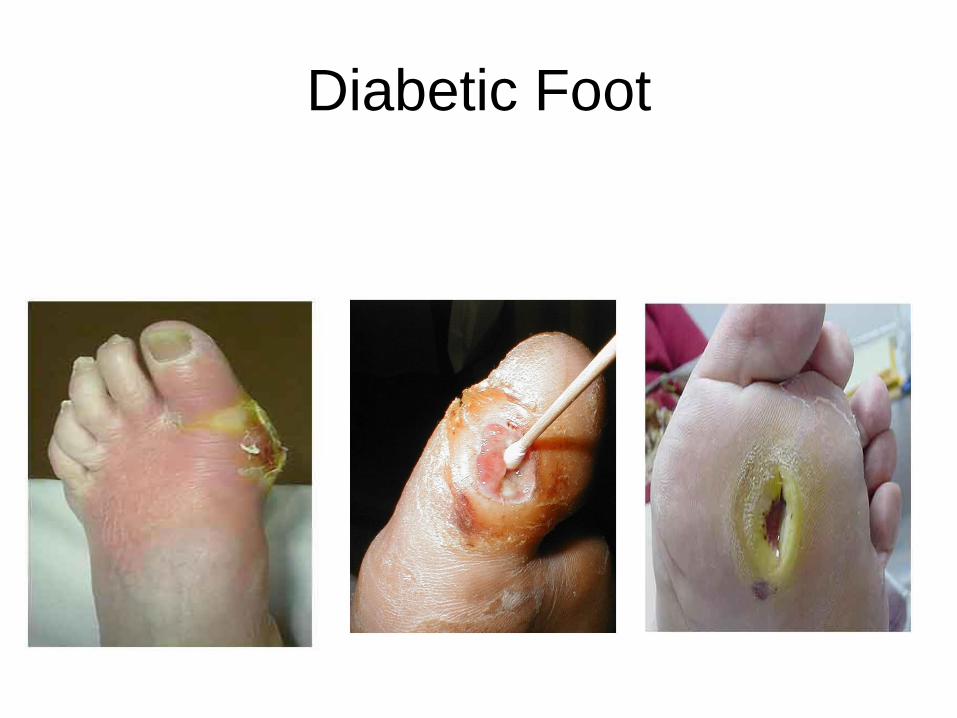
Diabetic Foot
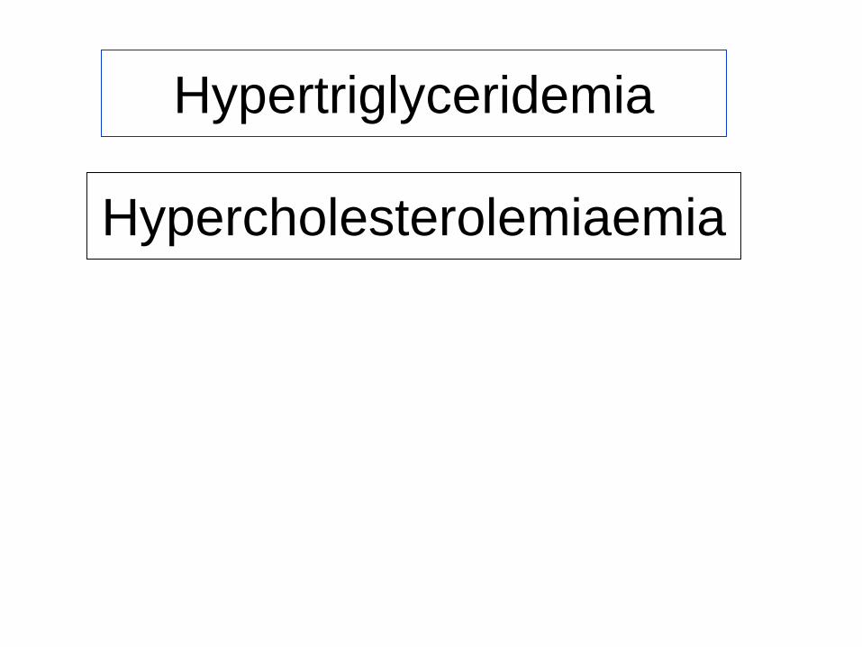
Hypertriglyceridemia
Hypercholesterolemiaemia
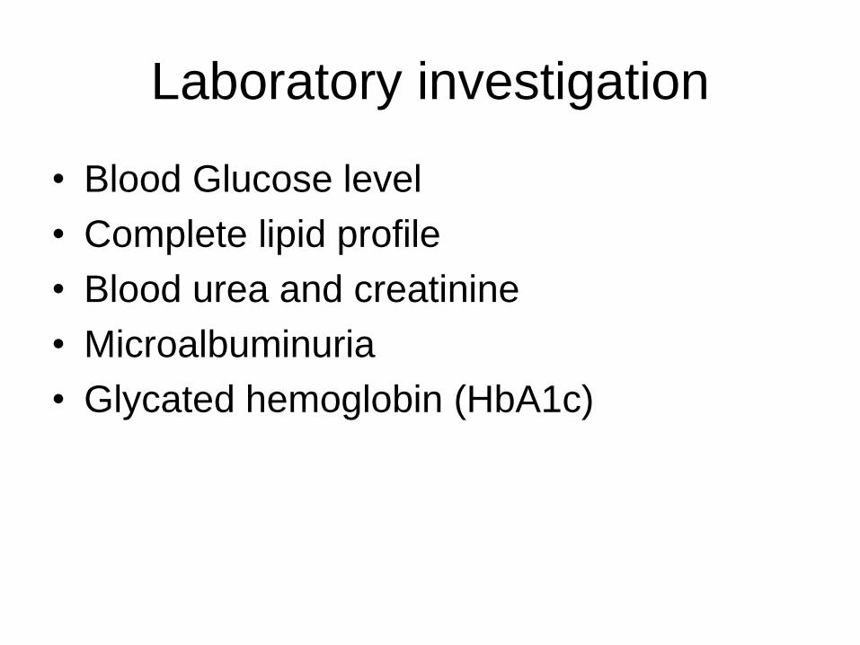
Laboratory investigation
• Blood Glucose level
• Complete lipid profile
• Blood urea and creatinine
• Microalbuminuria
• Glycated hemoglobin (HbA1c)
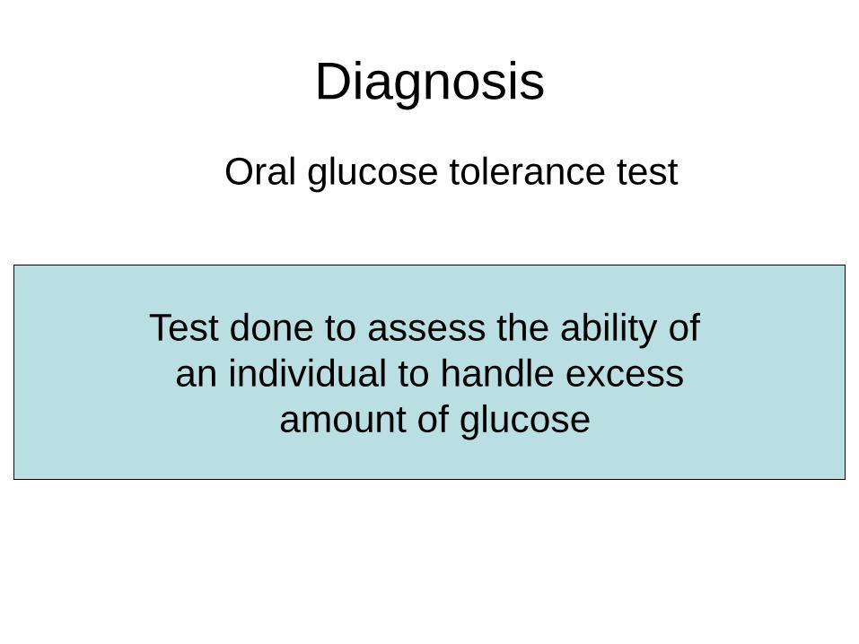
Diagnosis
Oral glucose tolerance test
Test done to assess the ability of
an individual to handle excess
amount of glucose
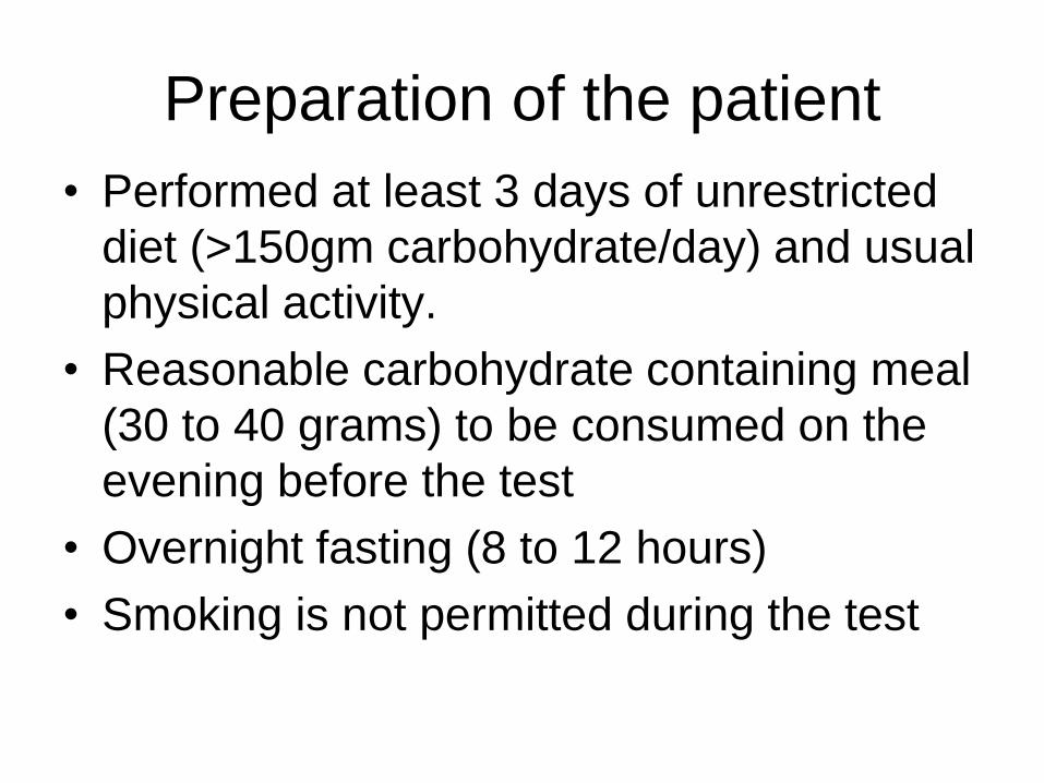
Preparation of the patient
• Performed at least 3 days of unrestricted
diet (>150gm carbohydrate/day) and usual
physical activity.
• Reasonable carbohydrate containing meal
(30 to 40 grams) to be consumed on the
evening before the test
• Overnight fasting (8 to 12 hours)
• Smoking is not permitted during the test
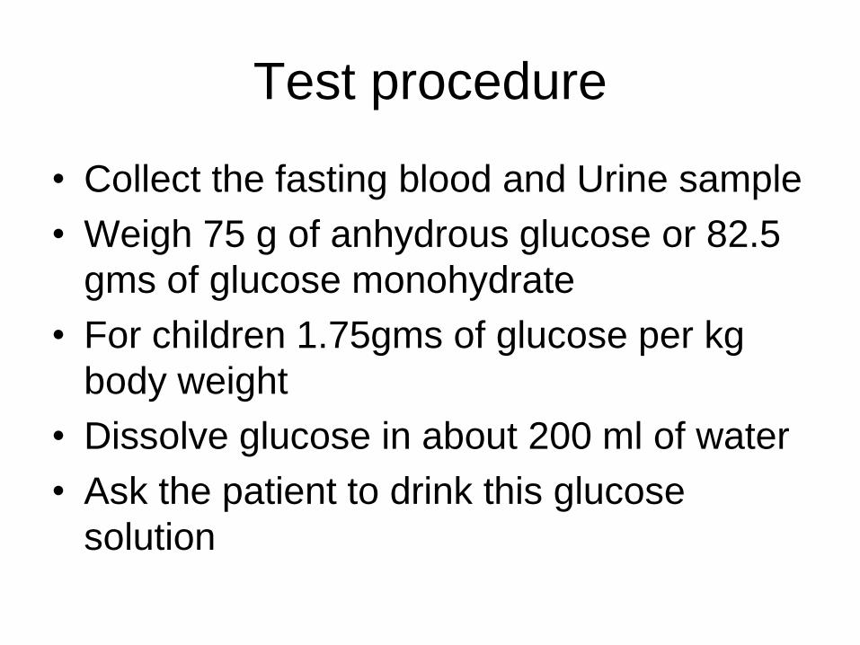
Test procedure
• Collect the fasting blood and Urine sample
• Weigh 75 g of anhydrous glucose or 82.5
gms of glucose monohydrate
• For children 1.75gms of glucose per kg
body weight
• Dissolve glucose in about 200 ml of water
• Ask the patient to drink this glucose
solution
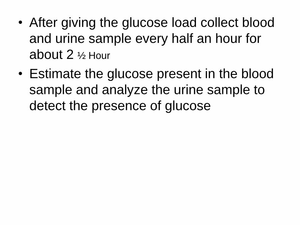
• After giving the glucose load collect blood
and urine sample every half an hour for
about 2 ½ Hour
• Estimate the glucose present in the blood
sample and analyze the urine sample to
detect the presence of glucose
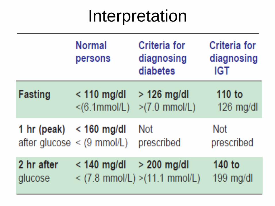
Interpretation
Diagnostic Criteria for Diabetes Mellitus
1. If the fasting plasma glucose is more than
126 mg/dl, on more than one occasion.
2. Or, if 2 hr post-glucose load value of OGTT is
more than 200 mg/dl (even at one occasion).
3. Or, if both fasting and 2 hr values are above
these levels, on the same occasion.
4. If the random plasma glucose level is more than
200 mg/dl, on more than one occasion.
Diagnosis should not be based on a single
random test alone; it should be repeated.
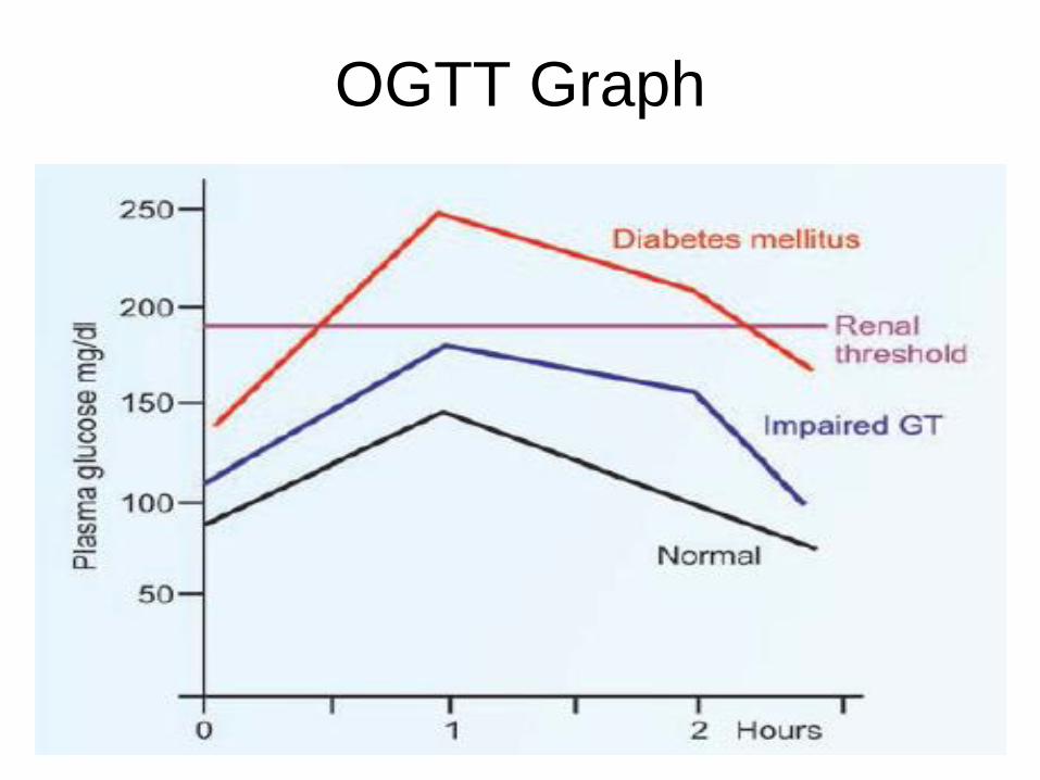
OGTT Graph
Glycated Hemoglobin
• When there is hyperglycemia, proteins in the body
may undergo glycation
• When glucose is attached to hemoglobin,
glucose is not removed from hemoglobin.
[ remains inside the erythrocyte, throughout the
life span of RBCs (120 days)]
• HbA1c - where glucose is attached to the N-
terminal valine of beta chain of hemoglobin
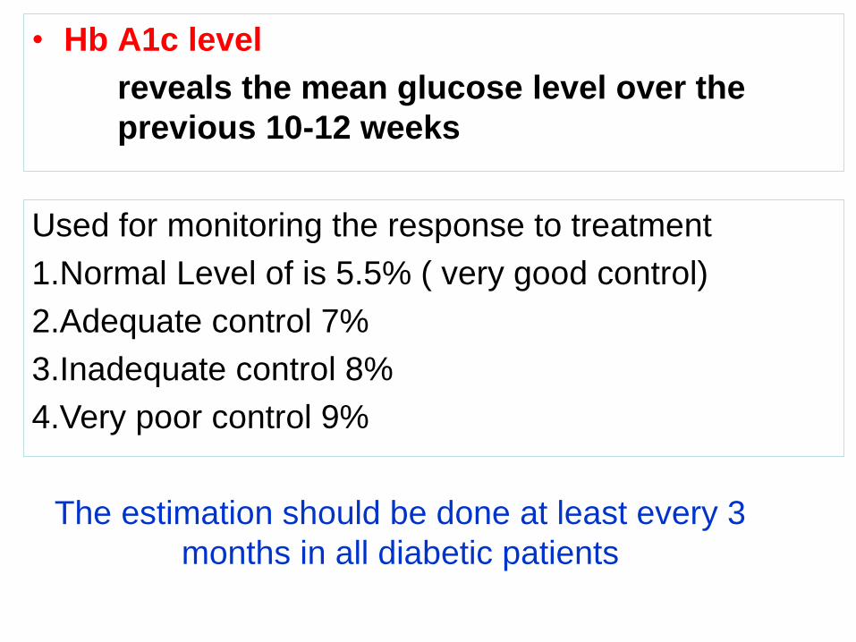
• Hb A1c level
reveals the mean glucose level over the
previous 10-12 weeks
Used for monitoring the response to treatment
1.Normal Level of is 5.5% ( very good control)
2.Adequate control 7%
3.Inadequate control 8%
4.Very poor control 9%
The estimation should be done at least every 3
months in all diabetic patients
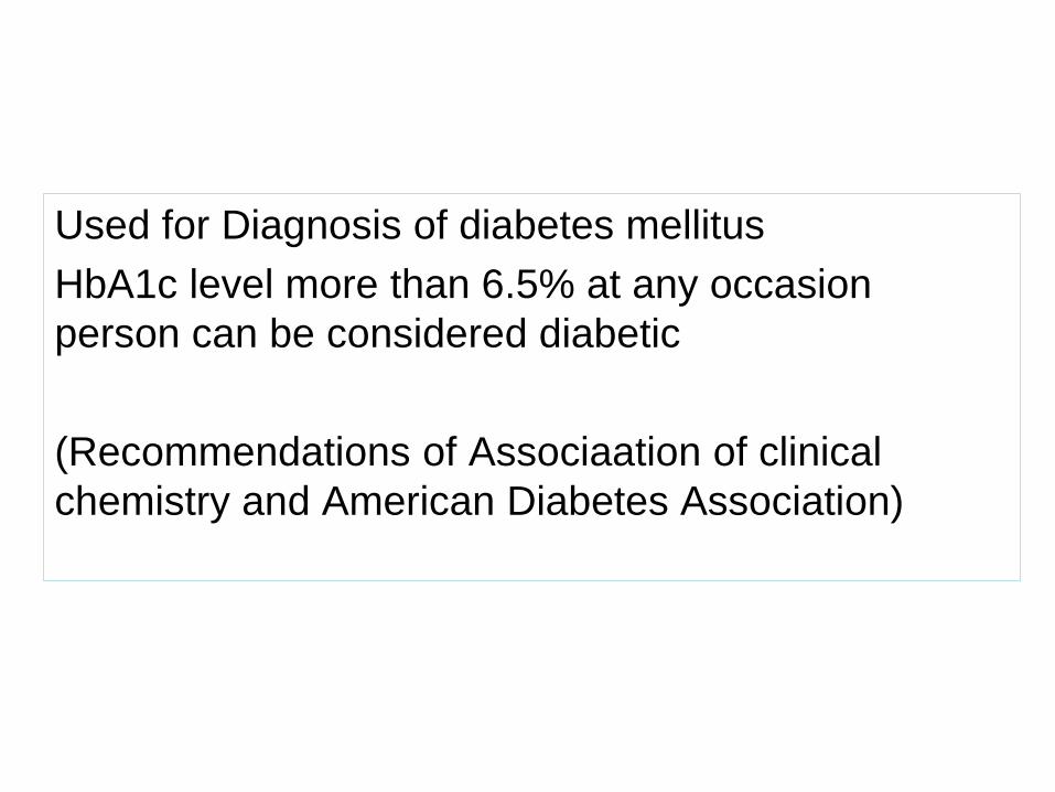
Used for Diagnosis of diabetes mellitus
HbA1c level more than 6.5% at any occasion
person can be considered diabetic
(Recommendations of Associaation of clinical
chemistry and American Diabetes Association)
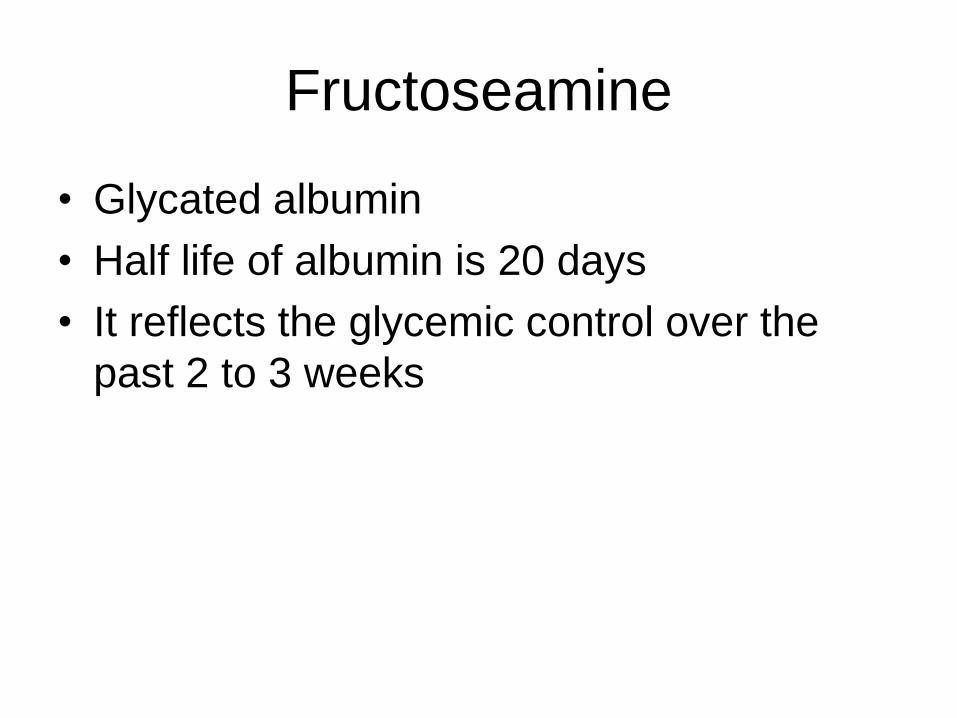
Fructoseamine
• Glycated albumin
• Half life of albumin is 20 days
• It reflects the glycemic control over the
past 2 to 3 weeks
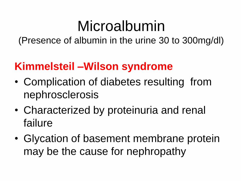
Microalbumin(Presence of albumin in the urine 30 to 300mg/dl)
Kimmelsteil –Wilson syndrome
• Complication of diabetes resulting from
nephrosclerosis
• Characterized by proteinuria and renal
failure
• Glycation of basement membrane protein
may be the cause for nephropathy
CASE 1
Hyperglycemia: Type I Diabetes Mellitus
David Mandel was diagnosed with type I (insulin-dependent)
diabetes mellitus when he was 12 years old . At the time of his
diagnosis, David was in middle school.
He was constantly thirsty and was urinating every 30 to 40 min.
Furthermore, despite a voracious appetite, he seemed to be
losing weight.
David’s parents panicked because they knew that these were
classic symptoms of diabetes mellitus. They took David to see
his pediatrician immediately.
The pediatrician performed a physical examination and ordered
laboratory tests
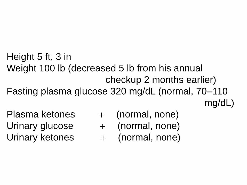
Height 5 ft, 3 in
Weight 100 lb (decreased 5 lb from his annual
checkup 2 months earlier)
Fasting plasma glucose 320 mg/dL (normal, 70–110
mg/dL)
Plasma ketones + (normal, none)
Urinary glucose + (normal, none)
Urinary ketones + (normal, none)
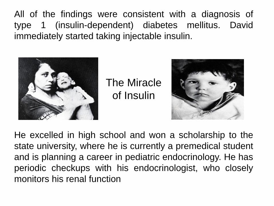
All of the findings were consistent with a diagnosis of
type 1 (insulin-dependent) diabetes mellitus. David
immediately started taking injectable insulin.
He excelled in high school and won a scholarship to the
state university, where he is currently a premedical student
and is planning a career in pediatric endocrinology. He has
periodic checkups with his endocrinologist, who closely
monitors his renal function
The Miracle
of Insulin
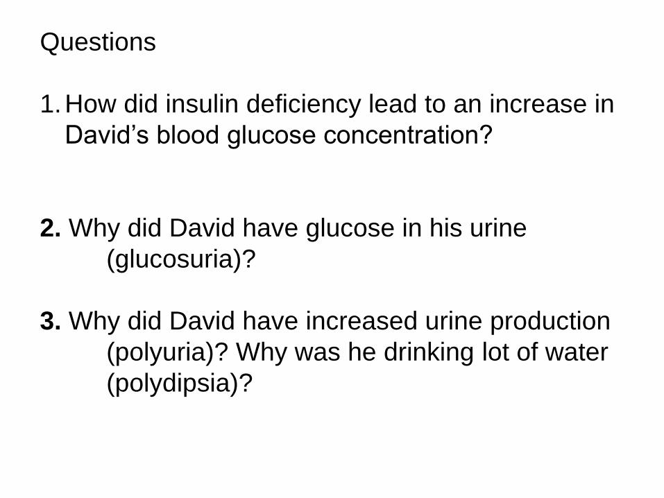
Questions
1.How did insulin deficiency lead to an increase in
David’s blood glucose concentration?
2. Why did David have glucose in his urine
(glucosuria)?
3. Why did David have increased urine production
(polyuria)? Why was he drinking lot of water
(polydipsia)?
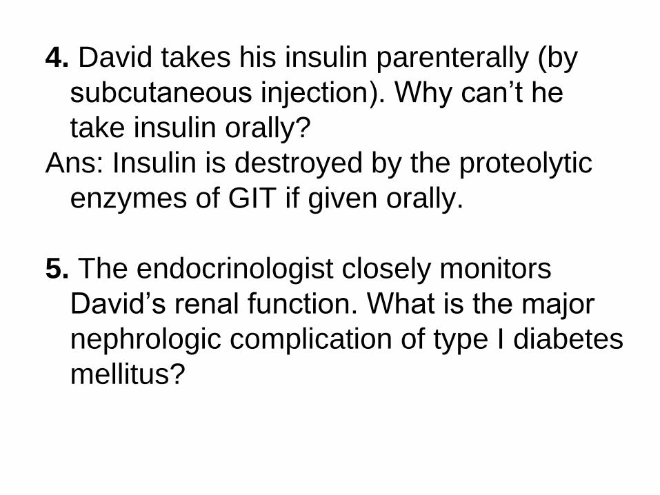
4. David takes his insulin parenterally (by
subcutaneous injection). Why can’t he
take insulin orally?
Ans: Insulin is destroyed by the proteolytic
enzymes of GIT if given orally.
5. The endocrinologist closely monitors
David’s renal function. What is the major
nephrologic complication of type I diabetes
mellitus?
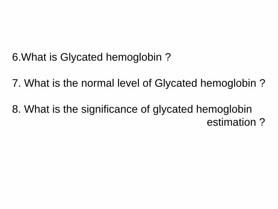
6.What is Glycated hemoglobin ?
7. What is the normal level of Glycated hemoglobin ?
8. What is the significance of glycated hemoglobin
estimation ?
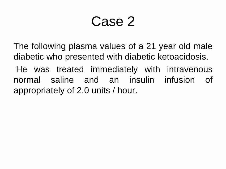
Case 2
The following plasma values of a 21 year old male
diabetic who presented with diabetic ketoacidosis.
He was treated immediately with intravenous
normal saline and an insulin infusion of
appropriately of 2.0 units / hour.
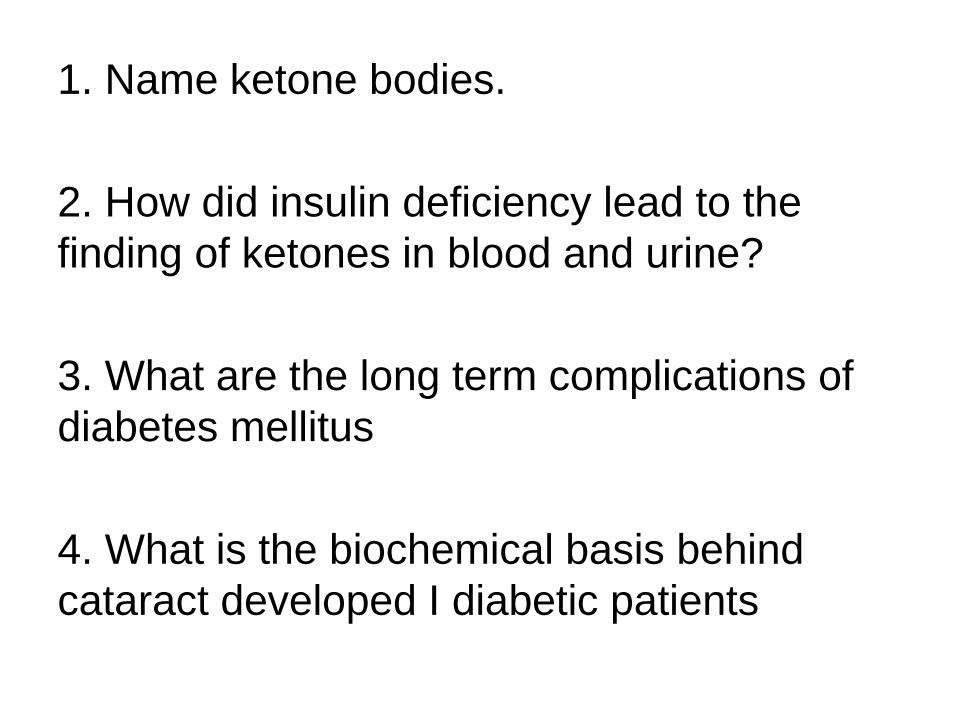
1. Name ketone bodies.
2. How did insulin deficiency lead to the
finding of ketones in blood and urine?
3. What are the long term complications of
diabetes mellitus
4. What is the biochemical basis behind
cataract developed I diabetic patients

• Thank you


