
15/04/2015
1
ANAT3231 Cytoskeleton Introduction
31st March 2015
COMMONWEALTH OF AUSTRALIA
Copyright Regulations 1969
WARNING
This material has been reproduced and communicated to you by and on behalf of the University of New South Wales pursuant to
Part VB of the Copyright Act 1968 (the Act).
The material in this communication may be subject to copyright under this Act. Any further reproduction of communication of
this material by you may be the subject of copyright protection under the Act.
Do not remove this notice

15/04/2015
2
Three filament systems: microtubules, microfilaments (actin filaments) and intermediate filaments
Image: The world of the Cell
Filament dimensions

15/04/2015
3
Functions of the cytoskeleton
• Functions based upon the filaments physical properties
• each filament system has different properties
• Integral strength
• Cell shape
• Motility
• inside the cell
• whole cell
• motor proteins associated with 2 filament systems
• Signal transduction
Note - the Extracellular Matrix has a similar structural role outside of the cell
Functions of the cytoskeleton
MBoC Fig 16-102
Signaling during neutrophil polarization Cytoskeleton driven force generation moving cells forward
MBoC Fig 16-86
Polarization of a cytotoxic T cell after target-cell recognition
MBoC Fig 16-103 Alberts et al. (2008)
Complex morphological changes during development
MBoC Fig 16-106
Alberts et al. (2008)

15/04/2015
4
Structure of the cytoskeleton
• Network of filamentous proteins
• filaments formed from a few proteins
• monomer protein forms polymer filaments
• Located in nucleus and cytoplasmic compartments
• not within organelles
• Location based upon cellular function
• Named on basis of physical size
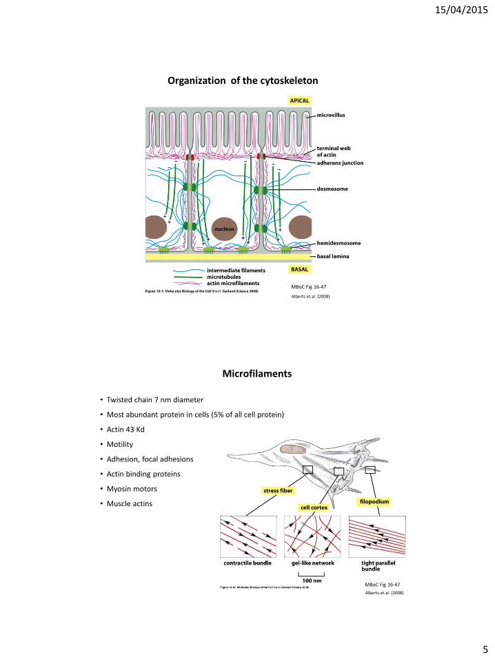
Organization of the cytoskeleton
• Cytoplasmic
• cortical meshwork under plasma membrane
• three dimensional meshwork through
cytoplasm
• Nuclear
• cortical meshwork under nuclear envelope
• Assembly
• some spontaneous
• assembly sites
• Dynamic
• variable stability
• high to low stability
• stability can be altered by associated proteins
and signals
• drugs can alter stability
15/04/2015
5
Organization of the cytoskeleton
MBoC Fig 16-47
Alberts et al. (2008)
Microfilaments
• Twisted chain 7 nm diameter
• Most abundant protein in cells (5% of all cell protein)
• Actin 43 Kd
• Motility
• Adhesion, focal adhesions
• Actin binding proteins
• Myosin motors
• Muscle actins
MBoC Fig 16-47
Alberts et al. (2008)
15/04/2015
6
Intermediate Filaments
• Different cell types, different intermediate filaments
• all eukaryotes nuclear cytoskeleton the same
• Resist stresses applied externally to the cell
• Cytoplasmic
• Anastomosed network
• Flexible intracellular scaffolding
• 10-nanometer diameter
• Cross-linking proteins allow interactions with other cytoskeletal networks
• Intermediate filament associated proteins (IFAPs)
• coordinate interactions between intermediate filaments and other
cytoskeletal elements and organelles
• Human disorders
• mutations weaken structural framework
• increase the risk of cell rupture
Microtubules
• 25 nm diameter, 14 nm internal channel
• Tubulin
• Cytoplasmic
• All cells contain
• Same core structure
• Same motors Dynein (-) and Kinesin (+)
• Different associated proteins
• Dynamic
• Continuous remodelling
• Movement
• Intracellular > cellular
• Cell division mitotic spindle
• Specialized structures
• centrosome, basal bodies, Spindle pole
• Cell processes - cilia (9+2)

15/04/2015
7
Polar versus non-polar
Intermediate Filaments
Image: MBoC Figure 16-11
Microtubules
Microfilaments
Image: MBoC Figure 16-12
No
n-p
olar
Po
lar
Alberts et al. (2008)
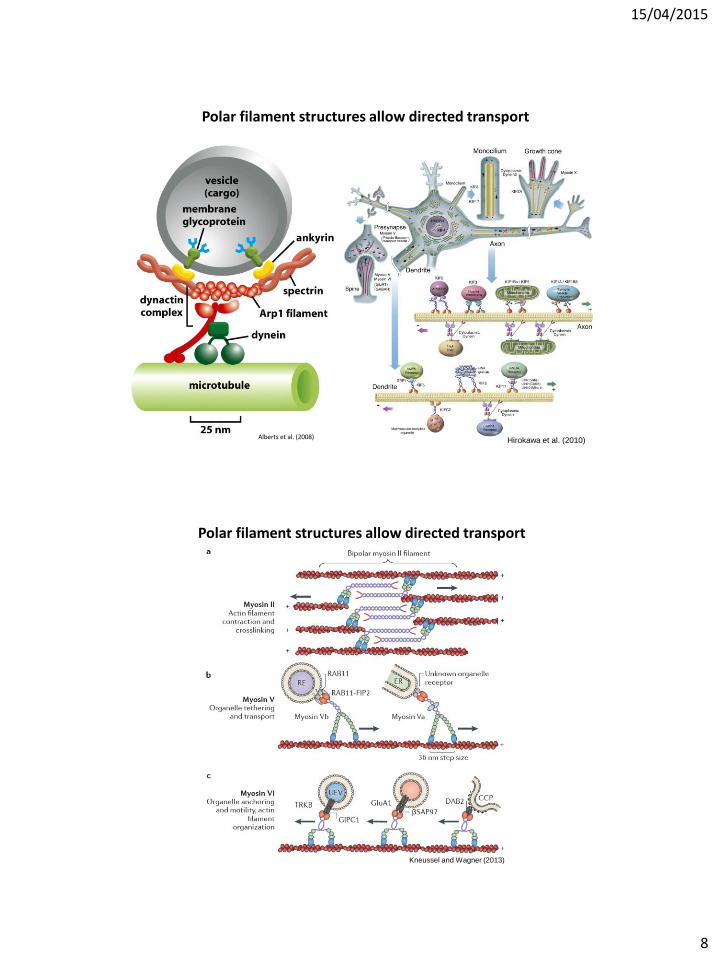
Polar filament structures allow directed transport
Albus et al. (2013)
15/04/2015
8
Polar filament structures allow directed transport
Alberts et al. (2008) Hirokawa et al. (2010)
Polar filament structures allow directed transport
Kneussel and Wagner (2013)

15/04/2015
9
Polar filament structures allow directed transport
• Polarized mRNA localization in the yeast bud tip
MBoC Fig 16-69
Alberts et al. (2008)
Prokaryotic Cytoskeleton Filaments
FtsZ ring
• Microtubule homolog
• Dynamic and exchanges subunits with the cytoplasmic pool
• Assembles into a ring at the future site of bacterial septum in cell division
MreB
• Microfilament (actin) homolog
• Dynamic and exchanges subunits with the cytoplasmic pool
• Assembles into helix-like structures
• Thought to spatially restrict cell growth activities during cell elongation
Crescentin
• Intermediate filament homolog
• Form stable filamentous structures
• Continuously incorporate subunits along their length
• Grow in a nonpolar fashion
• Stably anchored to the cell envelope

15/04/2015
10
How do we generate the diversity of the cytoskeleton?
• Large number of actin- and
microtubule associated
proteins
Alberts et al. (2008)
• Large number of actin- and
microtubule associated
proteins
How do we generate the diversity of the cytoskeleton?
Alberts et al. (2008)

15/04/2015
11
• Many proteins of the cytoskeleton are generated by alternative splicing
• Some examples are:
• lamins (nuclear intermediate filaments)
• tau (mictotubule-associated proteins)
• tropomyosins (actin-associated proteins)
How do we generate the diversity of the cytoskeleton?
(Blencowe, 2006)
Alberts et al. (2008)
Many proteins of the cytoskeleton are generated by alternative splicing
• The lamin protein family
Peter and Stick (2012)

15/04/2015
12
• The tau protein family
Wang and Liu (2008)
Many proteins of the cytoskeleton are generated by alternative splicing
• The tropomyosin protein family TPM1
TPM2
TPM3
TPM4
Many proteins of the cytoskeleton are generated by alternative splicing

15/04/2015
13
Abnormal Cytoskeleton
• Many mutations associated with human diseases
• Toxins can affect organization
Alberts et al. (2008)
References
Website:
http://php.med.unsw.edu.au/cellbiology/index.php?title=Cytoskeleton_Introduction
Articles:
• Alberts et al. (2008) Molecular Biology of the Cell, 5th Edition. • Albus et al. (2012) Cell length sensing for neuronal growth control. Trends Cell Biol, 23: 305-310. • Blencowe (2006) Alternative Splicing: New Insights from Global Analyses. Cell, 126: 37-47. • Hirokawa et al. (2010) Molecular Motors in Neurons: Transport Mechanisms and Roles in Brain
Function, Development, and Disease. Neuron, 18: 610-638. • Kneusel and Wagner (2013) Myosin motors at neuronal synapses: drivers of membrane transport
and actin dynamics. Nat Rev Neursci, 14: 233-247. • Peter and Strick (2012) Evolution of the lamin protein family. Nucleus, 3: 44-59. • Wang and Liu (2008) Microtubule-associated protein tau in development, degeneration and
protection of neurons. Progr Neurobiol, 85: 148-175.

15/04/2015
14
ANAT3231 Filament Dynamics and Research Methods to study the Cytoskeleton
2nd April 2015
Concepts of regulation of filament dynamics
[1] Microtubules [2] Microfilaments

15/04/2015
15
Microtubules
T. Wittmann [Nikon Small World, 2003]
• Cell organizing role
• Cytoskeleton – Largest fibre
– 25 nm diameter
– cytoplasmic
• All cells contain – Same core structure
– Same motors
– Different associated proteins
• Dynamic – Continuous remodelling
• Movement – Intracellular => cellular
– Cell division
Microtubules (EM)
Electron Microscope images transverse/longitudinal sections
Alberts et al. (2008)
15/04/2015
16
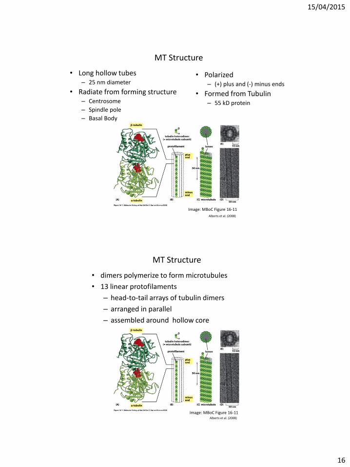
MT Structure
• Long hollow tubes – 25 nm diameter
• Radiate from forming structure – Centrosome
– Spindle pole
– Basal Body
Image: MBoC Figure 16-11
• Polarized – (+) plus and (-) minus ends
• Formed from Tubulin – 55 kD protein
Alberts et al. (2008)
MT Structure
Image: MBoC Figure 16-11
• dimers polymerize to form microtubules
• 13 linear protofilaments
– head-to-tail arrays of tubulin dimers
– arranged in parallel
– assembled around hollow core
Alberts et al. (2008)
15/04/2015
17
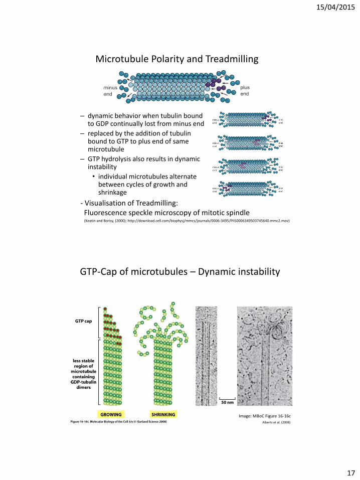
Microtubule Polarity and Treadmilling
– dynamic behavior when tubulin bound to GDP continually lost from minus end
– replaced by the addition of tubulin bound to GTP to plus end of same microtubule
– GTP hydrolysis also results in dynamic instability
• individual microtubules alternate between cycles of growth and shrinkage
- Visualisation of Treadmilling: Fluorescence speckle microscopy of mitotic spindle
(Keatin and Borisy, (2000); http://download.cell.com/biophysj/mmcs/journals/0006-3495/PIIS0006349503745640.mmc2.mov)
GTP-Cap of microtubules – Dynamic instability
Image: MBoC Figure 16-16c
Alberts et al. (2008)

15/04/2015
18
• Addition of tubulin adds GTP to end of protofilament – grows in linear conformation
readily packed into MT wall
– becoming stabilized
• Hydrolysis of GTP – changes subunits conformation
– force protofilament a curved shape
– less able to pack into the MT wall
– protofilaments with GDP-containing subunits forced linear conformation by lateral bonds within MT wall, mainly in stable cap of GTP-containing subunits
Image: MBoC Figure 16-6a
GTP-Cap of microtubules – Dynamic instability
Alberts et al. (2008)
Image: MBoC Figure 16-6b
• Addition of tubulin adds GTP to end of protofilament – grows in linear conformation
readily packed into MT wall
– becoming stabilized
• Hydrolysis of GTP – changes subunits conformation
– force protofilament a curved shape
– less able to pack into the MT wall
– protofilaments with GDP-containing subunits forced linear conformation by lateral bonds within MT wall, mainly in stable cap of GTP-containing subunits
GTP-Cap of microtubules – Dynamic instability
Alberts et al. (2008)
15/04/2015
19
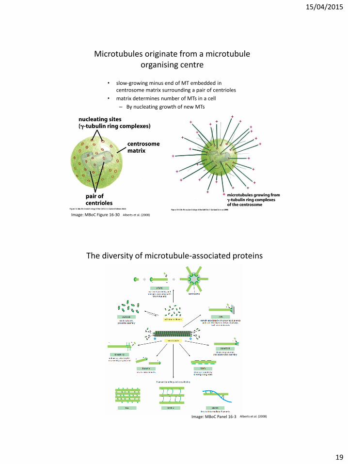
Microtubules originate from a microtubule organising centre
Image: MBoC Figure 16-30
• slow-growing minus end of MT embedded in centrosome matrix surrounding a pair of centrioles
• matrix determines number of MTs in a cell
– By nucleating growth of new MTs
Alberts et al. (2008)
The diversity of microtubule-associated proteins
Image: MBoC Panel 16-3 Alberts et al. (2008)

15/04/2015
20
Microfilaments
• Twisted chain 7 nm diameter
• Compared to MT – Thinner, more flexible, shorter
• Point in same direction
• Different organisation in different cellular regions
MBoC Figure16-49
Alberts et al. (2008)
Actin Types
• 6 Mammalian actin types (isoforms) – All are 43 Kd Protein
• 2 cytoskeletal isoforms in all non-muscle cells – Beta (b) 7p22-p12
– Gamma (g) 17q25
• 4 muscle isoforms in different muscle cells – Alpha (a) skeletal
– Alpha (a) cardiac
– Alpha (a) smooth
– Gamma (g) smooth

15/04/2015
21
Actin Protein
• Conserved in mammals
• Different ratios (b:g) in different cell types
• 374aa, 43 kD protein
• 4 aa difference between beta and gamma – at N- terminal
• Highly expressed gene – Promoter used in gene transfections
Actin Microfilament Formation
• Globular actin monomer (G-actin) polymerise to Filamentous actin (F-actin)
– Cells approx. 50:50
– Monomer can add to either (+ or - ) end
• Faster at + end
• Actin-ATP hydrolyzed (ADP) following addition
– Destabilizes (like MT)
Alberts et al. (2008)

15/04/2015
22
Nucleation/Elongation
• Nucleation – Two actin molecules bind weakly
– addition of a third (trimer) stabilizes the complex
– forms a "nucleation site”
• Elongation – Additional actin molecules form a long helical polymer
• Initial period of growth
• Then equilibrium phase reached
• Dynamic Equilibrium • Elongation ><Depolymerization controls filament length
Actin Binding Proteins
• Regulate polymerisation and create different structures
– Monomer binding protein • Sequester
• release
– Polymer binding proteins • Bundling
• cross-linking
• Severing
• contracting

15/04/2015
23
Actin Binding Proteins
Image: MBoC Figure 16-79 Alberts et al. (2008)
(Revenu et al. 2004)
Actin filament dynamics

15/04/2015
24
(Revenu et al. 2004)
Actin filament dynamics
(Baum et al. 2006)
How can we study the structure and function of the cytoskeleton?
3 Major Functions
• Spatial organization of the cellular contents • Physical and biochemical link to the external environment • Generation of forces for cell movement and reshaping
3 Levels at which to study the cytoskeleton
• In vitro • In vivo
• Intra vital

15/04/2015
25
Detailed imaging using a diverse range of imaging technologies
T. Wittmann [Nikon Small World, 2003] http://www.nhlbi.nih.gov/research/intramural/researchers/core/electron-microscopy-core/media-gallery
Electron microscopy Light microscopy
Reconstitute in vitro
Studying filament assembly outside of the cell

15/04/2015
26
Alberts et al. (2008)
Structure of microfilaments
Detailed imaging using a diverse range of imaging technologies
Structure of microtubules
Mandelkow et al. (1991)
In vitro analysis of filament assembly
Microtubules
Microtubule assembly assay
(Eidenmueller et al., 2000)
Pyrene polymerisation assay
Microfilaments
(Gupton et al., 2007)
15/04/2015
27
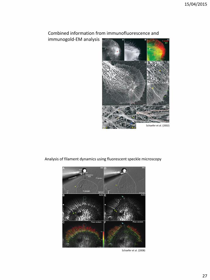
Combined information from immunofluorescence and immunogold-EM analysis
Schaefer et al. (2002)
Analysis of filament dynamics using fluorescent speckle microscopy
Schaefer et al. (2008)

15/04/2015
28
(Danuser and Waterman-Storer, 2006)
Analysis of filament dynamics using fluorescent speckle microscopy
(Svitkina, 2009)
Correlative phase-contrast and EM

15/04/2015
29
References
Articles:
• Baum et al. (2006) Regulation of apicomplexan actin-based motility. Nat Rev Microbiol, 4: 621-628. • Danuser and Waterman Storer (2006) Quantitative Fluorescent Speckle Microscopy of Cytoskeleton
Dynamics. Annu Rev Biophys Biomol Struct, 35:361–87. • Eidenmueller (2000) Structural and Functional Implications of Tau Hyperphosphorylation: Information • from Phosphorylation-Mimicking Mutated Tau Proteins. Biochemistry, 39, 13166-13175. • Gupton et al. (2007) mDia2 regulates actin and focal adhesion dynamics and organization in the
lamella for efficient epithelial cell migration. J Cell Sci, 120: 3475-3487 • Mandelkow et al. (1991) Microtubule Dynamics and Microtubule Caps: A Time-resolved Cryo-Electron
Microscopy Study. J Cell Biol, 114: 1991977-991. • Revenu et al. (2004) The co-workers of actin filaments: from cell structures to signals. Nat Rev Mol
Cell Biol, 5: 1-12. • Schaefer et al. (2002) Filopodia and actin arcs guide the assembly and transport of two populations of
microtubules with unique dynamic parameters in neuronal growth cones. J Cell Biol, 158: 139-152. • Schaefer et al. (2008) Coordination of Actin Filament and Microtubule Dynamics during Neurite
Outgrowth. Dev Cell, 15: 146–162 • Svitkina (2009) Imaging Cytoskeleton Components by Electron Microscopy. Methods Mol Biol. 2009 ;
586: 187–206.
ANAT3231 Mechanistic insights into the regulation of the cytoskeleton and how the cytoskeleton regulates biological functions
14th April 2015
Intermediate filaments &

15/04/2015
30
COMMONWEALTH OF AUSTRALIA
Copyright Regulations 1969
WARNING
This material has been reproduced and communicated to you by and on behalf of the University of New South Wales pursuant to
Part VB of the Copyright Act 1968 (the Act).
The material in this communication may be subject to copyright under this Act. Any further reproduction of communication of
this material by you may be the subject of copyright protection under the Act.
Do not remove this notice
Intermediate Filaments
Physical Characteristics
- 10 nm diameter
- Named by size relative to other cytoskeletal filaments
- intermediate filaments have no structural polarity
- Monomer - central α-helical domain
- Dimer - 2 monomers form parallel coiled coil
- Tetramer - pair of parallel dimers associates in an antiparallel staggered fashion
- tetramer is the soluble subunit (analogous to MT αβ-tubulin dimer, or MF actin
monomer)
- Provide rope-like resistance to mechanical stress
- In muscle: link Z discs of adjacent myofibrils
- Organization can be altered by phosphorylation

15/04/2015
31
Intermediate Filaments - Structure
(Hermann et al., 2007)
Intermediate Filaments - Structure
(Tang, 2008)

15/04/2015
32
Intermediate Filaments - Structure
ULF
(Hermann et al., 2007)
Type I (n = 28) Acidic keratins (pI < 5.7) 40–64 kDa
K9-28 (epithelia) K31-40 (hair/nail)
Type II (n = 26) Basic keratins (pI ≥ 6.0) 53–67 kDa
K1-8, K71-80 (epithelia) K81-86 (hair/nail)
Keratins form heterodimers that assemble into heteropolymeric keratin filaments
Intermediate Filament - Types

15/04/2015
33
Differential expression of Keratins
Dey et al. (2014)
Type III Desmin (cardiac, skeletal and smooth muscle) Vimentin (widespread distribution: leukocytes, blood vessels, endothelial, some epithelial and mesenchymal cells) 56 kDa Peripherin (neurons) 57 kDa Glial fibrillary acidic protein (GFAP) (astrocytes/glia) 50 kDa
Type III intermediate filament proteins can form both homo- and heteropolymeric filaments
Intermediate Filament - Types

15/04/2015
34
Intermediate Filament - Types
Type IV Neurofilament Low NF-L (neurons) 62 kDa Neurofilament Medium NF-M (neurons) 110 kDa Neurofilament High NF-H (neurons) 130 kDa
Neurofilaments form heteropolymers
α-internexin (CNS neurons) Synemins (muscle) Syncoilin (muscle) Nestin (stem cell marker) 220 kDa
Type V Lamin A/C (ubiquitous) 62–72 kDa Lamin B1/2 (ubiquitous) 65–68 kDa
Davidson and Lammerding (2014)
Intermediate Filament - Types
Type IV Neurofilament Low NF-L (neurons) 62 kDa Neurofilament Medium NF-M (neurons) 110 kDa Neurofilament High NF-H (neurons) 130 kDa
Neurofilaments form heteropolymers
α-internexin (CNS neurons) Synemins (muscle) Syncoilin (muscle) Nestin (stem cell marker) 220 kDa
Type V Lamin A/C (ubiquitous) 62–72 kDa Lamin B1/2 (ubiquitous) 65–68 kDa
Orphan Phakinin (lens) Filensin (lens)

15/04/2015
35
Intermediate Filament Associated Protein (IFAP)
Cross-link intermediate filaments with one another
forming a bundle (also called a tonofilament) or a network
IFAPs
Plectin 500 kDa Striated muscle, epithelia Nuclear envelop
Syncoilin 64 kDa Striated muscle
Nesprin-3 117 kDa Kerotinocytes
Paranemin 280 kDa
Desmuslin 230 kDa
Intermediate Filaments - Function
(Kottke et al. , 2006)

15/04/2015
36
Vikstrom et al. (1992)
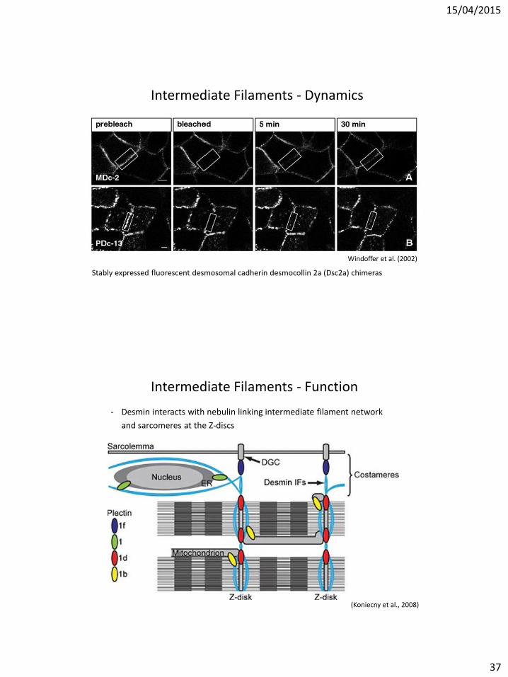
Intermediate Filaments - Dynamics
rhodamine-vimentin networks in 3T3 cells
Pre-bleach Bleach 13.5 min after bleach
Intermediate Filaments - Dynamics
(Moch et al – 2013)
FRAP in AK13-1 cells producing fluorescent HK13
15/04/2015
37
Intermediate Filaments - Dynamics
Windoffer et al. (2002)
Stably expressed fluorescent desmosomal cadherin desmocollin 2a (Dsc2a) chimeras
Intermediate Filaments - Function
- Desmin interacts with nebulin linking intermediate filament network
and sarcomeres at the Z-discs
(Koniecny et al., 2008)

15/04/2015
38
Intermediate Filaments Impact on dynamic cellular processes
Chung et al. (2013)
Chung et al. (2013)
Intermediate Filaments Impact on dynamic cellular processes

15/04/2015
39
(Huber et al., 2015)
Interplay between Microtubules, the Actin Cytoskeleton and Intermediate Filaments
(Baum et al. 2006)
How is Filament Dynamics and Stability Regulated?

15/04/2015
40
(Revenu et al. 2004)
How is Filament Dynamics and Stability Regulated?
How is Filament Dynamics and Stability Regulated?
Rho Rho GDP- GTP-
GAP
GEF
Rac Rac GTP- GDP-
GAP
GEF
ROCK PAK
Cofilin Cofilin -P
LIMK
Slingshot
Actin depolymerisation

15/04/2015
41
How is Filament Dynamics and Stability Regulated?
Gunning et al. (2005)
(Ridley, 2010)
Different signalling pathways lead to different spatial
organisation of the actin cytoskeleton
15/04/2015
42
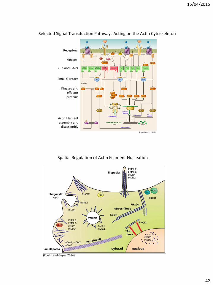
(Ligeti et al., 2012)
Selected Signal Transduction Pathways Acting on the Actin Cytoskeleton
Receptors
Kinases
GEFs and GAPs
Small GTPases
Kinases and effector proteins
Actin filament assembly and
disassembly
(Kuehn and Geyer, 2014)
Spatial Regulation of Actin Filament Nucleation

15/04/2015
43
Microtubules and the Actin Cytoskeleton in Cell Migration
(Blanchoin et al., 2014)
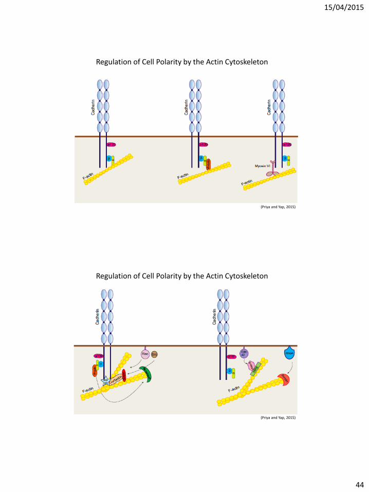
Regulation of Cell Polarity by the Actin Cytoskeleton
(Mack and Georgiou, 2012)
15/04/2015
44
(Priya and Yap, 2015)
Regulation of Cell Polarity by the Actin Cytoskeleton
(Priya and Yap, 2015)
Regulation of Cell Polarity by the Actin Cytoskeleton

15/04/2015
45
(Mack and Georgiou, 2012)
Regulation of Cell Polarity by the Actin Cytoskeleton
Regulation of Cell Polarity by the Actin Cytoskeleton
(Stiess and Bradke, 2011)

15/04/2015
46
Microtubules and the Actin Cytoskeleton in Neuronal Cell Morphogenesis
(Akhmanova and Hoogenraad, 2015)
Microtubules and the Actin Cytoskeleton Neurite Branching
(Spillane and Gallo, 2014)

15/04/2015
47
(Spillane and Gallo, 2014)
Microtubules and the Actin Cytoskeleton Neurite Branching
(Lamprecht, 2011)
Regulation of a Dynamic Actin Cytoskeleton at the Neuronal Synapse

15/04/2015
48
(Hanley, 2014)
Regulation of a Dynamic Actin Cytoskeleton at the Neuronal Synapse
AMPA receptor subunits
Endosomal system
The Cytoskeleton in the T-cell Response
Angus and Griffiths (2013)

15/04/2015
49
(Chircop, 2014)
Regulation of the Actin Cytoskeleton during Cell Division
Regulation of the Actin Cytoskeleton during Cell Division
(Chircop, 2014)
15/04/2015
50
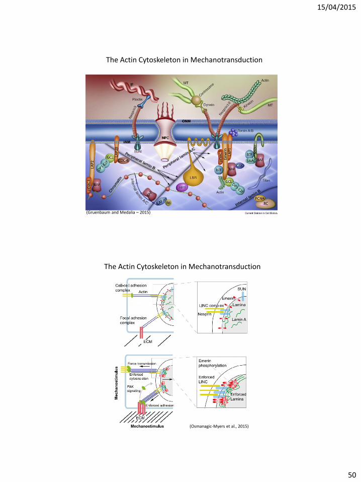
(Gruenbaum and Medalia – 2015)
The Actin Cytoskeleton in Mechanotransduction
The Actin Cytoskeleton in Mechanotransduction
(Osmanagic-Myers et al., 2015)

15/04/2015
51
Abnormal Cytoskeleton
• Many mutations associated with human diseases
• Toxins can affect organization
The Cytoskeleton in Disease
Goetz and Ittner (2008)
APP = Apolipoprotein
http://www.ahaf.org
http://med.kuleuven.be

15/04/2015
52
The Cytoskeleton in Disease
(Suchowerska and Fath, 2014)
(Toivola et al., 2015)
The Cytoskeleton in Disease

15/04/2015
53
Learning outcomes
After the Cytoskeleton lecture, you should be able to
• Describe the structure of the three cytoskeletal filament systems in eukaryotic cells, microtubules, intermediate filaments and microfilaments
• Identify the key regulatory mechanisms of the dynamics of these systems • Acquired an understanding of research methodologies to study the
function of the cytoskeleton • To name key cellular processes that are regulated by the cytoskeleton and
to be able to explain the functional role of the cytoskeleton in these processes and how these are disrupted in disease
References
Articles:
• Akhmanova and Hoogenraad (2015) Curr Biol, 25: R162-171. • Alberts et al. (2008) Molecular Biology of the Cell, 5th Edition. • Angus and Griffiths (2013) Curr Opin Cell Biol, 25: 85–91. • Baum et al. (2006) Nat Rev Microbiol, 4: 621-628. • Blanchoin et al. (2014) Phyysiol Rev, 94: 235-263. • Chircop (2014) Small GTPases, 5: e29770—1 - e29770—14. • Chung et al. (2013) Curr Opin Cell Biol, 25: 600–612. • Davidson and Lammerding (2014) Trends Cell Biol, 24: 247–256. • Dey et al. (2014) Diagnost Cytopath, DOI: 10.1002/dc.23132. • Goetz and Ittner (2008) Nat Rev Neurosci, 9: 532-544. • Gunning et al. (2005) Trends Cell Bio, 15: 333-341. • Gruenbaum and Medalia (2015) Curr Opin Cell Biol, 32:7–12. • Hanley (2014) Front Neurosci, 8: 1-8. • Herrmann et al. (2007) Nature Reviews - Mol Cell Biol, 8: 562-573. • Huber et al. (2015) Curr Opin Cell Biol, 32: 39–47. • Koniecny et al. (2008) J Cell Biol, 181, 667-681. • Kottke et al. (2006) J Cell Sci, 119, 797-806. • Kuehn and Geyer (2014) Small GTPases, 5: 1-15. • Lamprecht (2011) Progr Neurobiol, 117: 1–19. • Ligeti et al. (2012) Physiol Rev, 92: 237–272. • Mack and Georgiou (2012) Small GTPases, 5: 1-16. • Moch et al. (2013) Proc Nat Acad Sci USA, 110: 10664-10669. • Osmanagic-Myers et al. (2015) Genes & Dev 29:225–237. • Priya and Yap (2015) Curr Topics Dev Biol, 112: 65-102. • Revenu et al. (2004) Nat Rev Mol Cell Biol, 5: 1-5. • Ridley (2006) Trends Cell Biol, 16: 522-529. • Spillane and Gallo (2014) Small GTPases, 5, e279741-9. • Stiess and Bradke (2011) Dev Neurobiol, DOI 10.1002/dneu.20849 • Suchowerska and Fath (2014) Front Biol, 9: 5-17 • Tang (2008) Am J Physiol Cell Physiol, 2008: C869–C878. • Vikstrom et al. (1992) J Cell Biol, 118: 121–129. • Windoffer et al. (2002) J Cell Sci, 115: 1717-1732.
Book:
• Alberts et al. (2008) Molecular Biology of the Cell, 5th Edition.


