
Comparison of Two Lung Recruitment Strategies in Children with Acute Lung Injury John N Kheir, MD, Brian K Walsh, RRT-NPS, Craig D Smallwood, RRT-NPS, Jordan S Rettig, MD, John E Thompson, RRT-NPS, Camille Gómez-Laberge, PhD, Gerhard K Wolf, MD, John H Arnold, MD Boston Children’s Hospital, Harvard Medical School, 300 Longwood Avenue, Bader 6, Boston, MA 02115 From the Department of Cardiology (JNK), Department of Anesthesiology, Perioperative and Pain Medicine, Division of Critical Care Medicine (JNK, JSR, CGL, GKW, JHA) and the Department of Respiratory Care (BKW, CDS, JET) at Boston Children’s Hospital, Harvard Medical School, Boston, Massachusetts; and Department of Respiratory Care, Children's Medical Center, Dallas, Texas (BKW). Funding information: This study was funded by the Department of Anesthesiology, Perioperative and Pain Medicine, Division of Critical Care Medicine at Boston Children’s Hospital. The authors have not disclosed any conflicts of interest. For information regarding this article, please email [email protected].
RESPIRATORY CARE Paper in Press. Published on December 4, 2012 as DOI: 10.4187/respcare.01808
Epub ahead of print papers have been peer-reviewed and accepted for publication but are posted before being copy edited and proofread, and as a result, may differ substantially when published in final version in the online and print editions of RESPIRATORY CARE.
Copyright (C) 2012 Daedalus Enterprises

Abstract Background. Lung recruitment maneuvers are frequently used in the treatment of
children with lung injury. Here, we describe a pilot study to compare the acute effects of
two commonly used lung recruitment maneuvers on lung volume, gas exchange and
hemodynamic profiles in children with acute lung injury (ALI).
Methods. In a prospective, non-randomized, cross-over pilot study, n=10 intubated
pediatric patients with lung injury sequentially underwent (1) a period of observation, (2)
a sustained inflation (SI) maneuver of 40 cm H2O for 40 seconds and open lung
ventilation, (3) a staircase recruitment strategy (SRS) which utilized 5 cm H2O
increments in airway pressure from a starting plateau pressure of 30 cm H2O and PEEP
of 15 cm H2O, (4) a downwards PEEP titration and (5) a one hour period of observation
with PEEP set 2 cm H2O above closing PEEP.
Results. Arterial blood gases, lung mechanics, hemodynamics and functional residual
capacity were recorded following each phase of the study and following each increment
of the SRS. Both SI and SRS were effective in raising arterial oxygen tension and
functional residual capacity. During the SRS maneuver, we noted significant increases
in dead space ventilation, a decrease in CO2 elimination, an increase in arterial carbon
dioxide tension and a decrease in compliance of the respiratory system (Crs). Lung
recruitment was not sustained following the decremental PEEP titration.
Conclusions. SRS is effective in opening the lung in children with early ALI, and is
hemodynamically well tolerated. However, attention must be paid to carbon dioxide
tension during the SRS. Even minutes following lung recruitment, lungs may derecruit
when PEEP is lowered beyond the closing pressure.
RESPIRATORY CARE Paper in Press. Published on December 4, 2012 as DOI: 10.4187/respcare.01808
Epub ahead of print papers have been peer-reviewed and accepted for publication but are posted before being copy edited and proofread, and as a result, may differ substantially when published in final version in the online and print editions of RESPIRATORY CARE.
Copyright (C) 2012 Daedalus Enterprises

Keywords: acute lung injury, acute respiratory distress syndrome, recruitment
maneuver, functional residual capacity, staircase recruitment strategy
RESPIRATORY CARE Paper in Press. Published on December 4, 2012 as DOI: 10.4187/respcare.01808
Epub ahead of print papers have been peer-reviewed and accepted for publication but are posted before being copy edited and proofread, and as a result, may differ substantially when published in final version in the online and print editions of RESPIRATORY CARE.
Copyright (C) 2012 Daedalus Enterprises

Introduction
Acute lung injury (ALI) is a heterogenous lung disease in which regions of diseased lung
collapse in association with regional changes in lung surfactant, surface tension and
lung water.1 This leads to regions of intrapulmonary shunting and hypoxemia.
Decreases in aerated lung tissue may result from the weight of edematous lung2 or from
alveolar flooding3. Regardless of mechanism, it is agreed upon that increasing airway
pressures are required to recruit these regions. Because lung disease is heterogenous,
compliant alveoli are aerated first, followed by diseased alveoli. Maneuvers designed to
open the lung must be applied with sufficient pressure over sufficient time to reach
diseased lung. Recruitment maneuvers (RMs) may be used to reverse episodes of
profound hypoxemia, applied empirically following periods of derecruitment (e.g.
suctioning)4 or utilized as part of a ventilation strategy5.
Two lung recruitment maneuvers have been commonly described: sustained inflation
(SI) and the staircase recruitment strategy (SRS). SI is produced by applying high
continuous positive airway pressures (40-50 cm H2O) for a brief period of time (30-40
seconds). When combined with measures to maintain lung recruitment, SI raises
arterial oxygen tension in patients with ALI6, although this has not been shown to
improve oxygen delivery or long term outcomes.7-9 The SRS utilizes stepwise increases
in plateau and positive end-expiratory pressures (PEEP) to recruit the lung gradually
over time, and has demonstrated safety and efficacy in opening the lung in adult10-12
and pediatric13 patients with ALI. When compared to SI, SRS may impose less
afterload on the right ventricle and less impairment of cardiac output.14, 15 SI has been
RESPIRATORY CARE Paper in Press. Published on December 4, 2012 as DOI: 10.4187/respcare.01808
Epub ahead of print papers have been peer-reviewed and accepted for publication but are posted before being copy edited and proofread, and as a result, may differ substantially when published in final version in the online and print editions of RESPIRATORY CARE.
Copyright (C) 2012 Daedalus Enterprises
associated with temporary hemodynamic instability, desaturation and agitation in
pediatric patients16, whereas SRS has been shown to be well tolerated in pediatric
patients with ALI13. In our intensive care unit, patients with ALI are treated with
sustained inflations at the provider’s discretion, e.g. following suctioning or during a
desaturation episode. We performed a pilot study of the safety and efficacy of SRS
following SI in pediatric patients with ALI.
Methods
This study was approved by the Institutional Review Board (IRB) at Boston Children’s
Hospital. Our protocol was modified from that described by Borges11, which included a
sequential, non-randomized application of a sustained inflation and open lung approach
(OLA), followed by application of a staircase recruitment strategy. Although this
precluded direct comparison of the two maneuvers, we viewed this primarily as a safety
and feasibility study, and therefore adopted similar methodology.
Patients and Monitoring
Patients admitted to the Medical Surgical Intensive Care Unit at Boston Children’s
Hospital between October 1, 2008 and January 1, 2011 were screened for this study.
Inclusion criteria included patients age 44 weeks post conceptual age to 18 years
meeting criteria for ALI17 (PaO2/FiO2<300 on ABG obtained within 6 hours of screening,
acute onset of bilateral infiltrates on chest radiograph, and no evidence of left atrial
hypertension); demonstrated apnea due to neuromuscular blockade or deep sedation;
indwelling arterial line in situ; cuffed endotracheal tube in situ; conventional mechanical
RESPIRATORY CARE Paper in Press. Published on December 4, 2012 as DOI: 10.4187/respcare.01808
Epub ahead of print papers have been peer-reviewed and accepted for publication but are posted before being copy edited and proofread, and as a result, may differ substantially when published in final version in the online and print editions of RESPIRATORY CARE.
Copyright (C) 2012 Daedalus Enterprises
ventilation with current PEEP level between 5 and 15 cm H2O; written parental consent
obtained. Of note, we did not include spontaneously breathing patients in this study as
our institution’s review board disapproved of administering additional sedative or
neuromuscular blocking (NMB) agents for this pilot study, stating that NMBs are not part
of standard practice for lung recruitment maneuvers and therefore imposed an
additional risks to patients. Exclusion criteria were patients meeting criteria for ALI for >
72 hours; active hemodynamic instability (defined as >50% change in vasopressor
dosing over preceding 6 hours); history of prematurity (birth at post-conceptual age <37
weeks); clinically-recognized airway disease; known congenital heart disease;
congenital diaphragmatic hernia; recent history of intrathoracic instrumentation (e.g.,
orthopedic instrumentation, cardiac pacemaker, thoracostomy); known restrictive lung
disease, cystic fibrosis, or severe pulmonary hypertension (including patients on
pulmonary vasodilator therapy); severe brain injury; or the use of extra-corporeal life
support.
Measured endpoints included heart rate, saturations, invasive arterial blood pressure
and central venous pressure, which were continuously monitored. At the end of each
increment, vital signs, arterial blood gas (iStat CG8+, Abbott Point of Care, Princeton,
New Jersey), functional residual capacity by multiple breath nitrogen washout18, 19 (GE
Engstrom Carestation, GE Healthcare, United Kingdom), static compliance of the
respiratory system (Crs), and tidal volume were recorded. Dead space ventilation and
elimination of carbon dioxide (VCO2) were also measured using a mainstream breath
RESPIRATORY CARE Paper in Press. Published on December 4, 2012 as DOI: 10.4187/respcare.01808
Epub ahead of print papers have been peer-reviewed and accepted for publication but are posted before being copy edited and proofread, and as a result, may differ substantially when published in final version in the online and print editions of RESPIRATORY CARE.
Copyright (C) 2012 Daedalus Enterprises

analyzer (CO2SMO® Plus, Respironics Corporation, Amsterdam, Netherlands).
Experimental Protocol
All patients were in the supine position, sedated, and received 100% oxygen except
where specified. Prior to initiation of the study, intravascular volume status was
assessed by plethysmoghraphic variability index (PVI, Radical 7 pulse oximeter,
Masimo Corporation, Irvine, California).20, 21 Patients with high variability (PVI>15%)
received a 5 mL/kg, one-time fluid bolus prior to subsequent interventions. PVI was
followed but not intervened upon thereafter. Patients then underwent three pressure-
volume curve determinations (Avea, VIASYS Healthcare, CardinalHealth, Yorba Linda,
California) for identification of upper and lower inflection points. When identified, the
upper inflection point was set as the upper pressure limit for the subsequent phases of
the study. Complete lung opening was defined as the sum of PaO2 and PaCO2
exceeding 400 mm Hg on an arterial blood sample. In adults with ARDS undergoing
lung recruitment maneuvers, Borges et al11 found that patients meeting this endpoint
exhibited <5% collapse of total lung mass when studied by multislice CT scan. All
arterial blood gases were performed on FiO2 of 1.0. Study phases are outlined in
Figure 1.
1. Baseline Ventilation. Patients were observed on their baseline PEEP (i.e. the set
PEEP level at study enrollment) with volume control ventilation to achieve a tidal
volume of 6 mL/kg ideal body weight (IBW)22 for 10 minutes, followed by
measurements described above.
RESPIRATORY CARE Paper in Press. Published on December 4, 2012 as DOI: 10.4187/respcare.01808
Epub ahead of print papers have been peer-reviewed and accepted for publication but are posted before being copy edited and proofread, and as a result, may differ substantially when published in final version in the online and print editions of RESPIRATORY CARE.
Copyright (C) 2012 Daedalus Enterprises

2. Sustained inflation followed by open lung ventilation strategy (SI). A continuous
positive airway pressure of 40 cm H2O was applied for 40 seconds using a
ventilator-delivered inspiratory hold maneuver. Upon completion of this
maneuver, PEEP was set 2 cm H2O above the lower inflection point (LIP) as
previously identified on the pressure-volume curve. When a LIP was not
identified, PEEP was set 2 cm H2O above baseline PEEP. Patients were
ventilated using volume-controlled ventilation (VCV) using a tidal volume of 6
mL/kg IBW. Respiratory frequency was adjusted to ensure that measured
minute ventilation was maintained as measured at baseline. This ventilation
strategy was continued for 10 minutes followed by measurements.
3. Staircase recruitment strategy (SRS). Patients were then placed in pressure-
controlled ventilation (PCV) with a plateau pressure (Pplat) of 15 cm H2O above
PEEP. Inspiratory time remained unchanged from baseline. PEEP was initially
set at 15 cm H2O, which was maintained for 5 minutes, followed by the
measurements described above. If PaO2 plus PaCO2 was less than 400 mm Hg,
Pplat and PEEP were immediately increased by 5 cm H2O and ventilation
continued for another 5 minutes, followed by repeated measurements.
Measured minute ventilation was continually monitored, and respiratory
frequency was adjusted to achieve the baseline measured minute ventilation as
closely as possible. Inspiratory to expiratory (I:E) ratio was continuously
monitored to ensure that it did not decrease below 1; if it did, inspiratory time was
adjusted to maintain I:E ratio of 1. Spirometry was also examined to ensure that
expiratory flow reached zero following each breath. Following each arterial blood
RESPIRATORY CARE Paper in Press. Published on December 4, 2012 as DOI: 10.4187/respcare.01808
Epub ahead of print papers have been peer-reviewed and accepted for publication but are posted before being copy edited and proofread, and as a result, may differ substantially when published in final version in the online and print editions of RESPIRATORY CARE.
Copyright (C) 2012 Daedalus Enterprises

gas, respiratory frequency was further titrated as needed to maintain arterial pH
>7.25. The process was continued until either (a) PaO2 plus PaCO2 exceeded
400 mm Hg, which represented complete lung opening, or (b) Pplat reached
either 50 cm H2O or the measured upper inflection point (from the initial
pressure-volume curve).
4. PEEP Titration. Following SRS, PEEP was instantaneously decreased to 20 cm
H2O with tidal volumes of 6 mL/kg in VCV. Following ventilation for 5 minutes,
endpoints were measured. Whenever the sum of PaO2 and PaCO2 exceeded
400 mm Hg, PEEP was decreased by 2 cm H2O for 5 minutes, followed by
repeated measurements. Downward PEEP titration was continued until PaO2 +
PaCO2 was less than 380 (suggesting that the lung had reached a closing
pressure and begun the process of derecruitment11), at which point PEEP was
increased and maintained 2 cm H2O above closing PEEP for the one hour
followup period. No attempts at repeating lung recruitment were made prior to
this change.
5. Observation and Followup. PEEP was maintained at 2 cm H2O above closing
PEEP for 60 minutes, with measurements taken at 30 and 60 minutes to
evaluate maintenance of lung recruitment. Patients were followed after
completion of the protocol for 24 hours to monitor for development of
pneumothorax or subcutaneous emphysema. Patient disposition was
determined by subsequent chart review.
RESPIRATORY CARE Paper in Press. Published on December 4, 2012 as DOI: 10.4187/respcare.01808
Epub ahead of print papers have been peer-reviewed and accepted for publication but are posted before being copy edited and proofread, and as a result, may differ substantially when published in final version in the online and print editions of RESPIRATORY CARE.
Copyright (C) 2012 Daedalus Enterprises
Criteria for aborting the study at any phase included arterial pH below 7.00, decrease in
mean arterial blood pressure by 20% or more, increase in vasoactive support by more
than 50%, an arterial lactate greater than 2 mg/dL, or a decrease in arterial saturations
below 80%.
Data Management. All ventilator data, arterial blood gases values, pulmonary
mechanics, and functional residual capacity were hand recorded on data collection
forms in real-time, and subsequently entered into GraphPad Prism (Prism 5, Version
5.0b, GraphPad Software, Inc, LaJolla, California) for analysis. Vital signs were stored
by the hospital’s electronic medical record system, and collected off-line for analysis.
Arterial oxygen tension, functional residual capacity and lung mechanics (as shown in
Figure 4) following each phase were compared to baseline values using a Wilcoxon
matched pairs test, except as noted. IBW was used for all weight-based calculations.
RESPIRATORY CARE Paper in Press. Published on December 4, 2012 as DOI: 10.4187/respcare.01808
Epub ahead of print papers have been peer-reviewed and accepted for publication but are posted before being copy edited and proofread, and as a result, may differ substantially when published in final version in the online and print editions of RESPIRATORY CARE.
Copyright (C) 2012 Daedalus Enterprises

Results
During the study period, 58 patients with a definitive diagnosis of ALI (i.e. had an ABG
demonstrating PaO2/FiO2<300) were assessed for eligibility. Of these patients, 46 were
excluded and 12 were enrolled. One of these patients was then excluded due to the
development of heart block during baseline observations and a second was excluded
because we discovered a small pneumothorax on the most recent CXR during the
timeout immediately prior to the protocol. Therefore, 10 patients completed the protocol
(Table 1). The primary reason for exclusion was that patients were spontaneously
breathing (n=12). Others included prematurity (<37 weeks gestational age, n=6), known
restrictive lung disease (n=4), severe reactive airways disease (n=4), age >18 years
(n=4), airleak or presence of a thoracostomy tube (n=4), team member inavailability
(n=4), hemodynamic criteria (n=2), unilateral pneumonia (n=2), pleural or pericardial
effusions, PEEP > 16 cm H2O, primary pulmonary hypertension and tracheomalacia.
Of the 10 patients completing the protocol, all completed step 1 of the SRS (PEEP 15
cm H2O, delta P 15 cm H2O). Subsequently, one patient exited the protocol due to
severe respiratory acidosis, which was a stopping criteria for the study, and two others
met criteria for lung opening (i.e. PaO2 + PaCO2 ≧ 400 mmHg). Seven patients
completed step 2 of the SRS (PEEP 20 cm H2O), and one of these subsequently met
criteria for lung opening. Six patients completed step 3 (PEEP 25 cm H2O), none of
whom then met criteria for lung opening. Six patients completed step 4 (PEEP 30 cm
H2O), two of whom subsequently met criteria for lung opening, and one did not go on to
step 5 because this would have exceeded the measured upper inflection point. Three
RESPIRATORY CARE Paper in Press. Published on December 4, 2012 as DOI: 10.4187/respcare.01808
Epub ahead of print papers have been peer-reviewed and accepted for publication but are posted before being copy edited and proofread, and as a result, may differ substantially when published in final version in the online and print editions of RESPIRATORY CARE.
Copyright (C) 2012 Daedalus Enterprises

patients completed step 5 (PEEP 35 cm H2O), two of whom subsequently met criteria
for lung opening and the third did not. Therefore, of the 10 children studied, 7 achieved
complete lung recruitment, and all of these were discharged from the hospital. Three
remaining patients failed to achieve complete lung recruitment: the patient who
completed the 5 steps of the SRS without achieving lung opening was discharged from
the hospital. The patient who exited the protocol following step 4 due to the measured
upper inflection point died several days later. The third patient who exited the SRS
following step 1 due to severe respiratory acidosis died several days later. Except for
the patient who met stopping criteria (respiratory acidosis), all patients (n=9) completed
the PEEP titration and observation phases of the protocol.
Efficacy of RMs. Arterial oxygen tension at the end of the SRS phase (383 [95%
confidence interval, 247-519] mm Hg) was significantly higher than baseline values (226
[113-339] mm Hg, P=0.0195), and non-significantly higher than following SI (261 [128-395]
mm Hg, P=0.0547). PaO2 following SI was also significantly higher than baseline values
(P=0.0128). Following the decrease in airway pressures at the end of SRS, PaO2
increased further during the first several steps of the PEEP titration phase (Figure 2). PaO2
subsequently decreased with downward titration of PEEP throughout the PEEP titration
phase, and was not different from baseline values during the observation phase.
Functional residual capacity following SI was non-significantly higher than baseline values
(18.6 mL/kg IBW during SI vs 14.6 mL/kg IBW at baseline, P=0.0547), but increased
significantly to 34.8 mL/kg IBW at the end of SRS (P=0.0156). In contrast to the increase
RESPIRATORY CARE Paper in Press. Published on December 4, 2012 as DOI: 10.4187/respcare.01808
Epub ahead of print papers have been peer-reviewed and accepted for publication but are posted before being copy edited and proofread, and as a result, may differ substantially when published in final version in the online and print editions of RESPIRATORY CARE.
Copyright (C) 2012 Daedalus Enterprises

in PaO2 during the initial steps of the PEEP titration, FRC decreased to a plateau early in
the PEEP titration phase which was not significantly different from baseline values.
Safety of RMs. Three patients received a one-time, 5 mL/kg fluid bolus for PVI>15% prior
to entering the protocol. One patient exited the protocol due to severe respiratory acidosis
and mild hypotension during the first step of the SRS phase. This patient was placed on
HFOV, then ECMO support and subsequently died of respiratory failure. Two patients
experienced desaturation at the end of the sustained inflation procedure, which resolved
spontaneously upon the release of the maneuver. Two patients exhibited tachycardia
without hypotension during the SI, which resolved at the end of the maneuver. There were
no episodes of desaturation, tachycardia or hypotension associated with SRS. There
were no statistically significant differences in heart rate and mean arterial blood pressure
during the recruitment protocol (Figure 3). Central venous pressure transiently increased
during the later phases of the SRS. There were no episodes of pneumothorax or
subcutaneous emphysema.
Carbon dioxide tension increased throughout the SRS phase (46.1 [41.09-51.24] mm Hg
during the first increment of SRS to 64.33 [48.6-80.0] mm Hg during maximal lung opening,
P=0.0120, Mann Whitney), but was not associated with a significant change in arterial pH
(P=0.3510, Figure 4a). As patients progressed through the SRS phase, there were
significant increases in dead space fraction (Figure 4b). Immediately upon release of
airway pressures at the end of SRS (mean PEEP decreased from 27 to 18 cm H2O), dead
space fraction decreased to values similar to baseline. Similarly, CO2 elimination (VCO2)
RESPIRATORY CARE Paper in Press. Published on December 4, 2012 as DOI: 10.4187/respcare.01808
Epub ahead of print papers have been peer-reviewed and accepted for publication but are posted before being copy edited and proofread, and as a result, may differ substantially when published in final version in the online and print editions of RESPIRATORY CARE.
Copyright (C) 2012 Daedalus Enterprises

decreased significantly as patients progressed through SRS (P=0.0074, linear regression),
and was lower during the final phase of SRS (67.9 [28.4-107.4] mL CO2/min/kg IBW) than
following SI (109.4 [68.5-150.4] mL CO2/min/kg, P=0.0273, Wilcoxon signed rank test)
(Figure 4c). VCO2 during the SRS reached as low as 2 mL/min/kg in one patient, and 11
mL/min/kg in another, both during the final phase of SRS. These changes were paralleled
by a decrease in respiratory system compliance (Figure 4d) and tidal volume (Figure 4e)
throughout the SRS phase, all of which normalized with release of airway pressures during
the PEEP titration phase. In response to decreases in tidal volume, respiratory frequency
was significantly increased to 124% [112%-135%] of baseline by the end of the SRS.
Discussion
Our results suggest that in ventilated pediatric patients with ALI: (1) both SI and SRS
effectively raise arterial oxygen tension and functional residual capacity; (2) SI may be
associated with temporary desaturation in children; (3) SRS is associated with an increase
in carbon dioxide tension during the maneuver; (4) both SI and SRS are hemodynamically
well tolerated.
Several studies have noted that both SI and SRS are effective in raising arterial oxygen
tension in adults and children with ARDS.7, 11, 13, 23 When an SRS was applied following a
SI in non-randomized fashion, Borges11 noted that arterial oxygen tension raised further
following SRS. When assessed using computed tomography, Borges also noted regions
of lung collapse which had not opened following SI became aerated following SRS. This
is consistent with the increase in FRC we demonstrated following SRS compared to
RESPIRATORY CARE Paper in Press. Published on December 4, 2012 as DOI: 10.4187/respcare.01808
Epub ahead of print papers have been peer-reviewed and accepted for publication but are posted before being copy edited and proofread, and as a result, may differ substantially when published in final version in the online and print editions of RESPIRATORY CARE.
Copyright (C) 2012 Daedalus Enterprises

following SI. However, in both Borges’ and our study, the non-randomized, sequential
application of SRS after a SI precludes any conclusions regarding the efficacy of one
maneuver over the other; it is possible that serial applications of SI could have a similar
effect.
The increase in oxygen tension following lung recruitment was not sustained in our study
using a modification of the PEEP titration described by Borges11. It has been
demonstrated in both adults24 and children13 that in order to sustain improvements in
oxygenation following recruitment maneuvers, PEEP must be optimized following the
completion of the recruitment maneuver. In our protocol, we weaned PEEP following SRS
until we found evidence on arterial blood gas of partial lung ‘closing’, i.e. that the PaO2 plus
PaCO2 decreased below 380 mmHg, which most likely represented a decrease in PEEP
below the critical closing pressure of the lung. Because we did not then take measures to
re-recruit the lung following this step, it was not surprising that the improvement in
oxygenation was no longer sustained. Following a similar stepwise recruitment maneuver
followed by downward PEEP titration, Boriosi13 found that the addition of a ‘re-recruitment’
maneuver (ventilation at ‘opening pressures for 2 minutes’, i.e. the highest step used
during the SRS) afforded an improvement in PaO2/FiO2 ratio which persisted up to 12
hours post RM.
Consistent with prior reports13, 25, patients tolerated the sustained inflation with only
transient desaturation, and did not exhibit significant hemodynamic changes during SRS.
However, one very important potential adverse effect of SRS is severe hypercarbia during
RESPIRATORY CARE Paper in Press. Published on December 4, 2012 as DOI: 10.4187/respcare.01808
Epub ahead of print papers have been peer-reviewed and accepted for publication but are posted before being copy edited and proofread, and as a result, may differ substantially when published in final version in the online and print editions of RESPIRATORY CARE.
Copyright (C) 2012 Daedalus Enterprises

the maneuver itself. Boriosi13 described a protocol in children similar to the SRS described
here, though they utilized similar airway pressures (peak pressures of 45 versus 50 cm
H2O) over significantly shorter periods of time (1 minute versus 10 minutes per step), and
did not measure arterial blood gases during the SRS itself. They described an initial
cohort of patients who developed hypercarbia following the SRS, and they modified the
SRS to include a minimum tidal volume of 4 mL/kg, increased respiratory frequency during
the maneuver, and a stopping rule for increases in end tidal CO2. They also excluded
patients with pre-existing respiratory acidosis from enrollment. With these modifications,
the authors did not report significant changes in carbon dioxide tensions immediately
following the SRS. Our protocol did not include such modifications, though we carefully
monitored arterial blood gases, minute ventilation and carbon dioxide elimination during
each increment of the SRS. Although respiratory frequency was increased in most
patients as they progressed through the SRS to maintain near-baseline measured minute
ventilation, we still noted significant increases in arterial CO2 and decreases in CO2
elimination during the SRS. However, only two patients (one patient with hypercarbia and
one patient with an upper inflection point boundary) met predefined stopping rules which
precluded completion of the SRS maneuver. In patients in whom arterial carbon dioxide
tensions are not closely monitored, the safety of SRS may be compromised. Because the
mechanism of hypercarbia in this setting may be an increase in dead space ventilation, it
is possible that monitoring of end-tidal CO2 alone may be insufficient. Safety of the
maneuver may be further enhanced by utilizing regular recovery periods at lower
pressures (as utilized by Borges)11, utilizing lower airway pressures for shorter durations13,
26, or by use of larger driving pressures (PIP minus PEEP) during the maneuver. The use
RESPIRATORY CARE Paper in Press. Published on December 4, 2012 as DOI: 10.4187/respcare.01808
Epub ahead of print papers have been peer-reviewed and accepted for publication but are posted before being copy edited and proofread, and as a result, may differ substantially when published in final version in the online and print editions of RESPIRATORY CARE.
Copyright (C) 2012 Daedalus Enterprises

of esophageal pressure monitoring to quantify transpulmonary pressure27 may also
enhance the safety profile of SRS.
Our study has several limitations. (1) It is a single institution study, and our sample size
was significantly restricted by the requirements for indwelling arterial lines and for apnea
due to sedation or neuromuscular blockade. During the study period, most intubated
patients with ALI were kept spontaneously breathing and were monitored non-invasively
(i.e. without an arterial line), thus excluding them from study. (2) Because our patients
were heavily sedated or receiving neuromuscular blockade, we did not experience
problems with agitation or ventilator dyssynchrony during either SI or SRS. Therefore, the
safety of the SRS maneuver described here cannot be applied broadly to patients who are
less sedate and/or breathing spontaneously. This represents one important area of future
research before SRS can be applied broadly to the pediatric population. (3) As mentioned
above, the study protocol was modified from one previously described11 in which
patients were exposed first to a sustained inflation followed by ‘open lung approach
(OLA)’ and then to a similar SRS (which utilized higher airway pressures). Although the
authors concluded that ‘the proposed maximum-recruitment strategy recruited the lung
significantly better than the OLA,’ the sequential, non-randomized fashion in which both
experiments were performed precludes any conclusions regarding the efficacy of one
technique over the other. In our protocol, patients received several recruitment
maneuvers in series (including three PV curve measurements, SI and SRS), which likely
had a cumulative hysteretic effect. As the SRS was performed last in the experimental
protocol, the positive results may be biased by this sequence effect. Further, the
RESPIRATORY CARE Paper in Press. Published on December 4, 2012 as DOI: 10.4187/respcare.01808
Epub ahead of print papers have been peer-reviewed and accepted for publication but are posted before being copy edited and proofread, and as a result, may differ substantially when published in final version in the online and print editions of RESPIRATORY CARE.
Copyright (C) 2012 Daedalus Enterprises

measurements of lung opening following the SI were performed following a 5 minute
period of ‘open lung ventilation.’ Although this approach (at least theoretically) prevented
lung derecruitment during OLA ventilation by maintaining PEEP above the lower inflection
point, the measurements were completed on ventilating settings (rather than recruitment
settings, as in the SRS), which precludes comparison of the two maneuvers. (4) We
examined only for short term hemodynamic effects. If RMs are performed regularly, e.g.
as part of a ventilation protocol, they may lead to cumulative air trapping that could cause
hemodynamic compromise. (5) Our inclusion criteria excluded patients not adequately
resuscitated or with active hemodynamic instability. Our protocol also included steps to
optimize intravascular volume using an objective monitor of intravascular volume status.
The safety of SI or SRS in hemodynamically unstable patients is, therefore, unknown. (6)
The presence of ventilator associated lung injury associated with the protocol cannot be
completely ruled out, though the absence of air leak as a result of the protocol was
reassuring. (7) Finally, we studied the efficacy of lung opening in patients within 72 hours
of the diagnosis of ALI. Several authors have suggested that lung recruitment in later
stages may be less effective.28-30
In the majority of our patients, we noted a sequence of events associated with lung
recruitment. As patients approached the critical opening pressure during SRS, tidal
volumes decreased, compliance of the respiratory system decreased and carbon dioxide
elimination decreased, at times dramatically. It is possible that in order to recruit densely
consolidated lung, compliant alveoli must be markedly overdistended (and thus rendered
less compliant) in order to open less compliant regions. In doing so, perfusion to more
RESPIRATORY CARE Paper in Press. Published on December 4, 2012 as DOI: 10.4187/respcare.01808
Epub ahead of print papers have been peer-reviewed and accepted for publication but are posted before being copy edited and proofread, and as a result, may differ substantially when published in final version in the online and print editions of RESPIRATORY CARE.
Copyright (C) 2012 Daedalus Enterprises

compliant regions decreases31, which increases dead space ventilation and carbon dioxide
elimination, and decreases Crs.32 This phenomenon also shunts pulmonary blood flow
from compliant alveoli towards less compliant regions (which presumably exhibit a higher
alveolar-arterial oxygen gradient), which may further increase intrapulmonary shunting.
We hypothesize that oxygenation improves when diseased, poorly compliant regions are
recruited during the SRS, and improves further as airway pressures are released and
perfusion to healthy alveoli is restored.
Many questions remain regarding the utility of RMs in the care of patients with ALI. The
ability of RMs to impact patient outcome (e.g. ventilator-free days or mortality) has not
been demonstrated in either adult or pediatric populations.33 Before these clinical trials
can be appropriately designed, the safety of the SRS should be studied in a larger cohort
of children (ideally stratified by severity of lung injury), and the SRS and SI should be
compared in randomized fashion. Finally, the ability of RMs to achieve significant lung
recruitment in the later stages of ALI should be examined.
Conclusions
In heavily sedated, volume-replete pediatric patients, both SI and SRS appear to be
hemodynamically well-tolerated. Both SRS and SI are effective in immediately raising
arterial oxygen tension. Hypercarbia is an important risk of SRS which requires caution in
clinical use and merits further study. When followed by decremental PEEP titration without
attempts a lung re-recruitment, the lung recruiting effects of the SRS were transient.
RESPIRATORY CARE Paper in Press. Published on December 4, 2012 as DOI: 10.4187/respcare.01808
Epub ahead of print papers have been peer-reviewed and accepted for publication but are posted before being copy edited and proofread, and as a result, may differ substantially when published in final version in the online and print editions of RESPIRATORY CARE.
Copyright (C) 2012 Daedalus Enterprises

Acknowledgements
The authors thank the following companies for loaning equipment for the purposes of the
study: Abbott Point of Care for the iStat machine, GE Healthcare for the GE Engstrom
Carestation, Draeger Medical, Incorporated for the Electrical Impedance Tomography
device; Massimo Corporation for the Radical 7 pulse oximeter. We also thank Dionne
Graham, PhD, for assistance with statistical analysis.
RESPIRATORY CARE Paper in Press. Published on December 4, 2012 as DOI: 10.4187/respcare.01808
Epub ahead of print papers have been peer-reviewed and accepted for publication but are posted before being copy edited and proofread, and as a result, may differ substantially when published in final version in the online and print editions of RESPIRATORY CARE.
Copyright (C) 2012 Daedalus Enterprises
References
1. Ranieri VM, Rubenfeld GD, Thompson BT, Ferguson ND, Caldwell E, Fan E, et al. Acute respiratory distress syndrome: the Berlin Definition. Jama 2012;307(23):2526-2533.
2. Gattinoni L, D'Andrea L, Pelosi P, Vitale G, Pesenti A, Fumagalli R. Regional effects and mechanism of positive end-expiratory pressure in early adult respiratory distress syndrome. Jama 1993;269(16):2122-2127.
3. Hubmayr RD. Perspective on lung injury and recruitment: a skeptical look at the opening and collapse story. Am J Respir Crit Care Med 2002;165(12):1647-1653.
4. Constantin JM, Futier E, Cherprenet AL, Chanques G, Guerin R, Cayot-Constantin S, et al. A recruitment maneuver increases oxygenation after intubation of hypoxemic intensive care unit patients: a randomized controlled study. Crit Care 2010;14(2):R76.
5. Heinze H, Eichler W, Karsten J, Sedemund-Adib B, Heringlake M, Meier T. Functional residual capacity-guided alveolar recruitment strategy after endotracheal suctioning in cardiac surgery patients. Crit Care Med 2011;39(5):1042-1049.
6. Toth I, Leiner T, Mikor A, Szakmany T, Bogar L, Molnar Z. Hemodynamic and respiratory changes during lung recruitment and descending optimal positive end-expiratory pressure titration in patients with acute respiratory distress syndrome. Crit Care Med 2007;35(3):787-793.
7. Foti G, Cereda M, Sparacino ME, De Marchi L, Villa F, Pesenti A. Effects of periodic lung recruitment maneuvers on gas exchange and respiratory mechanics in mechanically ventilated acute respiratory distress syndrome (ARDS) patients. Intensive Care Med 2000;26(5):501-507.
8. Meade MO, Cook DJ, Guyatt GH, Slutsky AS, Arabi YM, Cooper DJ, et al. Ventilation strategy using low tidal volumes, recruitment maneuvers, and high positive end-expiratory pressure for acute lung injury and acute respiratory distress syndrome: a randomized controlled trial. Jama 2008;299(6):637-645.
9. Tugrul S, Akinci O, Ozcan PE, Ince S, Esen F, Telci L, et al. Effects of sustained inflation and postinflation positive end-expiratory pressure in acute respiratory distress syndrome: focusing on pulmonary and extrapulmonary forms. Crit Care Med 2003;31(3):738-744.
10. Povoa P, Almeida E, Fernandes A, Mealha R, Moreira P, Sabino H. Evaluation of a recruitment maneuver with positive inspiratory pressure and high PEEP in patients with severe ARDS. Acta Anaesthesiol Scand 2004;48(3):287-293.
11. Borges JB, Okamoto VN, Matos GF, Caramez MP, Arantes PR, Barros F, et al. Reversibility of lung collapse and hypoxemia in early acute respiratory distress syndrome. Am J Respir Crit Care Med 2006;174(3):268-278.
12. Moran I, Blanch L, Fernandez R, Fernandez-Mondejar E, Zavala E, Mancebo J. Acute physiologic effects of a stepwise recruitment maneuver in acute respiratory distress syndrome. Minerva Anestesiol 2011.
13. Boriosi JP, Sapru A, Hanson JH, Asselin J, Gildengorin G, Newman V, et al. Efficacy and safety of lung recruitment in pediatric patients with acute lung injury. Pediatr Crit Care Med 2011;12(4):431-436.
RESPIRATORY CARE Paper in Press. Published on December 4, 2012 as DOI: 10.4187/respcare.01808
Epub ahead of print papers have been peer-reviewed and accepted for publication but are posted before being copy edited and proofread, and as a result, may differ substantially when published in final version in the online and print editions of RESPIRATORY CARE.
Copyright (C) 2012 Daedalus Enterprises

14. Lim SC, Adams AB, Simonson DA, Dries DJ, Broccard AF, Hotchkiss JR, et al. Transient hemodynamic effects of recruitment maneuvers in three experimental models of acute lung injury. Crit Care Med 2004;32(12):2378-2384.
15. Iannuzzi M, De Sio A, De Robertis E, Piazza O, Servillo G, Tufano R. Different patterns of lung recruitment maneuvers in primary acute respiratory distress syndrome: effects on oxygenation and central hemodynamics. Minerva Anestesiol 2010;76(9):692-698.
16. Duff JP, Rosychuk RJ, Joffe AR. The safety and efficacy of sustained inflations as a lung recruitment maneuver in pediatric intensive care unit patients. Intensive Care Med 2007;33(10):1778-1786.
17. Bernard GR, Artigas A, Brigham KL, Carlet J, Falke K, Hudson L, et al. Report of the American-European consensus conference on ARDS: definitions, mechanisms, relevant outcomes and clinical trial coordination. The Consensus Committee. Intensive Care Med 1994;20(3):225-232.
18. Olegard C, Sondergaard S, Houltz E, Lundin S, Stenqvist O. Estimation of functional residual capacity at the bedside using standard monitoring equipment: a modified nitrogen washout/washin technique requiring a small change of the inspired oxygen fraction. Anesth Analg 2005;101(1):206-212, table of contents.
19. Zinserling J, Wrigge H, Varelmann D, Hering R, Putensen C. Measurement of functional residual capacity by nitrogen washout during partial ventilatory support. Intensive Care Med 2003;29(5):720-726.
20. Natalini G, Rosano A, Taranto M, Faggian B, Vittorielli E, Bernardini A. Arterial versus plethysmographic dynamic indices to test responsiveness for testing fluid administration in hypotensive patients: a clinical trial. Anesth Analg 2006;103(6):1478-1484.
21. Feissel M, Teboul JL, Merlani P, Badie J, Faller JP, Bendjelid K. Plethysmographic dynamic indices predict fluid responsiveness in septic ventilated patients. Intensive Care Med 2007;33(6):993-999.
22. Ventilation with lower tidal volumes as compared with traditional tidal volumes for acute lung injury and the acute respiratory distress syndrome. The Acute Respiratory Distress Syndrome Network. N Engl J Med 2000;342(18):1301-1308.
23. Villagra A, Ochagavia A, Vatua S, Murias G, Del Mar Fernandez M, Lopez Aguilar J, et al. Recruitment maneuvers during lung protective ventilation in acute respiratory distress syndrome. Am J Respir Crit Care Med 2002;165(2):165-170.
24. Lim CM, Jung H, Koh Y, Lee JS, Shim TS, Lee SD, et al. Effect of alveolar recruitment maneuver in early acute respiratory distress syndrome according to antiderecruitment strategy, etiological category of diffuse lung injury, and body position of the patient. Crit Care Med 2003;31(2):411-418.
25. Iannuzzi M, De Sio A, De Robertis E, Piazza O, Servillo G, Tufano R. Different patterns of lung recruitment maneuvers in primary acute respiratory distress syndrome: effects on oxygenation and central hemodynamics. Minerva Anestesiol 2010.
26. Lim CM, Koh Y, Park W, Chin JY, Shim TS, Lee SD, et al. Mechanistic scheme and effect of "extended sigh" as a recruitment maneuver in patients with acute respiratory distress syndrome: a preliminary study. Crit Care Med 2001;29(6):1255-1260.
RESPIRATORY CARE Paper in Press. Published on December 4, 2012 as DOI: 10.4187/respcare.01808
Epub ahead of print papers have been peer-reviewed and accepted for publication but are posted before being copy edited and proofread, and as a result, may differ substantially when published in final version in the online and print editions of RESPIRATORY CARE.
Copyright (C) 2012 Daedalus Enterprises

27. Talmor D, Sarge T, Malhotra A, O'Donnell CR, Ritz R, Lisbon A, et al. Mechanical ventilation guided by esophageal pressure in acute lung injury. N Engl J Med 2008;359(20):2095-2104.
28. Gattinoni L, Caironi P, Cressoni M, Chiumello D, Ranieri VM, Quintel M, et al. Lung recruitment in patients with the acute respiratory distress syndrome. N Engl J Med 2006;354(17):1775-1786.
29. Crotti S, Mascheroni D, Caironi P, Pelosi P, Ronzoni G, Mondino M, et al. Recruitment and derecruitment during acute respiratory failure: a clinical study. Am J Respir Crit Care Med 2001;164(1):131-140.
30. Grasso S, Mascia L, Del Turco M, Malacarne P, Giunta F, Brochard L, et al. Effects of recruiting maneuvers in patients with acute respiratory distress syndrome ventilated with protective ventilatory strategy. Anesthesiology 2002;96(4):795-802.
31. West JB, Dollery CT, Naimark A. Distribution of Blood Flow in Isolated Lung; Relation to Vascular and Alveolar Pressures. J Appl Physiol 1964;19:713-724.
32. Musch G, Harris RS, Vidal Melo MF, O'Neill KR, Layfield JD, Winkler T, et al. Mechanism by which a sustained inflation can worsen oxygenation in acute lung injury. Anesthesiology 2004;100(2):323-330.
33. Kacmarek RM, Kallet RH. Respiratory controversies in the critical care setting. Should recruitment maneuvers be used in the management of ALI and ARDS? Respir Care 2007;52(5):622-631; discussion 631-625.
RESPIRATORY CARE Paper in Press. Published on December 4, 2012 as DOI: 10.4187/respcare.01808
Epub ahead of print papers have been peer-reviewed and accepted for publication but are posted before being copy edited and proofread, and as a result, may differ substantially when published in final version in the online and print editions of RESPIRATORY CARE.
Copyright (C) 2012 Daedalus Enterprises

Figure Legends Figure 1. Study protocol Ventilation in phases 1, 2, 4 and 5 was performed using volume controlled (VC) ventilation.
Phase 3 used pressure control (PC) ventilation with a plateau pressure 15 cm H2O above
PEEP and an inspiratory time that was the patient’s baseline. Endpoints were measured
following each increment of each phase (see arrows). ‘P380’ denotes the PEEP
increment at which PaO2 + PaCO2 was less than 380 mmHg.
Figure 2. Efficacy of Recruitment Maneuvers
Arterial oxygen tension (PaO2, as measured on FiO2 of 1.0) is plotted throughout the
phases of study. PaO2 increased relative to baseline measurements (BL) following a
single sustained inflation (SI). As patients progressed through the staircase recruitment
strategy (SRS) consisting of sequentially increasing airway pressures, PaO2 decreased
initially then increased significantly as patients progressed through the SRS. Of note, in
Figures 2-4 the data within the SRS phase is organized such that the final SRS step
for each patient is aligned to the solid gray bar, with preceding increments being
shown progressively leftward. PaO2 increased even further during the first three steps
of the PEEP titration phase, then decreased to near-baseline levels as PEEP was weaned
beyond the closing pressure, and maintained there during the observation (Obs) phase.
Comparisons drawn between phase shown and baseline using Wilcoxon matched pairs
test. Dotted line represents quadratic polynomial regression line. *, P<0.05, **, P<0.01,
error bars = SEM.
RESPIRATORY CARE Paper in Press. Published on December 4, 2012 as DOI: 10.4187/respcare.01808
Epub ahead of print papers have been peer-reviewed and accepted for publication but are posted before being copy edited and proofread, and as a result, may differ substantially when published in final version in the online and print editions of RESPIRATORY CARE.
Copyright (C) 2012 Daedalus Enterprises
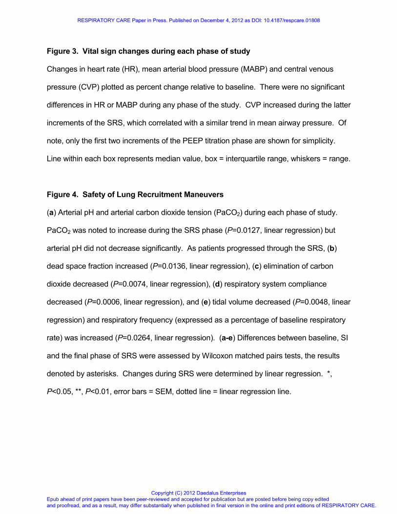
Figure 3. Vital sign changes during each phase of study
Changes in heart rate (HR), mean arterial blood pressure (MABP) and central venous
pressure (CVP) plotted as percent change relative to baseline. There were no significant
differences in HR or MABP during any phase of the study. CVP increased during the latter
increments of the SRS, which correlated with a similar trend in mean airway pressure. Of
note, only the first two increments of the PEEP titration phase are shown for simplicity.
Line within each box represents median value, box = interquartile range, whiskers = range.
Figure 4. Safety of Lung Recruitment Maneuvers
(a) Arterial pH and arterial carbon dioxide tension (PaCO2) during each phase of study.
PaCO2 was noted to increase during the SRS phase (P=0.0127, linear regression) but
arterial pH did not decrease significantly. As patients progressed through the SRS, (b)
dead space fraction increased (P=0.0136, linear regression), (c) elimination of carbon
dioxide decreased (P=0.0074, linear regression), (d) respiratory system compliance
decreased (P=0.0006, linear regression), and (e) tidal volume decreased (P=0.0048, linear
regression) and respiratory frequency (expressed as a percentage of baseline respiratory
rate) was increased (P=0.0264, linear regression). (a-e) Differences between baseline, SI
and the final phase of SRS were assessed by Wilcoxon matched pairs tests, the results
denoted by asterisks. Changes during SRS were determined by linear regression. *,
P<0.05, **, P<0.01, error bars = SEM, dotted line = linear regression line.
RESPIRATORY CARE Paper in Press. Published on December 4, 2012 as DOI: 10.4187/respcare.01808
Epub ahead of print papers have been peer-reviewed and accepted for publication but are posted before being copy edited and proofread, and as a result, may differ substantially when published in final version in the online and print editions of RESPIRATORY CARE.
Copyright (C) 2012 Daedalus Enterprises

Table 1. Patient factors upon meeting inclusion criteria
Pt Sex Age (yr)
IBW (kg)
Diagnosis Etiology
a FiO2 P/F
Ratiob
PEEP (cm H2O)
Crsc
OI
1 F 5.9 20 Parainfluenza pneumonia
P 0.7 87 12 0.71 16.1
2 M 4.2 16.2 Kawasaki Syndrome
NP 1 65 12 0.37 27.7
3 F 10.2 13.9 Sepsis, ascites
NP 0.6 192 12 0.45 8.9
4 F 5.6 34 Sepsis NP 0.5 224 8 0.44 6.7
5 F 10.4 38.5 Necrotizing pneumonia
P 0.6 123 14 0.21 17.0
6 F 13.9 54.7 Sepsis P 0.6 123 14 0.33 22.7
7 F 17.3 73.1 MAS NP 0.7 201 10 0.47 7.0
8 F 4.9 18.2 RSV pneumonitis
d P 1 60 15 0.50 30.0
9 M 11.7 43.1 Sepsis NP 0.4 268 12 0.47 6.4
10 F 9.4 38.5 Trauma NP 0.4 188 10 0.36 8.0
IBW = ideal body weight, OI = oxygenation index (FiO2 x mean airway pressure/PaO2), MAS = macrophage activation sydrome. aEtiology of ALI: P = pulmonary, NP = non-pulmonary. bP/F ratio upon meeting inclusion criteria cCrs = compliance of the respiratory system (mL/cm H2O/kg IBW) dThis patient with RSV did not exhibit signs of airways disease by spirometry.
RESPIRATORY CARE Paper in Press. Published on December 4, 2012 as DOI: 10.4187/respcare.01808
Epub ahead of print papers have been peer-reviewed and accepted for publication but are posted before being copy edited and proofread, and as a result, may differ substantially when published in final version in the online and print editions of RESPIRATORY CARE.
Copyright (C) 2012 Daedalus Enterprises

Figure 1. Study protocol
Ventilation in phases 1, 2, 4 and 5 was performed using volume controlled (VC) ventilation. Phase 3 used pressure control (PC) ventilation with a plateau pressure 15 cm H2O above PEEP and an inspiratory time that was the patient’s baseline. Endpoints were measured following each increment of each phase (see
arrows). ‘P380’ denotes the PEEP increment at which PaO2 + PaCO2 was less than 380 mm Hg.
230x133mm (72 x 72 DPI)
RESPIRATORY CARE Paper in Press. Published on December 4, 2012 as DOI: 10.4187/respcare.01808
Epub ahead of print papers have been peer-reviewed and accepted for publication but are posted before being copy edited and proofread, and as a result, may differ substantially when published in final version in the online and print editions of RESPIRATORY CARE.
Copyright (C) 2012 Daedalus Enterprises

Figure 2. Efficacy of Recruitment Maneuvers
Arterial oxygen tension (PaO2, as measured on FiO2 of 1.0) is plotted throughout the phases of study. PaO2 increased relative to baseline measurements (BL) following a single sustained inflation
(SI). As patients progressed through the staircase recruitment strategy (SRS) consisting of sequentially increasing airway pressures, PaO2 decreased initially then increased significantly as patients progressed
through the SRS. Of note, in Figures 2-4 the data within the SRS phase is organized such that the final SRS step for each patient is aligned to the solid gray bar, with preceding increments being shown progressively
leftward. PaO2 increased even further during the first three steps of the PEEP titration phase, then decreased to near-baseline levels as PEEP was weaned beyond the closing pressure, and maintained there
during the observation (Obs) phase. Comparisons drawn between phase shown and baseline using Wilcoxon matched pairs test. Dotted line represents quadratic polynomial regression line. *, P<0.05, **, P<0.01,
error bars = SEM. 94x48mm (300 x 300 DPI)
RESPIRATORY CARE Paper in Press. Published on December 4, 2012 as DOI: 10.4187/respcare.01808
Epub ahead of print papers have been peer-reviewed and accepted for publication but are posted before being copy edited and proofread, and as a result, may differ substantially when published in final version in the online and print editions of RESPIRATORY CARE.
Copyright (C) 2012 Daedalus Enterprises

Figure 3. Vital sign changes during each phase of study
Changes in heart rate (HR), mean arterial blood pressure (MABP) and central venous pressure (CVP) plotted as percent change relative to baseline. There were no significant differences in HR or MABP during any
phase of the study. CVP increased during the latter increments of the SRS, which correlated with a similar trend in mean airway pressure. Of note, only the first two increments of the PEEP titration phase are shown for simplicity. Line within each box represents median value, box = interquartile range, whiskers = range.
249x176mm (300 x 300 DPI)
RESPIRATORY CARE Paper in Press. Published on December 4, 2012 as DOI: 10.4187/respcare.01808
Epub ahead of print papers have been peer-reviewed and accepted for publication but are posted before being copy edited and proofread, and as a result, may differ substantially when published in final version in the online and print editions of RESPIRATORY CARE.
Copyright (C) 2012 Daedalus Enterprises

For Peer Review
Figure 4. Safety of Lung Recruitment Maneuvers
(a) Arterial pH and arterial carbon dioxide tension (PaCO2) during each phase of study. PaCO2 was noted to
increase during the SRS phase (P=0.0127, linear regression) but arterial pH did not decrease significantly. As patients progressed through the SRS, (b) dead space fraction increased (P=0.0136, linear
regression), (c) elimination of carbon dioxide decreased (P=0.0074, linear regression), (d) respiratory system compliance decreased (P=0.0006, linear regression), and (e) tidal volume decreased (P=0.0048, linear regression) and respiratory frequency (expressed as a percentage of baseline respiratory rate) was increased (P=0.0264, linear regression). (a-e) Differences between baseline, SI and the final phase of SRS were assessed by Wilcoxon matched pairs tests, the results denoted by asterisks. Changes during SRS were determined by linear regression. *, P<0.05, **, P<0.01, error bars = SEM, dotted line = linear regression
line. 276x414mm (300 x 300 DPI)
RESPIRATORY CARE Paper in Press. Published on December 4, 2012 as DOI: 10.4187/respcare.01808
Epub ahead of print papers have been peer-reviewed and accepted for publication but are posted before being copy edited and proofread, and as a result, may differ substantially when published in final version in the online and print editions of RESPIRATORY CARE.
Copyright (C) 2012 Daedalus Enterprises

For Peer Review
Figure 4. Safety of Lung Recruitment Maneuvers (a) Arterial pH and arterial carbon dioxide tension (PaCO2) during each phase of study. PaCO2 was noted to
increase during the SRS phase (P=0.0127, linear regression) but arterial pH did not decrease
significantly. As patients progressed through the SRS, (b) dead space fraction increased (P=0.0136, linear regression), (c) elimination of carbon dioxide decreased (P=0.0074, linear regression), (d) respiratory
system compliance decreased (P=0.0006, linear regression), and (e) tidal volume decreased (P=0.0048, linear regression) and respiratory frequency (expressed as a percentage of baseline respiratory rate) was increased (P=0.0264, linear regression). (a-e) Differences between baseline, SI and the final phase of SRS were assessed by Wilcoxon matched pairs tests, the results denoted by asterisks. Changes during SRS were determined by linear regression. *, P<0.05, **, P<0.01, error bars = SEM, dotted line = linear regression
line.
203x224mm (300 x 300 DPI)
RESPIRATORY CARE Paper in Press. Published on December 4, 2012 as DOI: 10.4187/respcare.01808
Epub ahead of print papers have been peer-reviewed and accepted for publication but are posted before being copy edited and proofread, and as a result, may differ substantially when published in final version in the online and print editions of RESPIRATORY CARE.
Copyright (C) 2012 Daedalus Enterprises


