University of Arkansas, Fayetteville University of Arkansas, Fayetteville
ScholarWorks@UARK ScholarWorks@UARK
Graduate Theses and Dissertations
5-2016
Comparative Efficacy of Foaming and Non-foaming Handsoap in Comparative Efficacy of Foaming and Non-foaming Handsoap in
Reduction of Microorganisms in Handwashing Reduction of Microorganisms in Handwashing
Danielle Marie Conover University of Arkansas, Fayetteville
Follow this and additional works at: https://scholarworks.uark.edu/etd
Part of the Community Health and Preventive Medicine Commons, and the Food Microbiology
Commons
Citation Citation Conover, D. M. (2016). Comparative Efficacy of Foaming and Non-foaming Handsoap in Reduction of Microorganisms in Handwashing. Graduate Theses and Dissertations Retrieved from https://scholarworks.uark.edu/etd/1504
This Thesis is brought to you for free and open access by ScholarWorks@UARK. It has been accepted for inclusion in Graduate Theses and Dissertations by an authorized administrator of ScholarWorks@UARK. For more information, please contact [email protected].

Comparative Efficacy of Foaming and Non-foaming Handsoap in Reduction of Microorganisms
in Handwashing
A thesis submitted in partial fulfillment
of the requirements for the degree of
Master of Science in Food Science
by
Danielle Conover
Kansas State University
Bachelor of Science in Food Science and Industry, 2014
May 2016
University of Arkansas
This thesis is approved for recommendation to the Graduate Council.
_________________________
Dr. Kristen Gibson
Thesis Director
_________________________
Dr. Philip Crandall
Committee Member
_________________________
Dr. John Marcy
Committee Member
Abstract
Handwashing (HW) is a long established method to prevent disease transmission. Ensuring
effectiveness of current HW methods is essential for optimal HW and enhanced disease
prevention. The objectives of this research were to 1) conduct a survey of soap type and volume
in food service establishments in Washington County, Arkansas; 2) investigate how soap type
impacts HW behavior; and 3) determine the difference in microbial reduction between foaming
(F) and liquid (L) handsoap. For Objective 1, food service establishments in Washington County,
AR were selected based on exclusion criteria and random number generations, and handsoap
samples were collected to determine soap type and average volume. For Objective 2, 12
volunteers applied 1.0 g of Glo Germ™ (GG) to their hands and washed their hands, and then
hands were swabbed in three locations to recover remaining GG. Swabs were eluted and
absorbance was measured at OD370nm to quantify remaining GG using a standard curve. For
Objective 3, hands of 24 volunteers were inoculated with approximately 108 CFU Escherichia
coli C3000 or 108 PFU MS2 bacteriophage. Following completion of a standard HW protocol,
microorganisms were recovered using a glove juice method, and culture assays were completed
to determine microorganisms remaining. For the Washington County soap survey, the average
volume of F and L handsoap was 0.64 ± 0.21 mL and 1.19 ± 0.46 mL, respectively. For
Objective 2, no significant difference in behavior was determined in terms of GG remaining, HW
time in the baseline HW and post GG HW, and baseline handrinsing time and post GG handrinse.
Average time for the baseline handwash was (F) 11.17 ± 3.93 s and (L) 13.83 ± 7.30 s, and for
the post GG handwash was (F) 13.33 ± 6.22 s and (L) 14.25 ± 7.70 s. For Objective 3, no
significant difference in efficacy of F and L in overall removal of E. coli and MS2 combined
occurred (p=0.56). However, F handsoap did remove significantly less MS2 when compared to

E. coli (p=0.0008). This research indicates that use of foaming soap in food service may need to
be reevaluated for control of foodborne viruses.

© 2016 Danielle Conover
All Rights Reserved
Acknowledgments
I would like to express my most sincere gratitude to my major professor, Dr. Kirsten Gibson for
her guidance, patience, time, and encouragement during my graduate research. Her passion and
drive throughout my research was infectious, and I am grateful to have had the opportunity to
work with such a driven and knowledgeable advisor. I would also like to thank Giselle Almeida,
laboratory assistant to Dr. Gibson, for her assistance and patience in my laboratory training, and
for her assistance and encouragement throughout my thesis work. I would also like to thank Dr.
Philip Crandall and Dr. John Marcy for serving on my committee and offering assistance on my
thesis work, as well as their general assistance and guidance in decisions impacting my future
career decisions. I would also like to express sincere gratitude to statistician Kevin Thompson for
his assistance in designing the experiment and in analyzing the data, and his patience and time in
working with me throughout the study. I would like to thank Mallory Hancock for her assistance
in experiments in chapters 4 and 5. Additionally, I would like to thank my fellow lab members,
committee members and friends for not only their help in preliminary experiments, but also their
support and encouragement throughout my graduate work. Without these people, I would not
have been able to complete this research: Cody Smith, Nicole Turnage, Adam Baker, Stephanie
Roto, Nathan Jarvis, Anisha Chowdhury, Zhaohao Shi, Sang In Lee, Jasmeet Braich, Pravin
Kaldone, Sabastine Arthur, Meghan Kelly, Colette Robinson, and Tung Pham. I would like to
thank my parents, siblings, friends, and Cody for their constant prayers, support, and
encouragement, throughout my graduate work, as well as their genuine interest and input in my
thesis work. Without their support, I would not have been able to complete this work. Last but
not least, I would like to thank God for providing me the opportunity to attend graduate school
and for the strength and support he provided to me as I completed my thesis work.
Dedication
I would like to dedicate this thesis to my late grandmother Almeda Faker. Although she was not
able to further her education past high school, my grandmother’s humble and honest character as
well as her passion for knowledge, continuous learning, hard work and service to others has
always been an inspiration to me. Although she has not been present throughout my time in
graduate school, I know she has been looking over me, and I am forever grateful to her for her
love, support, wisdom and encouragement throughout my life.

Table of Contents Chapter One: Literature Review ................................................................................................. 1
1. Handy hygiene and its impact on disease transmission .................................................. 1
2. Hand hygiene and its impact on the food industry ......................................................... 2
3. Pathogens commonly transferred by hands .................................................................... 3
3.1 Norovirus and handwashing ........................................................................................... 3
3.2 Enterobacteriaceae and handwashing ........................................................................... 5
4. Handwashing methods and their effectiveness................................................................ 6
4.1 Handwashing time ........................................................................................................... 6
4.2 Drying hands .................................................................................................................... 7
4.3 Antibacterial vs. non-antimicrobial handsoap .............................................................. 8
4.4 Soap volume ................................................................................................................... 10
5. Where is research lacking? ............................................................................................. 11
References: ................................................................................................................................ 13
Chapter 2: A Review of Methods for the Evaluation of Handwashing Efficacy .................. 16
Abstract .................................................................................................................................... 17
1. Introduction ...................................................................................................................... 18
2. Background ...................................................................................................................... 19
2.1 Handwashing and Impact on the Food Industry ........................................................ 19
2.2 Primary Factors Influencing Effective Handwashing .............................................. 21
3. Methods for Evaluation of Handwashing Efficacy ....................................................... 30
3.1 Hand Inoculation Techniques....................................................................................... 31
3.2 Methods to Recover Microorganisms from Hands .................................................... 38
4. Selection of Microorganisms ........................................................................................... 46
5. Conclusions and Recommendations ............................................................................... 47
References ................................................................................................................................. 50
Chapter 3: Survey of Soap Volume and Type in Washington County, Arkansas ................ 64
Abstract .................................................................................................................................... 65
1. Introduction: .................................................................................................................... 66
2. Materials and Methods: .................................................................................................. 66
2.1 Soap type and volume in Washington County, Arkansas .......................................... 66
2.2 Retail soap type and volume ......................................................................................... 67
3. Results: .............................................................................................................................. 68
4. Discussion: ........................................................................................................................ 68
References: ................................................................................................................................ 71
Chapter 4: Investigation of the Impact of Soap Type and Volume on Handwashing
Behavior ....................................................................................................................................... 74
Abstract .................................................................................................................................... 75
1. Introduction: .................................................................................................................... 76
2. Materials and Methods: .................................................................................................. 78
2.1. Study design .................................................................................................................. 78
2.2 Participant recruitment ................................................................................................ 78
2.3 Selection of soaps ........................................................................................................... 79
2.4 Baseline handwash ......................................................................................................... 79
2.5 Glo Germ™ Handwash ................................................................................................. 80
2.6 Swabbing Participant Hands ........................................................................................ 81
2.7 Determination of Sample Absorbance ......................................................................... 81
3. Results ............................................................................................................................... 82
4. Discussion.......................................................................................................................... 84
5. Conclusions ....................................................................................................................... 89
References ................................................................................................................................. 90
Chapter 5: Comparison of two plain soap types for removal of bacteria and viruses from
hands with specific focus on food service environments ......................................................... 97
Abstract .................................................................................................................................... 98
1. Introduction ...................................................................................................................... 99
2. Materials and Methods .................................................................................................. 101
2.1. Study design ............................................................................................................ 101
2.2 Participant recruitment and training ................................................................... 102
2.3 Preparation of inocula ................................................................................................. 102
2.4. Hand decontamination prior to inoculation ......................................................... 103
2.5. Inoculation of hands ............................................................................................... 104
2.6. Selection of soaps .................................................................................................... 104
2.7. Handwashing and drying ....................................................................................... 104
2.8. Recovery of microorganisms by GJM .................................................................. 105
2.9. Detection of microorganisms in recovered sampling solution ................................ 106
2.10. Statistical Analysis .............................................................................................. 106

3. Results ............................................................................................................................. 107
3.1. Efficacy of foaming and liquid handsoaps ........................................................... 107
3.2. Factors influencing HW efficacy ........................................................................... 107
4. Discussion........................................................................................................................ 108
5. Conclusions ..................................................................................................................... 113
Acknowledgements ................................................................................................................ 114
References ............................................................................................................................... 115
Chapter 6: Overall Conclusions .............................................................................................. 120
References: .............................................................................................................................. 124
Appendix .................................................................................................................................... 125
Figure 1: Schematic of experimental design (Chapter 5): ................................................. 125
IBC Approval Letter:............................................................................................................ 126
IRB Approval Letter:............................................................................................................ 127
List of Published or Under Review Articles:
1. Chapter 2: Conover, D.M., & Gibson, K.E. (2016). A review of methods for the
evaluation of handwashing efficacy. Food Control, 63, 53-64.
2. Chapter 5: Conover, D.M., & Gibson, K.E. (2016). Comparison of two plain soap types
for removal of bacteria and viruses from hands with specific focus on food service
environments. Food Control, DOI 10.1016/j.foodcont.2016.04.047.

1
Chapter One: Literature Review
1. Handy hygiene and its impact on disease transmission
Handwashing (HW) has long been established and accepted as a way to prevent disease
and reduce transmission of harmful bacteria and viruses. Hospitals, food industry employees, and
the general population require HW to promote a safe and hygienic environment. While HW is
accepted as a routine part of everyday life, the importance of this basic activity was not always
understood. The importance of hand hygiene has been documented as far back as 1199 by Jewish
philosopher and physician, Moses ben Maimon. Maimon wrote the Mishneh Torah, which was a
code of Jewish religious law, and included a chapter on hygiene where Mishneh wrote “Never
forget to wash your hands after having touched a sick person” (ECJ 2012). Although Mishneh
understood that HW was important, his attempt to influence others was limited as the discoveries
of Mishneh were primarily disregarded (ECJ 2012). Even though Mishneh’s discovery of hand
hygiene was essentially disregarded, another important hand hygiene breakthrough was made in
1847.
In 1847 Dr. Ignaz Semmelweis introduced the concept of hand antisepsis. Semmelweis
assisted in the maternity ward of a Viennese hospital and discovered that the cause of childbed
fever and thus a high mortality rate in a maternity ward was linked to cadaverous particles still
attached to the hands of examiners who had worked with cadavers before working in the
maternity wing of the hospital. Semmelweis found that the ordinary soap and water hand wash
was not sufficient to remove these cadaverous particles, and patients were becoming infected.
After introducing a chlorine wash, deaths from childbed fever decreased dramatically
(Semmelweis 1861). A breakthrough had been made in the hospital environment, and this
2
breakthrough continues to have an impact in disease prevention and transmission today. While
the importance of hand hygiene and all the variables associated with it have not always been
understood, numerous discoveries and breakthroughs have been made which have demonstrated
the importance of proper hand hygiene. Today, HW has become widely accepted as the number
one method available to prevent transmission of disease.
2. Hand hygiene and its impact on the food industry
Foodborne pathogens are estimated to cause 9.4 million illnesses, 55,961 hospitalizations,
and 1,351 deaths in the United States each year (Scallan et al. 2011). Although, foodborne
diseases will likely never be completely eliminated, there are certain practices which can be
followed to greatly reduce disease incidence. Similar to the medical field, one of the primary
prevention strategies is proper and consistent hand washing. Despite common knowledge of the
effectiveness of hand washing, the evidence continually shows that consumers and food-
preparation employees are failing to follow this simple rule, or are failing to wash hands
effectively. A recent report by the U.S. Food and Drug Administration (FDA) found that 38.8%
of employees in fast food restaurants are not in compliance with adequate HW, while 75.8% of
employees in full service restaurants are not in compliance with adequate HW (FDA 2009). A
recent study by Strohbehn et al. (2008) found that only 5% of restaurant employees were
compliant with Food Code recommendations in regards to frequency of washing during
production, service, and cleaning phases.
Currently, it is estimated that washing hands with soap has the potential to reduce
diarrheal disease by 42 to 47% (Curtis et al. 2003). The FDA Food Code, Section 2-301.12,
states that proper HW can result in a 2 to 3 log reduction in transient bacteria as well as a 2-log
3
reduction in transient viruses and protozoa (FDA 2013). Improving the HW of food workers is
critical to reducing foodborne illness outbreaks as transmission of pathogens from the hands of
food workers to food significantly contributes to the spread of foodborne illnesses (Green et al.
2006; Todd et al. 2010; Michaels et al. 2004; Edmonds et al. 2012; Pragle et al. 2007).
3. Pathogens commonly transferred by hands
Foodborne illnesses can be caused by a wide variety of microorganisms including
viruses, bacteria, parasites, fungus, and prions. Symptoms can range from mild (i.e.
asymptomatic) to severe (hospitalization or death) (Mead et al. 1999). Scallan et al. (2011)
reported that viruses, bacteria, and parasites caused an estimated 59, 39, and 2%, respectively, of
foodborne illnesses. While many pathogens can be introduced through natural vectors before a
plant or animal is harvested, improper food-handling techniques, more specifically improper
HW, leads to a significant percentage of foodborne illnesses. According to the FDA and the
CDC, there are five primary pathogens associated with transmission by food workers including
norovirus (NoV), Hepatitis A virus, Salmonella Typhi, Shigella spp., and Escherichia coli
0157:H7, or other Shiga toxin producing E. coli (FDA 2005).
3.1 Norovirus and handwashing
Of the pathogens associated with transmission by food workers, human noroviruses are
the most notable and significant contributors to foodborne illnesses via this transmission route.
Noroviruses are a family of non-enveloped, single stranded RNA viruses that causes acute
gastroenteritis. With a low infectious dose (as low as 18 to 100 viral particles) and high number
of infectious virus particles shed during and after illness, it is relatively easy for an infected food
handler to contaminate a ready-to-eat product with NoV (Teunis et al. 2008). The incubation

4
period of NoV is typically 24 to 48 hours after exposure, with symptoms lasting from 24 to72
hours (Forsythe, 2010). Overall, NoV is the leading cause of foodborne illness in the United
States, resulting in an estimated 58% of illnesses, 26% of hospitalizations, and 11% of the deaths
attributed to foodborne illness. (Scallan et al. 2011).
NoV infection is exceptionally contagious, with attack rates over 45% (Forsythe 2010).
The transmission of NoV can be quite extensive as a contaminated individual can shed NoV
during the incubation period before symptoms appear and can continue shedding NoV particles
for 10 or more days, while 30% of infected individuals can shed NoV even three weeks after
infection and symptoms have subsided (Forsythe 2010). Therefore, this prolonged shedding of
infectious virus particles increases the likelihood that a recently infected food worker will
contaminate foods. Ready-to-eat foods handled by an ill worker can become contaminated with
NoV if the food handler does not take the necessary precautions. While NoV can be transferred
to food or food contact surfaces in numerous ways, transmission through food from an infected
food handler is one transmission method that can be greatly reduced through proper and
consistent HW (Hall et al. 2014).
For instance, in October 2012, a NoV outbreak (GII.4 Sydney strain) occurred among 26
of 103 guests present at a wedding dinner in Austria. As reported in Maritschnik et al. (2012),
investigations of the food served at the wedding found that only one food item was linked to
NoV. The contaminated dish was a mushroom dish, which was garnished with parsley after the
dish was heated. While the mushroom dish was found to be the source of contamination for a
large portion of sick wedding guests, a specific food source of contamination could not be found
for 57% of those who fell ill with NoV. Based on further environmental investigation,
investigators believed handling of silverware by ill food workers likely led to the NoV exposure
5
in the additional 57% of the cases. Investigations into the source of contamination revealed that
no documented food safety training occurred for the kitchen staff. Additionally, it was found the
restroom used by the kitchen staff did not have operational hand hygiene facilities. A kitchen
worker was found to have been sick with the GII.4 strain. This particular worker assisted in
preparation for the wedding, despite being ill, and investigators believe that this symptomatic
worker spread the illness through hand contact in the kitchen environment (Maritschnik 2012).
3.2 Enterobacteriaceae and handwashing
Salmonella and E. coli are two types of enterobacteriaceae. Salmonella and E. coli are
gram-negative, facultative anaerobic, non-spore forming rods (Forsythe 2010). As previously
stated, both Salmonella and E. coli are among the top five pathogens associated with
transmission by food workers. While these pathogens are more commonly inherent to the food
rather than to the food handler, cross-contamination is of concern with these particular
microorganisms. Inadequate HW can result in cross-contamination of food and food-contact
surfaces that can assist in the transmission of these pathogens. Proper and consistent HW is one
preventative measure that can assist in reducing the transmission of these two pathogens.
While there are no published examples of the direct transmission of these bacteria from
hands to food, the following example related to petting zoos has been provided. Petting zoos
have commonly been implicated as a source of E. coli infection. These zoos allow direct contact
with animals that can often serve as vehicles for E. coli and Salmonella, but often do not provide
proper HW stations. Andrews et al. (2012) reported on an outbreak of E. coli O157:H7 from
2004 in which several children became infected attending the petting zoo at the state fair.

6
Investigations concluded that the animals at the petting zoo were the source of the outbreak. It
was concluded that the E. coli was transmitted directly from the hands to mouth.
4. Handwashing methods and their effectiveness
While HW is an effective method for disease control, its effectiveness hinges on the
ability to follow proper HW methods. There are many variables that determine the effectiveness
of HW including frequency, agent used, appropriateness, duration, and technique (Larson et al.
2006). While HW traditionally involves simply using soap and water and rubbing one’s hands
together, the concept of proper and effective HW has broadened throughout the years. Today
there are numerous options available for HW agents: non-antimicrobial handsoap, antibacterial
handsoap (e.g. triclosan), foaming and gel-based handsoaps (with or without antibacterial agent),
bar soap, and various hand sanitizing agents (typically alcohol based). As stated in the 2013 FDA
Food Code (Section 2-301.12), a 10-15 second scrub is necessary to remove transient pathogens
from hands. Additionally, the Food Code emphasizes the importance of every step in the
cleansing of hands, including scrubbing, rinsing, and drying. Failing to emphasize any of these
steps in the HW process can decrease the effectiveness of the HW episode (Food Code 2013).
4.1 Handwashing time
With respect to HW time, 20 seconds is generally considered to be a reasonable amount
of time to reduce microorganisms to an acceptable level. The 2013 Food Code ( section 2-
301.12) states that all food employees must wash hands and exposed portions of the arms for at
least 20 seconds, with 10 to 15 seconds of this total time dedicated to rubbing hands vigorously.
(Food Code 2013). Numerous organizations have continually shown that effective HW requires a
minimum of 20 seconds; however, on average, in both hospital settings as well as in public
7
restrooms, HW is often under 15 seconds (Soap and Detergent Association 2007). Even still, in a
study by Sickbert-Bennett et al. (2005), only a 10 second HW time was utilized as much of the
research available stated that people are continually washing hands shorter than the
recommended time. Perhaps surprisingly, the authors reported that shorter contact times led to a
reduction in transient hand flora which lead to an overall conclusion that more emphasis should
be placed on increasing HW compliance rather than increasing HW time as a shortened HW
time will likely aid in an increased compliance (Sickbert-Bennett et al. 2005).
4.2 Drying hands
Drying hands after washing is one critical step that can have a significant impact on the
overall effectiveness of HW. Bacteria are known to transfer more readily from wet or damp
surfaces rather than on dried surfaces (Fuls et al. 2008). A study conducted in 1997 found that
the drying of hands after washing has the potential to reduce microbial transfer to skin, tools, and
food by up to 99.8% (Patrick 1997). While HW is an effective method to reduce disease
transfer, it is essential to combine HW with careful drying of hands to limit the transfer of any
remaining microorganisms. A recent study stated that hand hygiene is a two-part process, and
adequate hand drying is as imperative as the initial HW (Miller 2011).
Numerous options for hand drying are available. Some common hand drying options
include paper towels, a cloth towel on a rotary dispenser, a mechanical air dryer featuring heated
air, and simply allowing hands to air dry naturally (Gustafson 2000). While there are numerous
methods available for hand drying, the research available on the most effective hand drying
technique for bacterial reduction is somewhat inconclusive. A recent study by Gustafson et al.
(2000) inoculated hands with Micrococcus luteus and then washed hands with a nonantibacterial

8
soap. Hands were then dried using four different methods: cloth towels on a rotation dispenser,
paper towels, a hot air drier, and spontaneous air evaporation. The results of the study indicated
that no significant difference in bacterial reduction occurred between the four HW methods.
Another study by Yamamoto et al. (2005) compared the effectiveness of paper towel drying with
warm air drying. This particular study found that the most effective method for hand drying was
the use of a warm air drier with ultraviolet light, while refraining from rubbing hands throughout
the drying process (Yamamoto et al. 2005). A recent review of various hand drying methods by
Huang et al. (2012) stated that hygienically speaking, paper towels are superior to electric air
dryers. Although research is not entirely conclusive as to which hand drying method is more
effective, the overall consensus is that handy-drying is essential to prevent the transfer of
microorganisms.
4.3 Antibacterial vs. non-antimicrobial handsoap
HW agent used is another variable which can have an impact on overall HW
effectiveness. There are two primary types of handsoap available, non-antimicrobial handsoap
(handsoap not containing any antimicrobial agents) and antibacterial handsoap. While both are
effective at reducing microorganisms found on hands, reports vary on the overall effectiveness of
each type of soap.
A recent study by Fuls et al. (2008) focusing on the effectiveness of antimicrobial and
non-antimicrobial soap found that the bacterial reductions associated with each type of soap were
affected by several variables including wash time, product type, and soap volume. In this study,
antimicrobial soap resulted in a greater reduction of bacteria when compared to non-
antimicrobial soap. In addition, the bacterial reduction achieved with antimicrobial soap

9
increased as wash time increased, whereas no additional increase in bacterial reduction occurred
for the non-antimicrobial soap (Fuls 2008). The authors stated that a non-antimicrobial soap
works primarily through its physical removal of bacteria. At a certain point, maximum removal
will be reached, and an increase in soap or wash time will not increase the removal further.
Because antimicrobial soaps allow for both physical removal as well as inactivation of the
microorganisms, the use of additional soap or added washing time can increase bacterial
reduction (Fuls 2008).
While non-antimicrobial soaps physically remove the pathogens and antimicrobial soaps
work through both physical removal and inactivation of pathogens, research has indicated that
these two soap types impact bacteria and viruses differently. A study by Sickbert-Bennnett et al.
(2005) compared the efficacy of hand hygiene agents in the reduction of bacteria and viruses.
The authors found that the most effective method to reduce MS2 bacteriophage—a surrogate for
the study of enteric viruses—was HW with tap water alone, while the second most effective
method was found to be non-antimicrobial soap. The data seemed to indicate that for viruses,
physical removal is more beneficial than inactivation of the virus (Sickbert-Bennett et al. 2005).
Moreover, most antibacterial and antimicrobial soaps do not use a compound capable of
inactivating viruses, specifically NoV (Liu 2009). For example, triclosan—the most common
active ingredient found in antimicrobial soap—functions as an antimicrobial agent by either
slowing down or inhibiting the growth of bacteria, fungi, and mildew (EPA 2010); however, its
effectiveness against viruses (specifically non-enveloped viruses) has been inconsistently
reported (Mbithi 1993). A study by Contreras (1999) reported similar findings to Sickbert-
Bennett et al. with results indicating that liquid hand dishwashing detergents were 100 times
more effective than antibacterial soaps in reducing respiratory syncytial virus.

10
4.4 Soap volume
In addition to the type of HW agent used, the volume of the HW agent applied can also
have an effect on the effectiveness of the HW episode. A study conducted by Larson et al. (1987)
focused on the quantity of soap as a variable in HW. The authors stressed the need to investigate
the efficacy of HW agents at various volumes since many studies simply utilized a standard 5
mL of HW agent. To address this, the authors used 1 and 3 mL quantities of select HW agents
including an antiseptic agent (4% chlorhexidine gluconate), 2 alcohol-based hand-rinses with
emollients, and a liquid, non-antimicrobial soap. The results of the study indicated that an
antiseptic soap would be beneficial in 3 to 5 mL amounts, while a nonantiseptic liquid soap
would likely not be beneficial in volumes exceeding 1 mL per HW (Larson et al. 1987). Similar
findings were reported by Fuls et al. (2008) which reported that increasing volumes of
antimicrobial soap resulted in increased bacterial reduction, while increased volumes of a non-
antimicrobial soap did not have the same result.
In addition to understanding the effectiveness of various volumes of soap, Larson et al.
(1987) also surveyed the amounts of soap used by each subject. The results of the study indicated
that the amount of soap used by each subject varied from 0.4 mL to 9.0 mL. Palm size of each
individual was recorded to account for a possible relationship between palm size and soap
volume used. Palm sizes ranged from 58 to 94.5 cm2, and no significant link between palm size
and amount of soap used was determined (Larson et al. 1987).
Mechanistically, non-antimicrobial soap works through the use of surfactants, which
reduce bacteria through physical removal. Therefore, a certain maximum amount of bacteria are
capable of being removed, and increased soap amount and wash time will not improve the
11
bacterial removal. Alternatively, antimicrobial soap can benefit from increased volumes as its
mechanism of action involves the combination of friction as well as through killing the bacteria
(Fuls et al. 2008).
Although soap volume can be an important variable in HW, it is also critical to pay
special attention to the time spent HW as indicated previously. A 2011 study by Miller et al.
focused on the use of time, hand-to-hand friction, and the use of non-antimicrobial handsoap for
hand decontamination. The authors found that the addition of soap in general to HW lead to an
initial delay in bacterial reduction in the first 5 to 10 seconds of HW. This delay was not present
at 15 or 20 seconds into the HW (Miller et al. 2011). The authors hypothesized that soap served
as a sort of lubricant in the HW process and thus the soap initially reduced the hand-to-hand
friction resulting in decreased bacterial reduction.
5. Where is research lacking?
Research on HW, the various methods available, and their effectiveness is readily
available. There are numerous studies detailing appropriate soaps, the effectiveness of
antimicrobial soaps versus non-antimicrobial soaps, and the efficacy of hand sanitizers instead of
or in addition to the use of handsoap. While HW has not changed dramatically throughout the
years, new technologies are continuing to appear, and the process of HW continues to evolve. In
recent years, foaming handsoaps have become increasingly common. Despite the plethora of
research available on HW, there is limited research available focusing on the effectiveness of
foaming handsoap, and even more limited research in comparing foaming handsoap to traditional
gel handsoap. While HW can be extremely beneficial in reducing disease transmission, it is vital
that the HW technique is optimized for utmost effectiveness. Considering this, it is important to
12
understand how people respond to the new developments in HW, as well as to understand how
these new developments (particularly foaming handsoap) may alter the proper HW technique
which will allow for continued disease prevention.
13
References:
Andrews, James. "The Petting Zoo Problem." Food Safety News, 2012. Print.
Contreras, P. A., Sami, I. R., Darnell, M. E.R., Ottolini, M.G., & Prince, G. A. (1999).
Inactivation of respiratory syncytial virus by generic hand dishwashing detergents and
antibacterial and soaps. Infection control and hospital epidemiology: the official journal of
the Society of Hospital Epidemiologists of America, 20, 57-58.
Curtis, V., & Cairncross, S. (2002). Effect of washing hands with soap on diarrhea risk in the
community: a systematic review. The Lancet Infectious Diseases, 3, 275-281.
Edmonds, S. L., McCormack, R. R., Zhou S.S., Macinga, D. R., & Fricker, C.M. (2012). Hand
hygiene regimens for the reduction of risk in food service environments. Journal of Food
Protection, 75, 1303-1309.
Ehrenkranz, N. J. (1992). Bland soap handwash or hand antisepsis? The pressing need for clarity.
Infection Control and Hospital Epidemiology, 13, 299-301.
Forsythe, S.J., (2010). The microbiology of safe food. (p. 157-165). United Kingdom: Wiley-
Blackwell.
Fuls, J. L., Rodgers, N. D., Fischler, G. E., Howard, J. M., Patel, M. Weidner, P. L., et al. (2008).
Alternative hand contamination technique to compare the activities of antimicrobial and
nonantimicrobial soaps under different test conditions. Applied and Environmental
Microbiology, 74, 3739-3744.
Green, L. R., Selman, C. A., Radke, V., Ripley, D., Mack, J. C., Reimann, D. W., et al. (2006)
Food worker hand washing practices: an observation study. Journal of Food Protection,
69, 2417-2423.
Gustafson, D. R., Vetter, E.A., Larson D.R., Ilstrup, D.M., Maker, M. D., Thompson, R. L., et al.
(2000). Effects of 4 hand drying methods for removing bacteria from washed hands: A
randomized trial. Mayo Clinic Proceedings, 75, 705-708.
Hall, A.J., Wikswo, M.E., Pringle, K., Gould, H., & Parashar, U.D. (2014). Vital signs:
Foodborne norovirus outbreaks — United States, 2009–2012." Centers for Disease Control
and Prevention. Accessed 11 Nov. 2014.
Huang, C. Ma, W., & Stack, S. (2012). The hygienic efficacy of different hand drying methods:
A review of the evidence." Mayo Clinic, 87, 791--798.
Larson, E. L., Eke, P.I. Wilder, M.P., & Laughon, B.E. (1987). Quantity of soap as a variable in
hand washing." Infection Control, 8, 371-375.
14
Larson, E. L. (1995). APIC guidelines for hand washing and hand antisepsis in health care
settings. American Journal of Infection Control, 23, 251-69.
Larson, E., & Lusk, E. (2006). Evaluating hand washing technique. Journal of advanced nursing
53, 46-50.
Liu, P., Yuen, Y., Hsiao, H., Jaykus, L. & Moe, C. (2010). Effectiveness of liquid soap and hand
sanitizer against Norwalk virus on contaminated hands. Applied and Environmental
Microbiology, 76, 394-399.
Maritschnik, S., Kanitz, E.E., Simons, E., Hohne, M., Neumann, H., Allerberger, F., et al.
(2013). A food handler-associated, foodborne norovirus GII. 4 Sydney 2012-outbreak
following a wedding dinner, Austria, October 2012." Food and environmental virology, 5,
220-225.
Mbithi, J.N., Springthorpe, V.S., & Sattar, S.A. (1993). Comparative in vivo efficiencies of
hand-washing agents against Hepatitis A Virus (HM-175) and Poliovirus Type 1 (Sabin).
Applied and Environmental Microbiology, 59, 3463-3469.
Mead, P. S., Slutsker, L., Dietz, V., McCaig, L.F., Bresee, J.S., Shapiro, C., et al. (1999). Food-
related illness and death in the United States." Emerging infectious diseases, 5, 607-625.
Michaels, B., Keller, C., Blevins, M., Paoli, G., Ruthman, T., Todd, E., et al. (2004). Prevention
of food worker transmission of foodborne pathogens: risk assessment and evaluation of
effective hygiene intervention strategies. Food Service Technology, 4, 31-49. Print.
Miller, T., Patrick, D., & Omrod, D. (2011). Hand decontamination: influence of common
variables on hand-washing efficiency. Healthcare Infection, 16, 18-23.
Patrick, D.R., Findon, G., & Miller, T.E. (1997). Residual moisture determines the level of
touch-contact-associated bacterial transfer following hand washing. Epidemiology and
infection, 119, 319-325.
Pittet, D., Mourouga, P., & Perneger, T.V. (1999). Compliance with hand washing in a teaching
hospital. Annals of Internal Medicine, 130, 126-130.
Pragle, A. S., Harding, A.K & Mack, J.C. (2007). Food workers' perspectives on handwashing
behaviors and barriers in the restaurant environment. Journal of Environmental Health,
69, 27-31.
Scallan, E., Hoekstra, R.M., Angulo, F.J., Tauxe, R.V., Widdowson, M.A., Roy, S.L. et al.
(2011). Foodborne illness acquired in the United States—major pathogens." Emerg Infect
Dis, 17, 1-21.
15
Semmelweis I. Die Aetiologie, der Begriff und die Prophylaxis des Kindbettfiebers. (1861).
[The etiology, concept and prophylaxis of childbed fever]. Pest, Wien und Leipzig, C.A:
Hartleben’s Verlag–Expedition.
Sickbert-Bennett, E. E., Weber, D. J., Gergen-Teague, M. F., Sobsey, M.D., Samsa, G. P., &
Rutala, W. A. (2005). Comparative efficacy of hand hygiene agents in the reduction of
bacteria and viruses. American Journal of Infection Control, 23, 67-77.
Soap and Detergent Association. (2007). Clean hands report card. Print.
Strohbehn, C., Sneed, J., Paola, P., & Meyer, J. (2008). Hand washing frequencies and
procedures used in retail food services. Journal of Food Protection®, 71, 1641-1650.
Teunis, P.F.M., Moe, C.L., Liu, P., Miller, S.E., Lindesmith, L., Baric, R.S., et al. (2008).
Norwalk virus: how infectious is it?" Journal of medical virology, 80, 1468-1476.
Todd, E. C.D., Michaels, B.S., Smith, D., Greig, J.D., & Bartleson, C.A. (2010). Outbreaks
where food workers have been implicated in the spread of foodborne disease. Part 9.
Washing and drying of hands to reduce microbial contamination." Journal of Food
Protection®, 73, 1937-1955.
"Triclosan: Facts." US EPA. March 2010.Web.
<http://www.epa.gov/oppsrrd1/REDs/factsheets/triclosan_fs.htm>. Accessed 30 Oct 2014.
USFDA. (2005). "Employee Health and Personal Hygiene Handbook-Introduction.". 3. Print.
USFDA. (2013) Food Code. (pp. 46-50; 388-389). Washington, D.C: U.S. Department of Health
and Human Services. Available from:
http://www.fda.gov/downloads/Food/GuidanceRegulation/RetailFoodProtection/FoodCo
de/UCM374510.pdf. Accessed 30 Oct 2014.
USFDA. (2009). Report on the Occurrence of Foodborne Illness Risk Factors in Selected
Institutional Foodservice, Restaurant, and Retail Food Service Facility Types. FDA
National Retail Food Team. Print.
"Why Wash Our Hands?" European Cleaning Journal. 11/28/12Web.
<http://www.europeancleaningjournal.com/magazine/articles/latest-news/why-wash-our-
hands>. Accessed 30 Oct 2014.
Yamamoto, Y., Ugai, K., & Takahashi, Y. (2005). Efficiency of hand drying for removing
bacteria from washed hands: Comparison of paper towel drying with warm air drying.
Infection control and hospital epidemiology: the official journal of the Society of Hospital
Epidemiologists of America, 26, 316-320.
16
Chapter 2: A Review of Methods for the Evaluation of Handwashing Efficacy
17
Abstract
Handwashing is relied upon in numerous fields as a primary means to prevent transmission of
harmful pathogens. While handwashing is a key step in disease prevention, the factors
controlling its effectiveness are not always well understood, and there are extensive variations in
the methodology used to assess each of these factors. This review summarizes the various factors
that can impact handwashing effectiveness as well as the methods and results of studies
evaluating each of these factors related to handwashing. Numerous methods are available to
inoculate hands as well as to recover microorganisms from hands, and for a given method,
experimental variables can be changed between researchers. These variations amongst methods
as well as variations in reporting experimental results can make it difficult to compare studies as
well as challenging to accurately interpret the results between studies. Standardization of
methods and reporting requirements are necessary to allow for comparison of studies so that
more accurate conclusions about the handwashing process can be made. Therefore, the need for
1) the development of more standardized handwashing test methods and 2) the formation of
guidelines on the minimal information required for publication of handwashing experiments are
considered and discussed.
18
1. Introduction
Handwashing is widely accepted as a primary means to limit the spread of pathogens and
aid in the prevention of infectious disease (Larson et al. 2000; Sickbert-Bennett et al. 2005).
Numerous fields from medicine to the food industry rely on proper and consistent handwashing
to promote a safe and hygienic environment for both employees and clients. While much of the
world’s population relies on handwashing as a daily method to maintain hygiene and prevent
disease transmission, the importance of hand hygiene has not always been understood.
In the food industry, strict guidelines are provided to minimize contamination of food and
aid in the production or preparation of a quality and safe product for consumers. The United
States Food and Drug Administration’s (FDA) Food Code describes in detail the appropriate
manner in which hands should be washed as well as the recommended times to wash hands when
preparing food, stating that food workers must wash hands immediately before handling: food,
clean equipment and utensils, and unwrapped single-service and single-use articles (USFDA
2013). Additionally the Food Code further describes particular situations where handwashing
should occur before handling food, including: after using the restroom, switching between raw
and ready-to-eat food, after touching bare human body parts, and before putting on gloves when
handling food (USFDA 2013).
Despite the vast focus of the food industry on proper handwashing, compliance with
proper handwashing often fails, and numerous foodborne disease outbreaks occur each year due
to improper handwashing (Green et al. 2006). Understanding and correcting this lack of
compliance is key to reduce foodborne illnesses caused by food handlers. In addition,
understanding the various factors involved in the handwashing process, and optimizing that
19
handwashing process are also beneficial in minimizing foodborne illness. Therefore, the
objectives of this review are to i) conduct a search of peer-reviewed publications available in the
field of handwashing; ii) summarize the different methodologies used in the evaluation of
handwashing efficacy; and iii) discuss the need for standardized methodologies and reporting
requirements to allow for comparison and consistency between handwashing studies. While a
previous meta-analysis by Montville and Schaffner (2011) focused on the effectiveness of
antimicrobial soaps along with the factors that may impact study results—including
methodologies—the primary focus of the present review is to discuss the experimental steps used
to determine the effectiveness of any given handwashing agent and highlight the need for
standard approaches and reporting requirements.
2. Background
2.1 Handwashing and Impact on the Food Industry
Proper hygiene and effective handwashing are essential to food safety. It is estimated that
foodborne pathogens, both major known pathogens as well as unspecified agents, cause 47.8
million illnesses, 127,830 hospitalizations, and 3,037 deaths in the U.S. each year (Scallan et al.
2011). While eliminating all foodborne disease is unrealistic, certain food safety practices such
as handwashing are an effective tool to reduce disease incidence. Section 2-301.12 of the FDA
Food Code states that proper handwashing can result in a 2 to 3 log reduction in transient
bacteria as well as a 2-log reduction in transient viruses and protozoa (USFDA 2013). Transfer
of pathogens from the hands of food workers to food significantly contributes to the spread of
foodborne illness, and the improvement of handwashing in food workers is critical to decrease
20
the amount of foodborne illness outbreaks (Edmonds et al. 2012; Green et al. 2006; Michaels et
al. 2004; Pragle et al. 2007; Todd et al. 2010a).
Although handwashing is heavily relied on in the food industry to limit microbial
contamination of food, and although clear guidelines on proper handwashing are provided, the
transmission of harmful microorganisms from food workers’ hands to food remains a significant
factor in transmission of foodborne illness (Green et al. 2006). Michaels et al. (2004) conducted
a study of 308 outbreaks attributed to ill or asymptomatic food handlers, and 59% of the
outbreaks were due to contamination of the food product through hand contact. Not surprisingly,
the majority of these outbreaks were due to foods that required a great deal of handling, such as
potato salad, salad mixed by hand, and guacamole. While these tasks involving more abundant
quantities of food are a significant contributor to foodborne illness, Michaels et al. (2004) also
found that much smaller tasks such as handling a slice of tomato or garnishing a dish before
serving can have a significant impact on foodborne illness.
One common strategy to aid in safe preparation of food is to utilize gloves when
handling foods. While gloves can be a great solution, they must be used properly and not as a
substitute for handwashing (Green and Selman 2005; Guzewich and Ross 1999; Michaels et al.
2004). Green et al. (2006) conducted an observational study on the handwashing practices of
321 food workers and found that appropriate handwashing rates decreased at a significant rate
when gloves were worn. The use of gloves in food production can present a false sense of
security, causing food handlers to practice unsafe food handling techniques such as washing their
hands less frequently or less often after high-risk tasks (i.e. handling raw meat) leading to
potential microbial contamination of foods (Todd et al. 2010b). Moreover, Montville et al.
(2001) conducted a study on glove usage and cross-contamination in food and demonstrated that

21
bacteria can transfer from food to the hands and from hands to food through the glove. The
results of this study further emphasize that gloves are not an ideal solution and can actually cause
a false sense of security.
While it has been established that food workers do contribute significantly to foodborne
illness outbreaks through improper handling of food, it is important to understand why proper
and consistent handwashing is not occurring. Pragle et al. (2007) conducted a study directly
asking food handlers about their knowledge, practices and barriers to handwashing in the
restaurant environment. The most significant barriers to handwashing included the availability of
supplies, accessibility of sinks, time pressure (i.e. not enough time to wash hands between tasks),
high volume of business, stress, lack of accountability, type of restaurant, insufficient training at
the restaurant, and inadequate food handler training (Pragle et al. 2007).
Even though handwashing is commonly relied on as one of the foremost methods to
prevent transmission of pathogens to food, it is clear that food is still frequently contaminated by
poor handwashing practices of food handlers. Having a clear understanding of how to make
handwashing optimally effective is essential for training employees and preventing
contamination of food from food handlers.
2.2 Primary Factors Influencing Effective Handwashing
While handwashing is a beneficial method to aid in disease control, there are a few key
variables that can impact the effectiveness of handwashing. Frequency, agent (e.g., soap or
sanitizer), appropriateness (i.e. whether the hands were washed when needed to be washed),
duration, and technique are all variables that determine the effectiveness of handwashing (Larson
et al. 2006). The 2013 FDA Food Code emphasizes the importance of every step in the cleansing

22
of hands, including scrubbing, rinsing, and drying, noting that failure to perform any of these
steps in the handwashing process can have a negative effect on the handwashing episode
(USFDA 2013). To improve handwashing, it is essential to understand the factors that make
handwashing effective.
2.2.1. Soap Type: Antimicrobial vs. Non-antimicrobial
There are two primary types of hand soap available: antimicrobial and non-antimicrobial
hand soap. Both types of hand soap lead to a reduction in microorganisms found on the hands,
but reports vary as to the overall effectiveness of each type of soap. Sickbert-Bennett et al.
(2005) conducted a study on the efficacy of hand hygiene agents using tap water, non-
antimicrobial soap, alcohol solutions, and various antimicrobial agents. Sickbert-Bennett et al.
(2005) and others concluded that while antimicrobial handwashing agents were the most
effective in bacterial removal, no handwashing agents were significantly superior to non-
antimicrobial soap or tap water alone. Edmonds et al. (2013) studied the effectiveness of hand
hygiene for removal of Clostridium difficile spores from hands. Handwashing agents in this
study included: tap water, 4% chlorhexidine gluconate (CHG) hand wash, non-antimicrobial
hand soap, 0.3% triclosan hand wash, and peracetic acid wipes which would not be used
routinely as they are too harsh on hands. Results of the study found a 0.76 log10 colony forming
units (CFU)/mL reduction in C. difficile spores with tap water alone. The non-antimicrobial hand
soap and the 4% chlorhexidine gluconate hand wash had similar reductions in C. difficile when
compared to tap water alone. The 0.3% triclosan hand wash had a slightly higher increase in C.
difficile removal at a 0.99 log10 CFU/mL reduction while the harsh peracetic acid wipes provided
the most significant reduction in C. difficile spores at a 1.1 log10 CFU/mL reduction.

23
Another study by Fuls et al. (2008) investigated the bacterial reduction associated with
antimicrobial and non-antimicrobial hand soaps as well as the change in bacterial reduction when
other variables such as wash time, product type, and soap volume were included. The authors
indicated that a greater reduction of bacteria occurred with the use of antimicrobial soap.
Additionally, an increase in wash time led to an increase in bacterial reduction with antimicrobial
soap while the same was not true for non-antimicrobial soap (Fuls et al. 2008). This lack of
correlation between wash time and increased microbial reduction may be explained since non-
antimicrobial hand soap relies only on the physical removal of bacteria from hands while
antimicrobial hand soap combines physical removal as well as antimicrobial inactivation of
bacteria.
Soap type can also impact viruses and bacteria differently. While most hand soaps will
aid in the physical removal of microorganisms, antimicrobial soap can inactivate pathogens
given a sufficient contact time as discussed previously. However, the majority of antimicrobial
soaps do not include compounds that are able to inactivate viruses, most notably human
norovirus—the primary cause of foodborne illness in the U.S. (Liu et al. 2009). In addition, some
antimicrobials such as CHG, are more effective against bacteria (especially vegetative forms)
than viruses and protozoa (McDonnell and Russell 1999). One reason that antimicrobials can
have a different effect on viruses, bacteria, and protozoa is microbial structure. Protozoa can
develop into their protective cyst forms under certain types of stress such as the presence of
antimicrobial compounds thus allowing the protozoa to remain dormant and unaffected by
chemicals (Nester et al. 2009). Some bacteria (Bacillus and Clostridium) are also capable of
forming spores that are extremely stable and resistant to stressful conditions such as heat and
toxic chemicals (Nester et al. 2009). Viruses are obligate intracellular infectious agents
24
composed of nucleic acid (either DNA or RNA) surrounded by a protein coat (capsid). Some
viruses have a lipid membrane (envelope) which surrounds the capsid, while other viruses—most
notably enteric viruses (e.g., human norovirus) transferred by the fecal-oral route—do not have
this membrane and are thus naked (Nester et al. 2009). These differences in structure lead to a
difference in how each type of microorganism is affected by antimicrobials and handwashing in
general.
Sickbert-Bennett et al. (2005) investigated the efficacy of hand hygiene agents against
viruses and bacteria. The results indicate that physical removal of non-enveloped viruses either
through use of water or a non-antimicrobial soap was more effective than using a hand soap with
an antimicrobial agent. Triclosan—one of the most common active ingredients in antimicrobial
soap—actually functions by slowing down or inhibiting the growth of bacteria, fungi, and
mildew (USEPA 2010). The effectiveness of triclosan against viruses—especially non-enveloped
viruses (i.e. enteric viruses)—remains somewhat unclear (Mbithi et al. 1993). Contreras et al.
(1999) echoed the findings of Sickbert-Bennett et al. (2005) with findings indicating that liquid
hand dishwashing detergents were 100 times more effective than antibacterial soaps in reducing
respiratory syncytial virus.
While individual studies have led to a somewhat inconclusive stance with respect to the
differences in effectiveness between antimicrobial and non-antimicrobial handsoap, a recent
meta-analysis conducted by Montville and Schaffner (2011) reported that antimicrobial soap
consistently resulted in a significantly greater reduction of microorganisms on hands than non-
antimicrobial handsoap. Although this difference does appear to be small (approximately a 0.5
log CFU reduction difference), this difference does exist and cannot be ignored (Montville and
Schaffner 2011). Moreover, Schaffner et al. (2014) showed possible reduction in incidence of

25
foodborne illness when antibacterial soaps are used; however, this is based on a risk simulation
and focused on reduction of bacterial pathogens and not viruses. It is important to take into
consideration that the active compounds in antibacterial and antimicrobial soaps are not
necessarily antiviral and thus may not have the same effect on viruses—or other pathogens such
as protozoa—as they do on bacteria.
2.2.2. Soap Volume
While the type of hand soap used can impact effectiveness, the volume of the
handwashing agent applied can also change the effectiveness of the handwashing episode.
Larson et al. (1987) conducted a study focused on the volume of soap as a variable in
handwashing. At that time, many studies were using a standard volume of 5 mL of handwashing
agent, and Larson et al. (1987) stressed the need to investigate the effect of volume on
handwashing effectiveness. The authors used two quantities of soap (1 and 3 mL) as well as
multiple handwashing agents (4% CHG antiseptic agent, two alcohol-based hand-rinses with
emollients, and a liquid non-antimicrobial hand soap). The authors found that a larger volume of
3 to 5 mL was beneficial for the antiseptic soap while a volume no greater than 1 mL was more
appropriate for the nonantiseptic, liquid soap (Larson et al. 1987). Fuls et al. (2008) reported
similar findings with respect to soap volume indicating an increase in bacterial reduction with
larger volumes of antimicrobial soap, while the same did not hold true for non-antimicrobial
hand soap. The meta-analysis by Montville and Schaffner (2011) found that while a strong
correlation does not exist between soap volume and effectiveness of soap (antimicrobial or non-
antimicrobial), there does seem to be an indication that it might be beneficial to use more than 1
26
mL of antimicrobial soap and that abnormally large volumes of soap (> 5 mL) are potentially
less effective.
As discussed previously (Section 2.2.1), non-antimicrobial soaps use physical removal to
reduce the level of bacteria on hands. This physical removal of bacteria occurs through the use of
surfactants, and because of this, there is a maximum amount of bacteria that are capable of being
removed. An increase in soap amount and wash time will not lead to increased bacterial removal
after the maximum removal from the use of surfactants is achieved (Fuls et al. 2008).
Antimicrobial soap however will benefit from increased volumes of soap as it combines the
surfactant abilities of the non-antimicrobial soap with the inactivation of microorganisms,
specifically bacteria (Fuls et al. 2008).
Relationships have also been linked between soap volumes, time spent washing, and
overall bacterial reduction of handwashing. Miller et al. (2011) completed a study on the use of
time, hand-to-hand friction, and the use of non-antimicrobial hand soap for hand
decontamination and found that adding soap actually caused an initial delay of bacterial
reduction within the first 10 seconds of handwashing. After the initial 10 seconds however, the
delay in bacterial reduction was no longer present (Miller et al. 2011). The authors discussed a
possible conclusion that the delayed bacterial reduction occurred as a result of a sort of lubricant
effect of the soap in the handwashing process, leading to an initial delay in the amount of
microorganisms removed from hands.
2.2.3. Handwashing Time
Time spent washing hands is another key variable. Generally, a 20 second hand wash is
considered sufficient to reduce microorganisms on hands. The 2013 Food Code (section 2-
301.12) requires food employees to wash hands as well as exposed portions of the arm for 20
27
seconds, designating 10 to 15 seconds of this handwashing to vigorous rubbing of the hands. If
performed properly, this handwashing regimen can result in a 2 to 3 log reduction in transient
bacteria as well as a 2-log reduction in transient viruses and protozoa (Food Code 2013).
Although many organizations including the World Health Organization, the Mayo Clinic, and
U.S. Centers for Disease Control and Prevention, recommend a handwashing time of a minimum
of 20 seconds for optimal removal of microbes, people in public restrooms as well as in hospitals
often wash their hands for 15 seconds or less (Soap and Detergent Association 2007). Munger
and Harris (1989) conducted a study testing the social influence on handwashing behavior in a
public restroom and found that observed participants washed their hands for an average of 5.2
seconds, while participants who were not observed washed their hands for an average of 4.7
seconds.
Sickbert-Bennett et al. (2005) utilized a 10 second handwash based on the assumption
that this amount of time was more representative of what people were actually practicing. The
authors of the study concluded that a significant reduction occurred in transient hand flora, and
therefore more focus should be placed on increasing handwashing compliance, as a shortened
handwashing time could aid in increasing handwashing compliance (Sickbert-Bennett et al.
2005). However, Montville and Schaffner (2011) showed that with a wash time of 30 seconds, a
significant difference occurred in reduction of microorganisms with antimicrobial and non-
antimicrobial soap. The authors reported that antimicrobial soap resulted in a 2.42 ± 0.88 log
reduction while non-antimicrobial soap had a reduction of 1.91 ± 0.75 log. Regardless, there is
still a need for additional studies looking at wash time and overall handwashing effectiveness, as
the research focusing on this particular factor is somewhat limited (Montville and Schaffner
2011).

28
Stroehbehn et al. (2008) conducted a study analyzing the handwashing practices of food
service employees in operations that serve ready-to-eat food to immunocompromised
individuals. The authors found that during food production in schools, of the 69 times out of the
300 times employees should have washed hands, soap was only used 62 times, and actual
lathering of soap in hands only occurred 37 times. When compared to employees in the food
production area, the employees were found to be more compliant with handwashing when first
entering the work area with 12 of 19 participants washing hands (Stroehbehn et al. 2008).
However, soap was only used 11 of 12 times, and lathering for 10 seconds only occurred nine
times (Stroehbehn et al. 2008). In restaurants, only six of 83 participants washed hands after
handling soiled equipment, and while all six participants used soap, only two actually lathered
for the 10 seconds recommended by the Food Code (Stroehbehn et al. 2008).
Poor compliance with proper handwashing time also occurs frequently in the medical
industry. Graham (1990) conducted a study on the frequency and duration of handwashing in an
intensive care unit and found that for observed handwashing episodes, the average handwash
duration time was 10 seconds (range of 3 to 45.2 seconds). Meengs et al. (1993) conducted a
study on handwashing frequency in an emergency department and found that for the 126 times
handwashing occurred with soap and water, the average duration was only 9.5 seconds.
The authors of the present review conducted a study to observe behavioral changes in 12
subjects when using foaming or liquid hand soap. The average wash time for individuals using
foaming hand soap was 13.6 seconds with a standard deviation of 6.1 seconds while the average
wash time for liquid hand soap was 15.3 seconds with a standard deviation of 6.6 seconds
(unpublished data). No significant difference in wash time occurred between foaming and liquid
hand soap. However, there was a large wash time range between the two soaps, with the shortest

29
time spent washing at 6 seconds and the longest at 26 seconds. With research studies indicating
that handwashing time is often well below the recommended 20 seconds, it seems that using a
more realistic amount of time in studies will produce data that will be more applicable to
reductions occurring in everyday handwashing.
2.2.4. Drying
As previously stated, all steps in the handwashing process are critical, and the last step of
drying is no less important. Bacteria transfer more efficiently from a wet surface than a dry
surface (CDC 2009; Fuls et al. 2008; Patrick et al. 1997). Patrick et al. (1997) observed that
drying hands after washing can decrease microbial transfer to skin, tools, and food by as much as
99.8%, or nearly 3-logs.
Various methods are available for hand drying including paper towels, cloth towels on
rotary dispensers, mechanical air dryers utilizing heated air, and simple air drying (Gustafson et
al. 2000). However, the answer to which drying method is the most effective is not entirely
clear. Gustafson et al. (2000) had participants wash hands with a non-antimicrobial hand soap
after inoculating one hand of each participant with 1 × 10⁷ bacterial cells of Micrococcus luteus
and then evaluated four different methods to dry hands including cloth towels on a rotation
dispenser, paper towels, a hot air dryer, and spontaneous air evaporation. The authors reported no
significant difference in overall bacterial reduction between the four drying methods evaluated.
However, Jensen (2015) mentions that Gustafson et al. (2000) reported the data in CFU rather
than in log CFU, and that if reported in log CFU, a 0.5 log CFU greater reduction occurred with
paper towel drying over air drying or drying with warm air. Yamamoto et al. (2005) evaluated
the effectiveness of paper towels and warm air drying and reported that a warm air dryer

30
combined with a 4 W ultraviolet light without rubbing hands together was the most effective.
The authors hypothesized that rubbing hands together could actually allow for an increase in
bacteria as bacteria are brought to the surface of skin from the hair follicles (Yamamoto et al.
2005). Conversely, Huang et al. (2012) reported results contrary to Yamamoto et al. (2005)
indicating that paper towels are superior to electric air dryers. Despite the fact that there does not
seem to be a conclusive answer to which hand drying method is optimum, removing residual
moisture from hands is essential to allow for optimum handwashing effectiveness and to prevent
unwanted transfer of microorganisms (Jumaa 2004).
3. Methods for Evaluation of Handwashing Efficacy
Since the 1980s, numerous studies have been conducted on handwashing. The focus of
these studies spans a wide range of variables from looking at different microorganisms (viruses
and bacteria) to handwashing time, soap volume, soap type, etc. While a common goal to
optimize handwashing effectiveness is the underlying premise in each study, the methods to
evaluate and achieve this goal are inconsistent across studies. Numerous methods to inoculate
and recover bacteria from hands are utilized, and different microorganisms are selected for
evaluations in each study. Table 1 summarizes the various studies (starting from 1985) along
with the inoculation and recovery methods and the microorganisms used. It is important to note
here that the authors did not include studies focused on cross contamination as these were out of
the scope of this review. In the following sections, the different inoculation and recovery
techniques will be discussed as well as the impact of selection of microorganisms on the reported
results.
31
3.1 Hand Inoculation Techniques
Numerous methods are used to inoculate the hands of participants (Table 1). Inoculation
methods include: hand contact with inoculated blotting paper, contact with public environmental
surfaces (i.e. natural inoculation), pouring of microbial suspension in cupped hands, direct
contamination of microbial suspension on fingertips, palmar-surface techniques of
contamination, and immersion of hands in microbial suspension.
3.1.1. Palmar Surface Methods
The palmar surface method (PSM) is one of the most common methods used to inoculate
hands. There are different variations of this method as highlighted in Table 1. American Society
for Testing and Materials (ASTM) Standard Test Method E2870-13 prescribes palmar surface
contamination to evaluate effectiveness of antimicrobial handwashing formulations (ASTM
2013a). The standard protocol for this procedure (ASTM E2870-13) is to inoculate each palm
with 100 µL of approximately 8 log CFU/mL Escherichia coli suspension. Subjects then spread
the inoculum across their palms and fingertips for 15 ± 1 seconds, and then hands are air-dried
for 30 ± 5 seconds. Bettin et al. (1994) used 100 µL of 6.7 log10 CFU/mL C. difficile suspension
pipetted onto the right palm and then gently rubbed onto the palmar surface of both hands for 10
seconds Edmonds et al. (2013) completed a similar procedure to Bettin et al. (1994) in which the
palms of participants’ hands were inoculated with 150 µL of C. difficile spore suspension
followed by rubbing the palms together for 15 seconds. Fuls et al. (2008) also used a PSM
though with significant modifications. Briefly, sterile paper towels were contaminated with 30
mL of a bacterial suspension (6 log CFU/mL), and hands were then inoculated by pressing on the
towels for five seconds Based on the sterile bag technique for recovery of microorganisms from
32
hands (Section 3.2.1), the calculated transfer of bacteria to hands was approximately 5.8 to 6.4
log CFU total depending on the bacteria (Fuls et al. 2008).
Although each individual method is still a variation of the PSM for inoculation of hands,
each method leads to different levels of inoculation. While Ansari et al. (1989), Bettin et al.
(1994), and Edmonds et al. (2013) used a more direct method of inoculation (Table 1), Fuls et al
(2008) used a more indirect method of the palmar surface inoculation technique. More
specifically, the paper towels were directly contaminated with the inoculum while the hands
were the secondary recipient of this inoculum. When interpreting the data in these studies, it will
be important to understand the actual number of microorganisms transferred to hands during the
inoculation procedure, so that accurate conclusions on handwashing effectiveness can be
determined.
3.1.2. Natural Inoculation
Another common method of inoculation is indirect inoculation (i.e. natural inoculation)
or inoculation through contact with everyday surfaces present in either public or controlled
environments. Larson et al. (1987) conducted a study that used this particular method (Table 1).
In this study hands were initially washed with a control soap, and a baseline hand culture was
obtained from each subject. Subjects washed their hands 15 times a day for five days, and hand
cultures were taken after the first and last handwash of days one and five to observe the effects of
initial and long-term use of a particular soap (Larson 1987). Burton et al. (2011) also used
natural inoculation in which participants were either taken to a large, frequently visited museum
or were instructed to travel on public transportation (e.g., bus or the “underground”). Participants
were instructed to intentionally wipe hands across commonly touched surfaces such as handrails,
door handles, and seats to obtain as much bacteria on their hands from the environment as

33
possible (Burton et al. 2011). The researchers found that washing with water reduced total
bacteria on hands from 44% to 23%, while washing with soap and water reduced the total from
44% to 8%. Another study by Amin et al. (2014) also used a type of natural inoculation
technique and focused on comparing the removal efficacy of water with barsoap or plain water.
Here, the authors did not artificially inoculate hands, but rather recovered microbes from one
hand of each participant prior to handwashing (Amin et al. 2014). The participants were then
instructed to perform either the wash with barsoap or the wash with water, and microorganisms
were recovered from the hand that was not initially sampled to determine the reduction in initial
bacterial levels achieved with each soap type (Amin et al. 2014).
Natural inoculation of hands can be beneficial in providing a realistic view of the
diversity and concentration of microorganisms that subjects can accumulate on their hands in
daily life. However, there are a number of limitations to this method of inoculation if the primary
goal is to test the effectiveness of a particular handwashing variable (i.e. handwashing time, soap
type, soap volume). For instance, there was an assumption in the study by Amin et al. (2014) that
microorganisms would be homogenously distributed between both hands without consideration
of possible bias in contamination of the participant’s dominant hand. Moreover, depending on
the surfaces that subjects are coming in contact with, the types and levels of microorganisms they
are coming in contact with have the potential to vary from day to day and even between the
hands of a single participant.
In the meta-analysis by Montville and Schaffner (2011), the authors observed that studies
using resident microflora on hands as opposed to inoculated transient bacteria (either gram-
positive or gram-negative bacteria) as an indicator of hand washing efficacy resulted in
significantly different reductions (i.e. 0.31 log CFU resident microflora and approximately 1.9
34
log CFU transient bacteria) on hands. Initially, these results may indicate that natural inoculation
methods would be preferable; however, there is no indication of the number of studies included
in each category of microorganism (resident vs. transient) or whether the recovery efficiencies of
each method applied were considered when reporting actual log reduction. Overall, if the goal of
the research is to understand how one particular variable can impact handwashing, using a more
direct and consistent method of contamination such as the PSM or the pouring methods (Section
3.1.3) will likely be more beneficial as they are more controlled and consistent methods.
3.1.3. Pouring Methods
Handwashing studies also utilize the pouring method of inoculation (PMI) which
involves pouring a microbial suspension into the participants’ hands and instructing the
participant to spread the suspension over their hands. Unlike the PSM, the PMI involves
dispersing the microorganism suspension over the entire hand (both ventral and dorsal surfaces)
rather than just the palmar (ventral) region. Sickbert-Bennett et al. (2005) poured a microbial
suspension (volume not specified) containing 8.5 log CFU/mL Serratia marcescens and 9.5 log
plaque forming units (PFU)/mL MS2 bacteriophage into the subjects’ cupped hands and subjects
spread the suspension over their hands for 45 seconds. Here, it is interesting to note that while
Sickbert-Bennett et al. (2005) referenced ASTM E-1174, stating the only modifications to the
procedure included the addition of MS2 bacteriophage and the adjustment to handwash time,
additional modification to the ASTM E-1174 occurred. In ASTM E-1174, the procedure states
that three 1.5 mL aliquots of the test organisms are to be poured into the subjects cupped hands,
with a 20 second spread onto the hands, and a 30 second air dry between each application
(ASTM 2013b); however, the PMI utilized by the authors clearly deviates from this standard
method (Sickbert-Bennett et al. 2005).

35
Edmonds et al. (2013) used two different inoculation methods—depending on the
microorganism—similar to Sickbert-Bennett et al. (2005). The authors inoculated the hands of
participants with 6 log10 spores of either Bacillus atrophaeus or C. sporogenes by placing a total
of 5 mL of the spore suspension onto the hands in three separate aliquots (1.5 mL, 1.5 mL, and
2.0 mL) with the participants spreading each aliquot over the entire hand surface for 45 seconds
(Edmonds et al. 2013). Edmonds et al. (2013) also used a PSM of contamination (Section 3.1.1).
In this instance, it appears that the different volumes were used to achieve the same inoculum
level as opposed to simply diluting the C. difficile spore stock to match the concentration of the
B. atrophaeus and C. sporogenes. However, one could speculate that inoculating the hands with
two different volumes would likely impact dispersion of the bacteria on the hands. Sasahara et
al. (2014) followed a similar procedure to Edmonds et al. (2013) spreading only 2.5 mL of B.
cereus suspension over the entire surface of each hand. Chamberlain et al. (1996) applied the
pouring method as well with slight modifications (Table 1).
3.1.4. Fingerpad Methods
Another technique frequently employed is limitation of contamination to the fingers.
Snyder, Jr. (2007) placed 10 µL of 11 log10 CFU/mL E. coli on the tips of the second and middle
fingers of the hand. Stowell et al. (2014) also completed a similar inoculation procedure,
pipetting 400 µL of cytomegalovirus suspension onto the ventral surface of participants’ fingers.
Ansari et al. (1989) also used a fingerpad method in which the five fingerpads of one hand were
each inoculated with 10 µL of rotavirus at approximately 4 log PFU or E. coli at approximately 6
log CFU, followed by the drying of hands for 20 minutes. Meanwhile, Miller et al. (2011)
adapted a variation on the FPM by inoculating a 25 × 6 cm section of blotting paper with a 6 mL
aliquot of E. coli and allowed the fingers and thumbs of the participants’ hands to contact the
36
contaminated blotting paper for 10 seconds. Stiles and Sheena (1985) performed an additional
variation on the FPM. Here, the finger and thumb tips were pushed into ground beef containing
naturally present coliforms, and then the hands were rubbed together until dry. Based on Table 1,
it is evident that the FPM of inoculation is one method featuring a large amount of variation
throughout the published literature.
3.1.5. Additional Methods
In addition to the methods described in Section 3.1, Jensen et al. (2014) chose two
different methods of inoculation not previously described. The first method was selected to
compare the efficacy of soap versus no soap (water only) in microbial removal on soiled hands.
To inoculate hands, study participants picked up and spread 5 g of ground beef inoculated with 6
log CFU/5 g Enterobacter aerogenes over their hands. Here, the researchers also evaluated
nonsoiled hands by inoculating the participants’ hands with two 0.5 mL aliquots of 6 log
CFU/mL E. aerogenes followed by dispersion when the subjects rubbed their hands together—a
possible variation of PMI though it is unclear. Stiles and Sheena (1985) also used ground beef to
inoculate hands. In this particular study, finger and thumb tips were pushed into ground beef
(inoculated at 6 to 8 and 7 to 9 log CFU/g ground beef of E. coli and Pseudomonas fluorescens,
respectively) for 5 seconds, and then hands were rubbed together up to the wrists until hands
were dry (Stiles and Sheena 1985). Toshima et al. (2001) also used ground beef to contaminate
hands, with participants working 200 grams of a ground meat blend in the palms of the hands at
two separate times for 30 seconds each time.
37
3.1.6. Inoculum Level
As evidenced in the preceding sections as well as in Table 1, it is clear that many
different techniques are used to inoculate hands with microorganisms in order to test numerous
variables and their impact on handwashing. Within these different inoculation methods are many
inconsistencies related to reporting inoculum concentrations. While some studies such as
Bartzokas et al. (1987), Nicoletti et al. (1990), and Bettin et al. (1994) clearly list the level of
microorganisms on inoculated hands, other studies do not make this as clear. For instance, Miller
et al. (2011) did not clearly state amount of E. coli but simply explained the process of preparing
the E. coli for suspension in a saline solution and inoculating blotting paper with the bacterial
suspension. Even though the baseline level of E. coli on participants’ is indicated, there is no
information provided in relation to the efficiency of the inoculation method used in transferring
E. coli to fingerpads—how much E. coli needs to be added to the blotting paper to result in the
baseline levels reported?
In another example, Toshima et al. (2001) inoculated hands by having participants work
200 grams of ground meat in their palms twice for 30 seconds each time. The authors do not
mention inoculation of the ground meat, but are studying the effect of handwashing on total
coliforms transferred from ground meat. In this particular case, if the ground meat was not
artificially inoculated, it is unlikely that coliform contamination in the ground meat will be
homogenous or consistent between subjects and replicates, making the full potential for coliform
reduction on hands due to handwashing difficult to determine. In Chamberlain et al. (1997) the
inoculation process is discussed, and the authors state that early stationary phase cultures of
Micrococcus sp. were diluted (1:50) into fresh broth which subjects then immersed their hands
into for one minute. Once again, the true effectiveness of the handwashing procedure in this
38
study is difficult to determine because the initial inoculum level was not reported. Not clearly
stating inoculum level is a common occurrence in the literature, and makes it difficult to draw
accurate conclusions about effectiveness of handwashing methods.
3.2 Methods to Recover Microorganisms from Hands
Numerous methods have been described for recovery of microorganisms from hands to
determine handwashing effectiveness. There are five primary methods that have been reported in
the published literature including the glove juice method (GJM), sterile bag technique (SBT),
swabbing of hands or fingertips (SWT), the glass bead method (GBM), handling surrogate
surfaces, and contact on a RODAC™ (Replicate Organism Detection and Counting) plate.
3.2.1. Sterile Bag Technique
Of the recovery methods listed, SBT is one of the most common—applied by five studies
evaluating handwashing efficacy (Table 1). Although there is a standard ASTM protocol (ASTM
E2870-13) for application of the SBT (ASTM 2013a), this method is often modified in studies
reported in the peer-reviewed literature. Larson et al. (1987) utilized the SBT to recover
microorganisms from hands. Here, the authors filled a sterile bag with 50 mL of sampling
solution and massaged participants’ hands for 3 minutes. Fuls et al. (2008) also utilized the SBT,
but chose to use 75 mL of a sampling solution in the sterile bag with a massage time of 1 minute.
Meanwhile, Amin et al. (2014) applied the SBT for recovery of microbes from hands and
utilized 200 mL of sterile Ringer’s solution as well as a 30 second massage—participants rubbed
their fingers against their palm for 15 seconds and then their hands were massaged for 15
seconds. While all of these studies had slightly different objectives, the overall goal of each
study was focused on handwashing, and the effect of a given handwashing variable on microbial
39
reduction. The slight modification of the SBT between studies makes it difficult to compare the
results to one another as differences in buffer type as well as hand massage type have the
potential to impact the bacterial recovery; however, to our knowledge, no systematic comparison
of these variables has occurred.
Buffer type and volume is a variable that can potentially have a significant impact on the
recovery efficiency of the microorganisms. Based on the studies listed in Table 1, numerous
sampling solutions are used to recover the microorganisms from hands of participants. Moreover,
the addition of a neutralizing solution was included in various studies as well—specifically those
evaluating antimicrobial soaps (Benton et al. 1990). Studies that employed the SBT selected a
wide range of buffer volumes (50 to 200 mL). This range of buffer volume potentially impacts
the ability to remove bacteria from the hands of the participants in order to determine an accurate
representation of the efficacy of the handwashing process being evaluated. For instance, the
higher the volume of elution buffer, the more dilute the microorganisms will be thus the
sensitivity of the chosen detection method is critical. Additionally, massage time ranges from 30
seconds to 3 minutes. While a massage time of 30 seconds may not be enough to remove
microorganisms from the hands of participants, a massage time of 3 minutes is likely more time
than what is needed. Standardization of this method is essential to provide consistency and allow
for more accurate comparison between studies.
3.2.2. Glove Juice Method
The GJM for recovery of microorganisms from hands is another method that varies
significantly between studies (Table 1). Sickbert-Bennett et al. (2005) utilized 75 mL of
sampling solution per glove and then massaged hands in nonsterile latex gloves for 30 seconds.
Edmonds et al. (2013) also used the GJM and followed ASTM standard method E1174-13

40
(previously ASTM E1174-06) for evaluating the effectiveness of health care personnel and
consumer handwash formulations. The ASTM protocol E1174-13 lists procedures for using S.
marcescens and recommends E. coli (American Type Culture Collection [ATCC] 11229) as an
alternative challenge organism (ASTM 2013c). After hands are washed and inoculated, the GJM
is applied. For this method, gloves (loose-fitting, powder-free, no antimicrobial properties) are
put on hands and filled with 75 mL of the ASTM E1174-13 sampling solution with validated
neutralizers. Hands are then massaged for 1 min ± 5 seconds, flipping the hands after 30 seconds
to ensure the palm as well as the back of the hand are massaged. After massaging, 3 to 5 mL of
the fluid in the glove is aseptically retrieved, diluted, and plated within 30 minutes of sampling.
Another study by Jensen et al. (2014) utilized the GJM. Here, the authors used 20 mL of
phosphate buffered saline (PBS) per glove with a 1 minute massage. Last, Sasahara et al. (2014)
used the GJM with 50 mL of sampling solution (0.04% KH2PO4, 1.01% Na2HPO4, and 0.10%
Triton X-100) per glove with a 1 minute massage.
Similar to the SBT, there are several variables that have been modified within the studies
applying the GJM. These variations in procedure can impact the resulting data as well as the
manner in which the data are analyzed. For instance, the authors of this review experimented
with various volumes of recovery solution while evaluating the GJM for use in an ongoing study.
It was observed that too low of a volume in the glove (20 to 30 mL) was not sufficient as the
volume did not cover the entire surface of the hand (especially a concern on larger hands) and
would not be able to adequately recover microorganisms from the entire surface of the hand
(unpublished data). Additionally, a larger volume in each glove (more than 40 to 50 mL) was too
high of a volume resulting in leakage of the sampling solution from the opening of the glove (a
concern for safety as well as for determining an accurate recovery of microorganisms). Based on

41
this, there is a need to optimize these variables to create consistency and assist with comparison
of data between various studies.
3.2.3. Swabbing Techniques
While the GJM and SBT are the two most popular methods used in handwashing
research, swabbing techniques (SWT) are also employed. For the SWT methods, various swab
types are used as well as swabbing protocols (Table 1). For instance, Chamberlain et al. (1996)
used a single, moistened, cotton-wool swab and passed the swab over the skin area (wrists,
dorsal and palmar surfaces, fingertips, and the interdigital spaces) in a single continuous
movement five times. Meanwhile Burton et al. (2011) used a sodium chloride soaked charcoal
swab that was passed only over the fingers of the dominant hand of participants. Another study
by Stowell et al. (2014) uniformly swabbed the entire contaminated finger surfaces with cotton-
tipped swabs that were premoistened with 100 µL of PBS. While these studies use a variety of
swab types, one consistent trend is that the swab is always pre-moistened. In a study
investigating the factors that influence recovery of microorganisms from surfaces, Moore and
Griffith (2002) found that a pre-moistened swab when used on a wet surface, increased
efficiency of the swabbing procedure when compared to a dry swab. Although swabbing is
sometimes used in handwashing studies, it is more commonly used in other areas of research
such as evaluation of surface sanitation practices and is a valuable tool in the food industry
(Davidson et al. 1998; Moore and Griffith 2002).
There are numerous variables involved in optimizing the effectiveness of the SWT, and
for this reason, it is possible that it may not be the best method for recovery of microorganisms
from hands in handwashing research. While swabbing is often selected based on the ease of
sampling, its accuracy depends on the ability of the swab (i.e. buffer and swab composition) to

42
pick up microorganisms from the sampling surface as well as the ability of the swab to release
those microorganisms (Moore and Griffith 2002). Moore and Griffith (2002) conducted a study
investigating the factors influencing the recovery of microorganisms from fomite surfaces by use
of traditional hygiene swabbing. In this study, stainless-steel squares were inoculated with
Salmonella spp. and then swabbed with sterile cotton swabs, Dacron swabs, or calcium alginate
swabs. Inoculated surfaces were sampled both wet and dry with swabs that were either wet or
dry and the results indicated that swabbing efficiency increased when a wet surface was sampled.
However, the authors noticed a general trend that if a swab was able to remove a large number of
microorganisms from a sampling surface, it was often difficult to then remove those
microorganisms from the swab surface (Moore and Griffith 2002). Overall, the many variables
present in ensuring efficiency and accuracy of the SWT caused Moore and Griffith (2002) to
question the reliability of swabbing as a method to monitor surface cleanliness. If the use of the
SWT as a tool for detecting microorganisms in small areas is questioned, then its reliability in
swabbing a larger area such as the surface of an entire hand should be considered as well.
3.2.4. Glass Bead Method
Another method less frequently utilized within the past decade is the GBM (Table 1). The
GBM typically involves pouring a sampling solution over fingers, and then massaging fingers on
glass beads to recover any remaining microorganisms from hands. Ayliffe et al. (1988) used the
GBM to compare various handwashing agents in hospital laboratories and wards. In this study,
after treatment with the specified handwashing agent, the fingers and thumbs were immersed in a
bowl containing 100 mL of nutrient broth with neutralizer and rubbed vigorously over glass
beads (3-5mm diameter) for 1 minute (Ayliffe et al. 1988). Nicoletti et al. (1990) utilized the
GBM for comparison of CHG detergents and soap. In this study, after treatment with the

43
specified soap samples, hands were sampled by rubbing fingertips for 2 minutes on 35 g of 3mm
glass beads in a bowl with 50 mL of saline (concentration not provided) with 3% Tween 80. The
most recent published study using a glass tube variation of the GBM was reported by
Guilhermetti et al. (2001) to investigate the effectiveness of hand-cleansing agents for removing
methicillin-resistant Staphylococcus aureus from contaminated hands. Here, eight fingers were
sampled from participants by rubbing for 3 minutes in short, flat-bottomed glass tubes containing
5 mL of 0.1% peptone water with neutralizer to prevent carryover inactivation.
3.2.5. Additional Techniques
In addition to the more commonly used recovery techniques, a few less commonly used
techniques are also mentioned in the handwashing literature and have been included in Table 1
as well. Bettin et al. (1994) used TCCFA (tau-rocholate-cycloserine-cefoxitin-fructose agar)
RODAC™ plates after hands were inoculated and treated with the particular handwashing
method followed by sampling in three locations on the TCCFA RODAC™ plates including the
fingertips and thumbtips, the palmar surfaces of the fingers, and the palms. Toshima et al.
(2001) used an agar stamping method in which participants stamped the fingers (including thumb
and all fingers except the little finger) from the fingertip up to the first phalangeal joint on
various selective media. Snyder, Jr. (2007) studied the removal of bacteria from fingertips and
the residual amount remaining on a handwashing nailbrush. To recover microorganisms from
hands, a rinse of 10 mL of letheen broth was applied to the fingertips in a 1-pint ziplock bag and
then participants rubbed the thumb against the second and middle fingers for 20 seconds
(Snyder, Jr. 2007). Ansari et al. (1989) eluted microorganisms from the fingertips by inverting
fingertips in a vial containing Earle balanced salt solution with 20% tryptose phosphate broth
(EBSS-TPB). In this particular method, fingertips were inverted in the vial for 40 seconds with
44
40 full inversions. In the same study, the authors also applied a technique in which 20 mL of the
EBSS-TPB was poured on the hands over a funnel while the hands were rubbed together. Miller
et al. (2011) used touch-transfer quantification to recover microorganisms from hands by
choosing two items for the participants to handle—licorice to represent a food item and soft
synthetic chamois to represent skin. The surrogate surfaces were picked up by the participant and
handled firmly for 5 seconds followed by transferring the surrogate surface to sterile 20 mL glass
containers containing 9 mL of saline and vortexed for 10 seconds to recover microorganisms
(Miller et al. 2011).
3.2.6. Recovery Efficiency and Baseline Inoculum
Although each recovery method has its own set of benefits, there is a need to standardize
1) how long the method should be applied (e.g., how long should hands be massaged in the
GJM?) and 2) the appropriate steps and reagents to be used for each method. Standardization of
the procedures for the various methods commonly used in handwashing research would allow for
easier comparison between studies as well as for more clear and accurate conclusions to be made
when comparing handwashing efficacy. Moreover, not only is methodological standardization
important, but handwashing researchers could benefit from the formation of guidelines on the
minimal information required for publication of handwashing experiments—these types of
guidelines have been established for common methods plagued by lack of standardization such
as real time, quantitative PCR (Bustin et al. 2008).
For instance, a major concern with handwashing studies is that the recovery efficiency of
the microbial recovery methods applied to hands are often not provided. Of the 13 studies
covered in Section 3.2, no researchers directly reported the recovery efficiency of the applied
method. However, two studies (Fuls et. al, 2008 and Sickbert-Bennett et al.2005) provided
45
enough information and data to allow for the recovery efficiency to be determined by the authors
of the present review. Knowing the recovery efficiency of a particular method is a key detail to
determine the true effectiveness of a particular handwashing variable. Variation in a particular
method from lab to lab, or even within one particular lab, can be significant, and thus the
recovery efficiency of a particular method is vital for determining the true effectiveness of a
particular handwashing regimen. Jensen (2015) stressed the importance of taking the sampling
method into consideration when designing experiments and comparing results as different
techniques can result in different observed log CFU or PFU reductions for similar initial
microbial concentrations.
Edmonds et al. (2013) did not detail the recovery efficiency of their method, but they do
mention that the baseline population was determined through the GJM prescribed by ASTM
E1174-13 (ASTM 2013b). Some methods such as ASTM E2870-13, which uses the PSM for
inoculation, do not require a baseline sampling of the hands because the method is designed to
determine the difference between handwashing products, rather than an overall reduction (ASTM
2013a). Additionally, the inoculum volume at such a low level (i.e. 100 µL), and the distribution
of the small volume of inoculum is controlled and limited to the palms and fingers, thus there is
limited opportunity for spillage of inoculum solution. While recovery efficiency is vital to report,
the baseline level is also an important detail that needs to be reported. Although baseline
sampling is not essential when a small, controlled volume is applied to hands (e.g. PSM of
inoculation), it is of more concern in inoculation methods such as the blotting paper method used
by Miller et al. (2011) to contaminate hands and the PMI used by other researchers (Table 1). In
the blotting paper technique, the hands are not directly inoculated with the microorganisms,
rather blotting paper is contaminated and hands then come in contact with the blotting paper,
46
leading to a lower inoculum level on the hands than on the blotting paper. The baseline level of
microorganisms here would be essential to know in order to calculate the recovery efficiency for
accurate determination of microbial reduction due to handwashing. Similarly, PMI would result
in comparable challenges for calculation of recovery efficiency.
4. Selection of Microorganisms
Numerous types of microorganisms (i.e. species, strains, bacteria [vegetative and spores],
viruses) are used in handwashing studies. As shown in Table 1, 14 different microorganisms
were used to inoculate hands with most being bacteria (n = 11) followed by one bacteriophage
and two viruses. Of the bacteria, E. coli was used most frequently (n = 8). The only other
organisms used in more than one study were C. difficile, Micrococcus, S. aureus, and S.
marcescens (Table 1). Eight of the studies did not artificially inoculate hands with
microorganisms, but rather used natural inoculation through environmental microbes which may
have included bacteria, viruses, and protozoa though analyses were only chosen for detection of
specific types of bacteria.
Some standard methods such as ASTM E1174-13 state the microorganisms that are to be
used for the particular procedure—in this case, S. marcescens (ATCC 14756) or E. coli (ATCC
11229) (ASTM 2013b). Other standard procedures, such as ASTM E2870-13 are more general.
ASTM Standard Test Method E2870-13 simply states that the microorganism should have
characteristics that allow it to be readily identified (ASTM 2013a). Although some standard
methods do list the microorganisms to be used for that particular method, numerous adjustments
to the standard protocol are typically made by researchers, and details such as the type of
microorganism to be used in a particular method are not always followed. However, this may not
necessarily be problematic. For instance, Edmonds et al. (2013) use ASTM E1174-06 which

47
states that either S. marcescens or E. coli are the microorganisms to be used in this method;
however, the authors used C. difficile spores as the test organism to focus on the dynamics of
removing spores during handwashing rather than vegetative bacteria. One possible option to
allow for consistency between studies would be to create a standardization of microorganisms,
where certain surrogate microbes could be chosen as standards to use to represent pathogenic
bacteria, viruses, and protozoa of human health concern. Consistency between microorganism
types used is not necessarily key, but rather reporting details such as recovery efficiency (which
could vary depending on the type of microorganism used) to ensure accuracy when determining
reduction of microorganisms on hands in the handwashing process is critical.
5. Conclusions and Recommendations
As evidenced in this review, handwashing research utilizes many different methods to
inoculate hands as well as to recover microorganisms from washed hands. In addition to this,
numerous microorganisms are used to inoculate hands. While there is a lot of variation in
different methods used to test handwashing efficacy, there is also extensive variation within the
various methods leading to some confusion in interpretation of published results as well as in
study reproducibility to determine handwashing effectiveness. Additionally, the vast amount of
variation that is occurring within the numerous studies makes it difficult to compare results of
studies and draw definitive conclusions from the data. For this reason, there is a need to create
standardized methods that can be routinely used by researchers. These standardized methods
will provide consistency and comparability between studies allowing for more transparent
conclusions to be drawn. Moreover, the formation of guidelines for the minimal information
required for publication of handwashing experiments could be an essential step in allowing
accurate comparisons of future published studies.

48
In preliminary, unpublished data from the authors of this review, the PSM was an ideal
method for inoculation. In addition, several commonly used recovery methods were evaluated
including the GJM, SWT, and SBT. From both a recovery efficiency standpoint and an ease of
use standpoint, the GJM with 35 mL of sampling solution and a 1 minute massage seemed to be
the optimum choice (unpublished data). Conversely, the SBT required a much higher volume of
sampling solution (around 200 mL), and as the sampling solution was in a larger area (i.e. a
plastic bag), it did not seem to thoroughly cover the hands. One issue observed with both the
SBT and the GJM was that the sampling solution would often leak and those recovered
microorganisms would be lost. Because the opening of the glove or the sterile bag was not
entirely closed around the participants’ lower arm, a slight change in angle, or an increase in
sampling solution volume often led to leakage at the opening of the glove or bag (both a concern
for subject safety and accuracy of recovery of microorganisms). For this reason, keeping the
sampling solution to a minimum volume of no more than about 50 mL is also optimal. With
respect to the SWT, the recovery efficiency of E. coli from hands was much lower at 0.47%
when compared to the GJM at 2.3% (unpublished data). As discussed previously, swabbing the
entire hand is not an ideal recovery efficiency method as the area is far too great compared to the
surface area of the swab.
Although the appropriate inoculation and recovery methods as well as the
microorganism(s) chosen will vary based on the objectives of a particular study, in the future,
consistency within an experimental method should occur to allow for consistency and assist in
ease of study comparisons. Additionally, details such as efficiency of the recovery method are
essential to report in the future as these types of details allow for a clear understanding of the true
reduction in microorganisms that is occurring due to handwashing. Reporting of essential details

49
such as recovery efficiency by researchers will aid in the determination of accurate conclusions
about the handwashing process to be made. Our conclusions support Jensen (2015) who stated
that although the factors that influence handwashing have been studied, comparing these studies
is difficult because of both methodological differences as well as statistical flaws in studies. It is
clear that inconsistencies between methods and between researchers is causing confusion in
interpreting dating and is making it difficult to develop accurate conclusions about what details
and methods will make handwashing most effective. Creating standardization in handwashing
research methodology as well as reporting guidelines is essential to allow for comparison
between studies so that researchers can accurately draw conclusions about the handwashing
process. Advancements in handwashing research will then allow for optimization of
handwashing and proper training of food-handlers so that handwashing effectiveness can be
maximized, and foodborne illness caused by improper handling due to food-handlers can be
minimized.

50
References
Amin, N., Pickering, A. J., Ram, P. K., Unicomb, L., Najnin, N., Homaira, N., et al. (2014).
Microbiological evaluation of the efficacy of soapy water to clean hands: a randomized,
non-inferiority field trial. American Journal of Tropical Medicine and Hygiene, 91, 415-
423.
Ansari, S. A., Sattar, S. A., Springthorpe, V. S., Wells, G.A., & Tostowaryk, W. (1989). In vivo
protocol for testing efficacy of hand-washing agents against viruses and bacteria:
experiments with rotavirus and Escherichia coli. Journal of Applied and Environmental
Microbiology, 55, 3113-3118.
ASTM. (2013a). ASTM E2870-13 Standard test method for evaluating relative effectiveness of
antimicrobial handwashing formulations using the palmar surface and mechanical hand
sampling. West Conshohocken: ASTM International.
ASTM. (2013b). ASTM E1174-13 Standard test method for evaluation of the effectiveness of
health care personnel handwash formulations. West Conshohocken: ASTM International.
Ayliffe, G. A. J., Bagg, J. R., Davies, J. G., & Lilly, H. A. (1988) Hand disinfection: a
comparison of various agents in laboratory and ward studies. Journal of Hospital
Infection. 11, 226-243.
Bartzokas, C. A. Corkill, J. E., & Makin, T. (1987). Evaluation of the skin disinfecting activity
and cumulative effect of chlorhexidine and triclosan handwash preparations on hands
artificially contaminated with Serratia marcescens. Infection Control. 8, 163-167.
Benson, L. Bush, L., & DeBlanc, D. (1990) Importance of Neutralizers in the stripping fluid in a
simulated healthcare personnel handwash. Infection Control and Hospital Epidemiology.
11, 595-599.
Bettin, K., Clabots, C., Mathie, P., Willard, K., & Gerding, D. N. (1994) Effectiveness of liquid
soap vs. chlorhexidine gluconate for the removal of clostridium difficile from bare hands
and gloved hands. Infection Control and Hospital Epidemiology. 15, 697-702.
Burton, M., Cobb, E., Donachie, P., Judah, G., Curtis, V., & Schmidt W. P. (2011). The effect of
handwashing with water or soap on bacterial contamination of hands. International
Journal of Environmental Research and Public Health. 8, 97-104.Centers for Disease
Control and Prevention. (2009). OPRP General Information on Hand Hygiene.
http://www.cdc.gov/nceh/vsp/cruiselines/hand_hygiene_general.htm
Bustin, S. A., Benes, V., Garson, J. A., Hellemans, J., Huggett, J., Kubista, M. et al. (2009). The
MIQE Guidelines: Minimum information for publication of quantitative real-time PCR
experiments. Clinical Chemistry, 55, 611-622.

51
Chamberlain, A. N., Halblab, M. A., Gould, D. J., & Miles, R. J. (1997). Distribution of bacteria
on hands and the effectiveness of brief and thorough decontamination procedures using
non-medicated soap. Zentralblatt fur Bakteriologie. 285, 565-575.
Contreras, P. A., Sami, I. R., Darnell, M. E.R., Ottolini, M. G., & Prince, G. A. (1999).
Inactivation of respiratory syncytial virus by generic hand dishwashing detergents and
antibacterial hand soaps. Infection Control and Hospital Epidemiology. 20, 57-58.
Davidson, C. A., Griffith, C. J., Peters, A. C., & Fielding, L. M. (1998). Evaluation of two
methods for monitoring surface cleanliness—ATP bioluminescence and traditional
hygiene swabbing. Luminescence. 14, 33-38.
Edmonds, S. L., McCormack, R. R., Zhou S. S., Macinga, D. R., & Fricker, C. M. (2012). Hand
Hygiene Regimens for the Reduction of Risk in Food Service Environments. Journal of
Food Protection. 75, 1303-1309.
Edmonds, S. L. Zapka, C., Kasper, D., Gerber, R., McCormack, R., Macinga, D., et al. (2013).
Effectiveness of hand hygiene for removal of Clostridium difficile spores from hands.
Infection Control and Hospital Epidemiology. 34, 302-305.
Fischler, G. E., Fuls, J. L., Dail, E. W., Duran, M. H., Rodgers, N. D., & Waggoner, A. L.
(2007). Effect of hand wash agents on controlling the transmission of pathogenic bacteria
from hands to food. Journal of Food Protection. 70, 2873-2877.
Fuls, J. L., Rodgers, N. D., Fischler, G. E., Howard, J. M., Patel, M. Weidner, P. L., et al. (2008).
Alternative hand contamination technique to compare the activities of antimicrobial and
nonantimicrobial soaps under different test conditions. Applied and Environmental
Microbiology. 74), 3739-3744.
Gibson, H., Taylor, J. H., Hall, K. E., & Holah, J. T. (1999). Effectiveness of cleaning techniques
used in the food industry in terms of the removal of bacterial biofilms. Journal of Applied
Microbiology. 87, 41-48.
Graham, M. (1990). Frequency and duration of handwashing in an intensive care unit. American
Journal of Infection Control. 18, 77-81.
Green, L., & Selman, C. 2005. Factors impacting food workers’ and managers’ safe food
preparation practices: a qualitative study. Food Protection Trends. 25), 981-990.
Green, L. R., Selman, C. A., Radke, V., Ripley, D., Mack, J. C., Reimann, D. W., et al. (2006)
Food worker handwashing practices: an observation study. Journal of Food Protection.
69, 2417-2423.
Guilhermetti, M. Hernandes, S. E. D., Fukushigue, Y., Garcia, L. B., & Cardoso, C. L. (2001).
Effectiveness of hand-cleansing agents for removing methicillin-resistant Staphylococcus
52
aureus from contaminated hands. Infection Control and Hospital Epidemiology. 22, 105-
108.
Gustafson, D. R., Vetter, E. A., Larson, D. R., Ilstrup, D. M., Maker, M.D. Thompson, R. L., et
al. (2000). Effects of 4 hand-drying methods for removing bacteria from washed hands: a
randomized trial. Mayo Clinic Proceedings. 75, 705-708.
Guzewich, J., & Ross, M. 1999. Evaluation of risks related to microbiological contamination of
ready-to-eat food by food preparation workers and the effectiveness of interventions to
minimize those risks. Available at:
http://citeseerx.ist.psu.edu/viewdoc/download?doi=10.1.1.410.843&rep=rep1&type=pdf.
Accessed 22 July 2015.
Huang, C., Wenjun, M., & Stack, S. (2012). The hygienic efficacy of different hand drying
methods: A review of the evidence. Mayo Clinic Proceedings. 87.8, 791-798.
Jensen, D. (2015). Addressing challenges and gaps in the hand hygiene literature using novel
quantitative approaches. Rutgers University, New Brunswick, N.J. P. 6, 65.
Jensen, D. A., Danyluk, M. D., Harris, L. J., & Schaffner, D. W. (2014). Quantifying the effect
of hand wash duration, soap use, ground beef debris, and drying methods on the removal
of Enterobacter aerogenes on hands. Journal of Food Protection. 78, 685-690.
Jumaa, P.A. (2004). Hand hygiene: simple and complex. International Journal of Infectious
Diseases. 9, 3-14.
Larson, E., Early, E., Cloonan, P. Sugrue, S. & Parides, M. (2000). An organizational climate
intervention associated with increased handwashing and decreased nosocomial infections.
Behavioral Medicine. 26, 14-22.
Larson, E. L., Eke, P. I., Wilder, M. P., & Laughon, B. E. (1987). Quantity of soap as a variable
in handwashing. Infection Control. , 371-375.
Larson, E., & Lusk, E. (2006). Evaluating handwashing technique. Journal of Advanced Nursing.
53.1, 46-50.
Larson, E., Mayur, K., & Laughon, B. A. (1989) Influence of two handwashing frequencies on
reduction in colonizing flora with three handwashing products used by health care
personnel. American Journal of Infection Control. 17, 83-88.
Liu, P., Yuen, Y., Hsiao, H. M., Jaykus, L. A., & Moe, C. (2009). Effectiveness of liquid soap
and hand sanitizer against Norwalk virus on contaminated hands. Applied and
Environmental Microbiology. 76, 394-399.
53
Mbithi, J. N., Springthorpe, V. S., & Sattar, S. A. (1993). Comparative in vivo efficiencies of
hand-washing agents against hepatitis A virus (HM-175) and poliovirus type 1 (Sabin).
Applied and Environmental Microbiology. 59.10, 3453-3469.
McDonnell, G., & Russell, A. D. (1999). Antiseptics and disinfectants: activity, action and
resistance. Clinical Microbiology Reviews. 12, 147-179.
Meengs, M. R., Giles, B. K., Chisholm, C. D., Cordell, W. H., & Nelson, D. R. (1994). Hand
washing frequency in an emergency department. Annals of Emergency Medicine. 23,
1307-1312.
Michaels, B., Keller, C., Blevins, M., Paoli, G., Ruthman, T., & Todd, E., Griffith, C. J. (2004).
Prevention of food worker transmission of foodborne pathogens: risk assessment and
evaluation of effective hygiene intervention strategies. Food Service Technology. 4.1, 31-
49.
Miller, M. L., James-Davis, L. A., & Milaneis, L. E. (1994). A field study evaluating the
effectiveness of different hand soaps and sanitizers. Dairy, Food, and Environmental
Sanitation. 14, 155-160.
Miller, T., Patrick, D., & Omrod, D. (2011). Hand decontamination: influence of common
variables on hand-washing efficacy. Healthcare Infection 16, 18-23.
Montville, R., Chen, Y., & Schaffner, D. W. (2001). Glove barriers to bacterial cross-
contamination between hands to food. Journal of Food Protection. 64, 845-849.
Montville, R., & Schaffner, D.W. (2011). A meta-analysis of the published literature on the
effectiveness of antimicrobial soaps. Journal of Food Protection. 74, 1875-1882.
Moore, G., & Griffith, C. (2002). Factors influencing recovery of microorganisms from surfaces
by use of traditional hygiene swabbing. Dairy, Food and Environmental Sanitation. 22,
410-421.
Moore, G., & Griffith, C. (2002). A comparison of surface sampling methods for detecting
coliforms on food contact surfaces. Food Microbiology. 19, 65-73.
Munger, K., & Harris, S.J. (1989). Effects of an observer on handwashing in a public restroom.
Perceptual and Motor Skills. 69, 733-734.
Nester, E. W., Anderson, D. G., Roberts, C. E. Jr., & Nester, M. T. (2009). Microbiology: a
human perspective. (p. 69, 287, 321). New York: McGraw-Hill.
Nicoletti, G., Boghossian, V., & Borland, R. (1990). Hygienic hand disinfection: a comparative
study with chlorhexidine detergents and soap. Journal of Hospital Infection. 15, 323-337.
54
Patrick, D.R., Findon, G., &Miller, T.E. (1997). Residual moisture determines the level of touch-
contact-associated bacterial transfer following hand washing. Epidemiology and
Infection. 119, 319-325.
Paulson, D.S. (1994). Comparative evaluation of five surgical hand scrub preparations. AORN
Journal. 60, 246-256.
Paulson, D. S., Riccardi, C., Beausoleil, C. M., Fendler, E. J., Dolan, M. J., Dunkerton, L. V., et
al. (1999). Efficacy evaluation of four hand cleansing regimens for food handlers. Dairy,
Food and Environmental Sanitation. 19, 680-684.
Pragle, A. S., Harding, A. K., & Mack, J. C. (2007) Food workers’ perspectives on handwashing
behaviors and barriers in the restaurant environment. Journal of Environmental Health.
69, 27-32.
Rusin, P., Maxwell, S., & Gerba, C. (2002). Comparative surface-to-hand and fingertip-to-mouth
transfer efficiency of gram-positive bacteria, gram-negative bacteria, and phage. Journal
of Applied Microbiology. 93, 585-592.
Sasahara, T., Hayashi, S., Hosoda, K., Morisawa, Y., & Hirai, Y. (2014). Comparison of hand
hygiene procedures for removing Bacillus cereus Spores. Biocontrol Science. 19, 129-
134.
Scallan, E., Griffin, P. M., Angulo F. J., Tauxe, R. V., & Hoekstra, R. M. (2011) Foodborne
illness acquired in the United States-major pathogens. Emerging Infectious Diseases. 17,
7-15.
Scallan, E., Griffin, P. M., Angulo F. J., Tauxe, R. V., & Hoekstra, R. M. (2011) Foodborne
illness acquired in the United States-unspecified agents. Emerging Infectious Diseases.
17, 16-22.
Schaffner, D. W., Bowman, J. P., English, D. J., Fischler, G. E., Fuls, J. L., Krowka, J. F., et al.
(2013). Quantitative microbial risk assessment of antibacterial hand hygiene products on
risk of shigellosis. Journal of Food Protection. 77, 574-582.
Sickbert-Bennett, E. E., Weber, D. J., Gergen-Teague, M. F., Sobsey, M.D., Samsa, G. P., &
Rutala, W. A. (2005). Comparative efficacy of hand hygiene agents in the reduction of
bacteria and viruses. American Journal of Infection Control. 23, 67-77.
Snyder, O. P., Jr. (2007). Removal of bacteria from fingertips and the residual amount remaining
on the hand washing nailbrush. Food Protection Trends. 27, 597-602.
Soap and Detergent Association. (2007). Clean hands report card. Print.
Stiles, M.E, Sheena, A.Z. (1985). Efficacy of low-concentration iodophors for germicidal hand
washing. Journal of Hygiene. 94, 269-277.
55
Stowell, J. D., Forlin-Passoni, D., Radford, K., Bate, S. L., Dollard, S. C., Bialek, S. R., et al.
(2014). Cytomegalovirus survival and transferability and the effectiveness of common
hand-washing agents against cytomegalovirus on live human hands. Applied and
Environmental Microbiology. 80, 455-461.
Strohbehn, C., Sneed, J., Paez, P., & Meyer, J. (2008). Hand washing frequencies and procedures
used in retail food services. Journal of Food Protection. 71, 1641-1650.
Todd, E. C. D., Michaels, B. S., Smith, D., Greig, J. D. & Bartleson, C. A. (2010a). Outbreaks
where food workers have been implicated in the spread of foodborne disease. Part 9.
Washing and drying of hands to prevent microbial contamination. Journal of Food
Protection. 73, 1937-1955.
Todd, E. C. D., Michaels, B. S., Smith, D., Greig, J. D., & Bartleson, C. A. (2010b). Outbreaks
where food workers have been implicated in the spread of foodborne disease. Part 8.
Gloves as barriers to prevent contamination of food by workers. Journal of Food
Protection. 73, 1762-1773.
Toshima, Y., Ojima, M., Yamada, H., Mori, H., Tonomura, M., Hioki, Y., et al. (2001).
Observation of everyday hand-washing behavior of Japanese, and effects of antibacterial
soap. International Journal of Food Microbiology. 68, 83-91.
USEPA. 2010. Triclosan facts. Available from:
http://archive.epa.gov/pesticides/reregistration/web/html/triclosan_fs.html Accessed on
November 9, 2015.
USFDA. 2013. Food Code. Washington, D.C.: U.S. Department of Health and Human Services.
pp. 46-50. Available from:
http://www.fda.gov/downloads/Food/GuidanceRegulation/RetailFoodProtection/FoodCo
de/UCM374510.pdf Accessed on November 9, 2015.
Yamamoto, Y., Ugai, K., & Takahashi, Y. (2005). Efficiency of hand drying for removing
bacteria from washed hands: Comparison of paper towel drying with warm air drying.
Infection Control and Hospital Epidemiology. 26, 316-32.

56
Table 1. Review of methods selected for evaluating handwashing efficacy.
Reference Purpose of the Study Inoculation Method Recovery Method Microorganisms
Used
(Concentration or
Total)
Stiles and
Sheena (1985)
To compare the efficacy of
iodophor products, CHG
detergents, and non-germicidal
soap for reducing
microorganisms on hands
1) NI
2) FPM: Finger and thumb
tips pushed into ground
beef inoculated with
bacteria; hands rubbed
together until dry
GBM: one hand immersed
in 100 mL letheen broth
in plastic bag containing
35 g of 4 mm sterile glass
beads; rubbed glass beads
20 times over palms
Bacteria naturally
present;
Escherichia coli (6
and 8 log CFU/g
beef),
Pseudomonas
fluorescens (7 and
9 log CFU/g beef)
Bartzokas et al.
(1987)
To evaluate initial and
cumulative efficacy of two
antiseptic HW preparations in
eliminating enterobacteria from
hands
PMI: 5 mL of bacterial
suspension poured into
cupped hands, rubbed over
hands, and air-dried for 60 s
GJM: 75 mL of sterile
tryptone water and 0.075
M phosphate buffer in
each glove; hands
massaged 60 s
Serratia
marcescens (9 log
CFU/mL in
suspension; 6.6 log
CFU total
baseline)
Larson et al.
(1987)
To determine effect of soap
volume on microbial reduction
during HW
NI
SBT: 50 mL of SSN in in
bag, hands massaged for 3
min
Bacteria naturally
present
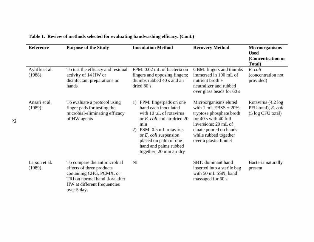
57
Table 1. Review of methods selected for evaluating handwashing efficacy. (Cont.)
Reference Purpose of the Study Inoculation Method Recovery Method Microorganisms
Used
(Concentration or
Total)
Ayliffe et al.
(1988)
To test the efficacy and residual
activity of 14 HW or
disinfectant preparations on
hands
FPM: 0.02 mL of bacteria on
fingers and opposing fingers;
thumbs rubbed 40 s and air
dried 80 s
GBM: fingers and thumbs
immersed in 100 mL of
nutrient broth +
neutralizer and rubbed
over glass beads for 60 s
E. coli
(concentration not
provided)
Ansari et al.
(1989)
To evaluate a protocol using
finger pads for testing the
microbial-eliminating efficacy
of HW agents
1) FPM: fingerpads on one
hand each inoculated
with 10 µL of rotavirus
or E. coli and air dried 20
min
2) PSM: 0.5 mL rotavirus
or E. coli suspension
placed on palm of one
hand and palms rubbed
together; 20 min air dry
Microorganisms eluted
with 1 mL EBSS + 20%
tryptose phosphate broth
for 40 s with 40 full
inversions; 20 mL of
eluate poured on hands
while rubbed together
over a plastic funnel
Rotavirus (4.2 log
PFU total), E. coli
(5 log CFU total)
Larson et al.
(1989)
To compare the antimicrobial
effects of three products
containing CHG, PCMX, or
TRI on normal hand flora after
HW at different frequencies
over 5 days
NI SBT: dominant hand
inserted into a sterile bag
with 50 mL SSN; hand
massaged for 60 s
Bacteria naturally
present
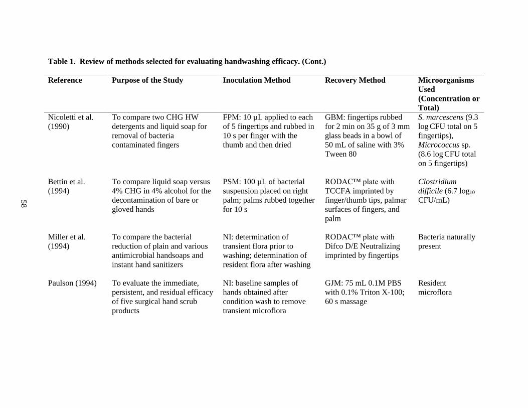
58
Table 1. Review of methods selected for evaluating handwashing efficacy. (Cont.)
Reference Purpose of the Study Inoculation Method Recovery Method Microorganisms
Used
(Concentration or
Total)
Nicoletti et al.
(1990)
To compare two CHG HW
detergents and liquid soap for
removal of bacteria
contaminated fingers
FPM: 10 µL applied to each
of 5 fingertips and rubbed in
10 s per finger with the
thumb and then dried
GBM: fingertips rubbed
for 2 min on 35 g of 3 mm
glass beads in a bowl of
50 mL of saline with 3%
Tween 80
S. marcescens (9.3
log CFU total on 5
fingertips),
Micrococcus sp.
(8.6 log CFU total
on 5 fingertips)
Bettin et al.
(1994)
To compare liquid soap versus
4% CHG in 4% alcohol for the
decontamination of bare or
gloved hands
PSM: 100 µL of bacterial
suspension placed on right
palm; palms rubbed together
for 10 s
RODAC™ plate with
TCCFA imprinted by
finger/thumb tips, palmar
surfaces of fingers, and
palm
Clostridium
difficile (6.7 log10
CFU/mL)
Miller et al.
(1994)
To compare the bacterial
reduction of plain and various
antimicrobial handsoaps and
instant hand sanitizers
NI: determination of
transient flora prior to
washing; determination of
resident flora after washing
RODAC™ plate with
Difco D/E Neutralizing
imprinted by fingertips
Bacteria naturally
present
Paulson (1994)
To evaluate the immediate,
persistent, and residual efficacy
of five surgical hand scrub
products
NI: baseline samples of
hands obtained after
condition wash to remove
transient microflora
GJM: 75 mL 0.1M PBS
with 0.1% Triton X-100;
60 s massage
Resident
microflora

59
Table 1. Review of methods selected for evaluating handwashing efficacy. (Cont.)
Reference Purpose of the Study Inoculation Method Recovery Method Microorganisms
Used
(Concentration or
Total)
Chamberlain et
al. (1997)
To investigate the effectiveness
of a HW procedure
PMI: Hands immersed in
bacterial suspension for 60 s
SWT: premoistened,
cotton-wool swab passed
over skin five times)
Micrococcus sp.
(concentration not
provided)
Paulson et al.
(1999)
To examine four HW regimens
for reduction of transient
microorganisms on hands
PMI: 5 mL of E. coli poured
into cupped hands in two 2.5
mL aliquots and spread over
both hands for 45 s; 2 min
air dry
GJM: sterile latex gloves
on each hand with 75 mL
of SSN in each glove,
wrists secured and hands
massaged for 60 s
E. coli (8 log
CFU/mL)
Guilhermetti et
al. (2001)
To investigate the effectiveness
of hand-cleansing agents for
removal of bacteria from hands
FPM: 0.02 mL of bacterial
suspension on 4 fingertips of
left hand and opposing
fingertips rubbed together
for 40 s and air dried 80 s
GBM: 8 fingers rubbed 3
min against 10 g of 3-5
mm sterile glass beads in
5 mL of 0.1% peptone +
neutralizer
methicillin-
resistant
Staphylococcus
aureus (3.8 and
6.8 log
CFU/fingertip)
Toshima et al.
(2001)
To investigate the efficacy of a
commercial antibacterial soap
NI: Ground meat (200 g)
massaged in the palm, two
times, 30 s each time
GJM: 30 mL SSN poured
into gloved hand and hand
massaged, covering each
side of the hand 10 times
Total coliforms
naturally present in
ground meat

60
Table 1. Review of methods selected for evaluating handwashing efficacy. (Cont.)
Reference Purpose of the Study Inoculation Method Recovery Method Microorganisms
Used
(Concentration or
Total)
Sickbert-Bennett
et al. (2005)
To evaluate efficacy of
antimicrobial agents in soap.
PMI: Microbial suspension
(volume not listed) poured
into the subject’s cupped
hands and spread over hands
for 45 s
GJM: 75mL of SSN in
one nonsterile glove per
hand; massaged for 30 s
S. marcescens (8.5
log CFU/mL),
MS2
bacteriophage (9.5
log PFU/mL)
Snyder, Jr.
(2007)
To determine efficacy of a
double HW procedure for
removal of bacteria from
fingertips
FPM: 10 µL of E. coli spread
on the tips of the second and
middle fingers
Modified SBT: Fingertips
rinsed in 10 mL letheen
broth in a plastic bag;
thumb rubbed against
second and middle fingers
for 20 s
E. coli (11 log
CFU/mL)
Fuls et al.
(2008)
To optimize ASTM E1174
method of hand contamination
technique and to measure effect
of time and soap volume on
antimicrobial and non-
antimicrobial soap effectiveness
PSM: sterile paper towels
inoculated with 30 mL of
bacterial suspension and
hands pressed on towels for
5 s
SBT: 75 mL of SSN in
bag, hands massaged
vigorously for 60 s
S. marcescens, S.
aureus, E. coli,
Shigella flexneri
(variable: 6 to 9
log CFU/mL)

61
Table 1. Review of methods selected for evaluating handwashing efficacy. (Cont.)
Reference Purpose of the Study Inoculation Method Recovery Method Microorganisms
Used
(Concentration or
Total)
Burton et al.
(2011)
To determine whether non-
antibacterial soap reduces
enteric bacteria better than
water alone
NI SWT: NaCl soaked
charcoal swab wiped
across fingers of dominant
hand
Bacteria naturally
present
Miller et al.
(2011)
To evaluate HW time, friction,
and soap for reduction in
bacterial translocation to skin
and food surfaces
Modified FPM: Fingertip
and thumb contact with
blotting paper inoculated
with 6 mL of E. coli
Handling Surrogate
Surfaces: surrogate placed
in sterile 20 mL glass
containers with 9 mL of
saline; vortexed for 10 s
to elute E. coli
E. coli
(concentration not
provided)
Edmonds et al.
(2013)
To compare the efficacy of
hand hygiene agents against C.
difficile
1) PMI: 5 mL of microbial
suspensions in 3 aliquots
(1.5, 1.5, and 2.0 mL)
placed in hands and
rubbed all over for 45 s
2) PSM: 150 µL of spore
suspension rubbed into
palms of hands for 15 s
GJM: 75 mL of SSN in
each glove, hands
massaged uniformly 1
min ± 5 s, flipping hands
after 30 s
C. difficile (7 log
CFU/mL), Bacillus
atrophaeus (6 log
CFU/mL), C.
sporogenes (6 log
CFU/mL)

62
Table 1. Review of methods selected for evaluating handwashing efficacy. (Cont.)
Reference Purpose of the Study Inoculation Method Recovery Method Microorganisms
Used
(Concentration or
Total)
Amin et al.
(2014)
To compare microbial efficacy
of soapy water with bar soap
and water alone
NI
SBT: 200 mL sterile
Ringer’s solution;
participant rubbed fingers
against palm for 15 s;
hands massaged for 15 s
Naturally present
thermotolerant
coliforms and C.
perfringens
Jensen et al.
(2014)
To determine effectiveness of a
minimal 5 s wash and a longer
U.S. FDA Model Food Code
compliant hand wash (20 s)
with and without food debris
1) Spread inoculated ground
beef on hands
2) Two 0.5 mL aliquots of
bacteria placed in each
hand and evenly
dispersed by rubbing
hands together
GJM: 20 mL of PBS per
glove; 60 s hand massage
Enterobacter
aerogenes (6 log
CFU/5 g beef or 6
log CFU/mL)
Sasahara et al.
(2014)
To determine the appropriate
HW procedure for removing B.
cereus spores
PMI: 2.5 mL of microbial
suspension spread on each
hand
GJM: 50 mL of sampling
solution in each glove;
hands massaged for 60 s
B. cereus (6 log
CFU/mL) , E. coli
(6 log CFU/mL)
Stowell et al.
(2014)
To evaluate the removal of
cytomegalovirus (CMV)
through HW
Modified FPM: 400 µL of
virus suspension applied to
ventral surface of fingers
SWT: ventral surface of
fingers swabbed with
cotton-tipped swabs
premoistened with 100 µL
of PBS
CMV (5 log
PFU/mL)
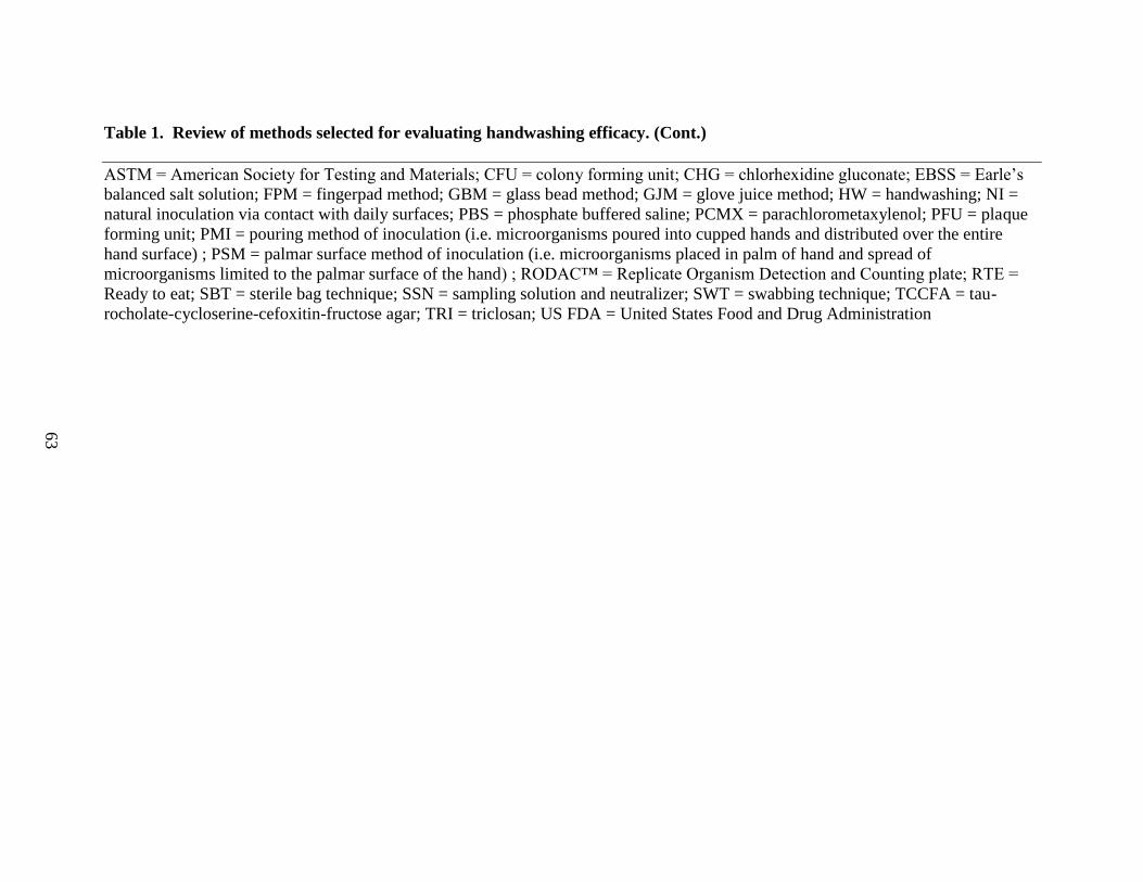
63
Table 1. Review of methods selected for evaluating handwashing efficacy. (Cont.)
ASTM = American Society for Testing and Materials; CFU = colony forming unit; CHG = chlorhexidine gluconate; EBSS = Earle’s
balanced salt solution; FPM = fingerpad method; GBM = glass bead method; GJM = glove juice method; HW = handwashing; NI =
natural inoculation via contact with daily surfaces; PBS = phosphate buffered saline; PCMX = parachlorometaxylenol; PFU = plaque
forming unit; PMI = pouring method of inoculation (i.e. microorganisms poured into cupped hands and distributed over the entire
hand surface) ; PSM = palmar surface method of inoculation (i.e. microorganisms placed in palm of hand and spread of
microorganisms limited to the palmar surface of the hand) ; RODAC™ = Replicate Organism Detection and Counting plate; RTE =
Ready to eat; SBT = sterile bag technique; SSN = sampling solution and neutralizer; SWT = swabbing technique; TCCFA = tau-
rocholate-cycloserine-cefoxitin-fructose agar; TRI = triclosan; US FDA = United States Food and Drug Administration
64
Chapter 3: Survey of Soap Volume and Type in Washington County, Arkansas

65
Abstract
Handwashing (HW) is one of the most significant methods used to prevent the spread of disease,
and numerous variables are present in the HW process that can impact the overall effectiveness
of a HW episode. Soap volume and soap type are two such variables, and numerous soap types
as well as soap volumes exist on the market today. The objectives of this chapter were to conduct
a survey of soap type and soap volume in food service establishments in Washington County,
Arkansas, to better understand the types of soap as well as the average soap volume used in food
service. The data from this chapter will then be used to determine representative volumes of
soap selected for use in chapters 4 and 5. First, a list of food service establishments in
Washington County, AR was obtained, and the list was narrowed down based on exclusion
criteria. Specific locations were selected for sampling through the use of random number
generations. Handsoap samples from restrooms in sampling locations were collected in triplicate
from 68 of the 75 selected food service locations to determine soap type and average volume.
One of the 68 restaurants had both foaming (F) and liquid (L) handsoap giving an overall
distribution of 54.4% (n = 37) and 47.06% (n = 32), respectively. The average volume of F and L
handsoap was 0.64 ± 0.21 mL and 1.19 ± 0.46 mL, respectively. This information was then used
for the selection of representative F and L handsoap volumes used in chapters 4 and 5.
66
1. Introduction:
Soap volume is a variable that can vary both between and within soap manufacturing
companies. As evidenced by my literature review (Chapter 2), soap volume is a variable that has
been shown to impact handwashing (HW) effectiveness (Fuls et al. 2008, Larson et al. 1987,
Montville and Schaffner 2011. The goal of this study was to conduct a survey of both soap type
and soap volume in food service environments to determine the range of soap volumes available
as well as the distribution of soap types. Soap volume was sampled from the restrooms of
licensed food service establishments in Washington County, Arkansas. I predicted that there
would be a similar ratio of foaming to liquid handsoap across restaurant restrooms in
Washington County, Arkansas, with an average soap volume of 1 mL for both foaming and
liquid handsoap. The data obtained from this soap survey were used for selection of
representative soap volumes for studies described in Chapters 4 and 5.
2. Materials and Methods:
2.1 Soap type and volume in Washington County, Arkansas
To determine the distribution of soap types available, as well as the range and average
amount of soap dispensed, 75 locations in Washington County, Arkansas were randomly selected
for sampling using a random sample generator. Briefly, for sample location selection, a list of all
licensed food service facilities in Washington County, Arkansas was obtained from the
Washington County Health Unit of the Arkansas Department of Health. The list of facilities was
then narrowed down through a set of exclusion criteria (Table 1), and 470 locations remained.
From these locations 10% plus an additional 30% (to account for any complications that could

67
arise in the initial location selections being unavailable for sampling) were chosen by random
sampling through the use of JMP® Pro 11.0 (SAS Institute, Cary, NC). Based on this, 75
locations were selected for sampling. At each food service location, one pump of the soap
available in the women’s restroom was dispensed in 50 or 15 mL conical tubes for foaming soap
and liquid soap, respectively. The soaps were collected in triplicate (three “pumps” from the
same soap dispenser on the same day) using this method. Following sample collection, the
samples were transported to the lab to quantify the volume of soap per pump. The soaps were
allowed to settle in their vials with caps on (to prevent evaporation) until all air bubbles settled.
Foaming soaps were transferred to 1.5 mL microcentrifuge tubes, and the volumes were
estimated using the graduations on the microcentrifuge tubes. Liquid soaps were left in their
original 15-mL conical tubes, and 2 mL of water were added to each vial to bring the volume up
to a readable level. The volume was then estimated by subtracting the additional 2 mL of water
from the total volume in the tube.
2.2 Retail soap type and volume
In addition to determining the volume and types of soap available at food service
establishments, the dispense volumes of commercial soaps were also determined. Three brands
of foaming soap and 3 brands of liquid soap were purchased from a local grocery store in
Fayetteville, Arkansas. Soaps were transported back to the lab, and one pump of each soap was
dispensed in 50 or 15 mL conical tubes for foaming soap and liquid soap, respectively. The
soaps were sampled in triplicate using this method. Similar to soap samples from food service
establishments, soaps were allowed to settle in the tubes with caps on (to prevent evaporation)
until all air bubbles settled. Foaming and liquid soaps were processed the same as described in

68
Section 2.1. The range of soap volumes was recorded, and the average soap volume for each type
of soap (foaming versus liquid) was calculated.
3. Results:
Of the 75 locations chosen for sampling, 68 were sampled while the remaining locations
were either closed, did not have consumer restrooms, or were not a traditional restaurant (some
were determined to be primarily bars or nightclubs rather than food service establishments). One
of the 68 restaurants had both foaming and liquid handsoap. Of the food service establishments
sampled, 54.4% (n = 37) had foaming handsoap and 47.06% (n = 32) had liquid handsoap. The
volume of foaming handsoap ranged from 0.2 to 1.21 mL with an average of 0.64 ± 0.21 mL,
and the volume of liquid handsoap ranged from 0.33 to 2.0 mL with an average volume of 1.19 ±
0.46 mL (Figure 1). One-way analysis of variance (ANOVA) of the soap volumes was
performed with a p-value of ≤ 0.05 considered significant. The difference between foaming and
liquid soap volume was significant (p = < 0.001). For the foaming and liquid soaps purchased at
retail, the volume of foaming soap ranged from 0.2 to 1.21mL with an average of 0.99 ± 0.48
mL. The volume for retail liquid soap ranged from 0.33 to 2.0 mL with an average of 1.83 ± 0.29
mL.
4. Discussion:
Although the significance of soap volume on HW effectiveness has been somewhat
disputed in the literature, some researchers have reported an increased efficacy with an increase
in soap volume, especially when using antimicrobial handsoap. In a study conducted with
hospital personnel, Larson et al. (1987) concluded that when using antiseptic soap, 3 to 5 mL per
69
HW should be used to reduce both colonizing and transient hand flora; however, using more than
1 mL of nonantiseptic liquid soap per HW is likely not advantageous. It is important to begin
understanding the range of soaps used to assist in understanding the impact of soap volume on
HW effectiveness. A meta-analysis on antimicrobial soaps conducted by Montville and
Schaffner (2011) found that while there is some evidence that a very small volume (e.g. < 1 mL
of antimicrobial soap) is less effective than a larger volume of antimicrobial soap, there were no
strong interactions between volume of soap and effectiveness of antimicrobial and non-
antimicrobial soap. Overall, research into soap volume as a variable in HW is somewhat limited.
Larson et al. (1987) discuss that the volume of antiseptic soap has the potential to be an
important determinant on the reduction of microorganisms in HW, noting that antibacterial
efficacy can vary between products; thus product manufacturers should provide specific
instructions regarding proper soap amount to be used to achieve maximum HW effectiveness.
This however may be a conflict of interest as a soap manufacturer would benefit financially from
recommending an increased soap volume. As evidenced by the data collected in Washington
County, Arkansas, soap volume does have a wide range depending on the soap type. Additional
research into optimal soap volume for microorganism removal for both antimicrobial and non-
antimicrobial soap as well as foaming and liquid soap types should be conducted to create a more
standardized soap volume on the market and to aid in maximizing HW effectiveness for not only
the food service industry, but also for the general population.
As evidenced by the results of my sampling survey, the volumes for liquid and foaming
soap (a mix of antimicrobial and non-antimicrobial soaps) in Washington County, Arkansas were
70
quite variable, with a significant difference in average soap volume between foaming and liquid
handsoap. The results of this preliminary study provided insight into the range of volumes of
soaps used in Washington County, Arkansas. This information was then used for the
standardization of soap volumes used in the studies described in Chapters 4 and 5.

71
References:
Fuls, J. L., Rodgers, N. D., Fischler, G. E., Howard, J. M., Patel, M. Weidner, P. L., et al. (2008).
Alternative hand contamination technique to compare the activities of antimicrobial and
nonantimicrobial soaps under different test conditions. Applied and Environmental
Microbiology, 74, 3739-3744.
Larson, E. L., Eke, P. I., Wilder, M. P., & Laughon, B. E. (1987). Quantity of soap as a variable
in handwashing. Infection Control, 8, 371-375.
Montville, R., & Schaffner, D.W. (2011). A meta-analysis of the published literature on the
effectiveness of antimicrobial soaps. Journal of Food Protection, 27, 1875-1882.

72
Table 1: Exclusion criteria of food service locations in Washington County, Arkansas
Locations to be excluded
Schools
Churches
Camps
Veterans affairs
Lodges
Nightclubs that are mainly a bar (don’t sell
food)
Hotels
Bowling alleys
Sports stadiums
Bookstore
Flea market
Self-pick orchard
Gym snack bars
Golf Courses

73
Figure 1: Boxplot of Average Volume (mL) by Soap Type in Food Service establishments in
Washington County, Arkansas
Figure 1 is a boxplot representing the distribution of both foaming and liquid handsoap collected
at the 68 different locations in Washington County, Arkansas. The horizontal line in the box
represents the median sample values in both foaming and liquid handsoap. The ends of the box
represent the 75th and 25th percentiles, or 3rd and 1st quartiles. The interquartile range (IQR) is the
difference between the 1st and 3rd quartiles. The whiskers (the area outside the box) extend to the
outermost data point within the computed ranges as follows: 3rd quartile + 1.5 × IQR and 1st
quartile + 1.5 × IQR. Any points beyond the whiskers are outliers in the data set.

74
Chapter 4: Investigation of the Impact of Soap Type and Volume on Handwashing
Behavior
75
Abstract
Handwashing (HW) is a tool used daily by the general population as well as the health care and
food industries to prevent the spread of disease. Although clear HW guidelines have been
established for HW in the food industry, numerous outbreaks due to improper handling by food
workers still occur each year. A variety of soaps are used in the food service industry, and more
recently, foaming soap was introduced onto the market. However, limited research into the
effectiveness of foaming (F) handsoap exists in the published literature when compared to liquid
(L) handsoap. Understanding how this soap may effect HW by food service workers is critical to
maximizing HW effectiveness and preventing transmission of foodborne illness. The primary
objective of this chapter is to understand how soap type impacts HW behavior. To complete this
objective, 12 volunteers completed a baseline HW, after hands were dry, volunteers then applied
a known amount of Glo Germ™ (GG) fluorescent lotion to their hands and washed their hands
without training in proper HW. Following both the baseline and GG HW, hands were swabbed in
three locations to recover remaining GG. Swabs were eluted and absorbance was measured at
OD370nm and remaining GG was quantified using a standard curve. No significant difference in
behavior was determined in terms of GG remaining, HW time in the baseline handwash and post
GG handwash, and baseline handrinsing time and post GG handrinse. Average HW time for the
baseline handwash was (F) 11.17 ± 3.93 s and (L) 13.83 ± 7.30 s, and for the post GG handwash
(F) 13.33 ± 6.22 s and (L) 14.25 ± 7.70 s. While no significant difference in behavior occurred
between F and L handsoap, a consistent increase in both wash time and rinse time for L
handsoap did occur, indicating that there may be a possible benefit to using liquid handsoap in
food service establishments.

76
1. Introduction:
Handwashing (HW) is one of the primary means to prevent transmission of infectious
diseases. While the general population uses HW as a daily tool to limit the spread of disease, it is
especially critical within a food service environment (Miller et al. 1994). Throughout the
production and preparation of food, food workers are presented with a variety of scenarios in
which hand contact (e.g. direct or indirect) can result in the contamination of food with harmful
microorganisms. For example, food worker hands’ may be contaminated with pathogens from
their own gastrointestinal tracts or through contact with objects or food that are contaminated
with pathogens (Paulson 2000). Despite the focus on proper HW in the food industry and the
establishment of clear guidelines for proper HW via the U.S. Food and Drug Administration
(USFDA) 2013 Food Code, food workers are still responsible for the transfer of harmful
pathogens to food resulting in a significant contribution to the incidence of foodborne illnesses,
especially norovirus (NoV) —the primary cause of foodborne outbreaks (Green et al. 2006, Hall
et al. 2012, Scallan et al. 2011). Recently an epidemiologic review of foodborne NoV outbreaks
occurring in the U.S. between 2001 and 2008 was conducted, and the authors found that 82% of
cases in which at least one food item was implicated involved food handler contact with ready-
to-eat (RTE) food, and a food handler was identified as the source of contamination in 53% of
outbreaks (Hall et al. 2012).
The 2013 Food Code (section 2-301.12) states that food employees must wash hands as
well as exposed portions of the arm for 20 seconds, designating 10-15 seconds of this HW
process to vigorous rubbing of the hands (USFDA 2013). Although the U.S. Food Code requires

77
a 20 second handwash and a minimum of 20 seconds is recommended by numerous other
organizations including the World Health Organization (WHO), the Mayo Clinic, and U.S.
Centers for Disease Control and Prevention (CDC), people in public restrooms as well as in
hospitals often wash hands for 15 seconds or less (Soap and Detergent Association, 2007). A
recent study conducted by Burton et al. (2011) found that when participants were instructed to
wash their hands as long and as thorough as they normally would do, participants averaged 12 ±
2.8 s for a handwash with water alone and 14 ± 2.3 s for a handwash with water and soap.
Meengs et al. (1994) studied HW in an emergency room department and found that the average
soap and water handwash was 9.5 s (n = 132). Strohbehn et al. (2008) conducted a study on HW
frequencies and procedures used in retail food services and reported that when restaurant
employees washed hands before engaging in food preparation, soap was used 61% of the time.
Moreover, when using soap, hands were not lathered for the full 10 s, and employees did not dry
hands properly 86% of the time.
In order for HW to be as effective as possible, it is essential to understand how all of the
different variables associated with HW will affect the end result. Numerous soaps are available
on the market today, and food service staff and the general population use these soaps daily.
Although liquid handsoaps have been used for a while, foaming handsoaps are relatively new. In
1999, Deb Group Limited introduced the first generic foaming soap system (Deb Group Ltd.
2014) and was described as having a greater convenience, efficiency, and reduced environmental
impact when compared to gel-based, or liquid, handsoaps. While these claims may be true, it is
important to understand if foaming handsoaps are equally as effective when compared with
traditional, liquid handsoaps. Additionally, it is important to determine how people respond to

78
these foaming handsoaps and to determine if HW behavior changes when different handsoap
types are used. Therefore, the primary goal of this objective was to determine if handsoap type
(foaming versus liquid) affects HW behavior. I hypothesized that HW behavior will change
between foaming and liquid handsoap and predict that participants will wash hands for a shorter
period of time when using foaming handsoap as compared to liquid.
2. Materials and Methods:
2.1. Study design
Studies were arranged in a paired t test design. To account for any possible confounding
factors, two blocking factors were incorporated into the statistical model. These included
sequence and participant. Twelve participants were recruited and completed the study. Two
experimental sequences occurred to alternate exposure of participants to soap type and to adjust
for any possible confounding factors (e.g., learning by either the researchers or the study
participants over the two weeks of the study).
2.2 Participant recruitment
Twelve participants (six men and six women), 18 years and older from the University of
Arkansas (Fayetteville, Arkansas) community were recruited as volunteers to wash their hands.
Participants were informed that in order to participate, they should have no known conditions of
the skin, and also should not have any broken skin. University of Arkansas Institutional Review
Board approval was obtained prior to participant recruitment, and all participants signed an
informed consent form prior to participation in the study. As this objective was behavioral based,
79
participants did not receive any training prior to participation, but rather were simply instructed
to wash hands as they normally would.
2.3 Selection of soaps
Two unscented, non-antimicrobial soaps were chosen for use in the study. The soaps did
not have identical formulations, but were determined to be representative of handsoaps used on
the market daily. Two automatic dispensers, one foaming (GOJO Industries, Akron, OH) and
one liquid (Epare, Staten Island, New York), were chosen to standardize the soap volume
dispensed and allow for ease of observation for the researcher. One dispense of foaming soap
was 0.9 mL (after foaming subsided), and one dispense of liquid handsoap was 1.5 mL. Data
collected in Chapter 3 determined that the average soap volume at 68 food service locations in
Washington County, Arkansas was 0.64 ± 0.22 mL and 1.19 ± 0.46 mL for foaming and liquid
handsoap, respectively. Based on these preliminary data, the soap dispensers selected for this
study were determined to be representative of the average soap volume used in food service
facilities.
2.4 Baseline handwash
On each study day, participants’ hands were inspected for any broken skin. Any jewelry
or items present on the wrist and hand were removed. Participants then completed a preliminary
handwash to remove any possible physical contamination (e.g., residues from hand lotions,
biological materials, etc.) present on their hands. Participants did not receive any direction as to
how to properly wash their hands. Participants first briefly wet their hands and then they were
provided the designated handsoap (either foaming or liquid) to wash their hands. Participants

80
were instructed to dispense the desired amount of soap into their hands, and the researcher
recorded how many pumps of soaps the participant dispensed. After briefly wetting their hands,
and then dispensing the soap into the hand, the participant then began lathering their hands.
Following the lathering of hands, the participants asked the researchers to turn on the water
(distilled tap water) and began rinsing their hands. As one researcher controlled the water for the
participant, the second researcher used a stopwatch to track the amount of time it took for the
participant to wash and rinse their hands. Following the handwash and rinse, participants were
instructed to flick their hands 10 times to remove excess water. Hands were then immediately
swabbed in the three locations discussed in Section 2.6, and the swabs were then processed
(Section 2.7). Swabs from this step were considered “baseline” swabs.
2.5 Glo Germ™ Handwash
Following the preliminary HW and drying, hands were air-dried for 30 s or until hands
appeared visibly dry. Participants were then provided with Glo Germ™ lotion which contains a
fluorescing compound approximately the size of bacteria (about 5 microns) (Glo Germ
Company, Moab, UT). The lotion is designed to be spread on hands to simulate the presence of
microorganisms and serves as a useful tool in training for both the healthcare and food industries
(Kilbride et al. 2003, Michaels 2002). Participants were provided with 1.0 ± 0.01 g (i.e.
approximately the size of a quarter) of Glo Germ™ lotion as recommended by the manufacturer.
Pre-aliquoted lotion on pieces of weigh paper was applied to the hands of participants, and
participants thoroughly rubbed the lotion into their hands (both palmar and dorsal sides) for 1
min until evenly distributed and absorbed. Next, hands were air dried for 30 s or until hands

81
appeared visibly dry. Once dry, participants completed a second handwash as described
previously in Section 2.4.
2.6 Swabbing Participant Hands
A study evaluating the HW technique of nurses by Taylor (1978) found that 89% of
participants missed some parts of the hand when HW with 56% of participants missing some part
of the thumb. Figure 1 illustrates the parts of the hands most frequently missed during HW.
Based on these findings, the skin between the thumb and index finger was chosen for swabbing
as well as the lower nail bed/skin region of the middle finger and the palmar side of the wrist.
After washing, the participants’ hands were swabbed using methods described in Gibson et al.
(2013) with modifications. To quantify the amount of Glo Germ™ remaining on hands after
washing, sterile, foam tipped swabs (VWR, Radnor, PA) were placed in a 15 mL centrifuge tube
containing 2 mL of 95% ethanol, and both hands were swabbed in 3 locations (Figure 2A-C)
using one swab for each location (i.e. 3 swabs total for each participant). Samples were then
processed within 30 min of sampling (Section 2.7).
2.7 Determination of Sample Absorbance
The three swabs were placed in their respective tubes and vortexed for 60 s to elute any
Glo Germ™ recovered from the participants’ hands. The sample was placed in a disposable
acrylic cuvette (VWR), and the absorbance of each swab was measured at an optical density of
370 nm by a DU® 640 spectrophotometer (Beckman Coulter, Inc., Brea, CA). The values from
each of the three swabs were combined to create a total fluorescence concentration for each
participant.
82
Prior to conducting this study, a standard curve of the Glo Germ™ (Figure 3) fluorescent
lotion was created by preparing a two-fold dilution of the fluorescent lotion in 95% ethanol and
subsequently measuring the absorbance at 370 nm with a DU® 640 spectrophotometer
(Beckman Coulter, Inc.). Concentration (y) of fluorescent compound remaining on participants
hands was determined by using the absorbance (x) measurement for each participant, and the
slope (m) and intercept (b) of the fit line of the standard curve through use of the slope intercept
equation (y = mx + b).
2.8 Statistical Analysis
JMP® Pro 11 (SAS, Cary, NC) statistical analysis software was used for all data analysis.
Difference in concentration between the post Glo Germ™ handwash and the baseline handwash
was calculated by first subtracting the total absorbance for the baseline absorbance from the total
absorbance for the post Glo Germ™ handwash. This value was then inserted into the slope-
intercept equation (Section 2.7) to solve for the total concentration. As the study was based on a
paired t test design, a fit model was used to test for significant differences in total concentration
of Glo Germ™ remaining. Sequence and week were incorporated into the model to account for
any variance that may have occurred due to learning by either the participants or researchers
throughout the study. Gender was also added as an effect in the model. Results were considered
statistically significant at p ≤ 0.05.
3. Results
All 12 participants completed the study within a two-week period. Participant age ranged
from 19 to 72 years with an average age of 30 years old. With respect to concentration of Glo
83
Germ™ fluorescent compound remaining on hands, no significant difference between foaming
and liquid handsoap occurred when the mean differences of the concentration of Glo Germ™
remaining between the post Glo Germ™ and baseline handwash were compared (p = 0.35).
Gender also did not have a significant impact on the difference of the concentration of Glo
Germ™ remaining between the post Glo Germ™ and baseline handwash (p = 0.40).
Wash time data are summarized in Table 1. No significant difference between foaming
and liquid handsoap occurred in terms of wash time for both the baseline (p = 0.29) and the post
Glo Germ™ handwash (p = 0.77). Gender did not have a significant impact on wash time for the
baseline (p = 0.89) or the post Glo Germ™ handwash (p = 0.53). Table 2 summarizes rinse time
data. No significant difference between foaming and liquid handsoap occurred in terms of rinse
time for both the baseline (p = 0.056) and the post Glo Germ™ handwash (p = 0.43). Gender did
not have a significant impact on wash time for the baseline rinse (p = 0.48) or the post Glo
Germ™ hand rinse (p = 0.34).
The minimum number of pumps dispensed for the baseline handwash for both foaming
and liquid handsoap was 1 pump, while the maximum was 2 pumps. The minimum number of
pumps dispensed for the post Glo Germ™ handwash for foaming and liquid handsoap was 1
pump, while the maximum was 3 and 2 pumps, respectively. No significant difference between
foaming and liquid handsoap occurred with respect to the number of pumps of soap for either the
baseline (p = 0.35) and the post Glo Germ™ handwash (p = 0.29). Gender did not have a
significant impact on the amount of soap dispensed in the baseline (p = 0.072) or in the post Glo
Germ™ handwash (p = 0.29).

84
Prior to conducting this study, another study with 12 different participants (4 men and 8
women) was conducted. Here, no baseline handwash was completed. The average wash time for
foaming and liquid handsoap was 13.58 ± 6.14 s and 15.33 ± 6.56 s, respectively. Average rinse
time for foaming and liquid handsoap was 9.08 ± 4.08 s and 10.92 ± 4.80 s, respectively. No
significant difference between foaming and liquid handsoap occurred in terms of wash time (p =
0.26), rinse time (p = 0.22), and total concentration Glo Germ™ remaining (p = 0.92). Gender
did not have a significant effect on total concentration of Glo Germ™ remaining on hands (p =
0.29) or on wash time (p = 0.74) or rinse time (p = 0.88). No participants used more than 1
pump of the liquid handsoap; however, five and six participants used 1 and 2 pumps of foaming
handsoap, respectively, while one participant used 4 pumps of foaming handsoap. No significant
difference was found in the concentration of Glo Germ™ remaining when number of pumps of
soap used changed (p = 0.68).
4. Discussion
Comparison of foaming and liquid handsoap, especially concerning behavior in response
to soap type, is extremely limited in the published literature. To our knowledge, no studies
investigating the effects of soap type on HW behavior are available in the published literature.
Overall, the results of the present study indicate that no significant difference exists in behavior
in terms of HW time, rinsing time, and amount of soap dispensed.
Despite the finding that no significant difference appears to exist in HW time and rinsing
time between foaming and liquid handsoap, on average participants consistently washed and
rinsed hands for a longer amount of time with liquid handsoap as compared to foaming
85
handsoap. However, it is important to note that while no statistically significant difference was
found in wash times or rinse times between the two soaps at an alpha level of 0.05, the p-value
for the baseline hand rinse at 0.056 is very close to alpha (0.05), indicating that it is nearly
significant. Having a p-value so close to alpha calls into question the potential for the finding of
a significant result, particularly if a larger sample size were used, resulting in increased statistical
power. While the lathering of soap in hands is a large component of the removal of
microorganisms from hands, rinsing adds additional physical removal of bacteria through the
water flow, primarily for transient microorganisms, (Price 1938, Todd et al. 2010). Miller et al.
(2011) conducted a study on reduction of Escherichia coli on skin with three different methods
of HW: 1) running water; 2) running water and friction; and 3) running water with friction and
soap. The authors found that simply holding contaminated hands passively under running water
was ineffective, but the friction from energetically rubbing hands together while holding hands
under water improved the decontamination of hands (Miller et al. 2011). Although the additional
reduction in microorganisms due to rinsing of hands is unclear in the published literature, a
significant increase in rinsing time between foaming and liquid handsoap could present an
opportunity for increased removal of microorganisms from hands; therefore, additional
investigation into the impact of soap type on rinse time may be warranted.
One inherent limitation with the present study is that it is an observational study. While
participants were not informed as to what researchers would be observing and recording prior to
their participation, it is entirely possible that participants inferred what was being studied and
then altered their HW behavior accordingly. It is known that people are responsive to the
presence of an outside observer (Ram 2013). Ram et al. (2010) conducted an observational
86
study in Bangladesh, and the researchers embedded acceleration sensors within the soap to assess
the reactivity of participants to structured observation. Results of the study indicated that on days
participants were observed, the use of the sensored soap increased by approximately 35%. This
led the authors to conclude that individuals substantially alter HW behavior in the presence of an
observer, particularly in participants who are aware of the social expectations of hand hygiene
(Ram et al. 2010). However, structured observation is still a commonly used method in HW
research, though it does require caution when interpreting data (Ram 2013). Even taking this
possible observation bias into consideration, the mean HW times for both foaming and liquid
handsoap in the present study are still well below the recommended 20 s HW time, indicating
that this time is more than likely not being met on a daily basis by the general population.
Additionally, although this cannot be determined, the possibility exists that if behavior was
changed because of the presence of an observer, that behavior would be equally changed for both
soap types, negating the effect.
As mentioned previously, an additional limitation of the present study is that of sample
size. Our study utilized the observations of 12 individuals (6 males and 6 females) over two
weeks. Although a significant difference in HW time was not found, a slight increase in HW
time consistently occurred for liquid handsoap, and it is possible that an increased sample size
would result in decreased variance and a corresponding finding of a significant difference in HW
behavior in response to soap type. Throughout the published literature, numerous studies have
low statistical power because of inadequate sample size or issues in the experimental design (Eng
2003, Freiman et al. 1978). Freiman et al. (1978) conducted a review of published clinical
literature (n = 71) with negative results and found that the alpha and beta remained almost
87
completely unmentioned in the studies reviewed; thus, concluding that many of the “negative”
trials had the potential to be false negatives. For many of these trials, an inadequate sample size
was chosen leading to a low power, and an increased possibility that a false negative conclusion
was observed. In my study, after consulting with a statistician, it was determined that the sample
size (n=12), though somewhat low, would be chosen because of allotment of available resources.
In originally setting up this experiment, one of the primary goals was to use Glo Germ™
as an indicator to determine if HW behavior was impacted by soap type (foaming versus liquid).
Glo Germ™, a lotion with a fluorescent compound, is more commonly used to qualitatively
observe HW behavior (Benoit et al. 2015, Biran, et al. 2009 Ling et al. 2012). Typically,
participants rub the fluorescent lotion onto their hands and then wash their hands. After washing
their hands, participants view their hands under a blacklight in order to visualize where the
fluorescent compound is remaining, thus indicating that these are the areas missed during HW.
Through this experiment, my goal was to take this qualitative tool and apply it in a quantitative
manner to determine if amount of the fluorescent compound remaining on hands would correlate
with a behavioral difference in participants between the two soap types. Establishment of a
standard curve allowed us to determine concentration of the fluorescent compound remaining on
participants’ hands. However, the remaining fluorescent compound on hands proved to be a
difficult tool to use as a measure of HW behavior since concentration of fluorescent compound
remaining on participant’s hands was rather low, and the concentration remaining varied greatly
from person to person.
88
Gibson et al. (2013) used Glo Germ™ powder fluorescent compound in a mock retail deli
environment to understand the impact of workers on cross contamination as well as to identify
critical areas of contamination. Similar to the present study, the results found by Gibson et al.
(2013) were rather variable for each participant, but the researchers were able to determine the
areas within the deli environment that were more likely for cross-contamination. Shaw et al.
(2015) used Glo Germ™ lotion to understand the spread of cross contamination by field workers
in a strawberry field not only on workers (from head to toe), but also onto the strawberries and in
the strawberry field. In this particular study, once nightfall was reached, pictures were taken of
field workers and the field to visualize where contamination took place. While this study was not
quantitative in terms of the amount of Glo Germ™ transferred to surfaces, it provided insight
into the quantity and spread of cross contamination that can occur during harvest (Shaw et al.
2015). Benoit et al. (2015) also used Glo Germ™ to quantitatively determine the spread of
Listeria monocytogenes between deli meats and product contact surfaces. In this study, Glo
Germ™ was spread on deli meats, and the transfer to product contact surfaces was measured by
photographing surfaces under UV-light and using image processing software to quantitatively
analyze the photos. These data were then compared to L. monocytogenes transfer data collected
under equivalent conditions. The authors found that Glo Germ™ was a reasonable surrogate to
rapidly quantify simulated L. monocytogenes cross-contamination (Benoit et al. 2015). While
Glo Germ™ can serve as a beneficial tool in HW training, based on my experiment, it appears to
be much more useful as a qualitative tool rather than as a quantitative tool, especially considering
the large amount of variance that can occur between participants. Time spent HW and rinsing
proved to be more reliable measurements of HW behavior in this experiment.
89
5. Conclusions
Known as one of the most effective methods in preventing disease transmission, the
importance of HW is especially critical in the food service industry as food handlers have a
multitude of contact points in the food preparation chain in which contaminated hands can
contaminate the food supply (Miller et al. 1994, Paulson 2000). Despite recommendations by the
WHO and CDC that HW should occur for a minimum of 20 s and strict guidelines established by
the USFDA in the Food Code, researchers have shown repeatedly that these guidelines are often
not being met, and people in daily life as well as in the healthcare industry and the food industry
are often washing hands for less time than the recommended 20 s handwash (Burton et al. 2011,
Meengs et al. 1994, Soap and Detergent Association 2007, Strohbehn et al. 2008). Foaming
handsoap is relatively new in the world of HW, and to my knowledge, limited research in the
published literature comparing the effectiveness of foaming to liquid handsoap exists.
Additionally, no studies have been conducted on the impact of soap type (foaming or liquid) on
HW behavior. It is essential to understand if HW behavior changes in response to the type of
soap used, as a significant change in HW behavior could result in a significant change in
effectiveness of soap in removing microorganisms during HW. The results of my study indicate
that no significant difference exists in HW time or rinsing time between foaming and liquid
handsoap.

90
References
Benoit, A.N., Marks, B.P., Ryser, E.T., Jeong, S. & Crandall, P.G. (2015). Image analysis of a
fluorescent physical surrogate for quantifying Listeria monocytogenes transfer between
delicatessen meats and product contact surfaces. Applied Engineering in Agriculture, 31,
939-948.
Biran, A., Schmidt, W.P., Wright, R., Jones, T., Seshadri, M., Isaac, P., et al. (2009). The effect
of a soap promotion and hygiene education campaign on handwashing behavior in rural
India: a cluster randomized trial. Tropical Medicine and International Health, 14, 1303-
1314.
Burton, M., Cobb, E., Donachie, P., Judah, G., Curtis, V., & Schmidt, W.P. (2011). The effect of
handwashing with water or soap on bacterial contamination of hands. International
Journal of Environmental Research and Public Health, 8, 97-104.
Deb Group Ltd. 2014. http://www.debgroup.com/us/technology-zone/foam-technology.
Accessed on November 9th, 2015.
Eng, J. (2003). Sample size estimation: how many individuals should be studied? Radiology,
227, 309-313.
Freiman, J.A., Chalmers, T.C., Smith, H., Kuebler, R.R. (1978). The importance of beta, the type
II error and sample size in the design and interpretation of the randomized control trial.
The New England Journal of Medicine, (299), 690-694.
Gibson, K.E., Koo, O.K., O’Bryan, C.A., Neal, J.A., Ricke, S.C., & Crandall, P.G. 2013.
Observation and relative quantification of cross-contamination within a mock retail
delicatessen environment. Food Control, 31, 116-124.
Glo Germ Company. Company History. Author Unknown. http://www.glogerm.com/Company-
History.html. Accessed January 13, 2016.
Graham, M. (1990). Frequency and duration of handwashing in an intensive care unit. American
Journal of Infection Control, 18, 77-81.
Green, L. R., Selman, C. A., Radke, V., Ripley, D., Mack, J. C., Reimann, D. W., et al. (2006)
Food worker handwashing practices: an observation study. Journal of Food Protection,
69, 2417-2423.
Hall, A.J. (2012). Noroviruses: the perfect human pathogens? Journal of Infectious Diseases,
205, 1622-1624.
Hall, A.J., Eisenbart, V.G., Etingue, A.L., Gould, H., Lopman, B.A., Parashar, U.D. (2012).
Epidemiology of foodborne norovirus outbreaks, United States, 2001-2008. Emerging
Infectious Diseases, 18, 1566-1573.
91
Handwashingforlife®. (2004). Parts of the hand often missed in handwashing. Consent for use
of image obtained from Jim Mann, Director of handwashingforlife® Libertyville, IL.
Image adapted from Taylor, L.J. (1978) An evaluation of handwashing techniques-1.
Nursing Times 74, 54-55.
Jensen, D.A., Danyluk, M.D., Harris, L.J., & Schaffner, D.W. (2014). Quantifying the effect of
hand wash duration, soap use, ground beef debris, and drying methods on the removal of
Enterobacter aerogenes on hands. Journal of Food Protection, 78, 685-690.
Kilbride, H.W., Wirtschafter, D.D., Powers, R.J., & Sheehan, M.B. (2003). Implementation of
evidence-based potentially better practices to decrease nosocomial infections. Pediatrics,
111, 519-533.
Ling, M.L., & How, K.B. (2012). Impact of a hospital-wide hand hygiene promotion strategy on
healthcare-associated infections. Antimicrobial Resistance and Infection Control, 1, 1-5.
Meengs, M.R., Giles, B.K., Chisholm, C.D., Cordell, W.H., & Nelson, D.R. (1994). Hand
washing frequency in an emergency department. Annals of Emergency Medicine, 23,
1307-1312.
Michaels, B.S. (2002). Handwashing: an effective tool in the food safety arsenal. Journal of
Food Quality, September 2002, 45-53.
Miller, M. L., James-Davis, L. A., & Milaneis, L. E. (1994). A field study evaluating the
effectiveness of different hand soaps and sanitizers. Dairy, Food, and Environmental
Sanitation. 14, 155-160.
Miller, T., Patrick, D., & Omrod, D. (2011). Hand decontamination: influence of common
variables on hand-washing efficiency. Healthcare Infection, 16, 18-23.
Montville, R., & Schaffner, D.W. (2011). A meta-analysis of the published literature on the
effectiveness of antimicrobial soaps. Journal of Food Protection. 27, 1875-1882.
Paulson, D. (2000). Handwashing, gloving, and disease transmission by the food preparer. Dairy,
Food and Environmental Sanitation, 103, 153-159.
Price, P.B. (1938). The bacteriology of normal skin: a new quantitative test applied to a study of
the bacterial flora and the disinfectant action of mechanical cleansing. The Journal of
Infectious Diseases, 63, 301-318.
Ram, P.K. (2013). Practical guidance for measuring handwashing behavior: 2013 update. Water
and Sanitation Program, 1-28.
Ram, P.K., Halder, A.K., Granger, S.P., Jones, T., Hall, P. Hitchcock, D., et al. (2010). Is
structured observation a valid technique to measure handwashing behavior? Use of
acceleration sensors embedded in soap to assess reactivity to structured observation.
American Journal of Tropical Medicine, 83, 1070-1076.
92
Scallan, E., Griffin, P. M., Angulo F. J., Tauxe, R. V., & Hoekstra, R. M. (2011) Foodborne
illness acquired in the United States-unspecified agents. Emerging Infectious Diseases,
17, 7-15.
Shaw, A. Svoboda, A., Jie, B., Daraba, A., Nonnecke, G. (2015). Importance of hand hygiene
during the harvesting of strawberries. Horticulture Technology, 25, 380-384.
Soap and Detergent Association. (2007). Clean Hands Report Card. Print.
Stroehbehn, C., Sneed, J., & Paez, P. (2008). Hand washing frequencies and procedures used in
retail food services. Journal of Food Protection, 71, 1641-1650.
Taylor, L.J. 1978. An evaluation of handwashing techniques-1. Nursing Times, 74, 54-55.
Todd, E.C. D., Michaels, B.S., Smith, D., Greig, J.D., & Bartleson, C.A. (2010). Outbreaks
where food workers have been implicated in the spread of foodborne disease. Part 9.
Washing and drying of hands to reduce microbial contamination. Journal of Food
Protection, 73 1937-1955.
USFDA. (2013) Food Code. (pp. 46-50; 388-389). Washington, D.C: U.S. Department of Health
and Human Services. Available from:
http://www.fda.gov/downloads/Food/GuidanceRegulation/RetailFoodProtection/FoodCo
de/UCM374510.pdf. Accessed 12.08.15.
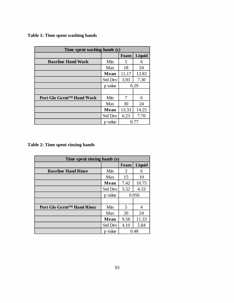
93
Table 1: Time spent washing hands
Table 2: Time spent rinsing hands
Foam Liquid
Baseline Hand Wash Min 5 6
Max 18 24
Mean 11.17 13.83
Std Dev 3.93 7.30
p value
Post Glo Germ™ Hand Wash Min 7 6
Max 30 24
Mean 13.33 14.25
Std Dev 6.23 7.70
p value
Time spent washing hands (s)
0.29
0.77
Foam Liquid
Baseline Hand Rinse Min 3 6
Max 15 19
Mean 7.42 10.75
Std Dev 3.32 4.33
p value
Post Glo Germ™ Hand Rinse Min 5 4
Max 20 24
Mean 9.58 11.33
Std Dev 4.10 5.84
p value
Time spent rinsing hands (s)
0.48
0.056
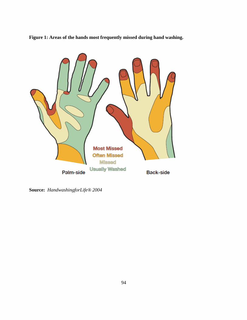
94
Figure 1: Areas of the hands most frequently missed during hand washing.
Source: HandwashingforLife® 2004
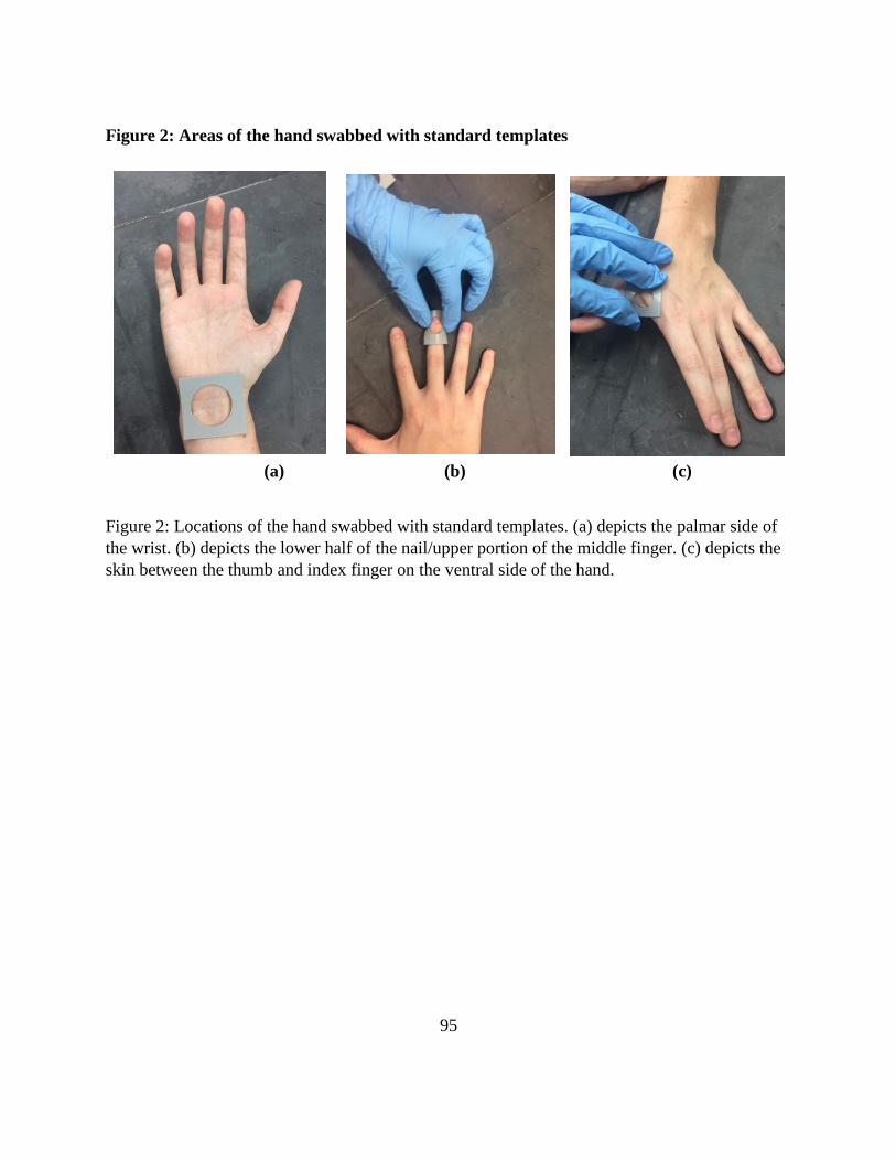
95
Figure 2: Areas of the hand swabbed with standard templates
(a) (b) (c)
Figure 2: Locations of the hand swabbed with standard templates. (a) depicts the palmar side of
the wrist. (b) depicts the lower half of the nail/upper portion of the middle finger. (c) depicts the
skin between the thumb and index finger on the ventral side of the hand.
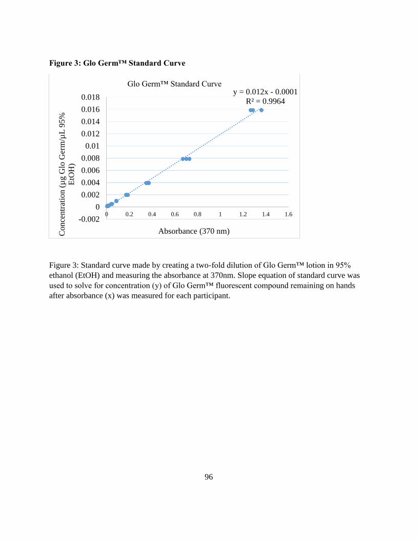
96
Figure 3: Glo Germ™ Standard Curve
Figure 3: Standard curve made by creating a two-fold dilution of Glo Germ™ lotion in 95%
ethanol (EtOH) and measuring the absorbance at 370nm. Slope equation of standard curve was
used to solve for concentration (y) of Glo Germ™ fluorescent compound remaining on hands
after absorbance (x) was measured for each participant.
y = 0.012x - 0.0001
R² = 0.9964
-0.002
0
0.002
0.004
0.006
0.008
0.01
0.012
0.014
0.016
0.018
0 0.2 0.4 0.6 0.8 1 1.2 1.4 1.6
Conce
ntr
atio
n (
µg G
lo G
erm
/µL
95%
EtO
H)
Absorbance (370 nm)
Glo Germ™ Standard Curve

97
Chapter 5: Comparison of two plain soap types for removal of bacteria and viruses from
hands with specific focus on food service environments

98
Abstract
Handwashing (HW) is a long established and widely accepted method to prevent disease
transmission. Ensuring effectiveness of current HW methods is essential for the optimization of
HW and enhanced disease prevention. The objective of this research was to determine the
difference in microbial reduction between plain foaming and liquid handsoap. The hands of 24
participants were inoculated by the palmar surface method with an average of 1.25 × 10⁸ CFU
Escherichia coli C3000 or 1.36 × 10⁸ PFU MS2 bacteriophage. Participants washed their hands
following a standard protocol with a standardized soap volume and a 10 s HW time. A glove
juice method was used to recover microorganisms from hands. Remaining microorganisms were
quantified by standard spread plate and plaque assays for E. coli and MS2, respectively. Hands
inoculated with E. coli had an average log reduction of 2.76 ± 0.70 and 2.52 ± 0.58 log CFU for
foaming and liquid handsoap, respectively. The mean log reduction for hands inoculated with
MS2 was 2.10 ± 0.57 and 2.23 ± 0.51 log PFU for foaming and liquid handsoap, respectively.
Data indicate no significant difference in overall microbial removal when comparing the efficacy
of plain foaming and liquid handsoap. However, regardless of soap type, the type of
microorganism impacted overall log reduction with a greater reduction for E. coli when
compared to MS2 with a significant difference (p = 0.0008) in reduction for foaming handsoap.
This study is the first comparison of the efficacy of plain liquid and foaming handsoap for
microbial reduction on hands during HW.
99
1. Introduction
It is estimated that foodborne pathogens, both major known pathogens as well as
unspecified agents, cause 47.8 million illnesses, 127,830 hospitalizations, and 3,037 deaths in the
U.S. each year with the leading causes of illness including noroviruses (58%), nontyphoidal
Salmonella spp. (11%), Clostridium perfringens (10%), and Campylobacter spp. (9%) (Scallan,
Griffin, Angulo, Tauxe, & Hoekstra, 2011). Pathogenic strains of Escherichia coli and
Salmonella are more commonly associated with raw meat (i.e. beef and poultry, respectively) as
animals are often hosts for these pathogens (Forsythe 2010). However, cross contamination of
pathogens between raw meat and ready-to-eat food products via food handlers’ hands is a
potential risk; therefore, proper handwashing (HW) is an essential control measure for risk
reduction (USFDA 2013). With respect to foodborne viruses, an epidemiologic investigation of
foodborne norovirus outbreaks in the U.S. from 2001-2008 found that 53% (473) of the 886
outbreaks were caused by food handler contamination (Hall et al., 2012). Additional analysis of
foodborne norovirus outbreaks from 2009 to 2012 confirmed these findings with food workers
implicated in 70% of 520 outbreaks, and bare hand contact was identified in 54% of the
outbreaks (Hall, Wikswo, Pringle, Gould, & Parashar, 2014). The recommended interventions
for preventing norovirus in a food service environment primarily include following US Food and
Drug Administration (FDA) Food Code guidelines for HW and glove use (Hall et al., 2012;
USFDA 2013).
The general population uses HW as an important step in disease prevention, and this is
especially critical within a food service environment (Miller, James-Davis, & Milaneis, 1994).

100
The hands of food service employees may become contaminated with foodborne pathogens
during critical stages in food service including after using the restroom, handling raw materials
(e.g., meats, vegetables, eggs, etc.) and after touching contaminated surfaces (Miller, James-
Davis, & Milaneis, 1994). Therefore, studies on the efficacy of HW agents are essential to ensure
that HW procedures are optimized for removal of pathogenic microorganisms from hands during
food service.
Numerous soaps (i.e. brands, types, formulations, etc.) are available on the market today,
and food service staff and the general population use these soaps daily. Plain (non-antimicrobial)
handsoap reduces soil, dirt, and in the case of food service, various physical and biological
materials on hands through physical removal with detergents. Meanwhile, antimicrobial
handsoap combines physical removal with the inactivation of microorganisms by antimicrobial
compounds in the soap that differentially affect viruses and bacteria (Fuls et al., 2008; Sickbert-
Bennett et al., 2006). While there have been numerous studies comparing the efficacy of
antimicrobial and plain handsoap (Fuls et al., 2008, Montville & Schaffner, 2011; Edmonds,
McCormack, Zhou, Macinga, & Fricker, 2013), the soaps used in these studies are typically
liquid handsoap. In a recent review by Conover and Gibson (2016), the methodologies and
results of 24 HW studies published since 1985 are discussed and despite the vast range of HW
agents tested in these studies, only one study evaluated foaming handsoaps (Fuls et al., 2008) and
none compared the efficacy of foaming and liquid handsoap. For this reason, the authors of the
present study selected to compare plain foaming and liquid handsoaps. One of the primary
differences between foaming and liquid handsoap is the level of surfactant. Foaming soaps
generally have a lower level of surfactants, and as a result, these soaps do not form micelles as
101
readily as liquid handsoap. Meanwhile liquid handsoaps typically have increased surfactant
levels as well as additional salts that allow for the formation of micelles (personal
communication provided by M. Caetta, VCI Formulation Specialist at GOJO Industries, Inc.)
that aid in the removal of dirt and oils as well as microorganisms.
With the increasing prevalence of foaming handsoap on the market and within food
service establishments, it is critical to determine if the associated microbial reductions are
comparable to that of traditional, plain liquid handsoap. For this study, we hypothesized that
there would be a significant difference in microbial reduction between foaming and liquid
handsoap. More specifically, we hypothesized that reduction of bacteria and virus on hands
would differ depending on soap type. Therefore, the overall goal of this study was to determine if
a difference exists in the efficacy of plain foaming and liquid handsoap by measuring the
reduction of microorganisms on hands inoculated with non-pathogenic E. coli and MS2
bacteriophage—a surrogate for the study of human enteric viruses such as norovirus.
2. Materials and Methods
2.1. Study design
The study was based on a Latin square design. The treatment structure was a two by two
factorial with microorganisms (E. coli C3000 and MS2) and soap type (foaming and liquid) as
the two different factors. Each participant visited one time per week over a four week period and
was randomly assigned to one of four sequences of treatment. Sequences were selected to
alternate exposure of participants to microorganism type and soap type and to adjust for any
possible confounding factors (e.g., learning by either the researchers or the experimental

102
participants or any carryover effects that could potentially be present throughout the four weeks
of the study).
2.2 Participant recruitment and training
Twenty-four participants (12 men and 12 women), 18 years and older, were recruited
from the University of Arkansas (Fayetteville, Arkansas) community. Participants had healthy
skin, with no presence of dermatitis, open wounds, cuts, burns, hangnails, or any additional
known disorders of the skin (ASTM 2013a). Institutional Review Board and Institutional
Biosafety Committee approval were obtained, and participants were informed about the safety of
microorganisms used in the study. All participants signed an informed consent form to
participate in the study. Sample size was determined based on a minimum power of 0.8 with the
following parameters: alpha = 0.05, standard deviation = 0.6, and a difference to detect of 0.5
log10 CFU or PFU.
To employ a standardized HW procedure throughout the study, prior to participating,
participants were trained on a standard HW protocol (Singapore Motherhood, 2012). Participants
were given 30 s to complete the HW procedure during training as well as throughout the
decontamination steps of the study. The actual experimental handwash was completed in 10 s
which is considered more representative of actual HW time occurring in daily life (discussed in
Section 2.7).
2.3 Preparation of inocula
2.3.1. Preparation of MS2 bacteriophage
103
A stock of MS2 bacteriophage (ATCC 15597-B1; American Type Culture Collection,
Manassas, VA) was prepared through propagation in E. coli C3000 followed by chloroform
extraction of the infected cell lysate as described previously by Gibson, Crandall, & Ricke
(2012). The stock concentration of MS2 bacteriophage was determined to be approximately 1011
PFU/mL by the double agar layer (DAL) method. One milliliter aliquots of MS2 were stored at -
80ºC. The phage stock was diluted with 0.1% peptone (Becton Dickinson and Company, Sparks,
Maryland) to approximately 6.78 × 10⁸ PFU/mL.
2.3.2. Preparation of E. coli C3000
Overnight stocks of E. coli C3000 (ATCC 15597; ATCC) was prepared in a culture flask
containing 25 mL of tryptic soy broth (Acumedia, Lansing, Michigan) incubated at 37ºC with
shaking at 110 rpm. Stock concentrations were determined by preparing a ten-fold dilution series
and plating 1 mL of each dilution in duplicate on 3M Petrifilm™ E. coli/coliform count plates
(3M, Maplewood, Minnesota). E. coli C3000 overnight culture (approximately 109 CFU/mL)
was diluted with 0.1% peptone (Becton Dickinson and Company) to approximately 6.26 ×10⁸
CFU/mL for inoculation on participants’ hands.
2.4. Hand decontamination prior to inoculation
To eliminate resident microorganisms on the hands of participants prior to inoculation
with test organisms, hands were treated with a conditioning wash as described by Fuls et al.
(2008) with modifications. Modifications included using 1 pump of antibacterial handsoap (The
Dial Corporation, Scottsdale, Arizona) with subjects scrubbing hands for 30 s and rinsing hands

104
for 10 s. Hands were also twice soaked in 70% ethyl alcohol and dried thoroughly before
inoculation with microorganisms.
2.5. Inoculation of hands
Hand inoculation of E. coli C3000 and MS2 was performed by the palmar surface
method (PSM) as described in the ASTM Standard Test Method E2870-13 with modifications.
One-hundred microliters of prepared E. coli or MS2 inoculum were pipetted onto the palm of
each hand (200 µl total) for an average of 1.25 × 10⁸ CFU total (hands combined) or 1.36 × 10⁸
PFU total (hand combined), respectively. The participants were asked to rub the palms and
fingers of each hand against each other for 10 ± 1 s in order to spread the inoculum on the palms
and fingers of each hand. Following inoculation, the hands were air-dried for 20 ± 5 s.
2.6. Selection of soaps
Two automatic dispensers, one foaming (GOJO Industries, Akron, OH) and one liquid
(Epare, Staten Island, New York), were selected for use in this study to limit potential
contamination of the soap dispenser from participants and to standardize the volume dispensed
each time. Based on the authors’ unpublished survey of soap volumes in food service
establishments, soap volumes were chosen as follows: one dispense of foaming soap was 0.9 mL
(i.e. after allowing ‘foam’ to subside), and one dispense of liquid handsoap was 1.5 mL.
2.7. Handwashing and drying
After allowing the inocula to dry, the participants were immediately asked to wash their
hands with one of the test soaps using the method described in Section 2.2. Following the 10 s
105
handwash, hands were rinsed for 10 s. Participants then dried hands for 10 s with three paper
towels, and hands were immediately sampled for recovery of microorganisms using the glove
juice method (GJM; Section 2.8.).
2.8. Recovery of microorganisms by GJM
Remaining microorganisms were recovered using the GJM described in ASTM E1174-13
with modifications (ASTM 2013b). Modifications included using 35 mL of 0.1% peptone
(Becton Dickinson and Company) sampling solution in each glove. As the soaps used did not
contain antimicrobial agents, no neutralizers were added to the sampling solution. An additional
modification included recovering all of the remaining liquid from each glove and transferring to
a sterile, glass sample jar (VWR) and using a portion of the remaining solution to dilute and
plate. Volumetric adjustments were made based on total volume recovered (Equation 1).
Prior to study commencement, the recovery efficiency of the GJM was determined to be
2.3 and 8.9% for E. coli C3000 and MS2, respectively. These values were based on two
experimental replicates with samples plated in duplicate. Recovery efficiency was calculated
using Equation 2. To ensure the true log reduction was determined, percent recovery efficiency
for each microorganism was then incorporated into calculating the recovery of microorganisms
(Equation 3). The log reduction that occurred due to HW with foaming and liquid handsoap was
then calculated (Equation 4). After the recovery of microorganisms from hands, hands were
again decontaminated following the procedure described in Section 2.4.
Equation 1: RM = T
V𝐴 × VR
Equation 2: RE = (RM / I) × 100
106
Equation 3: IRE = I × (RE/100)
Equation 4: LR = log10 (IRE/RM)
Where: RM = microorganisms recovered (CFU or PFU); T = total CFU or PFU counted; VA =
volume analyzed (mL); VR = total volume recovered by GJM (mL); RE = % recovery efficiency;
I = initial total inoculum (CFU or PFU); IRE = inoculum with recovery efficiency; LR = log10
reduction (CFU or PFU)
2.9. Detection of microorganisms in recovered sampling solution
For E. coli C3000, 1 mL aliquots of each dilution were plated onto 3M Petrifilm™ E.
coli/coliform count plates (3M) in duplicate and incubated at 37°C for 24 h. After 24 h, CFU
were counted. A negative control of 1mL of 0.1% peptone was plated as well as a positive
control of 1 mL of E. coli C3000 at a concentration of 101 to 102 CFU/mL. For MS2, the DAL
method was used as described previously (Gibson, Crandall, & Ricke, 2012). Following
incubation for 16-24 h at 37ºC PFU were counted, and the PFU/mL were determined. A negative
control with 100 µL of log phase E. coli C3000 plus 100 µL of 0.1% peptone was analyzed in
each set of experiments. A positive control of 100 µL of 1 × 10² PFU/mL MS2 and 100 µL of
log phase E. coli C3000 was also performed.
2.10. Statistical Analysis
JMP® Pro 11 (SAS, Cary, NC) statistical analysis software was used for all data analysis.
As the study was primarily based on a Latin Square design, with elements of a split plot design, a
mixed model was used to test the significance of differences that occurred in reduction of

107
microorganisms for each soap type (foaming or liquid) as well as for each microorganism type
(E. coli or MS2). Sequence and week were incorporated into the model to account for any
variance that may have occurred due to learning by either the participants or researchers
throughout the study as well as for any potential cross-over effect that may have occurred
between weeks of the study. Gender was also added as an effect in the model to determine any
significance. Multiple comparison of the interaction means was performed with Tukey’s honest
significant difference (HSD) test. Results were considered statistically significant at p ≤ 0.05.
3. Results
3.1. Efficacy of foaming and liquid handsoaps
All 24 participants completed all 4 weeks of the study. The average age of the
participants was 31 years of age. As stated in Section 2.5, the initial inoculum on hands was on
average 1.25 × 10⁸ CFU E. coli C3000 and 1.36 × 10⁸ PFU MS2. Table 1 reports the average
total log reductions for E. coli C3000 and MS2 on hands for foaming handsoap, liquid handsoap,
and the baseline wash with water. However, this baseline water only wash was not considered a
treatment in the mixed model used for statistical analyses (Section 2.10).
3.2. Factors influencing HW efficacy
Statistical analysis of log reductions indicates no significant difference in efficacy of
foaming and liquid handsoap for overall microbial removal (p = 0.56). However, the reduction of
microorganisms on hands was significantly different depending on the type of microorganism
with increased reductions occurring for hands inoculated with E. coli C3000 as compared to
hands inoculated with MS2. Based on Tukey’s HSD test, MS2 reduction was significantly less

108
with foaming soap when compared to E. coli reductions with foaming soap (p = 0.0008). Table 1
shows the comparison of log reduction for each microorganism by treatment. Gender did not
have a significant impact on the reduction of microorganisms on hands (p = 0.44).
4. Discussion
Even though the results presented here indicate no significant difference in overall
microbial removal between foaming and liquid handsoap, the type of microorganism on the hand
had a significant effect, with a greater reduction occurring for E. coli as compared to MS2. Of
particular interest is that while a greater reduction for E. coli was observed with foaming soap
(2.76 log CFU) as compared to liquid (2.52 log CFU), the opposite was true for MS2 with a 2.10
log PFU reduction using foaming soap and a 2.23 log PFU reduction using liquid soap.
Although the exact reasons behind these differences in reduction are not clear, it is
known that MS2 particle size (24-27 nm)—and human enteric viruses in general—is much
smaller than that of bacteria such as E. coli (500 nm in diameter) (Strauss & Sinsheimer, 1963;
Abbsazadegan, Mayer, Ryu, & Nwachuku, 2007). In addition, MS2 bacteriophage is strongly
hydrophobic and this property impacts its attachment to particulates (Bales, Li, Maguire, Yahya,
& Gerba, 1993; Shields & Farrah, 2002). With respect to lower reductions in MS2 when using
foaming soap when compared to E. coli, a possible theory is that the increased surfactant levels
of liquid soap create conditions in which the smaller, hydrophobic MS2 particles are surrounded
by micelles and are more readily removed from the hands. Conversely, the decreased surfactant
concentrations of the foaming handsoap creates conditions that do not allow for the same micelle
formation (personal communication provided by M. Caetta, VCI Formulation Specialist at GOJO

109
Industries, Inc.); therefore, limiting the ability to surround the particles with the micelle structure
and making it difficult to remove the smaller, hydrophobic MS2 particles present on hands.
The complex physiological state of the skin surface also likely plays a role into how
effectively microorganisms are removed from the hand. Numerous researchers have studied skin
physiology and have shown that microflora on hands can vary from person to person due to
numerous factors including environment, age, and sex (Larson 1985; Jumaa 2005). Each of
these variables can cause variation in skin flora, sebum (i.e. oily substance) production, skin
flexibility, and skin permeability all of which may impact microbial removal during HW (Noble,
1978; Leveque, deRigal, Agache, & Monneur, 1980).
With respect to MS2 removal reported in previously published studies, Sickbert-Bennett
et al. (2005) observed that tap water was actually the most effective for removal of MS2 from
hands with 1.89 to 2.56 log PFU reduction followed by plain, liquid soap with 1.54 to 2.03 log
PFU reduction. A baseline wash with tap water was completed in the present study, and although
this was not technically a ‘treatment’, the results reported here for tap water (1.20 log PFU
reduction) do not corroborate the findings by Sickbert-Bennett et al. (2005) while similar results
for plain, liquid handsoap are reported (2.23 log PFU reduction). Soap volume is one difference
between the present study and that of Sickbert-Bennett et al. (2005). The authors of that study
used 3 mL of liquid handsoap with a 10 s handwash, while the present study used half this
amount of soap (1.5 mL) and a 10 s handwash. Although both studies used a 10 s handwash
time, the difference in soap volumes may have an effect on the differences in reduction of MS2
by plain, liquid handsoap occurring between the two studies.

110
Another study by Burton et al. (2011) reported results that echo the findings of the
present study and demonstrated that HW with plain soap was more effective than HW with water
alone for reduction of naturally acquired bacteria on hands. Several additional studies have
investigated the reduction of non-pathogenic E. coli on hands via HW and have evaluated the
effect of numerous variables including soap type (plain versus antimicrobial) and HW time
(Ansari, Sattar, Springthorpe, Wells, & Tostowaryk, 1989; Ayliffe, Babb, Davies, & Lilly, 1987;
Fuls et al., 2008; Miller et al. 2011; Paulson et al. 1999; Sasahara, Hayashi, Hosoda, Morisawa,
& Hirai, 2014; Snyder 2007; Stiles & Sheena, 1985). In a meta-analysis by Montville and
Schaffner (2011), an average reduction of 1.93 ± 0.91 log CFU was reported for studies that
inoculated hands with gram-negative bacteria such as E. coli. This reduction is comparable to
what is reported in the present study for reduction of E. coli with plain foaming and liquid
handsoaps (2.76 ± 0.70 and 2.52 ± 0.58 log CFU, respectively).
Compared to studies on HW efficacy against bacteria, very few studies have been
reported on the reduction of human enteric viruses or viral surrogates achieved through HW with
either liquid or foaming handsoaps (Ansari, Sattar, Springthorpe, Wells, & Tostowaryk, 1989;
Lin et al., 2003; Liu, Yuen, Hsiao, Jaykus,, & Moe, 2010; Mbithi, Springthorpe, & Sattar, 1993;
Sickbert-Bennett et al. 2005; Stowell et al., 2014). Liu, Yuen, Hsiao, Jaykus, & Moe (2010)
investigated the efficacy of liquid soap for removal norovirus, using two different finger pad
protocols—the standard ASTM method (E 1838-2) and a modifications of this method. The
average log reductions achieved with a liquid handsoap containing 0.5% triclosan were 0.67 and
1.10 log for the standard and modified ASTM methods, respectively. Even though the described
study used a liquid handsoap containing an antimicrobial agent, the reported log reductions were

111
lower than those reported in the present study and more similar to the baseline water only wash.
Numerous differences occurred between the present study and that of Liu, Yuen, Hsiao, Jaykus,
& Moe (2010) including microorganisms selected, method of detection (molecular detection
versus plaque assay for infectivity), and the HW treatment method.
Lin et al. (2003) compared HW techniques to remove E. coli and caliciviruses under
natural fingernails. For nails inoculated with E. coli, reductions of 1.18 log CFU were reported
for both tap water and plain, liquid soap, respectively; meanwhile, reductions of feline calicivirus
(FCV) were 1.97 and 1.82 log PFU for tap water and plain, liquid soap, respectively. Reductions
reported by Lin et al. (2003) for FCV are comparable to the reductions reported for MS2 in the
present study, while the reductions achieved for E. coli are quite a bit lower than the reductions
reported in the present study. While the palmar surface method of inoculation was applied in the
present study, Lin et al. (2003) utilized contact of the underside of the nail with inoculated
ground beef to simulate real-life contamination of fingernails which may explain the difference
in log reduction values. Additional studies evaluating the efficacy of various HW agents
including plain, liquid soap in removal of enteric viruses (i.e. hepatitis A virus, poliovirus type 1,
human rotavirus) from hands have reported log reduction values ranging from 1 to 1.2 logs
(Ansari, Sattar, Springthorpe, Wells, & Tostowaryk, 1989; Mbithi, Springthorpe, & Sattar,
1993). Compared to the present study, these log reduction values are nearly 1-log lower than
those reported for MS2 in the present study and are more similar to the baseline water wash
removal. Again, many of these differences can be explained by variations in methodology even
though several standard methods for the evaluation of HW agents exist both in the U.S. and the
European Union (E.U.).

112
As evidenced by the papers referenced above, researchers test a variety of variables when
performing HW studies and numerous variables are changed between studies (especially HW
time, soap volume, and soap type). Even though there are numerous standard methods for the
evaluation of HW efficacy (ASTM (2013a), ASTM (2013b), Ayliffe, Babb, & Quoraishi, 1978,
CEN 2013a, CEN 2013b) based on what has been published in peer reviewed literature,
researchers often change numerous variables based on their experimental design or research
objectives. This further complicates direct comparison of data between studies. Additionally,
HW is a process that inherently has a great amount of variation each time it occurs, and
controlling or standardizing all parts of the HW process is a difficult task.
Here, it is important to acknowledge some of the limitations of the present study. In food
service facilities, food handlers’ hands are potentially covered with numerous organic materials
including oils and food particles as well as both resident and transient microorganisms. However,
to eliminate unnecessary variables for the initial evaluation of foaming versus liquid handsoap
participant hands were decontaminated at the beginning of each study day and then inoculated
with only one test microorganism at a time. Based on this, one limitation of the present study is
that the experimental design does not replicate the conditions that would be present in a food
service facility. To address this, future research on the effect of additional organic material on
hands on the reduction of both viruses and bacteria on hands when washed with foaming and
liquid handsoap would be beneficial. An additional limitation is the use of a short HW time in
our study (10 s wash and 10 s rinse). This HW time was selected to be more representative of
what has been observed and reported in the peer-reviewed literature as well as based on the
authors’ experience (Graham 1990; Meengs, Giles, Chisholm, Cordell, & Nelson, 1994; Munger
113
& Harris, 1989; Sickbert-Bennett et al. 2005; Strohbehn, Sneed, & Meyer, 2008). However, it
should be noted that the FDA Food Code 2013 recommends a 20 s HW time with vigorous
scrubbing for 10 to 15 s therefore the 20 s may include the rinse time as well. A final, potential
limitation is the low recovery efficiency of the GJM. In order to keep the recovery solution in the
glove, a volume of 35 mL was chosen for each glove. This lower volume may have had an
impact on the recovery efficiency. However, as discussed in section 2.8, recovery efficiency was
incorporated into the overall log reduction that occurred on hands to account for this. Moreover,
previously published studies that utilized a GJM rarely report recovery efficiency, and because of
this we were unable to accurately compare our recovery efficiency with that of other studies.
5. Conclusions
Results of the present study indicate that no significant difference in overall microbial
reduction occurs between foaming and liquid handsoaps. However, a significant difference does
exist between the types of microorganisms on hands (bacteria versus viruses). Based on this,
future research related to optimization of HW for increased virus removal is warranted. This is
especially important since poor hand hygiene is significant to the transmission of human
noroviruses in the food service industry as well as within healthcare and other settings commonly
implicated in human norovirus outbreaks (Barclay et al. 2014; Hall et al. 2012; Hall, Wikswo,
Pringle, Gould, & Parashar, 2014; Moe 2009).
114
Acknowledgements
This research was based upon work that is supported, in part, by the National Institute of Food
and Agriculture (NIFA), U.S. Department of Agriculture (USDA), under award number 2013-
68003-21288 as well as USDA NIFA Hatch Act funding. Additionally we would like to thank
statistician, Kevin Thompson, for his assistance in designing the experiment and analyzing the
data. We would also like to thank Giselle Almeida, Mallory Hancock, Adam Baker, Nicole
Turnage, Stephanie Roto, Zhaohao Shi, Sang In Lee, Nathan Jarvis, Jasmeet Braich, Pravin
Kaldhone, and Anisha Chowdhury for their assistance and willingness to participate in
preliminary research for the study.

115
References
Abbsazadegan, M., Mayer, B.K., Ryu, H., & Nwachuku, N. (2007). Efficacy of removal of CCL
viruses under enhanced coagulation conditions. Environmental Science and Technology,
41, 971-977.
Ansari, S.A., Sattar, S.A., Springthorpe, V.S., Wells, G.A., & Tostowaryk, W. (1989). In vivo
protocol for testing efficacy of hand-washing agents against viruses and bacteria:
experiments with Rotavirus and Escherichia coli. Applied and Environmental
Microbiology, 55, 3113-3118.
ASTM. (2013a). ASTM E2870-13 Standard test method for evaluating relative effectiveness of
antimicrobial handwashing formulations using the palmar surface and mechanical hand
sampling. West Conshohocken: ASTM International.
ASTM. (2013b). ASTM E1174-13 Standard test method for evaluation of the effectiveness of
health care personnel handwash formulations. West Conshohocken: ASTM
International.
Ayliffe, G.A.J., Babb, J.R., Davies, J.G., & Lilly, H.A. (1987). Hand disinfection: a comparison
of various agents in laboratory and ward studies. Journal of Hospital Infection, 11, 226-
243.
Ayliffe, G.A.J., Babb, J.R., & Quoraishi, A.H. (1978). A test for ‘hygienic’ hand disinfection.
Journal of Clinical Pathology, 31, 923-928.
Bales, R.C., Li, S., Maguire, K.M., Yahya, M.T., & Gerba, C.P. (1993). KMS-2 and poliovirus
transport in porous media: hydrophobic effects and chemical perturbations. Water
Resources Research, 29, 957-963.
Barclay, L., Park, G.W., Veg, E., Hall, A., Parashar, U., Vinje, J. & Lopman, B. (2014).
Infection control for norovirus. Clinical Microbiology and Infection, 20, 731-740.
Burton, M., Cobb, E., Donachie, P., Judah, G., Curtis, V., & Schmidt, W.P. (2011). The effect of
handwashing with water or soap on bacterial contamination of hands. International
Journal of Environmental Research and Public Health, 8, 97-104.
CEN. (2013a). EN1499. Chemical disinfectants and antiseptics - Hygienic handwash - Test
method and requirements (phase 2/step 2). Brussels. European Committee for
Standardization.
CEN. (2013b). EN1500. Chemical disinfectants and antiseptics - Hygienic handrub - Test
method and requirements (phase 2/step 2). Brussels. European Committee for
Standardization.
Conover, D.M., & Gibson, K.E. (2016). A review of methods for the evaluation of handwashing
efficacy. Food Control, 63, 53-64.
116
Edmonds, S. L., McCormack, R. R., Zhou S.S., Macinga, D. R., & Fricker, C.M. (2012). Hand
hygiene regimens for the reduction of risk in food service environments. Journal of Food
Protection, 75, 1303-1309.
Forsythe, S.J., (2010). The microbiology of safe food. (p. 157-174). United Kingdom: Wiley-
Blackwell.
Fuls, J. L., Rodgers, N. D., Fischler, G. E., Howard, J. M., Patel, M. Weidner, P. L., et al. (2008).
Alternative hand contamination technique to compare the activities of antimicrobial and
nonantimicrobial soaps under different test conditions. Applied and Environmental
Microbiology, 74, 3739-3744.
Gibson, K.E., Crandall, P.G., & Ricke, S.C. (2012). Removal and transfer of viruses on food
contact surfaces by cleansing cloths. Applied and Environmental Microbiology, 78, 3037-
3044.
Graham, M. (1990). Frequency and duration of handwashing in an intensive care unit. American
Journal of Infection Control, 18, 77-81.
Hall, A.J., Eisenbart, V.G., Etingue, A.L., Gould, H., Lopman, B.A., Parashar, U.D. (2012).
Epidemiology of foodborne norovirus outbreaks, United States, 2001-2008. Emerging
Infectious Diseases, 18, 1566-1573.
Hall, A.J., Wikswo, M.E., Pringle, K., Gould, H., & Parashar, U.D. (2014). Vital Signs:
Foodborne Norovirus Outbreaks-United States, 2009-2012. Morbidity and Mortality
Weekly Report, 63, 491-495.
Jensen, D. (2015). Addressing challenges and gaps in the hand hygiene literature using novel
quantitative approaches. 5, p. 99. New Brunswick, N.J. Rutgers University.
Jumaa, P.A. (2005). Hand hygiene: simple and complex. International Journal of Infectious
Diseases, 9, 3-14.
Larson, E. (1985). Handwashing and Skin: Physiologic and Bacteriologic Aspects. Infection
Control, 6, 14-23.
Leveque, J.L., deRigal, J., Agache, P.G., & Monneur, C. (1980). Influence of ageing on the in
vivo extensibility of human skin at a low stress. Archives of Dermatological Research,
269, 127-135.
Lin, C., Wu, F., Kim, H., Doyle, M.P., Michaels, B.S., & Williams, L.K. (2003). A comparison
of hand washing techniques to remove Escherichia coli and caliciviruses under natural or
artificial fingernails. Journal of Food Protection, 66, 2296-2301.
Liu, P., Yuen, Y., Hsiao, H., Jaykus, L. & Moe, C. (2010). Effectiveness of liquid soap and hand
sanitizer against Norwalk virus on contaminated hands. Applied and Environmental
Microbiology, 76, 394-399.

117
Mbithi, J.N., Springthorpe, V.S., & Sattar, S.A. (1993). Comparative in vivo efficiencies of
hand-washing agents against Hepatitis A Virus (HM-175) and Poliovirus Type 1 (Sabin).
Applied and Environmental Microbiology, 59, 3463-3469.
Meengs, M.R., Giles, B.K., Chisholm, C.D., Cordell, W.H., & Nelson, D.R. (1994). Hand
washing frequency in an emergency department. Annals of Emergency Medicine, 23,
1307-1312.
Miller, M.L., James-Davis, L.A., & Milaneis, L.E. (1994). A field study evaluating the
effectiveness of different hand soaps and sanitizers. Dairy, Food, and Environmental
Sanitation, 14, 155-160.
Miller, T., Patrick, D., & Omrod, D. (2011). Hand decontamination: influence of common
variables on hand-washing efficiency. Healthcare Infection, 16, 18-23.
Moe, C.L. (2009). Preventing Norovirus transmission: how should we handle food handlers?
Clinical Infectious Diseases, Editorial commentary, 38-40.
Montville, R., & Schaffner, D.W. (2011). A meta-analysis of the published literature on the
effectiveness of antimicrobial soaps. Journal of Food Protection, 27, 1875-1882.
Munger, K., & Harris, S.J. (1989). Effects of an observer on handwashing in a public restroom.
Perceptual and Motor Skills, 69, 733-734.
Noble, W.C., & Pitcher, D.G. (1978). Microbiology of Human Skin. In M. Alexander (Ed.),
Advances in Microbial Ecology (pp. 245-289). New York: Plenum Press.
Paulson, D.S., Riccardi, C., Beausoleil, C.M., Fendler, E.J., Dolan, M.J., Dunkerton, L.V., et al.
(1999). Efficacy evaluation of four hand cleansing regimens for food handlers. Dairy,
Food, and Environmental Sanitation, 19, 680-684.
Sasahara, T., Hayashi, S., Hosoda, K., Morisawa, Y., & Hirai, Y. (2014). Comparison of hand
hygiene procedures for removing Bacillus cereus spores. Biocontrol Science, 19, 129-
134.
Scallan, E., Griffin, P. M., Angulo F. J., Tauxe, R. V., & Hoekstra, R. M. (2011) Foodborne
illness acquired in the United States-unspecified agents. Emerging Infectious Diseases,
17, 7-15.
Shields, P.A., & Farrah, S.R. (2002). Characterization of Virus Adsorption by Using DEAE-
Sepharose and Octyl-Sepharose. Applied and Environmental Microbiology 68, 3965-
3968.
Sickbert-Bennett, E. E., Weber, D. J., Gergen-Teague, M. F., Sobsey, M.D., Samsa, G. P., &
Rutala, W. A. (2005). Comparative efficacy of hand hygiene agents in the reduction of
bacteria and viruses. American Journal of Infection Control, 23, 67-77.
Singapore Motherhood. (2012). Teach your child proper handwashing techniques. Available
from: http://singaporemotherhood.com/articles/2012/10/2873/. Accessed 03.12.15.
118
Snyder, O.P. (2007). Removal of bacteria from fingertips and the residual amount remaining on
the hand washing nailbrush. Food Protection Trends, 27, 597-602.
Stiles, M.E., & Sheena, A.Z. (1985). Efficacy of low-concentration iodophors for germicidal
hand washing. Journal of Hygiene, 94, 269-277.
Stowell, J.D., Forlin-Passoni, D., Raford, K., Bate, S.L., Dollard, S.C., Bialek, S.R., et al. (2014).
Cytomegalovirus survival and transferability and the effectiveness of common hand-
washing agents against cytomegalovirus on live human hands. Applied and
Environmental Microbiology, 80, 455-461.
Strauss, J.H., & Sinsheimer, R.L. (1963). Purification and properties of bacteriophage MS2 and
of its ribonucleic acid. Journal of Molecular Biology, 7, 43-54.
Strohbehn, C., Sneed, J., Paez, P., & Meyer, J. (2008). Hand washing frequencies and procedures
used in retail food services. Journal of Food Protection, 71, 1641-1650.
USFDA. (2013) Food Code. (pp. 46-50; 388-389). Washington, D.C: U.S. Department of Health
and Human Services. Available from:
http://www.fda.gov/downloads/Food/GuidanceRegulation/RetailFoodProtection/FoodCo
de/UCM374510.pdf. Accessed 12.08.15.

119
Table 1: Comparison of log reduction by treatment
Treatment
Total Log10 CFU or PFU Reduction (± SD)
Microorganism
E. coli MS2 p-value
Foaming 2.76 (0.70) 2.10 (0.57) 0.0008
Liquid 2.52 (0.58) 2.23 (0.51) 0.079
Water 2.45 (0.93) 1.20 (0.49) <0.0001

120
Chapter 6: Overall Conclusions
For quite some time now, handwashing (HW) has been accepted as a method to prevent
disease transmission not only in the general population, but especially in select industries
including the health care field and the food service industry. The food service industry has
established strict guidelines for employees to follow to prevent the transmission of harmful
pathogens to consumers. One such guideline is the United States Food and Drug
Administration’s (FDA) Food Code which details the steps needed for completion of a thorough
and appropriate handwash, as well as specific instances in which food service workers will need
to wash their hands (after using the restroom, switching between raw and ready-to-eat food, etc.)
(USFDA 2013).
Regardless of strict guidelines on proper HW, food service workers often fail to comply
with the established policies, and numerous foodborne illness outbreaks occur yearly because of
inadequate HW (Green et al. 2006). One important step in reducing foodborne illness caused by
food handlers is to understand and correct this lack of compliance. An additional method to
reduce foodborne illness is to understand and optimize the various factors that impact the HW
process (Conover and Gibson 2016). Soap type (foaming (F) versus liquid (L)) is once such
variable that may impact the HW process. To my knowledge no research studies have been
conducted comparing the effectiveness of foaming and liquid handsoap. Foaming handsoap is
relatively new in the realm of HW with the first generic system introduced in 1999 by Deb
Group Limited.
121
The primary objectives of my research were to I) conduct a survey of soap type and soap
volume in food service establishments in Washington County, Arkansas II) to determine if
handsoap type (foaming versus liquid) affects HW behavior and III) to determine if a difference
exists in the effectiveness of non-antimicrobial F and L handsoap by measuring the reduction of
microorganisms on hands inoculated with non-pathogenic E. coli and MS2 bacteriophage – a
surrogate for the study of human enteric viruses such as norovirus.
In order to begin this research, a list of 75 food service locations in Washington County,
Arkansas was generated based on a set of exclusion criteria as well as random number
generations. Soap samples were collected from these food service locations to determine average
soap volume as well as prevalence of soap type. Soap samples were collected from each location
in triplicate (three “pumps” from the same soap dispenser on the same day), and the volume was
measured. Sixty-eight of the 75 locations were sampled. I determined that 54.4% (n = 37) and
47.06% (n = 32) had F and L soap types, respectively. The average volume of F and L handsoap
was 0.64 ± 0.21 mL and 1.19 ± 0.46 mL, respectively. This information determined the selection
of representative F and L handsoap volumes used in chapters 4 and 5.
In order to understand HW behavior, 12 volunteers completed a series of handwashes in
which no training in proper HW was given. A baseline handwash was completed as the
participant entered the lab, while the second handwash involved the application of a known
amount of Glo Germ™ (GG) fluorescent lotion to hands prior to the handwash. Following both
the baseline and GG HW, hands were swabbed in three locations to recover remaining GG.
Swabs were eluted and absorbance was measured at OD370nm and remaining GG was quantified
122
using a standard curve. No significant difference in behavior was determined in terms of GG
remaining, HW time in the baseline handwash and post GG handwash, and baseline handrinsing
time and post GG handrinse. Average HW time for the baseline handwash was (F) 11.17 ± 3.93 s
and (L) 13.83 ± 7.30 s, and for the post GG handwash (F) 13.33 ± 6.22 s and (L) 14.25 ± 7.70 s.
While no statistically significant difference in behavior occurred between F and L handsoap, a
consistent increase in both wash time and rinse time for L handsoap did occur, indicating that
there may be a possible benefit to using liquid handsoap in food service establishments.
Additional research in this area may be beneficial to determine if this difference in wash time has
an impact on microbial reductions.
Finally, in order to compare the effectiveness of foaming and liquid handsoap, hands of
24 participants were inoculated by the palmar surface method with Escherichia coli C3000 or
MS2 bacteriophage. Participants washed their hands following a standard protocol with a
standardized soap volume and a 10 s HW time. Following this, remaining microorganisms were
recovered from hands and remaining microorganisms were quantified by standard spread plate
and plaque assays for E. coli and MS2, respectively. Hands inoculated with E. coli had an
average log reduction of 2.79 ± 0.71 and 2.52 ± 0.58 log CFU for foaming and liquid handsoap,
respectively. The mean log reduction for hands inoculated with MS2 was 1.98 ± 0.60 and 2.26 ±
0.49 log PFU for foaming and liquid handsoap, respectively. These data indicate that there is no
significant difference in the effectiveness between foaming and liquid handsoap in overall
microbial removal. However, regardless of soap type, the type of microorganism impacted
overall log reduction, with a greater log reduction occurring for hands inoculated with E. coli as
123
compared to hands inoculated with MS2. Future research into impact of soap type will be
beneficial to understand the potential decrease in reduction occurring for viruses with foaming
handsoap.
Overall, this research has provided insight into the impact different types of soap (F
versus L) have on handwashing behavior, as well as the difference in effectiveness between F
and L handsoap on microbial reduction on hands, particularly when focusing on the removal of
bacteria and viruses from hands. Additional research will be needed to better understand the
potential impact the difference in wash times between F and L handsoap has on microbial
reduction as well as the effect of F and L handsoap on bacteria and virus removal on hands and
the potential impact this may have on the food service industry.
124
References:
Conover, D.M., & Gibson, K.E. (2016). A review of methods for the evaluation of handwashing
efficacy. Journal of Food Control, 63, 53-64.
Deb Group Ltd. 2014. http://www.debgroup.com/us/technology-zone/foam-technology.
Accessed on November 9th, 2015.
Green, L. R., Selman, C. A., Radke, V., Ripley, D., Mack, J. C., Reimann, D. W., et al. (2006)
Food worker hand washing practices: an observation study. Journal of Food Protection,
69, 2417-2423.
USFDA. (2013) Food Code. (pp. 46-50; 388-389). Washington, D.C: U.S. Department of Health
and Human Services. Available from:
http://www.fda.gov/downloads/Food/GuidanceRegulation/RetailFoodProtection/FoodCo
de/UCM374510.pdf. Accessed 12.08.15.

125
Appendix
Figure 1: Schematic of experimental design (Chapter 5):
Figure 1: Schematic of experimental design of Chapter 5. Step 1 was completed before
participation in the study began. Steps 2-12 were completed each time (4 times total) a
participant completed the experiment.
126
IBC Approval Letter:

127
IRB Approval Letter:
April 15, 2015
MEMORANDUM TO: Kristen Gibson Danielle Conover FROM: Ro Windwalker IRB Coordinator RE: New Protocol Approval If you have questions or need any assistance from the IRB,
please contact me at 109 MLKG Building, 5-2208, or [email protected].


