Biomed Environ Sci, 2013; 26(1): 63‐69 63
*This research was supported by the National Key Technology R&D Program (No. 2012BAK01B00). #Correspondence should be addressed to FAN Yong Xiang. Tel: 86‐10‐87720035; E‐mail:[email protected] Biographical note of the first author: ZHANG Wen Zhong, male, born in 1976, Ph.D, associate professor, majoring in
molecular toxicology. Received: September 5, 2012; Accepted: October 22, 2012
Original Article
Combined Subchronic Toxicity of Bisphenol A and Dibutyl Phthalate on Male Rats*
ZHANG Wen Zhong, YONG Ling, JIA Xu Dong, LI Ning, and FAN Yong Xiang#
China National Center for Food Safety Risk Assessment, Beijing 100022, China
Abstract
Objective To evaluate the combined subchronic toxicity of bisphenol A (BPA) and dibutyl phthalate (DBP) in male Sprague Dawley (SD) rats.
Methods Forty 4‐week‐old male rats weighing 115‐125 g were randomly divided into BPA‐treated, DBP‐treated group, BPA+DBP‐treated and control groups and fed with a soy‐ and alfalfa‐free diet containing 285.4 ppm BPA, 285.4 ppm DBP, 285.4 ppm BPA plus 285.4 ppm DBP, and a control diet, respectively, for 90 consecutive days. At the end of the study, the animals were sacrificed by exsanguination via the carotid artery under diethyl etherane aesthesia and weighed. Organs, including liver, kidneys, spleen, thymus, heart, brain, and testis underwent pathological examination. The androgen receptor (AR), gonadotropin‐releasing hormone receptor (GNRHR), and progesterone hormone receptor (PR) genes from the hypothalamus were detected by real‐time PCR. The biomedical parameters were analyzed.
Results No significant difference was found in food intake, body weight, tissue weight, organ/brain weight ratio, and biomedical parameters among the four groups (P>0.05). However, BPA and DBP up‐regulated AR, PR and GNRHR expression levels in rats and showed a synergistic or an additive effect in the BPA+DBP group.
Conclusion The combined subchronic toxicity of BPA and DBP is synergistic or additive in male SD rats.
Key words: Bisphenol A; Dibutyl phthalate; Combined subchronic toxicity; Sprague dawley rat; Endocrine disruption
Biomed Environ Sci, 2013; 26(1):63‐69 doi: 10.3967/0895‐3988.2013.01.008 ISSN:0895‐3988
www.besjournal.com(full text) CN: 11‐2816/Q Copyright ©2013 by China CDC
INTRODUCTION
he endocrine disruptor hypothesis suggests that the global decrease in male reproductive function may be due to
background environmental exposures to hormonally active agents[1]. Endocrine disruptors, also known as endocrine‐disrupting chemical compounds (EDC), are exogenous substances that act like hormones in the endocrine system and disrupt the physiological
function of endogenous hormones[2]. However, this hypothesis has advocates and skeptics[1].
Bisphenol A (BPA) and dibutyl phthalate (DBP) are endocrine disruptors that are also abundant chemicals[3‐4]. Humans are mainly exposed to BPA and DBP through the ingestion of chemical‐contaminated food. Because of their widespread use and presence in ubiquitous and constant environments, human exposure is almost inevitable. It has been demonstrated that BPA and
T
64 Biomed Environ Sci, 2013; 26(1): 63‐69
DBP at doses equivalent to human exposure levels exert deleterious effects on the endocrine system in animals[4]. It has been reported that BPA exposure demonstrates effects on the reproductive system in adult animals, including oocyte chromosome abnormalities, recurrent miscarriage, and decreased semen[2] and that exposure to high DBP levels can induce fetal death, cancer, malformations, liver and kidney injury, and reproductive toxicity in animals[5‐8].
Because these compounds usually coexist in natural environments, their combined effects warrant further investigation. In fact, some reports are available on the combined effects of these compounds. Tan et al.[9] reported that 4‐NP and BPA exert their antagonistic effect on pubertal development in intact juvenile/peripubertal male Sprague Dawley (SD) rats. Duan et al.[10] reported that BPA and pentachlorophenol act in either a synergistic or an antagonistic mode depending on their different endpoints. Synergistic action can be displayed based on the endpoint of 24 h mortality, whereas an antagonistic effect can be demonstrated based on the endpoint of 72 h cardiac edema. Fent et al.[11] demonstrated that 37 compounds display either an additive or a synergistic effect on a yeast reporter gene. However, only a limited number of studies are available on the combined effect of BPA and DBP, and no study is available on their combined effect in vivo.
This study was designed to detect the combined effect of dietary BPA and DBP on male SD rats. It is estimated that the daily intake of BPA by children and adults is approximately 0.001‐0.1 μg/kg body weight (bw)/day[12], and that of DBP is 1‐4.3 μg/kg bw/day[8]. The dose we chose in this study was approximately 120‐fold of the daily intake of humans. The findings in this study can provide empirical evidence for the safety of BPA and DBP.
MATERIALS AND METHODS
Materials
The low temperature high‐speed centrifuge was from Beckman (Allegra X‐22R, Brea, CA, USA). The soy‐ and alfalfa‐free diet was supplied by the Institute of Laboratory Animal Sciences (Beijing, China) [license numbers: SCXK (JING) 2009‐0008]. Nutrient composition, anti‐nutrient and contaminant levels in the diets of the BPA+DBP‐treated, BPA‐treated, DBP‐treated and control groups were
analyzed to verify their suitability for use in the animal diets (Table 1). The diet composition met the requirements of Chinese national standards: laboratory animalsonutrients for formula feeds. BPA and DBP were dissolved in ethyl alcohol, sprayed and well distributed into raw food materials.
Table 1. Nutrient Composition, Anti‐nutrient and Contaminant Levels in Soy‐ and
Alfalfa‐free Diet (g/g, %)
BPA+DBP BPA DBP Control
Composition
Moisture 9.160 8.870 8.680 9.090
Crude protein 23.500 23.300 23.800 23.600
Carbohydrate 65.100 65.000 65.400 65.600
Crude fat 5.260 5.730 5.400 5.030
Ash 5.930 5.270 5.610 5.620
Crude fiber 4.630 4.450 4.770 4.790
Calcium 1.320 1.250 1.220 1.250
Total phosphorus 0.870 0.910 0.890 0.910
Calcium/total
phosphorus 1.350 1.430 1.420 1.370
Amino acids
Arg 1.410 1.460 1.350 1.430
His 0.673 0.668 0.622 0.654
Ile 0.979 0.938 0.921 0.956
Leu 1.850 1.830 1.920 1.800
Lys 1.410 1.440 1.390 1.380
Met+Cys 0.790 0.840 0.850 0.890
Phe+Tyr 1.510 1.530 1.500 1.480
Thr 0.979 1.110 1.140 1.020
Trp 0.273 0.288 0.291 0.265
Val 1.070 1.110 1.090 1.080
Animals and Housing Environment
Four‐week‐old SD rats, purchased from Vital River Laboratory Animal Technology Co. Ltd (Beijing, China) [license numbers: SCXK (Jing) 2006‐0009], were housed in stainless steel wire‐mesh cages at a room temperature of 24±1 °C and 50%±10% relative humidity in a 12:12 h light‐dark cycle with free access to food and tap water. This study was conducted in accordance with the Guiding Principles for the Use of Animals in Toxicology and approved by the Medical Ethics Committee.
Biomed Environ Sci, 2013; 26(1): 63‐69 65
Subchronic Toxicity
The subchronic toxicity study was performed according to the General Guidelines for Designing and Conducting Toxicity Studies (FDA, 2000)[13]. All animals underwent a physical examination for clinical signs of illness and were closely monitored in the first week upon their arrival. After the rats were weighed, they were randomly divided into BPA‐treated, DBP‐treated, BPA+DBP‐treated and control groups, and treated with a soy‐ and alfalfa‐free diet containing BPA (285.4 ppm), DBP (285.4 ppm), BPA+DBP (285.4 ppm), and a control diet, respectively, for 3 months.
At the end of the study, the rats were weighed again and sacrificed by exsanguination via the carotid artery under diethyl ether anesthesia. Blood was collected into heparin tubes and stored at ‐20 °C until further experiments. Liver, kidneys, spleen, thymus, heart, brain, and testis were excised, weighed immediately and fixed in formalin. The tissues were then embedded in paraffin and cut into 4‐μm thick sections, which were stained with H&E and examined by light microscopy.
Real‐Time PCR
The hypothalami were excised and ground into powder in liquid nitrogen (about ‐196 °C). Total RNA was isolated from hypothalami using a genomic RNA purification kit (BIO‐LAB, China) and reverse transcribed using a cDNA synthesis kit (BIO‐LAB). The relative number of gonadotropin‐releasing hormone receptor (GNRHR) gene, androgen receptor (AR) gene and progesterone hormone receptor (PR) gene transcripts was normalized with rat glyceraldehyde‐3‐phosphate dehydrogenase (GAPDH) gene transcripts in the same sample. The relative gene expression target was detected using a comparative cycle threshold (Ct)[14]. All samples were tested in triplicate. Real‐time PCR was performed on a mixture containing 10 μL of PCR Supermix (Bio‐Rad Laboratories, Hercules, CA, USA), 1 μL of forward and reverse primers (Sangon, China), 1 μL of template DNA, and 8 μL of distilled water.
Statistical Analysis
The data are expressed as the means±SE. Homogeneity of variance was examined by the Levene test. If the Levene test indicated no significant deviations from homogeneity in the variance, the data were analyzed by one‐way analysis of variance followed by Dunnett’s multiple
comparison to determine whether the difference was significant. In the case of significant deviations from variance with the Levene test, the significant difference between groups was identified by the Dunnett’s t‐test. The data were analyzed using SPSS for Windows (SPSS Inc., Chicago, IL, USA), and P<0.05 was considered statistically significant.
RESULTS
Food Intake, Body Weight, Tissue Weight, and Organ/Brain Weight Ratio
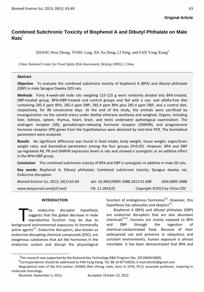
No abnormal changes were observed in behavior, posture or gait. No significant difference was observed in food intake, body weight, tissue weight, organ/brain weight ratio between the control group and different treatment groups (Figure 1, Tables 2, 3).
Figure 1. Body weight of male SD rats at different time points.
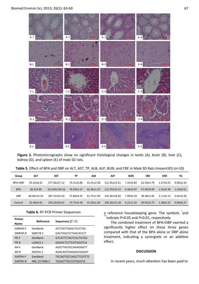
No significant histological changes were detected in heart, liver, kidneys, spleen, thymus, brain, and testis (Figure 2).
Biomedical Parameters
The blood biochemical parameters in Tables 4 and 5 were evaluated with a hematology analyzer (Beckman Coulter Ac. Tdiff2TM, Brea, CA, USA) and a Hitachi 7080 autochemistry analyzer (Hitachi, Tokyo, Japan), respectively. No significant difference in the biomedical parameters was found between the control group and different treatment groups (Tables 4, 5).
Real‐time PCR
GNRHR, PR, and AR genes were studied. The real‐time PCR primer sequences are listed in Table 6. The effect of BPA and DBP on GNRHR, PR and AR gene expression levels are shown in Table 7.
The data in Table 7 are 2‐ΔΔCt (n=9) with GAPDH as
66 Biomed Environ Sci, 2013; 26(1): 63‐69
Table 2. Effect of BPA and DBP on Food Intake of Male SD Rats at Different Time Points (mean±SD) (n=10)
Period BPA+DBP BPA DBP Control
1 week 96.57±11.04 98.31±9.50 97.66±8.77 97.14±10.43
2 week 140.15±7.35 141.24±10.67 138.69±11.39 142.65±10.28
3 week 168.36±11.17 176.38±11.64 171.99±13.27 173.89±13.38
4 week 183.92±14.47 179.36±12.98 178.26±14.94 178.55±12.89
5 week 189.72±13.93 188.83±14.42 192.23±15.88 188.48±9.63
6 week 191.63±11.59 190.56±12.53 193.74±10.56 192.24±10.23
7 week 194.27±14.58 193.83±13.57 194.56±12.37 194.73±11.85
8 week 196.53±15.52 197.43±13.05 198.47±12.67 198.27±11.38
9 week 197.58±12.69 199.78±11.35 198.63±10.37 199.65±10.84
10 week 191.25±13.02 199.54±10.54 199.52±11.26 196.58±11.54
11 week 198.62±10.68 198.78±13.59 199.43±12.26 200.48±11.47
12 week 203.15±11.85 201.67±13.03 200.82±12.64 205.98±12.63
13 week 203.81±13.05 205.84±11.46 206.46±10.41 205.15±14.47
Table 3. Effect of BPA and DBP on Tissue Weight and Organ/Brain Weight Ratio (mean±SE) (n=10)
Tissue BPA+DBP BPA DBP Control
Heart, g 1.65±0.08 1.70±0.07 1.56±0.06 1.69±0.06
Liver, g 12.38±0.57 13.59±0.78 11.85±0.53 11.70±0.41
Kidneys, g 3.12±0.64 3.08±0.62 3.11±0.65 3.07±0.67
Spleen, g 0.75±0.08 0.88±0.06 0.88±0.05 0.80±0.05
Thymus, g 0.43±0.03 0.52±0.07 0.46±0.03 0.52±0.04
Brain, g 2.05±0.06 2.02±0.04 2.03±0.09 2.10±0.04
Testis, g 3.27±0.07 3.31±0.09 3.24±0.08 3.53±0.13
Heart weight/brain, % 0.81±0.05 0.84±0.04 0.78±0.04 0.81±0.02
Liver weight/brain, % 6.08±0.29 6.71±0.35 5.92±0.30 5.59±0.20
Kidneys weight/brain, % 1.51±0.05 1.50±0.09 1.49±0.03 1.46±0.07
Spleen weight/brain, % 0.37±0.04 0.43±0.03 0.44±0.03 0.38±0.02
Thymus weight/brain, % 0.22±0.02 0.26±0.03 0.23±0.01 0.25±0.02
Testis weight/brain, % 1.61±0.06 1.64±0.05 1.63±0.08 1.69±0.06
Heart weight/brain, % 0.81±0.05 0.84±0.04 0.78±0.04 0.81±0.02
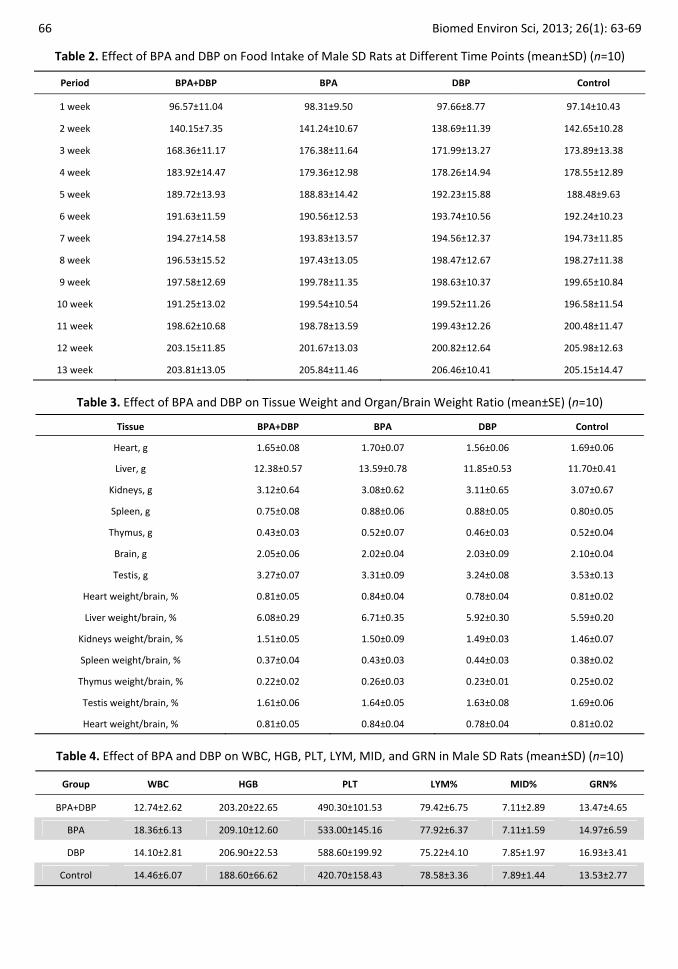
Table 4. Effect of BPA and DBP on WBC, HGB, PLT, LYM, MID, and GRN in Male SD Rats (mean±SD) (n=10)
Group WBC HGB PLT LYM% MID% GRN%
BPA+DBP 12.74±2.62 203.20±22.65 490.30±101.53 79.42±6.75 7.11±2.89 13.47±4.65
BPA 18.36±6.13 209.10±12.60 533.00±145.16 77.92±6.37 7.11±1.59 14.97±6.59
DBP 14.10±2.81 206.90±22.53 588.60±199.92 75.22±4.10 7.85±1.97 16.93±3.41
Control 14.46±6.07 188.60±66.62 420.70±158.43 78.58±3.36 7.89±1.44 13.53±2.77
Biomed Environ Sci, 2013; 26(1): 63-69 67
Figure 2. Photomicrographs show no significant histological changes in testis (A), brain (B), liver (C), kidney (D), and spleen (E) of male SD rats.
Table 5. Effect of BPA and DBP on ALT, AST, TP, ALB, ALP, BUN, and CRE in Male SD Rats (mean±SD) (n=10)
Group ALT AST TP ALB ALP BUN CRE CHO TG
BPA+DBP 35.20±8.42 277.40±37.12 74.31±6.80 41.41±2.58 112.56±23.61 7.45±0.82 62.50±4.79 1.37±0.25 0.90±0.33
BPA 36.3±4.90 312.60±139.16 76.59±3.37 42.46±1.55 112.70±19.14 6.36±0.97 67.60±9.99 1.31±0.30 1.24±0.61
DBP 40.40±10.54 287.10±35.50 73.86±4.91 41.75±1.94 105.40±18.40 7.09±0.50 58.48±3.45 1.11±0.13 0.81±0.26
Control 32.40±9.41 276.10±29.61 73.75±4.44 41.93±1.56 104.20±15.18 6.21±1.32 60.91±6.75 1.38±0.23 0.89±0.27
Table 6. RT-PCR Primer Sequences
Primer
Name Reference Sequences (5'-3')
GNRHR-F ATCTGCTGGACTCCCTAC
GNRHR-R
GenBank:
S68578.1 GACTGGCTCTGACACCCT
PR-F GTCATTCTACTCGCTGTGC
PR-R
GenBank:
L16922.1 GGAACTCCTCGTGGGTCA
AR-F AGGTTACGCCAAAGGGTT
AR-R
GenBank:
J05454.1 AGACAGTGAGGACGGGAT
GAPDH-F TGCAGTGCCAGCCTCGTCTC
GAPDH-R
GenBank:
NM_017008.3 TGAACTTGCCGTGGGTA
a reference housekeeping gene. The symbols *and **indicate P<0.05 and P<0.01, respectively.
The combined treatment of BPA+DBP exerted a significantly higher effect on these three genes compared with that of the BPA alone or DBP alone treatment, indicating a synergistic or an additive effect.
DISCUSSION
In recent years, much attention has been paid to
68 Biomed Environ Sci, 2013; 26(1): 63‐69
Table 7. Effect of BPA and DBP on Expression of GNRHR, PR and AR Genes in male SD Rats (mean±SE) (n=9)
GNRHR AR PR Group
Mean SE Mean SE Mean SE
BPA+DBP 613.91** 1.64 862.21** 1.58 352.09** 1.57
BPA 149.45** 1.75 150.38** 1.8 104.92** 1.76
DBP 10.41* 1.63 6.5* 1.75 6.63* 1.55
Control 1 2.2 1 2.07 1 1.56
whether exposure to environmental BPA affects human development and reproduction. It has been reported that BPA increases the health concern partly because of its effect on the endocrine system and its weak estrogen receptor‐binding capacity[12].
Estrogenic agents have been shown to work synergistically at the concentration below their NOEC[15], thus highlighting the necessity to study their combined subchronic toxicity in addition to the traditional focus on the effect of a single agent. In our study, the combined subchronic toxicity of the most widespread BPA and DBP was identified in rats.
The combined effects of EDC have been evaluated in in vitro and in vivo systems, showing that nonylphenol (NP) and DBP exert an additive effect on Sertoli cell toxicity[16].
It has been reported that EDCs (such as 4‐nonylphenol, 4‐t‐octylphenol, BPA and 2, 4‐dichlorophenol) cause an additive effect on male juvenile goldfish Carassius auratus[17]. A study on genotoxic effects has indicated that exposure to combined ozone, NNK and DBP induces stronger additive or synergistic genotoxic effects in mice (according to different test periods) than exposure to ozone alone[18]. Another study has reported that combined NNK, DBP and ozone treatment exerts an additive genotoxic effect in mice[19].
In contrast, a subchronic toxicity study has demonstrated that combined DBP and BPA treatment exerts antagonistic effects on the reproductive system of male SD rats[20], suggesting that the effects of exposure to mixtures of environmental EDC are unexpected and elusive.
In the present study, no significant difference was found in body weight, tissue weight, organ/brain weight ratio, food intake, and blood biochemical parameters between the control group and different BPA/DBP treatment groups. However, the gene expression levels of AR, GNRHR, and PR were significantly higher in the BPA/DBP treatment groups compared with that of the control group.
Moreover, combined BPA and DBP treatment exerted an additive or a synergistic effect.
EDC can affect the endocrine system by modulating the expression of hormone receptors. For instance, exposure to BPA (600 μg/L for 96 h) can up‐regulate the gene expression of GNRHR in hermaphroditic fish Kryptolebias marmoratus[21].
Monje et al.[22] has reported that BPA disrupts hypothalamic GNRH pre‐mRNA processing in adult rats, which is in agreement with recent reports that showed abnormalities in the estrous cycle when animals were exposed to 1.2 mg/kg BPA via drinking water[23] and 25.0‐62.5 mg/kg of BPA via subcutaneous injection[24].
In conclusion, the gene expression levels of AR, GNRHR and PR are higher in male SD rats after treatment with BPA and DBP (285.4 ppm) compared with that before treatment. BPA and DBP exert an additive or a synergistic effect. Further studies are needed to ascertain the findings in this study. However, the treatment did not significantly change the body weight, tissue weight, organ/brain weight ratio, and biomedical parameters of rats.
REFERENCES
1. Safe S. Bisphenol A and Related Endocrine Disruptors. Toxicological sciences, 2000; 56, 251‐2.
2. Chalupka S & Chalupka AN. The impact of environmental and occupational exposures on reproductive health. Journal of Obstetric, Gynecologic, & Neonatal Nursing, 2010; 39, 84‐102.
3. Ema M & Miyawaki E. Adverse effects on development of the reproductive system in male offspring of rats given monobutyl phthalate, a metabolite of dibutyl phthalate, during late pregnancy. Reproductive Toxicology, 2001; 15, 189‐94.
4. Groff T. Bisphenol A: invisible pollution. Curr Opin Pediatr, 2010; 22, 524‐9.
5. Latini G, Del Vecchio A, Massaro M, et al. In utero exposure to phthalates and fetal development. Curr Med Chem, 2006; 13, 2527‐34.
6. Hauser R & Calafat AM. Phthalates and human health. Occup Environ Med, 2005; 62, 806‐18.
7. Lovekamp‐Swan T & Davis BJ. Mechanisms of phthalate ester toxicity in the female reproductive system. Environ Health
Biomed Environ Sci, 2013; 26(1): 63‐69 69
Perspect, 2003; 111, 139‐45. 8. Lyche JL, Gutleb AC, Bergman Åke, et al. Reproductive and
developmental toxicity of phthalates. Journal of Toxicology and Environmental Health, Part B, 2009; 12, 225‐49.
9. Tan BL, Kassim NM, Mohd MA. Assessment of pubertal development in juvenile male rats after sub‐acute exposure to bisphenol A and nonylphenol. Toxicol Lett, 2003; 143, 261‐70.
10.Duan Z, Zhu L, Zhu L, et al. Individual and joint toxic effects of pentachlorophenol and bisphenol A on the development of zebrafish (Danio rerio) embryo. Ecotox Environ Safe, 2008; 71, 774‐80.
11.Fent K, Escher C, Caminada D. Estrogenic activity of pharmaceuticals and pharmaceutical mixtures in a yeast reporter gene system. Reprod Toxicol, 2006; 22, 175‐85.
12.Kamrin MA. Bisphenol A: A Scientific Evaluation. MedGenMed, 2004; 6(3), 7.
13.FDA Redbook 2000: IV.B.1 General Guidelines for Designing and Conducting Toxicity Studies November 2003.
14.Balboni A, Gallina L, 1 Palladini A, et al. A RealTime PCR Assay for Bat SARS‐Like Coronavirus Detection and Its Application to Italian Greater Horseshoe Bat Faecal Sample Surveys. The Scientific World Journal Vol. 2012, Article ID 989514, 8 pages, doi:10.1100/2012/989514.
15.Silva E, Rajapakse N & Kortenkamp A. Something from nothing‐eight weak estrogenic chemicals combined at concentrations below NOECs produce significant mixture effects. Environ Sci Technol, 2002; 36, 1751‐6.
16.Li DM, Hu Y, Shen XH, et al. Combined effects of two environmental endocrine disruptors nonyl phenol and di‐n‐butyl phthalate on rat Sertoli cells in vitro. Reproductive Toxicology, 2010; 30, 438‐45.
17.Li ZY, Zhang HL, Gibson M, et al. An evaluation of the combined effects of phenolic endocrine disruptors on vitellogenin induction in goldfish Carassius auratus. Ecotoxicology
Published online: 17 May 2012, doi: 10.1007/s10646‐012‐0925‐0.
18.Kim MY, Kim HW, Park JH, et al. Molecular analysis of hprt mutation in B6C3F1 mice exposed to ozone alone and combined treatment of 4‐(N‐methyl‐N‐nitrosamino)‐1‐ (3‐pyridyl)‐1‐butanone and‐or dibutyl phthalate for 32 and 52 weeks. J Vet Sci, 2004; 5(4), 379‐85.
19.Kim MY, Kim YC & Cho MH. Combined treatment with 4‐(N‐methyl‐N‐nitrosamino)‐1‐ (3‐pyridyl)‐1‐butanone and dibutyl phthalate enhances ozone‐induced genotoxicity in B6C3F1 mice. Mutagenesis, 2001; 17, 331‐6.
20.Chen XM, An H, Ao L, et al. The combined toxicity of dibutyl phthalate and benzo(a)pyrene on the reproductive system of male Sprague Dawley rats in vivo. Journal of Hazardous Materials, 2011; 186, 835‐41.
21.Rhee JS, Seo JS, Raisuddin S, et al. Gonadotropin‐releasing hormone receptor (GnRHR) gene expression is differently modulated in gender types of the hermaphroditic fish Kryptolebias marmoratus by endocrine disrupting chemicals. Comparative Biochemistry and Physiology, Part C, 2008; 147, 357‐65.
22.Monje L, Varayoud J, Muńoz‐de‐Toro M, et al. Exposure of neonatal female rats to bisphenol A disrupts hypothalamic LHRH re‐mRNA processing and estrogen receptor alpha expression in nuclei controlling estrous cyclicity. Reproductive Toxicology, 2010; 30, 625‐34.
23.Rubin BS, Murray MK, Damassa DA, et al. Perinatal exposure to low doses of bisphenol A affects body weight, patterns of estrous cyclicity, and plasma LH levels. Environ Health Perspect, 2001; 109, 675‐80.
24.Fernandez M, Bianchi M, Lux‐Lantos V, et al. Neonatal exposure to bisphenol A alters reproductive parameters and gonadotropin releasing hormone signaling in female rats. Environ Health Perspect, 2009; 117, 757‐62.
![ACUTE AND SUBCHRONIC ORAL TOXICITY STUDIES OF ETHYL ... · tempuyung, Apigenin 7-glucoside and Luteolin 7-glucoside, were suggested to have important role for such activity[1]. In](https://cdn.vdocuments.site/doc/165x107/5fc1601bb24ce869895f92cb/acute-and-subchronic-oral-toxicity-studies-of-ethyl-tempuyung-apigenin-7-glucoside.jpg)


