Fixed partial dentures
Color matching in dentistry. Part III. Color control*
Robert C. Sproull, D.D.S. El Paso, Texas
T he scene is a familiar one to the restorative dentist. Meticulous attention to detail has been the name of the game: You have made a careful diagnosis and treatment plan-an anterior tooth in need of a crown, preparation, impression, temporary crown, shade. The patient now sits expectantly in the dental chair waiting to receive the finished restoration. All is fine except for one thing --the color is wrong. The frustrated dentist sorrowfully shakes his head and silently entertains insecure feelings about his color matching ability. But is he really to blame? Why did he miss that shade ?
No single answer is possible. Although color matching techniques have improved over the years, frustration with color has been a problem in the past, is a problem now, and will be a problem in the future. When the problem of replacing a missing tooth was mainly a mechanical exercise in trying to get a substitute-any substitute --to maintain its position in the edentulous arch, an approximation of the correct
color was good enough. Color control of the pebbles, wood, ivory, bone, or ex- tracted teeth used to simulate the missing dentition was not a matter of great con- cern. The alternative to the bad color match -no replacement at all-created a situation in which color matching in dentistry did not command much attention. The patient did not expect, and usually did not receive, an esthetic replacement.
Such is not the situation today as improved materials and techniques and an informed public have all served to generate an increasingly sophisticated demand for esthetics. In placing a crown between natural anterior teeth, the artistic skills required of the dentist are exacting. The artist painting in oils may have free license to make the object of his endeavor any color or form he wishes, but the dentist has little artistic license. In most restorations, accurate color reproduction is required. Yet the artist has received an in-depth education in color, while the dentist has received little instruction in the subject. This state of affairs begins to throw light on how the dentist missed that shade. Clark’s’ statement some forty years ago, “We as dentists
Read before the American Academy of Restorative Dentistry, Chicago, Ill.
*Part I, J. PROSTKET. DENT. 29: 416-424, 1973; Part II, J. PROSTKET. DENT. 29: 556-566, 1973.
146
Volume- 31 Numbrr ?
Color matching in dentistry 147
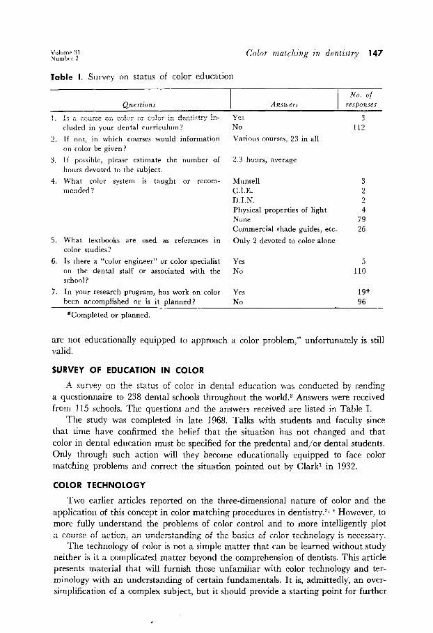
Table I. Survey on status of color education
No. of Questions ATlIlLWTS responses
1.
2.
3.
4.
5.
6.
7.
Is a course on color or color in dentistry in- cluded in your dental curriculum?
If not, in which courses would information on color be given?
If possible, please estimate the number of hours devoted to the subject.
What color system is taught or recom- mended?
What textbooks are used as references in color studies?
Munsell C.I.E. D.I.N. Physical properties of light None Commercial shade guides, etc.
Only 2 devoted to color alone
Is there a “color engineer” or color specialist on the dental staff or associated with the school?
Yes No
5 110
In your research program, has work on color Yes 19” been accomplished or is it planned? No 96
Yes NO Various courses, 23 in all
2.3 hours, average
3 112
3 2 2 4
79 26
*Completed or planned.
are not educationally equipped to approach a color problem,” unfortunately is still valid.
SURVEY OF EDUCATION IN COLOR
A survey on the status of color in dental education was conducted by sending a questionnaire to 238 dental schools throughout the world.2 Answers were received from 115 schools. The questions and the answers received are listed in Table I.
The study was completed in late 1968. Talks with students and faculty since that time have confirmed the belief that the situation has not changed and that color in dental education must be specified for the predental and/or dental students. Only through such action will they become educationally equipped to face color matching problems and correct the situation pointed out by Clark1 in 1932.
COLOR TECHNOLOGY
Two earlier articles reported on the three-dimensional nature of color and the application of this concept in color matching procedures in dentistry.“, * However, to more fully understand the problems of color control and to more intelligently plot a course of action, an understanding of the basics of color technology is necessary.
The technology of color is not a simple matter that can be learned without study neither is it a complicated matter beyond the comprehension of dentists. This article presents material that will furnish those unfamiliar with color technology and ter- minology with an understanding of certain fundamentals. It is, admittedly, an over- simplification of a complex subject, but it should provide a starting point for further
148 spr0d J. Prosthet. Dent.
February, 1974
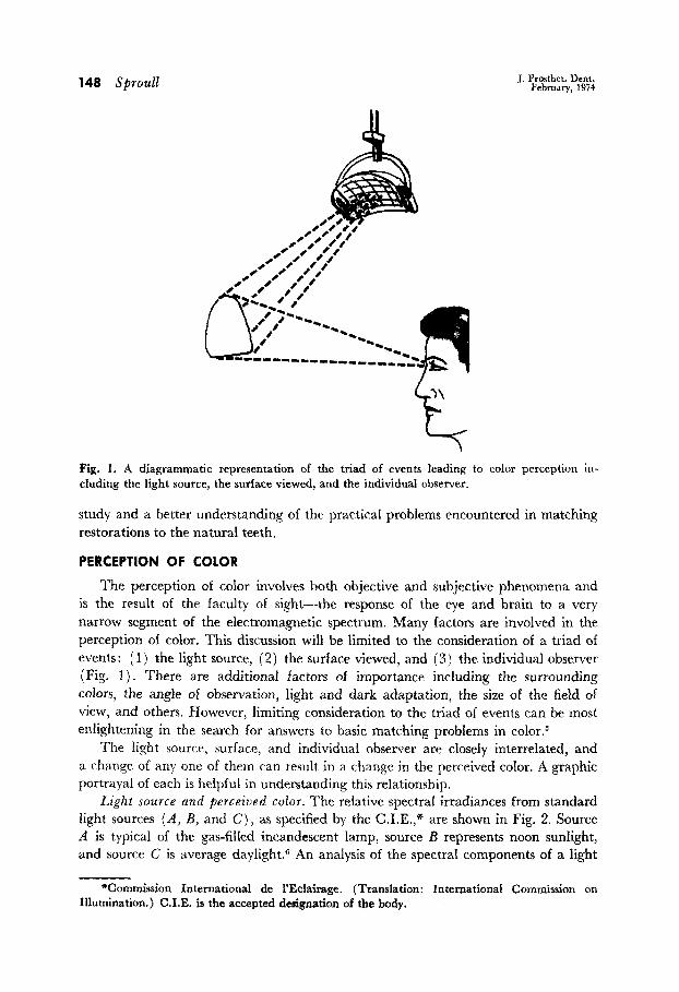
Fig. 1. A diagrammatic representation of the triad of events leading to color perception in- cluding the light source, the surface viewed, and the individual observer.
study and a better understanding of the practical problems encountered in matching restorations to the natural teeth.
PERCEPTION OF COLOR
The perception of color involves both objective and subjective phenomena and is the result of the faculty of sight-the response of the eye and brain to a very narrow segment of the electromagnetic spectrum. Many factors are involved in the perception of color. This discussion will be limited to the consideration of a triad of events: ( 1) the light source, (2) the surface viewed, and (3) the individual observer (Fig. 1). There are additional factors of importance including the surrounding colors, the angle of observation, light and dark adaptation, the size of the field of view, and others. However, limiting consideration to the triad of events can be most enlightening in the search for answers to basic matching problems in color.5
The light source, surface, and individual observer are closely interrelated, and a change of any one of them can result in a change in the perceived color. A graphic portrayal of each is helpful in understanding this relationship.
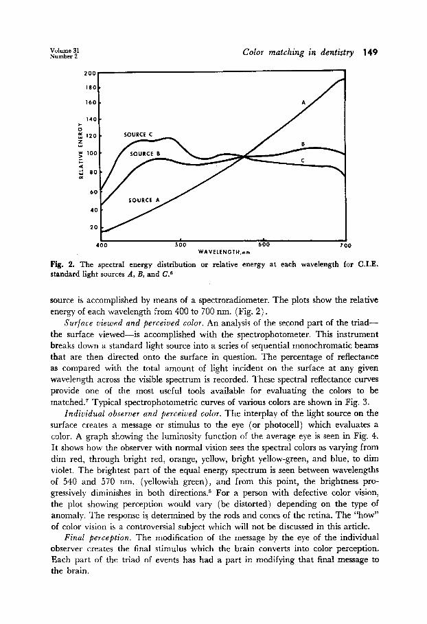
Light source and perceived color. The relative spectral irradiances from standard light sources (A, B, and C), as specified by the C.I.E.,” are shown in Fig. 2. Source A is typical of the gas-filled incandescent lamp, source Z3 represents noon sunIight, and source C is average daylight6 An analysis of the spectral components of a light
*Commission International de I’Eclairage. (Translation: International Commission on Illumination.) C.I.E. is the accepted designation of the body.
1 1 400 500 600 700
WAVELENGTH,nm
Fig. 2. The spectral energy distribution or relative energy at each wavelength for C.I.E. standard light sources A, B, and C.6
source is accomplished by means of a spectroradiometer. The plots show the relative energy of each wavelength from 400 to 700 nm. (Fig. 2).
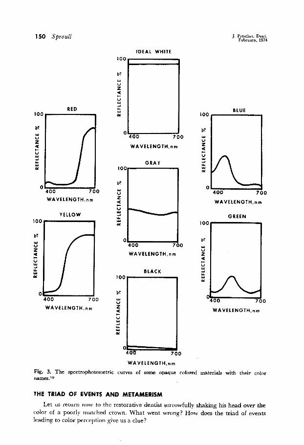
Surface viewed and perceived color. An analysis of the second part of the triad- the surface viewed-is accomplished with the spectrophotometer. This instrument breaks down a standard light source into a series of sequential monochromatic beams that are then directed onto the surface in question. The percentage of reflectance as compared with the total amount of light incident on the surface at any given wavelength across the visible spectrum is recorded. These spectral reflectance curves provide one of the most useful tools available for evaluating the colors to be matched.T Typical spectrophotometric curves of various colors are shown in Fig. 3.
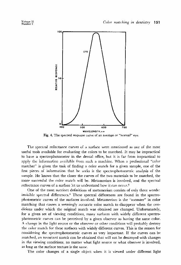
Individual observer and perceived color. The interplay of the light source on the surface creates a message or stimulus to the eye (or photocell) which evaluates a color. A graph showing the Iuminosity function of the average eye is seen in Fig. 4. It shows how the observer with normal vision sees the spectral colors as varying from dim red, through bright red, orange, yellow, bright yellow-green, and blue, to dim violet. The brightest part of the equal energy spectrum is seen between wavelengths of 540 and 570 nm. (yellowish green), and from this point, the brightness pro- gressively diminishes in both directions. * For a person with defective color vision, the plot showing perception would vary (be distorted) depending on the type of anomaly. The response is determined by the rods and cones of the retina. The “how” of color vision is a controversial subject which will not be discussed in this article.
Final perception. The modification of the message by the eye of the individual observer creates the final stimulus which the brain converts into color perception. Each part of the triad of events has had a part in modifying that final message to the brain.
150 Sprou11 J. Prosthet. Dent. February, 1974
RED
0 I( 400 7
WAVELENGTH,nm
YELLOW 100
0 El 400 700
WAVELENGTH,nm
IDEAL WHITE
400 700
WAVELENGTH,nm
GRAY 100
Is
u’
5 t !!i ii oz
0 El 400 700
WAVELENGTH,nm
100
L T- 400 7
a
BLUE
WAVELENGTH.nm
GREEN 100
BLACK
WAVELENGTH,nm
0
0
WAVELENGTH,nm
Fig. 3. The spectrophotometric curves of some opaque colored materials with their color names10
THE TRIAD OF EVENTS AND METAMERlSM
Let us return now to the restorative dentist sorrowfully shaking his head over the color of a poorly matched crown. What went wrong? How does the triad of events leading to color perception give us a clue?
Color matching in dentistry 151
500 600
WAVELENCTH,nm
Fig. 4. The spectral response curve of an average or “normal” eye.
The spectral reflectance curves of a surface were mentioned as one of the most useful tools available for evaluating the colors to be matched. It may be impractical to have a spectrophotometer in the dental office, but it is far from impractical to apply the information available from such a machine. When a professional “color matcher” is given the task of finding a color match for a given sample, one of the first pieces of information that he seeks is the spectrophotometric analysis of the sample. He knows that the closer the curves of the two materials to be matched, the more successful the color match will be. Metamerism is involved, and the spectral reflectance curves of a surface let us understand how it can occur.5
One of the most succinct definitions of metamerism consists of only three words: invisible spectral differences.g These spectral differences are found in the spectro- photometric curves of the surfaces involved. Metamerism is the “monster” in color matching that causes a seemingly accurate color match to disappear when the con- ditions under which the original match was obtained are changed. Unfortunately, for a given set of viewing conditions, many surfaces with widely different spectro- photometric curves can be perceived by a given observer as having the same color. A change in the light source or the observer or other conditions will probably destroy the color match for these surfaces with widely different curves. This is the reason for considering the spectrophotometric curves as very important. If the curves can be matched, an invariant match can be obtained that will not be destroyed with changes in the viewing conditions, no matter what light source or what observer is involved, as long as the surface texture is the same.
The color changes of a single object when it is viewed under different light
152 Sproull J. Prosthet. Dent. February, 1974
80
I.-
I .-
, -
) ‘-
c
1
3-
)- 400 500 600
WAVELENGTH, nm
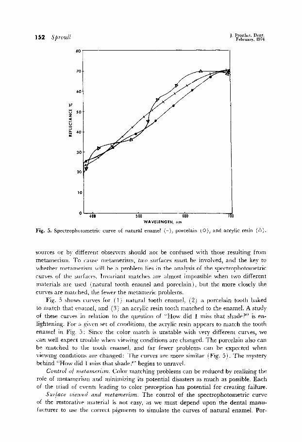
Fig. 5. Spectrophotometric curve of natural enamel (+), porcelain (0), and acrylic resin (A).
sources or by different observers should not be confused with those resulting from metamerism. To cause rnetamerism, two surfaces must be involved, and the key to whether metamerism \\ill be a problem lies in the analysis of the spectrophotometric curves of the surfaces. Invariant matches are almost impossible when two different materials are used (natural tooth enamel and porcelain), but the more closely the curves are matched, the fewer the metameric problems.
Fig. 5 shows curves for (1) natural tooth enamel, (2) a porcelain tooth baked to match that enamel, and (3) an acrylic resin tooth matched to the enamel. A study of these curves in relation to the question of “How did I miss that shade?” is en- lightening. For a given set of conditions, the acrylic resin appears to match the tooth enamel in Fig. 5: Since the color match is unstable with very different curves, we can well expect trouble when viewing conditions are changed. The porcelain also can be matched to the tooth enamel, and far fewer problems can be expected when viewing conditions are changed: The curves are more similar (Fig. 5). The mystery behind “How did I miss that shade?” begins to unravel.
Control of n~tamerism. Color matching problems can be reduced by realizing the role of metamerism and minimizing its potential disasters as much as possible. Each of the triad of events leading to color perception has potential for creating failure.
Surface uieraled and metamerism, The control of the spectrophotometric curve of the restorative material is not easy, as we must depend upon the dental manu- facturer to use the correct pigments to simulate the curves of natural enamel. Por-
Volume 3 1 Numhrr 2
Color matching in dentistry 153
celain appeared to have the advantage over most of the acrylic resins measured, but some resin teeth were excellent. A big advantage of porcelain is in the ease of modi- fying the original color with pigmented stains. Caution must be used, however, in selecting the stains. Grays attained by mixing complimentary colors are “complex” grays with erratic spectrophotometric curves that can introduce metameric prob- lems. Requesting information from the manufacturer concerning the curves of his product to facilitate comparison with those of natural enamel can help in eliminating those products with built-in metameric traps.
Indiuidual observer and metamerism. The role of the individual observer can be
tricky and elusive especially when it is the dentist’s own ocular mechanism that is at fault. Few dentists have been tested for color vision deficiencies. If the dentist’s color vision is defective, reliance may have to be placed on those having more normal vision. A simple device, the Davidson and Hemmendinger Color Rule,* permits a comparison of the color vision of different observers. The color rule consists of two highly metameric painted sliding color scales. By means of a “slide rule” adjustment of the scales, a match point is located. Differences in match points indicate differ- ences in color vision, and these differences can be quite helpful in locating trouble areas. If the color vision of the dentist and ceramist vary widely, differences of opinion on color matches can be expected. Although the Color Rule cannot correct vision deficiencies, its ability to spot them is invaluable.
Light source and metamerism. An adequate light source is essential to competent- ly handle color matching problems, but we must not lose sight of what a light source can and cannot accomplish. An advertisement for a single-color corrected light that “virtually eliminates costly errors and patient dissatisfaction too” ignores the problem of metamerism. The situations in which the spectral reflectance curve of a surface to be matched can be exactly duplicated in another material are rare. The color matcher knows this and, therefore, makes it a rule to check the color match under at least two and sometimes three widely different sources of 1ight.l” A match of colors that looks fairly good under a variety of lights is obviously better than a match that looks great under one source of light but terrible under others. The parallel with the dentist’s problem is apparent. If metamerism is a problem, it does no good for the dentist to use a single-color corrected light source in selecting the shade if the match disappears when the patient looks in the bathroom mirror at home.
The D and H Color Rule can be useful in this portion of our triad also. To check the similarity of one light source with another, simply set the match point under the one source in question and then go to the other. If the match point is no longer valid, the light sources are different.
UNDERSTANDING THE ROLE OF COLOR
Once the dentist possesses the foregoing information, color matching problems do not necessarily disappear. There are other problems. The inadequacy of available tooth shade guides was documented in an earlier article.” The need for a good working knowledge the tridimensional nature of color to survive with present guides and to intelliger plan for the future is another inescapable conclusion
*D and H Color Rule, Munsell Color Company, Inc., Baltimore, Md.
154 sprou11 J. l’rosthet. Dent. February, 1974
reached in previous articles:‘, ’ Color matching can be likened to a gigantic jigsaw
puzzle in which each piece must be properly oriented and positioned to achieve the desired result.
SUMMARY AND CONCLUSIONS
The dentist, as well as the artist, should be educated in color to be successful in working with color. To ensure that the dentists of the future have this background in color: such should be specified for predental and/or dental students. Color per- ception is a complicated process, but an understandin, w of the role of the light source, the surface viewed, and the individual observer gives the dentist a basis for planning a more intelligent and logical approach to color matching.
References
1. Clark, E. B.: 74th Annual Session of the American Dental Association, Buffalo, N. Y., Sept. 15, 1932.
2. Sproull, R. C.: A Survey of Color Education in the Dental Schools of the World, United States Army Research Report, El Paso, Texas, 1968.
3. Sproull, R. C.: Color Matching in Dentistry. Part I. The Three-Dimensional Nature of Color, J. PROSTHET. DENT. 29: 416-424, 1973.
4. Sproull, R. C.: Color Matching in Dentsitry. Part II. Practical Applications of the Or- ganization of Color, J. PROSTHET. DENT. 29: 556-566, 1973.
5. Billmeyer, F. W., Jr., and Saltzman, M.: Principles of Color Technology, New York, 1967, Interscience Publishers, Inc., pp. l-23.
6. Judd, D. B., and Wyszecki, G.: Color in Business, Science and Industry, ed. 2, New York, 1967, John Wiley & Sons, Inc., p. 110.
7. Ibid, pp. 95-106. 8. Ibid, pp. 72-76. 9. Little, A.: Shades of Meaning, Color Engineering 7: 10, 1969.
10. Billmeyer, F. W., Jr., and Saltzman, M.: Principles of Color Technology, New York, 1967, Interscience Publishers, Inc., p. 58.
2405 GAIRLOCH ST. EL PASO, TEXAS 79925


