Nucleic Acids Research, 2016 1doi: 10.1093/nar/gkw751
Co-dependence between trypanosome nuclear laminacomponents in nuclear stability and control of geneexpressionLuke Maishman1, Samson O. Obado2, Sam Alsford3, Jean-Mathieu Bart4, Wei-Ming Chen5,Alexander V. Ratushny5,6, Miguel Navarro4, David Horn1, John D. Aitchison5,6, BrianT. Chait2, Michael P. Rout2 and Mark C. Field1,*
1School of Life Sciences, University of Dundee, Dundee, Scotland, DD1 5EH, UK, 2The Rockefeller University, 1230York Avenue, New York, NY 10065, USA, 3London School of Hygiene and Tropical Medicine, Keppel Street, London,WC1E 7HT, UK, 4Instituto de Parasitologıa y Biomedicina Lopez-Neyra, Consejo Superior de InvestigacionesCientificas, 18100 Grenada, Spain, 5Center for Infectious Disease Research (formerly Seattle Biomedical ResearchInstitute), Seattle, WA 98109, USA and 6Institute for Systems Biology, Seattle, WA 98109, USA
Received April 26, 2016; Revised August 02, 2016; Accepted August 20, 2016
ABSTRACT
The nuclear lamina is a filamentous structure sub-tending the nuclear envelope and required for chro-matin organization, transcriptional regulation andmaintaining nuclear structure. The trypanosomatidcoiled-coil NUP-1 protein is a lamina componentfunctionally analogous to lamins, the major lam-ina proteins of metazoa. There is little evidence forshared ancestry, suggesting the presence of a dis-tinct lamina system in trypanosomes. To find addi-tional trypanosomatid lamina components we iden-tified NUP-1 interacting proteins by affinity captureand mass-spectrometry. Multiple components of thenuclear pore complex (NPC) and a second coiled-coilprotein, which we termed NUP-2, were found. NUP-2 has a punctate distribution at the nuclear periph-ery throughout the cell cycle and is in close proxim-ity to NUP-1, the NPCs and telomeric chromosomalregions. RNAi-mediated silencing of NUP-2 leads tosevere proliferation defects, gross alterations to nu-clear structure, chromosomal organization and nu-clear envelope architecture. Further, transcription isaltered at telomere-proximal variant surface glyco-protein (VSG) expression sites (ESs), suggesting arole in controlling ES expression, although NUP-2 si-lencing does not increase VSG switching. Transcrip-tome analysis suggests specific alterations to PolI-dependent transcription. NUP-1 is mislocalized inNUP-2 knockdown cells and vice versa, implying thatNUP-1 and NUP-2 form a co-dependent network and
identifying NUP-2 as a second trypanosomatid nu-clear lamina component.
INTRODUCTION
In metazoan cells the structural organization of the nucleusis maintained, at least in part, by the nuclear lamina, a stableprotein meshwork at the inner face of the nuclear envelope(NE), comprised of a small family of coiled-coil interme-diate filament lamins (1). Mutation or alterations in laminexpression causes abnormalities to nuclear architecture, in-cluding irregular protrusions into the cytoplasm, termednuclear blebs (1,2). Lamins are required for nuclear porecomplex (NPC) positioning, with defects leading to NPCclustering (3,4) and/or the absence of NPCs from nuclearblebs (2,5–6). The lamina also interacts with the cytoskele-ton through the linker of nucleoskeleton and cytoskeletoncomplex (LINC) (7,8) and is essential for mechano-signaltransduction (9). Lamins are also required for organizationof the genome into transcriptionally active euchromatin andrepressed heterochromatin (10), and influence transcrip-tional activity (1,2), interacting with a huge range of tran-scription factors (11). DNA replication is abolished at theinitiation and elongation phases when the lamin network isdisrupted (12,13), while lamin mutations lead to increasedDNA damage (14). For these and other reasons, hereditarymutations in lamin genes that cause laminopathies (1,5) areof significant clinical interest.
Lamin orthologs are widely distributed across Metazoaand social amoeba, and recently have been described ashaving broad presence, as well as, being absent from sev-eral major lineages (1,15–16). Yeast, which are evolution-arily closely related to animals, lack lamins and no laminastructure has been observed by electron microscopy (EM)
*To whom correspondence should be addressed. Tel: +44 0 751 550 7880; Email: [email protected]
C⃝ The Author(s) 2016. Published by Oxford University Press on behalf of Nucleic Acids Research.This is an Open Access article distributed under the terms of the Creative Commons Attribution License (http://creativecommons.org/licenses/by/4.0/), whichpermits unrestricted reuse, distribution, and reproduction in any medium, provided the original work is properly cited.
Nucleic Acids Research Advance Access published September 12, 2016 at R
ockefeller University on Septem
ber 23, 2016http://nar.oxfordjournals.org/
Dow
nloaded from
2 Nucleic Acids Research, 2016
(16,17). Instead, several proteins appear to have assumednucleoskeletal functions, e.g. Mlp 1 and 2, large (∼200 kDa)coiled-coil nuclear basket proteins orthologous to the mam-malian nuclear basket protein Tpr. Mlp1 and 2 maintain nu-clear architecture and NPC organization and interact withEsc1 (18), which itself has roles in telomeric silencing (19),chromatin tethering (20) and organizing the NPC basket(21). For example, over-expressing Esc1 in Saccharomycescerevisiae leads to nuclear blebbing, suggesting a structuralsystem is present in yeasts (22).
In plants a nucleoskeletal structure is also present, but themolecular identity is incompletely defined (23). Nuclear in-termediate filament proteins are immunologically identifiedcandidates that form 6–12 nm, lamin-like filaments in vitro(24). Another group of candidates are the nuclear matrixconstituent proteins at the nuclear periphery. These disas-semble and reassemble during mitosis similarly to lamins,affect nuclear size and shape and play a role in heterochro-matin organization (23). These examples from yeast andplants suggest that alternative, non-lamin, molecular sys-tems can construct a nuclear lamina.
A functional lamin analog, NUP-1, has been identified inthe highly divergent trypanosomatids, which reside withinthe Excavata supergroup. NUP-1 is a large coiled-coil pro-tein that forms a stable, fenestrated lattice at the edge of thenucleoplasm and expression of NUP-1 is essential for cor-rect nuclear architecture, NPC arrangement, heterochro-matin organization and the epigenetic regulation of geneexpression (25). A high molecular weight and extendedconformation within a relatively small nucleus means thatNUP-1 may have roles entirely distinct from lamins, in-cluding chromosomal segregation (26). As trypanosomesbranched early during eukaryotic evolution (27,28), theyare especially valuable for comparative studies.
Many features are conserved between metazoan and try-panosome nuclei, including the NPC transport system (29–33) and peripheral heterochromatin as a transcriptionallyrepressed portion of the genome (34). The trypanosomenuclear genome is physically segregated into eleven pairsof conventional megabase chromosomes (MBCs) that har-bor the majority of protein coding genes, up to five inter-mediate sized chromosomes (ICs) plus about 100 repetitivelower molecular weight minichromosomes (MCs). MBCsand MCs segregate during mitosis with differential kinet-ics, locations and possibly mechanisms (35). Transcrip-tion of housekeeping genes is polycistronic, with direc-tional gene clusters consisting of functionally unrelatedgenes (36), while mRNA levels are chiefly regulated post-transcriptionally.
A sophisticated mechanism for immune evasion operatesin mammalian infective trypanosomes, involving expressionof the variant surface glycoprotein (VSG). VSG expressionis monoallelic and exclusively via RNA Pol I transcriptionfrom telomere-proximal expression sites (ESs), present atboth MBC and IC telomeric regions (34). The surface coatis also developmentally regulated and, in early insect stagesVSG is replaced by procyclin, another superabundant sur-face protein. Several proteins mediate repression of inactiveVSG genes, including RAP1 (36), DAC3 (37) and NUP-1(25), while the single active VSG gene is transcribed exclu-
sively at the expression site body, an RNA polymerase I-richnuclear subdomain distinct from the nucleolus (38).
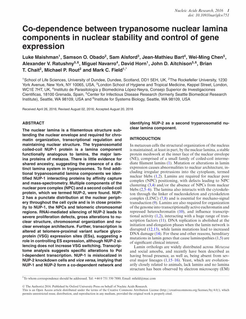
To further characterize the trypanosome lamina, we in-vestigated the NUP-1 interactome. Amongst many pu-tative interactions, we identified the protein product ofTb927.9.6460, a large, substantially coiled-coil protein es-sential in the bloodstream form (BSF) (39). We designatedTb927.9.6460 as NUP-2 for nuclear peripheral protein-2and found that it is widely distributed amongst kinetoplas-tids but restricted to this lineage. NUP-2 has functions anal-ogous to metazoan lamins, including maintenance of nu-clear architecture, chromatin organization and regulationof gene expression and exhibits functional interactions withNUP-1. We therefore define NUP-2 as a second componentof the trypanosomatid lamina.
MATERIALS AND METHODS
Bioinformatics
BLAST searches were used to identify candidate orthologsin the following predicted proteomes: T. congolense, T. vi-vax, T. brucei gambiense, T.grayii, T. cruzi, L. major, L.braziliensis, L. mexicana, L. infantum and Bodo saltanswere from geneDB (www.genedb.org/Homepage) and TriT-rypDB (http://tritrypdb.org/tritrypdb/) while Phytomonasserpens data were from (40). Access to pre-publication pre-dicted proteome data for T. borreli, T. carassii, T. theileri andEuglena gracilis was kindly provided by Steve Kelly (Ox-ford) or was from in house sequence data. The NCBI non-redundant protein database was used for searches in othereukaryotes.
JPred (http://www.compbio.dundee.ac.uk/jpred/index.html), COILS (41), InterProScan (https://www.ebi.ac.uk/interpro/search/sequence-search), Motif-Scan (http://myhits.isb-sib.ch/cgi-bin/motif scan), Phyre2 (42), cNLS Mapper (43) were used to predict domainsand secondary structure and phosphorylation sites werepredicted using Predict Protein (44). Peptide sequenceswere analyzed for identity and similarity using SIAS(http://imed.med.ucm.es/Tools/sias.html). Phylogenetictrees were created using both PhyML (45) and MrBayesv3.2.1 (46) using multiple sequence alignments generatedwith Merge Align (47). ProtTest version 2.4 (48) wasused to determine the lset rates parameters for MrBayesanalyses and the substitution model and gamma parameterfor PhyML analyses.
Trypanosome cell culture
BSF stage and procyclic culture form (PCF) Trypanosomabrucei brucei Lister 427 strain were cultured as previ-ously described (49). Single-marker BSF (SMB) and 2T1BSF cells were used for expression of tetracycline-inducibleRNA interference (RNAi) constructs as described (50,51).Reporter 2T1 cell lines (37) were used to investigate telom-eric transcriptional regulation.
In situ tagging
The pMOTag vector system (52) was used to intro-duce C-terminal in situ epitope tags, while the pN-PTP derivative vectors (53) were used to introduce
at Rockefeller U
niversity on September 23, 2016
http://nar.oxfordjournals.org/D
ownloaded from
Nucleic Acids Research, 2016 3
N-terminal tags. The primer sequences used were, forC-terminal tagging: NUP-2F CGAAGAGGTCGCACTTCCGGTGGGGCAGGTG GTCCCACTCCCGTTTCCATTACTGGCTCGCTTGGATTGAAGCCATCGGGT ACCGGGCCCCCCCTCGAG, NUP-2RAACTATTCAGTAACGCTTCCATATAATA GATAATATATATATATATATGTTTGGGTGTGTGCTCGTCGTCACGATGGCGGCCGCTCTAGAACTAGTGGAT,TbNup98F TGGGAATGCTTCAGCAAGTGGT GAAAAGAACAATGCTCCACGGAATCCCTTCTCATTTGGTGCCTCTTCTGGGAATGCTGGTACCGGGCCCCCCCTCGAG and TbNup98R ACTAAAGAAGGGTAGAAAACAAAGAAAACACCAAATAAGGTACCTGACGCAGC GGCAACACCACGTCGACTTGCTGGCGGCCGCTCTAGAACTAGTGGAT,and for N-terminal tagging; NUP-2F CTTAAGCTTCTATGATCGCTGCGGGCAATGAAAGC and NUP-2RCAGTAAGAATTCC AGCGGCTGAGAGCTGAGAA.All sequences are given in the 5′–3′ orientation. Linearpolymerase chain reaction (PCR) products were purifiedand sterilized by ethanol precipitation. Electroporationwas performed with 10–25 !g of DNA using an AmaxaNucleofector II for BSF trypanosomes and a Bio-RadGene Pulser II (1.5 kV and 25 !F) for PCF trypanosomes.Positive clones were assayed for correct insertion andexpression by Western blot.
Western blotting
Normally 1 × 10⁷ cells per lane were resolved on a 4–12% SDS–PAGE gel (Invitrogen) or a home-made 8% SDS-PAGE gel. Proteins were transferred to a nitrocellulosemembrane (Whatman). The following primary antibod-ies were used: HA mouse monoclonal (F7), at 1:10 000(from Santa Cruz, sc-7392), "-Tubulin mouse monoclonal(KMX-1) at 1:5000 (from Millipore, MAB3408), Detectionwas by chemiluminescence with luminol using rabbit anti-mouse peroxidase conjugate at 1:10 000 (Sigma, A9044).Images of developed films were analyzed using ImageJ (Na-tional Institutes of Health).
Isolation of protein complexes
Protein–protein interactions were analyzed by cryomillingof cell pellets and then immunoprecipitation, approximatelyas outlined in (54). In brief, ∼1010 PCF trypanosomes har-boring an endogenous GPF tag on the affinity handle pro-tein were harvested, frozen and then lysed by mechani-cal milling in a Retsch Planetary Ball Mill PM100 usingliquid nitrogen cooling (Retsch, UK). Aliquots of frozenpowder were thawed in buffer (see figure legends) con-taining protease inhibitors (Roche) and clarified by cen-trifugation. Tagged proteins were affinity isolated usingpolyclonal llama anti-GFP antibodies coupled to mag-netic beads (Dynabeads R⃝). Protein complexes were thenfractionated using 1D SDS–PAGE and gel bands excised,trypsin digested and identified by matrix-assisted laserdesorption/ionization – time of flight (MALDI-TOF) massspectrometry (29,55).
RNA interference
The online RNAit tool (56) was used to design primersfor NUP-2 RNAi, to avoid potential off-target effects. Thesequences used were, 5′ to 3′; NUP-2F TGAACAGCAAGGGCTCTTTT and NUP-2R GCCTCATGGCTTCTTAGCAC. For some experiments PCR products were clonedinto the p2T7TABlue plasmid (50) and transfected into sin-gle marker T7 RNAP/TetR BSF (SMB) cells (57). Alterna-tively, the pRPaiSL plasmid was used to generate targetedstem-loop constructs that were transfected into 2T1 BSFcells (51,58).
Immunofluorescence microscopy
Samples were prepared for microscopy as described in (59).The following primary antibodies were used: HA mousemonoclonal (F7) at 1:500 – 1:1000, (from Santa Cruz, sc-7392), "-Tubulin mouse monoclonal (KMX-1) at 1:1000(from Millipore, MAB3408), GFP lapine polyclonal at1:3000 (in house) and NUP-1 lapine polyclonal at 1:750–1:1500 (in house). Secondary antibodies for IFA were usedat 1:1000, and were goat anti-rabbit Oregon green (In-vitrogen, O-6381), goat anti-rabbit A568 (Invitrogen, A-11011), goat anti-mouse A568 (Invitrogen, A-11001 andA-11004). Wide-field microscopy was carried out using aNikon Eclipse E600 epifluorescence microscope with a cam-era and images captured using Metamorph software (Uni-versal Imaging Corporation). Confocal microscopy wascarried out on an SP2-visible inverted confocal microscope(Leica Microsystems) or an LSM 700 confocal microscope(Zeiss) and images deconvolved using Huygens Professionalprogram (Scientific Volume Imaging). Image quantificationwas carried out on raw images (wide-field microscopy) orraw deconvolved images (confocal microscopy) using Im-ageJ. Image processing for presentation was carried out us-ing Adobe Photoshop 7.0 (Adobe Systems). When measur-ing fluorescence intensity for NUP-2 or NUP-1, for eachcell quantified a separately focused image set was captured.The DAPI signal was used as a guide for the nucleus regionof interest, from which the target mean fluorescence inten-sity (MFI) was quantified using ImageJ. For each nucleus,local background MFI (outside the cell) was deducted fromthe nuclear measurement.
Fluorescence in situ hybridization
Telomere Fluorescence in situ hybridization (FISH) wasperformed as described (25) except that for co-stain withthe 177 base pair fragment that binds to mini-chromosomes,adjusted BMEB (20% v/v FCS in 100 mM maleic acid, 150mM NaCl, pH 7.5) was used in place of BMEB.
Cell-sorting
Mid-log phase cells were harvested by centrifugation andwashed extremely gently in Voorheis’s-modified phosphate-buffered saline (vPBS) (59,60). The cells were fixed in 70%methanol in phosphate buffered saline (PBS) at 4◦C for 1 h,washed in vPBS and stained in 30 !g/ml propridium iodide(PI) with 10 !g/ml RNAse A in PBS for 45 min at 37◦C.The samples were analyzed using a CyAn ADP MLE flowcytometer (Beckman Coulter).
at Rockefeller U
niversity on September 23, 2016
http://nar.oxfordjournals.org/D
ownloaded from
4 Nucleic Acids Research, 2016
Figu
re1.
The
tryp
anos
ome
lam
inan
alog
NU
P-1
inte
ract
swith
the
NPC
and
ano
velh
igh
mol
ecul
arw
eigh
tpro
tein
.(A
)NU
P-1
was
co-im
mun
opre
cipi
tate
dvi
aa
C-t
erm
inal
insi
tuG
FP
tag
in20
mM
HE
PES,
pH7.
4,25
0m
MN
aCl,
0.5%
Trito
n,0.
5%de
oxy-
Big
CH
AP
usin
gcr
yom
illin
gan
daf
finity
isol
atio
nw
ithan
ti-G
FP
antib
ody.
The
resu
lting
com
plex
esw
ere
frac
tiona
ted
by1D
SDS-
PAG
Ean
dvi
sual
ized
byst
aini
ngw
ithC
oom
assi
ebl
ue.T
heid
entit
yof
prot
ein
band
sw
asde
term
ined
bym
ass
spec
trom
etry
.The
mar
ker
posi
tions
ofa
co-e
lect
roph
ores
edpr
otei
nla
dder
are
indi
cate
dto
the
left
,val
ues
are
inki
loda
ltons
.Gre
yac
cess
ion
num
bers
indi
cate
prot
eins
with
nopu
blis
hed
orpr
edic
ted
func
tion.
(B)
Pred
icte
dar
chite
ctur
eof
NU
P-2
orth
olog
san
dth
em
ajor
pred
icte
dco
iled-
coil
dom
ains
and
nucl
ear
loca
lizat
ion
sign
al(N
LS)
mot
ifs.S
eque
nce
ribb
ons
are
alig
ned
toth
em
ajor
C-t
erm
inal
coile
d-co
ildo
mai
n,w
hich
isth
em
ostc
onse
rved
regi
on.T
heco
iled-
coil
dom
ains
and
tota
lle
ngth
ofea
chse
quen
cear
eap
prox
imat
ely
tosc
ale
onth
eho
rizo
ntal
axis.
Spec
ies
nam
esan
dto
tala
min
oac
idre
sidu
esar
ein
dica
ted
atle
ftan
dri
ght
resp
ectiv
ely.
(C)
Aps
eudo
-roo
ted
phyl
ogen
etic
tree
forN
UP-
2or
thol
ogsi
dent
ified
bypr
otei
nB
LA
STse
arch
es,T
.bor
relli
was
trea
ted
asan
out-
grou
p.B
oth
MrB
ayes
and
PhyM
Lal
gori
thm
swer
eus
edto
mod
elth
ere
latio
nshi
psbe
twee
nse
quen
ces,
and
the
PhyM
Lto
polo
gyis
show
n.M
rBay
esan
dPh
yML
post
erio
rpr
obab
ility
and
boot
stra
pva
lues
resp
ectiv
ely
are
show
nat
each
node
.The
key
indi
cate
sa
colo
rco
defo
rsp
ecie
sgr
oups
.
at Rockefeller U
niversity on September 23, 2016
http://nar.oxfordjournals.org/D
ownloaded from
Nucleic Acids Research, 2016 5
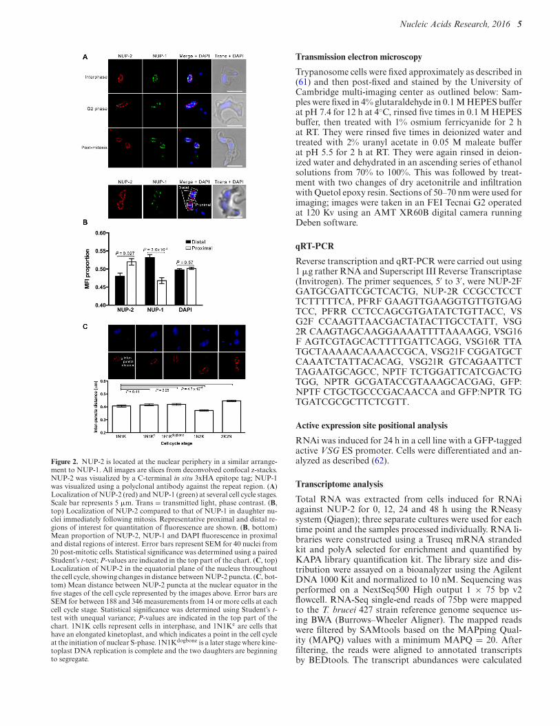
Figure 2. NUP-2 is located at the nuclear periphery in a similar arrange-ment to NUP-1. All images are slices from deconvolved confocal z-stacks.NUP-2 was visualized by a C-terminal in situ 3xHA epitope tag; NUP-1was visualized using a polyclonal antibody against the repeat region. (A)Localization of NUP-2 (red) and NUP-1 (green) at several cell cycle stages.Scale bar represents 5 !m. Trans = transmitted light, phase contrast. (B,top) Localization of NUP-2 compared to that of NUP-1 in daughter nu-clei immediately following mitosis. Representative proximal and distal re-gions of interest for quantitation of fluorescence are shown. (B, bottom)Mean proportion of NUP-2, NUP-1 and DAPI fluorescence in proximaland distal regions of interest. Error bars represent SEM for 40 nuclei from20 post-mitotic cells. Statistical significance was determined using a pairedStudent’s t-test; P-values are indicated in the top part of the chart. (C, top)Localization of NUP-2 in the equatorial plane of the nucleus throughoutthe cell cycle, showing changes in distance between NUP-2 puncta. (C, bot-tom) Mean distance between NUP-2 puncta at the nuclear equator in thefive stages of the cell cycle represented by the images above. Error bars areSEM for between 188 and 346 measurements from 14 or more cells at eachcell cycle stage. Statistical significance was determined using Student’s t-test with unequal variance; P-values are indicated in the top part of thechart. 1N1K cells represent cells in interphase, and 1N1Ke are cells thathave an elongated kinetoplast, and which indicates a point in the cell cycleat the initiation of nuclear S-phase. 1N1Kdogbone is a later stage where kine-toplast DNA replication is complete and the two daughters are beginningto segregate.
Transmission electron microscopy
Trypanosome cells were fixed approximately as described in(61) and then post-fixed and stained by the University ofCambridge multi-imaging center as outlined below: Sam-ples were fixed in 4% glutaraldehyde in 0.1 M HEPES bufferat pH 7.4 for 12 h at 4◦C, rinsed five times in 0.1 M HEPESbuffer, then treated with 1% osmium ferricyanide for 2 hat RT. They were rinsed five times in deionized water andtreated with 2% uranyl acetate in 0.05 M maleate bufferat pH 5.5 for 2 h at RT. They were again rinsed in deion-ized water and dehydrated in an ascending series of ethanolsolutions from 70% to 100%. This was followed by treat-ment with two changes of dry acetonitrile and infiltrationwith Quetol epoxy resin. Sections of 50–70 nm were used forimaging; images were taken in an FEI Tecnai G2 operatedat 120 Kv using an AMT XR60B digital camera runningDeben software.
qRT-PCR
Reverse transcription and qRT-PCR were carried out using1 !g rather RNA and Superscript III Reverse Transcriptase(Invitrogen). The primer sequences, 5′ to 3′, were NUP-2FGATGCGATTCGCTCACTG, NUP-2R CCGCCTCCTTCTTTTTCA, PFRF GAAGTTGAAGGTGTTGTGAGTCC, PFRR CCTCCAGCGTGATATCTGTTACC, VSG2F CCAAGTTAACGACTATACTTGCCTATT, VSG2R CAAGTAGCAAGGAAAATTTTAAAAGG, VSG16F AGTCGTAGCACTTTTGATTCAGG, VSG16R TTATGCTAAAAACAAAACCGCA, VSG21F CGGATGCTCAAATCTATTACACAG, VSG21R GTCAGAATTCTTAGAATGCAGCC, NPTF TCTGGATTCATCGACTGTGG, NPTR GCGATACCGTAAAGCACGAG, GFP:NPTF CTGCTGCCCGACAACCA and GFP:NPTR TGTGATCGCGCTTCTCGTT.
Active expression site positional analysis
RNAi was induced for 24 h in a cell line with a GFP-taggedactive VSG ES promoter. Cells were differentiated and an-alyzed as described (62).
Transcriptome analysis
Total RNA was extracted from cells induced for RNAiagainst NUP-2 for 0, 12, 24 and 48 h using the RNeasysystem (Qiagen); three separate cultures were used for eachtime point and the samples processed individually. RNA li-braries were constructed using a Truseq mRNA strandedkit and polyA selected for enrichment and quantified byKAPA library quantification kit. The library size and dis-tribution were assayed on a bioanalyzer using the AgilentDNA 1000 Kit and normalized to 10 nM. Sequencing wasperformed on a NextSeq500 High output 1 × 75 bp v2flowcell. RNA-Seq single-end reads of 75bp were mappedto the T. brucei 427 strain reference genome sequence us-ing BWA (Burrows–Wheeler Aligner). The mapped readswere filtered by SAMtools based on the MAPping Qual-ity (MAPQ) values with a minimum MAPQ = 20. Afterfiltering, the reads were aligned to annotated transcriptsby BEDtools. The transcript abundances were calculated
at Rockefeller U
niversity on September 23, 2016
http://nar.oxfordjournals.org/D
ownloaded from
6 Nucleic Acids Research, 2016
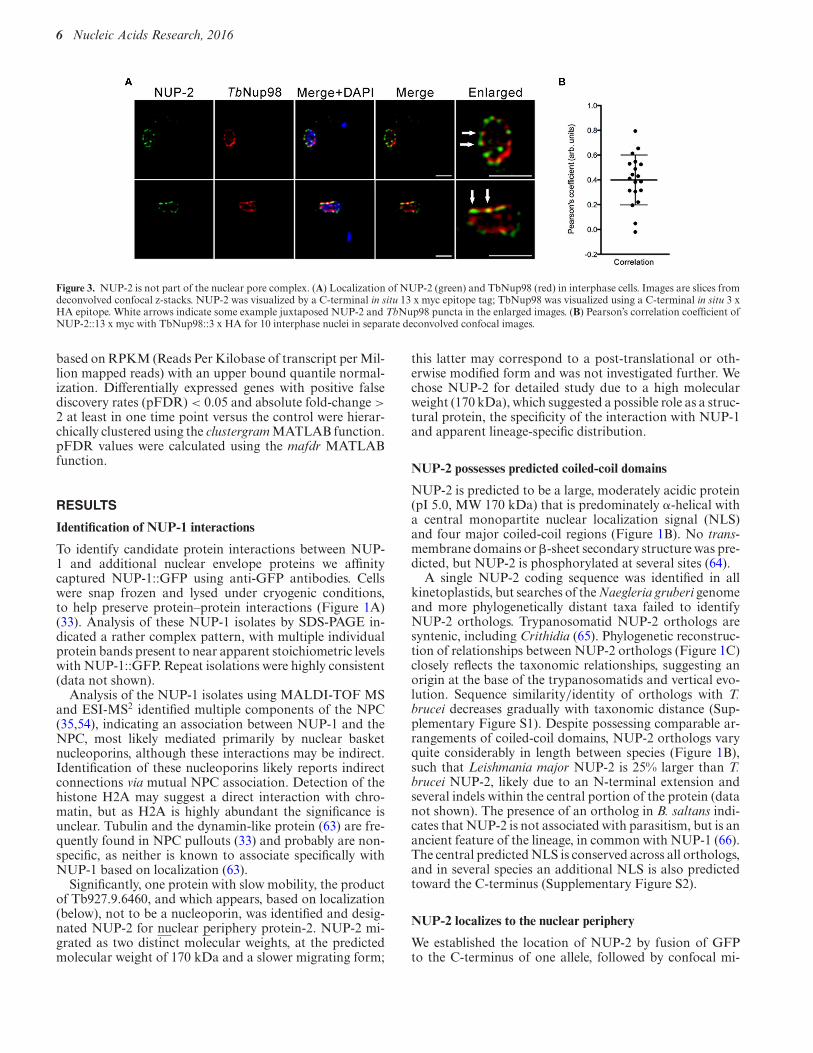
Figure 3. NUP-2 is not part of the nuclear pore complex. (A) Localization of NUP-2 (green) and TbNup98 (red) in interphase cells. Images are slices fromdeconvolved confocal z-stacks. NUP-2 was visualized by a C-terminal in situ 13 x myc epitope tag; TbNup98 was visualized using a C-terminal in situ 3 xHA epitope. White arrows indicate some example juxtaposed NUP-2 and TbNup98 puncta in the enlarged images. (B) Pearson’s correlation coefficient ofNUP-2::13 x myc with TbNup98::3 x HA for 10 interphase nuclei in separate deconvolved confocal images.
based on RPKM (Reads Per Kilobase of transcript per Mil-lion mapped reads) with an upper bound quantile normal-ization. Differentially expressed genes with positive falsediscovery rates (pFDR) < 0.05 and absolute fold-change >2 at least in one time point versus the control were hierar-chically clustered using the clustergram MATLAB function.pFDR values were calculated using the mafdr MATLABfunction.
RESULTS
Identification of NUP-1 interactions
To identify candidate protein interactions between NUP-1 and additional nuclear envelope proteins we affinitycaptured NUP-1::GFP using anti-GFP antibodies. Cellswere snap frozen and lysed under cryogenic conditions,to help preserve protein–protein interactions (Figure 1A)(33). Analysis of these NUP-1 isolates by SDS-PAGE in-dicated a rather complex pattern, with multiple individualprotein bands present to near apparent stoichiometric levelswith NUP-1::GFP. Repeat isolations were highly consistent(data not shown).
Analysis of the NUP-1 isolates using MALDI-TOF MSand ESI-MS2 identified multiple components of the NPC(35,54), indicating an association between NUP-1 and theNPC, most likely mediated primarily by nuclear basketnucleoporins, although these interactions may be indirect.Identification of these nucleoporins likely reports indirectconnections via mutual NPC association. Detection of thehistone H2A may suggest a direct interaction with chro-matin, but as H2A is highly abundant the significance isunclear. Tubulin and the dynamin-like protein (63) are fre-quently found in NPC pullouts (33) and probably are non-specific, as neither is known to associate specifically withNUP-1 based on localization (63).
Significantly, one protein with slow mobility, the productof Tb927.9.6460, and which appears, based on localization(below), not to be a nucleoporin, was identified and desig-nated NUP-2 for nuclear periphery protein-2. NUP-2 mi-grated as two distinct molecular weights, at the predictedmolecular weight of 170 kDa and a slower migrating form;
this latter may correspond to a post-translational or oth-erwise modified form and was not investigated further. Wechose NUP-2 for detailed study due to a high molecularweight (170 kDa), which suggested a possible role as a struc-tural protein, the specificity of the interaction with NUP-1and apparent lineage-specific distribution.
NUP-2 possesses predicted coiled-coil domains
NUP-2 is predicted to be a large, moderately acidic protein(pI 5.0, MW 170 kDa) that is predominately #-helical witha central monopartite nuclear localization signal (NLS)and four major coiled-coil regions (Figure 1B). No trans-membrane domains or "-sheet secondary structure was pre-dicted, but NUP-2 is phosphorylated at several sites (64).
A single NUP-2 coding sequence was identified in allkinetoplastids, but searches of the Naegleria gruberi genomeand more phylogenetically distant taxa failed to identifyNUP-2 orthologs. Trypanosomatid NUP-2 orthologs aresyntenic, including Crithidia (65). Phylogenetic reconstruc-tion of relationships between NUP-2 orthologs (Figure 1C)closely reflects the taxonomic relationships, suggesting anorigin at the base of the trypanosomatids and vertical evo-lution. Sequence similarity/identity of orthologs with T.brucei decreases gradually with taxonomic distance (Sup-plementary Figure S1). Despite possessing comparable ar-rangements of coiled-coil domains, NUP-2 orthologs varyquite considerably in length between species (Figure 1B),such that Leishmania major NUP-2 is 25% larger than T.brucei NUP-2, likely due to an N-terminal extension andseveral indels within the central portion of the protein (datanot shown). The presence of an ortholog in B. saltans indi-cates that NUP-2 is not associated with parasitism, but is anancient feature of the lineage, in common with NUP-1 (66).The central predicted NLS is conserved across all orthologs,and in several species an additional NLS is also predictedtoward the C-terminus (Supplementary Figure S2).
NUP-2 localizes to the nuclear periphery
We established the location of NUP-2 by fusion of GFPto the C-terminus of one allele, followed by confocal mi-
at Rockefeller U
niversity on September 23, 2016
http://nar.oxfordjournals.org/D
ownloaded from
Nucleic Acids Research, 2016 7
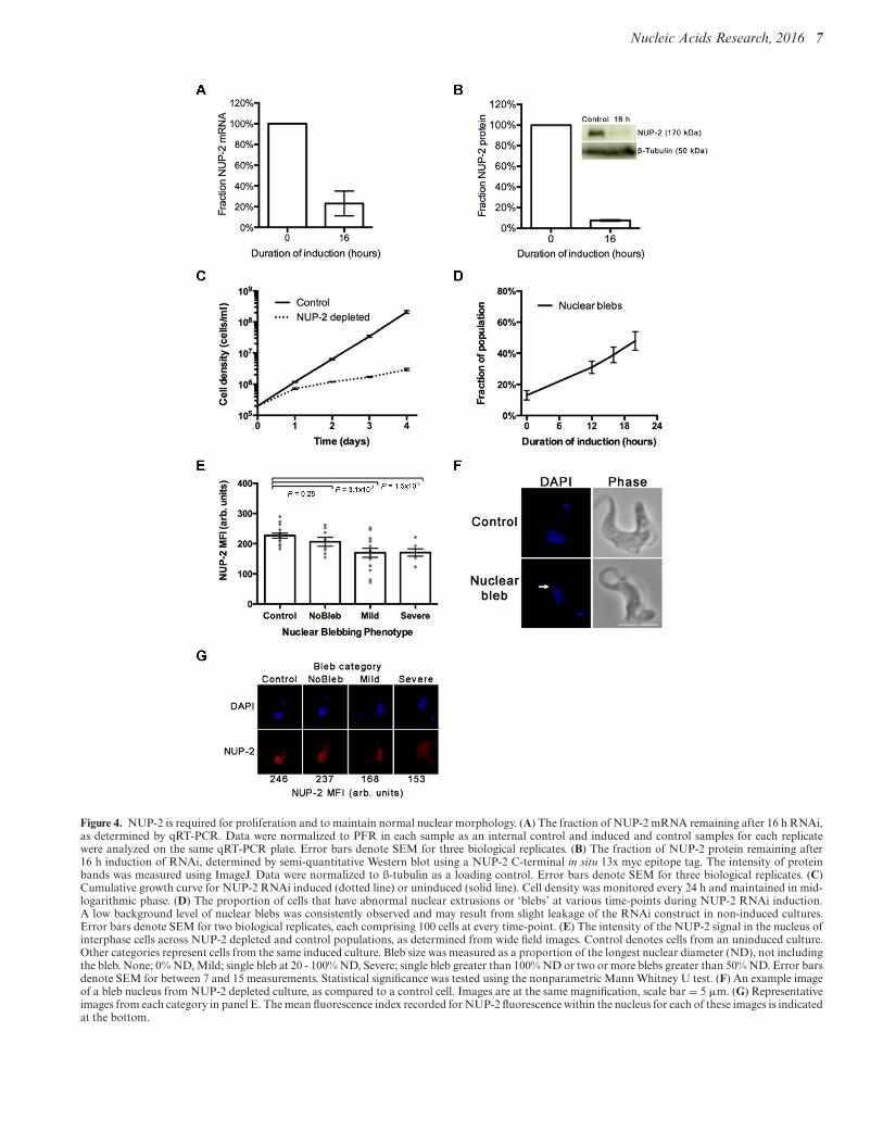
Figure 4. NUP-2 is required for proliferation and to maintain normal nuclear morphology. (A) The fraction of NUP-2 mRNA remaining after 16 h RNAi,as determined by qRT-PCR. Data were normalized to PFR in each sample as an internal control and induced and control samples for each replicatewere analyzed on the same qRT-PCR plate. Error bars denote SEM for three biological replicates. (B) The fraction of NUP-2 protein remaining after16 h induction of RNAi, determined by semi-quantitative Western blot using a NUP-2 C-terminal in situ 13x myc epitope tag. The intensity of proteinbands was measured using ImageJ. Data were normalized to ß-tubulin as a loading control. Error bars denote SEM for three biological replicates. (C)Cumulative growth curve for NUP-2 RNAi induced (dotted line) or uninduced (solid line). Cell density was monitored every 24 h and maintained in mid-logarithmic phase. (D) The proportion of cells that have abnormal nuclear extrusions or ‘blebs’ at various time-points during NUP-2 RNAi induction.A low background level of nuclear blebs was consistently observed and may result from slight leakage of the RNAi construct in non-induced cultures.Error bars denote SEM for two biological replicates, each comprising 100 cells at every time-point. (E) The intensity of the NUP-2 signal in the nucleus ofinterphase cells across NUP-2 depleted and control populations, as determined from wide field images. Control denotes cells from an uninduced culture.Other categories represent cells from the same induced culture. Bleb size was measured as a proportion of the longest nuclear diameter (ND), not includingthe bleb. None; 0% ND, Mild; single bleb at 20 - 100% ND, Severe; single bleb greater than 100% ND or two or more blebs greater than 50% ND. Error barsdenote SEM for between 7 and 15 measurements. Statistical significance was tested using the nonparametric Mann Whitney U test. (F) An example imageof a bleb nucleus from NUP-2 depleted culture, as compared to a control cell. Images are at the same magnification, scale bar = 5 !m. (G) Representativeimages from each category in panel E. The mean fluorescence index recorded for NUP-2 fluorescence within the nucleus for each of these images is indicatedat the bottom.
at Rockefeller U
niversity on September 23, 2016
http://nar.oxfordjournals.org/D
ownloaded from
8 Nucleic Acids Research, 2016
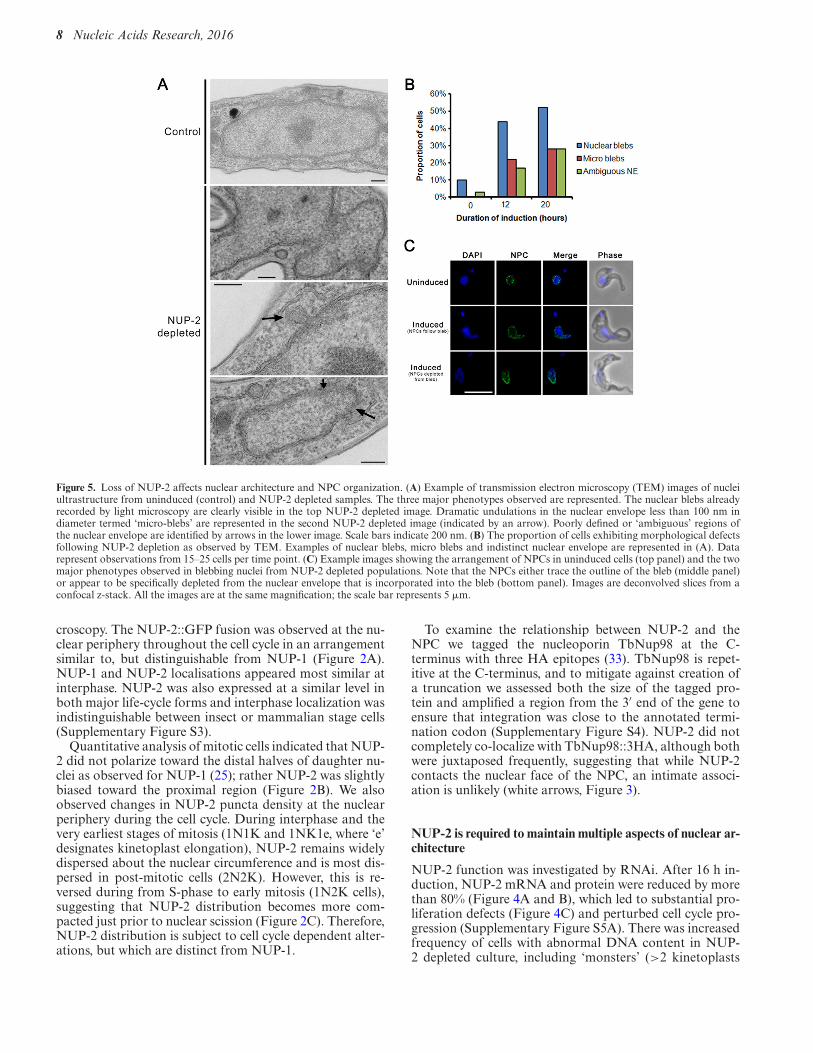
Figure 5. Loss of NUP-2 affects nuclear architecture and NPC organization. (A) Example of transmission electron microscopy (TEM) images of nucleiultrastructure from uninduced (control) and NUP-2 depleted samples. The three major phenotypes observed are represented. The nuclear blebs alreadyrecorded by light microscopy are clearly visible in the top NUP-2 depleted image. Dramatic undulations in the nuclear envelope less than 100 nm indiameter termed ‘micro-blebs’ are represented in the second NUP-2 depleted image (indicated by an arrow). Poorly defined or ‘ambiguous’ regions ofthe nuclear envelope are identified by arrows in the lower image. Scale bars indicate 200 nm. (B) The proportion of cells exhibiting morphological defectsfollowing NUP-2 depletion as observed by TEM. Examples of nuclear blebs, micro blebs and indistinct nuclear envelope are represented in (A). Datarepresent observations from 15–25 cells per time point. (C) Example images showing the arrangement of NPCs in uninduced cells (top panel) and the twomajor phenotypes observed in blebbing nuclei from NUP-2 depleted populations. Note that the NPCs either trace the outline of the bleb (middle panel)or appear to be specifically depleted from the nuclear envelope that is incorporated into the bleb (bottom panel). Images are deconvolved slices from aconfocal z-stack. All the images are at the same magnification; the scale bar represents 5 !m.
croscopy. The NUP-2::GFP fusion was observed at the nu-clear periphery throughout the cell cycle in an arrangementsimilar to, but distinguishable from NUP-1 (Figure 2A).NUP-1 and NUP-2 localisations appeared most similar atinterphase. NUP-2 was also expressed at a similar level inboth major life-cycle forms and interphase localization wasindistinguishable between insect or mammalian stage cells(Supplementary Figure S3).
Quantitative analysis of mitotic cells indicated that NUP-2 did not polarize toward the distal halves of daughter nu-clei as observed for NUP-1 (25); rather NUP-2 was slightlybiased toward the proximal region (Figure 2B). We alsoobserved changes in NUP-2 puncta density at the nuclearperiphery during the cell cycle. During interphase and thevery earliest stages of mitosis (1N1K and 1NK1e, where ‘e’designates kinetoplast elongation), NUP-2 remains widelydispersed about the nuclear circumference and is most dis-persed in post-mitotic cells (2N2K). However, this is re-versed during from S-phase to early mitosis (1N2K cells),suggesting that NUP-2 distribution becomes more com-pacted just prior to nuclear scission (Figure 2C). Therefore,NUP-2 distribution is subject to cell cycle dependent alter-ations, but which are distinct from NUP-1.
To examine the relationship between NUP-2 and theNPC we tagged the nucleoporin TbNup98 at the C-terminus with three HA epitopes (33). TbNup98 is repet-itive at the C-terminus, and to mitigate against creation ofa truncation we assessed both the size of the tagged pro-tein and amplified a region from the 3′ end of the gene toensure that integration was close to the annotated termi-nation codon (Supplementary Figure S4). NUP-2 did notcompletely co-localize with TbNup98::3HA, although bothwere juxtaposed frequently, suggesting that while NUP-2contacts the nuclear face of the NPC, an intimate associ-ation is unlikely (white arrows, Figure 3).
NUP-2 is required to maintain multiple aspects of nuclear ar-chitecture
NUP-2 function was investigated by RNAi. After 16 h in-duction, NUP-2 mRNA and protein were reduced by morethan 80% (Figure 4A and B), which led to substantial pro-liferation defects (Figure 4C) and perturbed cell cycle pro-gression (Supplementary Figure S5A). There was increasedfrequency of cells with abnormal DNA content in NUP-2 depleted culture, including ‘monsters’ (>2 kinetoplasts
at Rockefeller U
niversity on September 23, 2016
http://nar.oxfordjournals.org/D
ownloaded from
Nucleic Acids Research, 2016 9
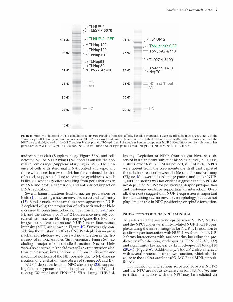
Figure 6. Affinity isolation of NUP-2 containing complexes. Proteins from each affinity isolation preparation were identified by mass spectrometry in theshown or parallel affinity capture preparations. NUP-2 is shown to interact with components of the NPC, and specifically, putative constituents of theNPC core scaffold, as well as the NPC nuclear basket protein TbNup110 and the nuclear lamina component NUP-1. Conditions for the isolation in leftpanels are 20 mM HEPES, pH 7.4, 250 mM NaCl, 0.5% Triton and for right panel 40 mM Tris, pH 7.4, 500 mM NaCl, 1% CHAPS.
and/or >2 nuclei) (Supplementary Figure S5A) and cellsdetected by FACS as having DNA content outside the nor-mal cell cycle range (Supplementary Figure S5C). The pres-ence of cells with abnormal DNA content and especiallythose with more than two nuclei, but the continued divisionof nuclei, suggests a failure to complete cytokinesis, whichis likely a secondary effect resulting from perturbations inmRNA and protein expression, and not a direct impact onDNA replication.
Several lamin mutations lead to nuclear protrusions orblebs (1), indicating a nuclear envelope structural deformity(15). Similar nuclear abnormalities were apparent in NUP-2 depleted cells; the proportion of cells with nuclear blebsincreased through time following induction (Figure 4D andF), and the intensity of NUP-2 fluorescence inversely cor-related with nuclear bleb frequency (Figure 4E). Exampleimages for nuclear defects and NUP-2 mean fluorescenceintensity (MFI) are shown in Figure 4G. Surprisingly, con-sidering the substantial effect of NUP-2 depletion on grossnuclear morphology, we observed no alteration in the fre-quency of mitotic spindles (Supplementary Figure S6), ex-cluding a major role in spindle formation. Nuclear blebswere also observed in knockdown cells by transmission elec-tron microscopy; invaginations <100 nm in diameter andill-defined portions of the NE, possibly due to NE disorga-nization or crenellation were observed (Figure 5A and B).
NUP-1 depletion leads to NPC clustering (25), suggest-ing that the trypanosomal lamina plays a role in NPC posi-tioning. We monitored TbNup98::3HA during NUP-2 si-
lencing. Depletion of NPCs from nuclear blebs was ob-served in a significant subset of blebbing nuclei (P = 0.006,Fisher’s exact test, n = 24 uninduced, n = 14 bleb). NPCswere absent from the bleb membrane itself and depletedfrom the intersection between the bleb and the nuclear rump(Figure 5C, lower induced image panel), and unlike NUP-1, NPC clustering was not evident suggesting that NPCs donot depend on NUP-2 for positioning, despite juxtapositionand proteomic evidence supporting an interaction. Over-all, these data suggest that NUP-2 expression is importantfor maintaining nuclear envelope morphology, but does notplay a major role in NPC positioning or spindle formation.
NUP-2 interacts with the NPC and NUP-1
To understand the relationships between NUP-2, NUP-1and the NPC further we affinity isolated NUP-2::GFP com-plexes using the same strategy as for NUP-1. In addition toconfirming an interaction with NUP-1, we found that NUP-2 forms interactions with nucleoporins including the pre-dicted scaffold-forming nucleoporins (TbNup82, 89, 132)and significantly the nuclear basket nucleoporin TbNup110(29,54) (Figure 6). Additionally, TbNUP-2 also interactswith several proteins of unknown function, which also lo-calize to the nuclear envelope (SO, MCF and MPR, unpub-lished).
The number of interactions recovered between NUP-2and the NPC are not as extensive as for NUP-1. We sug-gest that interactions with the NPC may be mediated via
at Rockefeller U
niversity on September 23, 2016
http://nar.oxfordjournals.org/D
ownloaded from
10 Nucleic Acids Research, 2016
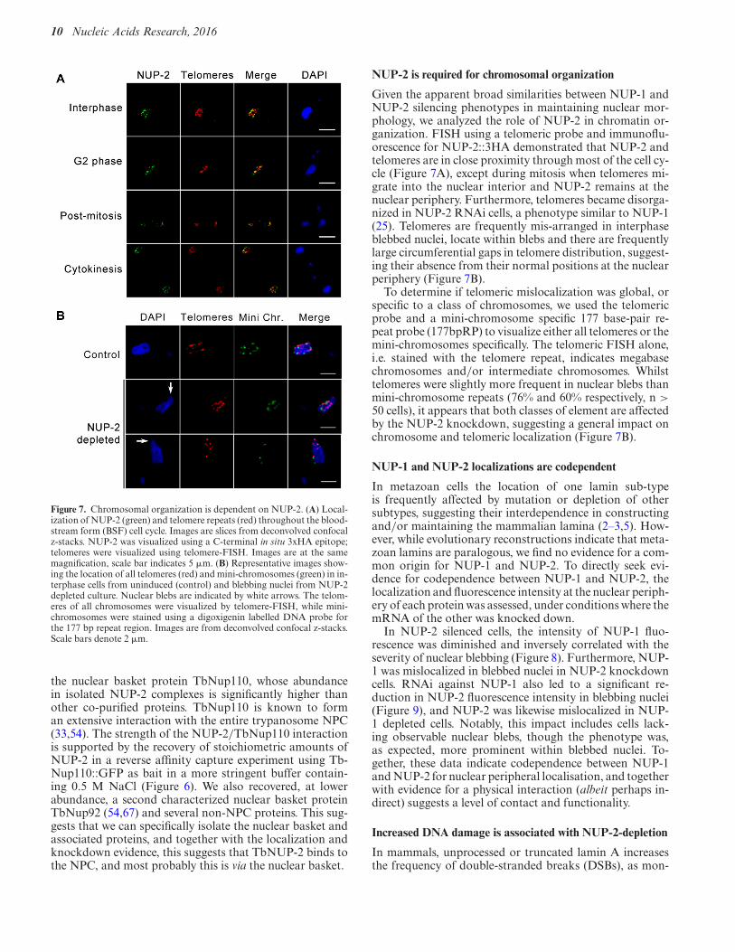
Figure 7. Chromosomal organization is dependent on NUP-2. (A) Local-ization of NUP-2 (green) and telomere repeats (red) throughout the blood-stream form (BSF) cell cycle. Images are slices from deconvolved confocalz-stacks. NUP-2 was visualized using a C-terminal in situ 3xHA epitope;telomeres were visualized using telomere-FISH. Images are at the samemagnification, scale bar indicates 5 !m. (B) Representative images show-ing the location of all telomeres (red) and mini-chromosomes (green) in in-terphase cells from uninduced (control) and blebbing nuclei from NUP-2depleted culture. Nuclear blebs are indicated by white arrows. The telom-eres of all chromosomes were visualized by telomere-FISH, while mini-chromosomes were stained using a digoxigenin labelled DNA probe forthe 177 bp repeat region. Images are from deconvolved confocal z-stacks.Scale bars denote 2 !m.
the nuclear basket protein TbNup110, whose abundancein isolated NUP-2 complexes is significantly higher thanother co-purified proteins. TbNup110 is known to forman extensive interaction with the entire trypanosome NPC(33,54). The strength of the NUP-2/TbNup110 interactionis supported by the recovery of stoichiometric amounts ofNUP-2 in a reverse affinity capture experiment using Tb-Nup110::GFP as bait in a more stringent buffer contain-ing 0.5 M NaCl (Figure 6). We also recovered, at lowerabundance, a second characterized nuclear basket proteinTbNup92 (54,67) and several non-NPC proteins. This sug-gests that we can specifically isolate the nuclear basket andassociated proteins, and together with the localization andknockdown evidence, this suggests that TbNUP-2 binds tothe NPC, and most probably this is via the nuclear basket.
NUP-2 is required for chromosomal organization
Given the apparent broad similarities between NUP-1 andNUP-2 silencing phenotypes in maintaining nuclear mor-phology, we analyzed the role of NUP-2 in chromatin or-ganization. FISH using a telomeric probe and immunoflu-orescence for NUP-2::3HA demonstrated that NUP-2 andtelomeres are in close proximity through most of the cell cy-cle (Figure 7A), except during mitosis when telomeres mi-grate into the nuclear interior and NUP-2 remains at thenuclear periphery. Furthermore, telomeres became disorga-nized in NUP-2 RNAi cells, a phenotype similar to NUP-1(25). Telomeres are frequently mis-arranged in interphaseblebbed nuclei, locate within blebs and there are frequentlylarge circumferential gaps in telomere distribution, suggest-ing their absence from their normal positions at the nuclearperiphery (Figure 7B).
To determine if telomeric mislocalization was global, orspecific to a class of chromosomes, we used the telomericprobe and a mini-chromosome specific 177 base-pair re-peat probe (177bpRP) to visualize either all telomeres or themini-chromosomes specifically. The telomeric FISH alone,i.e. stained with the telomere repeat, indicates megabasechromosomes and/or intermediate chromosomes. Whilsttelomeres were slightly more frequent in nuclear blebs thanmini-chromosome repeats (76% and 60% respectively, n >50 cells), it appears that both classes of element are affectedby the NUP-2 knockdown, suggesting a general impact onchromosome and telomeric localization (Figure 7B).
NUP-1 and NUP-2 localizations are codependent
In metazoan cells the location of one lamin sub-typeis frequently affected by mutation or depletion of othersubtypes, suggesting their interdependence in constructingand/or maintaining the mammalian lamina (2–3,5). How-ever, while evolutionary reconstructions indicate that meta-zoan lamins are paralogous, we find no evidence for a com-mon origin for NUP-1 and NUP-2. To directly seek evi-dence for codependence between NUP-1 and NUP-2, thelocalization and fluorescence intensity at the nuclear periph-ery of each protein was assessed, under conditions where themRNA of the other was knocked down.
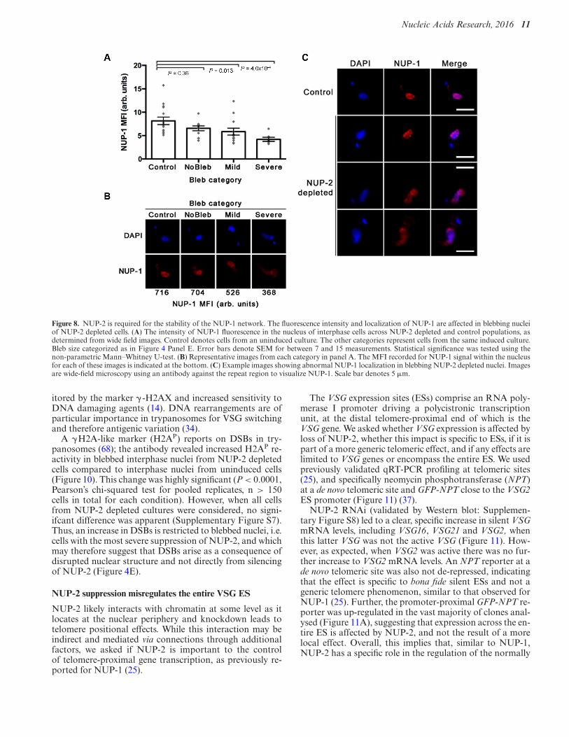
In NUP-2 silenced cells, the intensity of NUP-1 fluo-rescence was diminished and inversely correlated with theseverity of nuclear blebbing (Figure 8). Furthermore, NUP-1 was mislocalized in blebbed nuclei in NUP-2 knockdowncells. RNAi against NUP-1 also led to a significant re-duction in NUP-2 fluorescence intensity in blebbing nuclei(Figure 9), and NUP-2 was likewise mislocalized in NUP-1 depleted cells. Notably, this impact includes cells lack-ing observable nuclear blebs, though the phenotype was,as expected, more prominent within blebbed nuclei. To-gether, these data indicate codependence between NUP-1and NUP-2 for nuclear peripheral localisation, and togetherwith evidence for a physical interaction (albeit perhaps in-direct) suggests a level of contact and functionality.
Increased DNA damage is associated with NUP-2-depletion
In mammals, unprocessed or truncated lamin A increasesthe frequency of double-stranded breaks (DSBs), as mon-
at Rockefeller U
niversity on September 23, 2016
http://nar.oxfordjournals.org/D
ownloaded from
Nucleic Acids Research, 2016 11
Figure 8. NUP-2 is required for the stability of the NUP-1 network. The fluorescence intensity and localization of NUP-1 are affected in blebbing nucleiof NUP-2 depleted cells. (A) The intensity of NUP-1 fluorescence in the nucleus of interphase cells across NUP-2 depleted and control populations, asdetermined from wide field images. Control denotes cells from an uninduced culture. The other categories represent cells from the same induced culture.Bleb size categorized as in Figure 4 Panel E. Error bars denote SEM for between 7 and 15 measurements. Statistical significance was tested using thenon-parametric Mann–Whitney U-test. (B) Representative images from each category in panel A. The MFI recorded for NUP-1 signal within the nucleusfor each of these images is indicated at the bottom. (C) Example images showing abnormal NUP-1 localization in blebbing NUP-2 depleted nuclei. Imagesare wide-field microscopy using an antibody against the repeat region to visualize NUP-1. Scale bar denotes 5 !m.
itored by the marker $ -H2AX and increased sensitivity toDNA damaging agents (14). DNA rearrangements are ofparticular importance in trypanosomes for VSG switchingand therefore antigenic variation (34).
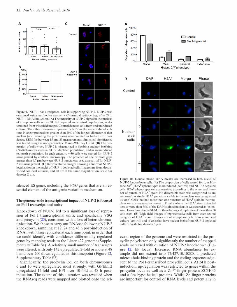
A $H2A-like marker (H2AP) reports on DSBs in try-panosomes (68); the antibody revealed increased H2AP re-activity in blebbed interphase nuclei from NUP-2 depletedcells compared to interphase nuclei from uninduced cells(Figure 10). This change was highly significant (P < 0.0001,Pearson’s chi-squared test for pooled replicates, n > 150cells in total for each condition). However, when all cellsfrom NUP-2 depleted cultures were considered, no signi-ifcant difference was apparent (Supplementary Figure S7).Thus, an increase in DSBs is restricted to blebbed nuclei, i.e.cells with the most severe suppression of NUP-2, and whichmay therefore suggest that DSBs arise as a consequence ofdisrupted nuclear structure and not directly from silencingof NUP-2 (Figure 4E).
NUP-2 suppression misregulates the entire VSG ES
NUP-2 likely interacts with chromatin at some level as itlocates at the nuclear periphery and knockdown leads totelomere positional effects. While this interaction may beindirect and mediated via connections through additionalfactors, we asked if NUP-2 is important to the controlof telomere-proximal gene transcription, as previously re-ported for NUP-1 (25).
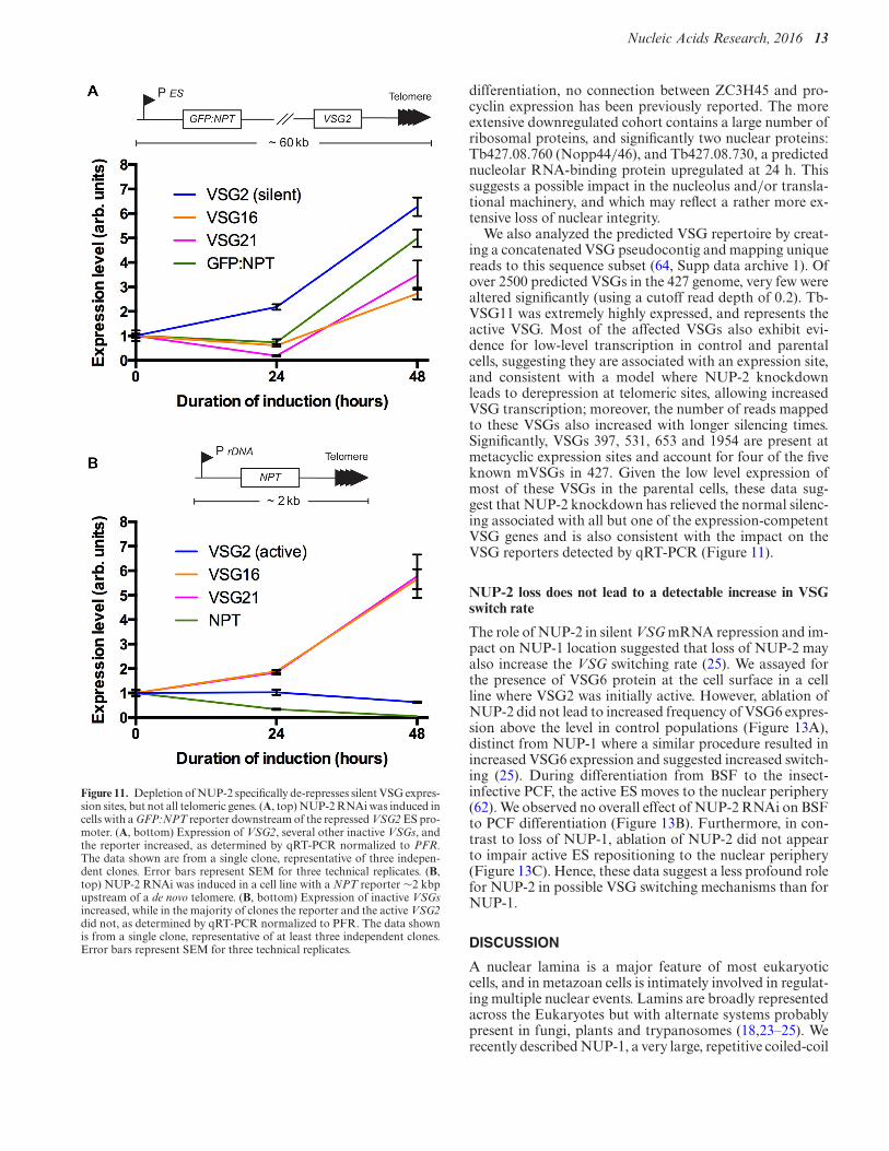
The VSG expression sites (ESs) comprise an RNA poly-merase I promoter driving a polycistronic transcriptionunit, at the distal telomere-proximal end of which is theVSG gene. We asked whether VSG expression is affected byloss of NUP-2, whether this impact is specific to ESs, if it ispart of a more generic telomeric effect, and if any effects arelimited to VSG genes or encompass the entire ES. We usedpreviously validated qRT-PCR profiling at telomeric sites(25), and specifically neomycin phosphotransferase (NPT)at a de novo telomeric site and GFP-NPT close to the VSG2ES promoter (Figure 11) (37).
NUP-2 RNAi (validated by Western blot: Supplemen-tary Figure S8) led to a clear, specific increase in silent VSGmRNA levels, including VSG16, VSG21 and VSG2, whenthis latter VSG was not the active VSG (Figure 11). How-ever, as expected, when VSG2 was active there was no fur-ther increase to VSG2 mRNA levels. An NPT reporter at ade novo telomeric site was also not de-repressed, indicatingthat the effect is specific to bona fide silent ESs and not ageneric telomere phenomenon, similar to that observed forNUP-1 (25). Further, the promoter-proximal GFP-NPT re-porter was up-regulated in the vast majority of clones anal-ysed (Figure 11A), suggesting that expression across the en-tire ES is affected by NUP-2, and not the result of a morelocal effect. Overall, this implies that, similar to NUP-1,NUP-2 has a specific role in the regulation of the normally
at Rockefeller U
niversity on September 23, 2016
http://nar.oxfordjournals.org/D
ownloaded from
12 Nucleic Acids Research, 2016
Figure 9. NUP-1 has a reciprocal role in supporting NUP-2. NUP-2 wasexamined using antibodies against a C-terminal epitope tag, after 24 hNUP-1 RNAi induction. (A) The intensity of NUP-2 signal in the nucleusof interphase cells across NUP-1 depleted and control populations, as de-termined from wide field images. Control denotes cells from and uninducedculture. The other categories represent cells from the same induced cul-ture. Nuclear protrusions greater than 20% of the longest diameter of thatnucleus (not including the protrusion) were counted as blebs. Error barsdenote SEM for between 15 and 23 measurements. Statistical significancewas tested using the non-parametric Mann–Whitney U-test. (B) The pro-portion of cells where NUP-2 is misarranged in blebbing and non blebbing(NoBleb) nuclei across a NUP-1 depleted population, and in an uninduced(control) population. In each category ∼30 cells were scored for NUP-2arrangement by confocal microscopy. The presence of one or more gapsgreater than 0.7 !m between NUP-2 puncta was used as a cut-off for NUP-2 misarrangement. (C) Representative images showing abnormal NUP-2localization in the nuclei of NUP-1 depleted cells. Images are from decon-volved confocal z-stacks, and all are at the same magnification, scale bardenotes 2 !m.
silenced ES genes, including the VSG genes that are an es-sential element of the antigenic variation mechanism.
The genome-wide transcriptional impact of NUP-2 is focusedon Pol I transcriptional units
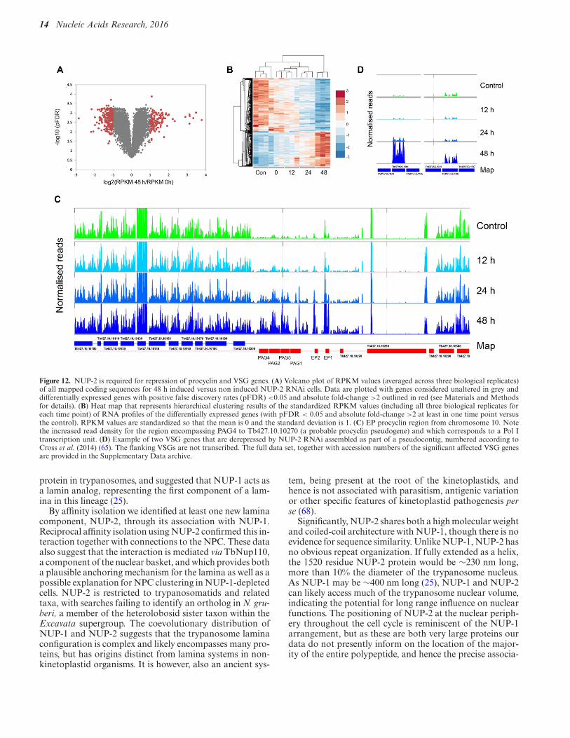
Knockdown of NUP-1 led to a significant loss of repres-sion of Pol I transcriptional units, and specifically VSGand procyclin (25), consistent with a loss of heterochroma-tinization. We chose to carry out RNAseq following NUP-2knockdown, sampling at 12, 24 and 48 h post-induction ofRNAi, with three replicates at each time point, in order thatwe could identify with confidence differentially regulatedgenes by mapping reads to the Lister 427 genome (Supple-mentary Table S1). A relatively small number of transcriptswere altered, with only 142 upregulated 2-fold or more at 48h and over 200 downregulated at this timepoint (Figure 12,Supplementary Table S2).
Significantly, the procyclin loci on both chromosomes6 and 10 were upregulated most strongly, with GPEET2upregulated 14-fold and EP1 over 10-fold at 48 h post-induction. The extent of this alteration was revealed whenthe RNAseq reads were mapped and plotted onto the rel-
Figure 10. Double strand DNA breaks are increased in bleb nuclei ofNUP-2 knockdown cells. (A) The proportion of cells scored for four His-tone 2AP (H2AP) phenotypes in uninduced (control) and NUP-2 depletedcells. H2AP phenotypes were categorized according to the extent and num-ber of puncta of H2AP stain: No discernible stain was categorized as ‘noresponse’. A single H2AP punctum visible in the nucleus was categorizedas ‘one’. Cells that had more than one punctum of H2AP stain in their nu-cleus were categorized as ‘several’. Finally, where the H2AP stain extendedacross more than 75% of the DAPI stained nucleus, it was scored as ‘exten-sive’. Error bars denote SEM for three biological replicates of more than 50cells each. (B) Wide-field images of representative cells from each scoredcategory of H2AP stain. Images are of interphase cells from uninducedculture (control) and of cells that have nuclear blebs from NUP-2 depletedculture. Scale bar denotes 5 !m.
evant region of the genome and were restricted to the pro-cyclin polycistron only; significantly the number of mappedreads increased with duration of NUP-2 knockdown (Fig-ure 12, EP locus). Increased RNA abundance for ex-ample did not extend into Tb427.10.10280, a predictedmicrotubule-binding protein and the coding sequence adja-cent to the Pol I-transcribed procyclin locus. At 24 h post-induction, up-regulation was restricted to genes within theprocyclin locus as well as a Zn2+-finger protein ZC3H45and a few hypothetical proteins. Whilst Zn finger proteinsare important for control of RNA levels and potentially in
at Rockefeller U
niversity on September 23, 2016
http://nar.oxfordjournals.org/D
ownloaded from
Nucleic Acids Research, 2016 13
Figure 11. Depletion of NUP-2 specifically de-represses silent VSG expres-sion sites, but not all telomeric genes. (A, top) NUP-2 RNAi was induced incells with a GFP:NPT reporter downstream of the repressed VSG2 ES pro-moter. (A, bottom) Expression of VSG2, several other inactive VSGs, andthe reporter increased, as determined by qRT-PCR normalized to PFR.The data shown are from a single clone, representative of three indepen-dent clones. Error bars represent SEM for three technical replicates. (B,top) NUP-2 RNAi was induced in a cell line with a NPT reporter ∼2 kbpupstream of a de novo telomere. (B, bottom) Expression of inactive VSGsincreased, while in the majority of clones the reporter and the active VSG2did not, as determined by qRT-PCR normalized to PFR. The data shownis from a single clone, representative of at least three independent clones.Error bars represent SEM for three technical replicates.
differentiation, no connection between ZC3H45 and pro-cyclin expression has been previously reported. The moreextensive downregulated cohort contains a large number ofribosomal proteins, and significantly two nuclear proteins:Tb427.08.760 (Nopp44/46), and Tb427.08.730, a predictednucleolar RNA-binding protein upregulated at 24 h. Thissuggests a possible impact in the nucleolus and/or transla-tional machinery, and which may reflect a rather more ex-tensive loss of nuclear integrity.
We also analyzed the predicted VSG repertoire by creat-ing a concatenated VSG pseudocontig and mapping uniquereads to this sequence subset (64, Supp data archive 1). Ofover 2500 predicted VSGs in the 427 genome, very few werealtered significantly (using a cutoff read depth of 0.2). Tb-VSG11 was extremely highly expressed, and represents theactive VSG. Most of the affected VSGs also exhibit evi-dence for low-level transcription in control and parentalcells, suggesting they are associated with an expression site,and consistent with a model where NUP-2 knockdownleads to derepression at telomeric sites, allowing increasedVSG transcription; moreover, the number of reads mappedto these VSGs also increased with longer silencing times.Significantly, VSGs 397, 531, 653 and 1954 are present atmetacyclic expression sites and account for four of the fiveknown mVSGs in 427. Given the low level expression ofmost of these VSGs in the parental cells, these data sug-gest that NUP-2 knockdown has relieved the normal silenc-ing associated with all but one of the expression-competentVSG genes and is also consistent with the impact on theVSG reporters detected by qRT-PCR (Figure 11).
NUP-2 loss does not lead to a detectable increase in VSGswitch rate
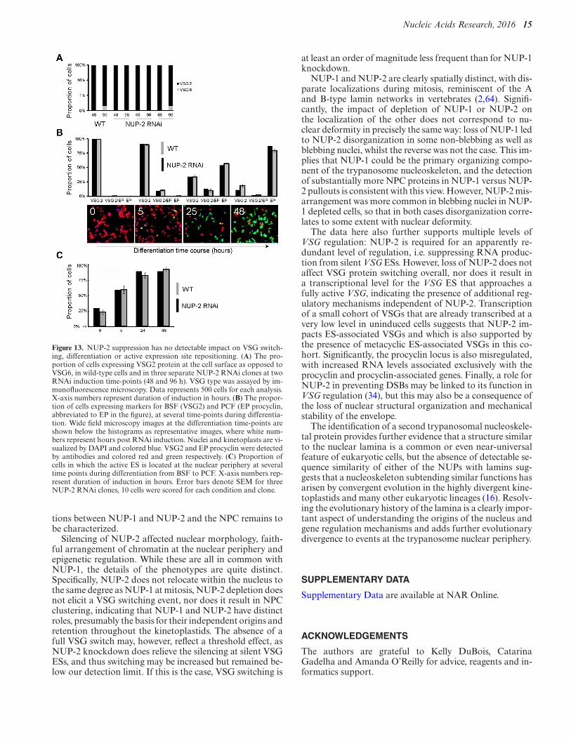
The role of NUP-2 in silent VSG mRNA repression and im-pact on NUP-1 location suggested that loss of NUP-2 mayalso increase the VSG switching rate (25). We assayed forthe presence of VSG6 protein at the cell surface in a cellline where VSG2 was initially active. However, ablation ofNUP-2 did not lead to increased frequency of VSG6 expres-sion above the level in control populations (Figure 13A),distinct from NUP-1 where a similar procedure resulted inincreased VSG6 expression and suggested increased switch-ing (25). During differentiation from BSF to the insect-infective PCF, the active ES moves to the nuclear periphery(62). We observed no overall effect of NUP-2 RNAi on BSFto PCF differentiation (Figure 13B). Furthermore, in con-trast to loss of NUP-1, ablation of NUP-2 did not appearto impair active ES repositioning to the nuclear periphery(Figure 13C). Hence, these data suggest a less profound rolefor NUP-2 in possible VSG switching mechanisms than forNUP-1.
DISCUSSION
A nuclear lamina is a major feature of most eukaryoticcells, and in metazoan cells is intimately involved in regulat-ing multiple nuclear events. Lamins are broadly representedacross the Eukaryotes but with alternate systems probablypresent in fungi, plants and trypanosomes (18,23–25). Werecently described NUP-1, a very large, repetitive coiled-coil
at Rockefeller U
niversity on September 23, 2016
http://nar.oxfordjournals.org/D
ownloaded from
14 Nucleic Acids Research, 2016
Figure 12. NUP-2 is required for repression of procyclin and VSG genes. (A) Volcano plot of RPKM values (averaged across three biological replicates)of all mapped coding sequences for 48 h induced versus non induced NUP-2 RNAi cells. Data are plotted with genes considered unaltered in grey anddifferentially expressed genes with positive false discovery rates (pFDR) <0.05 and absolute fold-change >2 outlined in red (see Materials and Methodsfor details). (B) Heat map that represents hierarchical clustering results of the standardized RPKM values (including all three biological replicates foreach time point) of RNA profiles of the differentially expressed genes (with pFDR < 0.05 and absolute fold-change >2 at least in one time point versusthe control). RPKM values are standardized so that the mean is 0 and the standard deviation is 1. (C) EP procyclin region from chromosome 10. Notethe increased read density for the region encompassing PAG4 to Tb427.10.10270 (a probable procyclin pseudogene) and which corresponds to a Pol Itranscription unit. (D) Example of two VSG genes that are derepressed by NUP-2 RNAi assembled as part of a pseudocontig, numbered according toCross et al. (2014) (65). The flanking VSGs are not transcribed. The full data set, together with accession numbers of the significant affected VSG genesare provided in the Supplementary Data archive.
protein in trypanosomes, and suggested that NUP-1 acts asa lamin analog, representing the first component of a lam-ina in this lineage (25).
By affinity isolation we identified at least one new laminacomponent, NUP-2, through its association with NUP-1.Reciprocal affinity isolation using NUP-2 confirmed this in-teraction together with connections to the NPC. These dataalso suggest that the interaction is mediated via TbNup110,a component of the nuclear basket, and which provides botha plausible anchoring mechanism for the lamina as well as apossible explanation for NPC clustering in NUP-1-depletedcells. NUP-2 is restricted to trypanosomatids and relatedtaxa, with searches failing to identify an ortholog in N. gru-beri, a member of the heterolobosid sister taxon within theExcavata supergroup. The coevolutionary distribution ofNUP-1 and NUP-2 suggests that the trypanosome laminaconfiguration is complex and likely encompasses many pro-teins, but has origins distinct from lamina systems in non-kinetoplastid organisms. It is however, also an ancient sys-
tem, being present at the root of the kinetoplastids, andhence is not associated with parasitism, antigenic variationor other specific features of kinetoplastid pathogenesis perse (68).
Significantly, NUP-2 shares both a high molecular weightand coiled-coil architecture with NUP-1, though there is noevidence for sequence similarity. Unlike NUP-1, NUP-2 hasno obvious repeat organization. If fully extended as a helix,the 1520 residue NUP-2 protein would be ∼230 nm long,more than 10% the diameter of the trypanosome nucleus.As NUP-1 may be ∼400 nm long (25), NUP-1 and NUP-2can likely access much of the trypanosome nuclear volume,indicating the potential for long range influence on nuclearfunctions. The positioning of NUP-2 at the nuclear periph-ery throughout the cell cycle is reminiscent of the NUP-1arrangement, but as these are both very large proteins ourdata do not presently inform on the location of the major-ity of the entire polypeptide, and hence the precise associa-
at Rockefeller U
niversity on September 23, 2016
http://nar.oxfordjournals.org/D
ownloaded from
Nucleic Acids Research, 2016 15
Figure 13. NUP-2 suppression has no detectable impact on VSG switch-ing, differentiation or active expression site repositioning. (A) The pro-portion of cells expressing VSG2 protein at the cell surface as opposed toVSG6, in wild-type cells and in three separate NUP-2 RNAi clones at twoRNAi induction time-points (48 and 96 h). VSG type was assayed by im-munofluorescence microscopy. Data represents 500 cells for each analysis.X-axis numbers represent duration of induction in hours. (B) The propor-tion of cells expressing markers for BSF (VSG2) and PCF (EP procyclin,abbreviated to EP in the figure), at several time-points during differentia-tion. Wide field microscopy images at the differentiation time-points areshown below the histograms as representative images, where white num-bers represent hours post RNAi induction. Nuclei and kinetoplasts are vi-sualized by DAPI and colored blue. VSG2 and EP procyclin were detectedby antibodies and colored red and green respectively. (C) Proportion ofcells in which the active ES is located at the nuclear periphery at severaltime points during differentiation from BSF to PCF. X-axis numbers rep-resent duration of induction in hours. Error bars denote SEM for threeNUP-2 RNAi clones, 10 cells were scored for each condition and clone.
tions between NUP-1 and NUP-2 and the NPC remains tobe characterized.
Silencing of NUP-2 affected nuclear morphology, faith-ful arrangement of chromatin at the nuclear periphery andepigenetic regulation. While these are all in common withNUP-1, the details of the phenotypes are quite distinct.Specifically, NUP-2 does not relocate within the nucleus tothe same degree as NUP-1 at mitosis, NUP-2 depletion doesnot elicit a VSG switching event, nor does it result in NPCclustering, indicating that NUP-1 and NUP-2 have distinctroles, presumably the basis for their independent origins andretention throughout the kinetoplastids. The absence of afull VSG switch may, however, reflect a threshold effect, asNUP-2 knockdown does relieve the silencing at silent VSGESs, and thus switching may be increased but remained be-low our detection limit. If this is the case, VSG switching is
at least an order of magnitude less frequent than for NUP-1knockdown.
NUP-1 and NUP-2 are clearly spatially distinct, with dis-parate localizations during mitosis, reminiscent of the Aand B-type lamin networks in vertebrates (2,64). Signifi-cantly, the impact of depletion of NUP-1 or NUP-2 onthe localization of the other does not correspond to nu-clear deformity in precisely the same way: loss of NUP-1 ledto NUP-2 disorganization in some non-blebbing as well asblebbing nuclei, whilst the reverse was not the case. This im-plies that NUP-1 could be the primary organizing compo-nent of the trypanosome nucleoskeleton, and the detectionof substantially more NPC proteins in NUP-1 versus NUP-2 pullouts is consistent with this view. However, NUP-2 mis-arrangement was more common in blebbing nuclei in NUP-1 depleted cells, so that in both cases disorganization corre-lates to some extent with nuclear deformity.
The data here also further supports multiple levels ofVSG regulation: NUP-2 is required for an apparently re-dundant level of regulation, i.e. suppressing RNA produc-tion from silent VSG ESs. However, loss of NUP-2 does notaffect VSG protein switching overall, nor does it result ina transcriptional level for the VSG ES that approaches afully active VSG, indicating the presence of additional reg-ulatory mechanisms independent of NUP-2. Transcriptionof a small cohort of VSGs that are already transcribed at avery low level in uninduced cells suggests that NUP-2 im-pacts ES-associated VSGs and which is also supported bythe presence of metacyclic ES-associated VSGs in this co-hort. Significantly, the procyclin locus is also misregulated,with increased RNA levels associated exclusively with theprocyclin and procyclin-associated genes. Finally, a role forNUP-2 in preventing DSBs may be linked to its function inVSG regulation (34), but this may also be a consequence ofthe loss of nuclear structural organization and mechanicalstability of the envelope.
The identification of a second trypanosomal nucleoskele-tal protein provides further evidence that a structure similarto the nuclear lamina is a common or even near-universalfeature of eukaryotic cells, but the absence of detectable se-quence similarity of either of the NUPs with lamins sug-gests that a nucleoskeleton subtending similar functions hasarisen by convergent evolution in the highly divergent kine-toplastids and many other eukaryotic lineages (16). Resolv-ing the evolutionary history of the lamina is a clearly impor-tant aspect of understanding the origins of the nucleus andgene regulation mechanisms and adds further evolutionarydivergence to events at the trypanosome nuclear periphery.
SUPPLEMENTARY DATA
Supplementary Data are available at NAR Online.
ACKNOWLEDGEMENTS
The authors are grateful to Kelly DuBois, CatarinaGadelha and Amanda O’Reilly for advice, reagents and in-formatics support.
at Rockefeller U
niversity on September 23, 2016
http://nar.oxfordjournals.org/D
ownloaded from
16 Nucleic Acids Research, 2016
FUNDING
This work was supported by the Wellcome Trust (programgrant 082813 to MCF, 093010 and 100320 to DH), theGates Cambridge Trust (studentship to LM) and the NIH(R21 AI096069, U54 GM103511 to BTC, JA and MPR,U01 GM098256, P41 GM109824 to MPR, R01 GM112108to JA and MPR, P50 GM076547 to JA and P41 GM103314to BTC). Funding for open access charge: Wellcome Trust.Conflict of interest statement. None declared.
REFERENCES1. Dittmer,T.A. and Misteli,T. (2011) The lamin protein family.
Genome Biol., 12, 222.2. Shimi,T., Pfleghaar,K., Kojima,S., Pack,C.G., Solovei,I.,
Goldman,A.E., Adam,S.A., Shumaker,D.K., Kinjo,M., Cremer,T.et al. (2008) The A- and B-type nuclear lamin networks:microdomains involved in chromatin organization and transcription.Genes Dev., 22, 3409–3421.
3. Lenz-Bohme,B., Wismar,J., Fuchs,S., Reifegerste,R., Buchner,E.,Betz,H. and Schmitt,B. (1997) Insertional mutation of theDrosophila nuclear lamin Dm0 gene results in defective nuclearenvelopes, clustering of nuclear pore complexes, and accumulation ofannulate lamellae. J. Cell Biol., 137, 1001–1016.
4. Liu,J., Rolef Ben-Shahar,T., Riemer,D., Treinin,M., Spann,P.,Weber,K., Fire,A. and Gruenbaum,Y. (2000) Essential roles forCaenorhabditis elegans lamin gene in nuclear organization, cell cycleprogression, and spatial organization of nuclear pore complexes.Mol. Biol. Cell, 11, 3937–3947.
5. Muchir,A., van Engelen,B.G., Lammens,M., Mislow,J.M.,McNally,E., Schwartz,K. and Bonne,G. (2003) Nuclear envelopealterations in fibroblasts from LGMD1B patients carrying nonsenseY259X heterozygous or homozygous mutation in lamin A/C gene.Exp. Cell Res., 291, 352–362.
6. Sullivan,T., Escalante-Alcalde,D., Bhatt,H., Anver,M., Bhat,N.,Nagashima,K., Stewart,C.L. and Burke,B. (1999) Loss of A-typelamin expression compromises nuclear envelope integrity leading tomuscular dystrophy. J. Cell Biol., 147, 913–920.
7. Caille,N., Thoumine,O., Tardy,Y. and Meister,J.J. (2002)Contribution of the nucleus to the mechanical properties ofendothelial cells. J. Biomech., 35, 177–187.
8. Guilak,F., Tedrow,J.R. and Burgkart,R. (2000) Viscoelasticproperties of the cell nucleus. Biochem. Biophys. Res. Commun., 269,781–786.
9. Houben,F., Ramaekers,F.C., Snoeckx,L.H. and Broers,J.L. (2007)Role of nuclear lamina-cytoskeleton interactions in the maintenanceof cellular strength. Biochim. Biophys. Acta, 1773, 675–686.
10. Mekhail,K. and Moazed,D. (2010) The nuclear envelope in genomeorganization, expression and stability. Nat. Rev. Mol. Cell Biol., 11,317–328.
11. Andres,V. and Gonzalez,J.M. (2009) Role of A-type lamins insignaling, transcription, and chromatin organization. J. Cell Biol.,187, 945–957.
12. Moir,R.D., Spann,T.P., Herrmann,H. and Goldman,R.D. (2000)Disruption of nuclear lamin organization blocks the elongationphase of DNA replication. J. Cell Biol., 149, 1179–1192.
13. Spann,T.P., Moir,R.D., Goldman,A.E., Stick,R. and Goldman,R.D.(1997) Disruption of nuclear lamin organization alters thedistribution of replication factors and inhibits DNA synthesis. J. CellBiol., 136, 1201–1212.
14. Liu,B., Wang,J., Chan,K.M., Tjia,W.M., Deng,W., Guan,X.,Huang,J.D., Li,K.M., Chau,P.Y., Chen,D.J. et al. (2005) Genomicinstability in laminopathy-based premature aging. Nat. Med., 11,780–785.
15. Kruger,A., Batsios,P., Baumann,O., Luckert,E., Schwarz,H.,Stick,R., Meyer,I. and Graf,R. (2012) Characterization of NE81, thefirst lamin-like nucleoskeleton protein in a unicellular organism.Mol. Biol. Cell, 23, 360–370.
16. Koreny,L. and Field,M.C. (2016) Ancient eukaryotic origin andevolutionary plasticity of nuclear lamina. Genome Biol Evol, evw087.
17. Strambio-de-Castillia,C., Blobel,G. and Rout,M.P. (1995) Isolationand characterization of nuclear envelopes from the yeastSaccharomyces. J. Cell Biol., 131, 19–31.
18. Niepel,M., Molloy,K.R., Williams,R., Farr,J.C., Meinema,A.C.,Vecchietti,N., Cristea,I.M., Chait,B.T., Rout,M.P. andStrambio-De-Castillia,C. (2013) The nuclear basket proteins Mlp1pand Mlp2p are part of a dynamic interactome including Esc1p andthe proteasome. Mol. Biol. Cell, 24, 3920–3938.
19. Andrulis,E.D., Zappulla,D.C., Ansari,A., Perrod,S., Laiosa,C.V.,Gartenberg,M.R. and Sternglanz,R. (2002) Esc1, a nuclear peripheryprotein required for Sir4-based plasmid anchoring and partitioning.Mol. Cell. Biol., 22, 8292–8301.
20. Taddei,A., Hediger,F., Neumann,F.R., Bauer,C. and Gasser,S.M.(2004) Separation of silencing from perinuclear anchoring functionsin yeast Ku80, Sir4 and Esc1 proteins. EMBO J., 23, 1301–1312.
21. Lewis,A., Felberbaum,R. and Hochstrasser,M. (2007) A nuclearenvelope protein linking nuclear pore basket assembly, SUMOprotease regulation, and mRNA surveillance. J. Cell Biol., 178,813–827.
22. Hattier,T., Andrulis,E.D. and Tartakoff,A.M. (2007) Immobility,inheritance and plasticity of shape of the yeast nucleus. BMC CellBiol., 8, 47.
23. Sakamoto,Y. and Takagi,S. (2013) LITTLE NUCLEI 1 and 4regulate nuclear morphology in Arabidopsis thaliana. Plant CellPhysiol., 54, 622–633.
24. Blumenthal,S.S., Clark,G.B. and Roux,S.J. (2004) Biochemical andimmunological characterization of pea nuclear intermediate filamentproteins. Planta, 218, 965–975.
25. DuBois,K.N., Alsford,S., Holden,J.M., Buisson,J., Swiderski,M.,Bart,J.M., Ratushny,A.V., Wan,Y., Bastin,P., Barry,J.D. et al. (2012)NUP-1 Is a large coiled-coil nucleoskeletal protein in trypanosomeswith lamin-like functions. PLoS Biol., 10, e1001287.
26. Field,M.C., Horn,D., Alsford,S., Koreny,L. and Rout,M.P. (2012)Telomeres, tethers and trypanosomes. Nucleus, 3, 478–486.
27. Adl,S.M., Simpson,A.G., Farmer,M.A., Andersen,R.A.,Anderson,O.R., Barta,J.R., Bowser,S.S., Brugerolle,G.,Fensome,R.A., Fredericq,S. et al. (2005) The new higher levelclassification of eukaryotes with emphasis on the taxonomy ofprotists. J. Eukaryot. Microbiol., 52, 399–451.
28. Cavalier-Smith,T. (2010) Kingdoms Protozoa and Chromista and theeozoan root of the eukaryotic tree. Biol. Lett., 6, 342–345.
29. DeGrasse,J.A., DuBois,K.N., Devos,D., Siegel,T.N., Sali,A.,Field,M.C., Rout,M.P. and Chait,B.T. (2009) Evidence for a sharednuclear pore complex architecture that is conserved from the lastcommon eukaryotic ancestor. Mol. Cell. Proteomics, 8, 2119–2130.
30. Hoek,M., Engstler,M. and Cross,G.A. (2000).Expression-site-associated gene 8 (ESAG8) of Trypanosoma brucei isapparently essential and accumulates in the nucleolus. J. Cell Sci.,113, 3959–3968.
31. Marchetti,M.A., Tschudi,C., Kwon,H., Wolin,S.L. and Ullu,E.(2000). Import of proteins into the trypanosome nucleus and theirdistribution at karyokinesis. J. Cell Sci., 113, 899–906.
32. O’Reilly,A.J., Dacks,J.B. and Field,M.C. (2011). Evolution of theKaryopherin-" family of nucleocytoplasmic transport factors;ancient origins and continued specialization. PLoS One, 6, e19308.
33. Obado,S.O., Brillantes,M., Uryu,K., Zhang,W., Ketaren,N.E.,Chait,B.T., Field,M.C. and Rout,M.P. (2016) Interactome mappingreveals the evolutionary history of the nuclear pore complex. PLoSBiol., 14, e1002365.
34. Daniels,J.P., Gull,K. and Wickstead,B. (2010) Cell biology of thetrypanosome genome. Microbiol. Mol. Biol. Rev., 74, 552–569.
35. Ersfeld,K. and Gull,K. (1997) Partitioning of large andminichromosomes in Trypanosoma brucei. Science, 276, 611–614.
36. Siegel,T.N., Hekstra,D.R., Wang,X., Dewell,S. and Cross,G.A.(2010) Genome-wide analysis of mRNA abundance in two life-cyclestages of Trypanosoma brucei and identification of splicing andpolyadenylation sites. Nucleic Acids Res., 38, 4946–4957.
37. Yang,X., Figueiredo,L.M., Espinal,A., Okubo,E. and Li,B. (2009)RAP1 is essential for silencing telomeric variant surface glycoproteingenes in Trypanosoma brucei. Cell, 137, 99–109.
38. Wang,Q.P., Kawahara,T. and Horn,D. (2010) Histone deacetylasesplay distinct roles in telomeric VSG expression site silencing inAfrican trypanosomes. Mol. Microbiol., 77, 1237–1245.
at Rockefeller U
niversity on September 23, 2016
http://nar.oxfordjournals.org/D
ownloaded from
Nucleic Acids Research, 2016 17
39. Navarro,M. and Gull,K. (2001) A pol I transcriptional bodyassociated with VSG mono-allelic expression in Trypanosomabrucei. Nature, 414, 759–763.
40. Alsford,S., Turner,D.J., Obado,S.O., Sanchez-Flores,A., Glover,L.,Berriman,M., Hertz-Fowler,C. and Horn,D. (2011) High-throughputphenotyping using parallel sequencing of RNA interference targetsin the African trypanosome. Genome Res., 21, 915–924.
41. Koreny,L., Sobotka,R., Kovarova,J., Gnipova,A., Flegontov,P.,Horvath,A., Obornik,M., Ayala,F.J. and Lukes,J. (2012) Aerobickinetoplastid flagellate Phytomonas does not require heme forviability. Proc. Natl. Acad. Sci. U.S.A., 109, 3808–3813.
42. Lupas,A., Van Dyke,M. and Stock,J. (1991) Predicting coiled coilsfrom protein sequences. Science, 252, 1162–1164.
43. Kelley,L.A. and Sternberg,M.J. (2009) Protein structure predictionon the Web: a case study using the Phyre server. Nat. Protoc., 4,363–371.
44. Kosugi,S., Hasebe,M., Tomita,M. and Yanagawa,H. (2009)Systematic identification of cell cycle-dependent yeastnucleocytoplasmic shuttling proteins by prediction of compositemotifs. Proc. Natl. Acad. Sci. U.S.A., 106, 10171–10176.
45. Rost,B., Yachdav,G. and Liu,J. (2004) The PredictProtein server.Nucleic Acids Res., 32, W321–W326.
46. Guindon,S., Dufayard,J.F., Lefort,V., Anisimova,M., Hordijk,W. andGascuel,O. (2010) New algorithms and methods to estimatemaximum-likelihood phylogenies: assessing the performance ofPhyML 3.0. Syst. Biol., 59, 307–321.
47. Ronquist,F., Teslenko,M., van der Mark,P., Ayres,D.L., Darling,A.,Hohna,S., Larget,B., Liu,L., Suchard,M.A. and Huelsenbeck,J.P.(2012) MrBayes 3.2: efficient Bayesian phylogenetic inference andmodel choice across a large model space. Syst. Biol., 61, 539–542.
48. Collingridge,P.W. and Kelly,S. (2012) MergeAlign: Improvingmultiple sequence alignment performance by dynamic reconstructionof consensus multiple sequence alignments. BMC Bioinformatics, 13,117.
49. Abascal,F., Zardoya,R. and Posada,D. (2005) ProtTest: selection ofbest-fit models of protein evolution. Bioinformatics, 21, 2104–2105.
50. Hirumi,H. and Hirumi,K. (1989) Continuous cultivation ofTrypanosoma brucei blood stream forms in a medium containing alow concentration of serum protein without feeder cell layers. J.Parasitol., 75, 985–989.
51. Alibu,V.P., Storm,L., Haile,S., Clayton,C. and Horn,D. (2005) Adoubly inducible system for RNA interference and rapid RNAiplasmid construction in Trypanosoma brucei. Mol. Biochem.Parasitol., 139, 75–82.
52. Alsford,S., Kawahara,T., Glover,L. and Horn,D. (2005) Tagging a T.brucei RRNA locus improves stable transfection efficiency andcircumvents inducible expression position effects. Mol. Biochem.Parasitol., 144, 142–148.
53. Oberholzer,M., Morand,S., Kunz,S. and Seebeck,T. (2006) A vectorseries for rapid PCR-mediated C-terminal in situ tagging ofTrypanosoma brucei genes. Mol. Biochem. Parasitol., 145, 117–120.
54. Kelly,S., Reed,J., Kramer,S., Ellis,L., Webb,H., Sunter,J., Salje,J.,Marinsek,N., Gull,K., Wickstead,B. et al. (2007) Functionalgenomics in Trypanosoma brucei: a collection of vectors for the
expression of tagged proteins from endogenous and ectopic gene loci.Mol. Biochem. Parasitol., 154, 103–109.
55. Holden,J.M., Koreny,L., Obado,S., Ratushny,A.V., Chen,W.M.,Chiang,J.H., Kelly,S., Chait,B.T., Aitchison,J.D., Rout,M.P. et al.(2014) Nuclear pore complex evolution: a trypanosome Mlpanalogue functions in chromosomal segregation but lackstranscriptional barrier activity. Mol. Biol. Cell, 25, 1421–1436.
56. Kalkum,M., Lyon,G.J. and Chait,B.T. (2003) Detection of secretedpeptides by using hypothesis-driven multistage mass spectrometry.Proc. Natl. Acad. Sci. U.S.A., 100, 2795–280.
57. Redmond,S., Vadivelu,J. and Field,M.C. (2003) RNAit: anautomated web-based tool for the selection of RNAi targets inTrypanosoma brucei. Mol. Biochem. Parasitol., 128, 115–118.
58. Wirtz,E., Leal,S., Ochatt,C. and Cross,G.A. (1999) A tightlyregulated inducible expression system for conditional geneknock-outs and dominant-negative genetics in Trypanosoma brucei.Mol. Biochem. Parasitol., 99, 89–101.
59. Alsford,S. and Horn,D. (2008) Single-locus targeting constructs forreliable regulated RNAi and trans-gene expression in Trypanosomabrucei. Mol. Biochem. Parasitol., 161, 76–79.
60. Field,M.C., Allen,C.L., Dhir,V., Goulding,D., Hall,B.S.,Morgan,G.W., Veazey,P. and Engstler,M. (2004) New approaches tothe microscopic imaging of Trypanosoma brucei. Microsc.Microanal., 10, 621–636.
61. Nolan,D.P., Jackson,D.G., Biggs,M.J., Brabazon,E.D., Pays,A., VanLaethem,F., Paturiaux-Hanocq,F., Elliott,J.F., Voorheis,H.P. andPays,E. (2000) Characterization of a novel alanine-rich proteinlocated in surface microdomains in Trypanosoma brucei. J. Biol.Chem., 275, 4072–4080.
62. Landeira,D. and Navarro,M. (2007) Nuclear repositioning of theVSG promoter during developmental silencing in Trypanosomabrucei. J. Cell Biol., 176, 133–131.
63. Morgan,G.W., Goulding,D. and Field,M.C. (2004) The singledynamin-like protein of Trypanosoma brucei regulatesmitochondrial division and is not required for endocytosis. J. Biol.Chem., 279, 10692–10107.
64. Delbarre,E., Tramier,M., Coppey-Moisan,M., Gaillard,C.,Courvalin,J.C. and Buendia,B. (2006) The truncated prelamin A inHutchinson-Gilford progeria syndrome alters segregation of A-typeand B-type lamin homopolymers. Hum. Mol. Genet., 15, 1113–1122.
65. Cross,G.A., Kim,H.S. and Wickstead,B. (2014) Capturing thevariant surface glycoprotein repertoire (the VSGnome) ofTrypanosoma brucei Lister 427. Mol. Biochem. Parasitol., 195, 59–73.
66. Aslett,M., Aurrecoechea,C., Berriman,M., Brestelli,J., Brunk,B.P.,Carrington,M., Depledge,D.P., Fischer,S., Gajria,B., Gao,X. et al.(2010) TriTrypDB: A functional genomic resource for theTrypanosomatidae. Nucleic Acids Res., 38, D457–D462.
67. Glover,L. and Horn,D. (2012) Trypanosomal histone gammaH2Aand the DNA damage response. Mol. Biochem. Parasitol., 183,78–83.
68. Jackson,A.P., Otto,T.D., Aslett,M., Armstrong,S.D., Bringaud,F.,Schlacht,A., Hartley,C., Sanders,M., Wastling,J.M., Dacks,J.B. et al.(2016) Kinetoplastid phylogenomics reveals the evolutionaryinnovations associated with the origins of parasitism. Curr. Biol., 26,161–172.
at Rockefeller U
niversity on September 23, 2016
http://nar.oxfordjournals.org/D
ownloaded from


