22 October 2014Claudia MoiaEarly Stage Researcher, Cranfield University (UK)
Cell type- and size-dependent in vitro toxicity of silica particles in human skin cells
Presentation Overview
• Introduction and Objectives
• Materials and Methods
• Characterization
• Cell Toxicity assessment
• Apoptosis assay on HaCaT cells
• Apoptosis assay on A375 cells
• Discussion
• Conclusions
• Further work2
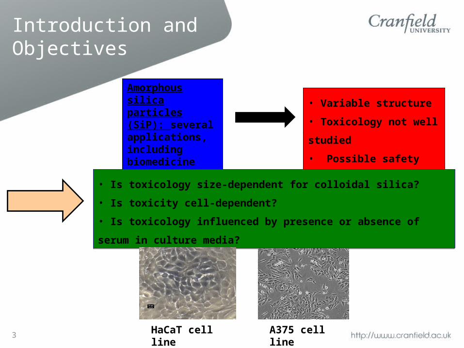
Introduction and Objectives
Amorphous silica particles (SiP): several applications, including biomedicine and biotechnology.
• Variable structure
• Toxicology not well studied
• Possible safety issues
• Is toxicology size-dependent for colloidal silica?
• Is toxicity cell-dependent?
• Is toxicology influenced by presence or absence of serum in culture media?
• Is toxicology size-dependent for colloidal silica?
• Is toxicity cell-dependent?
• Is toxicology influenced by presence or absence of serum in culture media?
HaCaT cell line A375 cell line3
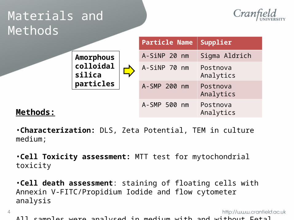
Materials and Methods
Particle Name Supplier
A-SiNP 20 nm Sigma Aldrich
A-SiNP 70 nm Postnova Analytics
A-SMP 200 nm Postnova Analytics
A-SMP 500 nm Postnova Analytics
4
Amorphous colloidal silica particles
Methods:
•Characterization: DLS, Zeta Potential, TEM in culture medium;
•Cell Toxicity assessment: MTT test for mytochondrial toxicity
•Cell death assessment: staining of floating cells with Annexin V-FITC/Propidium Iodide and flow cytometer analysis
All samples were analysed in medium with and without Fetal Bovine Serum (FBS) at concentrations between 10 and 200 µg/ml.
Characterization - DLS
5
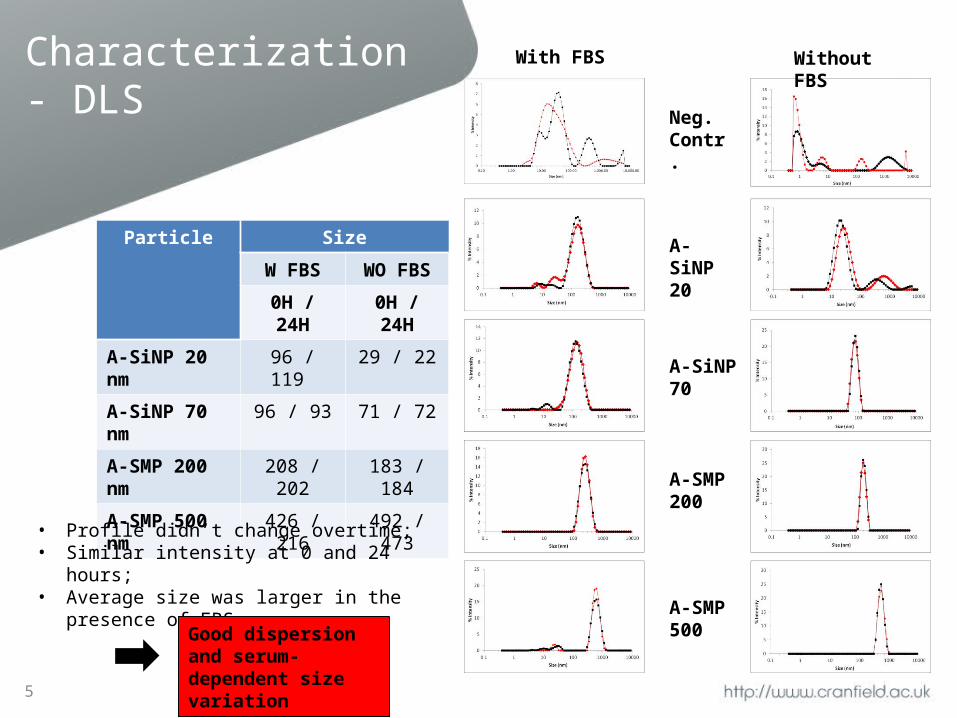
Particle Size
W FBS WO FBS
0H / 24H 0H / 24H
A-SiNP 20 nm 96 / 119 29 / 22
A-SiNP 70 nm 96 / 93 71 / 72
A-SMP 200 nm 208 / 202 183 / 184
A-SMP 500 nm 426 / 216 492 / 473
With FBS
Neg. Contr.
Without FBS
A-SiNP 20
A-SiNP 70
A-SMP 200
A-SMP 500
• Profile didn’t change overtime;• Similar intensity at 0 and 24 hours;• Average size was larger in the presence of FBS.
Good dispersion and serum-dependent size variation
Characterization – Zeta Potential and TEM
6
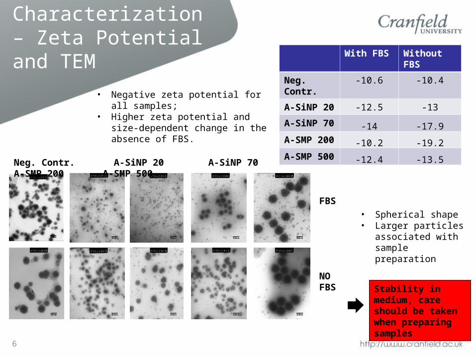
Neg. Contr. A-SiNP 20 A-SiNP 70 A-SMP 200 A-SMP 500
FBS
NO FBS
With FBS Without FBS
Neg. Contr. -10.6 -10.4
A-SiNP 20 -12.5 -13
A-SiNP 70 -14 -17.9
A-SMP 200 -10.2 -19.2
A-SMP 500 -12.4 -13.5
• Negative zeta potential for all samples;• Higher zeta potential and size-dependent
change in the absence of FBS.
• Spherical shape• Larger particles
associated with sample preparation
Stability in medium, care should be taken when preparing samples
Cell Toxicity assessment
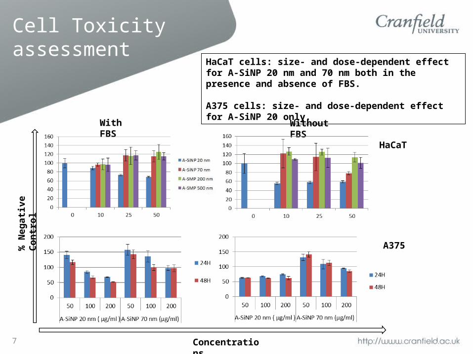
With FBS Without FBS
HaCaT
A375% N
egat
ive
Con
trol
Concentrations
HaCaT cells: size- and dose-dependent effect for A-SiNP 20 nm and 70 nm both in the presence and absence of FBS.
A375 cells: size- and dose-dependent effect for A-SiNP 20 only.
7
Apoptosis assay on HaCaT cells
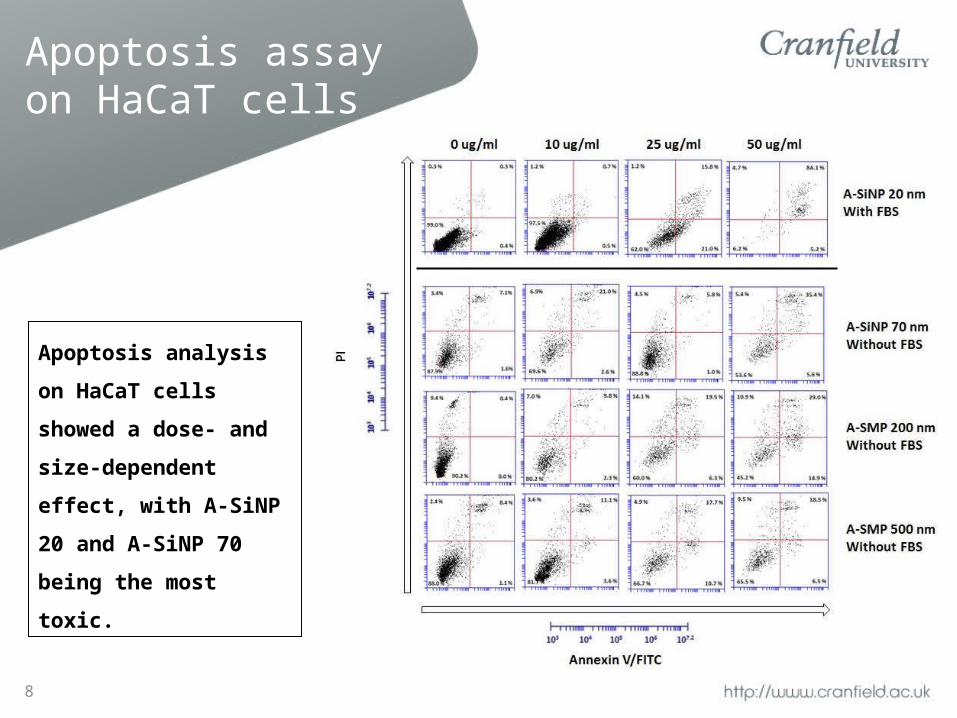
Apoptosis analysis on
HaCaT cells showed a dose-
and size-dependent effect,
with A-SiNP 20 and A-SiNP
70 being the most toxic.
8
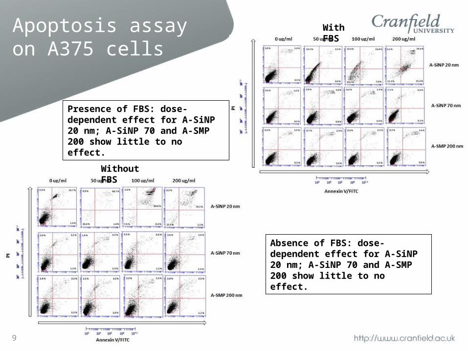
Apoptosis assay on A375 cells
Presence of FBS: dose-dependent effect for A-SiNP 20 nm; A-SiNP 70 and A-SMP 200 show little to no effect.
Absence of FBS: dose-dependent effect for A-SiNP 20 nm; A-SiNP 70 and A-SMP 200 show little to no effect.
9
Without FBS
With FBS
Discussion
• Characterization: Spherical shape, good dispersion and serum-dependent size variation.
• Cell Viability: A375 was sensitive only to A-SiNP 20, HaCaT to A-SiNP 20 and A-SiNP 70.
• Apoptotic cell death: detected for A-SiNP 20 nm both on HaCaT cells and A375 cells; other particles showed effect only on HaCaT cells in the absence of serum.
10
Conclusions
11
• Toxicity for colloidal silica is size-dependent in HaCaT cells, with smaller particles (A-SiNP 20 and 70 nm) being more toxic than its larger counterparts; on A375 cells, on the other hand, only A-SiNP 20 nm showed toxic effects;
• Toxicity is cell type dependent, as the same particles showed different behaviour on HaCaT and A375 cells. Cell death is mainly due to apoptosis;
• Nanoparticles characteristics and toxicity are influenced by serum, as particles were more toxic when it’s absent from the suspension medium.
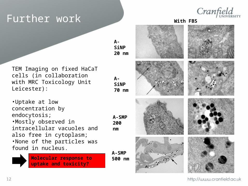
Further work
12
A-SiNP 20 nm
A-SiNP 70 nm
A-SMP 200 nm
A-SMP 500 nm
TEM Imaging on fixed HaCaT cells (in collaboration with MRC Toxicology Unit Leicester):
•Uptake at low concentration by endocytosis;•Mostly observed in intracellular vacuoles and also free in cytoplasm;•None of the particles was found in nucleus.
Molecular response to uptake and toxicity?
With FBS