
Carl B. Goodman, Ph.D.
Professor Pharmacology
College of Pharmacy & Pharmaceutical Sciences
Florida A&M University
308E FSH-SRC
599-3128


1. Most drugs are chemically synthesized (or at least modified, e.g. the penicillins) – the larger the molecules, the more difficult the synthesis, and the lower the yield will be.
2. Drugs need to reach their targets in the body, which means they need to be able to cross membrane barriers by diffusion. Diffusion becomes increasingly difficult with size.
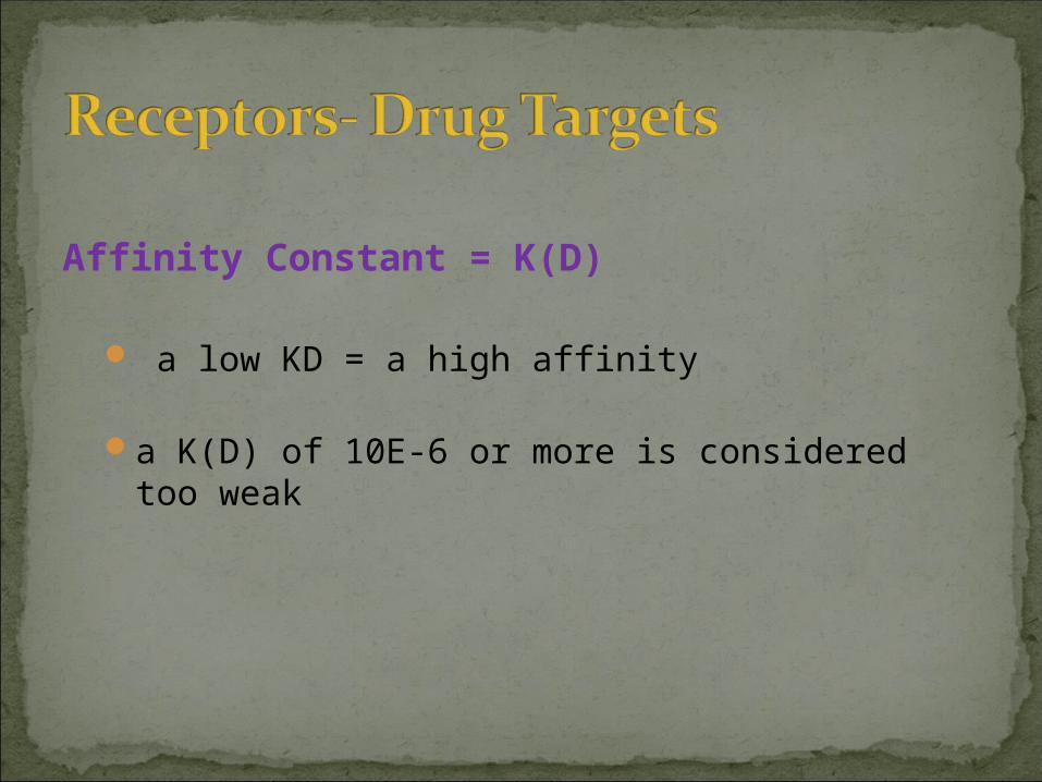
Affinity Constant = K(D)
a low KD = a high affinity
a K(D) of 10E-6 or more is considered too weak

Cell Surface Receptors
Receptors vs. Binding Sites7 transmembrane loopG protein-coupledSignaling Transduction PathwayEndogenous Ligand
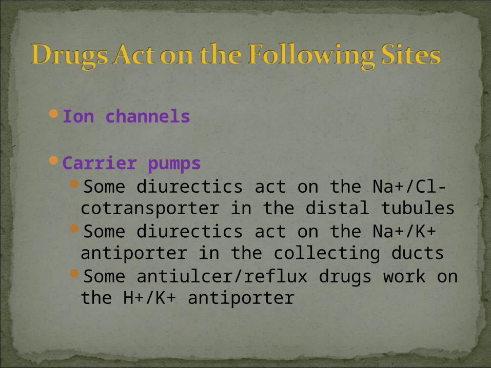
Ion channels
Carrier pumps Some diurectics act on the Na+/Cl-
cotransporter in the distal tubules Some diurectics act on the Na+/K+
antiporter in the collecting ducts Some antiulcer/reflux drugs work on
the H+/K+ antiporter

Enzymes NSAIDs inhibit cyclo-oxygenase which
blocks prostaglandin production
ACE inhibitors inhibit angiotensin converting enzyme to decrease blood pressure
HMGCoA reductase inhibitors inhibit this to reduce lipid concentration

Nuclear Receptors/RNA/DNA
Steroid Hormones- Estrogen and Androgens
Intracellular Structural Proteins

Inhibition Constant = K(I)
Measures degree of inhibition of an enzyme

Treatment of a disease can be by different targets:
Ex. to treat Gastric Hyperacidity: Neutralizing drug (NaHCO3) Proton pump inhibitor (Omeprazole) Histamine 2 receptor blocker
(Cimetidine)
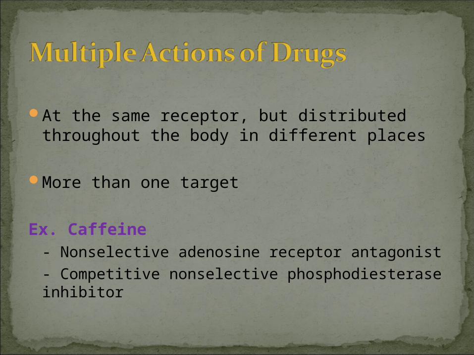
At the same receptor, but distributed throughout the body in different places
More than one target
Ex. Caffeine - Nonselective adenosine receptor antagonist
- Competitive nonselective phosphodiesterase inhibitor

Propranolol
Molecular: is a competitive inhibitor by binding to beta 1 receptors
Cellular: prevents increase in cAMP, protein phosphorylation
Physiological: reduces cardiac heart rate and contractile force
Therapeutic: used to treat angina
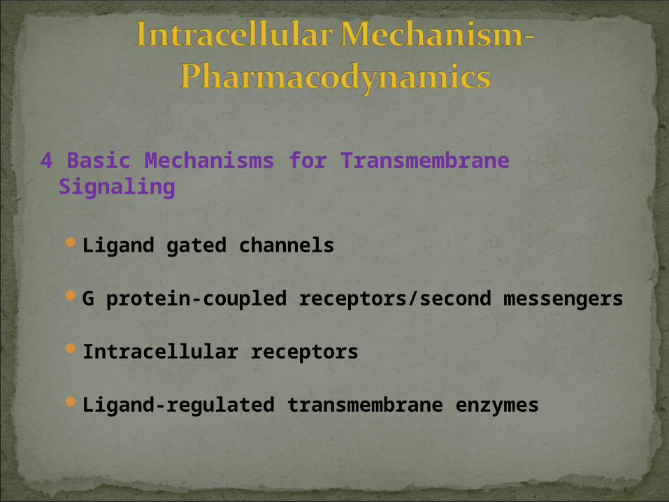
4 Basic Mechanisms for Transmembrane Signaling
Ligand gated channels
G protein-coupled receptors/second messengers
Intracellular receptors
Ligand-regulated transmembrane enzymes
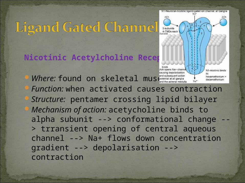
Nicotinic Acetylcholine Receptor
Where: found on skeletal muscle Function: when activated causes contraction Structure: pentamer crossing lipid bilayer Mechanism of action: acetycholine binds to
alpha subunit --> conformational change --> trransient opening of central aqueous channel --> Na+ flows down concentration gradient --> depolarisation --> contraction

Signaling system involves:
Extracellular drug binds to cell surface receptor
Receptor triggers activation of a G protein on cytoplasmic face of plasma membrane
Activated G protein alters activity of an effector element (e.g. enzyme or ion channel)
Effector element changes concentration of intracellular second messenger
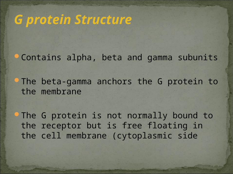
Contains alpha, beta and gamma subunits
The beta-gamma anchors the G protein to the membrane
The G protein is not normally bound to the receptor but is free floating in the cell membrane (cytoplasmic side
G protein Structure

1. When the receptor is not occupied by a drug: the G protein is in a resting state, where beta-gamma anchors G protein to membrane, and GDP occupies site on alpha subunit
2. The ligand binds to the receptor, which alters the conformation of the receptor, exposing the binding site for G protein
3. The G protein binds to the receptor, greatly weakening the affinity of the G protein for GDP
4. GDP dissociates, allowing GTP to bind to the alpha subunit
5. This causes the alpha subunit to change conformation, it now dissociates from the receptor and the beta and gamma subunits

6. The free alpha subunit now changes conformation so that it can bind with its target enzyme
7. This target enzyme acts, e.g. converts ATP-->cAMP, ion channel etc.
8. Hydrolysis of the GTP by the alpha subunit returns the subunit to its original conformation, causing it to dissociate from the target enzyme and reassociate with the beta and gamma subunits


Gs is a G protein that acts on adenyl cyclase, hence converts ATP-->cAMP
Gi is a G protein that inhibits adenyl cyclase, hence prevents ATP-->cAMP

1. cAMP many drugs/hormones act by increasing or
decreasing the adenyl cyclase which in turn increases or decreases cAMP
2. cGMP
3. Calcium-phosphoinositide pathway (IP3/DAG)

Mechanisms of this pathway:
Gs stimulates phospholipase C (a membrane enzyme)
Hydrolysis of IP2 into DAG (diacyclglycerol) and IP3 (inositol-1,4,5-triphosphate) DAG activates protein kinase C --> phosphorylates
intracellular proteins --> altered cellular function (e.g. contraction of smooth muscle)
IP3 triggers release of Ca++ from storage vesicles (in smooth muscle this will increase contraction)
These signals are terminated by the following: dephosphorylation of IP3 phosphorylating DAG to arachidonic acid actively removing the Ca++

Ligands for intracellular receptors include: steroids, vitamin D, thyroid hormone –regulate gene expression
Mechanism: ligand is lipid soluble, hence can cross plasma membrane binds with intracellular receptor intracellular receptors increases gene transcription by directly
or indirectly acting on promoter region
Drugs that act on intracellular receptors are slow acting because gene transcription takes time, but endure hours after the drug has been eliminated from the body

Protein Tyrosine Kinase
Mechanism: Ligand (includes insulin, growth factors) binds to
receptor
Enzymes (tyrosine kinases) located in the cytoplasmic domains (receptors have large extracellular and intracellular domains) are activated and cause phosphorylation of certain targets
These targets are activated or inactivated by this


