Cardiac Biomarkers in
Acute Chest Pain
Dr. K. Shanthi Naidu, Dr Maddury Jyotsna
Prof & HOD & Dept of Biochemistry, Prof & HOU-IV , Dept of Cardiology,
CARE Hospitals, Nizam’s institute of medical sciences,
Banjara Hills, Hyderabad. Panjagutta, Hyderabad-500082.
Cardiac biomarkers are substances that are released into the
blood when the heart is damaged or stressed.

The Ideal Biomarker Then Now
Sensitive and specific Either highly sensitive (diagnosis) OR
highly specific (treatment effect)
Reflects disease severity Reflects abnormal
Physiology/biochemistry
Correlates with prognosis Prognosis is most meaningful if level is
clinically actionable
Should aid in clinical decision making Should be used as a basis for specific
“biomarker guided-therapy”
Level should decrease following
effective therapy
“Bio-monitoring” during treatment is
an effective surrogate of improvement

Acute myocardial damage TnT, TnI, CK-MB
Ischemia and reprefusion ECG
GDF-15 ?
Clinical
background
Renal
Dysfunction Crea Cl
Cystatin C
Inflammatory Activity CRP, IL-6, Cd-40 lig
Left ventricular
Dysfunction BNP, NT-proBNP
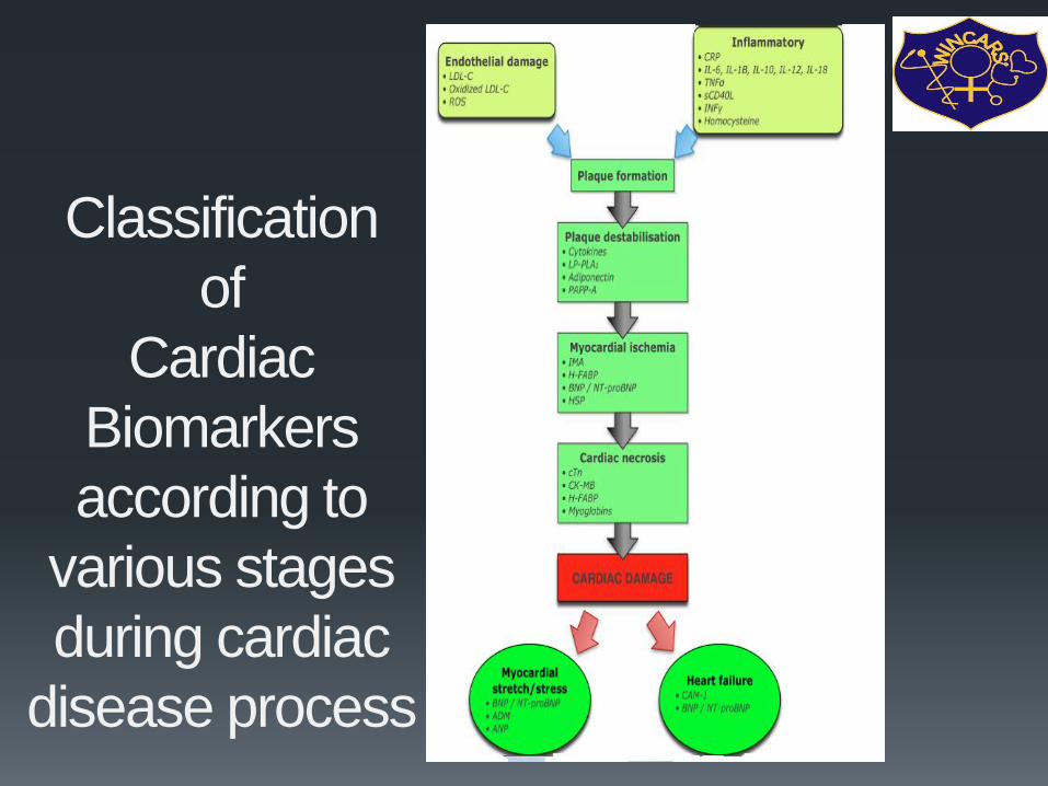
Classification
of
Cardiac
Biomarkers
according to
various stages
during cardiac
disease process
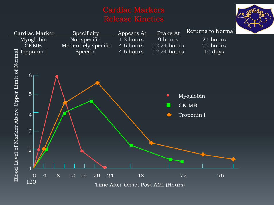
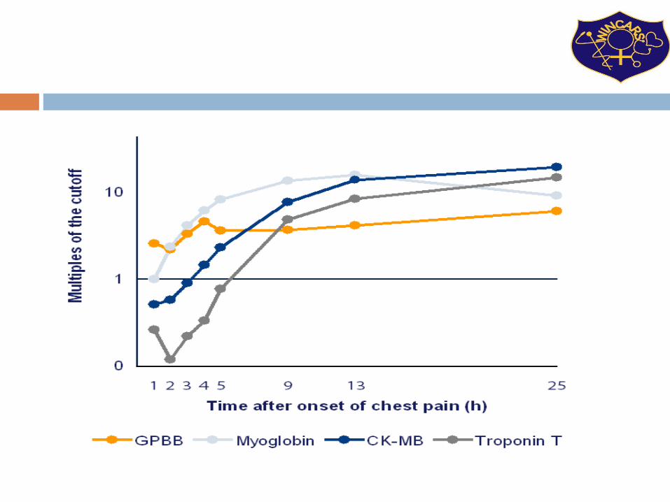
Cardiac Markers
Release Kinetics
2
1
6
5
4
3
0 4 8 12 16 20 24 48 72 96
120 Time After Onset Post AMI (Hours)
Blo
od L
evel of M
ark
er
Above U
pper
Lim
it o
f N
orm
al
Myoglobin
CK-MB
Troponin I
Cardiac Marker Specificity Appears At Peaks At Returns to Normal
Myoglobin Non - specific 1 - 3 hours 9 hours 24 hours CK - MB Moderately specific 4 - 6 hours 12 - 24 hours 72 hours
Troponin I Specific 4 - 6 hours 12 - 24 hours 10 days
Case capsule 1 54 yr old diabetic male, came to ED with chest pain
for 15 minutes with normal ECG.
Pt was hemodynamically stable.
What biomarkers should be done ?

Depends on time of arrival
Within one hour – Myoglobin
> 2hrs – CK MB/ Troponin

Myoglobin
Small-size heme protein found in all tissues mainly assists in oxygen transport
It is released from all damaged tissues
Its level rises more rapidly than cTn and CK-MB.
Released from damaged tissue within 1 hour
Normal value: 17.4-105.7 ng/ml Timing: Earliest Rise: 1-4 hrs Peak 6-9 hrs Return to normal: 12 hrs
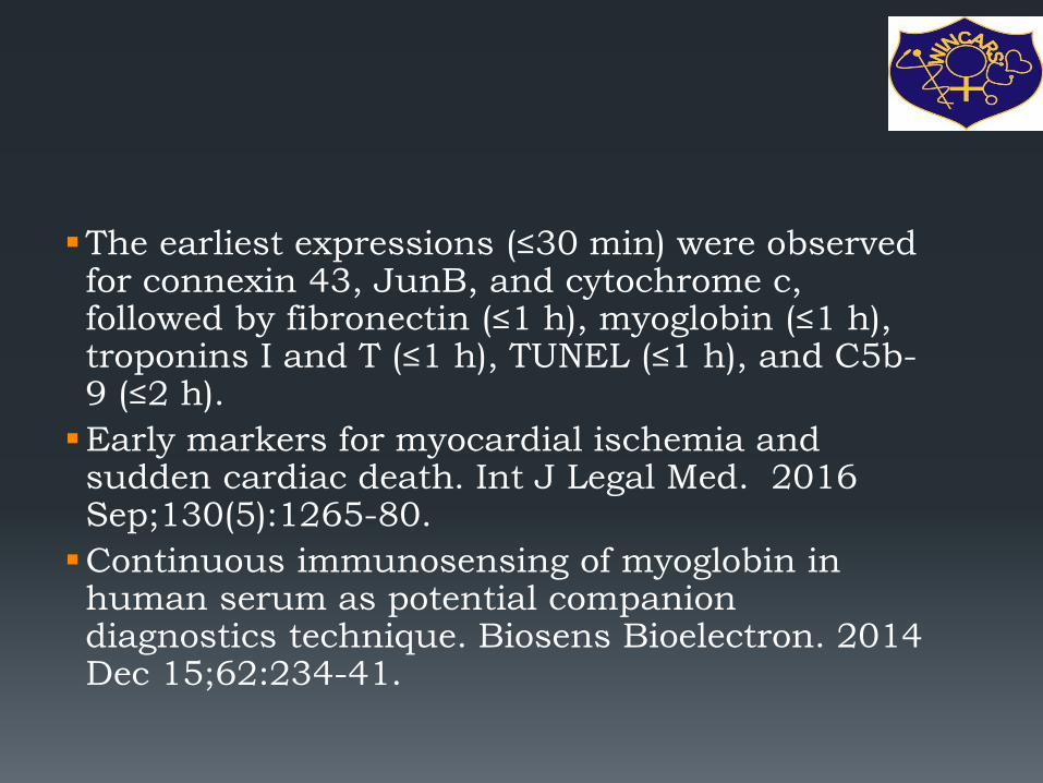
The earliest expressions (≤30 min) were observed for connexin 43, JunB, and cytochrome c, followed by fibronectin (≤1 h), myoglobin (≤1 h), troponins I and T (≤1 h), TUNEL (≤1 h), and C5b-9 (≤2 h).
Early markers for myocardial ischemia and sudden cardiac death. Int J Legal Med. 2016 Sep;130(5):1265-80.
Continuous immunosensing of myoglobin in human serum as potential companion diagnostics technique. Biosens Bioelectron. 2014 Dec 15;62:234-41.


AIM
Since limited numbers of studies have been
conducted, in this study the utility of H-FABP
as candidate markers for diagnosis of NSTE-
ACS.

EXCLUSION CRITERIA
Chest pain > 8 hours duration
non cardiac chest pain
recent injuries
renal failure
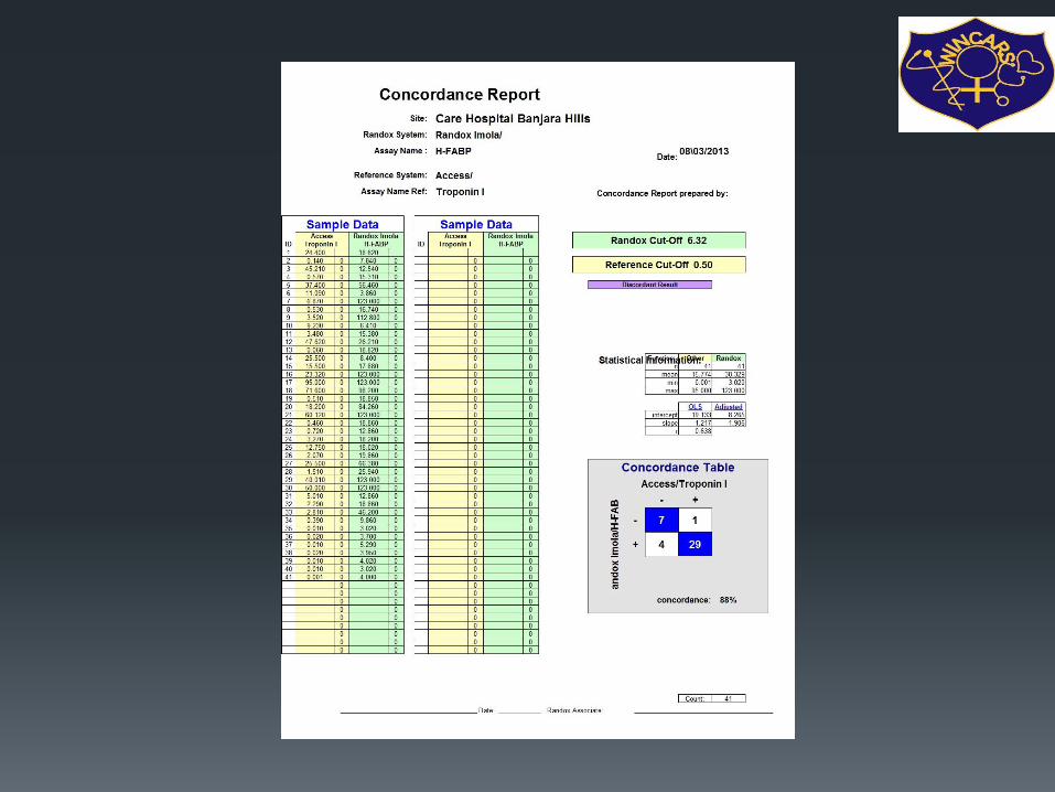
Laboratory Analysis Serum H-FABP : Quantitative immunoturbidimetric method
(Randox Laboratories, Ltd. Co., Antrim, United Kingdom)
cardiac Troponin T and Troponin I : Time resolved
immunofluorescence method (AQT90 FLEX, Radiometer,
Denmark).
CK-MB : Quantitative immunoturbidimetric method (Roche)
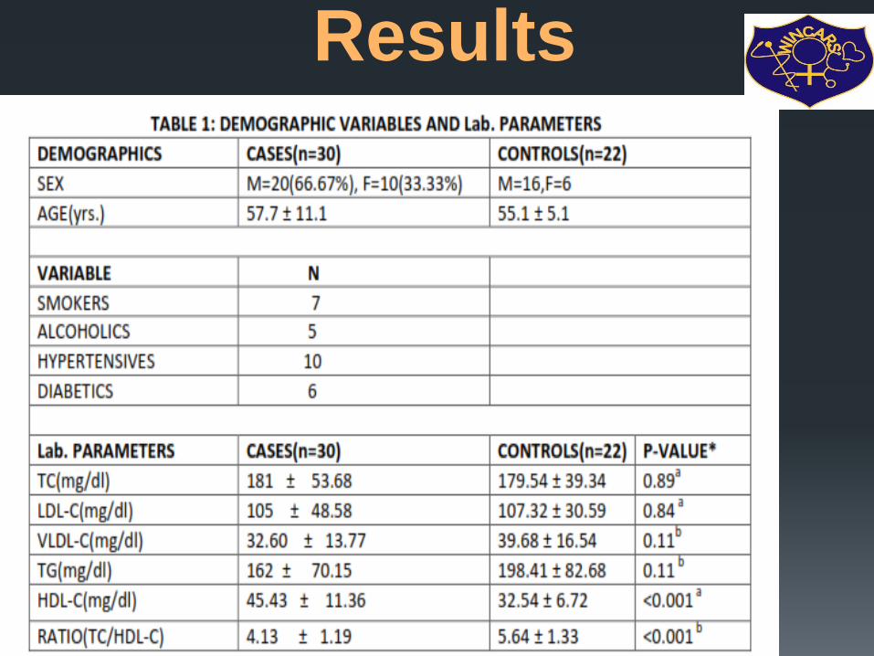
Results

CARDIAC
BIOMARKER
CASES(N=30) CONTROLS(N=2
2)
P VALUE
HFABP(ng/ml) 40.42±65.53 3.47±1.52 <0.001
cTnt(ng/L) 370±1007 8.59±0.5 <0.01
cTnI(μg/L) 0.18±0.35 0.008±0.0005 0.005
CARDIAC BIOMARKERS
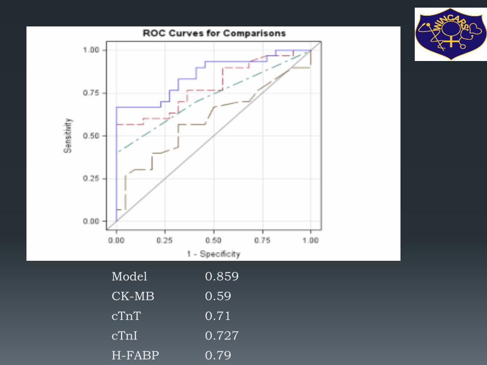
Model 0.859
CK-MB 0.59
cTnT 0.71
cTnI 0.727
H-FABP 0.79
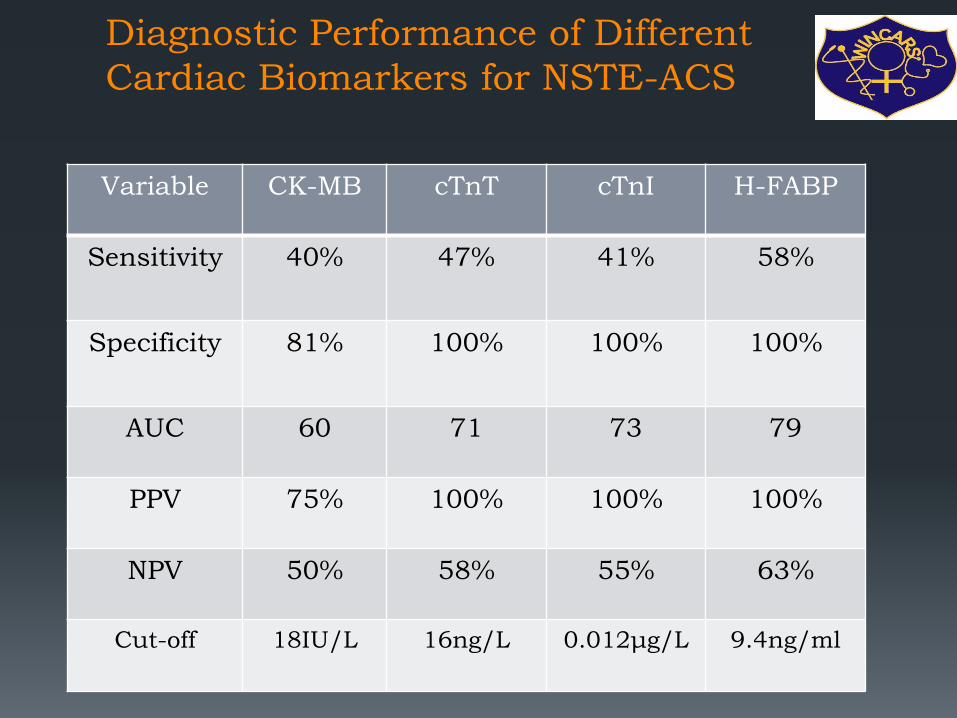
Diagnostic Performance of Different
Cardiac Biomarkers for NSTE-ACS
Variable CK-MB cTnT cTnI H-FABP
Sensitivity 40% 47% 41% 58%
Specificity 81% 100% 100% 100%
AUC 60 71 73 79
PPV 75% 100% 100% 100%
NPV 50% 58% 55% 63%
Cut-off 18IU/L 16ng/L 0.012μg/L 9.4ng/ml
DISCUSSION
H-FABP was elevated (>9.4 ng/mL) in 17 of the 30 patients (57%).
Among the cases, the mean level of H-FABP was 40 ng/mL,
where as in controls it was 3.47ng/ml and the difference was
statistically significant (p <0.001).
In a number of studies, H-FABP has been reported to be
particularly sensitive within the first few hours after the onset of
coronary occlusion and symptoms.
small molecular weight (15 kDa)
cytoplasmic unbound abundance
rapid release from damaged myocardial cells.
Kathrukha et al. proved in their study that H-FABP levels elevate
earlier than cTnI levels in patients with UA.
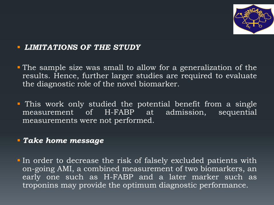
LIMITATIONS OF THE STUDY
The sample size was small to allow for a generalization of the results. Hence, further larger studies are required to evaluate the diagnostic role of the novel biomarker.
This work only studied the potential benefit from a single measurement of H-FABP at admission, sequential measurements were not performed.
Take home message
In order to decrease the risk of falsely excluded patients with on-going AMI, a combined measurement of two biomarkers, an early one such as H-FABP and a later marker such as troponins may provide the optimum diagnostic performance.
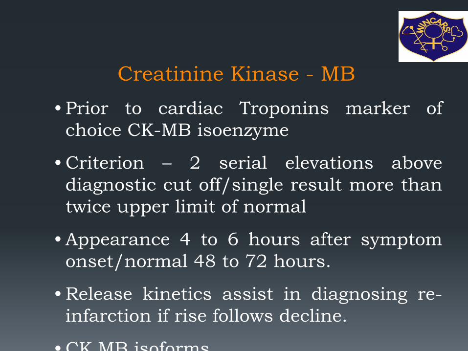
Creatinine Kinase - MB
• Prior to cardiac Troponins marker of
choice CK-MB isoenzyme
• Criterion – 2 serial elevations above
diagnostic cut off/single result more than
twice upper limit of normal
• Appearance 4 to 6 hours after symptom
onset/normal 48 to 72 hours.
• Release kinetics assist in diagnosing re-
infarction if rise follows decline.
• CK MB isoforms
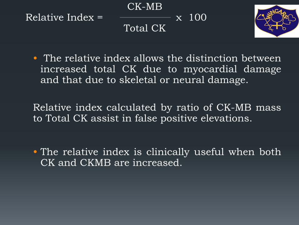
CK-MB Relative Index = χ 100 Total CK
• The relative index allows the distinction between increased total CK due to myocardial damage and that due to skeletal or neural damage.
Relative index calculated by ratio of CK-MB mass to Total CK assist in false positive elevations.
• The relative index is clinically useful when both CK and CKMB are increased.

CK-MB/CK relative index
A relative index exceeding 3 is indicative of AMI
Ratios between 3 and 5 represent a gray zone.
No definitive diagnosis can be established without serial determinations to detect a rise.
Note that the diagnosis of acute MI must not be based on an elevated relative index alone, because the relative index may be elevated in clinical settings when either the total CK or the CK-MB is within normal limits.
The relative index is only clinically useful when both the total CK and the CK-MB levels are increased.
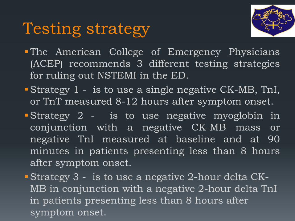
Testing strategy
The American College of Emergency Physicians
(ACEP) recommends 3 different testing strategies
for ruling out NSTEMI in the ED.
Strategy 1 - is to use a single negative CK-MB, TnI,
or TnT measured 8-12 hours after symptom onset.
Strategy 2 - is to use negative myoglobin in
conjunction with a negative CK-MB mass or
negative TnI measured at baseline and at 90
minutes in patients presenting less than 8 hours
after symptom onset.
Strategy 3 - is to use a negative 2-hour delta CK-
MB in conjunction with a negative 2-hour delta TnI
in patients presenting less than 8 hours after
symptom onset.

Testing strategy
ACEP’s recommendations on the use of delta CK-MB and delta TnI are based on determining the change in the level of TnI or CK-MB on samples drawn 2 hours apart.
However, the delta TnI evaluation is partially based on the use of older TnI assays and outdated WHO acute MI cutoffs in a retrospective study.
Therefore, ACEP’s recommendation to use a delta TnI in conjunction with a delta CK-MB may not be generalizable to other commercially available Troponin assays.
The ACC/AHA guidelines for the treatment of patients with unstable angina and NSTEMI recommend a baseline sample upon ED arrival and a repeat sample 6-9 hours after presentation.

Case 1 – follow up
Immediate Trop I (POC) testing after arrival
to ED was negative.
But CK MB which was sent to lab came as
elevated.
How can we interpret this discordant
Troponin and CK-MB results?

WHAT IS discordant Troponin
and CK-MB results
In the CRUSADE registry, a review of almost 30,000 patients
revealed that discordant Troponin and CK-MB results occurred in
28% of patients. However, patients who were Troponin negative but
CK-MB positive had in-hospital mortality rates that were not
significantly increased from patients who were negative for both
biomarkers.[30]
Similarly, in a report of more than 10,000 patients with ACS from
the multicenter GRACE registry, in-hospital mortality was highest
when both Troponin and CK-MB were positive, intermediate in
troponin-positive/CK-MB-negative patients, and lowest in patients
in whom both markers were negative and in those who were
troponin-negative/CK-MB-positive.[31] Thus, an isolated CK-MB
elevation has limited prognostic value in patients with a non-ST
elevation ACS.

Troponins
•Generally undetectable in healthy
patients ??
•? Sensitive assays available
•Absolute abnormal value – varies
depending on the clinical setting
•Above 99th percentile of healthy
population as cut off using an assay in
the acceptable precision

Features of serum markers of acute myocardial infarction
Sensitivity at:
Marker Time to
appearance
Duration
of
elevation
6hr 12hr Specificity Comments
MB2 Isoform 2 – 6 hr 1 – 2 d 95% 98-100% 95% Not widely
available
Myoglobin 1 – 2 hr < 1 d 85% 90% 80% Slightly improved
sensitivity early
in AMI when
added to
troponin/CK-MB
but not widely
used due to low
specificity
AMI = acute myocardial infarction; CK=creatine kinase
Courtesy: Cecil Textbook of Medicine, 22nd ed., Chapter 69 St.Elevation Acute
Myocardial Infarction and Complications of Myocardial Infarction

The recent developments in interpretation of
Troponin
• Cardiac Troponin – shows our understanding is still evolving
• Improved sensitivity – Compared to prior markers – Utilization
by Clinicians debatable ..Laboratory understanding of these
sensitive values is deter mental to help the physician
Heterogeneity of cut off values – MIXED MESSAGE – in its
usage, pushes in more individuals as AMI.. Not easy on the
clinician, Hence the situation must be circumstance where
the clinical signs, symptoms lead to a strong suspicion of AMI

Study from CARE hospital
Materials and Methods
100 patients were analysed for cardiac markers
79 were studied for CK-MB and Troponin T serially up
to 48 hours
23 patients were compared with Troponin T and
Troponin I
20 patients were compared with Troponin and NT
proBNP
Less than 15 were studied for myoglobin
HsCRP was found not to be effective in analysis

Serial Measurement of Cardiac
Markers after Chest pain
0
1
2
3
4
5
6
7
8
9
BASE 8hrs 16hrs 24hrs 0
500
1000
1500
2000
2500
TROP-T
CK-MB
N=79
0
1
2
3
4
5
6
7
8
9
BASE 8hrs 16hrs 24hrs 0
500
1000
1500
2000
2500
TROP-T
CK-MB
Trop T CK-MB
ng/ml U/L
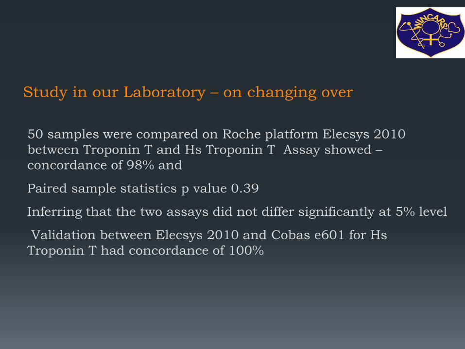
Study in our Laboratory – on changing over
50 samples were compared on Roche platform Elecsys 2010
between Troponin T and Hs Troponin T Assay showed –
concordance of 98% and
Paired sample statistics p value 0.39
Inferring that the two assays did not differ significantly at 5% level
Validation between Elecsys 2010 and Cobas e601 for Hs
Troponin T had concordance of 100%

Features of serum markers of acute
myocardial infarction
Sensitivity at:
Marker Time to
appearance
Duration of
elevation
6hr 12hr Specificity Comments
Troponin I 2 – 6 hr 5 – 10 d 75% 90-
100%
98% Generally regarded
as a test of choice
Troponin T 2 – 6 hr 5 – 14 d 80% 95-
100%
95% A test of choice.
Less specific than
Troponin I (elevated
in renal
insufficiency)
CK-MB 3 – 6 hr 2 – 4 d 65% 95% 95% Test of choice for
recurrent angina
once Troponin
elevated
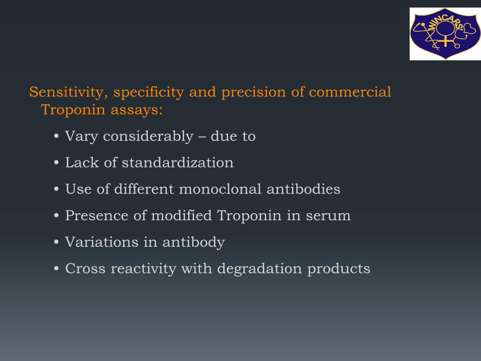
Sensitivity, specificity and precision of commercial
Troponin assays:
• Vary considerably – due to
• Lack of standardization
• Use of different monoclonal antibodies
• Presence of modified Troponin in serum
• Variations in antibody
• Cross reactivity with degradation products

Study in our Laboratory – on changing over
75 samples were validated Roche platform Elecsys 2010 and
Beckman Coulter Access for Hs Troponin T and Hs Troponin I
Assay showed – concordance of 89% and
Paired sample statistics p value 0.008
Inferring that the two variables of instrumentation and
methodology differ significantly at 5 % level.
Validation between Elecsys 2010 and Cobas e601 for Hs
Troponin T had concordance of 100%
Cobose601 and Access 2 for 20 samples showed concordance
90%.
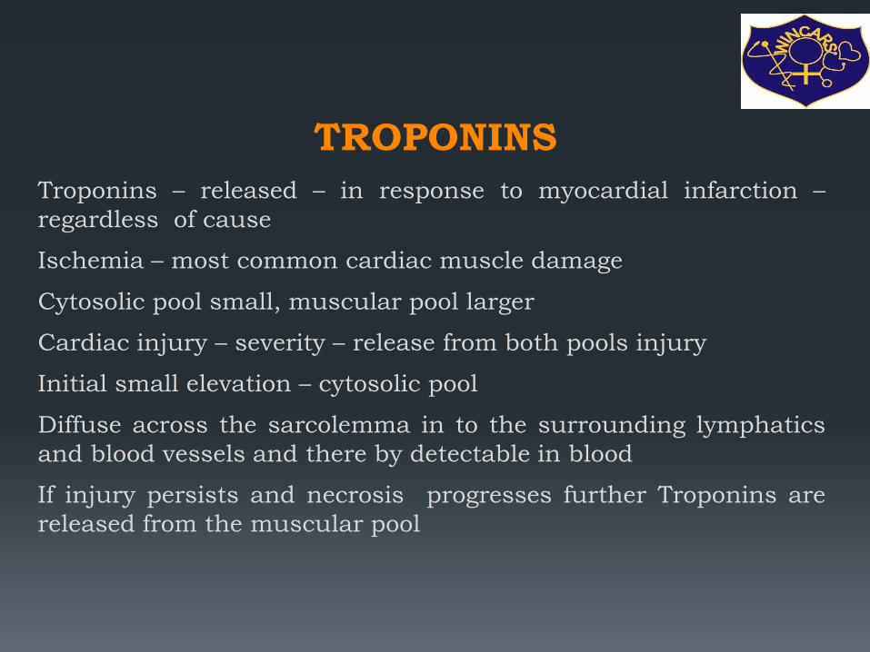
TROPONINS
Troponins – released – in response to myocardial infarction –
regardless of cause
Ischemia – most common cardiac muscle damage
Cytosolic pool small, muscular pool larger
Cardiac injury – severity – release from both pools injury
Initial small elevation – cytosolic pool
Diffuse across the sarcolemma in to the surrounding lymphatics
and blood vessels and there by detectable in blood
If injury persists and necrosis progresses further Troponins are
released from the muscular pool

TROPONINS
Levels of Troponin complex T & I
Not present in serum unless-cardiac necrosis Cardiac specific
Levels remain elevated from 3-14 days after MI, sensitivity high
when other markers have
returned to normal
Adv: Delay in seeking medical advice
Elevated levels are predictive of poor outcome in patients with
acute coronary syndrome
Disadvantage - Detection of Reinfarction
- Less sensitive in early stages of infarction

TROPONINS
Normal
Obtained Diagnostic Measurement
0
1
2
3
4
5
Trop T Trop I
0.69
3.9
0.3
? Ideal Marker
0
0.005
0.01
0.015
0.02
0.025
0.03
0.035
Trop T Trop I 0.00 0.50 1.00 1.50 2.00 2.50 3.00 3.50 4.00 4.50
Normal Measuredb N=23
Normal
Obtained Diagnostic Measurement
Troponin T Troponin I
Normal < 0.01 ng/ml 0.03 ng/ml
Cut off for AMI 0.1 ng/ml 0.5 ng/ml
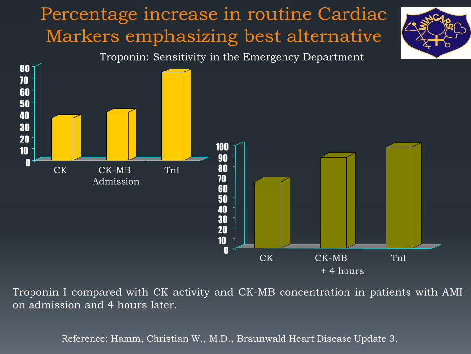
Percentage increase in routine Cardiac
Markers emphasizing best alternative
Reference: Hamm, Christian W., M.D., Braunwald Heart Disease Update 3.
0
10
20
30
40
50
60
70
80
CK CK-MB TnI
Admission
0 10
20 30 40 50 60 70 80 90
100
CK CK-MB TnI
+ 4 hours
Troponin I compared with CK activity and CK-MB concentration in patients with AMI
on admission and 4 hours later.
Troponin: Sensitivity in the Emergency Department

Evolution of the cardiac Troponin (cTn) assays and their diagnostic cut-offs.
Mahajan V S , Jarolim P Circulation 2011;124:2350-2354
Copyright © American Heart Association
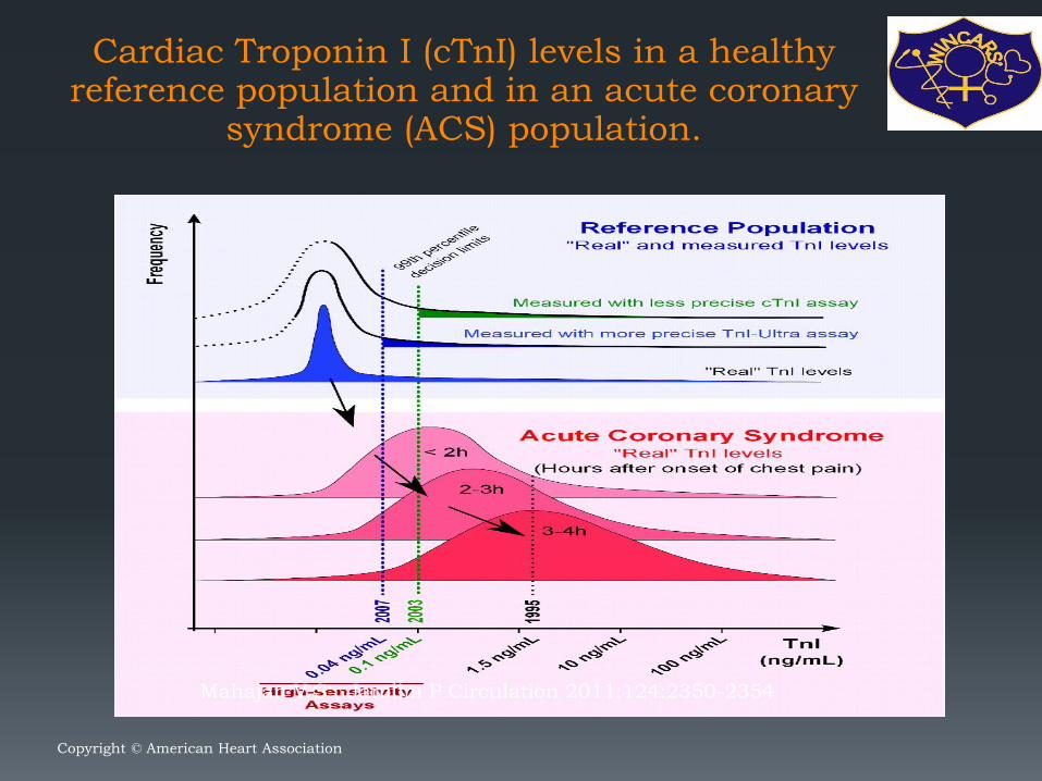
Cardiac Troponin I (cTnI) levels in a healthy reference population and in an acute coronary
syndrome (ACS) population.
Mahajan V S , Jarolim P Circulation 2011;124:2350-2354
Copyright © American Heart Association
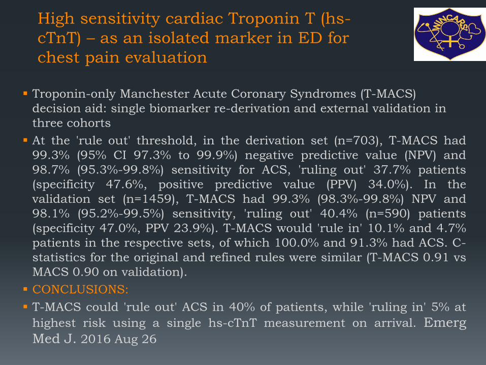
High sensitivity cardiac Troponin T (hs-
cTnT) – as an isolated marker in ED for
chest pain evaluation
Troponin-only Manchester Acute Coronary Syndromes (T-MACS)
decision aid: single biomarker re-derivation and external validation in
three cohorts
At the 'rule out' threshold, in the derivation set (n=703), T-MACS had
99.3% (95% CI 97.3% to 99.9%) negative predictive value (NPV) and
98.7% (95.3%-99.8%) sensitivity for ACS, 'ruling out' 37.7% patients
(specificity 47.6%, positive predictive value (PPV) 34.0%). In the
validation set (n=1459), T-MACS had 99.3% (98.3%-99.8%) NPV and
98.1% (95.2%-99.5%) sensitivity, 'ruling out' 40.4% (n=590) patients
(specificity 47.0%, PPV 23.9%). T-MACS would 'rule in' 10.1% and 4.7%
patients in the respective sets, of which 100.0% and 91.3% had ACS. C-
statistics for the original and refined rules were similar (T-MACS 0.91 vs
MACS 0.90 on validation).
CONCLUSIONS:
T-MACS could 'rule out' ACS in 40% of patients, while 'ruling in' 5% at
highest risk using a single hs-cTnT measurement on arrival. Emerg
Med J. 2016 Aug 26

Case capsule 2 63 years old female hypertensive patients with anterior STMI of 3 hours duration .
No contraindications for thrombolysis.
Hemodynamically stable.
What biomarker we should use?
Is there any difference in gender for biomarkers elevation timing and severity of elevation?

Are cardiac biomarkers are required for
STMI?
Note that cardiac markers are not
necessary for the diagnosis of patients
who present with ischemic chest pain
and diagnostic ECGs with ST-segment
elevation.

Clinical Effect of Sex-Specific Cutoff Values of High-Sensitivity Cardiac Troponin T in Suspected Myocardial Infarction. JAMA Cardiol 2016;Sep 21:[Epub ahead of print].
once using the uniform 99th percentile cutoff value level of 14 ng/L and once using sex-specific 99th percentile levels of hs-cTnT (women, 9 ng/L; men, 15.5 ng/L).
The diagnosis in two women was upgraded from unstable angina to AMI, and the diagnosis in one man was downgraded from AMI to unstable angina. These diagnostic results were confirmed when using two alternative pairs of uniform and sex-specific cutoff values.
Conclusions: The authors concluded that uniform 99th percentile should remain the standard of care when using hs-cTnT levels for the diagnosis of AMI.

Exceptions
Simple markers can distinguish Takotsubo cardiomyopathy
from ST segment elevation myocardial infarction. Int J Cardiol. 2016
Sep 15;219:417-20.
The concentration of NTproBNP was greater in pts with TTC than
STEMI (4702pg/ml vs 2138pg/ml). The concentration of TnI and CKMB
mass was greater in the STEMI group than in the TTC group (TnI:
2.1ng/ml and CK MB mass: 9.5ng/ml in pts with TTC vs TnI: 19ng/ml
and CK MB mass: 73.3ng/ml in pts with STEMI). The NTproBNP/TnI
ratio and NTproBNP/CKMB mass ratio were, respectively, 2235.2 and
678.2 in pts with TTC and 81.6 and 27.5 in pts with STEMI (p<0.001).
Moreover, the NTproBNP/EF ratio was also statistically significant
(110.4 in TTC group and 39.4 in STEMI group).
CONCLUSIONS:
NTproBNP/TnI, NTproBNP/CKMB mass and NTproBNP/EF ratios can
distinguish TTC from STEMI at an early stadium. The most accurate
marker is the NTproBNP/TnI ratio.

Toll Like Receptor 4 in Acute
Myocardial Infarction
Background:
Previous studies on Toll-like receptor 4 (TLR 4), which are identified as central innate immune receptors, were done for local (at plaque rupture site) and systemic expression of TLR 4 from mononuclear concentrate (MNC) in acute myocardial infarction (AMI) pts. TLR inhibition is considered as a emerging new therapeutic modality for LV remodeling. We want to study difference of expression of TLR 4 on MNC and plasma in AMI pts. Even though, for TLR 4 protein detection in plasma requires higher concentration on the lymphocytes and to be secreted into plasma , but still, if plasma detected TLR 4 has same prognostification importance as from TLR 4 of MNC then, TLR 4 detection from plasma may be used at bed side.

Methods
We recruited acute MI pts who presented with in 48 hrs of onset of
chest pain.
TLR4 estimation was done with TLR4 ELISA kit from Cusabio at the
time of admission.
Group 1 are AMI pts with TLR 4 measured from MNC (Ficoll paque
method) and Group 2 are AMI pts with TLR 4 measured from
plasma.
In two groups of Controls (Control A are volunteers without known
CAD and coronary risk factors and Control B are pts with obvious
sepsis), TLR4 was estimated in both plasma and from MNC.
According to that kit standards TLR 4 concentration < or = 0.03
ng/dl is considered as negative.
Killips class, adverse events in hospitals (including recurrent
angina, LVF, ventricular arrhythmias and death) and CPK levels
were correlated with TLR 4 levels.

Parameter AMI Controls
Group 1 (MNC) Group 2(Plasma) A(no CAD+RF) B (Infection)
Number 26 14 15 5
TLR 4 pos. 14 (53.8%) 2(14.3%) 0 5(100%)
Parameter Variable
Age (yrs) 54.1 ± 11.6
M:F 12:8
HTN 21 (52.5%)
DM 10 (25%)
Location of MI
Inferior MI 11 (27.5%)
Anterior MI 26 (65%)
Extensive MI 3 (7.5%)
Average Killips
class
1.7 ± 0.9
CPK levels 1652.2 ± 1294.6 units/l
Mean TLR 4 0.7 ± 0.4 ng/dl
Table 1: AMI
Table 2:
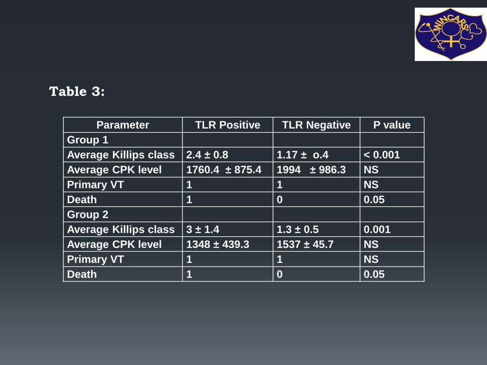
Parameter TLR Positive TLR Negative P value
Group 1
Average Killips class 2.4 ± 0.8 1.17 ± o.4 < 0.001
Average CPK level 1760.4 ± 875.4 1994 ± 986.3 NS
Primary VT 1 1 NS
Death 1 0 0.05
Group 2
Average Killips class 3 ± 1.4 1.3 ± 0.5 0.001
Average CPK level 1348 ± 439.3 1537 ± 45.7 NS
Primary VT 1 1 NS
Death 1 0 0.05
Table 3:

Conclusion
In Group 1, & group 2 also there is no correlation to TLR 4 concentration and occurrence of primary VT and CPK levels but strong association with Killips class and death.
Plasma TLR 4 detection was given same correlation with Killips class and dreaded prognostication of the patient ( that is mortality) in AMI like the TLR 4 detected from MNC. Therefore, kit to design using plasma of the AMI pt may be useful after larger AMI patients study.
Case capsule 3
44 year old gentleman admitted with road traffic accident
developed chest pain. What would be the preferred marker
for diagnosing infarction
Road traffic accident with rhadomyolysis
Trop
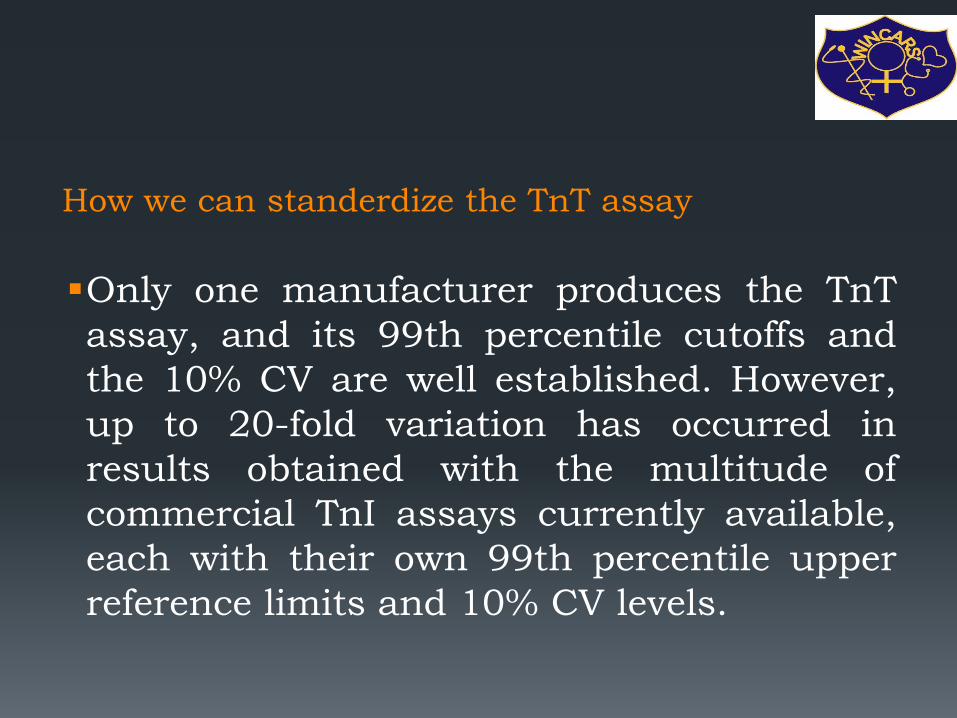
How we can standerdize the TnT assay
Only one manufacturer produces the TnT
assay, and its 99th percentile cutoffs and
the 10% CV are well established. However,
up to 20-fold variation has occurred in
results obtained with the multitude of
commercial TnI assays currently available,
each with their own 99th percentile upper
reference limits and 10% CV levels.
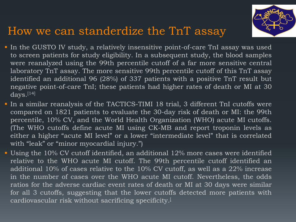
How we can standerdize the TnT assay
In the GUSTO IV study, a relatively insensitive point-of-care TnI assay was used
to screen patients for study eligibility. In a subsequent study, the blood samples
were reanalyzed using the 99th percentile cutoff of a far more sensitive central
laboratory TnT assay. The more sensitive 99th percentile cutoff of this TnT assay
identified an additional 96 (28%) of 337 patients with a positive TnT result but
negative point-of-care TnI; these patients had higher rates of death or MI at 30
days.[14]
In a similar reanalysis of the TACTICS-TIMI 18 trial, 3 different TnI cutoffs were
compared on 1821 patients to evaluate the 30-day risk of death or MI: the 99th
percentile, 10% CV, and the World Health Organization (WHO) acute MI cutoffs.
(The WHO cutoffs define acute MI using CK-MB and report troponin levels as
either a higher “acute MI level” or a lower “intermediate level” that is correlated
with “leak” or “minor myocardial injury.”)
Using the 10% CV cutoff identified, an additional 12% more cases were identified
relative to the WHO acute MI cutoff. The 99th percentile cutoff identified an
additional 10% of cases relative to the 10% CV cutoff, as well as a 22% increase
in the number of cases over the WHO acute MI cutoff. Nevertheless, the odds
ratios for the adverse cardiac event rates of death or MI at 30 days were similar
for all 3 cutoffs, suggesting that the lower cutoffs detected more patients with
cardiovascular risk without sacrificing specificity.[
How we can standerdize the TnT assay
The National Academy of Clinical Biochemistry (NACB) working with
the ACC/ESC guidelines has recommended adoption of the 99th
percentile upper reference limit as the recommended cutoff for a
positive troponin result. Ideally, the precision of the assay at this
cutoff level should be measured by a CV that is less than 10%.
However, most TnI assays are imprecise at the 99th percentile
reference limit.[17]Some have therefore recommended that the cutoff
level be raised to the slightly higher 10% CV level instead of the 99th
percentile reference limit to ensure adequate assay precision.
Is Point-of-care assays are available?
NACB recommendations specify that cardiac markers be
available on an immediate basis 24 h/d, 7 d/wk, with a
turnaround time of 1 hour.[18] Point-of-care (POC) devices that
provide rapid results should be considered in hospitals whose
laboratories cannot meet these guidelines.
POC assays for CK-MB, myoglobin, and the cardiac troponins
TnI and TnT are available. Only qualitative TnT assays are
available as POC tests, but both quantitative and qualitative
POC TnI assays are currently marketed.
Point-of-care assays
In a multicenter trial, the time to positivity was significantly
faster for the POC device than for the local laboratory (2.5 h
vs 3.4 h).[19]
In another multicenter study, which evaluated the i-STAT
POC TnI assay in comparison with the central laboratory in
2000 patients with suspected ACS, POC testing reduced the
length of stay by approximately 25 minutes for patients who
were discharged from the ED.[20, 21] The sensitivity of current
POC assays coupled with the benefit of rapid turnaround time
make the POC assays attractive clinical tools in the ED.

Point of care assays:
• NACB – Cardiac markers to be available 24 hrs/day, 7 d/week
• Ideal turn around time – 1 hour
• POC – when labs cannot meet this guidelines
• Qualitative TnT assay
• Qualitative and Quantitative for TnI assay
Ultrasensitive and low-volume point-of-care
diagnostics are available for trop T testing?
Sci Rep. 2016 Sep 16;6:33423. Ultrasensitive and
low-volume point-of-care diagnostics on flexible strips
- a study with cardiac Troponin biomarkers.
Shanmugam NR1, Muthukumar S2, Prasad S1.
A flexible, mechanically stable, and disposable
electrochemical sensor platform for
monitoring cardiac Troponin through the detection
and quantification of cardiac Troponin-T (cTnT). They
designed and fabricated nanostructured zinc oxide
(ZnO) sensing electrodes on flexible porous polyimide
substrates.
Additive biomarkers in AMI

Potential Biomarker Targets in ACS
Necrosis
Inflammation
Plaque Rupture
Thrombosis
Endothelial
Activation
Ischemia
Arrhythmias
Neurohormone
Activation
MMP’s, PAPP sCD40L,
PIGF
PAI-1, sCD40L, vWF,
D dimer
sICAM, pSelection
Hs-CRP, Ox LDL MCP-1,
MPO. IL18
cTnT, cTnl, Myo, CKMB, FABP
BNP, NE
IMA, uFFA
Midregional fragment of the N-terminal of pro-ANP (MR-proANP)
and 2 extracardiac biomarkers; the c-terminal provasopressin
(copeptin) and the midregional portion of proadrenomedullin
(MR-proADM).
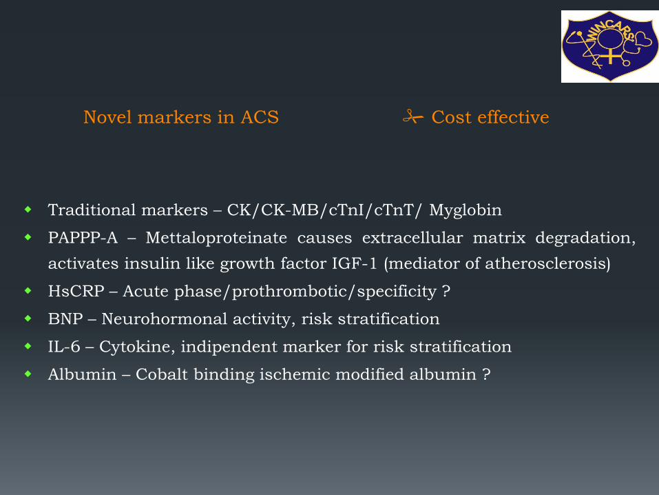
Novel markers in ACS Cost effective
Traditional markers – CK/CK-MB/cTnI/cTnT/ Myglobin
PAPPP-A – Mettaloproteinate causes extracellular matrix degradation,
activates insulin like growth factor IGF-1 (mediator of atherosclerosis)
HsCRP – Acute phase/prothrombotic/specificity ?
BNP – Neurohormonal activity, risk stratification
IL-6 – Cytokine, indipendent marker for risk stratification
Albumin – Cobalt binding ischemic modified albumin ?
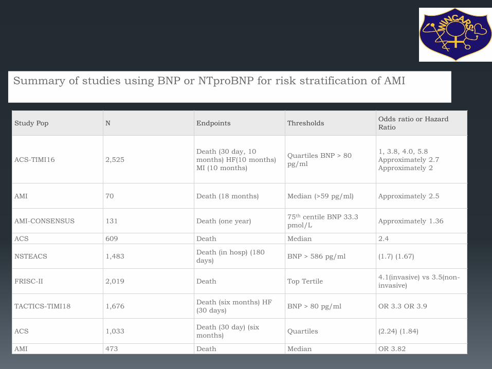
Study Pop N Endpoints Thresholds Odds ratio or Hazard
Ratio
ACS-TIMI16 2,525
Death (30 day, 10
months) HF(10 months)
MI (10 months)
Quartiles BNP > 80
pg/ml
1, 3.8, 4.0, 5.8
Approximately 2.7
Approximately 2
AMI 70 Death (18 months) Median (>59 pg/ml) Approximately 2.5
AMI-CONSENSUS 131 Death (one year) 75th centile BNP 33.3
pmol/L Approximately 1.36
ACS 609 Death Median 2.4
NSTEACS 1,483 Death (in hosp) (180
days) BNP > 586 pg/ml (1.7) (1.67)
FRISC-II 2,019 Death Top Tertile 4.1(invasive) vs 3.5(non-
invasive)
TACTICS-TIMI18 1,676 Death (six months) HF
(30 days) BNP > 80 pg/ml OR 3.3 OR 3.9
ACS 1,033 Death (30 day) (six
months) Quartiles (2.24) (1.84)
AMI 473 Death Median OR 3.82
Summary of studies using BNP or NTproBNP for risk stratification of AMI
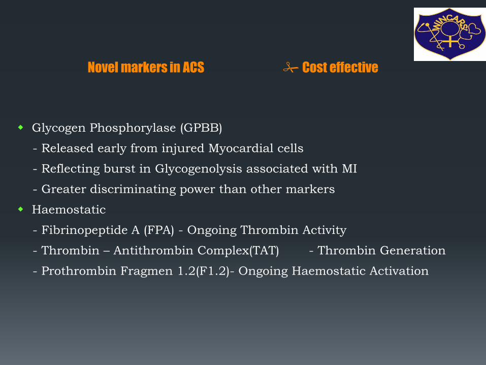
Glycogen Phosphorylase (GPBB)
- Released early from injured Myocardial cells
- Reflecting burst in Glycogenolysis associated with MI
- Greater discriminating power than other markers
Haemostatic
- Fibrinopeptide A (FPA) - Ongoing Thrombin Activity
- Thrombin – Antithrombin Complex(TAT) - Thrombin Generation
- Prothrombin Fragmen 1.2(F1.2)- Ongoing Haemostatic Activation
Novel markers in ACS Cost effective
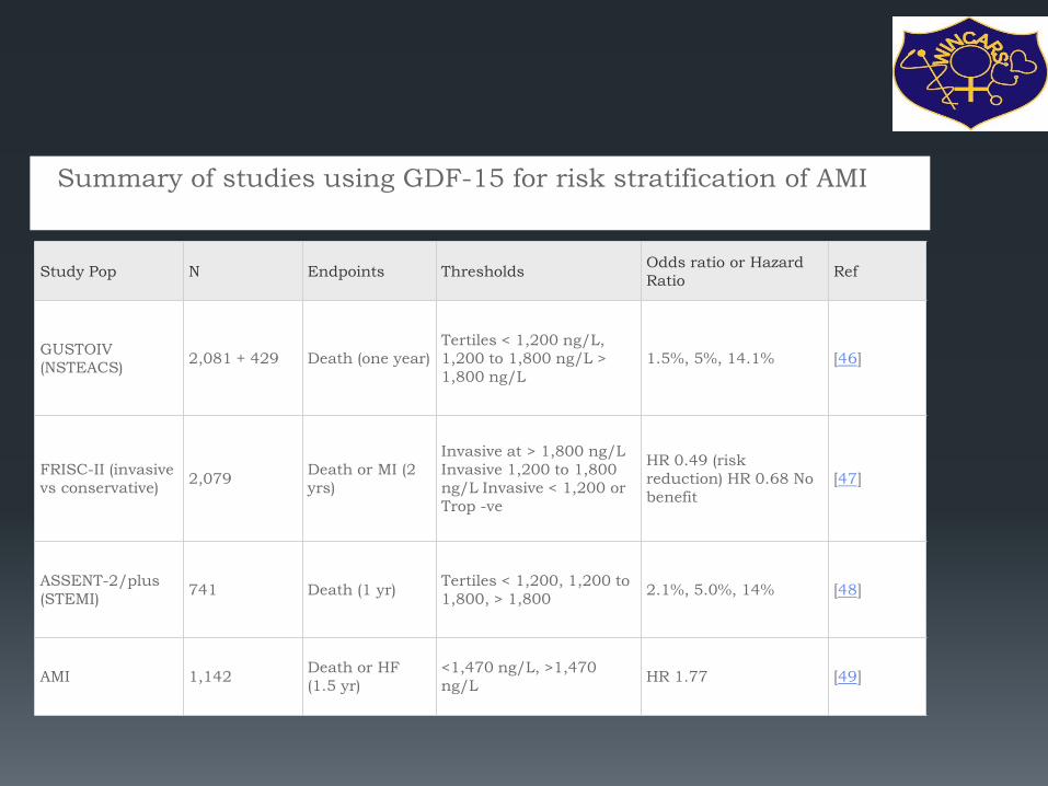
Study Pop N Endpoints Thresholds Odds ratio or Hazard
Ratio Ref
GUSTOIV
(NSTEACS) 2,081 + 429 Death (one year)
Tertiles < 1,200 ng/L,
1,200 to 1,800 ng/L >
1,800 ng/L
1.5%, 5%, 14.1% [46]
FRISC-II (invasive
vs conservative) 2,079
Death or MI (2
yrs)
Invasive at > 1,800 ng/L
Invasive 1,200 to 1,800
ng/L Invasive < 1,200 or
Trop -ve
HR 0.49 (risk
reduction) HR 0.68 No
benefit
[47]
ASSENT-2/plus
(STEMI) 741 Death (1 yr)
Tertiles < 1,200, 1,200 to
1,800, > 1,800 2.1%, 5.0%, 14% [48]
AMI 1,142 Death or HF
(1.5 yr)
<1,470 ng/L, >1,470
ng/L HR 1.77 [49]
Summary of studies using GDF-15 for risk stratification of AMI
Comprehensive Metabolomic Characterization of Coronary
Artery Diseases.
A total of 89 differential metabolites were identified. The altered metabolic pathways included reduced phospholipid catabolism, increased amino acid metabolism, increased short-chain acylcarnitines, decrease in tricarboxylic acid cycle, and less biosynthesis of primary bile acid.
Plasma metabolomics are powerful for characterizing metabolic disturbances. Differences in small-molecule metabolites may reflect underlying CAD and serve as biomarkers for CAD progression.
J Am Coll Cardiol. 2016 Sep 20;68(12):1281-93.

-Plasma D-Dimer - Activation of coagulation/ fibrinolysis
-S100 Protein- Levels at early & late ph
? Continuous Release after injury
? Future Marker
Novel markers in ACS Cost effective

Ack. JAPI, Volume 63, June 2015
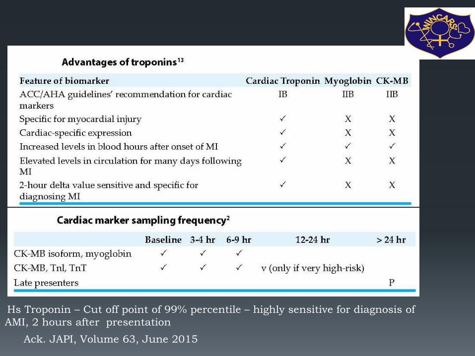
Ack. JAPI, Volume 63, June 2015
Hs Troponin – Cut off point of 99% percentile – highly sensitive for diagnosis of
AMI, 2 hours after presentation
Questions in thought process
• Diagnosing acute MI using high sensitive Troponin
• How to use high sensitive cardiac troponin in acute cardiac insult
• Groups with subclinical Ischemic heart disease and slightly elevated
troponin
• Studies of diagnostic performance study design influences sensitivity
and specificity
• Age cut off - ? Elderly
• ?? Cardiac – Troponin – A marker of Myocardial necrosis and not a
specific marker of AMI
• Constant values without diagnosis changes are likely to be marker of
chronic heart disease

The 10 commandments of troponin
• Collaborate with the laboratory and the emergency department
• Understand some analytical considerations
• Make the diagnosis of AMI based on cTn and the clinical scenario
• Rule out myocardial infarction differently than ruling it in
• Use common sense to interpret elevations of cTn in patients who are
critically ill
• Do not be intimidated by elevations in patients with renal failure
• Take the baseline value of cTn into account with percutaneous coronary
intervention
• It takes multiple parameters to make the diagnosis of AMI following bypass
surgery
• Do not forget drug toxicities as an aetiology for cTn elevations
• Be cautious with cTn elevations post-exercise
Heart 2011;97:940-946


