
Bb
HKC
a
ARRAA
KBSFB
0d
Progress in Polymer Science 37 (2012) 237– 280
Contents lists available at ScienceDirect
Progress in Polymer Science
j ourna l ho me pag e: ww w.elsev ier .com/ locate /ppolysc i
iodegradable synthetic polymers: Preparation, functionalization andiomedical application
uayu Tian, Zhaohui Tang, Xiuli Zhuang, Xuesi Chen ∗, Xiabin Jingey Laboratory of Polymer Ecomaterials, Changchun Institute of Applied Chemistry, Chinese Academy of Sciences, 5625 Renmin Street, Changchun 130022,hina
r t i c l e i n f o
rticle history:eceived 8 November 2009eceived in revised form 14 May 2011ccepted 30 June 2011vailable online 18 July 2011
eywords:
a b s t r a c t
Biodegradable polymers have been widely used and have greatly promoted the devel-opment of biomedical fields because of their biocompatibility and biodegradability. Thedevelopment of biotechnology and medical technology has set higher requirements forbiomedical materials. Novel biodegradable polymers with specific properties are in greatdemand. Biodegradable polymers can be classified as natural or synthetic polymers accord-ing to the source. Synthetic biodegradable polymers have found more versatile and diversebiomedical applications owing to their tailorable designs or modifications. This review
iodegradableynthetic polymersunctionalizationiomedical application
presents a comprehensivemers with reactive grouppreparation procedures antionalization and responsapplications. The possible
∗ Corresponding author. Tel.: +86 431 85262112; fax: +86 431 85262112.E-mail address: [email protected] (X. Chen).
Abbreviations: ADR, adriamycin; AP, 1,5-diamino pentane; APEG-DOX, polyPNIPAM; ASGPR, asialoglycoprotein receptor; ATQD, N-(4-aminophenyl)-N′-(4′
atom-transfer radical polymerization; BAA-NCA, �-benzyl aspartic acid N-carboxBECP, biodegradable electrically conducting polymer; BLA-NCA, benzyl-l-asparanhydride; BTMC, 5-benzyloxy-trimethylene carbonate; CaB, cathepsin B; CaD, ccell penetrating peptide; c(RGDfK)-PEG-b-P(Lys-MP), c(RGDfK)-poly(ethylene gltomography; DES, drug-eluting stents; DGBE, diethylene glycol bis(3-amino propytetraazacyclododecane-N,N′ ,N′′ ,N′′ ′-tetraacetic acid; DOX, doxorubicin; DPT, diprodocetaxel; EGFR, endothelial growth factor receptor; EPR, enhanced permeabilipoly(ethylene glycol)-co-poly(�-benzyl l-glutamate) block copolymer; Gd, gadN-hydroxylethylmaleimide; HO-R1-OH, di-hydroxyl compounds; ICG, indocyanalanine; LCST, lower critical solution temperature; LP-NCA, l-phenylalanine NCPLLA; MAL-PEG-PCL, maleimide-terminated poly(ethylene glycol)-poly(�-caprolamono-4-methoxybenzylidene-pentaerythritol carbonate; MMP-2, matrix metadoped superparamagnetic iron oxide; MP, 4-(3-aminopropyl) morpholine; MmPEG, poly(ethylene glycol) methyl ether; MRI, magnetic resonance imagingN-hydroxysuccinimide; NIPAM, N-isopropylacrylamide; NIR, near-infrared; NP(GA-co-BLG), poly[(l-glutamic acid)-co-(�-benzyl l-glutamate)]; PAGA, poly(�allylated PBLG; PBCLG, partially chlorinated PBLG; PBLG, poly(�-benzyl-l-glulated PBLG; Ppy, polypyrrole; PCL, poly(�-caprolactone); PCL-b-PBLG, poly(�-cPEI, polyethylenimine; PEG, polyethylene glycol; PEG-b-PEI, poly(ethylene glpoly(glutamic acid); PEG-b-P(LA-co-MCC/dtxel), poly(ethylene glycol)-block-PEG-b-PLA-b-PLG, poly(ethyl glycol)-b-polylactide-b-poly(l-glutamic acid); PEGP(Asp-Hyd-ADR), poly(ethylene glycol)-b-poly(aspartate-hydrazone-adriamycin)mono- and diethyleneglycol modified PLSer; PET, positron emission tomograph
079-6700/$ – see front matter © 2011 Elsevier Ltd. All rights reserved.oi:10.1016/j.progpolymsci.2011.06.004
introduction to various types of synthetic biodegradable poly-s and bioactive groups, and further describes their structure,d properties. The focus is on advances in the past decade in func-ive strategies of biodegradable polymers and their biomedical
future developments of the materials are also discussed.© 2011 Elsevier Ltd. All rights reserved.
acetal-doxorubicin; Apt, aptamers; AS-PNIPAM, amino-semitelechelic-(3-triethoxysilyl-propyl-ureido) phenyl-1,4-quinonenediimine); ATRP,y-anhydride; BMPCL, �-(2-bromo-2-methyl propionyl)-�-caprolactone;tate N-carboxyanhydride; BLG-NCA, �-benzyl l-glutamate N-carboxy-
athepsin D; CMMPL, �-chloromethyl-�-methyl-�-propionolactone; CPP,ycol)-b-poly[�-(3-mercaptopropino nyl)-lysine]; CT, computerized axiall) ether; DMSO, dimethyl sulfoxide; DOTA, designed macrocyclic 1,4,7,10-pylene triamine; DTPA-Gd, diethylenetriaminepentaacetic acid Gd; Dtxl,ty and retention; FOL, folic acid; gal-PEG-b-PBLG, galactose-conjugatedolinium; GSH, glutathione; HEMA, 2-Hydroxyethylmethacrylate; HEMI,ine green; IgG, immunoglobulin G; l-DOPA, l-3,4-dihydroxyphenyl-l-A; M-PCL, maleimido-terminated PCL; M-PLLA, maleimido-terminatedctone); MBC, 5-methyl-5-benzyloxycarbonyl-1,3-dioxan-2-one; MBPEC,
lloprotease-2; MMPs, matrix metalloproteinases; Mn-SPIO, manganeseP-g-OEI, multi-armed poly(l-glutamic acid)-graft-oligoethylenimine;
; NCA, N-carboxy-anhydride; NGF, neurotrophic growth factors; NHS,IRF, near-infrared fluorescent; NSCLC, non-small cell lung cancer;-(4-aminobutyl)-l-glycolic acid); PArg, polyarginine; PBALG, partially
tamate); PBN3LG, partially azidized PBLG; PBPLG, partially propargy-aprolactone)-b-poly(�-benzyl l-glutamate); PDI, polydispersity index;ycol)-b-polyethyleneimine; PEG-b-P(Glu-DP), poly(ethylene glycol)-b-poly(l-lactide-co-2-methyl-2-carboxyl-propylene carbonate/docetaxel;-P(Asp-Hyd), poly(ethylene glycol)-b-poly(aspartate-hydra zone); PEG-; PEG-PBLA, poly(ethylene glycol)-poly(�-benzyl-l-aspartate); PEGnLSer,y; PGS, planar gamma scintigraphy; PHB, poly[(R)-3-hydroxybutyrate];
238 H. Tian et al. / Progress in Polymer Science 37 (2012) 237– 280
Contents
1. Introduction . . . . . . . . . . . . . . . . . . . . . . . . . . . . . . . . . . . . . . . . . . . . . . . . . . . . . . . . . . . . . . . . . . . . . . . . . . . . . . . . . . . . . . . . . . . . . . . . . . . . . . . . . . . . . . . . . . . . . . . . 2392. Biopolymers with reactive groups . . . . . . . . . . . . . . . . . . . . . . . . . . . . . . . . . . . . . . . . . . . . . . . . . . . . . . . . . . . . . . . . . . . . . . . . . . . . . . . . . . . . . . . . . . . . . . . . . 239
2.1. Aliphatic polyesters . . . . . . . . . . . . . . . . . . . . . . . . . . . . . . . . . . . . . . . . . . . . . . . . . . . . . . . . . . . . . . . . . . . . . . . . . . . . . . . . . . . . . . . . . . . . . . . . . . . . . . . . . 2392.1.1. Aliphatic polyesters with carboxyl groups. . . . . . . . . . . . . . . . . . . . . . . . . . . . . . . . . . . . . . . . . . . . . . . . . . . . . . . . . . . . . . . . . . . . . . . . 2392.1.2. Aliphatic polyesters with amino groups . . . . . . . . . . . . . . . . . . . . . . . . . . . . . . . . . . . . . . . . . . . . . . . . . . . . . . . . . . . . . . . . . . . . . . . . . . 2392.1.3. Aliphatic polyesters with chloride groups . . . . . . . . . . . . . . . . . . . . . . . . . . . . . . . . . . . . . . . . . . . . . . . . . . . . . . . . . . . . . . . . . . . . . . . . 2392.1.4. Aliphatic polyesters with keto or hydroxyl groups . . . . . . . . . . . . . . . . . . . . . . . . . . . . . . . . . . . . . . . . . . . . . . . . . . . . . . . . . . . . . . . 2402.1.5. Aliphatic polyesters with bromide groups . . . . . . . . . . . . . . . . . . . . . . . . . . . . . . . . . . . . . . . . . . . . . . . . . . . . . . . . . . . . . . . . . . . . . . . . 2402.1.6. Aliphatic polyesters with C C groups . . . . . . . . . . . . . . . . . . . . . . . . . . . . . . . . . . . . . . . . . . . . . . . . . . . . . . . . . . . . . . . . . . . . . . . . . . . . 2402.1.7. Aliphatic polyesters with reactive groups by copolymerization . . . . . . . . . . . . . . . . . . . . . . . . . . . . . . . . . . . . . . . . . . . . . . . . . 240
2.2. Polycarbonate . . . . . . . . . . . . . . . . . . . . . . . . . . . . . . . . . . . . . . . . . . . . . . . . . . . . . . . . . . . . . . . . . . . . . . . . . . . . . . . . . . . . . . . . . . . . . . . . . . . . . . . . . . . . . . . 2412.3. Poly(amino acids) . . . . . . . . . . . . . . . . . . . . . . . . . . . . . . . . . . . . . . . . . . . . . . . . . . . . . . . . . . . . . . . . . . . . . . . . . . . . . . . . . . . . . . . . . . . . . . . . . . . . . . . . . . . 246
2.3.1. Poly(acidic amino acids) . . . . . . . . . . . . . . . . . . . . . . . . . . . . . . . . . . . . . . . . . . . . . . . . . . . . . . . . . . . . . . . . . . . . . . . . . . . . . . . . . . . . . . . . . . 2462.3.2. Poly(basic amino acid) . . . . . . . . . . . . . . . . . . . . . . . . . . . . . . . . . . . . . . . . . . . . . . . . . . . . . . . . . . . . . . . . . . . . . . . . . . . . . . . . . . . . . . . . . . . . 2502.3.3. Poly(neutral amino acid) . . . . . . . . . . . . . . . . . . . . . . . . . . . . . . . . . . . . . . . . . . . . . . . . . . . . . . . . . . . . . . . . . . . . . . . . . . . . . . . . . . . . . . . . . . 252
2.4. Polyphosphoesters . . . . . . . . . . . . . . . . . . . . . . . . . . . . . . . . . . . . . . . . . . . . . . . . . . . . . . . . . . . . . . . . . . . . . . . . . . . . . . . . . . . . . . . . . . . . . . . . . . . . . . . . . . 2522.5. Others . . . . . . . . . . . . . . . . . . . . . . . . . . . . . . . . . . . . . . . . . . . . . . . . . . . . . . . . . . . . . . . . . . . . . . . . . . . . . . . . . . . . . . . . . . . . . . . . . . . . . . . . . . . . . . . . . . . . . . . 254
3. Biopolymers with responsive activities . . . . . . . . . . . . . . . . . . . . . . . . . . . . . . . . . . . . . . . . . . . . . . . . . . . . . . . . . . . . . . . . . . . . . . . . . . . . . . . . . . . . . . . . . . . . 2543.1. Stimuli-responsive biopolymers . . . . . . . . . . . . . . . . . . . . . . . . . . . . . . . . . . . . . . . . . . . . . . . . . . . . . . . . . . . . . . . . . . . . . . . . . . . . . . . . . . . . . . . . . . . . 254
3.1.1. Temperature responsive . . . . . . . . . . . . . . . . . . . . . . . . . . . . . . . . . . . . . . . . . . . . . . . . . . . . . . . . . . . . . . . . . . . . . . . . . . . . . . . . . . . . . . . . . . 2553.1.2. pH-responsive biopolymers . . . . . . . . . . . . . . . . . . . . . . . . . . . . . . . . . . . . . . . . . . . . . . . . . . . . . . . . . . . . . . . . . . . . . . . . . . . . . . . . . . . . . . . 2553.1.3. Photo responsive biopolymers . . . . . . . . . . . . . . . . . . . . . . . . . . . . . . . . . . . . . . . . . . . . . . . . . . . . . . . . . . . . . . . . . . . . . . . . . . . . . . . . . . . . 2563.1.4. Redox responsive biopolymers . . . . . . . . . . . . . . . . . . . . . . . . . . . . . . . . . . . . . . . . . . . . . . . . . . . . . . . . . . . . . . . . . . . . . . . . . . . . . . . . . . . 257
3.2. Electroactive biomaterials . . . . . . . . . . . . . . . . . . . . . . . . . . . . . . . . . . . . . . . . . . . . . . . . . . . . . . . . . . . . . . . . . . . . . . . . . . . . . . . . . . . . . . . . . . . . . . . . . . 2583.3. Specific bonding biopolymers . . . . . . . . . . . . . . . . . . . . . . . . . . . . . . . . . . . . . . . . . . . . . . . . . . . . . . . . . . . . . . . . . . . . . . . . . . . . . . . . . . . . . . . . . . . . . . 2593.4. Biopolymers for tracing and bioimaging . . . . . . . . . . . . . . . . . . . . . . . . . . . . . . . . . . . . . . . . . . . . . . . . . . . . . . . . . . . . . . . . . . . . . . . . . . . . . . . . . . . 262
3.4.1. Biopolymers for optical tracing and bioimaging . . . . . . . . . . . . . . . . . . . . . . . . . . . . . . . . . . . . . . . . . . . . . . . . . . . . . . . . . . . . . . . . . . 2623.4.2. Biopolymers for MRI . . . . . . . . . . . . . . . . . . . . . . . . . . . . . . . . . . . . . . . . . . . . . . . . . . . . . . . . . . . . . . . . . . . . . . . . . . . . . . . . . . . . . . . . . . . . . . 2653.4.3. Other biopolymer-based tracing and bioimaging . . . . . . . . . . . . . . . . . . . . . . . . . . . . . . . . . . . . . . . . . . . . . . . . . . . . . . . . . . . . . . . . 266
4. Biomedical application . . . . . . . . . . . . . . . . . . . . . . . . . . . . . . . . . . . . . . . . . . . . . . . . . . . . . . . . . . . . . . . . . . . . . . . . . . . . . . . . . . . . . . . . . . . . . . . . . . . . . . . . . . . . . 2664.1. Medical devices . . . . . . . . . . . . . . . . . . . . . . . . . . . . . . . . . . . . . . . . . . . . . . . . . . . . . . . . . . . . . . . . . . . . . . . . . . . . . . . . . . . . . . . . . . . . . . . . . . . . . . . . . . . . . 266
4.1.1. Drug-eluting stents (DES) . . . . . . . . . . . . . . . . . . . . . . . . . . . . . . . . . . . . . . . . . . . . . . . . . . . . . . . . . . . . . . . . . . . . . . . . . . . . . . . . . . . . . . . . . 2664.1.2. Orthopedic devices . . . . . . . . . . . . . . . . . . . . . . . . . . . . . . . . . . . . . . . . . . . . . . . . . . . . . . . . . . . . . . . . . . . . . . . . . . . . . . . . . . . . . . . . . . . . . . . . 2674.1.3. Disposable medical devices . . . . . . . . . . . . . . . . . . . . . . . . . . . . . . . . . . . . . . . . . . . . . . . . . . . . . . . . . . . . . . . . . . . . . . . . . . . . . . . . . . . . . . . 2674.1.4. Other medical devices . . . . . . . . . . . . . . . . . . . . . . . . . . . . . . . . . . . . . . . . . . . . . . . . . . . . . . . . . . . . . . . . . . . . . . . . . . . . . . . . . . . . . . . . . . . . . 267
4.2. Tissue engineering . . . . . . . . . . . . . . . . . . . . . . . . . . . . . . . . . . . . . . . . . . . . . . . . . . . . . . . . . . . . . . . . . . . . . . . . . . . . . . . . . . . . . . . . . . . . . . . . . . . . . . . . . . 2674.3. Drug delivery and control release . . . . . . . . . . . . . . . . . . . . . . . . . . . . . . . . . . . . . . . . . . . . . . . . . . . . . . . . . . . . . . . . . . . . . . . . . . . . . . . . . . . . . . . . . . 2684.4. Gene delivery. . . . . . . . . . . . . . . . . . . . . . . . . . . . . . . . . . . . . . . . . . . . . . . . . . . . . . . . . . . . . . . . . . . . . . . . . . . . . . . . . . . . . . . . . . . . . . . . . . . . . . . . . . . . . . . . 269
4.4.1. Poly(l-lysine)-based degradable polymers . . . . . . . . . . . . . . . . . . . . . . . . . . . . . . . . . . . . . . . . . . . . . . . . . . . . . . . . . . . . . . . . . . . . . . . 2694.4.2. Poly(�-amino ester)s-based degradable polymers . . . . . . . . . . . . . . . . . . . . . . . . . . . . . . . . . . . . . . . . . . . . . . . . . . . . . . . . . . . . . . . 2694.4.3. Polyphosphoester-based degradable polymers. . . . . . . . . . . . . . . . . . . . . . . . . . . . . . . . . . . . . . . . . . . . . . . . . . . . . . . . . . . . . . . . . . . 2704.4.4. Polyethylenimine modified with degradable polymers . . . . . . . . . . . . . . . . . . . . . . . . . . . . . . . . . . . . . . . . . . . . . . . . . . . . . . . . . . 2704.4.5. Degradable polymers in siRNA delivery . . . . . . . . . . . . . . . . . . . . . . . . . . . . . . . . . . . . . . . . . . . . . . . . . . . . . . . . . . . . . . . . . . . . . . . . . . 2714.4.6. Other degradable polymers . . . . . . . . . . . . . . . . . . . . . . . . . . . . . . . . . . . . . . . . . . . . . . . . . . . . . . . . . . . . . . . . . . . . . . . . . . . . . . . . . . . . . . . 271
4.5. Bioseparation and diagnostics applications . . . . . . . . . . . . . . . . . . . . . . . . . . . . . . . . . . . . . . . . . . . . . . . . . . . . . . . . . . . . . . . . . . . . . . . . . . . . . . . . 271. . . . . . . .
5. Conclusions . . . . . . . . . . . . . . . . . . . . . . . . . . . . . . . . . . . . . . . . . . . . . . . . .Acknowledgments . . . . . . . . . . . . . . . . . . . . . . . . . . . . . . . . . . . . . . . . . . . . . . . . . .References . . . . . . . . . . . . . . . . . . . . . . . . . . . . . . . . . . . . . . . . . . . . . . . . . . . . . . . . . .
PHF-b-PEG, poly[(l-histidine)-co-(l-phenylalanine)]-block-poly(ethylene glycob-polylactide; PLCys, poly(l-cysteine); PLCys-b-PLLA, poly(l-cysteine)-b-polpoly(l-lysine); PLGA, poly(lactide-co-glycolide); PLHis, poly(l-histidine); PLL,PLL-co-PArg-b-PLLeu, poly(l-lysine)-co-polyarginine-b-poly(l-leucine); PLL-g-Ppoly(ethylene glycol); PLL-g-PLLA, poly(l-lysine)-g-poly(l-lactide); PLLA, poly(l-laPNIPAM, poly(N-isopropylacrylamide); PNIPAM-b-(HEMA-PCL), poly(N-isoproPNIPAM-b-PGA, poly(N-isopropylacrylamide)-block-poly(glutamic acid); PNIPglutamate)]; PPA, polyphosphoramidate; PPE, polyphosphoester; PPE-EA, p2, performance status; PSI, polysuccinimide; PTMC-b-PBLG, poly(trimethylenacid; PZLL-PDGBE-PZLL, poly(�-benzyloxycarbonyl l-lysine)-block-poly[diethyll-lysine); QDs, quantum dots; QD-strep, quantum dot-streptavidin; RGD, arginine-linked; siRNA, small interfering RNA; Sn(OTf)2, trifluoromethane sulfonate; SPDP,
iron oxide; SPECT, single photon emission computed tomography; Sr-PO,pentaerythritol carbonate; TSP50, testis-specific protease 50; VEGF, vascular enN-carboxyanhydride; ZLCys-NCA, �-benzyloxycarbonyl-l-cysteine N-carboxyanh
. . . . . . . . . . . . . . . . . . . . . . . . . . . . . . . . . . . . . . . . . . . . . . . . . . . . . . . . . . . . . . . . 272
. . . . . . . . . . . . . . . . . . . . . . . . . . . . . . . . . . . . . . . . . . . . . . . . . . . . . . . . . . . . . . . . 272. . . . . . . . . . . . . . . . . . . . . . . . . . . . . . . . . . . . . . . . . . . . . . . . . . . . . . . . . . . . . . . . 272
l); PLA, poly(lactic acid), polylactide; PLC-b-PLA, poly(l-cysteine)-y(l-lactide); PLDOPA, poly(l-DOPA); PLDOPA-PLL, poly(l-DOPA)-co-
poly(l-lysine); PLL-b-PPA, poly(l-lysine)-block-poly(l-phenylalanine);CL, poly(l-lysine)-g-poly(�-caprolactone); PLL-g-PEG, poly(l-lysine)-g-ctide); PLSer, poly(l-serine); PMDETA, pentamethyldiethylene-triamine;
pylacrylamide)-b-[2-hydroxyethyl methacrylate-poly(�-caprolactone)];AM-b-P(GA-co-BLG), PNIPAM-b-poly[(l-glutamic acid)-co-(�-benzyl-l-oly(2-aminoethyl propylene phosphate); PPZ, polyphosphazene; PSe carbonate)-b-poly(�-benzyl l-glutamate); p-TSA, p-toluenesulfonic
ene glycol bis(3-amino propyl) ether]-block-poly(�-benzyloxycarbonylglycine-aspartic acid; ROP, ring-opening polymerization; SCL, shell cross-
N-succinimidyl 3-(2-pyridyldithio)-propionate; SPIO, superparamagnetic amino isopropoxyl strontium; TMBPEC, 6-trimethoxybenzy-lidene-dothelial growth factor; Z2Arg-NCA, di-N-benzyloxycarbonyl-l-arginineydride1.

olymer S
1
im[Bttbmrbidoactpt
fctfamicbdipAib
rbaseibtmbicp
sp
2
2
p
H. Tian et al. / Progress in P
. Introduction
A biomaterial can be defined as a material intended tonterface with biological systems to evaluate, treat, aug-
ent or replace any tissue, organ or function of the body1]. Biomaterials play an important role in human health.iopolymers are the main type of biomaterials. Accordingo their degradation properties, biopolymers can be fur-her classified into biodegradable and non-biodegradableiopolymers. Many implants, such as bone substitutionaterials, some bone fixing materials, and dental mate-
ials, should possess long term stable performance in theody. In recent years, developments in tissue engineer-
ng, regenerative medicine, gene therapy, and controlledrug delivery have promoted the need of new propertiesf biomaterials with biodegradability. Biologically derivednd synthetic biodegradable biopolymers have attractedonsiderable attention [1]. Polysaccharides and protein areypical biologically derived biopolymers, while aliphaticolyesters and polyphosphoester (PPE) are typical syn-hetic biopolymers.
Biopolymers with diverse specific properties are neededor in vivo applications because of the diversity andomplexity of in vivo environments. Nowadays, syn-hetic biopolymers have become attractive alternativesor biomedical applications for the following reasons: (1)lthough most biologically derived biodegradable poly-ers possess good biocompatibility, some may trigger an
mmune response in the human body, possibly one thatould be avoided by the use of an appropriate syntheticiopolymer; (2) chemical modifications to biologicallyerived biodegradable polymers are difficult; (3) chem-
cal modifications likely cause the alteration of the bulkroperties of biologically derived biodegradable polymers.
variety of properties can be obtained and further mod-fications are possible with properly designed syntheticiopolymers wihout altering the bulk properties.
Specific properties are sometimes required for biomate-ials. For example, tissue engineering scaffolds should haveoth good biocompatibility and cell adhesive properties, inddition to needed biodegradable properties. Drug deliveryystems should be endowed with stimuli-responsive prop-rties for intelligent-control release. Functionalization isnevitable to improve the properties of traditional syntheticiopolymers. There are two commonly used functionaliza-ion strategies: (1) functional groups are introduced to the
onomers of polymers, sometimes in a protected formefore polymerization, to be deprotected after polymer-
zation; (2) functional groups are introduced to polymerhains by further chemical modification of the as-preparedolymers.
This review is focused on recent progress of differenttrategies of functionalization of synthetic biodegradableolymers and the applications of these.
. Biopolymers with reactive groups
.1. Aliphatic polyesters
Aliphatic polyesters, such as poly(lactic acid) (PLA),oly(glycolic acid), poly(�-caprolactone) (PCL) and their
cience 37 (2012) 237– 280 239
copolymers, have been widely investigated for biomedicalapplication because of their biodegradability, bioresorba-bility, and biocompatibility. Aliphatic polyesters withreactive groups have attracted attention because of thedemand of synthetic biopolymers with tunable properties,including features such as hydrophilicity, biodegradationrates, bioadhesion, drug/targeting moiety attachment, etc.[2]. In particular, polymeric biomaterials with propertiesthat can be tailored by introducing functional groups,such as carboxyl, hydroxyl, amino, ketal, bromo, chloro,carbon–carbon double bonds or triple bonds, etc., areneeded.
Aliphatic polyesters with reactive groups can be pre-pared by the homopolymerization or copolymerizationof cyclic monomers bearing protected functional groups(Fig. 1). Representative examples of the monomers and thepolymers are shown in Table 1.
2.1.1. Aliphatic polyesters with carboxyl groupsAliphatic polyesters with pendant carboxyl groups can
be prepared by the ring-opening polymerization (ROP)of cyclic esters bearing benzyl-protected carboxyl groups.Ouchi and Fujino prepared poly(�-malic acid) as a car-boxyl functional analogy of PLA by the ROP of malidedibenzyl ester followed by acid deprotection [3]. Kimuraet al. first reported the synthesis of poly[(�-malic acid)-alt-(glycolic acid)], a glycolide-based poly(ester) with pendantcarboxylic acid, by the ROP of 3(S)-[(benzyloxycarbonyl)-methyl]-1,4-dioxane-2,5-dione followed by debenzyla-tion. These aliphatic copolyesters are hydrolyzed morerapidly than PLA [4]. Weck and coworkers prepared side-chain-functionalized lactide analogues from commerciallyavailable amino acids. The resulting functionalized cyclicmonomers can be homopolymerized and copolymerizedwith lactides and then quantitatively deprotected, form-ing functional PLA-based materials with amino, hydroxylor carboxyl side chains [5]. Guerin et al. reported the syn-thesis and polymerization of benzyl malolactonate [14].The benzyl protecting groups could be readily removedby catalytic hydrogenolysis to give poly(�-malic acid).He et al. reported the synthesis of poly(l-lactide-co-�-malic acid) with a high molecular weight by thecopolymerization of l-lactide and benzyl malolactonate[15]. PCLs with pendant carboxylic acid groups wereprepared by Hedrick and coworkers via the ROP ofbenzyl �-(�-caprolactone)carboxylate or tert-butyl-�-(�-caprolactone)carboxylate followed by acid deprotection[6] (Fig. 2).
2.1.2. Aliphatic polyesters with amino groupsHedrick and coworkers synthesized
amino-functionalized PCL by the ROP of 4-trifluoroacetyl-7-oxo-1,4-oxazaperhydroepine followed by thedeprotection with NaBH4 [6]. Fiétier et al. reportedthe preparation of an aliphatic polyester bearing lateralamino groups by the ROP of N-tritylated serine �-lactones[16].
2.1.3. Aliphatic polyesters with chloride groupsLiu and coworkers synthesized a chloro-substituted
four-membered lactone, �-chloromethyl-�-methyl-�-

240 H. Tian et al. / Progress in Polymer Science 37 (2012) 237– 280
OR
O RO
O
n
R: reactive groups, including protected carboxyl, amino, chloro, ketal,ouble
Homopolymerization
OR
O + O(CH2)x
OR
OO
mCopolymerization
O
(CH2)xO n
tic poly
hydroxyl, bromo, carbon-carbon d
Fig. 1. Preparation of alipha
propionolactone (CMMPL). CMMPL was polymerized andcopolymerized with various amounts of �-caprolactone.The pendant chloromethyl groups of the copolymerswere converted into quaternary ammonium salts by thereaction with pyridine, which increased the hydrophilicityof the copolymer [7,17].
2.1.4. Aliphatic polyesters with keto or hydroxyl groupsAliphatic polyesters bearing keto groups were syn-
thesized by the ROP of 5-ethylene ketal �-caprolactonefollowed by deprotection [8,9,18,19]. The keto groups of thecopolymers were efficiently reduced into hydroxyl groupsby using NaBH4 in a mixture of CH2Cl2/EtOH at roomtemperature without any apparent chain degradation,resulting in aliphatic polyesters with pendant hydroxylgroups [9]. PCL containing pendant hydroxyl groups wereprepared by the ROP of �-caprolactone monomer bearingtriethylsilyloxy pendant groups that can be selectivelydeprotected into hydroxyl groups under mild conditions[20]. Hedrick and coworkers reported the synthesis andpolymerization of �-benzyloxy-�-caprolactone and �-2,2-bis(phenyldioxymethyl)propionate-�-caprolactone; thecatalytic hydrogenolysis of the benzyl protection groupof the products afforded PCL with pendant hydroxyl orbishydroxyl groups, respectively [6].
2.1.5. Aliphatic polyesters with bromide groupsHedrick and coworkers reported the preparation of
aliphatic polyesters with pendant bromide groups by theROP of a bromo-substituted cyclic ester, �-(2-bromo-2-methyl propionyl)-�-caprolactone (BMPCL) containing apendent-activated alkyl bromide functional group [10].
The pendent-activated alkyl bromide group could initi-ate controlled atom-transfer radical polymerization (ATRP)of methyl methacrylate; therefore, PCL-graft-poly(methylmethacrylate) copolymers were obtained in a simple one-Fig. 2. Preparation of PCL with pendaCopyright 2000, American Chemical Society. Reprinted with permission.
bonds, etc
esters with reactive groups.
step approach by the concurrent polymerization of an�-caprolactone, BMPCL, and methyl methacrylate with anappropriate initiator for the ROP and a catalyst for the ATRP.
2.1.6. Aliphatic polyesters with C C groupsUnsaturated aliphatic polyesters can be prepared by
the ROP of cyclic esters bearing double bonds. Hedrickand coworkers reported the preparation of unsaturatedaliphatic homopolyesters or random copolyesters bearingpendant double bonds by the ROP of 4-(acryloyloxy)-�-caprolactone, or 6-hydroxynon-8-enoic acid lactone with�-caprolactone and l-lactide [11,12]. Bizzarri and cowork-ers reported the preparation of aliphatic polyesters bearingdouble bonds by the ROP of four-membered lactones inthe presence of a quaternary ammonium salt as the ini-tiator [13,21]. Unsaturated aliphatic polyesters with innerdouble bonds were prepared by the ROP of unsaturated�-caprolactones with inner double bonds. Jérôme andcoworkers prepared unsaturated aliphatic polyesters bythe ROP of 6,7-dihydro-2(5H)-oxepinone and 6,7-dihydro-2(3H)-oxepinone using aluminum isopropoxide as theinitiator [22–24].
2.1.7. Aliphatic polyesters with reactive groups bycopolymerization
The copolymerization of morpholine-2,5-dione deriva-tives with lactide or lactones is a convenient way to preparealiphatic biopolymers bearing reactive groups. Feijen andcoworkers demonstrated the ROP of either �-caprolactoneor dl-lactide with morpholine-2,5-dione derivatives couldprotect functional substituents such as benzyl-protectedcarboxylic acid, benzyloxycarbonyl-protected amine and
p-methoxy-protected thiol groups. Polyesteramides withpendant carboxylic acid groups, pendant amine groups, orpendant thiol groups were obtained after deprotection ofthe copolymers [25].nt carboxylic acid groups [6].

olymer S
2
mppf
TA
H. Tian et al. / Progress in P
.2. Polycarbonate
Aliphatic polyesters and copolyesters are among the
ost commonly used degradable materials for thereparation of clinical devices. In this field, aliphaticolycarbonates are good materials because they possessunctionalizable side chains (OH, NH2, COOH, etc.) that
able 1liphatic polyesters and functional cyclic monomers.
Monomer Polyester
cience 37 (2012) 237– 280 241
can easily meet the need for functionalization of bio-materials. Moreover, aliphatic polycarbonates have goodbiocompatibility, low toxicity, and good biodegradability
[26,27]. High molecular weight aliphatic polycarbonatescan be prepared by the ROP of cyclic carbonates [28]. Themost commonly used cyclic carbonates for ROP are thefive- and six-membered cyclic monomers. PolymerizationReference
[3]
[4]
[5]
[5]
[5]
[6]
[6]

242 H. Tian et al. / Progress in Polymer Science 37 (2012) 237– 280
Table 1 (Continued)
Monomer Polyester Reference
[6]
[6]
[6]
[7]
[8]
[9]
H. Tian et al. / Progress in Polymer Science 37 (2012) 237– 280 243
Table 1 (Continued)
Monomer Polyester Reference
[10]
[11]
[12]
[13]
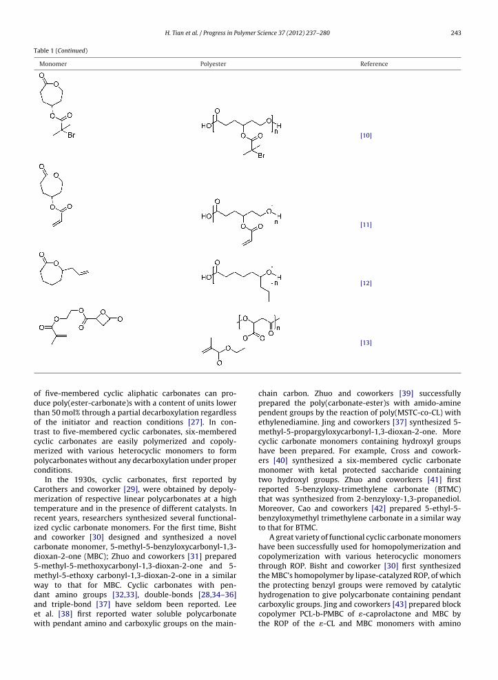
odtotcmpc
Cmtriacd5mwdaew
f five-membered cyclic aliphatic carbonates can pro-uce poly(ester-carbonate)s with a content of units lowerhan 50 mol% through a partial decarboxylation regardlessf the initiator and reaction conditions [27]. In con-rast to five-membered cyclic carbonates, six-memberedyclic carbonates are easily polymerized and copoly-erized with various heterocyclic monomers to form
olycarbonates without any decarboxylation under properonditions.
In the 1930s, cyclic carbonates, first reported byarothers and coworker [29], were obtained by depoly-erization of respective linear polycarbonates at a high
emperature and in the presence of different catalysts. Inecent years, researchers synthesized several functional-zed cyclic carbonate monomers. For the first time, Bishtnd coworker [30] designed and synthesized a novelarbonate monomer, 5-methyl-5-benzyloxycarbonyl-1,3-ioxan-2-one (MBC); Zhuo and coworkers [31] prepared-methyl-5-methoxycarbonyl-1,3-dioxan-2-one and 5-ethyl-5-ethoxy carbonyl-1,3-dioxan-2-one in a similaray to that for MBC. Cyclic carbonates with pen-
ant amino groups [32,33], double-bonds [28,34–36]nd triple-bond [37] have seldom been reported. Leet al. [38] first reported water soluble polycarbonateith pendant amino and carboxylic groups on the main-chain carbon. Zhuo and coworkers [39] successfullyprepared the poly(carbonate-ester)s with amido-aminependent groups by the reaction of poly(MSTC-co-CL) withethylenediamine. Jing and coworkers [37] synthesized 5-methyl-5-propargyloxycarbonyl-1,3-dioxan-2-one. Morecyclic carbonate monomers containing hydroxyl groupshave been prepared. For example, Cross and cowork-ers [40] synthesized a six-membered cyclic carbonatemonomer with ketal protected saccharide containingtwo hydroxyl groups. Zhuo and coworkers [41] firstreported 5-benzyloxy-trimethylene carbonate (BTMC)that was synthesized from 2-benzyloxy-1,3-propanediol.Moreover, Cao and coworkers [42] prepared 5-ethyl-5-benzyloxymethyl trimethylene carbonate in a similar wayto that for BTMC.
A great variety of functional cyclic carbonate monomershave been successfully used for homopolymerization andcopolymerization with various heterocyclic monomersthrough ROP. Bisht and coworker [30] first synthesizedthe MBC’s homopolymer by lipase-catalyzed ROP, of whichthe protecting benzyl groups were removed by catalytic
hydrogenation to give polycarbonate containing pendantcarboxylic groups. Jing and coworkers [43] prepared blockcopolymer PCL-b-PMBC of �-caprolactone and MBC bythe ROP of the �-CL and MBC monomers with amino
244 H. Tian et al. / Progress in Polymer Science 37 (2012) 237– 280
Table 2Polymers and functional cyclic carbonate monomers.
Monomer Polymer Referen-ce
OO
O
R1 R2 * O O O *
O
R1 R2 n
R1 = CH3; R2 = COOCH2Ph [30,41]
H2C O
OH2C
[43]R1 = H; R2 = OCH2CH CH2 [36]
H2C
H2C
[28]R1 = CH3; R2 = COOCH3 [31]R1 = CH3; R2 = COOCH2CH3 [31]
OO
O
R1 R2
O O O*
O
O
R1 R2
n
O O O*
O
O
R1 R2
*
nm
or
R1 = CH3; R2 = CH2OOCCH CHPh [44]R1 = CH3; R2 = COOCH2CH = CH2 [35]R1 = CH3; R2 = COOCH2C CH [35]R1 = H; R2 = NHCOOCH2Ph [33]
H2C
H2C
[34]
H2C O
OH2C
Ph
[45]
Q
T3 T4 or
RGIQ Q Q
,
Q
Q
T3 T4
n
RGI
Q Q Q,
Q
Q
nmT3 T4
R1 = CH3; R2 = COOCH2Ph [46]

H. Tian et al. / Progress in Polymer Science 37 (2012) 237– 280 245
Table 2 (Continued)
Monomer Polymer Referen-ce
H2C O
OH2C
Ph
[47]R1 = CH3; R2 = COOCH2CH CH2 [48]
OO
O
R1 R2O O
H
O
R1 R2
nPEG
R1 = CH3; R2 = CO2CH2Ph-o-NO2 [49]R1 = CH3; R2 = CO2CH2Ph [50]
OO
O
COOCH2PhCHO N
H
O C
O O H
O
O
CO2H
n
[38]
OO
O
O *
O O OO
*
O
O
O
O
nm
[51]
O
O
O
OO
O
O
*O
O
OO
O OO
OH OH
O * [39]
iccuteT
cst
O
O
sopropoxyl strontium (Sr-PO) as an initiator. Zhuo andoworkers [44] prepared poly[(5-benzyloxy-trimethylenearbonate)-co-(5,5-dimethyl-trimethylene carbonate)] bysing immobilized porcine pancreas lipase on silica par-icles with different sizes to catalyze ROP. Representativexamples of the monomers and the polymers are shown inable 2 [30,43,45–53,33–36,28,31,38,40].
Free functional pendant groups on poly(ester-arbonate)s are expected to facilitate further modificationsuch as attaching drug molecules and short pep-ides onto the functional groups of the polymers.
O On
Grinstaff and coworker [54] attached a nonsteroidalanti-inflammatory drug, 4-isobutylmethylphenylaceticacid, to the copolymer by esterification of free hydroxylgroups of 4-isobutylmethylphenylacetic acid. Jing andcoworkers successfully attached antitumor drugs pacli-taxel [55] and docetaxel (Dtxl) [56], biotin [57] andoligopeptide Gly-Arg-Gly-Asp-Ser-Tyr (RGD) [33] to
the pendants on the backbone of the copolymers.The results indicate further possible application ofpoly(ester-carbonate)s in specific drug delivery and tissueengineering.
246 H. Tian et al. / Progress in Polymer Science 37 (2012) 237– 280
(A) the
Fig. 3. Synthesis of functional PBLGs through the ester exchange reactionCopyright 2009, Elsevier Ltd. Reprinted with permission.2.3. Poly(amino acids)
Poly(amino acids) are an important kind of biocom-patible and biodegradable synthetic polymers and havebeen studied for biomedical application in many fields [58].However, their application is limited because of their insol-ubility or pH-dependent solubility and lack of functionalgroups [59]. This section summarizes recent develop-ments in the functional modifications of poly(amino acids)focusing on the preparation of materials with potentialapplications in medicine. Representative examples of thepoly(amino acids) before and after functionalization areshown in Table 3.
2.3.1. Poly(acidic amino acids)2.3.1.1. Poly(l-glutamic acid). Poly(l-glutamic acid) (PLG)is composed of naturally occurring l-glutamic acid residueslinked together through amide bonds with active carboxylgroups on the side. Methods can be used in function-alizing PLG include: (1) polymerizing or copolymerizingmonomers with functional groups, (2) modifying themonomer with functional molecules, (3) functional modifi-cation of the side groups, such as condensation, aminolysisand ester exchange, and (4) introduction of a second com-ponent to achieve a block, branch, hyper-branched ordendron-like architecture.
Functionalizing poly(�-benzyl-l-glutamate) (PBLG)through ester exchange with functional alcohols suchas 2-chloroethanol, 2-azidoethanol and poly(ethylene
glycol) methyl ether (mPEG) is a convenient method toobtain polymers that have controlled amounts of func-tional groups on the side chains without protection andde-protection processes. Huang and coworkers used theclick reaction, and the thiol-ene reaction of functional PBLGs (B) [60].
ROP of N-carboxy-anhydride (NCA) and ester exchangeto prepare functional PBLG with functional alcohols [60].PBLG was synthesized in anhydrous chloroform by the ROPof �-benzyl l-glutamate N-carboxy-anhydride (BLG-NCA)at room temperature using n-hexylamine as an initiator[73]. Functional PBLGs were then synthesized by the esterexchange reactions between PBLG and functional alcoholsin 1,2-dichloroethane using p-toluenesulfonic acid (p-TSA)as a catalyst [60]. Four kinds of functional PBLGs includingpartially chlorinated PBLG (PBCLG), partially azidizedPBLG (PBN3LG), partially allylated PBLG (PBALG) and par-tially propargylated PBLG (PBPLG) were synthesized. Theactivity of the functional groups on PBLG was examinedthrough click chemistry between PBN3LG or PBPLG andpropargyl mPEG2000 or 2-azidoethanol, or the thiol-enereaction between PBALG and thioglycol yielding PBPN3LG,PBN3LG-g-mPEG and PBALG-s-OH, respectively (Fig. 3). Ina similar manner, Lin and coworkers synthesized the graftcopolymer, PBLG-g-mPEG, through the ROP and the esterchange reaction [74].
Condensation reactions are a common method to func-tionalize PLG and its copolymers. Jing and coworkersreported the synthesis of RGD-grafted triblock copoly-mer poly(ethyl glycol)-b-polylactide-b-poly(l-glutamicacid)/RGD (PEG-b-PLA-b-PLG/RGD) by the combinationof a condensation reaction and ROP. The PEG-PLA-NH2macroinitiator was prepared by the ROP of l-lactide inthe presence of methoxy-poly(ethylene glycol) (Mn = 750)with stannous octanoate as the catalyst followed by the
replacement of the hydroxy end group by an amino groupvia a two-step reaction [75]. The resulting PEG-PLA-NH2was used as a macroinitiator for the living polymer-ization of �-benzyl-l-glutamate to eventually obtain
H. Tian et al. / Progress in Polymer Science 37 (2012) 237– 280 247
Table 3Poly(amino acids) before and after functionalization.
Monomer or Poly(amino acid) before functionalization Poly(amino acid) after functionalization Method for functionalization Reference
HN
O
O
O
R
n
HN
O
O
O
R1
n
Cl Ester Exchange [58]
Cl
N3
N3
R = H R1 = RGD Condensation Reaction [59]
OHO
H
H
HO
H
HOHH
HN
OH
NHO
[60]
HNSH
[61]
NH
OO
O
O
OO O Ring Opening Polymerization [62]
NH
OO
O
O
OO
O
OO
HN
O
n
OR
O HN
O
n
R1O

248 H. Tian et al. / Progress in Polymer Science 37 (2012) 237– 280
Table 3 (Continued)
Monomer or Poly(amino acid) before functionalization Poly(amino acid) after functionalization Method for functionalization Reference
R = H
NHN
OHO
OO
OH
OH
OH
O
O
OH NH2
O
Condensation Reaction [63,64]
HNHN
NH2Aminolysis Reaction [65]
NO
NH2[66]
NH2HN [67]
HN
O
n
NHR
HN
O
n
NHR1
R = HSH
OCondensation Reaction [68]
OO
nMichael Addition [69]
OO
n
HN
O
O
n
R
HN
O
O
n
R1
NH
OO
O
O OO Ring Opening Polymerization [70]

H. Tian et al. / Progress in Polymer Science 37 (2012) 237– 280 249
Table 3 (Continued)
Monomer or Poly(amino acid) before functionalization Poly(amino acid) after functionalization Method for functionalization Reference
OO
O
O
O
a(itPg(KfuPSpD[
twWm[rmegap
cdaogd[atpHLf(tiapt(sbam
NH
O O O
well-defined polymer-polypeptide triblock copolymerMw/Mn < 1.2). PEG-b-PLA-b-PLG was obtained by remov-ng the protective benzyl groups in PEG-b-PLA-b-PBLGhrough catalytic hydrogenation; RGD was grafted ontoEG-b-PLA-b-PLG by first activating the side chain carboxylroups of PEG-b-PLA-b-PLG with N-hydroxysuccinimideNHS) and then coupling them with RGD [61,76].iick and coworkers synthesized a family of galactose-
unctionalized PLA-based glycopolymers of various molec-lar weights using a condensation reaction betweenLG and N-(�-aminocaproyl)-�-d-galactosylamine [62].imilarly, Kataoka and coworkers synthesized thiolatedoly(ethylene glycol)-b-poly(glutamic acid) (PEG-b-P(Glu-P)) via ROP and a condensation reaction subsequently
63].Polymerizing or copolymerizing monomers with func-
ional groups is widely used in the preparation of polymersith well controlled molecular weight and architecture.u and coworkers used the ROP of NCA to synthesizeono- and diethyleneglycol functionalized PLGs directly
64]. EGn-l-glutamates were first prepared through theeaction of l-glutamic acid and monoethylene glycolonomethyl ether or di(ethylene glycol) monomethyl
ther; then the reaction of EGn-l-glutamates with triphos-ene yielded EGn-l-glutamate-NCAs. The formation of NCAllowed facile polymerization into high molecular weightolymers with a narrow polydispersity index (PDI) via ROP.
PLG may be functionalized by the incorporation ofomponents into the system to form copolymers withifferent architectures, such as block, graft, dendron-likend so on. Chen and coworkers synthesized a seriesf poly(N-isopropylacrylamide) (PNIPAM) and poly[(l-lutamic acid)-co-(�-benzyl l-glutamate)] (P(GA-co-BLG))iblock copolymers using radical polymerization and ROP77]. PNIPAM is a widely used polymer, with temper-ture sensitivity exhibiting a reversible coil-to-globuleransition at about 32 ◦C (the lower critical solution tem-erature, LCST). It is soluble in water below the LCST.owever, when the temperature increases above theCST, the polymer becomes insoluble and precipitatesrom its aqueous solution. Amino-semitelechelic PNIPAMAS-PNIPAM) was synthesized by monomer telomeriza-ion of N-isopropylacrylamide (NIPAM) with AIBN as thenitiator and AET·HCl as the chain transfer reagent. Temper-ture sensitive PNIPAM-b-PBLG diblock copolymers wererepared by the ROP of BLG-NCA using AS-PNIPAM ashe macroinitiator. PNIPAM-b-poly[(l-glutamic acid)-co-�-benzyl-l-glutamate)] (PNIPAM-b-P(GA-co-BLG)) were
ynthesized through partial debenzylation of PNIPAM--PBLG using HBr/CH3COOH. An alternative syntheticpproach was investigated by synthesizing graft copoly-ers instead of block copolymers, using the same materialsas those used in the preparation of the PLG-g-PNIPAM graftcopolymer [78].
Block copolymers prepared with biocompatible andbiodegradable components are targets for applications inthe medical field. Guillaume and coworkers reported anapproach to synthesize poly(trimethylene carbonate)-b-poly(�-benzyl l-glutamate) (PTMC-b-PBLG) and poly(�-caprolactone)-b-poly(�-benzyl l-glutamate) (PCL-b-PBLG)via the ROP of TMC or CL and BLG-NCA [79]. A PTMC-NH2 or a PCL-NH2 macroinitiator was synthesized by ROPin THF for TMC or in toluene for CL using diethyl zincas the catalyst and t-Boc-NH(CH2)3OH as the initiatorfollowed by removing t-Boc groups with trifluoroaceticacid at 0 ◦C for 45 min upon stirring. The well-definedPTMC-b-PBLG and PCL-b-PBLG diblock copolymers wereobtained using PTMC-NH2 or PCL-NH2 as a macroinitiatorvia ROP. A Dextran-b-PBLG block copolymer was syn-thesized via ROP and click chemistry in Schatz’s group[80]. First, the reducing end of dextran (Mn = 6600 g mol−1,PDI = 1.35) was modified with an alkyne group by reduc-tive amination with propargylamine in acetate buffer (pH5.0) in the presence of sodium cyanoborohydride, whichreduced double bonds in Schiff bases selectively; sec-ondly, PBLG that was end-functionalized with an azidegroup and had a degree of polymerization (DP) of 59 wasobtained through the ROP of BLG-NCA with 1-azido-3-aminopropane as the initiator; thirdly, the final dextronDextran-b-PBLG block copolymer was obtained via cou-pling of dextran and PBLG blocks in dimethyl sulfoxide(DMSO) at room temperature using a copper(I) cata-lyst (CuBr) and ligand pentamethyldiethylene-triamine(PMDETA). An extension in this chemistry was proposedby Jing and coworkers, reporting a well-defined Y-shapedcopolymer (poly(l-lactide))2-b-PBLG (PLLA-PBLG) via theconsecutive ROP of l-lactide and living NCA polymerization[81].
Multi-armed PBLGs were prepared via the ROP of BLG-NCA by Chen and coworkers [82,83]. The macroinitiatorpoly(ethylene glycol)-b-Polyethyleneimine (PEG-b-PEI)diblock copolymer was prepared via a two-step reaction:(1) mPEG was allowed to react with HMDI in large excessto obtain PEG-NCO; (2) PEG-NCO in CHCl3 was drop-wise added into a CHCl3 solution of hyper branched PEI.Then multi-armed PBLGs were synthesized via ROP withPEG-b-PEI diblock copolymer or PEI as the macroinitia-tor by dissolving the mixture in dried chloroform andstirring for 72 h at room temperature. Dong and cowork-ers synthesized dendron-like PBLG/linear poly(ethylene
oxide) block copolymers with both asymmetrical and sym-metrical topologies (i.e., ABn type Dm-PBLG-b-PEG andBnABn type Dm-PBLG-b-PEG-b-Dm-PBLG; n = 2m, m = 0, 1,2, and 3; Dm is the propargyl focal point of poly(amido
250 H. Tian et al. / Progress in Polymer Science 37 (2012) 237– 280
copolym
Fig. 4. Synthesis of dendron-like PBLG/linear poly(ethylene oxide) blocknation of ROP and click chemistry [84].Copyright 2009, American Chemical Society. Reprinted with permission.amine) type dendrons having 2m terminal primary aminegroups) via the combination of ROP and click chemistry[84]. Two synthesis methods, “arm-first” and “core-first”approaches, were used to prepare Dm-PBLG-b-PEG andDm-PBLG-b-PEG-b-Dm-PBLG. In the “arm-first” approach,the propargyl modified dendrons Dm with 2m terminal pri-mary amine groups were synthesized and then used asan initiator in the ROP of BLG-NCA, followed by couplingof the product with azide-terminated PEG (i.e., mPEG-N3or N3-PEG-N3) through click chemistry to produce thedendron-like/linear PBLG-b-PEG hybrid copolymers. In the“core-first” strategy, the dendrons Dm were click con-jugated with azide-terminated PEG to generate primaryamine-terminated PEG-dendrons (i.e., PEG-Dm), whichwere then used to initiate BLG-NCA to produce the targetedhybrid copolymers, with both asymmetrical and symmet-rical topologies (Fig. 4).
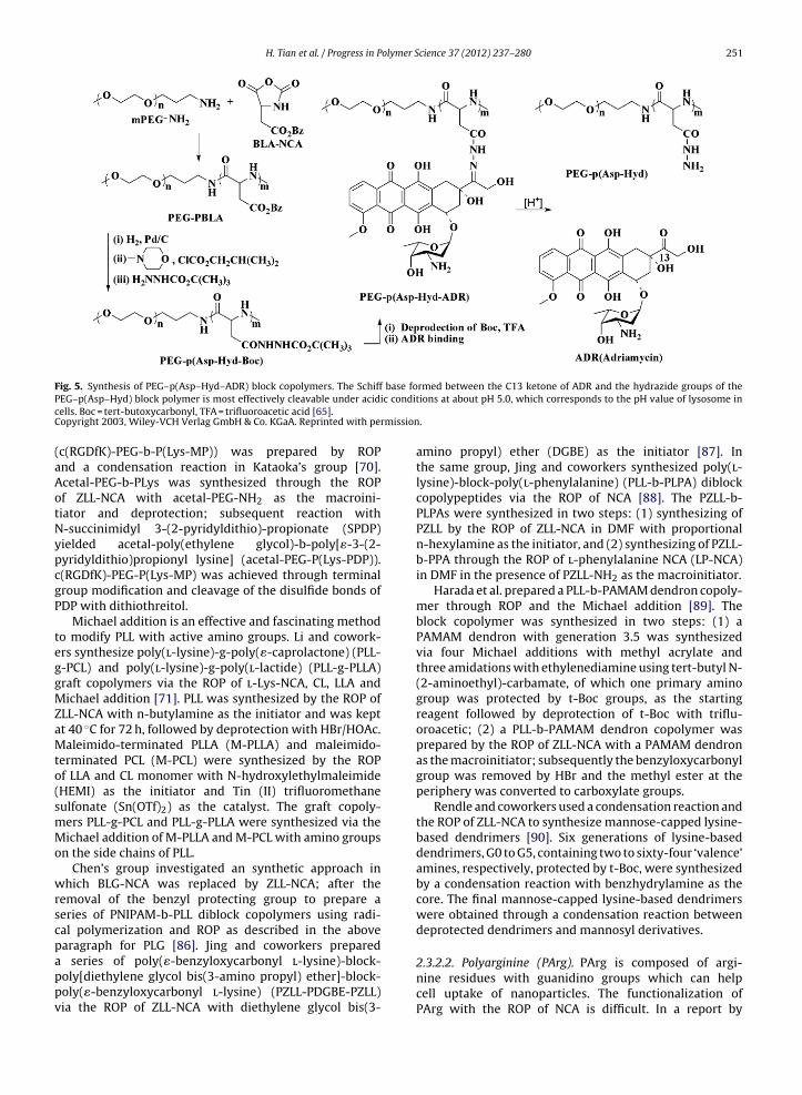
2.3.1.2. Poly(aspartic acid). Poly(aspartic acid) can be syn-thesized from aspartic acid by the ROP of �-benzyl asparticacid N-carboxy-anhydride (BAA-NCA) followed by removalof the protective benzyl groups. The two main approachesto modify poly(aspartic acid) are: (1) functional mod-ification of the side groups, such as condensation andaminolysis, and (2) introduction of a second componentto achieve different architectures.
Condensation is a simple and common methodto modify poly(aspartic acid) and its copolymers.Kataoka and coworkers reported the synthesis ofpoly(ethylene glycol)-b-poly(aspartate-hydrazone-adriamycin) (PEG-P(Asp-Hyd-ADR)) using poly(ethyleneglycol)-poly(�-benzyl-l-aspartate) (PEG-PBLA) as a tem-plate [65,66]. PEG-b-PBLA was synthesized via the ROP ofbenzyl-l-aspartate N-carboxyanhydride (BLA-NCA) with
mPEG-NH2 as the macroinitiator. Hydrazide groups wereattached to the end of the aspartate side carboxyl groupsof the block copolymer through an acid anhydride reactionafter removing the benzyl groups of PEG-b-PBLA. ADRers with both asymmetrical and symmetrical topologies via the combi-
was then conjugated to the polymer backbone through anacid-labile hydrazone bond between C13 of ADR and thehydrazide groups of the PEG-b-P(LA-Hyd) block copolymer(Fig. 5).
Aminolysis is extensively used in functionalizing PBAAwith functional amines, such as dipropylene triamine(DPT), 1,5-diamino pentane (AP), 4-(3-aminopropyl) mor-pholine (MP), etc.; it is a convenient and simple methodto obtain polymers with a controlled fraction of functionalgroups. PEG-b-poly-(3-[(3-aminopropyl)amino] propylaspartamide) was prepared through ROP and an aminoly-sis reaction by Kataoka and coworkers. PEG-b-PBLA wassynthesized by ROP and PEG-b-DPT was obtained by aside-chain aminolysis reaction of PEG-b-PBLA. In a similarway, poly([5-aminopentyl]-�,�-aspartamide) and PEG-b-poly[(3-morpholinopropyl) aspartamide]-b-poly-l-lysinewere synthesized by the same research group via the ROPof BLA-NCA and an aminolysis reaction using AP and MP,respectively [67–69].
2.3.2. Poly(basic amino acid)2.3.2.1. Polylysine. Poly(l-lysine) (PLL) with reactiveamido groups on the side chain can be prepared throughthe ROP of �-carbobenzoxy-l-lysine N-carboxyanhydride(ZLL-NCA) and deprotection. PLL is a polyelectrolyte (poly-cation) which displays pH-dependent solubility, limitedcirculation lifetime due to aggregation with oppositelycharged biopolymers, and high toxicity [59,85]. Similar tothose for PLG and poly(aspartic acid), several approachesare effective in functionalizing PLL: (1) functional modifi-cation of the side groups, such as condensation, Michaeladdition and so on, and (2) introduction of a second compo-nent to achieve block, branch, dendron-like architectures,etc.
A condensation reaction is a simple and convenientway to functionalize poly(l-lysine) directly. Cyclic RGDfunctional block copolymer c(RGDfK)-poly(ethyleneglycol)-b-poly[�-(3-mercaptopropionyl)-lysine]
H. Tian et al. / Progress in Polymer Science 37 (2012) 237– 280 251
F base foP c conditcC mission
(aAotNypcgP
teggMZaMto(smMo
wrscpappv
ig. 5. Synthesis of PEG–p(Asp–Hyd–ADR) block copolymers. The SchiffEG–p(Asp–Hyd) block polymer is most effectively cleavable under acidiells. Boc = tert-butoxycarbonyl, TFA = trifluoroacetic acid [65].opyright 2003, Wiley-VCH Verlag GmbH & Co. KGaA. Reprinted with per
c(RGDfK)-PEG-b-P(Lys-MP)) was prepared by ROPnd a condensation reaction in Kataoka’s group [70].cetal-PEG-b-PLys was synthesized through the ROPf ZLL-NCA with acetal-PEG-NH2 as the macroini-iator and deprotection; subsequent reaction with-succinimidyl 3-(2-pyridyldithio)-propionate (SPDP)ielded acetal-poly(ethylene glycol)-b-poly[�-3-(2-yridyldithio)propionyl lysine] (acetal-PEG-P(Lys-PDP)).(RGDfK)-PEG-P(Lys-MP) was achieved through terminalroup modification and cleavage of the disulfide bonds ofDP with dithiothreitol.
Michael addition is an effective and fascinating methodo modify PLL with active amino groups. Li and cowork-rs synthesize poly(l-lysine)-g-poly(�-caprolactone) (PLL--PCL) and poly(l-lysine)-g-poly(l-lactide) (PLL-g-PLLA)raft copolymers via the ROP of l-Lys-NCA, CL, LLA andichael addition [71]. PLL was synthesized by the ROP of
LL-NCA with n-butylamine as the initiator and was keptt 40 ◦C for 72 h, followed by deprotection with HBr/HOAc.aleimido-terminated PLLA (M-PLLA) and maleimido-
erminated PCL (M-PCL) were synthesized by the ROPf LLA and CL monomer with N-hydroxylethylmaleimideHEMI) as the initiator and Tin (II) trifluoromethaneulfonate (Sn(OTf)2) as the catalyst. The graft copoly-ers PLL-g-PCL and PLL-g-PLLA were synthesized via theichael addition of M-PLLA and M-PCL with amino groups
n the side chains of PLL.Chen’s group investigated an synthetic approach in
hich BLG-NCA was replaced by ZLL-NCA; after theemoval of the benzyl protecting group to prepare aeries of PNIPAM-b-PLL diblock copolymers using radi-al polymerization and ROP as described in the abovearagraph for PLG [86]. Jing and coworkers prepared
series of poly(�-benzyloxycarbonyl l-lysine)-block-oly[diethylene glycol bis(3-amino propyl) ether]-block-oly(�-benzyloxycarbonyl l-lysine) (PZLL-PDGBE-PZLL)ia the ROP of ZLL-NCA with diethylene glycol bis(3-
rmed between the C13 ketone of ADR and the hydrazide groups of theions at about pH 5.0, which corresponds to the pH value of lysosome in
.
amino propyl) ether (DGBE) as the initiator [87]. Inthe same group, Jing and coworkers synthesized poly(l-lysine)-block-poly(l-phenylalanine) (PLL-b-PLPA) diblockcopolypeptides via the ROP of NCA [88]. The PZLL-b-PLPAs were synthesized in two steps: (1) synthesizing ofPZLL by the ROP of ZLL-NCA in DMF with proportionaln-hexylamine as the initiator, and (2) synthesizing of PZLL-b-PPA through the ROP of l-phenylalanine NCA (LP-NCA)in DMF in the presence of PZLL-NH2 as the macroinitiator.
Harada et al. prepared a PLL-b-PAMAM dendron copoly-mer through ROP and the Michael addition [89]. Theblock copolymer was synthesized in two steps: (1) aPAMAM dendron with generation 3.5 was synthesizedvia four Michael additions with methyl acrylate andthree amidations with ethylenediamine using tert-butyl N-(2-aminoethyl)-carbamate, of which one primary aminogroup was protected by t-Boc groups, as the startingreagent followed by deprotection of t-Boc with triflu-oroacetic; (2) a PLL-b-PAMAM dendron copolymer wasprepared by the ROP of ZLL-NCA with a PAMAM dendronas the macroinitiator; subsequently the benzyloxycarbonylgroup was removed by HBr and the methyl ester at theperiphery was converted to carboxylate groups.
Rendle and coworkers used a condensation reaction andthe ROP of ZLL-NCA to synthesize mannose-capped lysine-based dendrimers [90]. Six generations of lysine-baseddendrimers, G0 to G5, containing two to sixty-four ‘valence’amines, respectively, protected by t-Boc, were synthesizedby a condensation reaction with benzhydrylamine as thecore. The final mannose-capped lysine-based dendrimerswere obtained through a condensation reaction betweendeprotected dendrimers and mannosyl derivatives.
2.3.2.2. Polyarginine (PArg). PArg is composed of argi-nine residues with guanidino groups which can helpcell uptake of nanoparticles. The functionalization ofPArg with the ROP of NCA is difficult. In a report by

olymer Science 37 (2012) 237– 280
P O
O
R
R1 On
252 H. Tian et al. / Progress in P
Deming’s group [91] the block copolymer poly(l-lysine)-co-polyarginine-b-poly(l-leucine) (PLL-co-PArg-b-PLLeu)was synthesized via the ROP of NCA in three steppreparation: (1) di-N-benzyloxycarbonyl-l-arginine N-carboxyanhydride (Z2Arg-NCA) was synthesized throughthe reaction between tri-N-benzyloxycarbonyl-l-arginineand �,�′-dichloromethylmethyl ether in dry methy-lene chloride, with a similar preparation for ZLL-NCA;(2) poly(di-N-benzyloxycarbonyl-l-arginine-random-N-benzyloxy-carbonyl-l-lysine)-b-Poly(l-leucine),PZLL-co-PZ2Arg-b-PLLeu, was synthesized by the ROPof ZLL-NCA and Z2Arg-NCA with Co(PMeB3B)B4 as thecatalyst, followed by addition of BLG-NCA in the dry box;(3) PLL-co-PArg-b-PLLeu was obtaind after deprotectionwith HBr.
2.3.2.3. Poly l-histidine. The electron lone pair on theunsaturated nitrogen of the imidazole ring endowspoly(l-histidine) (PLHis) with an amphoteric nature.Protonation–deprotonation on the side chain can facilitatesynthesize of LHis-NCA by a ROP, but the method is difficultand has limited application.
Bae and coworkers prepared PLHis and block copoly-mers with PEG, PLLA-b-PEG. PLHis was synthesized by theROP of protected l-Histidine NCA (i.e., Nim-DNP-l-histidineNCA) (NDLhis-NCA); the coupling of PLHis with PEG yieldedPLHis-b-PEG block copolymer after deprotection [92,93].The block copolymer was prepared in three steps as fol-lows: (1) NDLhis-NCA was obtained by a reaction betweenNim-DNP-l-histidine and thionyl chloride in THF at roomtemperature; (2) PLHis was synthesized by the ROP ofNDLhis-NCA with hexylamine or isopropylamine as the ini-tiator, followed by polymerization at room temperaturefor 72 h, with evolution of carbon dioxide; (3) PLHis-b-PEG was prepared via a coupling reaction with NHS andEDC under deprotection of 2-mercaptoethanol., A triblockcopolymer PLLA-b-PEG-b-PLHis was prepared by a simi-lar method with the ROP of NDLhis-NCA, via coupling anddeprotecting reactions [94].
In the same group, poly(l-histidine-co-phenylalanine)-b-poly(ethylene glycol) (i.e., PLHis-co-PPhe-b-PEG) wasprepared by the ROP of NDLhis-NCA and Phe-NCA, via acondensation reaction and deprotection as described above[95,96].
2.3.3. Poly(neutral amino acid)In the neutral amino acid family, there exist amino
acids with active groups, such as l-serine with a hydroxygroup, l-3,4-dihydroxyphenyl-l-alanine (l-DOPA) with adihydroxybenzyl group, and l-cysteine with a mercaptogroup.
Deming’s group prepared functionalized poly(l-serine) (PLSer), poly(l-cysteine) (PLCys) and poly(l-DOPA)(PLDOPA) through the ROP of modified monomersin combination with other components [72]. Mono-and diethyleneglycol modified PLSer (PEGnLSer) poly-mers were synthesized by the ROP of functionalized
LSer-NCA directly in three steps: (1) EGn-l-Serineswere obtained by coupling N�-tertbutyloxy-carbonyl-l-serine and 1-bromo-2-(2-methoxyethoxy) ethane or2-bromoethyl methyl ether with sodium hydride fol-2
Fig. 6. General structure of PPE.
lowed by deprotection with HCl; (2) EGn-l-Serine-NCAswere prepared with the reaction between EGn-l-Serinesand 1,1-diclorodimethylether; (3) PEGnLSers were pre-pared by the ROP of EGn-l-Serine-NCAs directly and thedegree of polymerization of the polymer was controlledwith a narrow PDI. In a similar way, well-controlleddiethyleneglycol-modified poly(l-cysteine) was pre-pared by using (2-(2-methoxyethoxy)ethyl)chloroformateinstead of 1-bromo-2-(2-methoxyethoxy) ethane.
A series of water soluble poly(l-DOPA)-co-poly(l-lysine) (PLDOPA-PLL) copolymers were prepared by theROP of �-amino acid NCA monomers [97]. PLDOPA-co-PLLcopolymers were synthesized via the ROP of N�-carbobenzyloxy-l-lysine NCA and O,O′-dicarbobenzyloxy-l-DOPA NCA with sodium tert-butoxide as the initiatorfollowed by the removal of carbobenzyloxy groups withHBr in acetic acid at room temperature.
In addition, synthesis of copolymers including PLCysand other components is an efficient approach to modifyPLCys. Jing and coworkers synthesized poly(l-cysteine)-b-poly(l-lactide) (PLCys-b-PLLA) diblock copolymer bythe ROP of NCA [98]. PLCys-b-PLLA was prepared intwo steps: (1) PLLA-NH2 was obtained through the ROPof l-lactide with stannous octoate as the catalyst andNH2-protected aminoethanol as the initiator followed bydeprotection; (2) the finial copolymer PLCys-b-PLLA wasprepared by the ROP of �-benzyloxycarbonyl-l-cysteineN-carboxyanhydride (ZLCys-NCA) with PLLA-NH2 as amacroinitiator and then removal of the t-Boc group.
2.4. Polyphosphoesters
PPEs with repeating phosphoester units in the backbone(Fig. 6) are attractive biocompatibile and biodegrad-able biomaterials because of their structural similarityto the naturally occurring nucleic acid and easy func-tionality as compared to conventional polyesters [99].The synthesis of PPE as the analogue of nucleic andteichoic acid was pioneered by Penczek and coworkersat the end of 1970s [100,101]. Since then, a num-ber of synthesis methodologies and mechanisms havebeen extensively investigated, including ROP, polyaddition,polycondensation, polytransesterification and enzyme-catalyzed polymerization [102–108]. PPEs with differentproperties, such as stimuli-responsiveness, photo-cross-linkability, and reactiveness, can be easily achieved byvarying the R1 or R2 group (Fig. 6). In the 1990s, Zhuo’s
group and Leong’s group further developed the synthesisstrategies of functional PPEs for various biomedical appli-cations such as tissue engineering scaffolds and drug/genedelivery vehicles [109,110]. Several functional groups such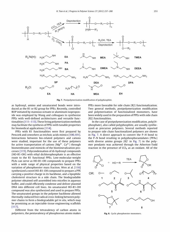
H. Tian et al. / Progress in Polymer Science 37 (2012) 237– 280 253
P O R1
O
H
On
P O R1
O
Cl
On
CCl 4, TEA
Deprotection
R' 2NH(H)
P O R1
O
(H)N
On
R'2
R2OH, TEA
DeprotectionP O R1
O
O
On
R2
Cl2 , CHCl3
R2'N(H): H2NNH EA
H2N NH
H2NNH
PA
BA
HN
NH
NNH
N+NH
NNH
MEA
DEEADMEA
TMEA
H2N NNH2 H2N
NNH2
NH2N NH2
DEA DPASP
R2O: H2NO
H2NO3
NH
OHO
O
n modifi
adRiPtmf
PIwfbc(rPwvsccpbDcTtmb[
p
the P–N bond resulting in polyphosphoramidates (PPAs)with diverse amino groups (R2′ in Fig. 7) in the poly-mer pendants was achieved through the Atherton-Toddreaction in the presence of CCl4 as an oxidant. All of the
POO
O OR
OR: O O
O
O
O
O
OOH
POO
O ClHO R
HN O
O
O
+
OO
n
TEA
OO
O
1 32 4
65
EA HA
Fig. 7. Postpolymerizatio
s hydroxyl, amino and unsaturated bonds were intro-uced as the R1 or R2 group for PPEs. Recently, controlledOP initiated by stannous octoate or aluminum isopropox-
de was employed by Wang and colleagues to synthesizePEs with well-defined architectures and versatile func-ionalities [111–113]. These living polymerization methods
ay facilitate the synthesis of PPEs with tunable propertiesor biomedical applications [114].
PPEs with R1 functionalities were first prepared byenczek and coworkers as teichoic acids mimics [100,101].nteractions between bio-related polymers and cations
ere studied, important for the use of these polymersor active transportation of cations (Mg2+
, Ca2+) throughiomembranes and mimicks of the biomineralization pro-esses [115]. Polycondensation of di-hydroxyl compoundsHO-R1-OH) with ethyl dichlorophosphate is an effectiveoute to the R1 functional PPEs. Low-molecular-weightLAs can serve as HO-R1-OH compounds to prepare PPEsith a wide range of physical properties based on the
ariation of phosphoester mass fraction. Wen at al. [116]ynthesized a novel HO-R1-OH compound to prepare a PPEarrying a positive charge in its backbone, and a lipophilicholesterol structure in a side chain. The biodegradableolymer obtained self-assembled into micelles in aqueousuffer, and could efficiently condense and deliver plasmidNA into different cell lines. An unsaturated HO-R1-OHompound was also synthesized and used to prepare PPEs.he unsaturated groups in the polymer backbone allowedhermally-induced free radical cross-linking between poly-
er chains to form a biodegradable gel in situ, which may
e promising as an injectable tissue engineering scaffolds117].Different from the tetravalency of carbon atoms inolyesters, the pentavalency of phosphorous atoms makes
MEA EH
cation of polyphosphite.
PPEs more favorable for side chain (R2) functionalization.Two general methods, postpolymerization modificationand polymerization of functionalized monomers, havebeen widely used in the preparation of PPEs with side chain(R2) functionalities.
In the case of postpolymerization modification, poly(H-phosphate), also called polyphosphite, are usually synthe-sized as precursor polymers. Several methods reportedto prepare side chain functionalized polymers are shownin Fig. 7. A direct approach to convert the P–H bond to
O87
Fig. 8. Cyclic phosphoester monomers.
olymer Science 37 (2012) 237– 280
254 H. Tian et al. / Progress in Psynthesized PPAs are biodegradable cationic polymers andhave been extensively studied as the non-viral gene car-riers [118–120]. Functional PPEs with P–O connected sidechains also could be obtained using polyphosphite as thestarting material. A chlorination process was adopted toconvert the P–H bond into the P–Cl bond followed by areaction with hydroxyl-compounds to generate PPEs withfunctional side chains (Fig. 7, R2). This method was Firstreported by Wang et al. to synthesize poly(2-aminoethylpropylene phosphate) (PPE-EA) with a biodegradable phos-phoester backbone and a �-aminoethoxy side chain thatwas shown to be an effective, nontoxic and biodegradablegene carrier [121]. Subsequently, PPEs with HA and MEAwere synthesized to study the effect of side chain struc-tures on the gene transfer efficiency [122]. Using the samemethod, Huang et al. also prepared a biodegradable PPEwith hydroxyl pendant groups as a nonionic noncondens-ing agent to enhance gene expressing in muscles [123].More recently, Koseva et al. [124] reported a new methodto prepare functional PPEs with reactive 1,3-dioxolan-2-one pendants through homolytic addition of P–H groups tothe C C double bond of 4-ethenyl-1,3-dioxolan-2-one. Thering-opening aminolysis of the cyclic carbonate in the sidechains led to the ability of the polymers to conjugate withpeptides/proteins or drug molecules conveniently, render-ing new functional PPEs as candidates for drug deliveryapplication.
ROP of cyclic phosphoester monomers provides anotherstrategy to prepare side chain-functionalized PPEs. Recentefforts on the controlled ROP of cyclic phosphoestermonomers have developed synthetic PPEs with vari-ous architectures and defined compositions [125–129].Functionalized PPEs can be achieved by the ROP of func-tionalized monomers. A monomer bearing vinyl group(monomer 5, Fig. 8) was employed in the synthesis ofvinyl group-functional PPEs that were used to preparehydrogels with different physical properties through cross-linking of the vinyl group in the pendants [130,131]. Unlikemonomers with a vinyl group, monomers with amino andhydroxyl groups need to be protected (monomer 6 and 7,Fig. 8) for them to be compatible with the polymerizationconditions. For example, an amphiphilic triblock copoly-mer PEG-b-PCL-b-PPEEA was synthesized by sequentialpolymerization of �-caprolactone and monomer 6, fol-lowed by deprotection to release amino groups. Anamphiphilic and cationic block copolymer self-assembledinto micelles as a promising delivery vehicle for small inter-fering RNA (siRNA) [132]. Similarly, Song et al. reporteda series of diblock PPEs bearing reactive hydroxyl groupsthat could self-assemble into either micelles or vesiclesin aqueous solution [133]. A novel unprotected hydroxylfunctionalized cyclic monomer 8 in Fig. 8) was recentlydesigned and synthesized by Liu et al. [134], and ahyperbranched PPE was successfully synthesized by self-condensing ROP of this monomer in the absence of acatalyst.
2.5. Others
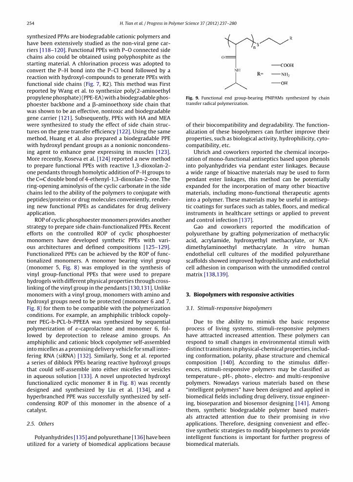
Polyanhydrides [135] and polyurethane [136] have beenutilized for a variety of biomedical applications because
Fig. 9. Functional end group-bearing PNIPAMs synthesized by chaintransfer radical polymerization.
of their biocompatibility and degradability. The function-alization of these biopolymers can further improve theirproperties, such as biological activity, hydrophilicity, cyto-compatibility, etc.
Uhrich and coworkers reported the chemical incorpo-ration of mono-functional antiseptics based upon phenolsinto polyanhydrides via pendant ester linkages. Becausea wide range of bioactive materials may be used to formpendant ester linkages, this method can be potentiallyexpanded for the incorporation of many other bioactivematerials, including mono-functional therapeutic agentsinto a polymer. These materials may be useful in antisep-tic coatings for surfaces such as tables, floors, and medicalinstruments in healthcare settings or applied to preventand control infection [137].
Gao and coworkers reported the modification ofpolyurethane by grafting polymerization of methacrylicacid, acrylamide, hydroxyethyl methacrylate, or N,N-dimethylaminoethyl methacrylate. In vitro humanendothelial cell cultures of the modified polyurethanescaffolds showed improved hydrophilicity and endothelialcell adhesion in comparison with the unmodified controlmatrix [138,139].
3. Biopolymers with responsive activities
3.1. Stimuli-responsive biopolymers
Due to the ability to mimick the basic responseprocess of living systems, stimuli-responsive polymershave attracted increased attention. These polymers canrespond to small changes in environmental stimuli withdistinct transitions in physical-chemical properties, includ-ing conformation, polarity, phase structure and chemicalcomposition [140]. According to the stimulus differ-ences, stimuli-responsive polymers may be classified astemperature-, pH-, photo-, electro- and multi-responsivepolymers. Nowadays various materials based on these“intelligent polymers” have been designed and applied inbiomedical fields including drug delivery, tissue engineer-ing, bioseparation and biosensor designing [141]. Amongthem, synthetic biodegradable polymer based materi-als attracted attention due to their promising in vivoapplications. Therefore, designing convenient and effec-
tive synthetic strategies to modify biopolymers to provideintelligent functions is important for further progress ofbiomedical materials.
olymer S
3
isttbstatLmhmbacptaftPfi
R[pmfhpOtwpa(sgCeesgacp
iPmmtshiAP
and the physiological pH ranges overlap each other [58].
H. Tian et al. / Progress in P
.1.1. Temperature responsiveTemperature is the most commonly studied among var-
ous environmental stimuli because of its physiologicalignificance. Most temperature-responsive polymers con-ain both hydrophilic and hydrophobic moieties. When theemperature changes to an appropriate range, the balanceetween these moieties is broken and reversible phaseeparation or precipitation can occur. PNIPAM is one ofhe most popular thermosensitive polymers, undergoing
rapid coil-to-globule (hydration-to-dehydration) transi-ion in an aqueous solution at its LCST of 31–32 ◦C [142]. TheCST can be appropriately elevated or reduced by copoly-erizing NIPAM with more hydrophilic monomer or more
ydrophobic monomers, respectively. Although there areany other temperature-responsive polymers synthesized
y the radical polymerization, PNIPAM is discussed heres a typical example. There are mainly two strategies toonjugate PNIPAM with synthetic biopolymers. One is torepare end-functionalized PNIPAM first for use as an ini-iator for the ROP of cyclic monomers or for coupling with
synthetic biopolymers. Another pathway is to synthesizeunctionalized biopolymer macroinitiators first for use inhe polymerization of NIPAM. The end functionalization ofNIPAM is conveniently achieved through the chain trans-er radical polymerization of NIPAM (structures are shownn Fig. 9).
Amine-terminated PNIPAM can also be obtained byAFT polymerization using an amine-bearing initiator143]. It is well known that most biodegradable syntheticolymers are prepared by the polymerization of cycliconomers initiated by hydroxyl or amine groups. There-
ore, various temperature-sensitive block copolymersave been obtained by this strategy. PNIPAM-b-PLA wasrepared through the ROP of lactide initiated by PNIPAM-H; PNIPAM-b-PLA self-assembled into micelles with
emperature-sensitive shells [144]. A similar approachas used in our group to prepare temperature- andH-responsive polypeptide-based block polymers suchs poly(N-isopropylacrylamide)-block-poly(glutamic acid)PNIPAM-b-PGA) and PNIPAM-b-PLL [77,86]. As demon-trated in the preceding paragraphs, many functionalroups can be incorporated into synthetic biopolymers.onjugation is a popular method to introduce functionalnd group-bearing PNIPAM to synthetic biopolymers. Forxample, PGA-g-PNIPAM and PLL-g-PNIPAM were synthe-ized through the condensation of amine and carboxylroups in the presence of carbodiimide [78,145]. However,
limitation of the conjugation reaction is that the purifi-ation of the final product is complicated by unwantedolymers.
Recent progress in controlled living radical polymer-zation provided more alternative routes to synthesis ofNIPAM-based biodegradable polymers. These techniquesade it possible to prepare well-defined and controlledolecular weight polymers not easily obtained by tradi-
ional radical polymerization. PLA-b-PNIPAAM-b-PLA wasynthesized by the ROP of lactide initiated from twoydroxyl groups of a RAFT agent and then used as an
nitiator for the RAFT polymerization of NIPAM [146].mphiphilic triblock copolymers with two hydrophilicNIPAM blocks flanking a central hydrophobic poly[(R)-
cience 37 (2012) 237– 280 255
3-hydroxybutyrate] (PHB) were prepared, in which thePNIPAM was initiated by the PHB macroinitiator throughATRP [147]. Polypeptide copolymers containing PNIPAMwere also synthesized using similar approaches. For exam-ple, PNIPAM-b-PGA was synthesized by a combination ofthe ROP of BLG-NCA and the RAFT polymerization of NIPAM[143]. It should be pointed out that the order of ROP andRAFT polymerization in this system could be interchanged,with a narrower PDI when PNIPAM is used as the initiator.
2-Hydroxyethylmethacrylate (HEMA) is a commonlyused monomer to promote favorable biocompatibilityof its polymers. Because of the pendent hydroxyl group,HEMA is also used as an initiator for the preparationof polyesters bearing double bonds at the end. Theresulting macromonomer can be copolymerized withNIPAM or initiated by the PNIPAM macroinitiator. Forexample, poly(N-isopropylacrylamide)-b-[2-hydroxyethylmethacrylate-poly(�-caprolactone)] (PNIPAM-b-(HEMA-PCL)) was synthesized by combining a macromonomermethod with RAFT polymerization [148]. The molecularweights of the macromonomers were generally low, whichfavors further polymerization.
Copolymers of polyester and PEG exhibiting reversiblesol–gel phase-transition in response to temperature havealso attracted considerable interest [149]. Their moleculararchitectures can be designed as ABA, BAB, AB and (AB)n
types, and they are also expanded into other structures,such a star-shape polymers [150]. Its thermo-responsiveproperties mainly depend on molecular parameters suchas the copolymer composition, hydrophilic/hydrophobicblock length and molecular weight. Recently, Lee andcoworkers prepared a series of this kind of in situ gellingcopolymers with pH sensitive segments. The gelling ofthese materials could be tuned by the combination of pHand temperature stimuli, which could expand the applica-tion of this kind of materials [149].
3.1.2. pH-responsive biopolymerspH is a well-studied stimulus because of pH vari-
ation within the body. For example, the pH in thestomach is acidic while in the intestine is more basic(pH 5–8). Generally, the pH in normal tissue and bloodis about 7.4, but in some tumors the pH is 0.5–1.0lower than the normal. When the polymers are takenup by cells there is also pH variation at different states.For example, in endosomes the pH is about 5.0–6.5,whereas lysosomes have an even lower pH (4.5–5.0)[151]. Thus, synthetic biodegradable polymers respon-sive to pH have promising application in drug delivery. Anumber of polypeptides bearing pendant ionizable groupsexhibit pH-responsive properties, such as poly(glutamicacid), poly(aspartic acid), poly(histidine), poly(lysine) andpoly(arginine). Among these, poly(glutamic acid) andpoly(aspartic acid) are acidic polypeptides while the oth-ers are basic., Poly(glutamic acid) and poly(histidine) arethe most practical pH-responsive polypeptides for in vivoapplication because their appropriate pH sensitivity ranges
Moreover, poly(glutamic acid) undergoes a sharp helix-to-coil conformational induced by pH changes, which canmimic the naturally occurring peptides to some extent.

256 H. Tian et al. / Progress in Polymer Science 37 (2012) 237– 280
amino-p
poly(l-lysine) containing ε-7-coumaryloxyacetyl-l-lysine
Fig. 10. Structure of
Poly(histidine) contains imidazole groups that can be eas-ily protonated at pH 6.5–5.0 to give a positively chargedpolyion, so this material can be used as a carrier for geneticmaterials. The pKa of polypeptides can be tuned by intro-ducing hydrophobic groups to expand their application. Forexample, Kim et al. synthesized poly[(l-histidine)-co-(l-phenylalanine)]-block-poly(ethylene glycol) (PHF-b-PEG)diblock copolymers to prepare pH-sensitive polymericmicelles [152]. It was found that the pKa value of thecopolymer can be controlled by adjusting the ratio ofhistidine to phenylalanine in the copolypeptide and byadjusting its molecular weight. In our previous work, theinfluence of hydrophobic benzyl groups on the phase tran-sition of PNIPAM-b-P(GA-co-BLG) copolymers was studied.The diblock copolymer responded sharply to a narrow pHchange in the region of pH 7.4–5.5 when the BLG contentin the P(GA-co-BLG) block was more than 30 mol% [77].
The introduction of pH-responsive properties can alsobe achieved by the conjugation of ionizable groups withthe polymer chain. For example, citraconic anhydridereacted with an amine modified PEG-b-PAsp was neg-atively charged owing to the carboxylate groups. Thecitraconic amide is stable at both neutral and basic pH, but itbecomes unstable at acidic pH and promptly degrades backto the cationic primary amine [153]. This approach can beused to prepare a charge-conversion polymer in responseto endosomal pH for gene delivery.
However, the disadvantage of biodegradable polyions isthat the excess charges can induce undesired interactionswith serum proteins leading to rapid elimination of thepolyions before reaching specific sites. One strategy toovercome this difficulty is to develop polymers bearingacid-labile groups, including mainly acetal/ketal andhydrazide. These groups are uncharged and cleavable inacidic media. Bae et al. designed acid-sensitive amphiphilicblock copolymers in which ADR was conjugated to thepolymer backbone through an acid-labile hydrazonebond between C13 of ADR and the hydrazide groups ofthe poly(ethylene glycol)-b-poly(aspartate-hydra zone)(PEG-P(Asp-Hyd)) block [65]. Tomlinson et al. [154] pre-pared water soluble and hydrolytically labile polyacetals,bearing pendant amine groups suitable for drug con-jugation (Fig. 10). Then doxorubicin was conjugated topolyacetal to get a polyacetal-doxorubicin (APEG-DOX).This polyacetals-drug conjugate displayed pH-dependentpolymer main-chain degradation. In mild degradationconditions, this conjugate can generate serinol-succ-DOX, which displayed antitumor activity in vitro. In vivobiodistribution studies in B16F10 tumor beared animalsshowed that APEG-DOX had prolonged plasma circula-
tion. Moreover, administration of APEG-DOX conjugatesled to significantly less deposition of DOX in liver andthe spleen. Polycarbonate and PEG diblock copolymersendent polyacetals.
comprising acid-labile groups were prepared by the ROPof mono-2,4,6-trimethoxybenzylidene-pentaerythritolcarbonate (TMBPEC) and mono-4-methoxybenzylidene-pentaerythritol carbonate (MBPEC). The resultant micellesshowed a high drug loading capacity and a significantlyfaster drug release rate at endosomal pH values than thatat the physiological pH [155]. However, a limitation ofthese micelles is that they are not stable at physiologicalpH level for a long time.
3.1.3. Photo responsive biopolymersLight is indispensable in human lives and also is a useful
stimulus for clinical operation. Therefore, synthesis of pho-tosensitive polymers has attracted great interest in recentyears. The most studied photo-chromic groups are azidegroups, cinnamoyl groups, spiropyran, coumarin and 2-nitrobenzyl groups (Fig. 11) [156–158].
Photosensitive properties can be applied as a trigger ofconformational change of polypeptides. For example, theconformation of PLL modified with azobenzene was inves-tigated in connection with their photochromic behaviorcaused by the trans � cis photoisomerization of the azogroups present in the side chains. These photosensitivepolypeptides exhibited photoinduced � helix changes,explained on the basis of the differences in geometry andhydrophobic character of the trans and the cis azoben-zene units [159]. Fissi et al. prepared spiropyran modifiedhigh molecular weight poly(glutamic acid). It was demon-strated that the photoisomerization of the photochromicside chains is able to trigger the coil/�-helix transitionof the macromolecular main chains only in a narrow“window” of solvent composition [160]. Besides polypep-tides, aliphatic polyesters with chromophoric units havealso been extensively investigated. For example, photo-cross-linkable polycarbonate was prepared by the ROPof a functionalized cyclic carbonate monomer containinga cinnamate moiety [46]. Li and coworkers reported awell-defined photosensitive polymer, with chromophoresconnected by pH-labile cyclic acetal linkages [161]. It wasdemonstrated that the stability of pH-labile cyclic acetallinkages could be tuned by the photoisomerization of cin-namyl chromophores, which makes these polycarbonatesinteresting in potential applications for photosensor devel-opment and light-triggered drug delivery.
Coumarin has widespread occurrence in plants andis used in biology, medicine, cosmetic and polymer sci-ence [162]. It is used as a photoinduced cross linker inbiomedical applications. Yamamoto et al. prepared co-
residues. When irradiated by light, the photo-cross-linkingreaction proceeded slowly between coumarin moieties inthe side chains to give a cis head-to-head cyclo coumarin

H. Tian et al. / Progress in Polymer Science 37 (2012) 237– 280 257
' or TR
N N
R R
N
N R(A)
N O
R2
R1
(B)
' N O
R2
R1
RO
O
RO
O
'
RO
O
RO
O
(C)
O
RO
O
O
O
OR
(D)
O
RO
O
O
O
OR
(E)
NO2
RO
NO
CHO
Fig. 11. Chemical structures and photo-induced transitions of chromophores. (A) Reversible trans (left) and cis (right) geometric isomers of azobenzene. (B)Reversible photodimerization of cinnamoyl groups. (C) Reversible photoisomerization of spirobenzopyran derivatives [156]. Copyright 2009, Royal Societyo oumariC rivativew
[ptpgmt
3
i(oDui
f Chemistry. Reprinted with permission. (D) Photo-cross-linking of the co. KGaA. Reprinted with permission. (E) Dissociation of 2-nitrobenzyl deith permission.
163]. Matsuda and coworker prepared a series of liquidolymers of coumarin-endcapped poly(ε-caprolactone-co-rimethylene carbonate) with different arms. These liquidhotocurable precursors were used to obtain desiredeometries of cross-linked biodegradable materials for theicrofabrication of medical devices and drug encapsula-
ion [164].
.1.4. Redox responsive biopolymersThe distinct redox potential difference between the
ntracellular space (reducing) and the extracellular spaceoxidizing) provides an opportunity for promising delivery
f drug based on disulfide-containing polymers [165].isulfide bonds are widespread covalent bonds in nat-ral peptides and proteins and play an important rolen the folding and stability of proteins. They are readily
n-modified polymers [157]. Copyright 2001, Wiley-VCH Verlag GmbH &s [158]. Copyright 2007, Wiley-VCH Verlag GmbH & Co. KGaA. Reprinted
cleaved in reducing conditions and reoxidized in oxi-dizing conditions. Due to these unique properties somemicelles sensitive to redox were prepared. In our previouswork, poly(l-cysteine)-b-polylactide (PLC-b-PLA) wasprepared. Because of the ease of disulfide exchange withthiols, the obtained micelles are reversible shell cross-linked (SCL) micelles [98]. Lu et al. designed macrocyclic1,4,7,10-tetraazacyclododecane-N,N′,N′′,N′′ ′-tetraaceticacid (DOTA) Gd(III) chelate and PGA conjugate containinga degradable disulfide spacer as a magnetic resonanceimaging (MRI) contrast agent. The degradable disulfidespacer between Gd(III)-DOTA and PGA is crucial for the
release and excretion of Gd(III) chelates, which showsgreat promise to solve the safety problems suffered bymacromolecular Gd(III) complexes [166]. The cleavage ofdisulfide bonds can also be designed as a trigger for drug
olymer S
258 H. Tian et al. / Progress in Pdelivery. Tang et al. prepared a disulfide-linked biodegrad-able diblock copolymer of PCL and poly(ethyl ethylenephosphate) to develop a micellar nanoparticle system forintracellular drug release triggered by glutathione (GSH)in tumor cells. As expected the intracellular DOX releasewas accelerated at a higher GSH concentration, which ledto more significant growth inhibition to A549 cells [167].Kataoka and coworkers prepared PEG-SS-P[Asp(DET)containing biocleavable disulfide linkage between PEGand polycation segments to trigger PEG detachment.They explained that the detachment of PEG at the cellsurface can increase the cellular uptake of the micelles.On the other hand, the detachment of PEG inside endo-somes would cause the interaction between the exposedcation segments and the endosomal membrane and/orwould increase endosomal pressure, enabling effectiveendosomal escape [168].
Many other stimuli-responsive polymers are used inthe biomedical fields including polymers that are respon-sive to glucose, electric or magnetic fields, ionic strengthresponsive polymers. However, little work has been doneto incorporate these stimuli-responsive polymers intosynthetic biodegradable polymers. Therefore, they notincluded here.
3.2. Electroactive biomaterials
After the discovery that electrical signals can reg-ulate cell attachment, proliferation and differentiation[169], many researchers sought to incorporate conduct-ing polymers into biomaterials to take advantage ofelectrical stimuli. In conducting polymers, polypyrrole(Ppy) has been widely studied in biomedical applications.Schmidt and coworkers made significant contributions tothe application of PPy in the biomedical field [170,171].They first employed PPy for tissue engineering purposes,demonstrating that an electrical stimulus in neurotrophicgrowth factors (NGF) induced PC-12 cells cultured onPPy significantly enhanced PC-12 neurite outgrowth andspreading. Moreover, they further studied the cause of thisenhancement and concluded that the electrical stimula-tion increased the adsorption of serum proteins, whichhelped improve the growth and proliferation of cells. Sub-sequently, Lakard et al. [172], George et al. [173] and severalother groups investigated cell adhesion and proliferationby culturing different cell lines on PPy [174–176].
Another conducting polymer, PANi, was studied byMattioli-Belmonte et al., demonstrating that PANi wasbiocompatible in vitro and in long-term animal studiesin vivo [177]. Wei and coworkers [178] reported that PANifilms functionalized with the bioactive laminin-derivedadhesion peptide YIGSR (Tyr-Ile-Gly Ser-Arg) exhibitedsignificant enhanced PC-12 cell attachment and differen-tiation.
Despite the advantage of these conducting polymers,some issues related to their application still exist: poorsolubility in most common solvents, poor polymer-cell
interaction and the lack of biodegradability. Therefore, itis a very important and challenging task to overcome theselimitations if conductive polymers are to be applied as tis-sue engineering scaffolds.cience 37 (2012) 237– 280
As models of conducting polymers, oligomers showedmany advantages over polymers, such as good solubilityand easier synthesis. Because of the same redox behavior,oligomers were used instead of conductive polymersto obtain electroactive biomaterials. In 2002, Rivers etal. first incorporated pyrrole and thiophene oligomerswith aliphatic chains using degradable ester linkages tofabricate a biodegradable electrically conducting poly-mer (BECP). The polymer showed good biocompatibilityin vitro and in vivo as shown in Fig. 12 [169]. Guo et al.[179] demonstrated that the electroactive silsesquioxaneprecursor, N-(4-aminophenyl)-N′-(4′-(3-triethoxysilyl-propyl-ureido) phenyl-1,4-quinonenediimine) (ATQD),containing aniline trimer covalently modified by oligopep-tide could be a kind of promising biomaterial for tissueengineering. Bioactive material ATQD-RGD could supportPC-12 cell adhesion and proliferation and could stimulatespontaneous neuritogenesis in PC-12 cells in the absenceof NGF as shown in Fig. 13.
Based on the above work, Chen and coworkers., choseaniline oligomers (especially aniline pentamer with dicar-boxyl end groups) as an electroactivity resource, whichwas incorporated with degradable polymers such as PLA,PCL and natural biopolymer chitosan, to prepare newbiodegradable electroactive biomaterials. First, triblockand multiblock copolymers of PLLA and aniline pentamerwere prepared by a condensation polymerization reaction[180,181]. These copolymers possessed good electroactiv-ity, solubility and biodegradability similar to pure PLA.In vitro cell evaluation showed that the electroactivecopolymers were innocuous and could indeed promotethe attachment and growth of rat C6 glioma cells. More-over, in comparison experiments with and without appliedelectrical potentials, the doped electroactive copolymersimproved the differentiation of PC-12 cells, as shown inFig. 14.
Most electroactive polymers containing oligomers can-not dissolve in water, hindering their application in vivo.A new kind of water-soluble electroactive polymer, anilinepentamer cross-linking chitosan, was prepared by Chen’sgroup [182,183]. This new polymer showed good elec-troactivity even in aqueous solution. The MTT assay, celladhesion test, and degradation assessment in the presenceof enzyme confirmed that these polymers had good bio-compatibility and biodegradability. Electroactive polymerscan improve the neuronal differentiation of PC-12 cells,even without the extra electrical stimulation, as shown inFig. 15.
For conducting polymers, high conductivity is consid-ered to affect the growth and differentiation of cells;however, oligomers without high conductivity and poly-mers containing oligomers also showed improvement incell differentiation even without extra electrical stim-ulation. In order to find the reason, aniline pentamercross-linking chitosan with a low molecular weight wasprepared [183]. Adding this electroactive polymer inculture medium can promote the differentiation and pro-
liferation of the cells because the cells readily exhibitedneural-like phenotype (Fig. 16(a and b)). While the cellsshowed only proliferation without electroactive polymerin culture medium (Fig. 16(c and d)). In the culture medium,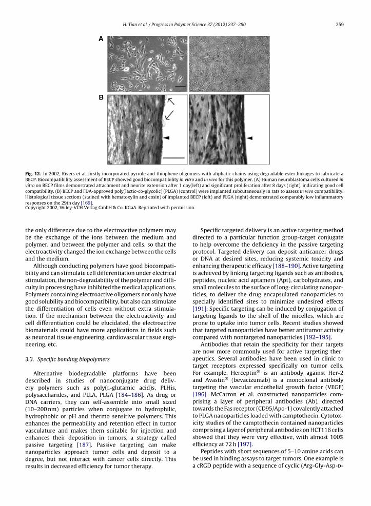
H. Tian et al. / Progress in Polymer Science 37 (2012) 237– 280 259
Fig. 12. In 2002, Rivers et al. firstly incorporated pyrrole and thiophene oligomers with aliphatic chains using degradable ester linkages to fabricate aBECP. Biocompatibility assessment of BECP showed good biocompatibility in vitro and in vivo for this polymer. (A) Human neuroblastoma cells cultured invitro on BECP films demonstrated attachment and neurite extension after 1 day(left) and significant proliferation after 8 days (right), indicating good cellcompatibility. (B) BECP and FDA-approved poly(lactic-co-glycolic) (PLGA) (control) were implanted subcutaneously in rats to assess in vivo compatibility.Histological tissue sections (stained with hematoxylin and eosin) of implanted BECP (left) and PLGA (right) demonstrated comparably low inflammatoryrC mission
tbpea
bscPgttcban
3
depD(hevepndr
esponses on the 29th day [169].opyright 2002, Wiley-VCH Verlag GmbH & Co. KGaA. Reprinted with per
he only difference due to the electroactive polymers maye the exchange of the ions between the medium andolymer, and between the polymer and cells, so that thelectroactivity changed the ion exchange between the cellsnd the medium.
Although conducting polymers have good biocompati-ility and can stimulate cell differentiation under electricaltimulation, the non-degradability of the polymer and diffi-ulty in processing have inhibited the medical applications.olymers containing electroactive oligomers not only haveood solubility and biocompatibility, but also can stimulatehe differentiation of cells even without extra stimula-ion. If the mechanism between the electroactivity andell differentiation could be elucidated, the electroactiveiomaterials could have more applications in fields suchs neuronal tissue engineering, cardiovascular tissue engi-eering, etc.
.3. Specific bonding biopolymers
Alternative biodegradable platforms have beenescribed in studies of nanoconjugate drug deliv-ry polymers such as poly(l-glutamic acid)s, PLHis,olysaccharides, and PLLA, PLGA [184–186]. As drug orNA carriers, they can self-assemble into small sized
10–200 nm) particles when conjugate to hydrophilic,ydrophobic or pH and thermo sensitive polymers. Thisnhances the permeability and retention effect in tumorasculature and makes them suitable for injection andnhances their deposition in tumors, a strategy called
assive targeting [187]. Passive targeting can makeanoparticles approach tumor cells and deposit to aegree, but not interact with cancer cells directly. Thisesults in decreased efficiency for tumor therapy..
Specific targeted delivery is an active targeting methoddirected to a particular function group-target conjugateto help overcome the deficiency in the passive targetingprotocol. Targeted delivery can deposit anticancer drugsor DNA at desired sites, reducing systemic toxicity andenhancing therapeutic efficacy [188–190]. Active targetingis achieved by linking targeting ligands such as antibodies,peptides, nucleic acid aptamers (Apt), carbohydrates, andsmall molecules to the surface of long-circulating nanopar-ticles, to deliver the drug encapsulated nanoparticles tospecially identified sites to minimize undesired effects[191]. Specific targeting can be induced by conjugation oftargeting ligands to the shell of the micelles, which areprone to uptake into tumor cells. Recent studies showedthat targeted nanoparticles have better antitumor activitycompared with nontargeted nanoparticles [192–195].
Antibodies that retain the specificity for their targetsare now more commonly used for active targeting ther-apeutics. Several antibodies have been used in clinic totarget receptors expressed specifically on tumor cells.For example, Herceptin® is an antibody against Her-2and Avastin® (bevacizumab) is a monoclonal antibodytargeting the vascular endothelial growth factor (VEGF)[196]. McCarron et al. constructed nanoparticles com-prising a layer of peripheral antibodies (Ab), directedtowards the Fas receptor (CD95/Apo-1) covalently attachedto PLGA nanoparticles loaded with camptothecin. Cytotox-icity studies of the camptothecin contained nanoparticlescomprising a layer of peripheral antibodies on HCT116 cellsshowed that they were very effective, with almost 100%
efficiency at 72 h [197].Peptides with short sequences of 5–10 amine acids canbe used in binding assays to target tumors. One example isa cRGD peptide with a sequence of cyclic (Arg-Gly-Asp-d-
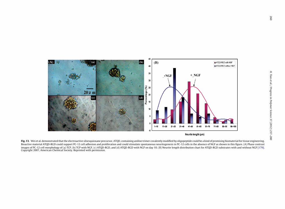
260H
. Tian
et al.
/ Progress
in Polym
er Science
37 (2012) 237– 280
Fig. 13. Wei et al. demonstrated that the electroactive silsesquioxane precursor, ATQD, containing aniline trimer covalently modified by oligopeptide could be a kind of promising biomaterial for tissue engineering.Bioactive material ATQD-RGD could support PC-12 cell adhesion and proliferation and could stimulate spontaneous neuritogenesis in PC-12 cells in the absence of NGF as shown in this figure. (A) Phase contrastimages of PC-12 cell morphology of (a) TCP, (b) TCP with NGF, (c) ATQD-RGD, and (d) ATQD-RGD with NGF on day 10; (B) Neurite length distribution chart for ATQD-RGD substrates with and without NGF [179].Copyright 2007, American Chemical Society. Reprinted with permission.

H. Tian et al. / Progress in Polymer Science 37 (2012) 237– 280 261
Fig. 14. Triblock and multiblock copolymers of PLA and aniline pentamer possessed good electroactivity, solubility and biodegradability similar to purePLA. In vitro cell evaluation showed that the electroactive copolymers were innocuous and could indeed promote the attachment and growth of rat C6glioma cells. Moreover, in the comparison experiments with and without applying electrical potentials, the doped electroactive copolymers had the abilityof improving the differentiation of PC-12 cells. (A) Representative fluorescence micrographs of PC-12 cells for the substrates (a) TCPS (−) without electricalstimulation, (b) TCPS (+) exposed to electrical stimulation, (c) EM PLAAP (−) doped with CSA without electrical stimulation, (d) EM PLAAP (+) doped withC gth of PPC
Pcgmewt
acrtt
FswC
SA exposed to electrical stimulation on day 4; (B) the mean neurite lenLAAP (+) on day 4 [181].opyright 2008, American Chemical Society. Reprinted with permission.
he-Lys), which targets the �v�3 integrin. Nasongkla et al.onjugated cRGD to maleimide-terminated poly(ethylenelycol)-PCL (MAL-PEG-PCL) copolymer with a fluorescentarker in the micelle core [198]. The result by flow cytom-
try showed that the percentage of cell uptake increasedith increasing cRGD density on the micelle surface and
here was a 30-fold increase with 76% cRGD attachment.Nucleic acid ligands such as Apt and spiegelmers
re DNA or RNA oligonucleotides that represent novellasses of target agents. In vivo studies were car-
ied out by Farokhzad and coworkers [194] by intra-umoral injection of xenografted nude mice with LNCaPumor cells using Dtxl-encapsulated nanoparticles ofig. 15. Aniline pentamer cross-linking chitosan can obviously improve the netimulation. The visualization of PC-12 neurite outgrowth by micrographs are givith electroactivity (aniline pentamer cross-linking chitosan) on the fifth day [18opyright 2008, American Chemical Society. Reprinted with permission.
C-12 cells cultured on the substrates of EM PLAAP (−), TCPS (+), and EM
poly(d,l-lactic-co-glycolic acid)-block-poly(ethylene gly-col) copolymer with the A10 2′-fluoropyrimidine RNAApt. The result showed that five of seven xenograftednude mice demonstrated complete tumor reductionwith Dtxl-Nanoparticles-Apt bioconjugates injection whileonly two of seven xenografted nude mice demon-strated complete tumor reduction with Dtxl-Nanparticlesinjection. This result demonstrates the potential util-ity of nanoparticle-Apt bioconjugates for cancer ther-apy.
Carbohydrates such as galactose and mannose are foundto be specific ligands to the asialoglycoprotein receptor(ASGPR), which is overexpressed in hepatocellular carci-
uronal differentiation of PC-12 cells even without the extra electricalen here for the substrates (A) without electroactivity (chitosan), and (B)3].
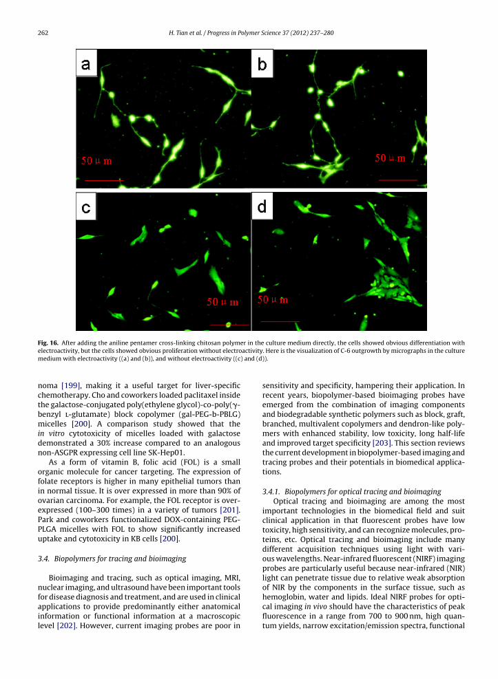
262 H. Tian et al. / Progress in Polymer Science 37 (2012) 237– 280
Fig. 16. After adding the aniline pentamer cross-linking chitosan polymer in the culture medium directly, the cells showed obvious differentiation withactivity) and (d
electroactivity, but the cells showed obvious proliferation without electromedium with electroactivity ((a) and (b)), and without electroactivity ((c
noma [199], making it a useful target for liver-specificchemotherapy. Cho and coworkers loaded paclitaxel insidethe galactose-conjugated poly(ethylene glycol)-co-poly(�-benzyl l-glutamate) block copolymer (gal-PEG-b-PBLG)micelles [200]. A comparison study showed that thein vitro cytotoxicity of micelles loaded with galactosedemonstrated a 30% increase compared to an analogousnon-ASGPR expressing cell line SK-Hep01.
As a form of vitamin B, folic acid (FOL) is a smallorganic molecule for cancer targeting. The expression offolate receptors is higher in many epithelial tumors thanin normal tissue. It is over expressed in more than 90% ofovarian carcinoma. For example, the FOL receptor is over-expressed (100–300 times) in a variety of tumors [201].Park and coworkers functionalized DOX-containing PEG-PLGA micelles with FOL to show significantly increaseduptake and cytotoxicity in KB cells [200].
3.4. Biopolymers for tracing and bioimaging
Bioimaging and tracing, such as optical imaging, MRI,nuclear imaging, and ultrasound have been important tools
for disease diagnosis and treatment, and are used in clinicalapplications to provide predominantly either anatomicalinformation or functional information at a macroscopiclevel [202]. However, current imaging probes are poor in. Here is the visualization of C-6 outgrowth by micrographs in the culture)).
sensitivity and specificity, hampering their application. Inrecent years, biopolymer-based bioimaging probes haveemerged from the combination of imaging componentsand biodegradable synthetic polymers such as block, graft,branched, multivalent copolymers and dendron-like poly-mers with enhanced stability, low toxicity, long half-lifeand improved target specificity [203]. This section reviewsthe current development in biopolymer-based imaging andtracing probes and their potentials in biomedical applica-tions.
3.4.1. Biopolymers for optical tracing and bioimagingOptical tracing and bioimaging are among the most
important technologies in the biomedical field and suitclinical application in that fluorescent probes have lowtoxicity, high sensitivity, and can recognize molecules, pro-teins, etc. Optical tracing and bioimaging include manydifferent acquisition techniques using light with vari-ous wavelengths. Near-infrared fluorescent (NIRF) imagingprobes are particularly useful because near-infrared (NIR)light can penetrate tissue due to relative weak absorptionof NIR by the components in the surface tissue, such as
hemoglobin, water and lipids. Ideal NIRF probes for opti-cal imaging in vivo should have the characteristics of peakfluorescence in a range from 700 to 900 nm, high quan-tum yields, narrow excitation/emission spectra, functional
olymer S
gsrmcab
3bndcNta
picbeeAmpo(ublcl(raAsmsiccs
otmditsClM(l
ahb
H. Tian et al. / Progress in P
roups for chemical conjugations, high chemical and phototability, excellent biocompatibility, biodegradability, exc-etability, etc. [204]. Biopolymer based NIRF probes areore available in clinic because of their long half-life in vivo
irculation, stability, low toxicity, high targeting ability and low background signal. In this part, the development ofiopolymer based NIRF probes is summarized.
.4.1.1. Biopolymer-based organic probes. Most NIRFiopolymer-based organic probes are similar to indocya-ine green (ICG) in structure. ICG is a tricarbocyanineye, approved for clinical ophthalmic retinal angiography,ardiac function, and liver function testing by FDA. ManyIRF cyanine dyes have been synthesized, and several of
hem including Cy5.5 and Alexa 680 are commerciallyvailable [205].
The most widely-applied biopolymer-based organicrobes, for which some barriers such as rapid clearance
n vivo were avoided, were developed by Weissleder andolleagues. This group explored the use of biocompati-le and optically quenched NIRF imaging probes with annzymatically cleavable polymer backbone that can gen-rate a strong NIRF signal after enzyme activation [206].
graft copolymer consisting of PLL sterically grafted byultiple MPEG chains was used as a vehicle of quenched
robes to tumors. Each PLL backbone includes an averagef 92 MPEG molecules and 11 Cy5.5 molecules, yieldingCy5.5)11-PLL-g-MPEG92. The graft copolymer contains 44nmodified lysines on the backbone as sites for cleavagey enzymes, such as trypsin and cathepsines with lysine-
ysine specificity. In in vivo experiments, the NIRF probearrier accumulated in solid tumors due to its long circu-ation time and the enhanced permeability and retentionEPR) effect. EPR effects exit in solid tumors. They haveich blood vessels which have irregular and imcompleterchitectures. Large gaps exit between endothelial cells.t same time, impaired lymphatic clearance also exit inolid tumors. All these factors lead to high selective per-eability and retention of macromolecules and lipids in
olid tumors, which was defined as EPR effect [187]. In vivomaging showed a 12-fold increase in NIRF signal after theopolymer was cleaved by lysosomal proteases in tumorells, allowing the detection of tumors with submillimeter-ized diameters.
The family of matrix metalloproteinases (MMPs) that isverexpressed in tumor comprising over 20 enzyme sub-ypes is an important target site for NIRF probes. Matrix
etalloprotease-2 (MMP-2) (i.e., gelatinase) can cause theegradation of the extracellular matrix, and is involved
n tumor infiltration and tumor-induced neovasculariza-ion. The MMP-2-activatable NIRF probes with the MMP-2ubstrate peptide Gly-Pro-Leu-Gly-Val-Arg-Gly-Lys(FITC)-ys-NH2 can be used to distinguish an MMP-2-positive cell
ine, the human fibrosarcoma cell line (HT1080), from anMP-2-negative cell line, the human breast cancer cell line
adenocarcinoma, BT20), and to determine the expressionevel of tumoral MMP-2 in vivo [207].
As MMPs, cathepsins including cathepsin D (CaD)nd cathepsin B (CaB) are overexpressed in tumor andave potential use as tumor imaging targeting sites. Aiopolymer PLL-g-MPEG based NIRF probe with CaD-
cience 37 (2012) 237– 280 263
specific peptide Gly-Pro-Ile-Cys(Et)-Phe-Phe-Arg-Leu-Gly-Lys(FITC)-Cys-NH2 was prepared by Weissleder andcoworkers, and used to demonstrate for the first time thatCaD enzyme has activity directly in vivo [208].
3.4.1.2. Biopolymer-based inorganic probes. Comparedwith organic probes, inorganic probes such as quantumdots (QDs) and gold nanoparticles (AuNPs) have severaladvantages, including tunable excitation and emissionwavelengths, high quantum yields, specific targetingability, high quality photos and chemical stability [209].Biopolymer conjugated inorganic probes have long cir-culation time, low immunogenicity, low toxicity and theability to penetrate leaky endothelial barriers to overcomethe limitation of nude inorganic probes [204].
QDs are among the most promising and fascinatingfluorescent labels for biotracing and bioimaging. Wu andcoworkers used amphiphilic PEG-b-PLL diblock copoly-mer coated QDs that could highly specifically link toimmunoglobulin G (IgG) and streptavidin to label thebreast cancer marker Her2 on the surface of fixed and livecancer cells. Compared with organic dyes such as Alexa 488,functional QDs are more specific, bright and photostable.This group simultaneously detected two cellular targetswith one excitation wavelength through functional QDswith different emission spectra [210].
Chen and coworkers reported a biopolymer based probelabeled with arginine-glycine-aspartic acid (RGD) peptideusing PEG as the linker (Fig. 17) [211]. The probe wasdemonstrated to target integrin ˛vˇ3 overexpressed by themajority of tumor vasculature in vitro, ex vivo, and in livingmice. After six hours of the injection of the QD705-PEG-RGD probe, the maximum fluorescence signal intensity wasshown in tumor tissue, with good contrast to nude QD705and Cy5.5-RGD.
Park and colleagues prepared polyethylene glycol(PEG) modified-12 nm quantum dot-streptavidin (QD-strep) nanoparticles with a biotin-cell penetrating peptide(CPP) bound to the surface via biotin-streptavidin interac-tions, which could be specifically dePEGylated in responseto the presence of the matrix MMP-2 enzyme (Fig. 18)[212]. More than nine PEG chains per single QD wereneeded to effectively inhibit the cellular uptake of modi-fied QD particles. The cellular uptake of modified QD wasdown to around 20% compared with that of a nude QD. Afterthe cleavage of the MMP-2-specific substrate in the immo-bilized PEG chains, the cells took up the QDs by exposingcell-penetrating peptides to the cell membrane.
AuNPs are also potential fluorescent agents for bio-tracing and imaging. Mason and coworkers prepared abiopolymer based fluorescent probe using 15 nm AuNPsstabilized by heterobifunctional PEG and covalently com-bined with F19 monoclonal antibodies [213]. Darkfieldmicroscopy was used to image the tissue samples nearthe nanoparticle resonance scattering maximum (560 nm).Tumor tissue samples treated with gold nanoparticleswith nonspecific control antibodies and healthy pancreatic
tissue treated with mAb-F19-conjugated gold nanoparti-cles both exhibited correctly negative results and showedno tissue imaging. Similarly, gold nanoparticles and goldnanorods immobilized by a PLGA-g-MPEG graft copolymer
264 H. Tian et al. / Progress in Polymer Science 37 (2012) 237– 280
Fig. 17. A biopolymer based probe was labeled with RGD peptide using PEGCopyright 2006, American Chemical Society. Reprinted with permission.
Fig. 18. Schematic presentation of MMP-2-enzyme-specifiCopyright 2009, American Chemical Society. Reprinted with permission.
as the linker. PEG denotes poly(ethylene glycol) (Mw = 2000) [211].
c dePEGylation and intracellular QD delivery [212].

olymer S
spTf[
3
pamnciiieb(o
3nubbb
awtcbwa[1PtwTi2t
bmepGiecaGufaght
H. Tian et al. / Progress in P
howed excellent stability in aqueous solution at differentH values and elevated temperatures as well as in serum.hese characteristics make these very powerful materialsor in vivo applications including drug delivery or imaging214].
.4.2. Biopolymers for MRIMRI can provide superb anatomical information and
roduce high quality imaging in vivo with high spatialnd temporal resolutions. Compared with other imagingodalities, MRI yields several advantages such as being
on-invasive, non-ionizing radiation, excellent soft tissueontrast, high sensitivity to blood flow and discriminationn any imaging plane. Biopolymer-based MRI bioimag-ng probes have some advantages such as low toxicity,ncreasing contrast, long half-life of in vivo circulation andasy functionalization. They have promising potentials iniomedical applicaition [202,203]. Several paramagneticGadolinium (Gd) based) and superparamagnetic (ironxide) MRI probes are discussed in detail below.
.4.2.1. Biopolymer-based paramagnetic probes. Paramag-etic or positive contrast MRI probes are metal ions withnpaired electrons, such as Gd3+, Mn2+, etc. Biopolymer-ased paramagnetic MRI probes are available in clinicsecause of their advantages, such as low toxicity and sta-ility.
Gd is an excellent MRI probe because of its short T1nd ferromagnetic properties. Gd-chelate probes modifiedith biocompatible synthetic polymers such as polypep-
ides and PEG have unique pharmacological properties andan be used in vivo. Gupta et al. reported a biopolymer-ased MRI probe that was Gd-labeled and functionalizedith a PLL-g-MPEG-DTPA (diethylenetriaminepentaacetic
cid) graft copolymer to modulate functional properties215]. In their experiments, twelve rats were treated with.5-T MRI after intravenous injection of Gd labeled MPEG-LL-DTPA with a dose of 35 �mol kg−1. The vasculatures ofhe infected and contralateral normal legs were depictedell immediately after intravenous injection of the probe.
he biopolymer-based probe was accumulated at the site ofnfection 12 h after injection and was more pronounced at4 h; the signal intensity at inflammation sites went downo the baseline after 72 h.
Lu and colleagues synthesized biodegradableiopolymer-based Gd-DTPA l-cystine bisamide copoly-ers (GCAC) as safe and effective probes for MRI and
valuated their biodegradability and efficacy in MR bloodool imaging in an animal model [216]. The polymericd(III) chelates readily degraded into smaller molecules
n incubation with 15 mM cysteine via disulfide-thiolxchange reactions in vitro and in vivo and showed strongontrast enhancement in the blood pool, major organsnd tissues of rats at a dose of 0.1 mmol Gd kg−1. TheCAC MRI probe, which can degrade into low molec-lar weight Gd(III) chelates and can be rapidly clearedrom the body, has potential for use in cardiovascular
nd tumor MRI. In a similar manner and by the sameroup, PLGA-cystamine-(Gd-DO3A) was synthesized inigh yield with 55% Gd-DO3A conjugation efficiency andhe contrast-enhanced MRI was investigated in micecience 37 (2012) 237– 280 265
bearing MDA-MB-231 breast carcinoma xenografts [217].The PLGA-cystamine-(Gd-DO3A) MRI probe resulted insignificant contrast enhancement in the blood pool, majororgans and tumor tissue, but minimal long-term tissueretention.
Biodegradable polysuccinimide (PSI) derivatives conju-gated with diethylenetriaminepentaacetic acid Gd (DTPA-Gd) [PSI-g-mPEG-C16-(DTPA-Gd)] were synthesized asbiopolymer-based MRI probes by Cho and colleagues [218].In vitro MRI tests with a concentration of Gd below9.4 × 10−4 M showed an image contrast better than that ofOmniscan®, a commercial product; the signal intensity ofPSI-mPEG-C16-(DTPA-Gd) at 1.2 × 10⁻⁴ M (Gd) was similarto the signal intensity of Omniscan at 4.7 × 10⁻⁴ M (Gd).
3.4.2.2. Biopolymer-based superparamagnetic probes.Superparamagnetic or negative contrast MRI probes basedon iron oxides can provide higher contrast and good bio-compatibility, and are easily produced. Biopolymer-basedsuperparamagnetic MRI probes are fascinating for theirstability, low toxicity and good contrast [219].
Riffle and coworkers immobilized 8.8 nm superpara-magnetic iron oxide (SPIO) particles with hydrophilictriblock copolymers containing controlled concentrationsof carboxylic acid groups in the central segments andpoly(ethylene oxide) tails (PEO-b-COOH-b-PEO). This MRIprobe was stable at the physiological pH (7.4) and lower pHvalues than 7.4, suggesting that it will be stable in blood.The saturation magnetization of this probe was approxi-mately 65–70 emu g−1, which was better than others [220].
Superparamagnetic polymeric micelles with SPIOs sta-bilized by amphiphilic MPEG-b-PCL were prepared by Gaoand colleagues as a new MRI probe with high sensitivity[221]. The hydrophilic PEG corona made the MRI probesstable in aqueous solution with an ultrasensitive MRIdetection limit of 5.2 �g mL−1 (5 nM). Similarly, an alterna-tive synthetic approach was investigated with manganesedoped superparamagnetic iron oxide (Mn-SPIO) nanopar-ticles in place of SPIO to form ultrasensitive MRI contrastagents for liver imaging by Ai’s group [222]. The MPEG-b-PCL based MRI probes had a T2 relaxivity of 270 (Mn+Fe)mM−1 s−1. With these probes, the liver contrast signalintensity changed 80% at 5 min after intravenous injectionand the time window for enhanced-MRI could reach at least36 h.
Gao and coworkers prepared multifunctional poly-meric micelles from cRGD-PEG-b-PLA, composed of DOXthat can be released through a pH-dependent mech-anism. In this process cRGD ligands first target �v�3integrins on tumor endothelial cells. and subsequentlyinduce receptor-mediated endocytosis for cell uptake, andthen SPIO nanoparticles are loaded inside the hydrophobiccore for MRI detection [223]. With the biopolymer-basedmulitifunctional MRI probes, efficient �v�3-mediatedendocytosis led to a more significant darkening contrast
of MRI from cRGD-encoded micelles compared with thatwithout cRGD. Specifically, at a level of 6.25 Fe �g mL−1, theMRI signal intensity decreased from 73.8 ± 7.0 for micelleswithout cRGD to 30.2 ± 3.5 for cRGD-encoded micelles.
olymer S
addition, stents coated with polyurethane as drug control
266 H. Tian et al. / Progress in P
3.4.3. Other biopolymer-based tracing and bioimagingIn addition to optical bioimaging and tracing, nuclear
bioimaging including planar gamma scintigraphy (PGS),single photon emission computed tomography (SPECT),positron emission tomography (PET), ultrasound imaging,and X-ray computerized axial tomography (CT) are alsoimportant imaging technologies used in clinic.
Nuclear imaging techniques have the advantages ofexcellent sensitivity, so that a minute quantity of tracermolecules is needed and rich biochemical information onpathological conditions. Therefore, these techniques arewidely used in clinics. PGS can compress the complexanatomical structure of organs into a two dimensionalrepresentation, with quantification of tissue distributionas a percentage of the injected dose. Biopolymer-basedPGS probes have been developed with high stability andspecificity. To increase targeting, Torchilin prepared apolychelating agent-biotin conjugate through the interac-tion of biotin-maleimide with PLL modified with multipleresidues of diethylenetriaminepentaacetic acid, which con-tained amino-terminal pyridylthio-propionate groups. Itcan be easily loaded with multiple metal atoms such,as 111In, and can interact specifically with avidin (asagarose) [224]. 111In-loaded DTPA-PLL-Biotin could bedelivered to an avidin-containing matrix with almost 15times more radioactivity than DTPA-biotin under the sameconditions. Similarly, Li and colleagues prepared a PGSradiotracer 111In-DTPA-PEG-C225 using PEG as a linkerbetween the monoclonal antibody and metal chelatorDTPA. The probes can be selectively localized to A431tumor xenografts, in which the endothelial growth fac-tor receptor (EGFR) is overexpressed 3-fold higher thanin MDA-MB-468 xenografts. They probes also can reduceliver uptake level, resulting in improved visualization ofEGFR-positive tumors [225,226].
Single photon emission computed tomography (SPECT)can be used to obtain three-dimensional informationwith the same probes as those for PGS, and PET, offer-ing more accurate imaging data with the limitation ofshort half-life of PET probes. 99mTechnetium-labeled DTPA-PEG-folate targeting the lymphatic system of metastatictumors was prepared and tested by Lu and coworkers[227]. The biopolymer based SPECT radionuclide enteredKB cells through the folate receptor endocytosis pathwayin vitro. DTPA-PEG-folate was in excess of 98% in radiola-beled yield while specific activity of 7.4 kBq (0.2 �Ci �g−1)was achieved. After subcutaneous injection, the probesexhibited an initial increase and subsequent decline ofaccumulation in popliteal nodes in normal Wistar rats.A fast accumulation and clearance was observed with aradioactivity amount of 5.91 ± 1.55% ID g−1 in the lymphnodes at 15 min post-injection; it increased to the maxi-mum (13.43 ± 2.21% ID g−1) at 1 h and then decreased to2.31 ± 0.28% ID g−1 at 4 h. Except for the kidney, uptakeof [99mTc]DTPA-PEG-folate by other tissues was rather lit-tle. The lymphatic vessels were readily visualized by SPECTwith this probe in a normal rabbit imagine study.
Ultrasound bioimaging has many advantages, such as
versatility, being noninvasive, low risk and being cost-effective, so it is widely used in clinics. The most usedapproach of ultrasound bioimaging is the intravenouscience 37 (2012) 237– 280
injection of microbubbles, based on the principle of usingsound waves to detect a difference in density betweenthe probe (microbubbles) and the surrounding medium(blood or soft tissue) at different time points during theexamination. Many biocompatible, biodegradable and non-toxic polymers such as PLA, PCL, poly(d,l-lactic-co-glycolicacid) and even polypeptides can be used to encapsu-late microbubbles used as ultrasound bioimaging probes[228,229].
Like MRI, CT has high spatial and temporal resolutionsand can provide superb anatomical information. Therefore,it is one of the most useful diagnostic tools. Current con-trast agents for CT are based on iodinated small moleculesbecause of their high X-ray absorption coefficient, withthe limitation of short imaging times. Biopolymer-basedCT probes are more available in clinics for their long cir-culation time in plasma and good efficacy/safety profilein vivo. AuNPs coated with PEG imparted with antibiofoul-ing properties were prepared as a CT probe by Jon andcoworkers [230]. The X-ray absorption coefficient of this CTprobe was 5.7 times higher than that of Ultravist®, a currentiodine-based CT contrast probe. This new probe showed amuch longer blood circulation time (>4 h) than Ultravist(<10 min) after intravenous injection, and accumulated inthe organs containing phagocytic cells, such as the spleenand the liver. In addition, a high contrast (2-fold) betweenhepatoma and the normal liver tissue on CT imaging wasachieved after intravenous injection of this new CT probe.
4. Biomedical application
4.1. Medical devices
Synthetic biodegradable polymers have attracted con-siderable attention for applications in medical devices, andwill play an important role in the design and function ofmedical devices. The general criteria of polymer materi-als used for medical devices include mechanical propertiesand adegradation time appropriate to the medical pur-pose. In addition, the materials should not evoke toxic orimmune responses, and they should be metabolized in thebody after fulfilling their tasks. According to these require-ments, various synthesized biodegradable polymers havebeen designed and used. Some synthesized biodegradablepolymers that have been used or show potential in selectedfields are summarized below.
4.1.1. Drug-eluting stents (DES)DES have been widely used as a default treatment
for patients with coronary artery disease. Biodegradablepolymers are always used as a biodegradable and biore-sorbable coatings on stents to control the release of drugs[231]. Studies of some stainless steel stents coated withsirolimus and PLA, such as Excel® (JW Medical System,China), Cura® (Orbus Neich, Fort Lauderdale, Florida) andSupralimus® (Sahajanand Medical Technologies, India),showed some interesting preliminary results [231,232]. In
layers were also reported [233].Beside being used as biodegradable coatings, biodegrad-
able polymers are also candidate materials for fully
olymer Science 37 (2012) 237– 280 267
bepiaI
4
pSaPu
hTiacrmotdti
4
adprPPmeamc
4
pd[
4
atrTlrctpi
Table 4Some applications and potential applications of synthetic biopolymers.
Polymer Tissue engineeringPolyanhydrides Bone tissue engineering [262]
Polyurethane Vascular tissue engineering [263]Bone tissue engineering [264]
Polyelectroactive materials Nerve tissue engineering [177]Polyphosphoester Bone tissue engineering [266]
H. Tian et al. / Progress in P
iodegradable stents [234] because of their suitable prop-rties for controlled drug release and good mechanicalerformance to prevent stents from deforming or fractur-
ng. PLLA was used to prepare a fully degradable stent [235]nd an everolimus-PLLA stent (BVS®, Abbott Laboratories,L, USA), which were under clinical evaluations [236].
.1.2. Orthopedic devicesIn the 1960s, poly(glycolide) was used to prepare com-
letely biodegradable and bioresorbable sutures [237].ince then, poly(glycolide), poly(lactide) and other materi-ls such as poly(dioxanone), poly(trimethylene carbonate),CL and poly [d,l-(lactide-co-glycolide)] have been widelysed for medical devices [238].
Orthopedic devices made from biodegradable materialsave advantages over metal or nondegradable materials.hey can transfer stress over time to the damaged area ast heals, allowing of the tissues, and there is no need of
second surgery to remove the implanted devices. Manyommercial orthopedic fixation devices such as pins andods for bone fracture fixation, and screws and plates foraxillofacial repair are made of PLLA, poly(glycolide) and
ther biodegradable polymers [238,239]. As summarized inhe review by Middleton and Tipton [238], many orthope-ic fixation devices are commercially available. However,he research on devices for load-bearing bone repair andmplantable medical devices still has a long way to go.
.1.3. Disposable medical devicesIn the 21st century, environment factors concern
ll manufacturing industries. Many disposable medicalevices, such as syringes, injection pipes, surgical gloves,ads, etc., are usually made of non-degradable plastics,esulting in serious environmental and economic issues.LA, poly(glycolide), poly[d,l-(lactide-co-glycolide)] andCL are all biodegradable. Therefore, they are promisingaterials for use in disposable medical devices meeting
nvironmental friendly requirements. These biodegrad-ble polymers have been used to prepare some disposableedical devices and will likely have a widening commer-
ial application.
.1.4. Other medical devicesBiodegradable polymers have also been used to pre-
are anastomosis rings used for intestinal resection [240],rug delivery devices [241–243], in situ forming implants244,245] and stents used in urology [246].
.2. Tissue engineering
Tissue engineering is an interdisciplinary field thatpplies the principles of engineering and life sciencesowards the development of biological substitutes used toestore, maintain or improve tissue functions [247,248].he main purpose of tissue engineering is to overcome theack of tissue donors and the immune repulsion betweeneceptors and donors. In the process of tissue engineering,
ells are cultured on a scaffold to form a natural tissue, andhen the formed tissue is implanted in the defect part in theatients. In some cases, a scaffold or a scaffold with cells ismplanted in vivo directly, and the host’s body works as
Poly(propylene fumarate) Bone tissue engineering [273]Polyesterurathane Genitourinary tissue engineering [272]
a bioreactor to construct new tissues (Fig. 19). A success-ful tissue engineering implant largely depends on the roleplayed by three-dimensional porous scaffolds. The idealscaffolds should be biodegradable and bioabsorbable tosupport the replacement of new tissues. In addition, thescaffolds must be biocompatible without inflammation orimmune reactions and possess proper mechanical proper-ties to support the growth of new tissues.
Synthetic biopolymers such as PLLA, PCL, PGA,poly(glycolide) and poly[d,l-(lactide-co-glycolide)] haveexcellent biocompatibility and good mechanical propertiesand have been licensed by FDA for in vivo applications, sothey have been the most widely used materials for tissueengineering scaffolds [249]. Considerable research hasbeen carried out about PLLA, PCL, PGA, poly(glycolide) andpoly[d,l-(lactide-co-glycolide)] used in bone tissue engi-neering [249–252], cartilage tissue engineering [253,254],cardiovascular tissue engineering [255], arterial replace-ment [256], heart valve tissue engineering [257], smallintestine tissue engineering [258], nerve regenerationtissue engineering [259–261], engineering of dermal sub-stitutes for skin regeneration [262], ligament replacement[263], genitourinary tissue engineering [264,265] andother fields. Other synthetic polymers such as polyan-hydride [266], polyurethane [267,268], polyelectroactivematerials [181], PPE [269,270] polycarbonate [33,56],poly(ester amide) [271], poly(amino acid) [272,273]biodegradable hydrogels [274,275] polyesterurathane[276], poly(propylene fumarate) [277] are also biodegrad-able and have shown many potential applications in tissueengineering. Table 4 lists the application or potentialapplications of these biodegradable polymers in tissueengineering.
A limitation of these synthetic polymers is that thematerials lack biological cues that can promote cell adhe-sion, proliferation and tissue recovery. In order to improvethe bioproperties of synthesized polymers and to enhancetheir interactions with cells, composites of syntheticbiodegradable polymers and natural polymers or naturalpolymer modified synthetic biodegradable polymers, andbiodegradable polymers blends have been used to pre-pare tissue engineering scaffolds [278–282]. In addition,biopolymers with functional groups or synthesized poly-mers modified with different methods are showing manypotential applications. For example, Deng et al. [76] pre-pared a new type of triblock copolymer poly(glutamic
acid)-b-poly(l-lactide)-b-poly(glutamic acid), with PLLAchains as the hydrophobic part and poly(glutamic acid) asthe hydrophilic part. RGD was connected to the polymer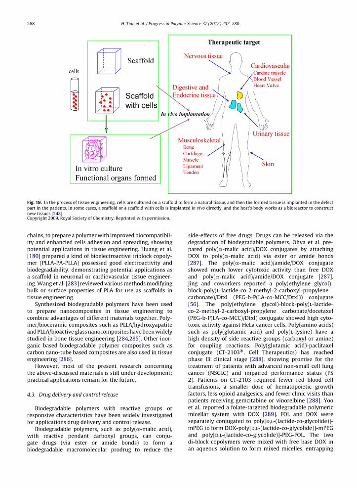
268 H. Tian et al. / Progress in Polymer Science 37 (2012) 237– 280
Fig. 19. In the process of tissue engineering, cells are cultured on a scaffold to form a natural tissue, and then the formed tissue is implanted in the defectplante
part in the patients. In some cases, a scaffold or a scaffold with cells is imnew tissues [248].Copyright 2009, Royal Society of Chemistry. Reprinted with permission.
chains, to prepare a polymer with improved biocompatibil-ity and enhancied cells adhesion and spreading, showingpotential applications in tissue engineering. Huang et al.[180] prepared a kind of bioelectroactive triblock copoly-mer (PLLA-PA-PLLA) possessed good electroactivity andbiodegradability, demonstrating potential applications asa scaffold in neuronal or cardiovascular tissue engineer-ing. Wang et al. [283] reviewed various methods modifyingbulk or surface properties of PLA for use as scaffolds intissue engineering.
Synthesized biodegradable polymers have been usedto prepare nanocomposites in tissue engineering tocombine advantages of different materials together. Poly-mer/bioceramic composites such as PLLA/hydroxyapatiteand PLLA/bioactive glass nanocomposites have been widelystudied in bone tissue engineering [284,285]. Other inor-ganic based biodegradable polymer composites such ascarbon nano-tube based composites are also used in tissueengineering [286].
However, most of the present research concerningthe above-discussed materials is still under development;practical applications remain for the future.
4.3. Drug delivery and control release
Biodegradable polymers with reactive groups orresponsive characteristics have been widely investigatedfor applications drug delivery and control release.
Biodegradable polymers, such as poly(�-malic acid),with reactive pendant carboxyl groups, can conju-gate drugs (via ester or amide bonds) to form abiodegradable macromolecular prodrug to reduce the
d in vivo directly, and the host’s body works as a bioreactor to construct
side-effects of free drugs. Drugs can be released via thedegradation of biodegradable polymers. Ohya et al. pre-pared poly(�-malic acid)/DOX conjugates by attachingDOX to poly(�-malic acid) via ester or amide bonds[287]. The poly(�-malic acid)/amide/DOX conjugateshowed much lower cytotoxic activity than free DOXand poly(�-malic acid)/amide/DOX conjugate [287].Jing and coworkers reported a poly(ethylene glycol)-block-poly(l-lactide-co-2-methyl-2-carboxyl-propylenecarbonate)/Dtxl (PEG-b-P(LA-co-MCC/Dtxl)) conjugate[56]. The poly(ethylene glycol)-block-poly(l-lactide-co-2-methyl-2-carboxyl-propylene carbonate/docetaxel(PEG-b-P(LA-co-MCC)/Dtxl) conjugate showed high cyto-toxic activity against HeLa cancer cells. Poly(amino acids)such as poly(glutamic acid) and poly(l-lysine) have ahigh density of side reactive groups (carboxyl or amine)for coupling reactions. Poly(glutamic acid)-paclitaxelconjugate (CT-2103®, Cell Therapeutics) has reachedphase III clinical stage [288], showing promise for thetreatment of patients with advanced non-small cell lungcancer (NSCLC) and impaired performance status (PS2). Patients on CT-2103 required fewer red blood celltransfusions, a smaller dose of hematopoietic growthfactors, less opioid analgesics, and fewer clinic visits thanpatients receiving gemcitabine or vinorelbine [288]. Yooet al. reported a folate-targeted biodegradable polymericmicellar system with DOX [289]. FOL and DOX wereseparately conjugated to poly[d,l-(lactide-co-glycolide)]-
mPEG to form DOX-poly[d,l-(lactide-co-glycolide)]-mPEGand poly[d,l-(lactide-co-glycolide)]-PEG-FOL. The twodi-block copolymers were mixed with free base DOX inan aqueous solution to form mixed micelles, entrapping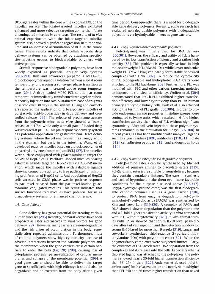
olymer S
Dmeuasutdsa
w[dtttttoeptfrwheidpwAgasicttsd
4
hpdacoatrcbggd
H. Tian et al. / Progress in P
OX aggregates within the core while exposing FOL on theicellar surface. The folate-targeted micelles exhibited
nhanced and more selective targeting ability than folatenconjugated micelles in vitro tests. The results of in vivonimal experiments with the folate-targeted micellarystem also showed significant regression in tumor vol-me and an increased accumulation of DOX in the tumorissue. These results indicate that cellular-specific drugelivery systems can be obtained by attaching specific-ite-targeting groups to biodegradable polymers withctive groups.
Stimuli-responsive biodegradable polymers, have beenidely explored as potential drug-delivery systems
290–293]. Kim and coworkers prepared a MPEG-PCLiblock copolymer aqueous solution that was a sol at roomemperature, undergoing a sol-to-gel phase transition ashe temperature was increased above room tempera-ure [294]. A drug-loaded MPEG-PCL solution at roomemperature immediately transformed into a gel on subcu-aneously injection into rats. Sustained release of drug wasbserved over 30 days in the system. Huang and cowork-rs reported the application of pH-responsive micelles ofoly(acrylic acid-b-dl-lactide) in drug delivery and con-rolled release [295]. The release of prednisone acetaterom the polymeric micelles in vitro showed a “burst”elease at pH 7.4, while only a small part of loaded drugas released at pH 1.4. This pH-responsive delivery systemas potential application for gastrointestinal tract deliv-ry systems, where the pH environment is strongly acidicn the stomach, but basic in the intestine. Wang et al.eveloped reactive micelles based on diblock copolymer ofoly(ethyl ethylene phosphate) and PCL [127]. The micellesere surface-conjugated with galactosamine to target theSGPR of HepG2 cells. Paclitaxel-loaded micelles bearingalactose ligands targeted HepG2 cells via ASGP-R medi-tion, which made the micelles with galactose ligandshowing comparable activity to free paclitaxel for inhibit-ng proliferation of HepG2 cells. And population of HepG2ells arrested in G2/M phase was in positive responseo paclitaxel released from the paclitaxel-loaded galac-osamine conjugated micelles. This result indicates thaturface functionalized micelles have potential for us asrug delivery systems for enhanced chemotherapy.
.4. Gene delivery
Gene delivery has great potential for treating variousuman diseases [296]. Recently, nonviral vectors have beenroposed as safer alternatives to viral vectors for geneelivery [297]. However, many carriers are non-degradablend the risk arises of accumulation in the body, espe-ially after repeated administration. Furthermore, mostf cationic polymers show high cytotoxicity because ofdverse interactions between the cationic polymers andhe membranes when the gene carriers cross certain bar-iers to enter the cells (Fig. 20) [298], causing loss ofytoplasmic proteins, permeabilization of cellular mem-
ranes and collapse of the membrane potential [299]. Aood gene carrier should be able to deliver the targetene to specific cells with high efficacy; it should also beegradable and be excreted from the body after a givencience 37 (2012) 237– 280 269
time period. Consequently, there is a need for biodegrad-able gene delivery polymers. Recently, some research hasevaluated non-degradable polymers with biodegradablepolycations via hydrolyzable linkers as gene carriers.
4.4.1. Poly(l-lysine)-based degradable polymersPoly(l-lysine) was initially used for DNA delivery
[300,301]. However, the efficacy and utility of PLL is ham-pered by its low transfection efficiency and a rather hightoxicity [85]. This problem is especially serious in highmolecular weight PLL (Mw 25 kDa), while lower molecularweight PLL (Mw 3 kDa) can hardly form stable nanosizedcomplexes with DNA [302]. To reduce the cytotoxicityof PLL, biodegradable and hydrophobic PLGA grafts wereattached to the PLL backbone [303]. Furthermore, PLL wasmodified with PEG and other various targeting moietiesto improve its transfection efficiency. Wolfert et al. [304]demonstrated that PEG-b-PLL exhibited higher transfec-tion efficiency and lower cytotoxicity than PLL in humanprimary embryonic kidney cells. Park et al. also attachedPEG to the termini of PLL grafts [305,306]. In order to pro-vide endosomal escape properties, histidine groups wereconjugated to lysine units, which resulted in 6-fold highertransfection activity than that of PLL without significantcytotoxicity. After tail vein injection, these polymer sys-tems remained in the circulation for 3 days [307,308]. Inrecent years, PLL has been modified with many cell ligandssuch as sugar residues [309], antibodies [310,311], folate[312], cell adhesion peptides [313], and endogenous lipids[314].
4.4.2. Poly(ˇ-amino ester)s-based degradable polymersPoly(�-amino ester)s can be synthesized by Michael
addition of primary amines to diacrylate esters [315].Poly(�-amino ester)s are suitable for gene delivery becausethey contain degradable linkages. The ease in synthesisand lack of byproducts make them even more favorablecandidates for the purpose discussed above [316,317].Poly(4-hydroxy-l-proline ester) was the first biodegrad-able cationic polymer used as a gene carrier [318],to protect DNA from enzyme degradation. Poly(�-(4-aminobutyl)-l-glycolic acid) (PAGA) was synthesized byKim and coworkers [319,320]. A complex of PAGA andDNA showed slower degradation than the polymer aloneand a 3-fold higher transfection activity in vitro comparedwith PLL, without cytotoxicity [320]. In vivo animal stud-ies with PAGA showed that serum IL-10 level peaked 5days after tail vein injection and the detection window forserum IL-10 lased for more than 9 weeks [319]. Langer andcoworkers synthesized thiol-reactive 2-(pyridyldithio)-ethylamine (PDA) with poly(amino ester) [321]. When thepolymers/DNA complexes were subjected intracellularly,the existence of GSH accelerated DNA separation from thecomplexes and its release into the cells. Especially when athiolated ligand was attached to the polyplexes, the poly-
mers showed nearly 20-fold higher transfection efficiencythan PEI-25k in vitro [322]. Anderson et al. used poly(�-amino ester) for in vivo evaluation and nearly 4 times higherthan PEI-25k and 26 times higher transfection than naked
270 H. Tian et al. / Progress in Polymer Science 37 (2012) 237– 280
Fig. 20. Barriers to gene delivery. Design requirements for gene delivery systems include the ability to (I) package therapeutic genes, (II) gain entry intor releas
cells, (III) escape from the endo-lysosomal pathway, (IV) affect DNA/vectogene expression [298].Copyright 2007, Elsevier Ltd. Reprinted with permission.
DNA was observed when poly(�-amino ester) was intratu-morally administrated [323].
4.4.3. Polyphosphoester-based degradable polymersPPE-based degradable polymers such as PPA, PPE and
polyphosphazene (PPZ) are known to be biodegradableand biocompatible in gene delivery. The polymers canbe obtained by the ROP and subsequent derivatizationof 4-methyl-2-oxo-2-hydro-1,3,2,-dioxaphospholane withspermidine and aminohexyl or(methyl-)aminoethyl sidechains [121,324]. PPE-EA consists of a phosphoester back-bone and aminoethoxy side chains [30]. PPE-EA couldcondense plasmid DNA efficiently and provide pDNA resis-tance against attacks from nucleases. After 4–9 days,complete DNA was observed to be released from theAE-PPE polyplexes at a suitable polymer/DNA ratio. Thetransfection efficiencies of PPE-AE polyplexes were abouttwo-fold higher than that of pLL-mediated transfection.PPE-EA based polyplexes also showed enhanced geneexpression in vivo [31]. PPA consists of a phosphoesterbackbone and different pendant chains via phosphorami-date bonds, and its molecular weight is about 40–50 K[118]. PPZs were prepared with dimethylaminoethyl sidechains connected to the backbones either by oxygen(DMAE-PPZ) or nitrogen (DMAEA-PPZ) [325]. DMAEA-
PPZ was carried out successfully in vivo and a highexpression level of the reporter gene in tumor wasobserved, while very low levels were seen in organs[326].e, (V) travel through the cytoplasm and into the nucleus, and (VI) enable
4.4.4. Polyethylenimine modified with degradablepolymers
PEI has been used for gene delivery under bothin vitro [327,328] and in vivo [329] conditions. How-ever, many studies demonstrated that the cytotoxicityof PEI may be due to a large excess of free polymercomplexation with pDNA [330,331] owing to a lack ofbiodegradability [332,333]. Therefore, it is important tomodify PEI with degradable polymers that can retain thehigh transfection efficiency of PEI. Many studies haveestablished that hydrophobic moieties affect transfec-tion activity of cationic polymers [334,335]. Kwon andcoworkers reported the synthesis of a peptide-based(–NHCHCO–) PEI-25k analogue with higher transfec-tion efficiency and greater biocompatibility as comparedwith PEI-25k in vivo [336]. Tian et al. investigated thehydrophobic amino acid poly(�-benzyl l-glutamate) seg-ments at the hyperbranched chain ends. The polymercould effectively condense pDNA and improve transfec-tion efficiency significantly relative to that for PEI-25kin HeLa cells [337]. Transferrin, an 80 kDa glycopro-tein, is a suitable ligand for tumor targeting becauseits receptors are over-expressed in cancers. Thereby,transferrin-PEI was used as a gene carrier in vivo, resultingin 100–500 times higher luciferase reporter gene expres-sion in tumors compared with that in other organs [338].
Chen et al. reported a series of multi-armed poly(l-glutamicacid)-graft-oligoethylenimine (MP-g-OEI) copolymers thatpossessed different charge densities. All the MP-g-OEIcopolymers exhibited lower cytotoxicity and higher gene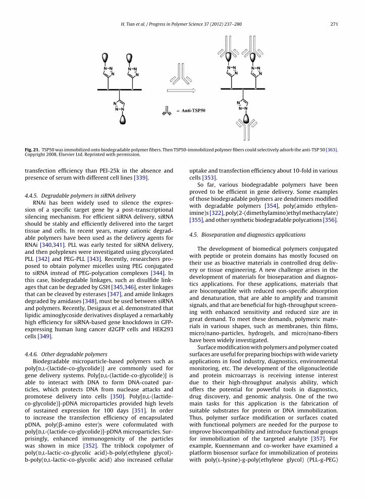
H. Tian et al. / Progress in Polymer Science 37 (2012) 237– 280 271
F TSP50-imC
tp
4
ssstaRaPpttatdalhec
4
pgatpcotpppwpb
ig. 21. TSP50 was immobilized onto biodegradable polymer fibers. Then
opyright 2008, Elsevier Ltd. Reprinted with permission.
ransfection efficiency than PEI-25k in the absence andresence of serum with different cell lines [339].
.4.5. Degradable polymers in siRNA deliveryRNAi has been widely used to silence the expres-
ion of a specific target gene by a post-transcriptionalilencing mechanism. For efficient siRNA delivery, siRNAhould be stably and efficiently delivered into the targetissue and cells. In recent years, many cationic degrad-ble polymers have been used as the delivery agents forNAi [340,341]. PLL was early tested for siRNA delivery,nd then polyplexes were investigated using glycosylatedLL [342] and PEG-PLL [343]. Recently, researchers pro-osed to obtain polymer micelles using PEG conjugatedo siRNA instead of PEG-polycation complexes [344]. Inhis case, biodegradable linkages, such as disulfide link-ges that can be degraded by GSH [345,346], ester linkageshat can be cleaved by esterases [347], and amide linkagesegraded by amidases [348], must be used between siRNAnd polymers. Recently, Desigaux et al. demonstrated thatipidic aminoglycoside derivatives displayed a remarkablyigh efficiency for siRNA-based gene knockdown in GFP-xpressing human lung cancer d2GFP cells and HEK293ells [349].
.4.6. Other degradable polymersBiodegradable microparticle-based polymers such as
oly[d,l-(lactide-co-glycolide)] are commonly used forene delivery systems. Poly[d,l-(lactide-co-glycolide)] isble to interact with DNA to form DNA-coated par-icles, which protects DNA from nuclease attacks andromotese delivery into cells [350]. Poly[d,l-(lactide-o-glycolide)]-pDNA microparticles provided high levelsf sustained expression for 100 days [351]. In ordero increase the transfection efficiency of encapsulatedDNA, poly(�-amino ester)s were coformulated witholy[d,l-(lactide-co-glycolide)]-pDNA microparticles. Sur-
risingly, enhanced immunogenicity of the particlesas shown in mice [352]. The triblock copolymer ofoly(d,l-lactic-co-glycolic acid)-b-poly(ethylene glycol)--poly(d,l-lactic-co-glycolic acid) also increased cellularmobilized polymer fibers could selectively adsorb the anti-TSP 50 [363].
uptake and transfection efficiency about 10-fold in variouscells [353].
So far, various biodegradable polymers have beenproved to be efficient in gene delivery. Some examplesof those biodegradable polymers are dendrimers modifiedwith degradable polymers [354], poly(amido ethylen-imine)s [322], poly(2-(dimethylamino)ethyl methacrylate)[355], and other synthetic biodegradable polycations [356].
4.5. Bioseparation and diagnostics applications
The development of biomedical polymers conjugatedwith peptide or protein domains has mostly focused ontheir use as bioactive materials in controlled drug deliv-ery or tissue engineering. A new challenge arises in thedevelopment of materials for bioseparation and diagnos-tics applications. For these applications, materials thatare biocompatible with reduced non-specific absorptionand denaturation, that are able to amplify and transmitsignals, and that are beneficial for high-throughput screen-ing with enhanced sensitivity and reduced size are ingreat demand. To meet these demands, polymeric mate-rials in various shapes, such as membranes, thin films,micro/nano-particles, hydrogels, and micro/nano-fibershave been widely investigated.
Surface modification with polymers and polymer coatedsurfaces are useful for preparing biochips with wide varietyapplications in food industry, diagnostics, environmentalmonitoring, etc. The development of the oligonucleotideand protein microarrays is receiving intense interestdue to their high-throughput analysis ability, whichoffers the potential for powerful tools in diagnostics,drug discovery, and genomic analysis. One of the twomain tasks for this application is the fabrication ofsuitable substrates for protein or DNA immobilization.Thus, polymer surface modification or surfaces coatedwith functional polymers are needed for the purpose toimprove biocompatibility and introduce functional groups
for immobilization of the targeted analyte [357]. Forexample, Kuennemann and co-worker have examined aplatform biosensor surface for immobilization of proteinswith poly(l-lysine)-g-poly(ethylene glycol) (PLL-g-PEG)
olymer S
272 H. Tian et al. / Progress in P[358,359]. The PLL-g-PEG coated sensor surface showedvery low non-specific absorption and its structure couldbe fine-tuned by varying the polymer compositions. As aresult, the density of the proteins on the surface was almostquantitatively controlled, which is significant for improv-ing sensitivity of the sensors. Moreover, the reversibilityof the surface immobilization enables the regeneration ofthe biosensors. Therefore, such fine-tunable platforms arepromising for use as biosensors.
Bioresponsive hydrogels that change properties inresponse to selective biological recognition events haverecently gained increasing interest for application in drugdelivery, diagnostics, and tissue engineering [360]. Fordiagnostics applications, the biological events are repre-sented by the macroscopic volume changes that generatesignals for detection. For example, Miyata et al. havereported tumor marker (�-fetoprotein, AFP) responsivegels fabricated by a biomolecular imprinting technology[361]. Lectin Con A and polyclonal anti-AFP antibodies wereconjugated into the gels to introduce the recognition sitesfor specific biomarker binding. The shrinking behavior ofthe gels in response to AFP molecules enables the visibleand accurate detection of biomarker molecules, indicatingthat the biomolecule imprinted gels have potential appli-cation in sensing application for diagnostics.
Polymeric micro/nano-spheres and micro/nano-fibershave attracted increasing attention because they can beused as substrates to immobilize biomolecules for biomed-ical applications, such as controlled drug delivery, tissueengineering, diagnostics and bioseparation. Due to theirlarge specific surface areas and relatively small size,these materials are suited to the immobilization of morecompact biomolecules with a reduced device size, butenhanced sensitivity. Jiang and coworkers have reportedthat using electrospinning polymer nanofibrous mem-branes as the solid substrates for microfludic immunoassaycan dramatically improve the sensitivity and signal-to-noise ratio as compared to the commercially availablepolymer membranes [362]. Jing and co-workers inves-tigated the immobilization of testis-specific protease 50(TSP50) on biodegradable polymer fibers [363]. The resultsshowed that the TSP50-immobilized polymer fibers couldselectively adsorb the anti-TSP 50 (Fig. 21), even in the pres-ence of high concentration of BSA (104 times). The anti-TSP50 can then be eluted simply by changing the pH, and thepolymer fibers are reusable.
5. Conclusions
Compared to biologically derived biodegradable poly-mers, synthetic biodegradable polymers do not haveimmunogenicity, but it is easier for them to be chemicallymodified and functionalized. Functionalization of syntheticbiodegradable polymers has extended the applicationscope for these biomaterials and has greatly promotedthe development in the biomedical field. The developingtrends in the functionalization of synthetic biodegradable
polymers can be predicted as followings: (1) Functional-ization processes will become easier and highly efficientas functionalization processes with mild reaction con-ditions and without harmful effects on bulk propertiescience 37 (2012) 237– 280
of polymers are pursued; (2) Functionalization will beincreasingly related to biomimetics, such that syntheticbiodegradable polymers will not simply combine differ-ent functions into one polymer, but instead, the differentfunctions should have synergistic actions; (3) The applica-tion of synthetic biodegradable polymers will be furtherexpanded, including promising potential for in vivo appli-cations. Since development in synthetic biodegradablepolymers are closely related to chemistry, materials scienceand biomedical science, any new technology in these fieldswill promote the development of synthetic biodegradablepolymers. Synthetic biodegradable polymers have beenvery important and will make more contribution to thedevelopment of biomedical science in the future.
Acknowledgments
The authors thank Jun Hu, Changwen Zhao, JunchaoWei, Chunsheng Xiao, Jianxun Ding, Yadong Liu, Jie Chen,Zhaopei Guo for their help in this review. The authorsare thankful to the National Natural Science Founda-tion of China (20604028, 20774092, 50873102, 20974109,21074129 and Key Program No: 50733003), and theNational Natural Science Foundation of China-A3 ForesightProgram (20921140264), the International Cooperationfund of Science and Technology (Key project 20071314)and Support Project (2007BAE42B02) from the Ministryof Science and Technology of China, the KnowledgeInnovation Project of Chinese Academy of Sciences (KGCX-YW-208) for financial support to this work.
References
[1] Nair LS, Laurencin CT. Biodegradable polymers as biomaterials. ProgPolym Sci 2007;32:762–98.
[2] Lou X, Detrembleur C, Jérôme R. Novel aliphatic polyestersbased on functional cyclic (di) esters. Macromol Rapid Commun2003;24:161–72.
[3] Ouchi T, Fujino A. Synthesis of poly (�-malic acid) and its hydrolysisbehavior in vitro. Die Makro Chemie 2003;190:1523–30.
[4] Kimura Y, Shirotani K, Yamane H, Kitao T. Ring-opening poly-merization of 3 (S)-[(benzyloxycarbonyl) methyl]-1, 4-dioxane-2,5-dione: a new route to a poly (.alpha.-hydroxy acid) with pendantcarboxyl groups. Macromolecules 1988;21:3338–40.
[5] Gerhardt WW, Noga DE, Hardcastle KI, Garcia AJ, Collard DM, WeckM. Functional lactide monomers: methodology and polymeriza-tion. Biomacromolecules 2006;7:1735–42.
[6] Trollsas M, Lee VY, Mecerreyes D, Lowenhielm P, Moller M, MillerRD, Hedrick JL. Hydrophilic aliphatic polyesters: design, synthe-sis, and ring-opening polymerization of functional cyclic esters.Macromolecules 2000;33:4619–27.
[7] Liu XQ, Li ZC, Du FS, Li FM. Ring-opening copolymerizationof alpha-chloromethyl-alpha-methyl-beta-propionolactone withepsilon-caprolactone. Macromol Rapid Commun 1999;20:470–4.
[8] Tian D, Dubois P, Grandfils C, Jérôme R. Ring-opening polymer-ization of 1, 4, 8-trioxaspiro [4.6]-9-undecanone: a new route toaliphatic polyesters bearing functional pendent groups. Macro-molecules 1997;30:406–9.
[9] Tian D, Dubois P, Jérôme R. Macromolecular engineering ofpolylactones and polylactides. 22. Copolymerization of [epsilon]-caprolactone and 1, 4, 8-trioxaspiro [4. 6]-9-undecanone initiatedby aluminum isopropoxide. Macromolecules 1997;30:2575–81.
[10] Mecerreyes D, Atthoff B, Boduch KA, Trollsas M, Hedrick JL. Uni-
molecular combination of an atom transfer radical polymerizationinitiator and a lactone monomer as a route to new graft copolymers.VI. Macromolecules 1999;32:5175–82.[11] Mecerreyes D, Humes J, Miller RD, Hedrick JL, Detrembleur C,Lecomte P, Jérôme R, San Roman J. First example of an unsymmetri-

olymer S
H. Tian et al. / Progress in Pcal difunctional monomer polymerizable by two living/controlledmethods. Macromol Rapid Commun 2000;21:779–84.
[12] Mecerreyes D, Miller RD, Hedrick JL, Detrembleur C, Jérôme R.Ring-opening polymerization of 6-hydroxynon-8-enoic acid lac-tone: novel biodegradable copolymers containing allyl pendentgroups. J Polym Sci Part A Polym Chem 2000;38:870–5.
[13] Bizzarri R, Chiellini F, Ober CK, Saltzman WM, Solaro R. Influ-ence of structural parameters on the ring-opening polymerizationof new alkyl malolactonate monomers and on the biocompat-ibility of polymers therefrom. Macromol Chem Phys 2002;203:1684–93.
[14] Guerin P, Vert M, Braud C, Lenz RW. Optically active poly (beta-malic acid). Polym Bull 1985;14:187–92.
[15] He B, Wan Y, Bei J, Wang S. Synthesis and cell affinity of func-tionalized poly(l-lactide-co-beta-malic acid) with high molecularweight. Biomaterials 2004;25:5239–47.
[16] Fiétier I, Borgne A, Spassky N. Synthesis of functional polyestersderived from serine. Polym Bull 1990;24:349–53.
[17] Liu XQ, Wang MX, Li ZC, Li FM. Synthesis and ring-opening polymer-ization of a-chloromethyl-a-methyl-b-propiolactone. MacromolChem Phys 1999;200:468–73.
[18] Tian D, Dubois P, Jérôme R. Macromolecular engineering ofpolylactones and polylactides. 23. Synthesis and characterizationof biodegradable and biocompatible homopolymers and blockcopolymers based on 1,4,8-trioxa[4.6]spiro-9-undecanone. Macro-molecules 1997;30:1947–54.
[19] Tian D, Halleux O, Dubois P, Jérôme R, Sobry R, Van den Boss-che G. Poly(2-oxepane-1,5-dione): a highly crystalline modifiedpoly(epsilon-caprolactone) of a high melting temperature. Macro-molecules 1998;31:924–7.
[20] Stassin F, Halleux O, Dubois P, Detrembleur C, Lecomte P, Jérôme R.Ring opening copolymerization of epsilon-caprolactone, gamma-(triethylsilyloxy)-epsilon-caprolactone and gamma-ethyleneketal-epsilon-caprolactone: a route to hetero-graft copolyesters.Macromol Symp 1999;153:27–39.
[21] Bizzarri R, Chiellini F, Solaro R, Chiellini E, Cammas-Marion S,Guerin P. Synthesis and characterization of new malolactonatepolymers and copolymers for biomedical applications. Macro-molecules 2002;35:1215–23.
[22] Lou X, Detrembleur C, Lecomte P, Jérôme R. Living ring-opening(co)polymerization of 6,7-dihydro-2(5H)-oxepinone into unsatu-rated aliphatic polyesters. Macromolecules 2001;34:5806–11.
[23] Lou XD, Detrembleur C, Lecomte P, Jérôme R. Novel unsaturatedepsilon-caprolactone polymerizable by ring-opening and ring-opening metathesis mechanisms. E-Polym 2002;034:1–12.
[24] Lou XD, Detrembleur C, Lecomte P, Jérôme R. Controlled synthesisand chemical modification of unsaturated aliphatic (co)polyestersbased on 6,7-dihydro-2(3H)-oxepinone. J Polym Sci Part A PolymChem 2002;40:2286–97.
[25] Veld in’t PJA, Dijkstra PJ, Feijen J. Synthesis of biodegradablepolyesteramides with pendant functional groups. Die MakroChemie 1992;193:2713–30.
[26] Zhu KJ, Hendren RW, Jensen K, Pitt CG. Synthesis, properties,and biodegradation of poly(1,3-trimethylene carbonate). Macro-molecules 1991;24:1736–40.
[27] Rokicki G. Aliphatic cyclic carbonates and spiroorthocarbonates asmonomers. Prog Polym Sci 2000;25:259–342.
[28] Chen XH, McCarthy SP, Gross RA. Synthesis, characterization,and epoxidation of an aliphatic polycarbonate from 2,2-(2-pentene-1,5-diyl)trimethylene carbonate ((c)HTC) ring-openingpolymerization. Macromolecules 1997;30:3470–6.
[29] Spanagel EW, Carothers WH. Macrocyclic esters. J Am Chem Soc1935;57:929–34.
[30] Al-Azemi TF, Bisht KS. Novel functional polycarbonate bylipase-catalyzed ring-opening polymerization of 5-methyl-5-benzyloxycarbonyl-1,3-dioxan-2-one. Macromolecules1999;32:6536–40.
[31] Liu ZL, Zhou Y, Zhuo RX. Synthesis and properties of func-tional aliphatic polycarbonates. J Polym Sci Part A Polym Chem2003;41:4001–6.
[32] Sanda F, Kamatani J, Endo T. Synthesis and anionic ring-openingpolymerization behavior of amino acid-derived cyclic carbonates.Macromolecules 2001;34:1564–9.
[33] Hu XL, Chen XS, Xie ZG, Cheng HB, Jing XB. Aliphatic poly(ester-
carbonate)s bearing amino groups and its RGD peptide grafting. JPolym Sci Part A Polym Chem 2008;46:7022–32.[34] Chen XH, McCarthy SP, Gross RA. Synthesis, modification, andcharacterization of l-lactide/2,2-[2-pentene-1,5-diyl]trimethylenecarbonate copolymers. Macromolecules 1998;31:662–8.
cience 37 (2012) 237– 280 273
[35] Mullen BD, Tang CN, Storey RF. New aliphaticpoly(ester-carbonates) based on 5-methyl-5-allyloxycarbonyl-1,3-dioxan-2-one. J Polym Sci Part A Polym Chem 2003;41:1978–91.
[36] He F, Wang YP, Liu G, Jia HL, Feng J, Zhuo RX. Synthesis, character-ization and ring-opening polymerization of a novel six-memberedcyclic carbonate bearing pendent allyl ether group. Polymer2008;49:1185–90.
[37] Lu CH, Shi Q, Chen XS, Lu TC, Xie ZG, Hu XL, Ma J, Jing XB. Sugars-grafted aliphatic biodegradable poly(l-lactide-co-carbonate)s byclick reaction and their specific interaction with lectin molecules. JPolym Sci Part A Polym Chem 2007;45:3204–17.
[38] Lee RS, Yang JM, Lin TF. Novel, biodegradable, functional poly(ester-carbonate)s by copolymerization of trans-4-hydroxy-l-prolinewith cyclic carbonate bearing a pendent carboxylic group. J PolymSci Part A Polym Chem 2004;42:2303–12.
[39] Zhou Y, Zhuo R-X, Liu Z-L. Synthesis and characterization of novelaliphatic poly(carbonate-ester)s with functional pendent groups.Macromol Rapid Commun 2005;26:1309–14.
[40] Shen YQ, Chen XH, Gross RA. Aliphatic polycarbonates withcontrolled quantities of d-xylofuranose in the main chain. Macro-molecules 1999;32:3891–7.
[41] Wang X-L, Zhuo R-X, Liu L-J, He F, Liu G. Synthesis and characteri-zation of novel aliphatic polycarbonates. J Polym Sci Part A PolymChem 2002;40:70–5.
[42] Yang J, Hao QH, Liu XY, Ba CY, Cao A. Novel biodegradablealiphatic poly(butylene succinate-co-cyclic carbonate)s with func-tionalizable carbonate building blocks. 1. Chemical synthesis andtheir structural and physical characterization. Biomacromolecules2004;5:209–18.
[43] Guan HL, Xie ZG, Tang ZH, Xu XY, Chen XS, Jing XB. Preparationof block copolymer of epsilon-caprolactone and 2-methyl-2-carboxyl-propylene carbonate. Polymer 2005;46:2817–24.
[44] He F, Wang YX, Feng J, Zhuo RX, Wang XL. Synthesis ofpoly [(5-benzyloxy-trimethylene carbonate)-co-(5,5 dimethyl-trimethylene carbonate)] catalyzed by immobilized lipase on silicaparticles with different size. Polymer 2003;44:3215–9.
[45] Vandenberg EJ, Tian D. A new, crystalline high meltingbis(hydroxymethyl)polycarbonate and its acetone ketal forbiomaterial applications. Macromolecules 1999;32:3613–9.
[46] Hu XL, Chen XS, Cheng HB, Jing XB. Cinnamate-functionalizedpoly(ester-carbonate): synthesis and its uv irradiation-inducedphoto-crosslinking. J Polym Sci Part A Polym Chem 2009;47:161–9.
[47] Xie ZG, Lu CH, Chen XS, Chen L, Wang Y, Hu XL, Shi Q, JingXB. Synthesis and characterization of novel poly(ester carbon-ate)s based on pentaerythritol. J Polym Sci Part A Polym Chem2007;45:1737–45.
[48] Guan HL, Xie ZG, Zhang PB, Wang X, Chen XS, Wang XH,Jing XB. Synthesis and characterization of novel biodegradableblock copolymer poly(ethylene glycol)-block-poly (l-lactide-co-2-methyl-2-carboxyl-propylene carbonate). J Polym Sci Part A PolymChem 2005;43:4771–80.
[49] Hu XL, Liu S, Chen XS, Mo GJ, Xie ZG, Jing XB. Biodegradableamphiphilic block copolymers bearing protected hydroxylgroups: synthesis and characterization. Biomacromolecules2008;9:553–60.
[50] Hu XL, Chen XS, Xie ZG, Liu S, Jing XB. Synthesis and characterizationof amphiphilic block copolymers with allyl side-groups. J Polym SciPart A Polym Chem 2007;45:5518–28.
[51] Xie ZG, Hu XL, Chen XS, Mo GJ, Sun J, Jing XB. A novel biodegradableand light-breakable diblock copolymer micelle for drug delivery.Adv Eng Mater 2009;11:B7–11.
[52] Xie ZG, Guan HL, Chen L, Tian HY, Chen XS, Jing XB.Novel biodegradable poly(ethylene glycol)-block-poly(2-methyl-2carboxyl-propylene carbonate) copolymers: synthesis, character-ization, and micellization. Polymer 2005;46:10523–30.
[53] Cheng SX, Miao ZM, Wang LS, Zhuo RX. Synthesis andcharacterization of novel biodegradable copolymers of 5-benzyloxy-1,3-dioxan-2-one and glycolide. Macromol RapidCommun 2003;24:1066–9.
[54] Ray WC, Grinstaff MW. Polycarbonate and poly(carbonate-ester)ssynthesized from biocompatible building blocks of glycerol andlactic acid. Macromolecules 2003;36:3557–62.
[55] Xie ZG, Hu XL, Chen XS, Sun J, Shi Q, Jing XB. Synthe-
sis and characterization of novel biodegradable poly(carbonateester)s with photolabile protecting groups. Biomacromolecules2008;9:376–80.[56] Xie ZG, Hu XL, Chen XS, Lu TC, Liu S, Jing XB. A biodegradablediblcok copolymer poly(ethylene glycol)-block-poly(l-lactide-co-

olymer S
274 H. Tian et al. / Progress in P2-methyl-2-carboxyl-propylene carbonate): docetaxel and RGDconjugation. J Appl Polym Sci 2008;110:2961–70.
[57] Xie Z, Guan H, Lü C, Chen X, Jing X. Synthesis and characterizationof novel biotinylated biodegradable poly (ethylene glycol)-b-poly(carbonate-lactic acid) copolymers. Acta Biomater 2005;1:635–41.
[58] Deming TJ. Synthetic polypeptides for biomedical applications.Prog Polym Sci 2007;32:858–75.
[59] Yu M, Nowak AP, Deming TJ, Pochan DJ. Methylated mono-and diethyleneglycol functionalized polylysines: nonionic,alpha-helical, water-soluble polypeptides. J Am Chem Soc1999;121:12210–1.
[60] Guo JS, Huang YB, Jing XB, Chen XS. Synthesis and characteriza-tion of functional poly(gamma-benzyl-l-glutamate) (PBLG) as ahydrophobic precursor. Polymer 2009;50:2847–55.
[61] Deng C, Tian HY, Zhang PB, Sun J, Chen XS, Jing XB. Synthe-sis and characterization of RGD peptide grafted poly(ethyleneglycol)-b-poly(l-lactide)-b-poly(l-glutamic acid) triblock copoly-mer. Biomacromolecules 2006;7:590–6.
[62] Polizzotti BD, Maheshwari R, Vinkenborg J, Kiick KL. Effects ofsaccharide spacing and chain extension on toxin inhibition byglycopolypeptides of well-defined architecture. Macromolecules2007;40:7103–10.
[63] Cabral H, Nakanishi M, Kumagai M, Jang WD, Nishiyama N, KataokaK. A photo-activated targeting chemotherapy using glutathionesensitive camptothecin-loaded polymeric micelles. Pharm Res2009;26:82–92.
[64] Xu X, Wu G, Zhang J, Wang Y, Fan Y, Ma J. Synthesis and amphiphilicproperty of ethyleneglycol functionalized polyglutamate. ActaChim Sin 2008;66:1102–6.
[65] Bae Y, Fukushima S, Harada A, Kataoka K. Design of environment-sensitive supramolecular assemblies for intracellular drug deliv-ery: polymeric micelles that are responsive to intracellular pHchange. Angew Chem Int Ed 2003;42:4640–3.
[66] Bae Y, Nishiyama N, Fukushima S, Koyama H, Yasuhiro M, KataokaK. Preparation and biological characterization of polymeric micelledrug carriers with intracellular pH-triggered drug release prop-erty: tumor permeability, controlled subcellular drug distribution,and enhanced in vivo antitumor efficacy. Bioconjugate Chem2005;16:122–30.
[67] Dong WF, Kishimura A, Anraku Y, Chuanoi S, Kataoka K.monodispersed polymeric nanocapsules: spontaneous evolutionand morphology transition from reducible hetero-PEG PICmi-celles by controlled degradation. J Am Chem Soc 2009;131:3804–5.
[68] Fukushima S, Miyata K, Nishiyama N, Kanayama N, Yamasaki Y,Kataoka K. PEGylated polyplex micelles from triblock catiomerswith spatially ordered layering of condensed pDNA and buffer-ing units for enhanced intracellular gene delivery. J Am Chem Soc2005;127:2810–1.
[69] Oana H, Kishimura A, Yonehara K, Yamasaki Y, Washizu M,Kataoka K. Spontaneous formation of giant unilamellar vesiclesfrom microdroplets of a polyion complex by thermally inducedphase separation. Angew Chem Int Ed 2009;48:4613–6.
[70] Oba M, Aoyagi K, Miyata K, Matsumoto Y, Itaka K, Nishiyama N,Yarnasaki Y, Koyama H, Kataoka K. Polyplex micelles with CyclicRGD peptide ligands and disulfide cross-links directing to theenhanced transfection via controlled intracellular trafficking. MolPharmaceutics 2008;5:1080–92.
[71] Xu N, Du FS, Li ZC. Synthesis of poly(l-lysine)-graft-polyestersthrough Michael addition and their self-assemblies in aque-ous solutions. J Polym Sci Part A Polym Chem 2007;45:1889–98.
[72] Hwang JY, Deming TJ. Methylated mono- and di(ethyleneglycol)-functionalized beta-sheet forming polypeptides. Biomacro-molecules 2001;2:17–21.
[73] Deng MX, Wang R, Rong GZ, Sun JR, Zhang XF, Chen XS,Jing XB. Synthesis of a novel structural triblock copolymer ofpoly(gamma-benzyl-l-glutamic acid)-b-poly(ethylene oxide)-b-poly(epsilon-caprolactone). Biomaterials 2004;25:3553–8.
[74] Tang DM, Lin JP, Lin SL, Zhang SN, Chen T, Tian XH. Self-assembly ofpoly(gamma-benzyl l-glutamate)-graft-poly(ethylene glycol) andits mixtures with poly(gamma-benzyl l-glutamate) homopolymer.Macromol Rapid Commun 2004;25:1241–6.
[75] Deng C, Rong GZ, Tian HY, Tang ZH, Chen XS, Jing XB. Synthesis
and characterization of poly(ethylene glycol)-b-poly (l-lactide)-b-poly(l-glutamic acid) triblock copolymer. Polymer 2005;46:653–9.[76] Deng C, Chen XS, Sun J, Lu TC, Wang WS, Jing XB. RGDpeptide grafted biodegradable amphiphilic triblock copolymer
cience 37 (2012) 237– 280
poly(glutamic acid)-b-poly(l-lactide)-b-poly(glutamic acid): syn-thesis and self-assembly. J Polym Sci Part A Polym Chem2007;45:3218–30.
[77] He CL, Zhao CW, Chen XS, Guo ZJ, Zhuang XL, Jing XB. NovelpH- and temperature-responsive block copolymers with tun-able pH-responsive range. Macromol Rapid Commun 2008;29:490–7.
[78] He CL, Zhao CW, Guo XH, Guo ZJ, Chen XS, Zhuang XL, Liu SY, JingXB. Novel temperature- and pH-responsive graft copolymers com-posed of poly(l-glutamic acid) and poly(N-isopropylacrylamide). JPolym Sci Part A Polym Chem 2008;46:4140–50.
[79] Le Hellaye M, Fortin N, Guilloteau J, Soum A, LecommandouxS, Guillaume SM. Biodegradable polycarbonate-b-polypeptideand polyester-b-polypeptide block copolymers: synthesis andnanoparticle formation towards biomaterials. Biomacromolecules2008;9:1924–33.
[80] Schatz C, Louguet S, Le Meins JF, Lecommandoux S. Polysaccharide-block-polypeptide copolymer vesicles: towards synthetic viralcapsids. Angew Chem Int Ed 2009;48:2572–5.
[81] Sun J, Chen XS, Guo JS, Shi Q, Xie ZG, Jing XB. Synthesis and self-assembly of a novel Y-shaped copolymer with a helical polypeptidearm. Polymer 2009;50:455–61.
[82] Tian HY, Deng C, Lin H, Sun JR, Deng MX, Chen XS, JingXB. Biodegradable cationic PEG-PEI-PBLG hyperbranched blockcopolymer: synthesis and micelle characterization. Biomaterials2005;26:4209–17.
[83] Tian HY, Chen XS, Lin H, Deng C, Zhang PB, Wei Y, JingXB. Micellization and reversible pH-sensitive phase transfer ofthe hyperbranched multiarm PEI-PBLG copolymer. Chem-Eur J2006;12:4305–12.
[84] Peng SM, Chen Y, Hua C, Dong CM. Dendron-like polypeptide/linearpoly(ethylene oxide) biohybrids with both asymmetrical andsymmetrical topologies synthesized via the combination of clickchemistry and ring-opening polymerization. Macromolecules2009;42:104–13.
[85] Choi YH, Liu F, Kim JS, Choi YK, Park JS, Kim SW. Polyethylene glycol-grafted poly-l-lysine as polymeric gene carrier. J Controlled Release1998;54:39–48.
[86] Zhao CW, Zhuang XL, He CL, Chen XS, Jing XB. Synthesis of novelthermo- and pH-responsive poly(l-lysine)-based copolymer and itsmicellization in water. Macromol Rapid Commun 2008;29:1810–6.
[87] Sun J, Shi Q, Chen XS, Guo JS, Jing XB. Self-assembly of ahydrophobic polypeptide containing a short hydrophilic middlesegment: vesicles to large compound micelles. Macromol ChemPhys 2008;209:1129–36.
[88] Sun J, Chen XS, Deng C, Yu HJ, Xie ZG, Jing XB. Direct for-mation of giant vesicles from synthetic polypeptides. Langmuir2007;23:8308–15.
[89] Harada A, Nakanishi K, Ichimura S, Kojima C, Kono K. Spontaneousformation of narrowly-distributed self-assembly from polyami-doamine dendron-poly(l-lysine) block copolymers through helix-coil transition of poly(l-lysine) block. J Polym Sci Part A Polym Chem2009;47:1217–23.
[90] Greatrex BW, Brodie SJ, Furneaux RH, Hook SM, McBurney WT,Painter GF, Rades T, Rendle PM. The synthesis and immunestimulating action of mannose-capped lysine-based dendrimers.Tetrahedron 2009;65:2939–50.
[91] Holowka EP, Sun VZ, Kamei DT, Deming TJ. Polyarginine segmentsin block copolypeptides drive both vesicular assembly and intra-cellular delivery. Nat Mater 2007;6:52–7.
[92] Lee ES, Na K, Bae YH. Super pH-sensitive multifunctional polymericmicelle. Nano Lett 2005;5:325–9.
[93] Lee ES, Shin HJ, Na K, Bae YH. Poly(l-histidine)-PEG block copoly-mer micelles and pH-induced destabilization. J Controlled Release2003;90:363–74.
[94] Lee ES, Gao ZG, Kim D, Park K, Kwon IC, Bae YH. Super pH-sensitive multifunctional polymeric micelle for tumor pH(e)specific TAT exposure and multidrug resistance. J ControlledRelease 2008;129:228–36.
[95] Kim D, Lee ES, Oh KT, Gao ZG, Bae YH. Doxorubicin-loadedpolymeric micelle overcomes multidrug resistance of cancer bydouble-targeting folate receptor and early endosomal pH. Small2008;4:2043–50.
[96] Lee ES, Kim D, Youn YS, Oh KT, Bae YH. A virus-mimetic nanogel
vehicle. Angew Chem Int Ed 2008;47:2418–21.[97] Yu ME, Deming TJ. Synthetic polypeptide mimics of marine adhe-sives. Macromolecules 1998;31:4739–45.
[98] Sun J, Chen XS, Lu TC, Liu S, Tian HY, Guo ZP, Jing XB.Formation of reversible shell cross-linked micelles from the

olymer S
H. Tian et al. / Progress in Pbiodegradable amphiphilic diblock copolymer poly(l-cysteine)-block-poly(l-lactide). Langmuir 2008;24:10099–106.
[99] Zhao Z, Wang J, Mao HQ, Leong KW. Polyphosphoesters in drug andgene delivery. Adv Drug Delivery Rev 2003;55:483–99.
[100] Kaluzynski K, Libisowski J, Penczek S. A new class of synthetic poly-electrolytes. Macromolecules 1976;9:365–7.
[101] Libiszowski J, Kałuzynski K, Penczek S. Polymerization of cyclicesters of phosphoric acid. VI. Poly(alkyl ethylene phosphates).Polymerization of 2-alkoxy-2-oxo-1,3,2-dioxaphospholans andstructure of polymers. J Polym Sci Polym Chem Ed 1978;16:1275–83.
[102] Biedron T, Kaluzynski K, Pretula J, Kubisa P, Penczek S, Loont-jens T. Synthesis of high molar mass poly(alkylene phosphate)sby polyaddition of diepoxides to difunctional phosphoric acids:unusual elimination of the side reactions. J Polym Sci Part A PolymChem 2001;39:3024–33.
[103] Feng J, Zhuo RX, He F. Enzymatic ring-opening copolymerizationof trimethylene carbonate and ethylene ethyl phosphate. Sci ChinaSer B Chem 2003;46:160–7.
[104] Klosinski P, Penczek S. Synthesis of models of teichoic-acids by ring-opening polymerization. Macromolecules 1983;16:316–20.
[105] Penczek S, Duda A, Kaluzynski K, Lapienis G, Nyk A, SzymanskiR. Thermodynamics and kinetics of ring-opening polymerizationof cyclic alkylene phosphates. Makromol Chem, Macromol Symp1993;73:91–101.
[106] Penczek S, Pretula J, Kaluzynski K. Polyesters of phospho-ric acid: synthesis and kinetics of hydrolysis. Macromol Symp1998;130:305–17.
[107] Pretula J, Kaluzynski K, Szymanski R, Penczek S. Polycondensa-tion route leading to poly(alkylene phosphate)s. Macromol Symp1997;122:269–74.
[108] Pretula J, Kaluzynski K, Szymanski R, Penczek S. Transesterificationof oligomeric dialkyl phosphonates, leading to the high-molecular-weight poly-H-phosphonates. J Polym Sci Part A Polym Chem1999;37:1365–81.
[109] Leong KW, Mao HQ, Zhuo RX. Biodegradable polymerswith a phosphoryl-containing backbone: tissue engineeringand controlled drug delivery applications. Chin J Polym Sci1995;13:289–314.
[110] Dahiyat BI, Richards M, Leong KW. Controlled release frompoly(phosphoester) matrices. J Controlled Release 1995;33:13–21.
[111] Chen DP, Wang J. Synthesis and characterization of blockcopolymer of polyphosphoester and poly(epsilon-caprolactone).Macromolecules 2006;39:473–5.
[112] Wang YC, Shen SY, Wu QP, Chen DP, Wang J, SteinhoffG, Ma N. Block copolymerization of epsilon-caprolactone and2-methoxyethyl ethylene phosphate initiated by aluminumisopropoxide: Synthesis, characterization, and kinetics. Macro-molecules 2006;39:8992–8.
[113] Xiao CS, Wang YC, Du JZ, Chen XS, Wang J. Kinetics and mech-anism of 2-ethoxy-2-oxo-1,3,2-dioxaphospholane polymerizationinitiated by stannous octoate. Macromolecules 2006;39:6825–31.
[114] Wang YC, Yuan YY, Du JZ, Yang XZ, Wang J. Recent progress inpolyphosphoesters: from controlled synthesis to biomedical appli-cations. Macromol Biosci 2009;9:1154–64.
[115] Penczek S, Pretula J, Kaluzynski K. Poly(alkylene phosphates):from synthetic models of biomacromolecules and biomembranestoward polymer—inorganic hybrids (mimicking biomineraliza-tion). Biomacromolecules 2005;6:547–51.
[116] Wen J, Mao HQ, Li WP, Lin KY, Leong KW. Biodegradablepolyphosphoester micelles for gene delivery. J Pharm Sci 2004;93:2142–57.
[117] Qiu JJ, Liu CM, Hu F, Guo XD, Zheng QX. Synthesis of unsaturatedpolyphosphoester as a potential injectable tissue engineering scaf-fold materials. J Appl Polym Sci 2006;102:3095–101.
[118] Wang J, Gao SJ, Zhang PC, Wang S, Mao HQ, Leong KW. Polyphos-phoramidate gene carriers: effect of charge group on gene transferefficiency. Gene Ther 2004;11:1001–10.
[119] Zhang PC, Wang J, Leong KW, Mao HQ. Ternary complexes com-prising polyphosphoramidate gene carriers with different types ofcharge groups improve transfection efficiency. Biomacromolecules2005;6:54–60.
[120] Zhang XQ, Wang XL, Zhang PC, Liu ZL, Zhuo RX, Mao HQ, LeongKW. Galactosylated ternary DNA/polyphosphoramidate nanopar-
ticles mediate high gene transfection efficiency in hepatocytes. JControlled Release 2005;102:749–63.[121] Wang J, Mao HQ, Leong KW. A novel biodegradable gene car-rier based on polyphosphoester. J Am Chem Soc 2001;123:9480–1.
cience 37 (2012) 237– 280 275
[122] Wang J, Huang SW, Zhang PC, Mao HQ, Leong KW. Effect ofside-chain structures on gene transfer efficiency of biodegradablecationic polyphosphoesters. Int J Pharm 2003;265:75–84.
[123] Huang SW, Wang J, Zhang PC, Mao HQ, Zhuo RX, Leong KW. Water-soluble and nonionic polyphosphoester: synthesis, degradation,biocompatibility and enhancement of gene expression in mousemuscle. Biomacromolecules 2004;5:306–11.
[124] Koseva N, Bogomilova A, Atkova K, Troev K. New functionalpolyphosphoesters: design and characterization. React FunctPolym 2008;68:954–66.
[125] Du JZ, Chen DP, Wang YC, Xiao CS, Lu YJ, Wang J, Zhang GZ. Syn-thesis and micellization of amphiphilic brush-coil block copolymerbased on poly(epsilon-caprolactone) and PEGylated polyphospho-ester. Biomacromolecules 2006;7:1898–903.
[126] Wang YC, Li Y, Yang XZ, Yuan YY, Yan LF, Wang J. Tunablethermosensitivity of biodegradable polymer micelles of poly(epsilon-caprolactone) and polyphosphoester block copolymers.Macromolecules 2009;42:3026–32.
[127] Wang YC, Liu XQ, Sun TM, Xiong MH, Wang J. Functional-ized micelles from block copolymer of polyphosphoester andpoly(epsilon-caprolactone) for receptor-mediated drug delivery. JControlled Release 2008;128:32–40.
[128] Wang YC, Tang LY, Li Y, Wang J. Thermoresponsive blockcopolymers of poly(ethylene glycol) and polyphosphoester:thermo-induced self-assembly, biocompatibility, and hydrolyticdegradation. Biomacromolecules 2009;10:66–73.
[129] Iwasaki Y, Wachiralarpphaithoon C, Akiyoshi K. Novel thermore-sponsive polymers having biodegradable phosphoester backbones.Macromolecules 2007;40:8136–8.
[130] Du JZ, Sun TM, Weng SQ, Chen XS, Wang J. Synthesis and char-acterization of photo-cross-linked hydrogels based on biodegrad-able polyphosphoesters and poly(ethylene glycol) copolymers.Biomacromolecules 2007;8:3375–81.
[131] Wachiralarpphaithoon C, Iwasaki Y, Akiyoshi K. Enzyme-degradable phosphorylcholine porous hydrogels cross-linked withpolyphosphoesters for cell matrices. Biomaterials 2007;28:984–93.
[132] Sun TM, Du JZ, Yan LF, Mao HQ, Wang J. Self-assembled biodegrad-able micellar nanoparticles of amphiphilic and cationic blockcopolymer for siRNA delivery. Biomaterials 2008;29:4348–55.
[133] Song WJ, Du JZ, Liu NJ, Dou S, Cheng J, Wang J. Functionalized diblockcopolymer of poly(epsilon-caprolactone) and polyphosphoesterbearing hydroxyl pendant groups: synthesis, characterization, andself-assembly. Macromolecules 2008;41:6935–41.
[134] Liu J, Huang W, Zhou Y, Yan D. Synthesis of hyperbranchedpolyphosphates by self-condensing ring-opening polymerizationof HEEP without catalyst. Macromolecules 2009;42:4394–9.
[135] Tamada J, Langer R. The development of polyanhydrides fordrug delivery applications. J Biomater Sci Polym Ed 1992;3:315–53.
[136] Grad S, Kupcsik L, Gorna K, Gogolewski S, Alini M. The use ofbiodegradable polyurethane scaffolds for cartilage tissue engineer-ing: potential and limitations. Biomaterials 2003;24:5163–71.
[137] Prudencio A, Carbone AL, Griffin J, Uhrich KE. A novel approach forincorporation of mono-functional bioactive phenols into polyan-hydrides. Macromol Rapid Commun 2009;30:1101–8.
[138] Zhu YB, Gao CY, Guan JJ, Shen JC. Promoting the cytocompatibility ofpolyurethane scaffolds via surface photo-grafting polymerizationof acrylamide. J Mater Sci Mater Med 2004;15:283–9.
[139] Ma ZW, Mao ZW, Gao CY. Surface modification and property anal-ysis of biomedical polymers used for tissue engineering. ColloidsSurf, B 2007;60:137–57.
[140] Alarcon CDH, Pennadam S, Alexander C. Stimuli responsive poly-mers for biomedical applications. Chem Soc Rev 2005;34:276–85.
[141] Jeong B, Gutowska A. Lessons from nature: stimuli-responsivepolymers and their biomedical applications. Trends Biotechnol2002;20:305–11.
[142] Schild HG. Poly (N-Isopropylacrylamide)—experiment, theory andapplication. Prog Polym Sci 1992;17:163–249.
[143] Zhang XQ, Li JG, Li W, Zhang A. Synthesis and characteriza-tion of thermo- and pH-responsive double-hydrophilic diblockcopolypeptides. Biomacromolecules 2007;8:3557–67.
[144] Kohori F, Sakai K, Aoyagi T, Yokoyama M, Sakurai Y, Okano T.Preparation and characterization of thermally responsive blockcopolymer micelles comprising poly(N-isopropylacrylamide-b-dl-
lactide). J Controlled Release 1998;55:87–98.[145] Konak C, Reschel T, Oupicky D, Ulbrich K. Thermally con-trolled association in aqueous solutions of poly(l-lysine)grafted with poly (N-isopropylacrylamide). Langmuir 2002;18:8217–22.

olymer S
276 H. Tian et al. / Progress in P[146] You YZ, Hong CY, Wang WP, Lu WQ, Pan CY. Preparation andcharacterization of thermally responsive and biodegradable blockcopolymer comprised of PNIPAAM and PLA by combination of ROPand RAFT methods. Macromolecules 2004;37:9761–7.
[147] Loh XJ, Zhang ZX, Wu YL, Lee TS, Li J. Synthesis of novelbiodegradable thermoresponsive triblock copolymers based onpoly[(R)-3-hydroxybutyrate] and poly(N-isopropylacrylamide)and their formation of thermoresponsive micelles. Macromolecules2009;42:194–202.
[148] Zhu JL, Zhang XZ, Cheng H, Li YY, Cheng SX, Zhuo RX. Syn-thesis and characterization of well-defined, AmphiphilicPoly(N-isopropylacrylamide)-b-[2-hydroxyethyl methacrylate-poly(epsilon-caprolactone)](n) graft copolymers by RAFTpolymerization and macromonomer method. J Polym Sci Part APolym Chem 2007;45:5354–64.
[149] He CL, Kim SW, Lee DS. In situ gelling stimuli-sensitive blockcopolymer hydrogels for drug delivery. J Controlled Release2008;127:189–207.
[150] Jeong B, Choi YK, Bae YH, Zentner G, Kim SW. New biodegradablepolymers for injectable drug delivery systems. J Controlled Release1999;62:109–14.
[151] Bawa P, Pillay V, Choonara YE, du Toit LC. Stimuli-responsivepolymers and their applications in drug delivery. Biomed Mater2009;4:1–15.
[152] Kim GM, Bae YH, Jo WH. pH-induced micelle formation ofpoly(histidine-co-phenylalanine)-block-poly(ethylene glycol) inaqueous media. Macromol Biosci 2005;5:1118–24.
[153] Lee Y, Fukushima S, Bae Y, Hiki S, Ishii T, Kataoka K. A proteinnanocarrier from charge-conversion polymer in response to endo-somal pH. J Am Chem Soc 2007;129:5362–3.
[154] Tomlinson R, Heller J, Brocchini S, Duncan R. Polyacetal-doxorubicin conjugates designed for pH-dependent degradation.Bioconjugate Chem 2003;14:1096–106.
[155] Chen W, Meng F, Li F, Ji S-J, Zhong Z. pH-responsive biodegradablemicelles based on acid-labile polycarbonate hydrophobe: syn-thesis and triggered drug release. Biomacromolecules 2009;10:1727–35.
[156] Li MH, Keller P. Stimuli-responsive polymer vesicles. Soft Matter2009;5:927–37.
[157] Ohkawa K, Shoumura K, Yamada M, Nishida A, Shirai H,Yamamoto H. Photoresponsive peptide and polypeptide sys-tems, 14. Biodegradation of photocrosslinkable copolypeptidehydrogels containing l-ornithine and �-7-coumaryloxyacetyl-l-ornithine residues. Macromol Biosci 2001;1:149–56.
[158] Zhao Y. Rational design of light-controllable polymer micelles.Chem Rec 2007;7:286–94.
[159] Fissi A, Pieroni O, Ciardelli F. Photoresponsivepolymers—azobenzene-containing poly(l-lysine). Biopolymers1987;26:1993–2007.
[160] Fissi A, Pieroni O, Angelini N, Lenci F. Photoresponsive polypep-tides, photochromic and conformational behavior of spiropyran-containing poly(l-glutamate)s under acid conditions. Macro-molecules 1999;32:7116–21.
[161] Cui WG, Qi MB, Li XH, Huang SZ, Zhou SB, Weng J. Electrospunfibers of acid-labile biodegradable polymers with acetal groups aspotential drug carriers. Int J Pharm 2008;361:47–55.
[162] Trenor SR, Shultz AR, Love BJ, Long TE. Coumarins in polymers: fromlight harvesting to photo-cross-linkable tissue scaffolds. Chem Rev2004;104:3059–77.
[163] Yamamoto H, Kitsuki T, Nishida A, Asada K, Ohkawa K. Pho-toresponsive peptide and polypeptide systems. 13. Photoinducedcross-linked gel and biodegradation properties of copoly(l-lysine)containing epsilon-7-coumaryloxyacetyl-l-lysine residues. Macro-molecules 1999;32:1055–61.
[164] Kwon IK, Matsuda T. Photo-polymerized microarchitectural con-structs prepared by microstereolithography (mu SL) using liquidacrylate-end-capped trimethylene carbonate-based prepolymers.Biomaterials 2005;26:1675–84.
[165] Meng FH, Hennink WE, Zhong Z. Reduction-sensitive poly-mers and bioconjugates for biomedical applications. Biomaterials2009;30:2180–98.
[166] Lu ZR, Wang XH, Parker DL, Goodrich KC, Buswell HR. Poly(l-glutamic acid) Gd(III)-DOTA conjugate with a degradable spacer formagnetic resonance imaging. Bioconjugate Chem 2003;14:715–9.
[167] Tang LY, Wang YC, Li Y, Du JZ, Wang J. Shell-detachable micellesbased on disulfide-linked block copolymer as potential carrier forintracellular drug delivery. Bioconjugate Chem 2009;20:1095–9.
[168] Takae S, Miyata K, Oba M, Ishii T, Nishiyama N, Itaka K, YamasakiY, Koyama H, Kataoka K. PEG-detachable polyplex micelles based
cience 37 (2012) 237– 280
on disulfide-linked block catiomers as bioresponsive nonviral genevectors. J Am Chem Soc 2008;130:6001–9.
[169] Rivers TJ, Hudson TW, Schmidt CE. Synthesis of a novel, biodegrad-able electrically conducting polymer for biomedical applications.Adv Funct Mater 2002;12:33–7.
[170] Kotwal A, Schmidt CE. Electrical stimulation alters protein adsorp-tion and nerve cell interactions with electrically conductingbiomaterials. Biomaterials 2001;22:1055–64.
[171] Schmidt CE, Shastri VR, Vacanti JP, Langer R. Stimulation of neuriteoutgrowth using an electrically conducting polymer. Proc Natl AcadSci USA 1997;94:8948–53.
[172] Lakard S, Herlem G, Valles-Villareal N, Michel G, Propper A, GharbiT, Fahys B. Culture of neural cells on polymers coated surfaces forbiosensor applications. Biosens Bioelectron 2005;20:1946–54.
[173] George PM, Lyckman AW, LaVan DA, Hegde A, Leung Y, Avasare R,Testa C, Alexander PM, Langer R, Sur M. Fabrication and biocom-patibility of polypyrrole implants suitable for neural prosthetics.Biomaterials 2005;26:3511–9.
[174] Wan Y, Wu H, Wen DJ. Porous-conductive chitosan scaffolds fortissue engineering, 1—Preparation and characterization. MacromolBiosci 2004;4:882–90.
[175] Castano H, O’Rear EA, McFetridge PS, Sikavitsas VI. Polypyr-role thin films formed by admicellar polymerization support theosteogenic differentiation of mesenchymal stem cells. MacromolBiosci 2004;4:785–94.
[176] Jiang XP, Marois Y, Traore A, Tessier D, Dao LH, Guidoin R, Zhang Z.Tissue reaction to polypyrrole-coated polyester fabrics: an in vivostudy in rats. Tissue Eng 2002;8:635–47.
[177] Mattioli-Belmonte M, Giavaresi G, Biagini G, Virgili L, Giacomini M,Fini M, Giantomassi F, Natali D, Torricelli P, Giardino R. Tailoringbiomaterial compatibility: in vivo tissue response versus in vitrocell behavior. Int J Artif Organs 2003;26:1077–85.
[178] Wei Y, Lelkes PI, MacDiarmid AG, Guterman E, Cheng S, PalouianK, Bidez PR. Electroactive polymers and nanostructured materialsfor neural tissue engineering. In: Zhou QF, Cheng ZD, editors. Elec-troactive polymers and nanostructured materials for neural tissueengineering in contemporary topics in advanced polymer scienceand technology. Beijing China: Peking University Press; 2004. p.430–6.
[179] Guo Y, Li MY, Mylonakis A, Han JJ, MacDiarmid AG, ChenXS, Lelkes PI, Wei Y. Electroactive oligoaniline-containingself-assembled monolayers for tissue engineering applications.Biomacromolecules 2007;8:3025–34.
[180] Huang LH, Hu J, Lang L, Wang X, Zhang PB, Jing XB, Wang XH,Chen XS, Lelkes PI, MacDiarmid AG, Wei Y. Synthesis and charac-terization of electroactive and biodegradable ABA block copolymerof polylactide and aniline pentamer. Biomaterials 2007;28:1741–51.
[181] Huang LH, Zhuang XL, Hu J, Lang L, Zhang PB, Wang YS, Chen XS, WeiY, Jing XB. Synthesis of biodegradable and electroactive multiblockpolylactide and aniline pentamer copolymer for tissue engineeringapplications. Biomacromolecules 2008;9:850–8.
[182] Hu J, Huang LH, Zhuang XL, Chen XS, Wei Y, Jing XB. A new oxidationstate of aniline pentamer observed in water-soluble electroactiveoligoaniline-chitosan polymer. J Polym Sci Part A Polym Chem2008;46:1124–35.
[183] Hu J, Huang LH, Zhuang XL, Zhang PB, Lang L, Chen XS, Wei Y, JingXB. Electroactive aniline pentamer cross-linking chitosan for stim-ulation growth of electrically sensitive cells. Biomacromolecules2008;9:2637–44.
[184] Haider M, Megeed Z, Ghandehari H. Genetically engineered poly-mers: status and prospects for controlled release. J ControlledRelease 2004;95:1–26.
[185] Li C. Poly (l-glutamic acid)-anticancer drug conjugates. Adv DrugDelivery Rev 2002;54:695–713.
[186] Sugahara SI, Kajiki M, Kuriyama H, Kobayashi TR. Paclitaxel deliverysystems: the use of amino acid linkers in the conjugation of pacli-taxel with carboxymethyldextran to create prodrugs. Biol PharmBull 2002;25:632–41.
[187] Maeda H, Greish K, Fang J. The EPR effect and polymeric drugs: aparadigm shift for cancer chemotherapy in the 21st century. AdvPolym Sci 2006;193:103–21.
[188] Allen TM. Ligand-targeted therapeutics in anticancer therapy. NatRev Cancer 2002;2:750–63.
[189] Marcucci F, Lefoulon F. Active targeting with particulate drug car-riers in tumor therapy: fundamentals and recent progress. DrugDiscovery Today 2004;9:219–28.
[190] Schrama D, Reisfeld RA, Becker JC. Antibody targeted drugs as can-cer therapeutics. Nat Rev Drug Discovery 2006;5:147–59.

olymer S
H. Tian et al. / Progress in P[191] Brannon-Peppas L, Blanchette JO. Nanoparticle and targetedsystems for cancer therapy. Adv Drug Delivery Rev 2004;56:1649–59.
[192] Kirpotin DB, Drummond DC, Shao Y, Shalaby MR, Hong K,Nielsen UB, Marks JD, Benz CC, Park JW. Antibody targeting oflong-circulating lipidic nanoparticles does not increase tumorlocalization but does increase internalization in animal models.Cancer Res 2006;66:6732–40.
[193] Bartlett DW, Su H, Hildebrandt IJ, Weber WA, Davis ME. Impactof tumor-specific targeting on the biodistribution and efficacy ofsiRNA nanoparticles measured by multimodality in vivo imaging.Proc Natl Acad Sci USA 2007;104:15549–54.
[194] Farokhzad OC, Cheng JJ, Teply BA, Sherifi I, Jon S, Kantoff PW,Richie JP, Langer R. Targeted nanoparticle-aptamer bioconju-gates for cancer chemotherapy in vivo. Proc Natl Acad Sci USA2006;103:6315–20.
[195] Gu F, Zhang L, Teply BA, Mann N, Wang A, Radovic-Moreno AF,Langer R, Farokhzad OC. Precise engineering of targeted nanoparti-cles by using self-assembled biointegrated block copolymers. ProcNatl Acad Sci USA 2008;105:2586–91.
[196] Kabbinavar F, Hurwitz HI, Fehrenbacher L, Meropol NJ, NovotnyWF, Lieberman G, Griffing S, Bergsland E. Phase II, randomizedtrial comparing bevacizumab plus fluorouracil (FU)/leucovorin (LV)with FU/LV alone in patients with metastatic colorectal cancer. JClin Oncol 2003;21:60–5.
[197] McCarron PA, Marouf WM, Quinn DJ, Fay F, Burden RE, OlwillSA, Scott CJ. Antibody targeting of camptothecin-loaded PLGAnanoparticles to tumor cells. Bioconjugate Chem 2008;19:1561–9.
[198] Nasongkla N, Shuai X, Ai H, Weinberg BD, Pink J, Boothman DA, GaoJM. cRGD-functionalized polymer micelles for targeted doxorubicindelivery. Angew Chem Int Edit 2004;43:6323–7.
[199] Ross JF, Chaudhuri PK, Ratnam M. Differential regulation of folatereceptor isoforms in normal and malignant-tissues in-vivo and inestablished cell-lines—physiological and clinical implications. Can-cer 1994;73:2432–43.
[200] Jeong Y-I, Seo S-J, Park I-K, Lee H-C, Kang I-C, Akaike T,Cho C-S. Cellular recognition of paclitaxel-loaded polymericnanoparticles composed of poly([gamma]-benzyl l-glutamate) andpoly(ethylene glycol) diblock copolymer endcapped with galactosemoiety. Int J Pharm 2005;296:151–61.
[201] Wands JR, Blum HE. Primary hepatocellular-carcinoma. N Engl JMed 1991;325:729–31.
[202] Khemtong C, Kessinger CW, Gao JM. Polymeric nanomedicinefor cancer MR imaging and drug delivery. Chem Commun2009:3497–510.
[203] Kim JH, Park K, Nam HY, Lee S, Kim K, Kwon IC. Polymers forbioimaging. Prog Polym Sci 2007;32:1031–53.
[204] Funovics M, Weissleder R, Tung CH. Protease sensors for bioimag-ing. Anal Bioanal Chem 2003;377:956–63.
[205] Lee S, Park K, Kim K, Choi K, Kwon IC. Activatable imaging probeswith amplified fluorescent signals. Chem Commun 2008:4250–60.
[206] Weissleder R, Tung CH, Mahmood U, Bogdanov A. In vivo imaging oftumors with protease-activated near-infrared fluorescent probes.Nat Biotechnol 1999;17:375–8.
[207] Bremer C, Bredow S, Mahmood U, Weissleder R, Tung CH. Opticalimaging of matrix metalloproteinase-2 activity in tumors: feasibil-ity study in a mouse model. Radiology 2001;221:523–9.
[208] Tung CH, Mahmood U, Bredow S, Weissleder R. In vivo imagingof proteolytic enzyme activity using a novel molecular reporter.Cancer Res 2000;60:4953–8.
[209] Michalet X, Pinaud FF, Bentolila LA, Tsay JM, Doose S, Li JJ, Sundare-san G, Wu AM, Gambhir SS, Weiss S. Quantum dots for live cells,in vivo imaging, and diagnostics. Science 2005;307:538–44.
[210] Wu XY, Liu HJ, Liu JQ, Haley KN, Treadway JA, Larson JP, Ge NF, PealeF, Bruchez MP. Immunofluorescent labeling of cancer marker Her2and other cellular targets with semiconductor quantum dots. NatBiotechnol 2003;21:41–6.
[211] Cai WB, Shin DW, Chen K, Gheysens O, Cao QZ, Wang SX, Gamb-hir SS, Chen XY. Peptide-labeled near-infrared quantum dots forimaging tumor vasculature in living subjects. Nano Lett 2006;6:669–76.
[212] Mok H, Bae KH, Ahn CH, Park TG. PEGylated and MMP-2 specificallyDePEGylated quantum dots: comparative evaluation of cellularuptake. Langmuir 2009;25:1645–50.
[213] Eck W, Craig G, Sigdel A, Ritter G, Old LJ, Tang L, Bren-nan MF, Allen PJ, Mason MD. PEGylated gold nanoparticlesconjugated to monoclonal F19 antibodies as targeted labelingagents for human pancreatic carcinoma tissue. ACS Nano 2008;2:2263–72.
cience 37 (2012) 237– 280 277
[214] Prencipe G, Tabakman SM, Welsher K, Liu Z, Goodwin AP, ZhangL, Henry J, Dai HJ. PEG branched polymer for functionalization ofnanomaterials with ultralong blood circulation. J Am Chem Soc2009;131:4783–7.
[215] Gupta H, Wilkinson RA, Bogdanov AA, Callahan RJ, WeisslederR. Inflammation: imaging with methoxy poly(ethylene glycol)-poly-l-lysine-DTPA, a long-circulating graft copolymer. Radiology1995;197:665–9.
[216] Kaneshiro TL, Ke T, Jeong EK, Parker DL, Lu ZR. Gd-DTPAl-cystine bisamide copolymers as novel biodegradable macro-molecular contrast agents for MR blood pool imaging. Pharm Res2006;23:1285–94.
[217] Ke TY, Feng Y, Guo JY, Parker DL, Lu ZR. Biodegradable cystaminespacer facilitates the clearance of Gd(III) chelates in poly(glutamicacid) Gd-DO3A conjugates for contrast-enhanced MR imaging.Magn Reson Imaging 2006;24:931–40.
[218] Lee HY, Jee HW, Seo SM, Kwak BK, Khang G, Cho SH. Diethylene-triaminepentaacetic acid-gadolinium (DTPA-Gd)-conjugated poly-succinimide derivatives as magnetic resonance imaging contrastagents. Bioconjugate Chem 2006;17:700–6.
[219] Bulte JWM, Kraitchman DL. Iron oxide MR contrast agents formolecular and cellular imaging. NMR Biomed 2004;17:484–99.
[220] Harris LA, Goff JD, Carmichael AY, Riffle JS, Harburn JJ, St PierreTG, Saunders M. Magnetite nanoparticle dispersions stabilized withtriblock copolymers. Chem Mater 2003;15:1367–77.
[221] Ai H, Flask C, Weinberg B, Shuai X, Pagel MD, Farrell D, DuerkJ, Gao JM. Magnetite-loaded polymeric micelles as ultrasensitivemagnetic-resonance probes. Adv Mater 2005;17:1949–52.
[222] Lu J, Ma SL, Sun JY, Xia CC, Liu C, Wang ZY, Zhao XN, Gao FB, GongQY, Song B, Shuai XT, Ai H, Gu ZW. Manganese ferrite nanoparticlemicellar nanocomposites as MRI contrast agent for liver imaging.Biomaterials 2009;30:2919–28.
[223] Nasongkla N, Bey E, Ren JM, Ai H, Khemtong C, Guthi JS, ChinSF, Sherry AD, Boothman DA, Gao JM. Multifunctional polymericmicelles as cancer-targeted, MRI-ultrasensitive drug delivery sys-tems. Nano Lett 2006;6:2427–30.
[224] Torchilin VP. Biotin-conjugated polychelating agent. BioconjugateChem 1999;10:146–9.
[225] Wen XX, Wu QP, Lu Y, Fan Z, Charnsangavej C, Wallace S, Chow D,Li C. Poly(ethylene glycol)-conjugated anti-EGF receptor antibodyC225 with radiometal chelator attached to the termini of polymerchains. Bioconjugate Chem 2001;12:545–53.
[226] Wen XX, Wu QP, Ke S, Ellis L, Charnsangavej C, Delpassand AS, Wal-lace S, Li C. Conjugation with In-111-DTPA-poly(ethylene glycol)improves imaging of Anti-EGF receptor antibody C225. J Nucl Med2001;42:1530–7.
[227] Liu M, Xu W, Xu LJ, Zhong GR, Chen SL, Lu WY. Synthesis and biologi-cal evaluation of diethylenetriamine pentaacetic acid-polyethyleneglycol-folate: a new folate-derived, Tc-99m-based radiopharma-ceutical. Bioconjugate Chem 2005;16:1126–32.
[228] Jain RA. The manufacturing techniques of various drug loadedbiodegradable poly(lactide-co-glycolide) (PLGA) devices. Biomate-rials 2000;21:2475–90.
[229] Kim SH, Jeong JH, Chun KW, Park TG. Target-specific cellular uptakeof PLGA nanoparticles coated with poly(l-lysine)-poly(ethyleneglycol)-folate conjugate. Langmuir 2005;21:8852–7.
[230] Kim D, Park S, Lee JH, Jeong YY, Jon S. Antibiofouling polymer-coatedgold nanoparticles as a contrast agent for in vivo X-ray computedtomography imaging. J Am Chem Soc 2007;129:7661–5.
[231] Kukreja N, Onuma Y, Daemen J, Serruys PW. Thefuture of drug-eluting stents. Pharmacol Res 2008;57:171–80.
[232] Chong E, Lee CH, Shen L, Tan HC. Comparison of one-year clinicaloutcome of endothelial progenitor cell capture stent and sirolimus-eluting bioarsorbable polymer-coated stent in patients undergoingprimary percutaneous coronary intervention for acute myocardialinfarction. Int J Cardiol 2007;122:109–19.
[233] Radeleff B, Grenacher L, Christoph P, Sommer CM, Stampfl U, Ram-sauer S, Henn T, Kurz P, Lopez-Benitez R, Berger I, Richter GM.Comparison of a microporous thermoplastic polyurethane-coveredstent with a self-expanding bare nitinol stent in a porcine iliacartery model. J Vasc Interv Radiol 2009;20:927–35.
[234] Wykrzykowska JJ, Onuma Y, Serruys PW. Advances in stent drugdelivery: the future is in bioabsorbable stents. Expert Opin Drug
Delivery 2009;6:113–26.[235] Tamai H, Igaki K, Kyo E, Kosuga K, Kawashima A, Matsui S, KomoriH, Tsuji T, Motohara S, Uehata H. Initial and 6-month results ofbiodegradable poly-l-lactic acid coronary stents in humans. Circu-lation 2000;102:399–404.

olymer S
278 H. Tian et al. / Progress in P[236] Tanimoto S, Serruys PW, Thuesen L, Dudek D, de Bruyne B, Cheva-lier B, Ormiston JA. Comparison of in vivo acute stent recoilbetween the bioabsorbable everolimus-eluting coronary stent andthe everolimus-eluting cobalt chromium coronary stent: insightsfrom the ABSORB and SPIRIT trials. Catheter Cardiovasc Interv2007;70:515–23.
[237] Schmitt EE, Polistina RA. Surgical sutures. US Pat 3297033; 1967.[238] Middleton JC, Tipton AJ. Synthetic biodegradable polymers as
orthopedic devices. Biomaterials 2000;21:2335–46.[239] Peltoniemi H, Ashammakhi N, Kontio R, Waris T, Salo A, Lindqvist
C, Gratz K, Suuronen R. The use of bioabsorbable osteofixationdevices in craniomaxillofacial surgery. Oral Surg Oral Med OralPathol 2002;94:5–14.
[240] Joji S, Muneshige H, Ikuta Y. Experimental study of mechanicalmicrovascular anastomosis with new biodegradable ring device.Br J Plast Surg 1999;52:559–64.
[241] Park JH, Allen MG, Prausnitz MR. Polymer microneedles forcontrolled-release drug delivery. Pharm Res 2006;23:1008–19.
[242] Farokhzad OC, Dimitrakov JD, Karp JM, Khademhosseini A, FreemanMR, Langer R. Drug delivery systems in urology—getting “smarter”.Urology 2006;68:463–9.
[243] Yasukawa T, Kimura H, Tabata Y, Ogura Y. Biodegradable scle-ral plugs for vitreoretinal drug delivery. Adv Drug Delivery Rev2001;52:25–36.
[244] Eliaz RE, Wallach D, Kost J. Delivery of soluble tumor necrosis factorreceptor from in-situ forming PLGA implants: in-vivo. Pharm Res2000;17:1546–50.
[245] Kempe S, Metz H, Mader K. Do in situ forming PLG/NMP implantsbehave similar in vitro and in vivo? A non-invasive and quantita-tive EPR investigation on the mechanisms of the implant formationprocess. J Controlled Release 2008;130:220–5.
[246] Talja M, Valimaa T, Tammela T, Petas A, Tormala P. Bioabsorbableand biodegradable stents in urology. J Endourol 1996;11:391–7.
[247] Langer R, Vacanti JP. Tissue engineering. Science 1993;260:920–6.[248] Place ES, George JH, Williams CK, Stevens MM. Synthetic polymer
scaffolds for tissue engineering. Chem Soc Rev 2009;38:1139–51.[249] Rezwan K, Chen QZ, Blaker JJ, Boccaccini AR. Biodegradable and
bioactive porous polymer/inorganic composite scaffolds for bonetissue engineering. Biomaterials 2006;27:3413–31.
[250] Ren TB, Ren J, Jia XZ, Pan KF. The bone formation in vitro andmandibular defect repair using PLGA porous scaffolds. J BiomedMater Res Part A 2005;74:562–9.
[251] Salgado AJ, Coutinho OP, Reis RL. Bone tissue engineering: state ofthe art and future trends. Macromol Biosci 2004;4:743–65.
[252] Liu XH, Ma PX. Polymeric scaffolds for bone tissue engineering. AnnBiomed Eng 2004;32:477–86.
[253] Mercier NR, Costantino HR, Tracy MA, Bonassar LJ. Poly(lactide-co-glycolide) microspheres as a moldable scaffold for cartilage tissueengineering. Biomaterials 2005;26:1945–52.
[254] Lee SH, Kim BS, Kim SH, Kang SW, Kim YH. Thermally producedbiodegradable scaffolds for cartilage tissue engineering. MacromolBiosci 2004;4:802–10.
[255] Venkatraman S, Boey F, Lao LL. Implanted cardiovascular poly-mers: natural, synthetic and bio-inspired. Prog Polym Sci2008;33:853–74.
[256] Isenberg BC, Williams C, Tranquillo RT. Small-diameter artificialarteries engineered in vitro. Circ Res 2006;98:25–35.
[257] Neuenschwander S, Hoerstrup SP. Heart valve tissue engineering.Transplant Immunol 2004;12:359–65.
[258] Gupta A, Dixit A, Sales KM, Winslet MC, Seifalian AM. Tissue engi-neering of small intestines—current status. Biomacromolecules2006;7:2701–9.
[259] Ciardelli G, Chiono V. Materials for peripheral nerve regeneration.Macromol Biosci 2006;6:13–26.
[260] Bini TB, Gao SJ, Wang S, Ramakrishna S. Development of fibrousbiodegradable polymer conduits for guided nerve regeneration. JMater Sci Mater Med 2005;16:367–75.
[261] Yang F, Murugan R, Ramakrishna S, Wang X, Ma YX, Wang S. Fabri-cation of nano-structured porous PLLA scaffold intended for nervetissue engineering. Biomaterials 2004;25:1891–900.
[262] Venugopal J, Ramakrishna S. Biocompatible nanofiber matrices forthe engineering of a dermal substitute for skin regeneration. TissueEng 2005;11:847–54.
[263] Cooper JA, Lu HH, Ko FK, Freeman JW, Laurencin CT. Fiber-
based tissue-engineered scaffold for ligament replacement: designconsiderations and in vitro evaluation. Biomaterials 2005;26:1523–32.[264] Atala A, Koh C. Applications of tissue engineering in the genitouri-nary tract. Expert Rev Med Devices 2005;2:119–26.
cience 37 (2012) 237– 280
[265] Atala A. Tissue engineering for the replacement of organ functionin the genitourinary system. Am J Transplant 2004;4:58–73.
[266] Burkoth AK, Burdick J, Anseth KS. Surface and bulk modifications tophotocrosslinked polyanhydrides to control degradation behavior.J Biomed Mater Res 2000;51:352–9.
[267] Grenier S, Sandig M, Mequanint K. Polyurethane biomaterials forfabricating 3D porous scaffolds and supporting vascular cells. JBiomed Mater Res Part A 2007;82:802–9.
[268] Bil M, Ryszkowska J, Roether JA, Bretcanu O, Boccaccini AR. Bioac-tivity of polyurethane-based scaffolds coated with Bioglass((R)).Biomed Mater 2007;2:93–101.
[269] Huang SW, Zhuo RX. Recent advances in polyphosphoester andpolyphosphoramidate-based biomaterials. Phosphorus Sulfur Sil-icon Relat Elem 2008;2:340–8.
[270] Li Q, Wang J, Shahani S, Sun DDN, Sharma B, Elisseeff JH, Leong KW.Biodegradable and photocrosslinkable polyphosphoester hydrogel.Biomaterials 2006;27:1027–34.
[271] Hemmrich K, Salber J, Meersch M, Wiesemann U, Gries T, PalluaN, Klee D. Three-dimensional nonwoven scaffolds from a novelbiodegradable poly(ester amide) for tissue engineering applica-tions. J Mater Sci Mater Med 2008;19:257–67.
[272] Hsieh CY, Tsai SP, Wang DM, Chang YN, Hsieh HJ. Preparation ofgamma-PGA/chitosan composite tissue engineering matrices. Bio-materials 2005;26:5617–23.
[273] Yoshida H, Klinkhammer K, Matsusaki M, Moller M, Klee D, AkashiM. Disulfide-crosslinked electrospun poly(gamma-glutamic acid)nonwovens as reduction-responsive scaffolds. Macromol Biosci2009;9:568–74.
[274] Jiang ZQ, You YJ, Deng XM, Hao JY. Injectable hydrogelsof poly(epsilon-caprolactone-co-glycolide)-poly(ethylene glycol)-poly(epsilon-caprolactone-co-glycolide) triblock copolymer aque-ous solutions. Polymer 2007;48:4786–92.
[275] Matsusaki M, Akashi M. Novel functional biodegradable polymerIV: pH-Sensitive controlled release of fibroblast growth factor-2 from a poly(gamma-glutamic acid)-sulfonate matrix for tissueengineering. Biomacromolecules 2005;6:3351–6.
[276] Danielsson C, Ruault S, Simonet M, Neuenschwander P, Frey P.Polyesterurethane foam scaffold for smooth muscle cell tissue engi-neering. Biomaterials 2006;27:1410–5.
[277] Lin CY, Schek RM, Mistry AS, Shi XF, Mikos AG, Krebsbach PH, Hollis-ter SJ. Functional bone engineering using ex vivo gene therapy andtopology-optimized, biodegradable polymer composite scaffolds.Tissue Eng 2005;11:1589–98.
[278] Sahoo S, Cho-Hong JG, Siew-Lok T. Development of hybrid polymerscaffolds for potential applications in ligament and tendon tissueengineering. Biomed Mater 2007;2:169–73.
[279] Rowlands AS, Lim SA, Martin D, Cooper-White JJ.Polyurethane/poly(lactic-co-glycolic) acid composite scaffoldsfabricated by thermally induced phase separation. Biomaterials2007;28:2109–21.
[280] Yoo HS, Lee EA, Yoon JJ, Park TG. Hyaluronic acid modifiedbiodegradable scaffolds for cartilage tissue engineering. Biomate-rials 2005;26:1925–33.
[281] He W, Yong T, Teo WE, Ma ZW, Ramakrishna S. Fabricationand endothelialization of collagen-blended biodegradable polymernanofibers: potential vascular graft for blood vessel tissue engi-neering. Tissue Eng 2005;11:1574–88.
[282] Sarazin P, Roy X, Favis BD. Controlled preparation and properties ofporous poly(l-lactide) obtained from a co-continuous blend of twobiodegradable polymers. Biomaterials 2004;25:5965–78.
[283] Wang SG, Cui WJ, Bei JZ. Bulk and surface modifications of polylac-tide. Anal Bioanal Chem 2005;381:547–56.
[284] Zhang PB, Hong ZK, Yu T, Chen XS, Jing XB. In vivo mineralizationand osteogenesis of nanocomposite scaffold of poly (lactide-co-glycolide) and hydroxyapatite surface-grafted with poly(l-lactide).Biomaterials 2009;30:58–70.
[285] Liu A, Hong Z, Zhuang X, Chen X, Cui Y, Liu Y, Jing X. Surface mod-ification of bioactive glass nanoparticles and the mechanical andbiological properties of poly (l-lactide) composites. Acta Biomater2008;4:1005–15.
[286] Shi XF, Hudson JL, Spicer PP, Tour JM, Krishnamoorti R, Mikos AG.Injectable nanocomposites of single-walled carbon nanotubes andbiodegradable polymers for bone tissue engineering. Biomacro-molecules 2006;7:2237–42.
[287] Ohya Y, Hirai K, Ouchi T. Synthesis and cytotoxic activity of aox-orubicin bound to poly(alpha-malic acid) via eater or amide bonds.Macromol Chem Phys 1992;193:1881–7.
[288] O’Brien MER, Socinski MA, Popovich AY, Bondarenko IN, TomovaA, Bilynskyi BT, Hotko YS, Ganul VL, Kostinsky IY, Eisenfeld AJ,

olymer S
H. Tian et al. / Progress in PSandalic L, Oldham FB, Bandstra B, Sandler AB, Singer JW. Random-ized phase III trial comparing single-agent paclitaxel poliglumex(CT-2103, PPX) with single-agent gemcitabine or vinorelbine forthe treatment of PS 2 patients with chemotherapy-naive advancednon-small cell lung cancer. J Thorac Oncol 2008;3:728–34.
[289] Yoo HS, Park TG. Folate receptor targeted biodegradable polymericdoxorubicin micelles. J Controlled Release 2004;96:273–83.
[290] Klouda L, Mikos AG. Thermoresponsive hydrogels in biomedicalapplications. Eur J Pharm Biopharm 2008;68:34–45.
[291] Lee SJ, Bae Y, Kataoka K, Kim D, Lee DS, Kim SC. In vitro releaseand in vivo anti-tumor efficacy of doxorubicin from biodegradabletemperature-sensitive star-shaped PLGA-PEG block copolymerhydrogel. Polym J 2008;40:171–6.
[292] Hatefi A, Amsden B. Biodegradable injectable in situ forming drugdelivery systems. J Controlled Release 2002;80:9–28.
[293] Jeong B, Bae YH, Lee DS, Kim SW. Biodegradable block copoly-mers as injectable drug-delivery systems. Nature 1997;388:860–2.
[294] Hyun H, Kim YH, Song IB, Lee JW, Kim MS, Khang G, Park K, LeeHB. In vitro and in vivo release of albumin using a biodegrad-able MPEG-PCL diblock copolymer as an in situ gel-forming carrier.Biomacromolecules 2007;8:1093–100.
[295] Xue YN, Huang ZZ, Zhang JT, Liu M, Zhang M, Huang SW, Zhuo RX.Synthesis and self-assembly of amphiphilic poly(acrylic acid-b-dl-lactide) to form micelles for pH-responsive drug delivery. Polymer2009;50:3706–13.
[296] Merdan T, Kopeek J, Kissel T. Prospects for cationic polymers in geneand oligonucleotide therapy against cancer. Adv Drug Delivery Rev2002;54:715–58.
[297] Cook SE, Park IK, Kim EM, Jeong HJ, Park TG, Choi YJ, Akaike T,Cho CS. Galactosylated polyethylenimine-graft-poly (vinyl pyrroli-done) as a hepatocyte-targeting gene carrier. J Controlled Release2005;105:151–63.
[298] Wong SY, Pelet JM, Putnam D. Polymer systems forgene delivery—past, present, and future. Prog Polym Sci2007;32:799–837.
[299] Fischer D, Li Y, Ahlemeyer B, Krieglstein J, Kissel T. In vitro cytotox-icity testing of polycations: influence of polymer structure on cellviability and hemolysis. Biomaterials 2003;24:1121–31.
[300] Zauner W, Ogris M, Wagner E. Polylysine-based transfection sys-tems utilizing receptor-mediated delivery. Adv Drug Delivery Rev1998;30:97–113.
[301] Schatzlein AG. Non-viral vectors in cancer gene therapy: principlesand progress. Anti-Cancer Drugs 2001;12:275–304.
[302] Kwoh DY, Coffin CC, Lollo CP, Jovenal J, Banaszczyk MG, Mullen P,Phillips A, Amini A, Fabrycki J, Bartholomew RM. Stabilization ofpoly-l-lysine/DNA polyplexes for in vivo gene delivery to the liver.Biochim Biophys Acta Gene Struct Expression 1999;1444:171–90.
[303] Jeong JH, Park TG. Poly (l-lysine)-g-poly (d,l-lactic-co-glycolic acid)micelles for low cytotoxic biodegradable gene delivery carriers. JControlled Release 2002;82:159–66.
[304] Wolfert MA, Schacht EH, Toncheva V, Ulbrich K, Nazarova O, Sey-mour LW. Characterization of vectors for gene therapy formed byself-assembly of DNA with synthetic block co-polymers. Hum GeneTher 1996;7:2123–33.
[305] Park S, Healy KE. Nanoparticulate DNA packaging using terpoly-mers of poly (lysine-g-(lactide-b-ethylene glycol)). BioconjugateChem 2003;14:311–9.
[306] Park S, Healy KE. Compositional regulation of poly (lysine-g-(lactide-b-ethylene glycol))–DNA complexation and stability. JControlled Release 2004;95:639–51.
[307] Bikram M, Ahn CH, Chae SY, Lee M, Yockman JW, Kim SW.Biodegradable poly (ethylene glycol)-co-poly (l-lysine)-g-histidinemultiblock copolymers for nonviral gene delivery. Macromolecules2004;37:1903–16.
[308] Bikram M, Lee M, Chang CW, Janát-Amsbury MM, Kern SE, KimSW. Long-circulating DNA-complexed biodegradable multiblockcopolymers for gene delivery: degradation profiles and evidenceof dysopsonization. J Controlled Release 2005;103:221–33.
[309] Hashida M, Takemura S, Nishikawa M, Takakura Y. Targeted deliv-ery of plasmid DNA complexed with galactosylated poly (l-lysine).J Controlled Release 1998;53:301–10.
[310] Shimizu N, Chen J, Gamou S, Takayanagi A. Immunogene approachtoward cancer therapy using erythrocyte growth factor receptor-
mediated gene delivery. Cancer Gene Ther 1996;3:113–20.[311] Kang HC, Kim S, Lee M, Bae YH. Polymeric gene carrierfor insulin secreting cells: poly (l-lysine)-g-sulfonylurea forreceptor mediated transfection. J Controlled Release 2005;105:164–76.
cience 37 (2012) 237– 280 279
[312] Mislick KA, Baldeschwieler JD, Kayyem JF, Meade TJ. Transfection offolate-polylysine DNA complexes: evidence for lysosomal delivery.Bioconjugate Chem 1995;6:512–5.
[313] Kim JS, Maruyama A, Akaike T, Kim SW. Terplex DNAdelivery system as a gene carrier. Pharm Res 1998;15:116–21.
[314] Incani V, Lin X, Lavasanifar A, Uludag H. Relationship between theextent of lipid substitution on poly (l-lysine) and the dna deliveryefficiency. ACS Appl Mater Interfaces 2009;1:841–8.
[315] Lynn DM, Langer R. Degradable poly ([beta]-amino esters): synthe-sis, characterization, and self-assembly with plasmid DNA. J AmChem Soc 2000;122:10761–8.
[316] Lynn DM, Anderson DG, Putnam D, Langer R. Accelerated discoveryof synthetic transfection vectors: parallel synthesis and screeningof a degradable polymer library. J Am Chem Soc 2001;123:8155–6.
[317] Akinc A, Lynn DM, Anderson DG, Langer R. Parallel synthesis andbiophysical characterization of a degradable polymer library forgene delivery. J Am Chem Soc 2003;125:5316–23.
[318] Lim Y, Choi YH, Park J. A self-destroying polycationic polymer:biodegradable poly (4-hydroxy-l-proline ester). J Am Chem Soc1999;121:5633–9.
[319] Koh JJ, Ko KS, Lee M, Han S, Park JS, Kim SW. Degradable polymericcarrier for the delivery of IL-10 plasmid DNA to prevent autoim-mune insulitis of NOD mice. Gene Ther 2000;7:2099–104.
[320] Lim YB, Han SO, Kong HU, Lee Y, Park JS, Jeong B, Kim SW.Biodegradable polyester, poly [�-(4-aminobutyl)-l-glycolic acid],as a non-toxic gene carrier. Pharm Res 2000;17:811–6.
[321] Zugates GT, Anderson DG, Little SR, Lawhorn IEB, Langer R. Synthe-sis of poly ([beta]-amino ester) s with thiol-reactive side chains forDNA delivery. J Am Chem Soc 2006;128:12726–34.
[322] Christensen LV, Chang CW, Kim WJ, Kim SW, Zhong Z, Lin C,Engbersen JFJ, Feijen J. Reducible poly (amido ethylenimine) sdesigned for triggered intracellular gene delivery. BioconjugateChem 2006;17:1233–40.
[323] Anderson DG, Peng W, Akinc A, Hossain N, Kohn A, Padera R, LangerR, Sawicki JA. A polymer library approach to suicide gene therapyfor cancer. Proc Natl Acad Sci USA 2004;101:16028–33.
[324] Wang J, Zhang PC, Lu HF, Ma N, Wang S, Mao HQ, Leong KW. Newpolyphosphoramidate with a spermidine side chain as a gene car-rier. J Controlled Release 2002;83:157–68.
[325] Luten J, Van Steenis JH, Van Someren R, Kemmink J, Schuurmans-Nieuwenbroek NME, Koning GA, Crommelin DJA, Van NostrumCF, Hennink WE. Water-soluble biodegradable cationic polyphos-phazenes for gene delivery. J Controlled Release 2003;89:483–97.
[326] de Wolf HK, Luten J, Snel CJ, Oussoren C, Hennink WE, StormG. In vivo tumor transfection mediated by polyplexes based onbiodegradable poly (DMAEA)-phosphazene. J Controlled Release2005;109:275–87.
[327] Godbey WT, Wu KK, Mikos AG. Poly (ethylenimine) and its role ingene delivery. J Controlled Release 1999;60:149–60.
[328] Remy JS, Abdallah B, Zanta MA, Boussif O, Behr JP, Demeneix B.Gene transfer with lipospermines and polyethylenimines. Adv DrugDelivery Rev 1998;30:85–95.
[329] Bragonzi A, Boletta A, Biffi A, Muggia A, Sersale G, Cheng SH, Bor-dignon C, Assael BM, Conese M. Comparison between cationicpolymers and lipids in mediating systemic gene delivery to thelungs. Gene Ther 1999;6:1995–2004.
[330] Jeong GJ, Byun HM, Kim JM, Yoon H, Choi HG, Kim WK, Kim SJ, OhYK. Biodistribution and tissue expression kinetics of plasmid DNAcomplexed with polyethylenimines of different molecular weightand structure. J Controlled Release 2007;118:118–25.
[331] Boeckle S, von Gersdorff K, van der Piepen S, Culmsee C, WagnerE, Ogris M. Purification of polyethylenimine polyplexes high-lights the role of free polycations in gene transfer. J Gene Med2004;6:1102–11.
[332] Thomas M, Ge Q, Lu JJ, Chen J, Klibanov A. Cross-linked smallpolyethylenimines: while still nontoxic, deliver DNA efficiently tomammalian cells in vitro and in vivo. Pharm Res 2005;22:373–80.
[333] Godbey WT, Wu KK, Mikos AG. Poly(ethylenimine)-mediated genedelivery affects endothelial cell function and viability. Biomaterials2001;22:471–80.
[334] Wang D, Narang AS, Kotb M, Gaber AO, Miller DD, KimSW, Mahato RI. Novel branched poly (ethylenimine)-cholesterolwater-soluble lipopolymers for gene delivery. Biomacromolecules
2002;3:1197–207.[335] Kono K, Akiyama H, Takahashi T, Takagishi T, Harada A. Transfec-tion activity of polyamidoamine dendrimers having hydrophobicamino acid residues in the periphery. Bioconjugate Chem2005;16:208–14.
olymer S
280 H. Tian et al. / Progress in P[336] Xiong MP, Laird Forrest M, Ton G, Zhao A, Davies NM, Kwon GS.Poly(aspartate-g-PEI800), a polyethylenimine analogue of low tox-icity and high transfection efficiency for gene delivery. Biomaterials2007;28:4889–900.
[337] Tian H, Xiong W, Wei J, Wang Y, Chen X, Jing X, Zhu Q. Gene trans-fection of hyperbranched PEI grafted by hydrophobic amino acidsegment PBLG. Biomaterials 2007;28:2899–907.
[338] Kircheis R, Wightman L, Schreiber A, Robitza B, Rossler V, KursaM, Wagner E. Polyethylenimine/DNA complexes shielded by trans-ferrin target gene expression to tumors after systemic application.Gene Ther 2001;8:28–40.
[339] Chen L, Tian H, Chen J, Chen X, Huang Y, Jing X. Multi-armed poly(l-glutamic acid)-graft-oligoethylenimine copolymers as efficientnonviral gene delivery vectors. J Gene Med 2009;12:64–76.
[340] Farrell LL, Pepin J, Kucharski C, Lin X, Xu Z, Uludag H. A compar-ison of the effectiveness of cationic polymers poly-l-lysine (PLL)and polyethylenimine (PEI) for non-viral delivery of plasmid DNAto bone marrow stromal cells (BMSC). Eur J Pharm Biopharm2007;65:388–97.
[341] Köping-Höggård M, Tubulekas I, Guan H, Edwards K, Nilsson M,Vårum KM, Artursson P. Chitosan as a nonviral gene deliverysystem. Structure-property relationships and characteristics com-pared with polyethylenimine in vitro and after lung administrationin vivo. Gene Ther 2001;8:1108–21.
[342] Leonetti JP, Rayner B, Lemaitre M, Gagnor C, Milhaud PG, ImbachJL, Lebleu B. Antiviral activity of conjugates between poly (l-lysine) and synthetic oligodeoxyribonucleotides. Gene 1988;72:323–32.
[343] Harada A, Togawa H, Kataoka K. Physicochemical properties andnuclease resistance of antisense-oligodeoxynucleotides entrappedin the core of polyion complex micelles composed of poly (ethy-lene glycol)–poly (l-lysine) block copolymers. Eur J Pharm Sci2001;13:35–42.
[344] Kim SH, Jeong JH, Lee SH, Kim SW, Park TG. PEG conjugatedVEGF siRNA for anti-angiogenic gene therapy. J Controlled Release2006;116:123–9.
[345] Lee SH, Kim SH, Park TG. Intracellular siRNA delivery system usingpolyelectrolyte complex micelles prepared from VEGF siRNA-PEGconjugate and cationic fusogenic peptide. Biochem Biophys ResCommun 2007;357:511–6.
[346] Kim SH, Jeong JH, Lee SH, Kim SW, Park TG. Local and sys-temic delivery of VEGF siRNA using polyelectrolyte complexmicelles for effective treatment of cancer. J Controlled Release2008;129:107–16.
[347] Oishi M, Sasaki S, Nagasaki Y, Kataoka K. pH-responsiveoligodeoxynucleotide (ODN) poly (ethylene glycol) conjugatethrough acid-labile [beta]-thiopropionate linkage: preparationand polyion complex micelle formation. Biomacromolecules2003;4:1426–32.
[348] Jeong JH, Kim SW, Park TG. A new antisense oligonucleotide deliv-ery system based on self-assembled ODN–PEG hybrid conjugatemicelles. J Controlled Release 2003;93:183–91.
[349] Desigaux L, Sainlos M, Lambert O, Chevre R, Letrou-Bonneval E,Vigneron JP, Lehn P, Lehn JM, Pitard B. Self-assembled lamellar
cience 37 (2012) 237– 280
complexes of siRNA with lipidic aminoglycoside derivatives pro-mote efficient siRNA delivery and interference. Proc Natl Acad SciUSA 2007;104:16534–9.
[350] Cui Z, Mumper RJ. Microparticles and nanoparticles as deliv-ery systems for DNA vaccines. Crit Rev Ther Drug Carrier Syst2003;20:103–37.
[351] Jang JH, Shea LD. Intramuscular delivery of DNA releasing micro-spheres: microsphere properties and transgene expression. JControlled Release 2006;112:120–8.
[352] Little SR, Lynn DM, Ge Q, Anderson DG, Puram SV, Chen J, Eisen HN,Langer R. Poly-� amino ester-containing microparticles enhancethe activity of nonviral genetic vaccines. Proc Natl Acad Sci USA2004;101:9534–9.
[353] Jeong JH, Kim SW, Park TG. Biodegradable triblock copolymer ofPLGA-PEG-PLGA enhances gene transfection efficiency. Pharm Res2004;21:50–4.
[354] Mumper RJ, Duguid JG, Anwer K, Barron MK, Nitta H, Rolland AP.Polyvinyl derivatives as novel interactive polymers for controlledgene delivery to muscle. Pharm Res 1996;13:701–9.
[355] van de Wetering P, Schuurmans-Nieuwenbroek NME, HenninkWE, Storm G. Comparative transfection studies of human ovar-ian carcinoma cells in vitro, ex vivo and in vivo with poly(2-(dimethylamino) ethyl methacrylate)-based polyplexes. J GeneMed 1999;1:156–65.
[356] Park JH, Lee S, Kim JH, Park K, Kim K, Kwon IC. Polymericnanomedicine for cancer therapy. Prog Polym Sci 2008;33:113–37.
[357] Goddard JM, Hotchkiss JH. Polymer surface modificationfor the attachment of bioactive compounds. Prog Polym Sci2007;32:698–725.
[358] Zhen GL, Eggli V, Voros J, Zammaretti P, Textor M, Glockshuber R,Kuennemann E. Immobilization of the enzyme beta-lactamase onbiotin-derivatized poly(l-lysine)-g-poly(ethylene glycol)-coatedsensor chips: a study on oriented attachment and surface activ-ity by enzyme kinetics and in situ optical sensing. Langmuir2004;20:10464–73.
[359] Zhen GL, Falconnet D, Kuennemann E, Voros J, Spencer ND,Textor M, Zurcher S. Nitrilotriacetic acid functionalized graftcopolymers: a polymeric interface for selective and reversiblebinding of histidine-tagged proteins. Adv Funct Mater 2006;16:243–51.
[360] Ulijn RV, Bibi N, Jayawarna V, Thornton PD, Todd SJ, MartRJ, Smith AM, Gough JE. Bioresponsive hydrogels. Mater Today2007;10:40–8.
[361] Miyata T, Jige M, Nakaminami T, Uragami T. Tumor marker-responsive behavior of gels prepared by biomolecular imprinting.Proc Natl Acad Sci USA 2006;103:1190–3.
[362] Yang DY, Niu X, Liu YY, Wang Y, Gu X, Song LS, Zhao R, Ma LY,Shao YM, Jiang XY. Electrospun nanofibrous membranes: a novel
solid substrate for microfluidic immunoassays for HIV. Adv Mater2008;20:4770–5.[363] Shi Q, Chen X, Lu T, Jing X. The immobilization of proteins onbiodegradable polymer fibers via click chemistry. Biomaterials2008;29:1118–26.


