Download - Atrophy. Dystrophy
Atrophy. Dystrophy.
II. practical training
2rd year Dentistry
Lucie Tučková
Atrophy
Decrease in size of the cell or organ
Reduction in cell size and/or cell number, or both
Atrophic cells may have diminished function, but they are NOT death
However, atrophy may be mediated by apoptosis
PHYSIOLOGICAL ATROPHY
• Morphogenesis
• Physiological involution of organs (thymus)
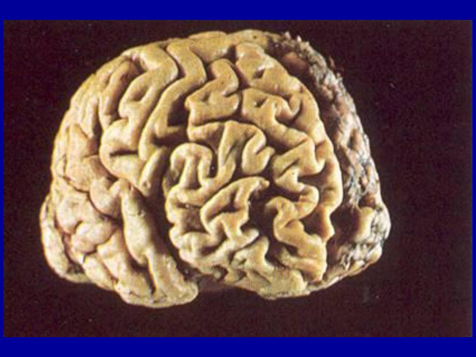
• Ageing (brown atrophy)
PATHOLOGICAL ATROPHY
• Decreased function (muscle atrophy due to immobilisation)
• Loss of innervation
• Loss of blood supply or blood stagnation (epidermal atrophy due to
tissue hypoxia)
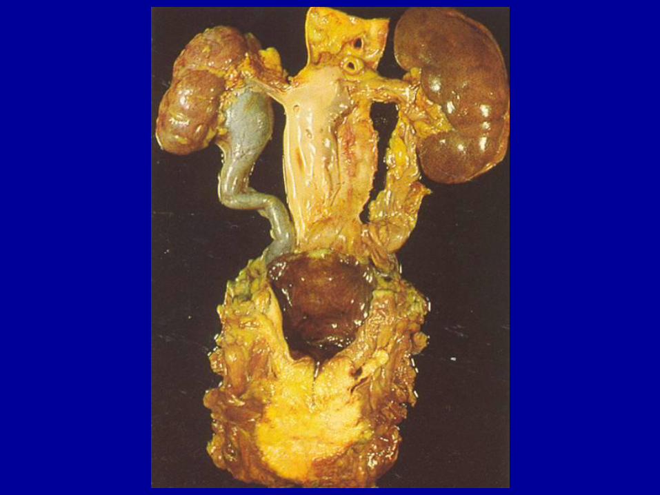
• Pressure atrophy (decubitus)
• Lack of nutrition (cachexia)
• Lack of endocrine stimulation (menopause)
• Idiopathic (myopathies)
Hypoplasia • Failure of the organ to attain its normal size
Agenesis (aplasia) • Failure of development of the organ
• Renal agenesis
• Anencephaly – incompatible with life
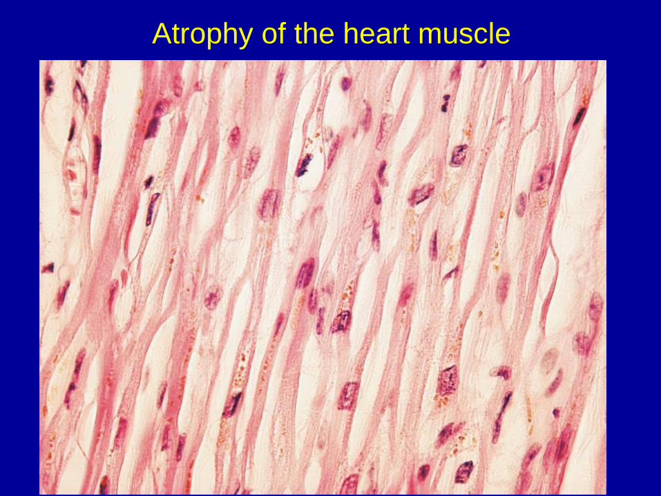
Atrophy of the heart muscle
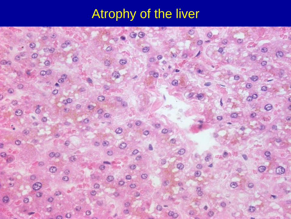
Atrophy of the liver
Dystrophy
Degeneration of tissue resulting from metabolic
disorders and abnormal intracellular accumulation of
metabolites
May be harmless or may cause varied degrees of injury
Localized in the cytoplasm, in the organelles (lysosomes),
or in the nucleus
3 types of accumulated substances:
1) normal endogenous substances
2) abnormal endogenous substances
3) exogenous substances
Steatosis (fatty change)
Abnormal intracellular accumulation of lipids
May have or may not have effect on cellular function
Process is reversible
Most commonly seen in the liver and in the heart
Causes:
1) hypoxia („tiger heart“)
LIPOMATOSIS – extracellular fat deposition
2) toxic (alcohol, mushroom, bacterial toxins)
3) lipidoses – congenital defects of enzymes of fat metabolism
(Niemann-Pick…sfingomyelin)
4) starvation, diabetes
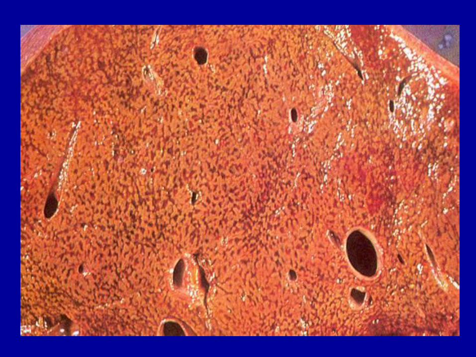
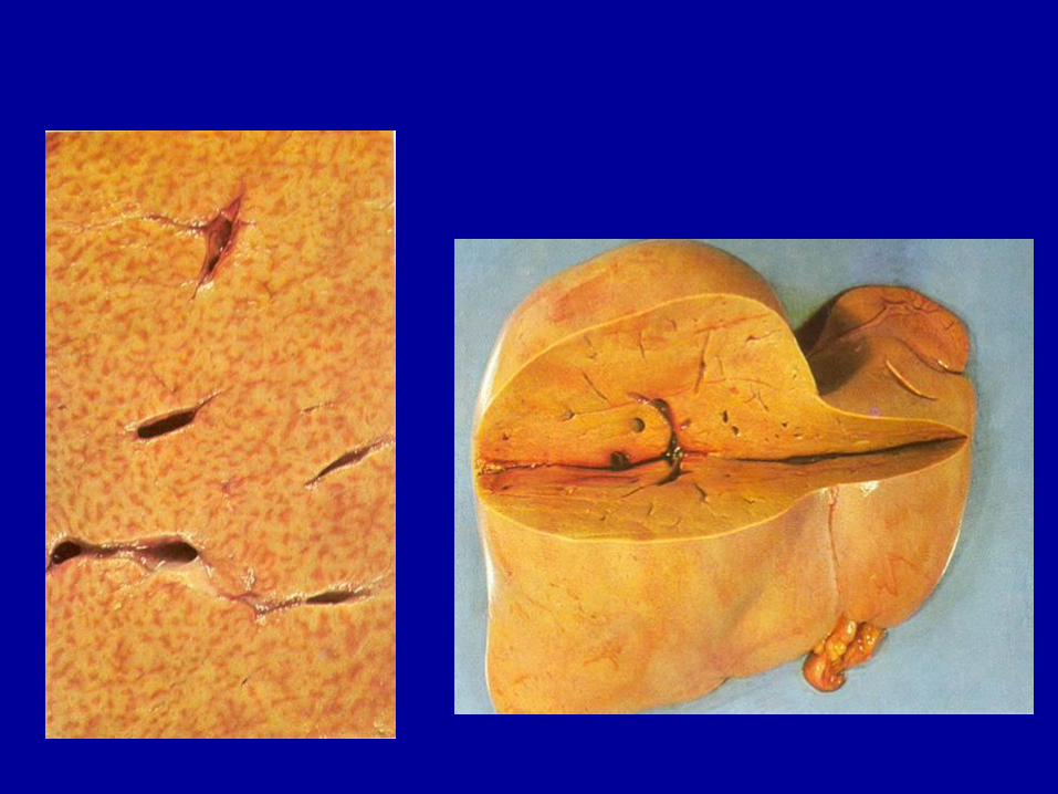
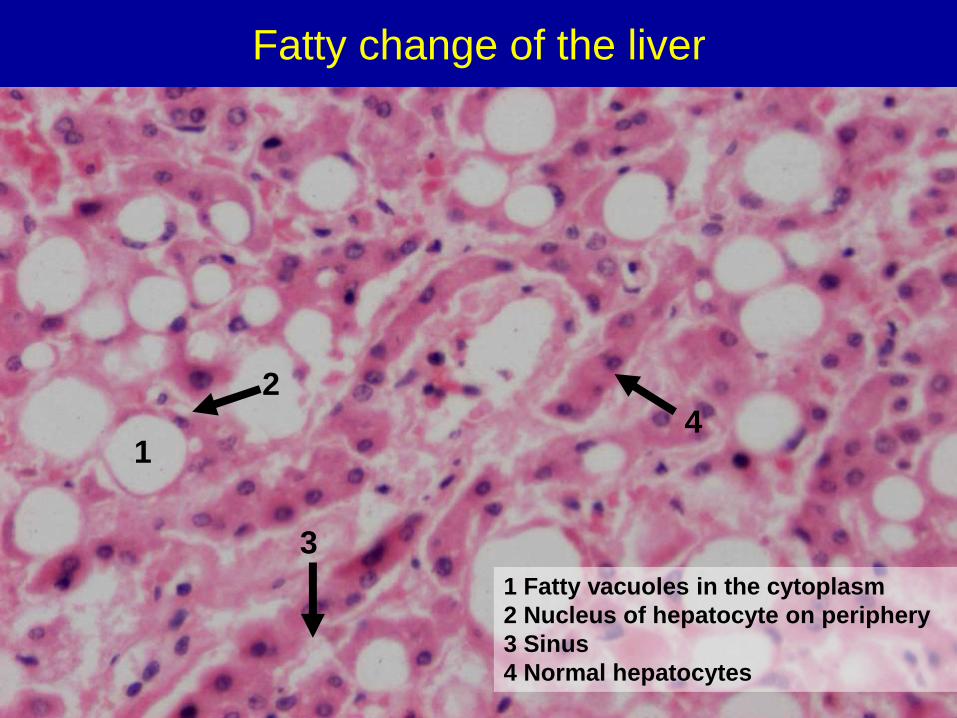
Fatty change of the liver
3
1
1 Fatty vacuoles in the cytoplasm
2 Nucleus of hepatocyte on periphery
3 Sinus
4 Normal hepatocytes
2
4
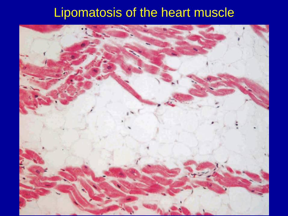
Lipomatosis of the heart muscle
Intracellular accumulation of proteins
HYALINE
• Large amount of protein is presented to the cells or the cells
synthetize excessive amounts
• Usually not visible
• Microscopically, eosinophilic amorphous droplets, appears as
inclusion
• In the kidneys (nephrotic syndromes), in the liver (Mallory bodies –
„alcoholic hyaline“), in the brain (Lewy bodies, neurofibrillary
tangles)
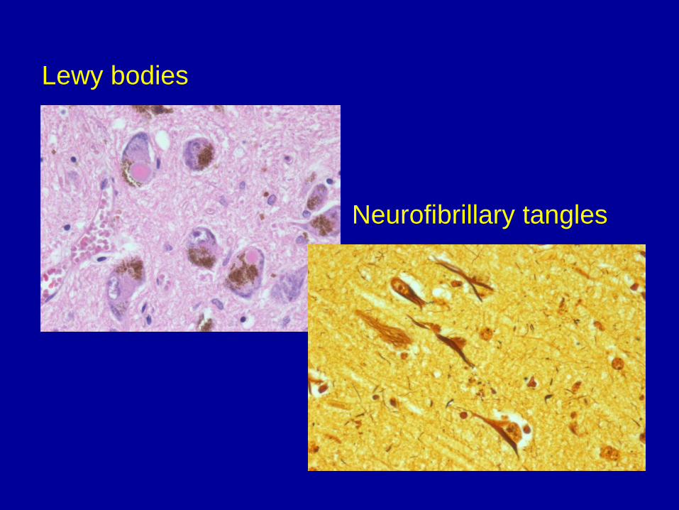
Lewy bodies
Neurofibrillary tangles
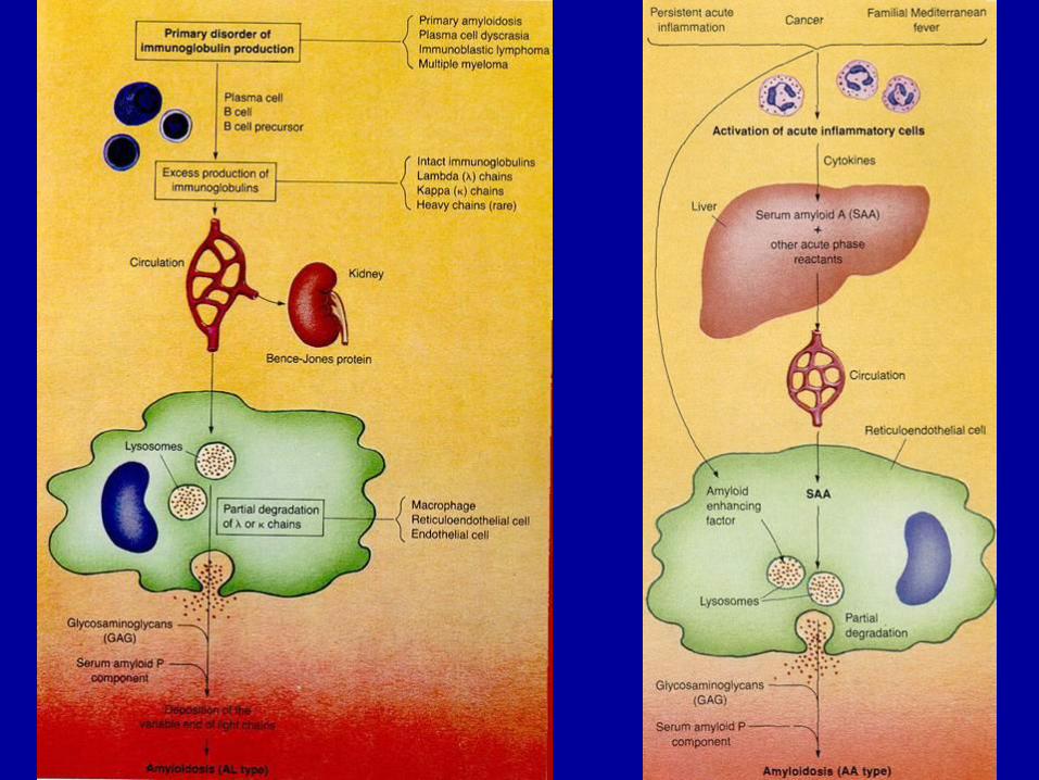
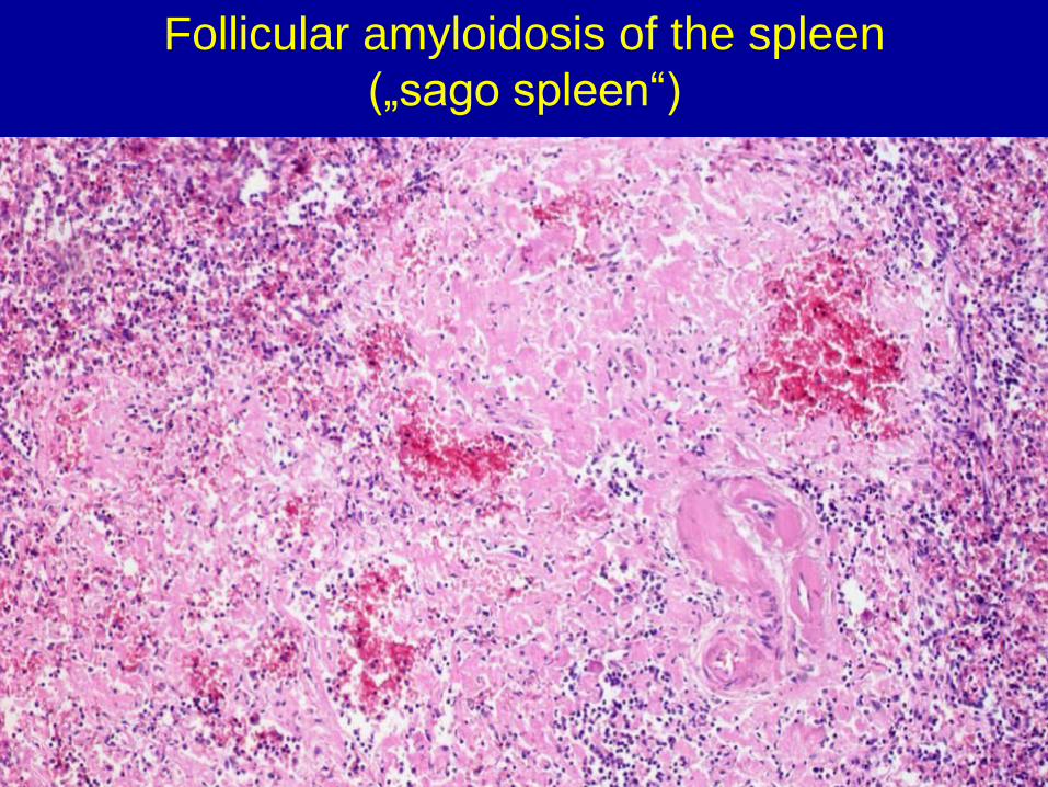
AMYLOID
• EXTRACELLULAR deposition of the fibrous protein aggregates
• Chemically differ but share some specific structural traits
• Grey-white colour, waxy appearance, elastic consistency
• Microscopically, eosinophilic amorphous substance
• Congo Red…stains red…shows green dichroism in polarized light
• Ultrastructurally, fibrillar bundles of the protein (β-sheets)
Extracellular accumulation of proteins
Types of amyloid:
1) AL amyloid (myeloma - Bence-Jones protein in the urine)
Kidneys, vessels, myocardium, spleen, GIT, adrenal gland
2) AA amyloid (chronic inflammatory diseases, neoplasms)
Adrenal glands, spleen, liver, lymph nodes, kidneys, bowel
3) AE amyloid (medullary carcinoma of the thyroid)
4) AS (senile) amyloid
Myocardium
5) AD (dermal) amyloid
Clinical classification:
1) Primary amyloidosis (≈AL amyloidosis)
2) Secondary (reactive) amyloidosis (≈AA amyloidosis)
3) hemodialysis-associated amyloidosis
4) Hereditary amyloidosis - rare
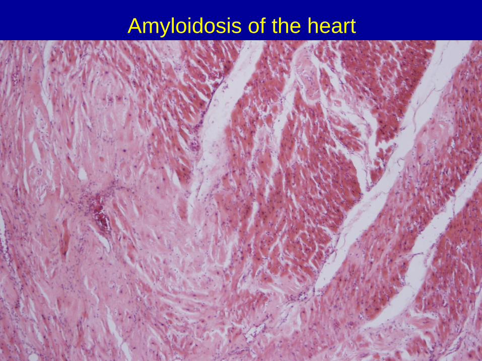
Amyloidosis of the heart
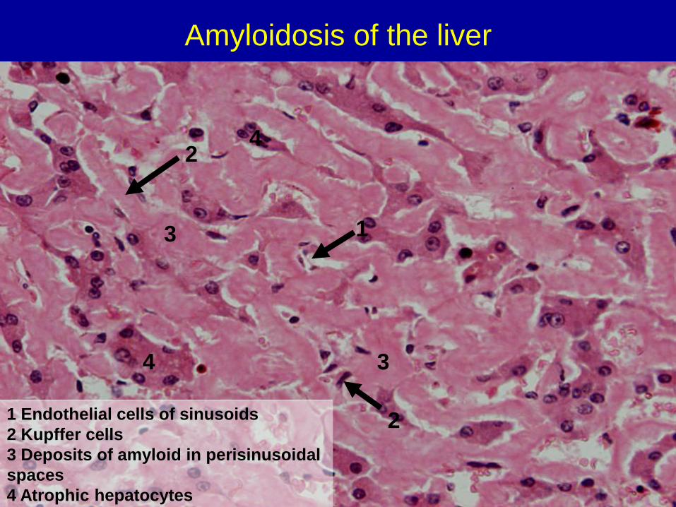
Amyloidosis of the liver
1 Endothelial cells of sinusoids
2 Kupffer cells
3 Deposits of amyloid in perisinusoidal
spaces
4 Atrophic hepatocytes
2
4
2
1 3
3
4
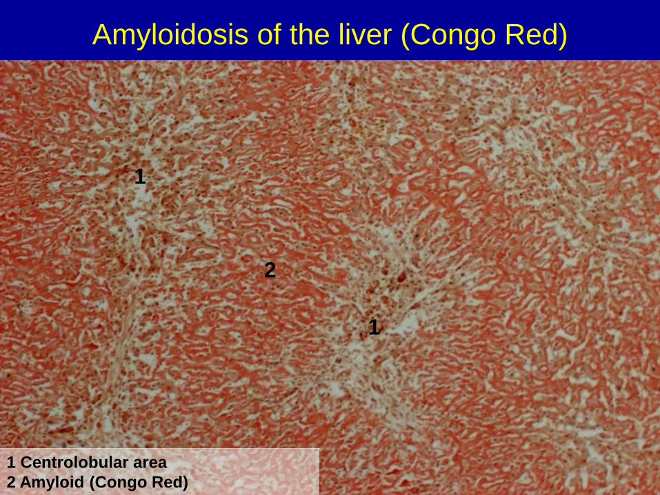
Amyloidosis of the liver (Congo Red)
1 Centrolobular area
2 Amyloid (Congo Red)
2
1
1

Follicular amyloidosis of the spleen
(„sago spleen“)
Follicular amyloidosis of the spleen
(„sago spleen“)
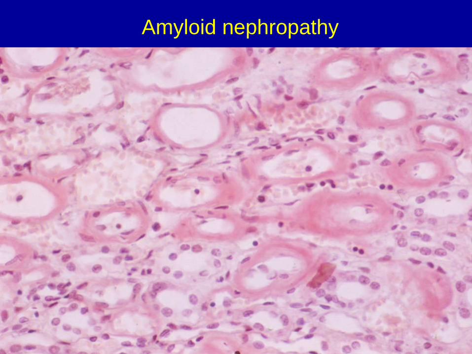
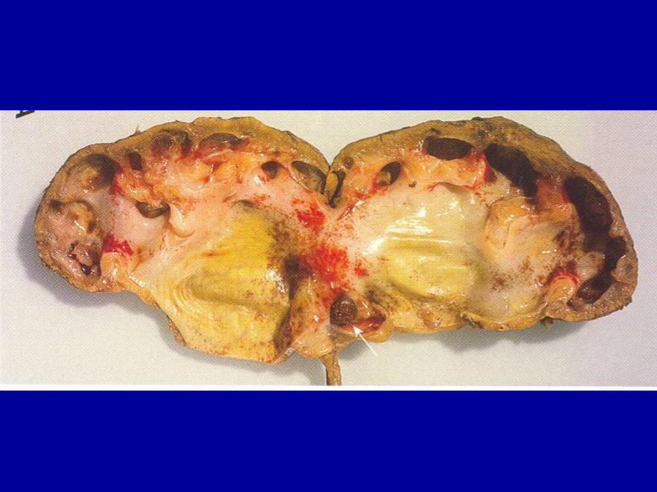
Amyloid nephropathy
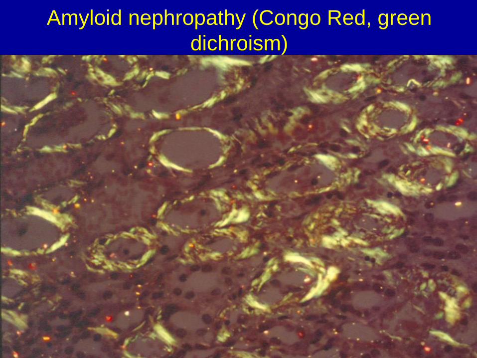
Amyloid nephropathy (Congo Red, green
dichroism)
ACCUMULATION OF MUCOID SUBSTANCES
• Ganglion cyst (swelling around joints and tendons in the hand or
foot)
• Myxoedema – hyperthyroidism (pretibial myxoedema),
hypothyroidism
• Mucopolysaccharidoses – congenital defects of enzymes
CYSTIC FIBROSIS (MUCOVISCIDOSIS)
• AR disorder. Defect of secretory process of exocrine glands
• Salty sweat
• Abnormally viscid mucus blocks the airways (secondary infections)
and pancreatic ducts (malabsorption)
• Newborns: meconium ileus
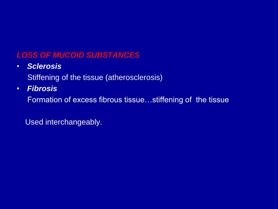
LOSS OF MUCOID SUBSTANCES
• Sclerosis
Stiffening of the tissue (atherosclerosis)
• Fibrosis
Formation of excess fibrous tissue…stiffening of the tissue
Used interchangeably.
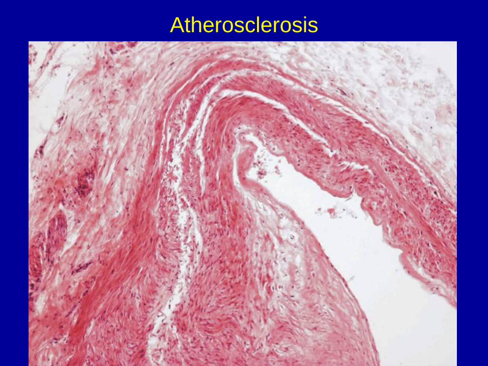
Atherosclerosis
Intracellular accumulation of
carbohydrates
DIABETES MELLITUS
Impaired glucose homeostasis resulting from a relative or
absolute insufficiency of insulin
Hyperglycaemia, glycosuria
• Type I (IDDM) – juvenile, genetic predisposition
• Type II (NIDDM) – adult
Complications:
• Macroangiopathy – accelerated atherosclerosis, gangrene
• Microangiopathy – diabetic retinopathy, glomerulosclerosis
• Neuropathy – focal demyelination, diabetic polyneuropathy
• Increased susceptibility to infections
• Diabetic ketoacidosis
• Hyperosmolar nonketotic coma
Intracellular accumulation of
carbohydrates (storage diseases)
GLYKOGENOSIS
• Congenital defects of enzymes of sugar metabolism
• Most frequent Von Gierke and Pompe
FRUCTOSE INTOLERANCE
• Liver
GALACTOSAEMIA (MILK INTOLERANCE)
• Liver
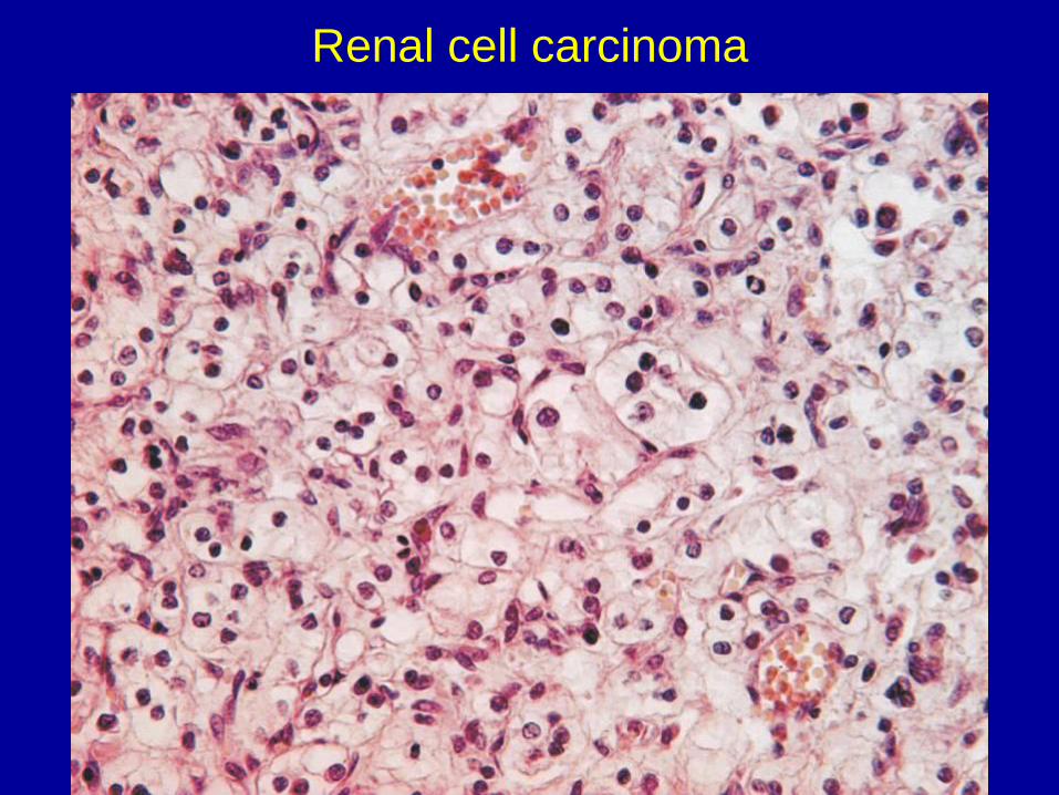
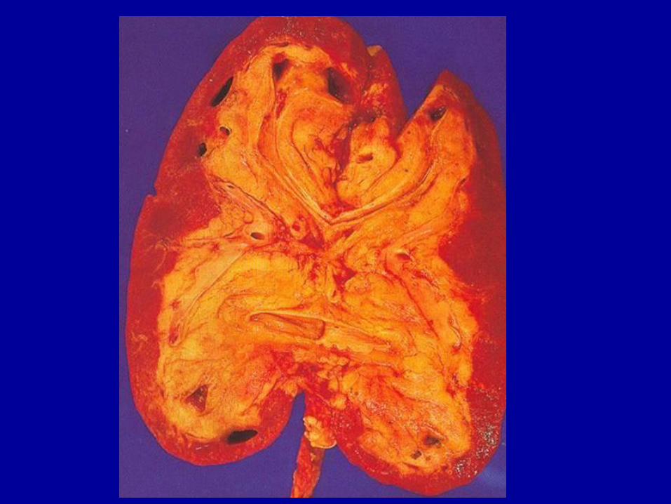
Renal cell carcinoma
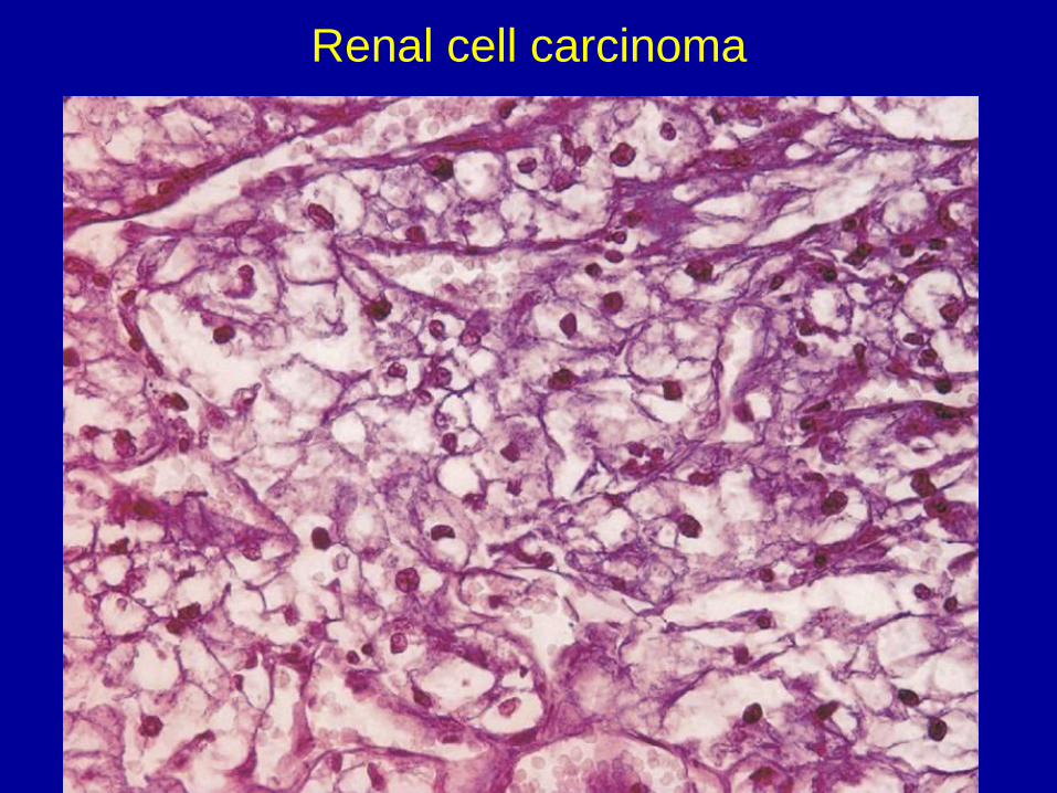
Renal cell carcinoma


