
APPROPRIATE USE CRITERIA FOR TREATMENT
OF DISTAL RADIUS FRACTURES
Adopted by the American Academy of Orthopaedic Surgeons
Board of Directors
March 18, 2013

Disclaimer Volunteer physicians from multiple medical specialties created and categorized these Appropriate Use Criteria. These Appropriate Use Criteria are not intended to be comprehensive or a fixed protocol, as some patients may require more or less treatment or different means of diagnosis. These Appropriate Use Criteria represent patients and situations that clinicians treating or diagnosing musculoskeletal conditions are most likely to encounter. The clinician’s independent medical judgment, given the individual patient’s clinical circumstances, should always determine patient care and treatment.
Disclosure Requirement In accordance with American Academy of Orthopaedic Surgeons policy, all individuals whose names appear as authors or contributors to this document filed a disclosure statement as part of the submission process. All authors provided full disclosure of potential conflicts of interest prior to participation in the development of these Appropriate Use Criteria. Disclosure information for all panel members can be found in Appendix C.
Funding Source The American Academy of Orthopaedic Surgeons exclusively funded development of these Appropriate Use Criteria. The American Academy of Orthopaedic Surgeons received no funding from outside commercial sources to support the development of these Appropriate Use Criteria.
FDA Clearance Some drugs or medical devices referenced or described in this document may not have been cleared by the Food and Drug Administration (FDA) or may have been cleared for a specific use only. The FDA has stated that it is the responsibility of the physician to determine the FDA clearance status of each drug or device he or she wishes to use in clinical practice.
Copyright All rights reserved. Reproduction, storage in a retrieval system, or transmission, in any form, or by any means, electronic, mechanical, photocopying, recording, or otherwise, of any part of this document, requires prior written permission from the American Academy of Orthopaedic Surgeons.
Published 2013 by the American Academy of Orthopaedic Surgeons 6300 North River Road Rosemont, IL 60018 First Edition Copyright 2013 by the American Academy of Orthopaedic Surgeons

To access the AUC web-based application, please visit www.aaos.org/aucapp.

Table of Contents Treatment of Distal Radius Fractures AUC Writing Panel ......................................................... i Treatment of Distal Radius Fractures AUC Review Panel .......................................................... i Treatment of Distal Radius Fractures AUC Voting Panel ........................................................... i Voting Panel Round Two Discussion Moderators...................................................................... ii AUC Section Leader ................................................................................................................... ii AAOS AUC Section ................................................................................................................... ii AAOS Staff ................................................................................................................................. ii
I. INTRODUCTION ...................................................................................................................1
Overview ..................................................................................................................................... 1
II. METHODS ..............................................................................................................................3
Developing Criteria ..................................................................................................................... 3 Formulating Indications and Scenarios ................................................................................... 4 Creating Definitions and Assumptions ................................................................................... 5
Literature Review........................................................................................................................ 6 Reviewing Scenarios ................................................................................................................... 6 Determining Appropriateness ..................................................................................................... 7
Treatment of Distal Radius Fractures AUC Voting Panel ...................................................... 7 Rating Appropriateness ........................................................................................................... 7 Round One Voting .................................................................................................................. 8 Round Two Voting ................................................................................................................. 8 Final Ratings ........................................................................................................................... 9
Revision Plans ........................................................................................................................... 10 Disseminating Appropriate Use Criteria ................................................................................... 10
III. DEFINITIONS AND ASSUMPTIONS ................................................................................11
Definitions................................................................................................................................. 11 General Assumptions ................................................................................................................ 13
IV. RESULTS OF APPROPRIATENESS RATINGS ................................................................14
V. LITERATURE REVIEW FINDINGS ...................................................................................21
List of Tables ............................................................................................................................ 22 Chapter 1: Fused epiphysis ....................................................................................................... 23 Chapter 2: Post reduction radial shortening >3mm, dorsal tilt >10° ........................................ 27 Chapter 3: Surgical treatment of distal radius fracture ............................................................. 29 Chapter 4: C1, C2, C3 Arthroscopic evaluation in patients with other associated injuries. ..... 42 Chapter 5: Concurrent treatment of distal radioulnar joint instability in patients with operatively treated distal radius fracture. .................................................................................. 44 Chapter 6: Fixation of ulnar styloid fractures associated with distal radius fractures. ............. 46 Chapter 7: Age > 55 years ........................................................................................................ 48 Chapter 8: Early wrist immobilization ...................................................................................... 50 Literature Review References ................................................................................................... 52

VI. APPENDICES .......................................................................................................................56
Appendix A. Documentation of Approval ............................................................................ 57 Appendix B. List of Clinical Scenarios ................................................................................ 58 Appendix C. Disclosure Information .................................................................................... 64 Appendix D. References ....................................................................................................... 67

TREATMENT OF DISTAL RADIUS FRACTURES AUC WRITING PANEL Julie E. Adams, MD, MS American Academy of Orthopaedic Surgeons Brett D. Crist, MD, FAAOS, FACS Orthopaedic Trauma Association Charles A. Goldfarb, MD American Society of Surgery of the Hand John J. McGraw, MD American Academy of Orthopaedic Surgeons
Miguel Pirela-Cruz, MD, FACS American Association for Hand Surgery David C. Ring, MD, PhD American Society for Surgery of the Hand Jaiyoung Ryu, MD American Association for Hand Surgery Paul Tornetta, III, MD Orthopaedic Trauma Association
TREATMENT OF DISTAL RADIUS FRACTURES AUC REVIEW PANEL Jeffrey E. Budoff, MD American Society for Surgery of the Hand Peter J. Evans, MD, PhD American Society for Surgery of the Hand American Association for Hand Surgery American Association of Hip and Knee Surgeons Daren Forward, MD, FRCS American Society for Surgery of the Hand Jeffrey B. Friedrich, MD, FACS American Society of Plastic Surgeons M. Felix Freshwater, MD American College of Occupational and Environmental Medicine Kenneth Koval, MD American Society for Surgery of the Hand
Donald H. Lee, MD American Society for Surgery of the Hand Jose J. Monsivais, MD, FACS American Society for Surgery of the Hand Jay Pomerance, MD American Society for Surgery of the Hand J. Andrew Trenholm, MD, FRCSC American Society for Surgery of the Hand Orthopaedic Trauma Association Boris A. Zelle, MD Orthopaedic Trauma Association American Academy of Orthopaedic Surgeons Dan A. Zlotolow, MD American Society for Surgery of the Hand
TREATMENT OF DISTAL RADIUS FRACTURES AUC VOTING PANEL Alan M. Adelman, MD, MS American Academy of Family Physicians Henry Backe, MD American Association of Hip and Knee Surgeons George W. Balfour, MD American Academy of Orthopaedic Surgeons Warren C. Hammert, MD American Society for Surgery of the Hand Robert Charles Kramer, MD
American Academy of Orthopaedic Surgeons David “Dirk” Leu, MD Pediatric Orthopaedic Society of North America Peter Stern, MD American Society for Surgery of the Hand Steven Strode, MD, MPH, Med American Academy of Family Physicians Walter H. Truong, MD Pediatric Orthopaedic Society of North America
i

VOTING PANEL ROUND TWO DISCUSSION MODERATORS William C. Watters, III, MD James O. Sanders, MD AUC SECTION LEADER William C. Watters III, MD AAOS AUC SECTION Joseph A. Bosco III, MD Brent Graham, MD Michael H. Heggeness, MD
Michael Warren Keith, MD Charles T. Mehlman, DO, MPH
COMMITTEE ON EVIDENCE-BASED QUALITY AND VALUE CHAIR David S. Jevsevar, MD, MBA COUNCIL ON RESEARCH AND QUALITY CHAIR Kevin J. Bozic, MD, MBA AAOS STAFF Deborah Cummins PhD Director of Research and Scientific Affairs Phone: (847) 384-4326 E-mail: [email protected]
Jayson Murray MA Manager, Evidence-Based Medicine Unit Phone: (847) 384-4316 E-mail: [email protected]
William Martin, III, MD Medical Director
Nilay Patel, MA Lead Research Analyst
Anne Woznica MLS Medical Librarian
Leeaht Gross MPH Evidence-Based Medicine Coordinator
Yasseline Martinez Evidence-Based Medicine Administrative Assistant Former Staff: Kevin Boyer, MPH
ii

I. INTRODUCTION OVERVIEW The American Academy of Orthopaedic Surgeons (AAOS) has developed these Appropriate Use Criteria (AUC) to determine appropriateness of Treatment for Distal Radius Fractures. An “appropriate” healthcare service is one for which the expected health benefits exceed the expected negative consequences by a sufficiently wide margin.1 Evidence-based information, in conjunction with the clinical expertise of physicians from multiple medical specialties, was used to develop the criteria in order to improve patient care and obtain the best outcomes while considering the subtleties and distinctions necessary in making clinical decisions. The foundation for this AUC is the 2009 Treatment of Distal Radius Fractures Clinical Practice Guideline which can be accessed via the following link: http://www.aaos.org/research/guidelines/drfguideline.pdf.
The purpose of the AUC is to help determine the appropriateness of clinical practice guideline recommendations for the heterogeneous patient population routinely seen in practice. The best available scientific evidence is synthesized with collective expert opinion on topics where gold standard randomized clinical trials are not available or are inadequately detailed for identifying distinct patient types. When there is evidence corroborated by consensus that expected benefits substantially outweigh potential risks exclusive of cost, a procedure is determined to be appropriate. The AAOS uses the RAND/UCLA Appropriateness Method (RAM).1 Our process includes these steps: reviewing the results of the evidence analysis, compiling a list of clinical vignettes, and having an expert panel comprised of representatives from multiple medical specialties determine the appropriateness of each of the clinical indications for treatment as “Appropriate,” “May be Appropriate,” or “Rarely Appropriate.” To access an intuitive and more user-friendly version of the appropriate use criteria for this topic online, please use our AUC web-based application at www.aaos.org/aucapp.
These criteria should not be construed as including all indications or excluding indications reasonably directed to obtaining the same results. The criteria intend to address the most common clinical scenarios facing all appropriately trained surgeons and all qualified physicians managing patients under consideration for treatment of distal radius fractures. The ultimate judgment regarding any specific criteria should address all circumstances presented by the patient and the needs and resources particular to the locality or institution. It is also important to state that these criteria were developed as guidelines and are not meant to supersede clinician expertise and experience or patient preference.
INTERPRETING THE APPROPRIATENESS RATINGS To prevent misuse of these criteria, it is extremely important that the user of this document understands how to interpret the appropriateness ratings. The appropriateness rating scale ranges from one to nine and there are three main range categories that determine how the median rating is defined (i.e. 1-3 = “Rarely Appropriate”, 4-6 = “May Be Appropriate”, and 7-9 = “Appropriate”). Before these appropriate use criteria are consulted, the user should read through and understand all contents of this document.
1 AAOS Evidence-Based Medicine Unit
PATIENT POPULATION This document addresses the treatment of acute distal radius fracture in adults (defined as patients 19 years of age and older). ETIOLOGY Fracture of the distal radius is the result of trauma. There is a bimodal distribution of distal radius fractures where high-energy fractures occur in younger persons (predominately male) and high and low-energy fractures occur in older persons (Predominately female).2, 3
INCIDENCE Distal radius fracture is one of the most common fractures seen by orthopaedic surgeons, with an incidence of 195.2/100,000 persons per year.3
BURDEN OF DISEASE As one of the most common fractures seen by orthopaedic surgeons, distal radius fractures result in significant financial burden. Costs related to distal radius fractures are mostly service related and at least $164,000,000 was spent on hospitalizations related to distal radius fractures in 2007.4,5
EMOTIONAL AND PHYSICAL IMPACT Acute distal radius fracture results in pain, tenderness, swelling and potential deformity. Patients may be faced with substantial morbidity if fracture healing is delayed or results in clinically significant deformity. Additionally, there are known complications in the treatment of distal radius fracture. The recovery period for distal radius fracture can be substantial and the impact of the method of fixation on activities and daily living can be significant. POTENTIAL BENEFITS, HARMS, AND CONTRAINDICATIONS The aim of treatment is pain relief and maintenance of the patient’s functional status. Most treatments are associated with some known risks, especially invasive and operative treatments. In addition, contraindications vary widely based on the treatment administered. Therefore, discussion of available treatments and procedures applicable to the individual patient rely on mutual communication between the patient and physician, weighing the potential risks and benefits for that patient.
2 AAOS Evidence-Based Medicine Unit

II. METHODS These AUC for Treatment of Distal Radius Fractures are based on a review of the available literature regarding treatment of distal radius fractures and a list of clinical scenarios (i.e. criteria) constructed and voted on by experts in orthopaedic surgery and other relevant medical fields. This section describes the methods adapted from the RAND/UCLA Appropriateness Method (RAM).1 This section also includes the activities and compositions of the various panels that developed, defined, reviewed, and voted on the criteria.
Members of the Treatment of Distal Radius Fractures AUC Writing Panel developed a list of 240 patient scenarios and 10 treatments. The Treatment of Distal Radius Fractures AUC Review Panel reviewed these scenarios and treatments independently to ensure that they were representative of patients and scenarios clinicians are likely to encounter. The Treatment of Distal Radius Fractures Voting Panel participated in two rounds of voting. During the first round of voting, the voting panel was given approximately one month to independently rate the appropriateness of the 10 treatments for the 240 patient scenarios as ‘Appropriate’, ‘May Be Appropriate’, or ‘Rarely Appropriate’ via an electronic ballot. After the first round of appropriateness ratings were submitted, AAOS staff calculated the median ratings for each patient scenario and specific treatment. Three one and a half hour conference calls were held on January 6th, 14th, and 17th, of 2013 with participating Voting Panel members to address the scenarios/treatments which resulted in disagreement (definition of disagreement can be found in Table 3). After this discussion, the second round of electronic voting occurred. The Voting Panel determined appropriateness by rating scenarios (i.e. criteria) as ‘Appropriate’, ‘May Be Appropriate’, or ‘Rarely Appropriate’. There was no attempt to obtain consensus about appropriateness.
AAOS Appropriate Use Criteria Section, the AAOS Council on Research and Quality, and the AAOS Board of Directors sequentially approved the Appropriate Use Criteria for Treatment of Distal Radius Fractures. AAOS submits AUC to the National Guidelines Clearinghouse and in accordance with the National Guidelines Clearinghouse criteria will update or retire this AUC every five years.
DEVELOPING CRITERIA Members of the Treatment of Distal Radius Fractures AUC Writing Panel, who are orthopaedic specialists in treatment of distal radius fractures, developed clinical scenarios using the following guidelines:
• Include a broad spectrum of patients that may be eligible for treatment of distal radius fractures [comprehensive]
• Classify patients into a unique scenario [mutually exclusive] • Consistently classify similar patients into the same scenario [reliable, valid
indicators] The Writing Panel developed the scenarios by categorizing patients in terms of indications evident during the clinical decision making process (Figure 1). These scenarios relied upon definitions and general assumptions, mutually agreed upon by the Writing Panel during the development of the scenarios. These definitions and assumptions were necessary to provide
3 AAOS Evidence-Based Medicine Unit

consistency in the interpretation of the clinical scenarios among experts voting on the scenarios and readers using the final criteria.
FORMULATING INDICATIONS AND SCENARIOS The scenarios began development with the Treatment of Distal Radius Fractures AUC Writing Panel identifying clinical indications typical of patients commonly presenting for treatment of distal radius fractures in clinical practice. Indications are most often parameters observable by the clinician, including symptoms or results of diagnostic tests. Additionally “human factor” (e.g. activity level) or demographic variables can be considered.
Indications identified in clinical trials (derived from patient selection criteria) included in AAOS Clinical Practice Guidelines served as a starting point for the Treatment of Distal Radius Fractures AUC Writing Panel and ensured that these Appropriate Use Criteria referred to the evidence base for the Treatment of Distal Radius Fractures AUC. The Writing Panel considered this initial list and other indications based on their clinical expertise and selected the most clinically relevant indications (Table 1). The Writing Panel then defined distinct classes for each indication in order to stratify/categorize the indication (Table 1).
The Writing Panel organized these indications into a matrix of clinical scenarios (Appendix B) that addressed all combinations of the classifications. The Writing Panel was given the opportunity to remove any scenarios that never occur in clinical practice; however, they agreed that all 240 scenarios could present themselves in clinical practice, thus no scenarios were
Indication: Observable/appreciable patient
parameter
Classification: Class/category of an indication;
standardized by definitions*
Clinical Scenario: Combination of a single
classification from each indication; assumptions assist interpretation*
Chapter: Group of scenarios based on the major clinical indication
Major clinical indication
Figure 1. Developing Criteria
Criteria: A unique clinical scenario with a final appropriateness rating
4 AAOS Evidence-Based Medicine Unit

removed. The major clinical decision making indications chosen by the Writing Panel divided the matrix of clinical scenarios into chapters. AO fracture type, mechanism of injury, functional demands, ASA status, and associated injuries served as the major clinical decision making indications for the chapters presented in Table 1.
CREATING DEFINITIONS AND ASSUMPTIONS The Treatment of Distal Radius Fractures AUC Writing Panel constructed concise and explicit definitions for the indications and classifications. This standardization helped ensure that how the Writing Panel defined AO fracture types, mechanisms of injury, functional demands, ASA statuses, and associated injuries was consistent among those reading the clinical scenario matrix or the final criteria. Definitions drew explicit boundaries when possible and were based on standard medical practice or existing literature.
Additionally, the Writing Panel formulated a list of general assumptions in order to provide more consistent interpretations of a scenario. These assumptions differed from definitions in that they identified circumstances that exist outside of the control of the clinical decision making process. Examples of such can be the assumption that diagnostic exams were appropriately conducted (x-rays, labs) or that mitigating factors do not complicate clinical scenarios (e.g. do not resuscitate, non-compliance). Assumptions also addressed the use of existing published literature regarding the effectiveness of treatment and/or the procedural skill level of physicians. Additionally, assumptions highlighted intrinsic methods described in this document such as the role of cost considerations in rating appropriateness or the validity of the definition of appropriateness. The main goal of assumptions was to focus scenarios so that they apply to the average patient presenting to an average physician at an average facility.1
The definitions and assumptions provided all readers with a common starting point in interpreting the clinical scenarios. This list of definitions and assumptions accompanied the matrix of clinical scenarios in all stages of the development of this AUC and appears in the Definitions and Assumptions section.
VOTING PANEL MODIFICATIONS TO WRITING PANEL MATERIALS The original indications table constructed by the Writing Panel was modified by the Voting Panel during the round two discussions. The original list of associated injuries created by the Writing Panel included carpus injuries; however, it was the consensus of the Voting Panel to remove all patient scenarios which included “Carpus Injuries” as an indication. The rationale behind this decision was that, as Voting Panel members, they would need much more information concerning the carpus injury to properly rate the appropriateness of treatment for the distal radius fracture.
The Voting Panel also agreed to amend the original indication labeled, “Open Wound”, as they expressed concern about rating a treatment for a patient presenting with an open wound without knowing whether that open wound was a Grade I, II, or III open fracture as defined by the Gustilo Open Fracture Classification. The Voting Panel separated the patient scenarios reflecting high-energy fractures into two categories: Grade I or II versus Grade III open fractures. Patient scenarios reflecting low-energy fractures and open wounds were specified as having a Grade I or II open fracture.
5 AAOS Evidence-Based Medicine Unit

The final modification to the indications was the deletion of ASA Status 5 from the indications list. The original scenarios were grouped by ASA 1-3 and ASA 4-5, the revised groupings are ASA 1-3 and ASA 4. The rationale behind the deletion of ASA 5 was that treatment for a distal radius fracture in a moribund patient would be unnecessary.
Additionally the Voting Panel agreed to add two new assumptions concerning open fractures to the assumptions list. Assumptions 13 and 14 were added by the Voting Panel during the round two discussions.
Table 1. Indications and Classifications
Indication Classification(s)
AO/OTA Fracture Type A B C
Mechanism of Injury High energy Low Energy
Functional Demands Homebound Independent Normal High
ASA Status (co-morbidities) Associated Injuries
ASA 1-3 ASA 4 No associated injuries Grade I or II Open Fracture Grade III Open Fracture Median Nerve Injury Other Ipsilateral Injury
LITERATURE REVIEW Concurrent with the Writing Panel developing the criteria, the AAOS Appropriate Use Criteria Unit undertook a literature review based on the results of the AAOS clinical practice guideline and all literature published after the release of the clinical practice guideline related to the treatment of distal radius fractures. This literature review informed the decisions relevant to the indications identified by the Writing Panel when they were available and necessary. The literature review also considered lower quality evidence when the best available evidence (i.e. randomized control trials) did not contain information relevant to the clinical scenarios. The full results of the literature review appear in the Literature Review Findings section.
REVIEWING SCENARIOS After the Writing Panel developed the scenarios, the Treatment of Distal Radius Fractures AUC Review Panel reviewed the proposed chapters in order to ensure that they were representative of patients and scenarios clinicians are likely to encounter. The Review Panel was comprised of
6 AAOS Evidence-Based Medicine Unit

orthopaedic surgeons who routinely perform treatments for distal radius fractures and other specialties who may refer patients with distal radius fractures to a specialist. No member of this panel participated in the Writing Panel’s initial development of the scenarios or participated in the appropriateness rating of the scenarios.
Review Panel members considered the lists of scenarios, definitions, assumptions and the literature review associated with each scenario. Each independent reviewer suggested to the Writing Panel, potential modifications to the content or structure of the lists and literature review. The Writing Panel provided final determination of modifications to the indications, scenarios, assumptions and literature review.
DETERMINING APPROPRIATENESS TREATMENT OF DISTAL RADIUS FRACTURES AUC VOTING PANEL A multidisciplinary panel of clinicians assembled to determine the appropriateness of treatments for distal radius fractures. This group consisted of approximately 50% specialists and 50% generalists. Two non-voting moderators who are orthopaedic surgeons but are not specialists in treatment of distal radius fractures facilitated the Voting Panel. The moderators were familiar with the methods and procedures of AAOS Appropriate Use Criteria and led the panel (as non-voters) in discussions. Additionally, no member of the voting panel was involved in the development (Writing Panel) or independent review (Review Panel) of the scenarios.
The Voting Panel used a modified Delphi procedure to determine appropriateness ratings. The Voting Panel participated in two rounds of voting while considering evidence-based information provided in the literature review. While cost is often a relevant consideration, panelists focused their appropriateness ratings on the effectiveness of treatment for distal radius fractures.
RATING APPROPRIATENESS When rating the appropriateness of a scenario, the Voting Panel considered the following definition:
“An appropriate treatment for distal radius fractures is one for which the treatment is generally acceptable, is a reasonable approach for the indication, and is likely to improve the patient’s health outcomes or survival.”
They then rated each scenario using their best clinical judgment, taking into consideration the available evidence, for an average patient presenting to an average physician at an average facility as follows:
Table 2. Appropriateness Ratings Rating Explanation
7-9
Appropriate: Appropriate for the indication provided, meaning treatment is
generally acceptable and is a reasonable approach for the indication and is likely to improve the patient’s health outcomes
or survival.
7 AAOS Evidence-Based Medicine Unit

4-6
May Be Appropriate: Uncertain for the indication provided, meaning treatment may
be acceptable and may be a reasonable approach for the indication, but with uncertainty implying that more research and/or patient information is needed to further classify the
indication.
1-3
Rarely Appropriate: Rarely an appropriate option for management of patients in this
population due to the lack of a clear benefit/risk advantage; rarely an effective option for individual care plans; exceptions
should have documentation of the clinical reasons for proceeding with this care option (i.e. procedure is not generally acceptable and is not generally reasonable for the indication).
Each panelist uses the scale below to record their response for each scenario:
Appropriateness of [Topic]
1 2 3 4 5 6 7 8 9 ROUND ONE VOTING The first round of voting occurred after completion of the independent review of the scenarios by the Review Panel and approval of the final indications, scenarios, and assumptions by the Writing Panel. The Voting Panel rated the scenarios electronically using a personalized ballot created by AAOS staff using SNAP 10 Survey Software. There was no interaction between panel members while completing the first round of voting. Panelists considered the following materials:
• The instructions for rating appropriateness • The completed literature review, that is appropriately referenced when evidence is
available for a scenario • The list of indications, definitions and assumptions, to ensure consistency in the
interpretation of the clinical scenarios ROUND TWO VOTING The second round of voting occurred after a series of 3 conference calls, which were led by a non-voting moderator. Before the discussions, each panelist received a personalized document that included their first round ratings along with summarized results of the first-round ratings that resulted in disagreement. These results indicated the frequency of ratings for a scenario for all panelists. The document contained no identifying information for other panelists’ ratings. The moderator also used a document that summarized the results of the panelists first round voting. These personalized documents served as the basis for discussions of scenarios which resulted in disagreement.
May Be Appropriate Appropriate Rarely Appropriate
8 AAOS Evidence-Based Medicine Unit

After all of the disagreed upon scenarios were discussed, the Voting Panel performed a second round of voting for only those scenarios. After the round two ratings were submitted, AAOS staff calculated the median values and level of agreement for all voting items, after which the Voting Panel examined the ratings for anomalies. There was no attempt to obtain consensus among the panel members.
FINAL RATINGS Using the median value of the second round ratings, AAOS determined the final levels of appropriateness. Disagreement among raters can affect the final rating. Agreement and disagreement were determined using the BIOMED definitions of Agreement and Disagreement as reported in the RAND/UCLA Appropriate Method User’s Manual for a panel of 8-10 voting members (see Table 3 below). For this panel size, disagreement is defined as when ≥ 3 member’s appropriateness ratings fell within the appropriate (7-9) and rarely appropriate (1-3) ranges for any scenario (i.e. ≥ 3 member’s ratings fell between 1-3 and ≥ 3 member’s ratings fell between 7-9 on any given scenario and its treatment).If there is still disagreement in the Voting Panel ratings after the second round of voting, that voting item is labeled as “5” regardless of median score. Agreement is defined as ≤ 2 panelists rated outside of the 3-point range containing the median.
Table 3. Defining Agreement and Disagreement for Appropriateness Ratings Disagreement Agreement
Panel Size Number of panelists rating in each extreme (1-3 and 7-9)
Number of panelists rating outside the 3-point region
containing the median (1-3, 4-6, 7-9)
8,9,10 ≥ 3 ≤ 2
11,12,13 ≥ 4 ≤ 3
14,15,16 ≥ 5 ≤ 4 Adapted from RAM 1
The classifications in the table below determined final levels of appropriateness.
Table 4. Interpreting Final Ratings of Criteria
Level of Appropriateness Description
Appropriate • Median panel rating between 7-9 and no disagreement
May Be Appropriate • Median panel rating between 4-6 or • Median panel rating 1-9 with disagreement
Rarely Appropriate • Median panel rating between 1-3 and no disagreement
9 AAOS Evidence-Based Medicine Unit

REVISION PLANS These criteria represent a cross-sectional view of current use of treatments for distal radius fractures and may become outdated as new evidence becomes available or clinical decision making indicators are improved. AAOS will update or withdraw these criteria in five years in accordance with the standards of the National Guideline Clearinghouse. AAOS will issue updates in accordance with new evidence, changing practice, rapidly emerging treatment options, and new technology.
DISSEMINATING APPROPRIATE USE CRITERIA Publication of the Appropriate Use Criteria (AUC) document is on the AAOS website at [http://www.aaos.org/auc]. This document provides interested readers with full documentation about the development of Appropriate Use Criteria and further details of the criteria ratings.
AUCs are first announced by an Academy press release and then published on the AAOS website. AUC summaries are published in the AAOS Now and the Journal of the American Academy of Orthopaedic Surgeons (JAAOS). In addition, the Academy’s Annual Meeting showcases the AUCs on Academy Row and at Scientific Exhibits.
The dissemination efforts of AUC include web-based mobile applications, webinars, online modules for the Orthopaedic Knowledge Online website, Radio Media Tours, and Media Briefings. In addition AUCS are also promoted in relevant Continuing Medical Education (CME) courses and distributed at the AAOS Resource Center.
Other dissemination efforts outside of the AAOS include submitting AUCs to the National Guideline Clearinghouse and to other medical specialty societies’ meetings.
10 AAOS Evidence-Based Medicine Unit

III. DEFINITIONS AND ASSUMPTIONS DEFINITIONS The following definitions standardized the interpretation of the clinical scenarios presented in the Treatment of Distal Radius Fractures Appropriate Use Criteria. This standardization ensures that those responsible for rating the appropriateness of a scenario and those reading these scenarios are using the same parameters to address the scenario.
AO/OTA Fracture Type: • A: Extra-articular fracture • B: Partial articular fracture • C: Complete articular fracture of the radius
Figure 2. AO/OTA Fracture Classification of the Distal Radius6
Mechanism of Injury: • High energy: Injury due to trauma, such as a fall from higher than standing height,
motor vehicle accident, or industrial accident where velocity at impact results in high compression forces; assuming significant displacement and comminution.
• Low energy: Injury due to chronic conditions that weaken the strength of the bone and low velocity at impact results in bending forces; assuming minimal comminution and displacement.
11 AAOS Evidence-Based Medicine Unit

Functional Demands: • Homebound: Effort required for patient to leave their residency is taxing and
unsafe. Require human assistance to leave home. • Independent: Routinely completes activities of daily living with assistance of
ambulation devices (canes, walkers, etc). • Normal: Completes activities of daily living without assistance. • High: Patients experiencing substantial stress/stain on their wrist on a regular
basis (e.g. high-level athletics, heavy labor jobs).
American Society of Anesthesiologist’s (ASA) Status (co-morbidities) • ASA 1: Normal, healthy patient • ASA 2: Patient with mild systemic disease • ASA 3: Patient with severe systemic disease • ASA 4: Patient with severe systemic disease that is a constant threat to life
Associated injuries
• No associated injuries: Isolated distal radius fracture. • Open fracture: Wound caused by penetration or puncture of the skin (e.g. foreign
objects, fractured bones) of the same arm as the distal radius fracture. Open fractures are defined using the following two Gustilo classifications:
a) Grade I or II open fracture b) Grade III open fracture
• Median nerve injury: Damage/dysfunction of the median nerve on the same arm as the distal radius fracture, determined by physical examination or electrodiagnostic testing.
• Other ipsilateral injury: Injuries to ligaments, bones, or soft tissue of the same arm/hand as the distal radius fracture.
12 AAOS Evidence-Based Medicine Unit

GENERAL ASSUMPTIONS The following assumptions clarified the interpretation of the clinical scenarios presented in the Treatment of Distal Radius Fractures Appropriate Use Criteria. This standardization ensures that those responsible for rating the appropriateness of a scenario and those reading these scenarios are using the same parameters to address the scenario.
Before these AUC are consulted, it is assumed that:
1. The patient is healthy enough to undergo surgery if indicated.
2. An adequate physical exam of the patient has been conducted.
3. Adequate radiographs have been obtained and examined by the clinician.
4. The patient history is available and has been reviewed by the clinician.
5. The patient has given adequate and informed consent.
6. The surgeon is trained and capable of performing all operative techniques with equal effectiveness.
7. The fracture is not so complex, and/or the patient’s comorbidities or social situation such a factor, as to represent an exception to these scenarios (e.g. C3.3 fracture that might be optimally treated with a distraction plate).
8. There is not a clear advantage (i.e. evidence for or against) for one procedure based on fracture pattern (e.g. volar plate for volar shearing fracture).
9. The surgery, when indicated, will be performed in a timely fashion to allow ideal treatment of the fracture.
10. The surgeon will perform the surgery in the most appropriate location (i.e., ASC, outpatient, inpatient) based on the health of the patient and other injuries rather the nature of the fracture. Open fractures and associated injuries may dictate that surgery should be inpatient.
11. The surgeon will choose a cost effective treatment based on the nature of the fracture and expectations after surgery.
12. The facility has each type of implant/equipment available and capable support personnel.
13. In the event that the patient has an open wound, it is assumed that the clinician has cleaned the wound before considering treatment.
14. It is assumed that a low-energy open fracture is a Grade I or II open fracture.
13 AAOS Evidence-Based Medicine Unit

IV. RESULTS OF APPROPRIATENESS RATINGS The following appropriate use criteria tables contain the final appropriateness ratings assigned by the nine members of the voting panel. The appropriate use criteria tables are formatted by AO fracture type (i.e. A, B, or C) and mechanism of injury (i.e. high mechanism of injury versus low mechanism of injury). Additional patient characteristics are found under the column titled “Patient Characteristics”. The appropriate use criteria for each patient scenario can be found under each of the 10 treatment columns. These criteria are formatted by appropriateness labels (i.e. “R”=Rarely Appropriate, “M”=May Be Appropriate, and “A”=Appropriate), median score (in parentheses), and + or - indicating agreement or disagreement, respectively. Out of 2160 total voting items (i.e. 216 patient scenarios x 10 treatments), 440 (20%) voting items were rated as “Rarely Appropriate”, 953 (44%) voting items were rated as “May Be Appropriate”, and 767 (36%) voting items were rated as “Appropriate” (Figure 1). Additionally, the voting panel members were in agreement on 730 (34%) voting items and were in disagreement on 10 (0.5%) voting items. The appropriate use criteria can also be accessed online via our AUC web-based application at www.aaos.org/aucapp.
14 AAOS Evidence-Based Medicine Unit

Immobilization without reduction
Reduction and Immobilization
Percutaneous Pinning
Spanning External Fixation
Non-Spanning External Fixation
Distraction Plate
Volar lock ing Plate
Dorsal Plate
Fragment Specific Fixation
Intramedullary Nail
1 R (2)+ A (7)+ M (6) A (7)+ R (3) R (3) A (7) A (7)+ M (6) A (7)2 R (2)+ M (5) A (7) A (7)+ M (6) M (5) A (7)+ A (7) M (6) M (6)3 R (1)+ R (3)+ A (7) A (7)+ A (7) M (5)+ A (7) M (6) M (6) M (6)4 R (1)+ A (7) A (7) A (7) M (6) M (5) A (7)+ M (5) A (7) M (6)5 R (2)+ A (7) M (6) A (7) M (6) M (5) A (7) M (6) A (7) M (6)6 R (2)+ A (7)+ A (7) A (7) M (5)+ M (4) M (6) M (5) M (5) M (5)7 R (2)+ M (6) A (7) A (7) M (6) M (5)+ A (7)+ A (7) M (6) M (6)8 R (1)+ M (4)+ A (7) A (7)+ A (7) M (5)+ M (6) M (6) M (6)+ M (5)9 R (3) A (7)+ A (7) A (7) M (6) M (5) M (6) M (4) M (6) M (6)10 R (3) A (7)+ A (7)+ A (7) M (5) M (5) A (7) M (5) M (6) M (5)11 R (2)+ A (7) M (6) A (7) R (3) M (5) A (7) A (7) A (7) M (6)12 R (1)+ R (3)+ A (7) A (7) A (7)+ M (5) A (7)+ A (7)+ A (7) M (6)13 R (1)+ R (3)+ A (7) A (7) A (7) M (5)+ A (7)+ A (7) M (6) M (5)14 R (1) R (3) A (7) M (6) M (5) M (5) A (8) M (6) A (7) A (7)15 R (2)+ A (7) A (7) M (6) M (5) M (5) A (8)+ A (7) A (7) A (7)+16 R (1)+ A (7)+ A (7) A (7) M (5) M (5) A (7) M (6) M (5) M (6)17 R (1)+ M (4)+ A (7)+ A (7)+ A (7) M (5)+ A (7) A (7) M (6) M (6)18 R (1)+ M (4)+ A (7) A (7)+ M (6) M (5)+ M (6) M (6) M (6) M (6)19 R (3)+ A (7)+ A (7) A (7) M (6) M (5) A (7)+ M (6) M (6)+ M (6)20 R (2) A (7) A (7)+ A (7) M (6) M (6)+ A (7) M (6) M (6) M (6)21 R (2)+ A (7)+ A (7) A (7) M (6) M (6) A (8)+ A (7) A (7)+ A (7)22 R (1)+ R (1)+ A (7) A (7)+ A (7) M (5)+ A (7)+ A (7)+ A (7) A (7)23 R (1)+ R (3)+ A (7)+ A (7)+ A (7) M (5)+ A (7) A (7) M (6) M (5)24 R (1)+ M (5) A (7)+ A (7) M (6) M (5) A (8)+ M (6) A (7)+ A (7)25 R (1)+ A (7) A (7) A (7) M (6) M (5) A (8)+ M (6) A (7)+ M (6)26 R (3) A (7)+ A (7)+ M (6) M (6) M (5) A (7) M (6) M (6)+ A (7)27 R (1)+ R (3)+ A (7) A (7)+ M (6) M (5)+ M (6) M (6) M (6)+ M (6)28 R (1)+ M (4)+ A (7) A (7)+ A (7) M (5)+ M (6) M (6) M (6)+ M (5)29 R (1)+ A (7)+ A (7)+ A (7) M (6) M (5) A (8) M (5) M (6) M (5)30 R (1) A (7)+ A (7)+ A (7) M (6) M (5) A (7) M (6) M (6) M (6)31 R (2)+ A (7)+ M (6) M (6) A (7) M (4) A (8)+ M (6) A (7) A (7)+32 R (1)+ R (2)+ A (7) A (7)+ A (7) M (5)+ A (7)+ A (7)+ M (6) M (6)33 R (1)+ R (1)+ A (7) A (7)+ A (7) M (6)+ A (7) A (7) M (6) M (6)34 R (1)+ A (7) A (7) M (6) M (6) M (5) A (8)+ M (6) A (7) M (6)35 R (1)+ A (7) A (7) M (6) M (6) M (6) A (8)+ M (6) A (7) A (7)36 R (1)+ A (7)+ A (7)+ A (7) M (6) M (5) A (7) M (6) M (6) M (5)37 R (1)+ R (2)+ A (7) A (7)+ A (7) M (5)+ A (7) A (7) M (6)+ M (6)38 R (1)+ R (2)+ A (7) A (7)+ A (7) M (5)+ M (6) M (6) M (6)+ M (6)39 R (1)+ A (7)+ A (7)+ A (7) M (6) M (4) A (8) M (6) M (6) M (6)40 R (1) A (7)+ A (7)+ A (7) M (6) M (5) A (8) M (6) M (6) M (6)
AO/OTA A Fracture Type; High Energy Mechanism of Injury
* R=Rarely Appropriate, M=May be Appropriate, A=Appropriate; Numbers in parentheses indicate median rating of voting panel; "+" =Agreement between voting panel members, "-" = Disagreement between voting panel members
PATIENT CHARACTERISTICS
TREATMENTS
High Functional Demands, ASA 4, Median nerve injuryHigh Functional Demands, ASA 4, Other Ipsilateral Injury
Normal Functioning, ASA 4, Other Ipsilateral InjuryHigh Functional Demands, ASA 1-2-3, No associated injuriesHigh Functional Demands, ASA 1-2-3, Grade I or II Open Fracture
High Functional Demands, ASA 1-2-3, Median nerve injuryHigh Functional Demands, ASA 1-2-3, Other Ipsilateral Injury
Home-bound, ASA 1-2-3, No associated injuriesHome-bound, ASA 1-2-3, Grade I or II Open Fracture
Home-bound, ASA 1-2-3, Median nerve injuryHome-bound, ASA 1-2-3, Other Ipsilateral Injury
Normal Functioning, ASA 1-2-3, Median nerve injury
Home-bound, ASA 4, Median nerve injuryHome-bound, ASA 4, Other Ipsilateral InjuryFunctionally Independent, ASA 1-2-3, No associated injuriesFunctionally Independent, ASA 1-2-3, Grade I or II Open Fracture
Functionally Independent, ASA 1-2-3, Median nerve injury
Functionally Independent, ASA 4, Median nerve injuryFunctionally Independent, ASA 4, Other Ipsilateral InjuryNormal Functioning, ASA 1-2-3, No associated injuriesNormal Functioning, ASA 1-2-3, Grade I or II Open Fracture
Normal Functioning, ASA 4, Median nerve injury
Normal Functioning, ASA 1-2-3, Other Ipsilateral Injury
Functionally Independent, ASA 1-2-3, Other Ipsilateral InjuryFunctionally Independent, ASA 4, No associated injuriesFunctionally Independent, ASA 4, Grade I or II Open Fracture
SCENARIO #
Home-bound, ASA 1-2-3, Grade III Open Fracture
Home-bound, ASA 4, Grade III Open Fracture
Functionally Independent, ASA 1-2-3, Grade III Open Fracture
Functionally Independent, ASA 4, Grade III Open Fracture
Normal Functioning, ASA 1-2-3, Grade III Open Fracture
Normal Functioning, ASA 4, Grade III Open Fracture
High Functional Demands, ASA 1-2-3, Grade III Open Fracture
High Functional Demands, ASA 4, Grade III Open Fracture
Home-bound, ASA 4, No associated injuriesHome-bound, ASA 4, Grade I or II Open Fracture
High Functional Demands, ASA 4, No associated injuriesHigh Functional Demands, ASA 4, Grade I or II Open Fracture
Normal Functioning, ASA 4, No associated injuriesNormal Functioning, ASA 4, Grade I or II Open Fracture
15 AAOS Evidence-Based Medicine Unit

Immobilization without reduction
Reduction and Immobilization
Percutaneous Pinning
Spanning External Fixation
Non-Spanning External Fixation
Distraction Plate
Volar locking Plate
Dorsal Plate
Fragment Specific Fixation
Intramedullary Nail
41 M (5) A (8)+ A (7)+ M (5) M (4) R (3)+ A (7)+ M (5) M (6) A (7)42 R (1)+ R (3)+ A (7) A (7)+ A (7) M (5)+ A (7)+ A (7)+ M (6) A (7)43 R (3)+ A (7)+ A (7) M (6) M (5) R (3) A (7)+ M (5) M (6) M (5)44 R (3) A (7)+ A (7)+ M (6) M (5)+ M (4) A (7)+ M (6) M (6) M (5)45 A (7) A (8)+ M (6) M (4) R (3)+ R (3)+ M (6) M (6) M (6) M (5)46 R (1)+ M (4)+ A (7) A (7) M (6) M (5)+ M (6) M (6) M (6) M (6)47 M (5) A (7)+ A (7) M (5) R (3) R (3)+ M (5) M (5)+ M (5) M (5)48 R (3) A (7)+ A (7) M (6) R (3) R (3)+ M (6) M (5) M (5) M (5)49 R (2)+ A (7)+ A (7)+ M (6) M (4) R (3) A (7)+ M (6) M (6) A (7)50 R (1)+ R (3)+ A (7) A (7)+ A (7) M (5) A (7)+ A (7)+ M (6) A (7)51 R (3)+ A (7)+ A (7)+ M (6) M (5) R (3) A (7)+ M (5) M (6) A (7)52 R (3)+ A (7)+ A (7)+ M (6) M (5) R (3) A (8)+ M (6) M (6) A (7)53 R (2) A (7)+ A (7)+ M (6) M (4) R (3)+ A (7) M (6)+ M (5)+ M (6)54 R (1)+ M (4)+ A (7) A (7) M (6) M (5)+ M (6) M (6) M (6)+ M (5)55 R (1)+ A (7)+ A (7) M (6) M (5) R (3) A (7) M (5) M (6) M (5)56 R (3) A (7)+ A (7) M (6) M (5) R (3) A (7) M (5) M (6) M (6)57 R (1)+ A (8)+ A (7)+ M (6) M (6) R (3) A (7)+ M (6) A (7) A (7)58 R (1)+ R (3)+ A (7) A (7)+ A (7) M (5) A (7)+ A (7)+ M (6) A (7)59 R (1) A (7)+ A (7)+ M (6) M (6) M (4) A (8)+ M (6) A (7) A (7)60 R (1)+ A (7)+ A (7)+ M (6) M (6) M (4) A (8)+ M (6) A (7) A (7)61 R (3)+ A (7)+ A (7)+ M (6)+ M (5)+ R (3) A (7) M (6) M (6) M (6)62 R (1)+ M (4)+ A (7) A (7) M (6) M (5)+ A (7) A (7) M (6)+ M (6)63 R (1)+ A (7)+ A (7)+ M (6) M (5) M (4) A (8) M (5) M (6) M (6)64 R (2) A (7)+ A (7)+ M (6) M (6) M (4) A (7) M (6) M (6) M (6)65 R (1)+ A (7)+ A (7)+ A (7) M (6) R (3) A (8)+ M (6) A (7) A (7)66 R (1)+ R (3)+ A (7) A (7) A (7) M (5) A (7)+ A (7)+ M (6) A (7)67 R (1)+ A (7)+ A (7)+ M (5) M (5) M (5) A (8)+ M (6) A (7) A (7)68 R (1)+ A (7)+ A (7)+ M (6) M (6) M (4) A (8)+ M (6) M (6) A (7)69 R (3) A (7)+ A (7)+ A (7) M (6) R (3)+ A (7) M (5) M (5) M (6)70 R (1)+ M (4)+ A (7) A (7) M (6) M (5)+ A (7) A (7) M (6)+ M (6)71 R (1)+ A (7)+ A (7)+ M (5) M (5) R (3) A (7) M (5) M (5) M (6)72 R (1) A (7)+ A (7)+ M (5) M (5) M (4) A (7) M (6) M (6) M (6)* R=Rarely Appropriate, M=May be Appropriate, A=Appropriate; Numbers in parentheses indicate median rating of voting panel; "+" =Agreement between voting panel members, "-" = Disagreement between voting panel members
High Functional Demands, ASA 4, No associated injuriesHigh Functional Demands, ASA 4, Grade I or II Open Fracture High Functional Demands, ASA 4, Median nerve injuryHigh Functional Demands, ASA 4, Other Ipsilateral Injury
High Functional Demands, ASA 1-2-3, Other Ipsilateral Injury
Normal Functioning, ASA 1-2-3, Median nerve injuryNormal Functioning, ASA 1-2-3, Other Ipsilateral InjuryNormal Functioning, ASA 4, No associated injuriesNormal Functioning, ASA 4, Grade I or II Open Fracture Normal Functioning, ASA 4, Median nerve injury
Functionally Independent, ASA 1-2-3, No associated injuriesFunctionally Independent, ASA 1-2-3, Grade I or II Open Fracture
TREATMENTS
High Functional Demands, ASA 1-2-3, No associated injuriesHigh Functional Demands, ASA 1-2-3, Grade I or II Open Fracture
Functionally Independent, ASA 4, Grade I or II Open Fracture Functionally Independent, ASA 4, Median nerve injury
Home-bound, ASA 4, Grade I or II Open Fracture Home-bound, ASA 4, Median nerve injuryHome-bound, ASA 4, Other Ipsilateral Injury
Home-bound, ASA 4, No associated injuries
Functionally Independent, ASA 4, No associated injuries
Home-bound, ASA 1-2-3, No associated injuries
Functionally Independent, ASA 1-2-3, Median nerve injury
High Functional Demands, ASA 1-2-3, Median nerve injury
Normal Functioning, ASA 4, Other Ipsilateral Injury
Functionally Independent, ASA 4, Other Ipsilateral InjuryNormal Functioning, ASA 1-2-3, No associated injuriesNormal Functioning, ASA 1-2-3, Grade I or II Open Fracture
Home-bound, ASA 1-2-3, Median nerve injuryHome-bound, ASA 1-2-3, Other Ipsilateral Injury
PATIENT CHARACTERISTICS
Functionally Independent, ASA 1-2-3, Other Ipsilateral Injury
AO/OTA A Fracture Type; Low Energy Mechanism of Injury
Home-bound, ASA 1-2-3, Grade I or II Open Fracture
SCENARIO #
16 AAOS Evidence-Based Medicine Unit

Immobilization without reduction
Reduction and Immobilization
Percutaneous Pinning
Spanning External Fixation
Non-Spanning External Fixation
Distraction Plate
Volar locking Plate
Dorsal Plate
Fragment Specific Fixation
Intramedullary Nail
73 R (1)+ M (4) A (7) M (6) M (5)+ M (5) A (9)+ M (6) A (8) M (4)74 R (1)+ R (2)+ A (7) M (6) M (5)+ M (5) A (8)+ A (7)+ A (7) M (4)+75 R (1)+ R (3)+ M (4) A (7) M (5) M (5) A (7) A (7) A (7) M (5)+76 R (1) M (6) A (7) M (6) M (5) M (5) A (7) M (6) A (7) M (5)77 R (1)+ M (5)- A (7) M (6) M (5) M (5)+ A (8)+ M (6) A (8)+ M (5)78 R (3) A (7)+ M (6) M (6) M (5) M (5)+ A (8) M (6) M (6) M (5)79 R (1)+ M (4)+ M (6)+ M (6) M (5)+ M (5)+ M (6) M (6) M (6) M (5)+80 R (1)+ R (3)+ M (5)- A (7) M (5) M (5) M (6) M (6) M (6) M (4)+81 R (2)+ A (7) M (6) M (6) M (5) M (5) A (8) M (5) A (8) M (4)82 R (3) A (7) M (6) M (6) M (5) M (5) A (8) M (6) A (7) M (5)83 R (1)+ R (2) A (7) M (6) M (5) M (5)+ A (8)+ M (6) A (8) M (5)84 R (1)+ R (3)+ A (7) A (7) M (5) M (6) A (7)+ A (7)+ A (7)+ M (5)+85 R (1)+ R (3) M (6) A (7) M (5) M (6)+ A (7) M (6) A (7) M (5)+86 R (1) R (2) A (7) M (6) M (5) M (4) A (9)+ M (6) A (8)+ M (5)87 R (1)+ R (2) M (6) M (6) M (5) M (5) A (8)+ A (7)+ A (8)+ M (5)88 M (4) A (7) M (6) M (6) M (5) R (2) A (7) M (6) A (7) M (5)89 R (1)+ R (3)+ M (6) M (6) M (5) M (5) A (7)+ A (7)+ A (7)+ M (5)+90 R (1)+ R (3)+ M (6) A (7) M (5) M (6) M (6) M (6) M (6) M (5)+91 R (1)+ M (5) A (7) M (6) M (5) M (4) A (8) M (6) A (8) M (5)92 R (3) R (3) M (6) M (6) M (5) M (5) A (8) M (6) A (8) M (5)93 R (1)+ R (3) A (7) M (6) M (5) M (4) A (9)+ A (7)+ A (9)+ M (5)94 R (1)+ R (3)+ M (6) M (6) M (5) M (5)+ A (8)+ A (7)+ A (7)+ M (5)+95 R (1)+ R (3)+ A (7) A (7) M (5) M (5) A (7) A (7) A (7) M (5)+96 R (1)+ R (3) A (7) M (6) M (4) M (4) A (9)+ A (7) A (8)+ M (5)97 R (1)+ R (3) A (7) M (6) M (5) M (4) A (8)+ A (7) A (8)+ M (5)98 R (3) M (5) M (6) M (6) M (5) M (5) A (8) M (6) A (8) M (5)99 R (1)+ R (3)+ M (6) M (6)+ M (5)+ M (5)+ A (7) A (7) A (7) M (5)+100 R (1)+ R (3) M (6) A (7) M (5) M (5) A (7) A (7) A (7) M (5)101 R (2)+ R (3) M (6) M (6) M (5) M (4) A (8) M (5) A (8) M (5)102 R (2) M (5) A (7) M (6) M (5) M (5) A (8) M (6) A (8) M (5)103 R (1)+ R (3) M (6) M (5) M (4) R (3) A (9)+ A (7)+ A (8)+ M (5)104 R (1)+ R (3)+ M (6) A (7) M (4)+ M (5) A (7)+ A (7)+ A (7)+ M (5)+105 R (1)+ R (3)+ M (6) A (7) M (5)+ M (5)+ A (7) A (7) A (7) M (5)+106 R (1)+ R (3) M (6) M (6) M (5) R (3) A (9)+ A (7) A (8) R (3)+107 R (1)+ R (3) M (6) M (6) M (5) M (5) A (9)+ A (8)+ A (8)+ R (3)108 R (3)+ M (5)- M (6) M (6) M (5) R (3) A (7) M (6) A (7) M (4)+109 R (1)+ R (3)+ M (6) A (7) M (5)+ M (5)+ A (7) A (7) M (6) M (5)110 R (1)+ R (3)+ M (6) A (7) M (5) M (5)+ A (7) M (6) A (7) M (5)+111 R (2)+ M (5)- M (6) M (5) M (4) R (3) A (8) M (6) A (7) M (4)112 R (2)+ M (5) M (6) M (5) M (4) M (4) A (8) M (6) A (8) M (4)* R=Rarely Appropriate, M=May be Appropriate, A=Appropriate; Numbers in parentheses indicate median rating of voting panel; "+" =Agreement between voting panel members, "-" = Disagreement between voting panel members
AO/OTA B Fracture Type; High Energy Mechanism of Injury
High Functional Demands, ASA 4, Median nerve injury
Normal Functioning, ASA 4, Grade I or II Open Fracture
Normal Functioning, ASA 4, Median nerve injuryNormal Functioning, ASA 4, Other Ipsilateral InjuryHigh Functional Demands, ASA 1-2-3, No associated injuriesHigh Functional Demands, ASA 1-2-3, Grade I or II Open Fracture
High Functional Demands, ASA 1-2-3, Median nerve injuryHigh Functional Demands, ASA 1-2-3, Other Ipsilateral InjuryHigh Functional Demands, ASA 4, No associated injuriesHigh Functional Demands, ASA 4, Grade I or II Open Fracture
High Functional Demands, ASA 4, Other Ipsilateral Injury
Functionally Independent, ASA 1-2-3, No associated injuriesFunctionally Independent, ASA 1-2-3, Grade I or II Open Fracture
Functionally Independent, ASA 1-2-3, Median nerve injury
Normal Functioning, ASA 4, No associated injuries
Functionally Independent, ASA 1-2-3, Other Ipsilateral InjuryFunctionally Independent, ASA 4, No associated injuriesFunctionally Independent, ASA 4, Grade I or II Open Fracture
Functionally Independent, ASA 4, Median nerve injuryFunctionally Independent, ASA 4, Other Ipsilateral InjuryNormal Functioning, ASA 1-2-3, No associated injuriesNormal Functioning, ASA 1-2-3, Grade I or II Open Fracture
Normal Functioning, ASA 1-2-3, Median nerve injuryNormal Functioning, ASA 1-2-3, Other Ipsilateral Injury
TREATMENTS
Home-bound, ASA 1-2-3, No associated injuriesHome-bound, ASA 1-2-3, Grade I or II Open Fracture
Home-bound, ASA 1-2-3, Median nerve injury
SCENARIO #
Home-bound, ASA 1-2-3, Grade III Open Fracture
Home-bound, ASA 4, Grade III Open Fracture
Functionally Independent, ASA 1-2-3, Grade III Open Fracture
Functionally Independent, ASA 4, Grade III Open Fracture
Normal Functioning, ASA 1-2-3, Grade III Open Fracture
Normal Functioning, ASA 4, Grade III Open Fracture
High Functional Demands, ASA 1-2-3, Grade III Open Fracture
High Functional Demands, ASA 4, Grade III Open Fracture
PATIENT CHARACTERISTICS
Home-bound, ASA 1-2-3, Other Ipsilateral InjuryHome-bound, ASA 4, No associated injuriesHome-bound, ASA 4, Grade I or II Open Fracture
Home-bound, ASA 4, Median nerve injuryHome-bound, ASA 4, Other Ipsilateral Injury
17 AAOS Evidence-Based Medicine Unit

Immobilization without reduction
Reduction and Immobilization
Percutaneous Pinning
Spanning External Fixation
Non-Spanning External Fixation
Distraction Plate
Volar locking Plate
Dorsal Plate
Fragment Specific Fixation
Intramedullary Nail
113 R (3) A (7)+ A (7) M (5) M (4) R (3) A (7)+ A (7) A (7) M (4)114 R (1)+ R (3)+ A (7) M (6) M (5) M (5)+ A (7)+ A (7)+ A (7)+ M (4)+115 R (2) M (5)- A (7) M (5) M (5) R (3) A (7) M (6) A (7) M (5)116 R (3) A (7) A (7) M (5) M (4) R (3) A (7)+ A (7) A (7) M (5)117 M (5) A (7)+ M (6) M (5)+ M (4) R (3)+ A (7) M (6)+ A (7) M (5)118 R (1)+ R (3)+ A (7) M (6) M (5) M (5)+ A (7) A (7) A (7) M (5)+119 R (3) M (5) M (6) M (5)+ R (3) R (3) A (7) M (5)+ A (7) M (5)120 R (3) A (7)+ M (6) M (5) M (4) R (3)+ A (7) M (6)+ A (7) M (4)121 R (2)+ M (5)- A (7) M (5)+ M (4) R (3)+ A (8)+ M (6) A (8)+ M (5)122 R (1)+ R (2)+ A (7)+ M (6) M (5)+ M (5)+ A (7)+ A (7)+ A (7) M (5)+123 R (2)+ M (5) A (7) M (5) M (4) R (3)+ A (8)+ M (5) A (7)+ M (5)124 R (3)+ M (5) A (7) M (5) M (4) R (3)+ A (8)+ M (6) A (8)+ M (5)125 M (4) A (7) A (7) M (5) M (5) R (3)+ A (8)+ M (6) A (8) M (5)126 R (1)+ R (3)+ A (7) M (6) M (5) M (5)+ A (7) A (7) A (7) M (5)+127 R (2) M (5)- A (7) M (5) M (4) R (3)+ A (8)+ M (6) A (7) M (5)128 R (2) A (7) A (7) M (5) M (4) R (3)+ A (8)+ M (6) A (8) M (4)129 R (2)+ M (6) A (7)+ M (5) M (4) R (3) A (8)+ M (6) A (8)+ M (5)130 R (1)+ R (2)+ A (7) M (6) M (5)+ M (5)+ A (8)+ A (7)+ A (7)+ M (5)+131 R (2) M (5) A (7) M (5) R (3) R (3)+ A (8)+ M (6) A (7)+ M (5)132 R (2)+ M (4) A (7) M (5) R (2) R (3) A (8)+ M (6) A (8)+ M (5)133 R (3) A (7) A (7) M (5) M (4) R (3) A (7)+ M (6) A (7)+ M (4)134 R (1)+ R (3)+ A (7) M (6) M (5) M (5)+ A (7) A (7) A (7) M (5)135 R (2) M (5)- A (7) M (5) M (4) R (3) A (7) M (6) A (7)+ M (4)136 M (4) M (6) A (7) M (5) M (4) R (3)+ A (8)+ M (6) A (8) M (5)137 R (1)+ M (4) M (6) M (5) M (5) R (3) A (9)+ M (6) A (8)+ M (5)138 R (1)+ R (2)+ M (6) M (6) M (5)+ M (5)+ A (7)+ A (7)+ A (7)+ M (5)+139 R (1)+ R (3) M (6) M (5) M (4) R (3)+ A (8)+ M (6) A (8)+ M (5)140 R (1)+ R (3) M (6) M (5) M (4) R (3) A (8)+ M (6) A (8)+ M (5)141 R (1)+ M (4) A (7) M (5) M (4) R (2)+ A (7)+ M (6) A (7) M (4)142 R (1)+ R (3)+ M (6) A (7) M (5) M (5)+ A (7) A (7) A (7) M (5)+143 R (1)+ M (5) A (7) M (5) M (4) R (3)+ A (7)+ M (6) A (7) M (5)144 R (1)+ M (5)- A (7) M (5) M (4) R (3)+ A (7)+ M (6) A (7) M (5)
Home-bound, ASA 4, No associated injuries
Functionally Independent, ASA 4, No associated injuries
* R=Rarely Appropriate, M=May be Appropriate, A=Appropriate; Numbers in parentheses indicate median rating of voting panel; "+" =Agreement between voting panel members, "-" = Disagreement between voting panel members
High Functional Demands, ASA 4, Grade I or II Open Fracture High Functional Demands, ASA 4, Median nerve injuryHigh Functional Demands, ASA 4, Other Ipsilateral Injury
High Functional Demands, ASA 1-2-3, No associated injuriesHigh Functional Demands, ASA 1-2-3, Grade I or II Open Fracture High Functional Demands, ASA 1-2-3, Median nerve injuryHigh Functional Demands, ASA 1-2-3, Other Ipsilateral InjuryHigh Functional Demands, ASA 4, No associated injuries
Normal Functioning, ASA 1-2-3, Other Ipsilateral InjuryNormal Functioning, ASA 4, No associated injuriesNormal Functioning, ASA 4, Grade I or II Open Fracture Normal Functioning, ASA 4, Median nerve injury
Functionally Independent, ASA 4, Grade I or II Open Fracture
Home-bound, ASA 1-2-3, Median nerve injury
PATIENT CHARACTERISTICS
Normal Functioning, ASA 4, Other Ipsilateral Injury
Functionally Independent, ASA 4, Other Ipsilateral InjuryNormal Functioning, ASA 1-2-3, No associated injuriesNormal Functioning, ASA 1-2-3, Grade I or II Open Fracture Normal Functioning, ASA 1-2-3, Median nerve injury
Functionally Independent, ASA 4, Median nerve injury
Home-bound, ASA 4, Grade I or II Open Fracture Home-bound, ASA 4, Median nerve injuryHome-bound, ASA 4, Other Ipsilateral InjuryFunctionally Independent, ASA 1-2-3, No associated injuriesFunctionally Independent, ASA 1-2-3, Grade I or II Open Fracture Functionally Independent, ASA 1-2-3, Median nerve injuryFunctionally Independent, ASA 1-2-3, Other Ipsilateral Injury
Home-bound, ASA 1-2-3, Other Ipsilateral Injury
AO/OTA B Fracture Type; Low Energy Mechanism of InjuryTREATMENTS
Home-bound, ASA 1-2-3, No associated injuriesHome-bound, ASA 1-2-3, Grade I or II Open Fracture
SCENARIO #
18 AAOS Evidence-Based Medicine Unit

Immobilization without reduction
Reduction and Immobilization
Percutaneous Pinning
Spanning External Fixation
Non-Spanning External Fixation
Distraction Plate
Volar locking Plate
Dorsal Plate
Fragment Specific Fixation
Intramedullary Nail
145 R (2)+ A (7) M (6) A (7) M (5) M (6) A (8)+ M (6) A (8)+ R (2)146 R (1)+ R (3)+ M (6) A (7)+ M (5)+ A (7) A (7)+ A (7)+ A (7)+ M (5)+147 R (1)+ R (2)+ M (6) A (7)+ M (5)+ M (6) A (7) A (7) A (7) M (5)+148 R (2)+ A (7) M (6) A (7) M (5) M (6)+ A (8)+ M (6) A (8)+ R (3)149 R (2) A (7) M (6) M (6) M (5) M (6) A (8)+ A (7) A (8)+ R (3)150 M (4) A (7) M (6) A (7) M (4) M (5) A (7) M (5) A (7) R (2)151 R (1)+ M (4)+ M (6) A (7)+ M (5)+ M (5)+ A (7) A (7) M (6) M (5)152 R (1)+ M (4)+ M (6) A (7)+ M (5)+ M (5) A (7) A (7) A (7) M (5)+153 R (2)+ A (7) M (6) A (7) R (3) M (5) A (7) M (5) A (7) R (3)154 R (3) A (7) M (6) A (7) R (3) M (6) M (6) M (6) M (6) R (3)155 R (2)+ R (2) M (6) A (7)+ M (5)+ M (6) A (8)+ M (6) A (8)+ R (2)156 R (1)+ R (2)+ M (6) A (7) M (5)+ A (7) A (7)+ A (7)+ A (7)+ M (5)+157 R (1)+ R (2)+ M (5) A (7)+ M (5)+ M (5) A (7) A (7) A (7) M (4)158 R (1)+ R (3) M (6) A (7)+ M (5) M (6) A (8)+ M (6) A (8)+ R (2)159 R (1) R (2)+ M (6) A (7)+ R (3) M (6) A (8)+ M (6) A (8)+ R (3)160 R (2) A (7) M (6) A (7) M (4) M (5) A (7) M (6) A (7) R (2)161 R (1)+ R (3)+ M (6) A (7) M (5)+ M (5)+ A (7) A (7) M (6) M (5)162 R (1)+ R (2)+ M (6) A (7)+ M (5)+ M (6)+ M (6) M (6) M (6) M (5)+163 R (1)+ A (7) M (6) A (7) R (3) M (5) A (8) M (5) A (8) R (3)164 R (1) A (7) M (6) A (7)+ R (3) M (5) A (8) M (6) A (8) R (3)165 R (1)+ R (3) M (6) A (7) R (3) M (6) A (9)+ A (7) A (9)+ R (2)166 R (1)+ R (2)+ M (6) A (7)+ M (5)+ M (5) A (7)+ A (7)+ A (7)+ M (4)+167 R (1)+ R (2)+ A (7) A (7)+ M (5) M (6) A (7) A (7) A (7) M (4)+168 R (1)+ M (4) M (6) A (7) R (3) M (6) A (9)+ M (6) A (8)+ R (2)169 R (1)+ R (3) M (6) A (7)+ R (3) M (6) A (8)+ M (6) A (8) R (2)170 R (2)+ M (6) M (6) A (7)+ M (5) M (6) A (8)+ M (6) A (8) R (2)171 R (1)+ M (4)+ A (7) A (7) M (5)+ M (5)+ A (7) M (6) M (6) M (4)+172 R (1)+ R (3)+ M (6) A (7)+ M (5) M (5) M (6) M (6) M (6) M (4)+173 R (1)+ M (5) M (6) A (7) R (3) M (5) A (8)+ M (5) A (8) R (2)174 R (2)+ M (6) M (6) A (7) M (5) M (5) A (8)+ M (6) A (8) R (2)175 R (1)+ R (3) M (6) M (6) R (3) M (6) A (9)+ M (6) A (9)+ R (2)176 R (1)+ R (1) M (6)+ A (7)+ M (5) A (7)+ A (7) A (7)+ A (7)+ M (4)+177 R (1)+ R (2) M (6)+ A (7) M (5) M (6) A (7) A (7) A (7) M (4)+178 R (1)+ M (5) M (6) A (7) R (3) M (6) A (8)+ M (6) A (8)+ R (2)179 R (1)+ R (3) M (6) A (7)+ M (5) M (6) A (8)+ M (6) A (8) R (2)180 R (2)+ A (7) M (6) A (7) R (3) M (5) A (7) M (6) A (8) R (2)181 R (1)+ R (3)+ M (6) A (7) M (5)+ M (5)+ A (7) M (6) M (6) M (4)+182 R (1) R (3) M (6) A (7) M (5) M (6) M (6) M (6) M (6) M (4)183 R (1)+ R (3) M (6) A (7) R (2) M (5) A (8)+ M (5) A (8) R (2)184 R (1)+ A (7) M (6) A (7)+ R (2) M (5) A (8) M (6) A (8) R (2)
AO/OTA C Fracture Type; High Energy Mechanism of Injury
High Functional Demands, ASA 4, Other Ipsilateral Injury* R=Rarely Appropriate, M=May be Appropriate, A=Appropriate; Numbers in parentheses indicate median rating of voting panel; "+" =Agreement between voting panel members, "-" = Disagreement between voting panel members
High Functional Demands, ASA 4, Median nerve injury
Normal Functioning, ASA 4, Grade I or II Open Fracture
Normal Functioning, ASA 4, Median nerve injuryNormal Functioning, ASA 4, Other Ipsilateral InjuryHigh Functional Demands, ASA 1-2-3, No associated injuriesHigh Functional Demands, ASA 1-2-3, Grade I or II Open Fracture
High Functional Demands, ASA 1-2-3, Median nerve injuryHigh Functional Demands, ASA 1-2-3, Other Ipsilateral InjuryHigh Functional Demands, ASA 4, No associated injuriesHigh Functional Demands, ASA 4, Grade I or II Open Fracture
Functionally Independent, ASA 1-2-3, No associated injuriesFunctionally Independent, ASA 1-2-3, Grade I or II Open Fracture
Functionally Independent, ASA 1-2-3, Median nerve injury
Normal Functioning, ASA 4, No associated injuries
Functionally Independent, ASA 1-2-3, Other Ipsilateral InjuryFunctionally Independent, ASA 4, No associated injuriesFunctionally Independent, ASA 4, Grade I or II Open Fracture
Functionally Independent, ASA 4, Median nerve injuryFunctionally Independent, ASA 4, Other Ipsilateral InjuryNormal Functioning, ASA 1-2-3, No associated injuriesNormal Functioning, ASA 1-2-3, Grade I or II Open Fracture
Normal Functioning, ASA 1-2-3, Median nerve injuryNormal Functioning, ASA 1-2-3, Other Ipsilateral Injury
TREATMENTS
Home-bound, ASA 1-2-3, No associated injuries Home-bound, ASA 1-2-3, Grade I or II Open Fracture
Home-bound, ASA 1-2-3, Median nerve injury
SCENARIO #
Home-bound, ASA 1-2-3, Grade III Open Fracture
Home-bound, ASA 4, Grade III Open Fracture
Functionally Independent, ASA 1-2-3, Grade III Open Fracture
Functionally Independent, ASA 4, Grade III Open Fracture
Normal Functioning, ASA 1-2-3, Grade III Open Fracture
Normal Functioning, ASA 4, Grade III Open Fracture
High Functional Demands, ASA 1-2-3, Grade III Open Fracture
High Functional Demands, ASA 4, Grade III Open Fracture
PATIENT CHARACTERISTICS
Home-bound, ASA 1-2-3, Other Ipsilateral InjuryHome-bound, ASA 4, No associated injuriesHome-bound, ASA 4, Grade I or II Open Fracture
Home-bound, ASA 4, Median nerve injuryHome-bound, ASA 4, Other Ipsilateral Injury
19 AAOS Evidence-Based Medicine Unit

Immobilization without reduction
Reduction and Immobilization
Percutaneous Pinning
Spanning External Fixation
Non-Spanning External Fixation
Distraction Plate
Volar locking Plate
Dorsal Plate
Fragment Specific Fixation
Intramedullary Nail
185 R (3) A (7)+ M (6) M (6) M (4) M (5) A (7)+ M (6)+ A (7) R (2)186 R (1)+ R (3)+ A (7) A (7)+ M (5)+ A (7) A (7)+ A (7) A (7) R (3)+187 R (3) A (7) M (6) A (7) R (3) M (5) A (8)+ M (6)+ A (8) R (2)188 R (3)+ M (5) M (6) A (7) R (3) M (5) A (7) M (6) A (7) R (2)189 R (3) A (7)+ M (6) A (7) R (3) M (5) A (7) M (6)+ A (7) R (2)+190 R (1)+ R (3)+ A (7) A (7)+ M (5)+ M (5)+ A (7) M (6) M (6) R (3)+191 R (2) A (7)+ M (6) A (7) R (3) M (5) A (8) M (6) A (7) R (2)+192 R (3) A (7)+ M (6) A (7)+ R (3) M (5) A (7) M (6) A (7) R (2)+193 R (1)+ R (3) M (6) A (7) R (3) M (5) A (8)+ A (7) A (8) R (2)194 R (1) R (3)+ M (6) A (7)+ M (5) M (6) A (7)+ A (7) A (7)+ R (3)+195 R (1)+ R (3) A (7) A (7) R (2) M (5) A (8)+ M (6) A (7) R (2)+196 R (1) R (3) A (7) A (7) R (3) M (5) A (8)+ M (6) A (8) R (2)+197 R (2) A (7) M (6) A (7) R (3) M (4) A (7) M (6) A (7) R (2)198 R (1)+ R (3)+ M (6) A (7)+ M (5) M (5) M (6) M (6) M (6) R (3)+199 R (1)+ A (7) M (6) M (6) R (2) M (4) A (7)+ M (6) A (7) R (2)200 R (1) A (7) M (6) M (6) R (3) M (5) A (7) M (6) A (7) R (2)+201 R (2)+ R (3) M (6) M (6) M (5) M (6) A (8)+ M (6) A (8) R (2)202 R (1)+ R (2)+ M (6)+ A (7)+ M (5)+ M (6) A (7)+ A (7) A (7)+ R (3)+203 R (1)+ R (3) A (7) A (7) R (2) M (6) A (8)+ M (6) A (8) R (2)204 R (1) M (4) M (6) M (6) R (2) M (6) A (8)+ M (6) A (8) R (2)+205 R (2) A (7) M (6) A (7) R (2) M (5) A (7) M (6) A (7) R (2)+206 R (1)+ R (3)+ A (7) A (7)+ M (5) M (5)+ A (7) M (6) M (6) R (3)+207 R (1)+ A (7) M (6) A (7) R (2) M (5) A (8) M (5) A (8) R (2)+208 R (1) A (7) M (6) A (7) R (3) M (5) A (8) M (6) A (8) R (2)+209 R (1)+ R (3) M (6) A (7) M (4) M (6) A (9)+ M (6) A (9)+ R (2)+210 R (1)+ R (1)+ M (6) A (7)+ M (5)+ M (6) A (7)+ A (7) A (7)+ R (3)+211 R (1)+ R (3) M (6) A (7) R (2) M (5) A (8)+ M (6) A (8) R (2)+212 R (1)+ M (4) M (6) A (7) R (2) M (6) A (8)+ M (6) A (9) R (2)+213 R (2)+ A (7) M (6) A (7) R (2) M (5) A (7) M (6)+ A (8) R (2)+214 R (1)+ R (3)+ M (6) A (7)+ M (5)+ M (6)+ A (7) M (6) M (6) M (4)+215 R (1)+ A (7) M (6) A (7) R (2) M (5) A (8) M (6) A (8) R (2)+216 R (2)+ A (7) M (6) A (7) M (4) M (6) A (8) M (6)+ A (7) R (3)+
Functionally Independent, ASA 4, Median nerve injury
Home-bound, ASA 4, Grade I or II Open Fracture Home-bound, ASA 4, Median nerve injuryHome-bound, ASA 4, Other Ipsilateral InjuryFunctionally Independent, ASA 1-2-3, No associated injuriesFunctionally Independent, ASA 1-2-3, Grade I or II Open Fracture
High Functional Demands, ASA 1-2-3, Median nerve injuryHigh Functional Demands, ASA 1-2-3, Other Ipsilateral InjuryHigh Functional Demands, ASA 4, No associated injuries
Normal Functioning, ASA 4, Median nerve injuryNormal Functioning, ASA 4, Other Ipsilateral Injury
* R=Rarely Appropriate, M=May be Appropriate, A=Appropriate; Numbers in parentheses indicate median rating of voting panel; "+" =Agreement between voting panel members, "-" = Disagreement between voting panel members
High Functional Demands, ASA 4, Grade I or II Open Fracture High Functional Demands, ASA 4, Median nerve injuryHigh Functional Demands, ASA 4, Other Ipsilateral Injury
AO/OTA C Fracture Type; Low Energy Mechanism of Injury
PATIENT CHARACTERISTICS
High Functional Demands, ASA 1-2-3, No associated injuriesHigh Functional Demands, ASA 1-2-3, Grade I or II Open Fracture
Functionally Independent, ASA 4, Other Ipsilateral InjuryNormal Functioning, ASA 1-2-3, No associated injuriesNormal Functioning, ASA 1-2-3, Grade I or II Open Fracture Normal Functioning, ASA 1-2-3, Median nerve injury
Functionally Independent, ASA 1-2-3, Median nerve injuryFunctionally Independent, ASA 1-2-3, Other Ipsilateral InjuryFunctionally Independent, ASA 4, No associated injuriesFunctionally Independent, ASA 4, Grade I or II Open Fracture
Normal Functioning, ASA 1-2-3, Other Ipsilateral InjuryNormal Functioning, ASA 4, No associated injuriesNormal Functioning, ASA 4, Grade I or II Open Fracture
Home-bound, ASA 4, No associated injuries
TREATMENTS
Home-bound, ASA 1-2-3, No associated injuriesHome-bound, ASA 1-2-3, Grade I or II Open Fracture Home-bound, ASA 1-2-3, Median nerve injuryHome-bound, ASA 1-2-3, Other Ipsilateral Injury
SCENARIO #
20 AAOS Evidence-Based Medicine Unit

V. LITERATURE REVIEW FINDINGS
APPROPRIATE USE CRITERIA FOR TREATMENT OF DISTAL RADIUS FRACTURES
Literature Review
21 AAOS Evidence-Based Medicine Unit

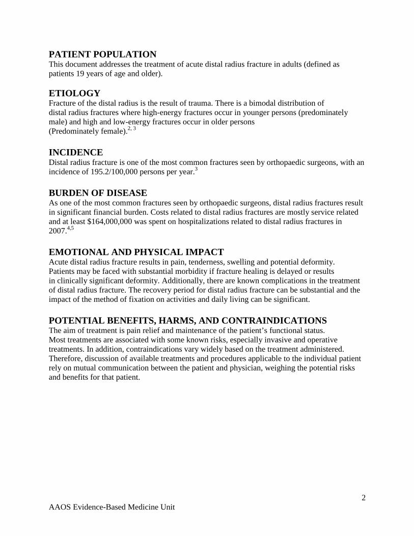
LIST OF TABLES Table 1. Fused Radial Epiphysis ................................................................................................... 24 Table 2. Fused Radial Epiphysis ................................................................................................... 25 Table 3. Post Reduction Radial Shortening >3mm, Dorsal Tilt >10° .......................................... 28 Table 4. Closed Reduction and Percutaneous Fixation vs. ORIF ................................................. 31 Table 5. Non-bridging vs. Bridging External Fixation ................................................................. 32 Table 6. Augmented Bridging External Fixation vs. Percutaneous Pinning ................................ 33 Table 7. Augmented Bridging External Fixation vs. Bridging External Fixation ........................ 33 Table 8. VLP vs. Conservative Treatment of Cast ....................................................................... 34 Table 9. Augmented Bridging External Fixation vs. Plate(s) ....................................................... 35 Table 10. Augmented Bridging External Fixation vs. Volar Locking Plate ................................. 36 Table 11. Closed Reduction and Percutaneous Pinning vs. Open Reduction and Internal Fixation (VLP) ............................................................................................................................................ 37 Table 12. External Fixation vs. Volar Locking Plate.................................................................... 38 Table 13. VLP With or Without Calcium Phosphate Bone Cement............................................. 39 Table 14. Bridging External Fixation vs. Medullary Pinning ....................................................... 40 Table 15. Bridging External Fixation vs. Pins and Plaster ........................................................... 41 Table 16. Dorsal Locking Plate vs. Dual Plating .......................................................................... 41 Table 17. C1, C2, C3 Arthroscopic Evaluation in Patients With Other Associated Injuries ........ 43 Table 18. Concurrent Surgical Treatment of Distal Radioulnar Joint Instability in Patients With Operatively Treated Distal Radius Fracture ................................................................................. 45 Table 19. Fixation of Ulnar Styloid Fractures Associated With Distal Radius Fractures ............ 47 Table 20. Age>55 ......................................................................................................................... 49 Table 21. Wrist Immobilization .................................................................................................... 51
22 AAOS Evidence-Based Medicine Unit

CHAPTER 1: FUSED EPIPHYSIS Patients in chapter one of this AUC had fused radial epiphysis (see Tables 1 and 2). All patients were candidates for conservative treatment. The systematic review looked at which treatment led to a better patient’s outcome, rigid cast or less rigid immobilization (such as removable wrap or brace). Five Level II randomized controlled trials met the inclusion criteria. There were significant differences in pain at 5-6, 8, and 24 weeks, in favor of casting. All other durations of follow-up did not have significant differences between patients treated with rigid immobilization and those treated with less-rigid immobilization.
23 AAOS Evidence-Based Medicine Unit

Table 1. Fused Radial Epiphysis
Study n Patient's Profile Level of Evidence
Outcome Domain
Duration of Follow-
up Favored
Treatment
Tumia 329 Age: 60, 281F 68M, Fused radial epiphysis, a unilateral colles fracture, Dorsal angulation more than 3mm and
radial loss of more than 4° required manipulation II
Pain
10 days ○*
Moir 79 Age: 21-86, 70F 9M, Fused radial epiphysis Median Frykman score V-VI in cast group, II 10 - 14
days ○*
Tumia 182
Same as above
II 5 weeks ○* Moir 79 II 5 - 6 weeks cast* Moir 79 II 8 weeks cast*
Tumia 182 II 8 weeks ○* Tumia 182 II 12 weeks ○* Moir 79 II 13 weeks ○*
Tumia 182 II 24 weeks cast* Moir 79 II 26 weeks ○* Moir 79 Same as above II
Complications n/a cast Ledingham 57 Age: 60, 25F 5M, Fused radial epiphysis requiring
manipulation/reduction II
Bunger 136 Sex: 125F 20M, Frykmann's classification, I and II: 43, III and IV:24, V and VI: 30, VIII and VIII:39 II
Stewart 235 Age:60, Sex 207F 36M, II
24 AAOS Evidence-Based Medicine Unit

Table 2. Fused Radial Epiphysis
Study n Patient's Profile Level of Evidence
Outcome Domain
Duration of Follow-up
Favored Treatment
O’Connor 66 Age: 57, Sex: 44F 22M, Colles fracture not
requiring manipulation II
Pain
1 week ○
Tumia 329
Age: 60, 281F 68M, Fused radial epiphysis, a unilateral colles fracture, Dorsal angulation
more than 3mm and radial loss of more than 4° required manipulation II 10 days ○
Abbaszadegan 68
Undisplaced or minimally displaced Colles fracture,Axial shortening less than 2mm,
Frykman's Classification I and II:19, III and IV:10, V and VI:30, VII and VIII:9 II 11 days ○
O’Connor 66 Same as above II 2 weeks cast
Davis 52 Age: 55, Sex: 43F 11M, <10° of dorsal
angulation II 4 weeks ○ Abbaszadegan 68
Same as above
II 4 weeks ○ Tumia 329 II 5 weeks ○
O’Connor 66 II 6 weeks brace Davis 52 II 6 weeks ○
Abbaszadegan 68 II 8 weeks brace* Tumia 329 II 8 weeks ○ Tumia 329 II 12 weeks ○ Tumia 329 II 24 weeks ○
Abbaszadegan 68 II 1 year ○ Davis 52 II Function > 5 weeks ○
25 AAOS Evidence-Based Medicine Unit

Table 2. Fused Radial Epiphysis
Study n Patient's Profile Level of Evidence
Outcome Domain
Duration of Follow-up
Favored Treatment
O’Connor 66 II Complications
n/a ○
Tumia 329 II Abbaszadegan 68 II
Davis 52 II * Reported by study author(s); ○ No significant difference
26 AAOS Evidence-Based Medicine Unit

CHAPTER 2: POST REDUCTION RADIAL SHORTENING >3MM, DORSAL TILT >10° Chapter two of this AUC contained operative treatment options for fractures with post reduction radial shortening >3mm and dorsal tilt of >10 degrees, or fractures with intra-articular displacement or step off >2mm as opposed to cast fixation (see Table 3). Five randomized clinical trials that compared fixation to cast immobilization are included as evidence for this chapter. All trials had at least one methodological flaw and were downgraded to Level II. All trials mixed articular fractures and extra-articular fractures in a manner which did not allow for separate analysis. There were no age criteria. The average patient age in these trials was similar to trials that address treatment in older-aged patients. There were differences in pain at 24 and 52 weeks, but not at 8 and 12 weeks in one study, differences in motion at 52 weeks in one study, and differences in complications overall, in 4 studies. The differences were all in favor of operative treatment. Complications included carpal tunnel syndrome, thumb pain, ulnar nerve symptoms, and malunion. The moderate strength of the data is, therefore, based primarily on differences in complications, which can be somewhat variably defined.
The amount of pain in patients treated with external fixation was not significantly different from the pain in patients treated with casting at 8 or 12 weeks post-operatively, but was significantly lower at 24 weeks post-operatively and remained significant up to 1 year. However, the number of patients that were pain free after 7 years was not significantly different between external fixation and casting. Additionally, function was significantly better in patients treated by external fixation after 1 year, but not significantly different at 8, 12, or 24 weeks.
There are several complications reported in the studies. Seven of the eleven studies reported that complications did not occur significantly more or less in patients treated with cast or external fixation. Statistically significant differences between patients treated with operative fixation and those treated with casting favored patients treated with external fixation (fewer occurrences of malunion, CTS, ulnar nerve symptoms, and thumb pain). However, there are complications unique to external fixation that patients treated with casting are not exposed to.
27 AAOS Evidence-Based Medicine Unit

Table 3. Post Reduction Radial Shortening >3mm, Dorsal Tilt >10°
Study n Patient's Profile Level of Evidence
Outcome Domain
Duration of
Follow-up
Favored Treatment
Abbaszadegan 46
Age: 63, Sex: 36F, 11M Fracture type: Older 3 and 4, Radial compression of 5mm or more
II
Pain
8 weeks ○
Abbaszadegan 46 II 12 weeks ○
Abbaszadegan 46 II 24 weeks Ex-fix*
Abbaszadegan 46 II 52 weeks Ex-fix*
Young
85
Age: 29-82, Sex: 66F 19M, >10° dorsal angulation or greater than 2mm radial shortening, Frykman grade 1&2: 38
3&4: 19, 5&6:12, 7&8:16
II 7 years ○
Abbaszadegan 46
Age: 63, Sex: 36F, 11M Fracture type: Older 3 and 4, Radial compression of 5mm or more
II
Function
8 weeks ○
Abbaszadegan 46 II 12 weeks ○
Abbaszadegan 46 II 24 weeks ○
Abbaszadegan 46 II 52 weeks Ex-fix*
McQueen 120 Age: 63, Sex: 107F 13M, >10° dorsal angulation or greater than 3mm radial shortening, AO Classifications: A3.2:48,
C2.1:46, C2.2:4, C2.3:2, C3.2:20 II
Complications n/a Ex-fix Howard 50 >30° dorsal angulation or greater than 1cm radial shortening II
Young 85 Age: 29-82, Sex: 66F 19M, >10° dorsal angulation or
greater than 2mm radial shortening, Frykman grade 1&2: 38 3&4: 19, 5&6:12, 7&8:16
II
Pring 76 NR II
* Reported by study author(s), Ex-fix: External fixation, ○: No significant difference
28 AAOS Evidence-Based Medicine Unit

CHAPTER 3: SURGICAL TREATMENT OF DISTAL RADIUS FRACTURE Fourteen clinical trials are included for the chapter on surgical procedure for this AUC: 8 combined intra and extra-articular fractures, 5 studied only intra-articular fractures, and one studied only extra-articular fractures (see Tabled 4-16). No studies evaluated shearing/articular rim fractures or radiocarpal fracture-dislocations. Inclusion was based on inadequate radiographic alignment after initial adequate closed reduction and splint immobilization. Thus, the included studies did not allow for stratification by fracture type. Only two comparisons were made by more than one study, making meta-analysis impossible. All had at least one methodological flaw and were downgraded to Level II.
The included studies in this recommendation do not address many important aspects of the operative treatment of distal radius fractures, including different operative treatments for different fracture types. Therefore, it is not possible to come to an evidence-based conclusion for the optimal operative treatment of distal radius fractures.
Only three of 14 studies had statistically significant findings. In one study, there was only a statistically significant difference in complications. In another study, there was a possibly clinically important difference in DASH at 1 year but not at 3 or 6 months. In the third study, there was statistically significant better function at 2 years for percutaneous fixation over ORIF. All other outcomes evaluated by the included studies were not statistically significant.
Chapter: Closed reduction and percutaneous fixation vs. open reduction and internal fixation. Review identified one randomized controlled trial comparing closed reduction and percutaneous fixation to open reduction and internal fixation (ORIF). Patients treated with closed reduction and percutaneous fixation had significantly better function and no difference in pain. Tendon rupture occurred significantly less in patients treated with closed reduction and percutaneous fixation. No other complication reported by the authors was statistically significant.
Chapter: Non-bridging vs. bridging external fixation. The review identified three randomized controlled trials comparing non-bridging external fixation to bridging external fixation. Function and pain determined by the DASH score showed no significant difference between groups. This result is a true negative, suggesting that patients treated with non-bridging external fixation or bridging external fixation had similar outcomes 1 year after surgery. Pain measured by visual analog scale (VAS) up to 1 year was not significantly different in patients treated with non-bridging or bridging external fixation pain at different anatomic locations on or around the distal radius was not significantly different in patients treated with non-bridging or bridging external fixation. Statistically significant complications occurred in favor of non-bridging external fixation and in favor of bridging external fixation. Tendon rupture occurred significantly more in patients treated with non-bridging external fixation. Malunion occurred significantly more in patients treated with bridging external fixation. Chapter: Augmented bridging external fixation vs. bridging external fixation. One randomized controlled trial compared augmented bridging external fixation (by addition of a fifth external fixator pin) to bridging external fixation. The study did not report validated patient oriented outcomes. There were no statistically significant differences in the occurrence of
29 AAOS Evidence-Based Medicine Unit

complications between patients treated with augmented bridging external fixation or bridging external fixation. Chapter: Augmented bridging external fixation vs. plate We identified one randomized controlled trial comparing augmented bridging external fixation to plate fixation. Activity at final follow-up was similar in patients treated with augmented external fixation or plate fixation. Statistically significant complications occurred in favor of augmented bridging external fixation and in favor of plate(s) Median nerve symptoms occurred significantly more often in patients treated with plate fixation, and radial nerve symptoms occurred significantly more often in patients treated with external fixation. Additionally, infection of pin tracts was reported in patients treated with external fixation. Patients treated by plate fixation are not subject to this complication. Chapter: Augmented bridging external fixation vs. volar locking plate One randomized controlled trial compared augmented bridging external fixation to volar locking plate fixation. At 1 year, the DASH results were possibly clinically important and patients treated with volar locking plates had better function than patients treated with augmented external fixation. However, pain at 1 year was not significantly different between these groups of patients. There were no statistically significant differences in complications between patients treated with augmented bridging external fixation and patients treated with volar locking plate fixation However, both interventions have unique complications.
Chapter: Bridging external fixation vs. Medullary pinning. We identified one randomized controlled trial comparing bridging external fixation to medullary pinning. The study did not report validated patient oriented outcomes, but reports complications. There were significantly more occurrences of infection, median nerve symptoms, and shoulder pain in patients treated with external fixation than those treated with medullary pinning.
Chapter: Dorsal locking plates vs. dual plates We identified one randomized controlled trial comparing dorsal locking plate fixation to dual plate fixation. Pain at 6 months after surgery was similar between the two groups and there was no statistically significant difference in return to activities for patients treated with a dorsal locking plate or dual plates.
30 AAOS Evidence-Based Medicine Unit

Table 4. Closed Reduction and Percutaneous Fixation vs. ORIF
Study n Patient's Profile Level of Evidence
Outcome Domain
Duration of Follow-up
Favored Treatment
Kreder 118 Age: 40, Sex: 70F 48M, AO classification AOB:24, AO-C:155
II Function 2 years
Closed reduction &
percutaneous fixation
Kreder 118 II Pain 2 years ○
31 AAOS Evidence-Based Medicine Unit

Table 5. Non-bridging vs. Bridging External Fixation
Study n Patient's Profile Level of Evidence
Outcome Domain
Duration of Follow-
up Favored
Treatment
Krukhaug 71
>10° of dorsal angulation, and/or radial shortening of more than 2mm, 32 patients treated with plaster cast which failed
to maintain reduction (Malailignment more than 5° or a shortening radius of more than 2mm) at 10 day follow up.
II Function 1 year true negative
Krishnan 60 Age: 57, Sex: 41F 19M, AO calcification A3.2: 3. B2.1:1, AO C:56, C3.2:32, Other AO C 24
II
Pain
6 weeks ○
Krishnan 60 II 26 weeks ○
McQueen 56 Same as below II 1 year ○
Krukhaug 71
>10° of dorsal angulation, and/or radial shortening of more than 2mm, 32 patients treated with plaster cast which failed
to maintain reduction (Malailignment more than 5° or a shortening radius of more than 2mm) at 10 day follow up.
II
Complications n/a ○ Krishnan 60 Same as above II
McQueen 56 Age: 61 Sex: 55F 5M, AO classification system: A3.2: 44, C2.1:14, A3.3: 2 II
32 AAOS Evidence-Based Medicine Unit

Table 6. Augmented Bridging External Fixation vs. Percutaneous Pinning
Study n Patient's Profile
Level of Evidence
Outcome Domain
Duration of Follow-up
Favored Treatment
Harley 41
Age: 42, Sex 28F 22M, AO classidication: A3: 9 C2:17, C3:24
II Function 6 months true negative
Harley 41 II Physical health
6 months ○ Harley 33 II 12 months ○
Harley 41 II Mental health
6 months ○ Harley 33 II 12 months ○
Table 7. Augmented Bridging External Fixation vs. Bridging External Fixation
Study n Patient's Profile Level of Evidence Outcome Domain
Duration of Follow-up
Favored Treatment
Werber 50 Age: 58, Sex 35F 15M, AO classification: A2.2:3, A3.1:3, A3.2:14, C2.1:11, C2.2:17, C2.3:2 II Complications n/a ○
33 AAOS Evidence-Based Medicine Unit

Table 8. VLP vs. Conservative Treatment of Cast
Study n Patient's Profile Level of Evidence Outcome Domain
Duration of follow-up
Favored treatment
Arora 73
Age:76, F55, M18, AO classification A2:6, A3:16, C1:15, C2:20,C3:16 II
Extension 12 Months ○ Flexion 12 Months ○ Supination 12 Months ○ Pronation 12 Months ○ Ulnar deviation 12 Months ○ Radial deviation 12 Months ○ Grip Strength 12 Months VLP Pain at rest (VAS) 12 Months ○
Pain under stress (VAS) 12 Months ○
DASH 12 Months ○ PRWE 12 Months ○
○: no significant difference
34 AAOS Evidence-Based Medicine Unit

Table 9. Augmented Bridging External Fixation vs. Plate(s)
Study n Patient's Profile Level of Evidence Outcome Domain
Duration of Follow-up
Favored Treatment
Leung 144 Age: 45, Sex: 52F 85M, AO classification: C1:36, C2:50, C3:58
II Activity Final follow-up ○
Leung 144 II Complications n/a ○
○: No significant difference
35 AAOS Evidence-Based Medicine Unit

Table 10. Augmented Bridging External Fixation vs. Volar Locking Plate
Study n Patient's Profile Level of Evidence
Outcome Domain
Duration of Follow-up
Favored Treatment
Egol 77
Age:50, Sex: 47F 30M, AO classifications: AoA:46, B:3 C:43
II
Function
3 months true negative
Egol 77 II 6 months true negative
Egol 77 II 1 year
possibly clinically important favoring
VLP
Egol 77 II
Pain
3 months ○
Egol 77 II 6 months ○
Egol 77 II 1 year ○
Egol 77 II Complications n/a ○
36 AAOS Evidence-Based Medicine Unit

Table 11. Closed Reduction and Percutaneous Pinning vs. Open Reduction and Internal Fixation (VLP)
Study n Patient's Profile Level of Evidence
Outcome Domain
Duration of Follow-up
Favored Treatment
Rozental 2009
42
Age: 51.5, Sex: 33F,11M, AO classification: A2:6, A3:10, C1:8 C2:20
II Grip strength (kg)
6 weeks VLP
9 weeks VLP 12 weeks ○
1 year ○
II Pinch strength (kg)
6 weeks VLP 9 weeks VLP
12 weeks ○ 1 year ○
II Digital motion to palm (mm)
6 weeks ○ 9 weeks ○
12 weeks ○ 1 year ○
II
DASH score (points)
6 weeks VLP 9 weeks VLP
12 weeks ○ 1 year ○
McFadyen 2011
56
Age: 18-80, Sex: 33F, 23M, Dorsal angulation >20°, radial shortening 4mm. AO classification: AO A:56 II
DASH 3 months VLP
6 months VLP
Gartland and Werley 3 months VLP
6 months VLP
37 AAOS Evidence-Based Medicine Unit
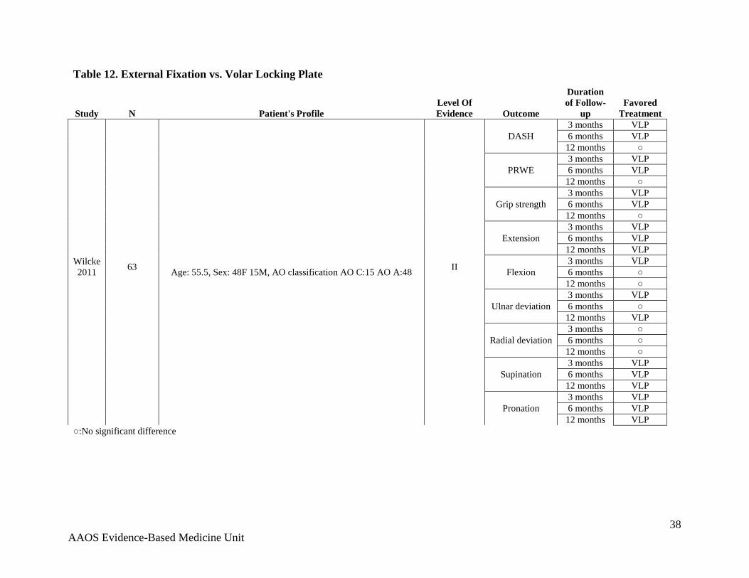
Table 12. External Fixation vs. Volar Locking Plate
Study N Patient's Profile Level Of Evidence Outcome
Duration of Follow-
up Favored
Treatment
Wilcke 2011
63 Age: 55.5, Sex: 48F 15M, AO classification AO C:15 AO A:48 II
DASH 3 months VLP 6 months VLP
12 months ○
PRWE 3 months VLP 6 months VLP
12 months ○
Grip strength 3 months VLP 6 months VLP
12 months ○
Extension 3 months VLP 6 months VLP
12 months VLP
Flexion 3 months VLP 6 months ○
12 months ○
Ulnar deviation 3 months VLP 6 months ○
12 months VLP
Radial deviation 3 months ○ 6 months ○
12 months ○
Supination 3 months VLP 6 months VLP
12 months VLP
Pronation 3 months VLP 6 months VLP
12 months VLP ○:No significant difference
38 AAOS Evidence-Based Medicine Unit

Table 13. VLP With or Without Calcium Phosphate Bone Cement
Study N Patient's Profile Level Of Evidence Outcome
Duration of Follow-up
Favored Treatment
Kim
50
Age: 73, Sex:40F 8M, AO classification A3:20, B3:3, C1:5, C2:18, C3:4
II
Flexion 3 months ○ 1 year ○
Extension 3 months ○ 1 year ○
Supination 3 months ○ 1 year ○
Pronation 3 months ○ 1 year ○
Grip strength (Kg)
3 months ○ 1 year ○
VAS score 3 months ○ 1 year ○
DASH score
3 months ○ 1 year ○
Comparing VLP alone and VLP and calcium phosphate bone cement ○:No significant difference
39 AAOS Evidence-Based Medicine Unit

Table 14. Bridging External Fixation vs. Medullary Pinning
Study n Patient's Profile Level of Evidence Outcome Domain
Duration of Follow-up
Favored Treatment
Pritchett 100 Age: 66 medain, Sex: 55F, 45M, Frykman type VIII, II Complications n/a Medullary pinning
40 AAOS Evidence-Based Medicine Unit

Table 15. Bridging External Fixation vs. Pins and Plaster
Study n Patient's Profile Level of Evidence
Outcome Domain
Duration of Follow-
up Favored
Treatment
Raskin 60 Age: 45, AO group C
II Activity Final follow-up ○
Raskin 60 II Complications
n/a ○
Hutchinson 85 Age: 65, Dorsal angulation greater than 20°, Frykman type V to VIII II
Table 16. Dorsal Locking Plate vs. Dual Plating
Study n Patient's Profile Level of Evidence
Outcome Domain
Duration of Follow-
up Favored
Treatment Hahnloser 46 Age: 55, Sex: 35F,11MDorsal angulation greater than 20° Loss
of radial length >10mm, AO classification AO A3:18, C1:8, C2:7, C3:13
II Pain 6 months ○
Hahnloser 46 II Activity 6 months ○
41 AAOS Evidence-Based Medicine Unit

CHAPTER 4: C1, C2, C3 ARTHROSCOPIC EVALUATION IN PATIENTS WITH OTHER ASSOCIATED INJURIES. Only one of two studies was sufficiently powered to detect the minimum clinically important difference (see Table 17). In this study, arthroscopy assisted reduction of the articular surface. In the arthroscopy group, the DASH scores were clinically improved at the three-month interval. Regardless of arthroscopy, the difference in function as determined by DASH scores was inconclusive at 1 and 2 years post-operatively.
42 AAOS Evidence-Based Medicine Unit

Table 17. C1, C2, C3 Arthroscopic Evaluation in Patients With Other Associated Injuries
Study n Patient's Profile Level of Evidence
Outcome Domain
Duration of Follow-up
Favored Treatment
Varitimidis 40 Age:46, Sex: 23F 17M, AO fracture types C1, C2, or C3
II Function
3 months Arthroscopic*
Varitimidis 40 II 12 months inconclusive Varitimidis 40 II 24 months inconclusive
* Clinically important difference compared to fluoroscopic
43 AAOS Evidence-Based Medicine Unit

CHAPTER 5: CONCURRENT TREATMENT OF DISTAL RADIOULNAR JOINT INSTABILITY IN PATIENTS WITH OPERATIVELY TREATED DISTAL RADIUS FRACTURE. Two studies investigated the functional outcome of DRUJ injuries (see Table 18). The instabilities were identified at the conclusion of treatment. Therefore, no instabilities were treated at the time of surgery. Although the patients with instability had poorer outcomes, neither study addressed the question of whether early operative intervention is indicated.
We identified one prospective non-randomized study that compared patients with unstable DRUJ after distal radius fixation to patients with a stable DRUJ after distal radius fixation. Patients with unstable DRUJ had significantly more pain at rest and on loading than those patients with a stable DRUJ.
We also identified an additional prospective non-randomized study comparing patients with a distal radius fracture involving the DRUJ to those with a fracture not involving the DRUJ. Significantly more patients without involvement of the DRUJ were pain free 6 weeks after treatment of their distal radius fracture. The authors report that this significant difference remained at 6 months and 1 year (chi-square, p < 0.05).
44 AAOS Evidence-Based Medicine Unit
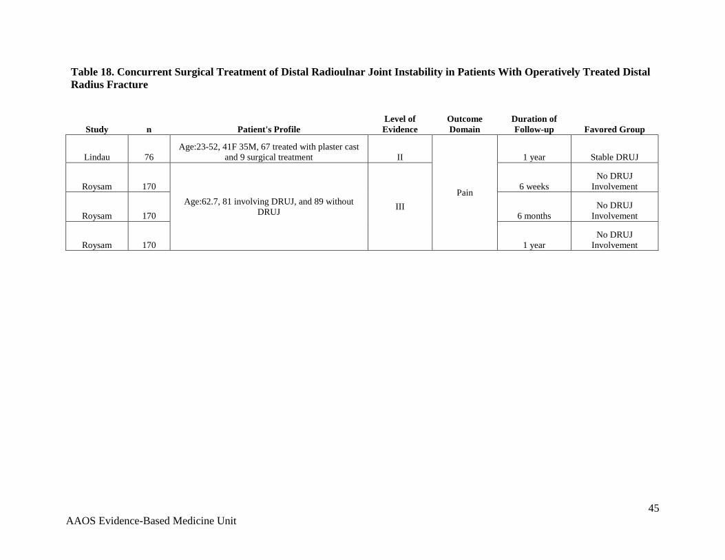
Table 18. Concurrent Surgical Treatment of Distal Radioulnar Joint Instability in Patients With Operatively Treated Distal Radius Fracture
Study n Patient's Profile Level of Evidence
Outcome Domain
Duration of Follow-up Favored Group
Lindau 76 Age:23-52, 41F 35M, 67 treated with plaster cast
and 9 surgical treatment II
Pain
1 year Stable DRUJ
Roysam 170 Age:62.7, 81 involving DRUJ, and 89 without
DRUJ III
6 weeks No DRUJ
Involvement
Roysam 170 6 months No DRUJ
Involvement
Roysam 170 1 year No DRUJ
Involvement
45 AAOS Evidence-Based Medicine Unit

CHAPTER 6: FIXATION OF ULNAR STYLOID FRACTURES ASSOCIATED WITH DISTAL RADIUS FRACTURES. One study found no difference between treatment (fixation) and no treatment (see Table 19). The other study identified ulna styloid fractures after treatment was completed. The study found that there were clinically important differences between patients with and without styloid fractures. Therefore, no ulna styloid fractures were treated at the time of surgery. Although the patients with ulna styloid fractures had poorer outcomes, the study did not address the question of whether early operative intervention is indicated. Therefore, we found no conclusive evidence to recommend operative or non-operative treatment for the ulna styloid fracture.
We identified one randomized controlled trial comparing patients treated operatively for a concomitant ulna fracture to patients treated conservatively for a concomitant ulnar fracture. There was no significant difference between the numbers of patients that were pain free 2 years after treatment.
Additionally, we identified a single prospective non-randomized study that compared patients without a concomitant ulna fracture to patients with a concomitant fracture of the base or tip of the ulna. The patients with concomitant ulnar fractures did not receive any treatment for their ulnar fracture. Therefore, the comparison between the groups in this study determines the effect of an untreated concomitant ulnar fracture on the outcome. Patients without a concomitant ulna fracture had clinically important differences in function, as determined by the DASH instrument, compared to those with a concomitant ulna fracture. Additionally, there was no significant difference between patients with and without a concomitant ulna fracture in the occurrence of ulnar wrist pain at final follow-up.
46 AAOS Evidence-Based Medicine Unit

Table 19. Fixation of Ulnar Styloid Fractures Associated With Distal Radius Fractures
Study n Patient's Profile Level of Evidence Outcome Domain Duration of Follow-up Favored Group
Ekenstam 40
Age: 51, Sex: 31F 10M, 19 Treated with closed manipulation followed by suturing of the triangular stabilization of the ulnar styloid, 22 patients treated with closed reduction and plaster cast.
II
Pain
2 years ○
Zenke 118 Age: 64.1, Sex: 90F 28M, Treated
with internal fixation VLP, >5° dorsal tilt, <20°radial inclination angle,
>2mm ulnar variance contralateral or intra-articular step of >1mm. AO
Classification AO A2:29, A3:31. AO C1:17 C2:31 C3:10
II
Final follow-up ○
Zenke 118
Function
1 week no ulnar fx* Zenke 118 2 weeks no ulnar fx* Zenke 118 4 weeks no ulnar fx* Zenke 118 8 weeks no ulnar fx* Zenke 118 12 weeks no ulnar fx* Zenke 118 24 weeks no ulnar fx*
* Clinically important difference compared to patients with ulnar fracture ○: No significant difference
47 AAOS Evidence-Based Medicine Unit

CHAPTER 7: AGE > 55 YEARS We were interested in determining the role of operative treatment compared to non-operative treatment of patients, defined by the published literature as “elderly”, and that distinguished patients based on infirmity, functional demands, bone quality, or energy of injury. Three clinical trials met the inclusion criteria (see Table 20). Two trials compared external fixation to cast immobilization and one trial compared percutaneous pinning to cast immobilization. All trials had at least one methodological flaw and were downgraded to Level II. One trial addressed extra-articular fractures, one addressed articular fractures, and one addressed both. Age criteria included age over 55 in 2 studies and over age 60 in one study. We selected the age of 55 because these included studies that enrolled patients no younger than 55 years. We were unable to identify studies that distinguished patients based on infirmity, functional demands, bone quality, or energy of injury. Inclusion was based on redisplacement in one study, initial radiographic alignment in one study, and instability not otherwise defined in one study. There were no differences in pain, function, complications or SF-36 at any time point. The amount of pain experienced after 1 year was not significantly different in patients treated with percutaneous pinning or with a cast. There was also no significant difference in overall mental or physical health, as determined by the SF-36 score, and no significant difference in the occurrence of complications in patients treated with percutaneous pinning or cast. However, percutaneous pinning does have complications that those patients treated with casts are not subject to.
Both randomized controlled trials that compared patients treated with external fixation to those treated with cast and over the age of 55 found no statistically significant differences for pain experienced at different anatomic locations on or around the distal radius or with different amounts of activity after 1 year. No statistically significant differences, for various functional activities, between patients treated with external fixation or casting and over the age of 55 were present after 1 year.
48 AAOS Evidence-Based Medicine Unit

Table 20. Age>55
Study n Patient's Profile Level of Evidence Outcome Domain
Duration of Follow-up
Favored Treatment
Azzopardi 57
Age: 72, Sex: 48F 6M, AO Classification AO-A3 or Frykman types I and II II
Pain 1 year ○*
Azzopardi 57 Mental health 1 year ○*
Azzopardi 57 Physical health 1 year ○*
Azzopardi 57 Complications n/a ○*
Hegeman 32 Same as below II
Pain
6 weeks ○**
Hegeman 32 3 months ○**
Roumen 101 Age: 70, 93F 8M, Frykman type: II:2, V:22, VI:36, VII:9,
VIII:32 II 26 weeks ○**
Hegeman 32 Age: 70 29F 3M, Dorsal angulation of more than 10° and
radial inclination of less than 20°, and a positive ulnar variance of more than 3mm
II
1 year ○**
Hegeman 32 Function
6 weeks ○**
Hegeman 32 3 months ○**
Hegeman 32 1 year ○**
Foldhazy 59
Age:72, Sex: 53F 6M, AO classification A2:6, A3:21, C1:1, C2:15, C3:8 II
Green & Obrian/Conney
score 1 year ○**
Foldhazy 59 VAS
2 months ○**
Foldhazy 59 6 months ○**
Foldhazy 59 1 year ○**
Foldhazy 59 Grip strength
2 months ○**
Foldhazy 59 6 months ○**
Foldhazy 59 1 year ○**
Wong 60 Age: 70, 49F 11M, Frykman's classification: I:36, II:25 II Overall WHOQoL 1 year ○*
Wong 60 Total score (100) 6 year ○*
*Comparing percutaneous pinning to casting ** comparing external fixation to casting ○: No significant difference
49 AAOS Evidence-Based Medicine Unit

CHAPTER 8: EARLY WRIST IMMOBILIZATION Three studies were included for this chapter. Each study investigated different operative treatment methods: volar plate, trans-styloid fixation, or external fixation (see Table 21). Mobilization was commenced at different times. In the two internal fixation studies, therapy started approximately at 1 week. In the external fixation study, mobilization commenced at 3 weeks. In 2 studies, the control group was either casted or immobilized with a fixator. In the volar plating study, the control group was immobilized by a thermoplastic splint that they were instructed to remove for showering and, therefore, are not a reliable control group.
The outcome measures used were pain and function (DASH) and/or complications. None of the outcomes were significantly different between early motion and late motion. These data support the recommendation that patients do not need to begin early wrist motion after stable fracture fixation.
There is no difference (true negative) in function determined by the DASH score up to 6 months between patients with early wrist motion and those with late wrist motion. There is also no significant difference between the amount of pain experienced by patients with early wrist motion and those with late wrist motion. No significant differences in the occurrence of complications between patients with early wrist motion or patients with late wrist motion were seen in any of the trials.
50 AAOS Evidence-Based Medicine Unit

Table 21. Wrist Immobilization
Study n Patient's Profile Level of Evidence
Outcome Domain
Duration of
Follow-up
Favored Group
Allain 60 Age: 53, Sex: 49F 11M, AO classification: A2:28, A3:17, C1:8, C2:7, K wire and Cast II
Pain
4 weeks ○
Lozano-Calderon 56
Age:55, Sex:39F, 21M, AO classification C:29, A:22, B:8
II 3 months true negative
Lozano-Calderon 54 II 6 months true
negative
Allain 60
Same as above
II 1 year ○
Lozano-Calderon 56 II
Function
3 months ○
Lozano-Calderon 54 II 6 months ○
Lozano-Calderon 56 II
Complications
n/a ○
Allain 60 II ○
McQueen 54 II ○
○: No significant difference
51 AAOS Evidence-Based Medicine Unit

LITERATURE REVIEW REFERENCES Abbaszadegan H, Conradi P, Jonsson U. Fixation not needed for undisplaced Colles' fracture. Acta Orthop Scand 1989;60(1):60-62. Abbaszadegan H, Jonsson U. External fixation of plaster cast for severely displaced Colles' fractures? Prospective 1-year study of 46 patients. Acta Orthopaedica Scandinavica 1990;61(6):528-530. Allain J, le GP, Le MS, Goutallier D. Trans-styloid fixation of fractures of the distal radius. A prospective randomized comparison between 6- and 1-week postoperative immobilization in 60 fractures. Acta Orthop Scand 1999;70(2):119- 123. Arora R, Lutz M, Deml C, Krappinger D, Haug L, Gabl M. A prospective randomized trial comparing nonoperative treatment with volar locking plate fixation for displaced and unstable distal radial fractures in patients sixty-five years of age and older. J Bone Joint Surg Am 2011;93-A(23):2146-2153. Azzopardi T, Ehrendorfer S, Coulton T, Abela M. Unstable extra-articular fractures of the distal radius: a prospective, randomised study of immobilisation in a cast versus supplementary percutaneous pinning. J Bone Joint Surg Br 2005;87(6):837-840. Bunger C, Solund K, Rasmussen P. Early results after Colles' fracture: functional bracing in supination vs dorsal plaster immobilization. Arch Orthop Trauma Surg 1984;103(4):251-256. Davis TR, Buchanan JM. A controlled prospective study of early mobilization of minimally displaced fractures of the distal radial metaphysis. Injury 1987;18(4):283-285. Egol K, Walsh M, Tejwani N, McLaurin T, Wynn C, Paksima N. Bridging external fixation and supplementary Kirschner-wire fixation versus volar locked plating for unstable fractures of the distal radius: a randomised, prospective trial. J Bone Joint Surg Br 2008;90(9):1214-1221. af Ekenstam F., Jakobsson OP, Wadin K. Repair of the triangular ligament in Colles' fracture. No effect in a prospective randomized study. Acta Orthop Scand 1989;60(4):393-396. Foldhazy Z, Ahrengart L. External fixation versus closed treatment of displaced distal radial fractures in elderly patients: a randomized controlled trial. Current Orthopaedic Practice 2010;21(3):288-295. Hahnloser D, Platz A, Amgwerd M, Trentz O. Internal fixation of distal radius fractures with dorsal dislocation: pi-plate or two 1/4 tube plates? A prospective
52 AAOS Evidence-Based Medicine Unit

randomized study. J Trauma 1999;47(4):760-765. Harley BJ, Scharfenberger A, Beaupre LA, Jomha N, Weber DW. Augmented external fixation versus percutaneous pinning and casting for unstable fractures of the distal radius--a prospective randomized trial. J Hand Surg [Am] 2004;29(5):815-824. Hegeman JH, Oskam J, van der PJ, Ten Duis HJ, Vierhout PAM. Primary external fixation versus plaster immobilization of the intra-articular unstable distal radial fracture in the elderly. Aktuelle Traumatologie 2004;34(2):64-70. Howard PW, Stewart HD, Hind RE, Burke FD. External fixation or plaster for severely displaced comminuted Colles' fractures? A prospective study of anatomical and functional results. Journal of Bone and Joint Surgery - Series B 1989;71(1):68-73. Hutchinson DT, Strenz GO, Cautilli RA. Pins and plaster vs external fixation in the treatment of unstable distal radial fractures. A randomized prospective study. J Hand Surg [Br ] 1995;20(3):365-372. Kim JK, Koh YD, Kook SH. Effect of calcium phosphate bone cement augmentation on volar plate fixation of unstable distal radius fractures in the elderly. J Bone Joint Surg Am 2011;93-A(7):609-14. Kreder HJ, Hanel DP, Agel J et al. Indirect reduction and percutaneous fixation versus open reduction and internal fixation for displaced intra-articular fractures of the distal radius: a randomised, controlled trial. J Bone Joint Surg Br 2005;87(6):829-836. Krishnan J, Wigg AE, Walker RW, Slavotinek J. Intra-articular fractures of the distal radius: a prospective randomised controlled trial comparing static bridging and dynamic non-bridging external fixation. J Hand Surg [Br ] 2003;28(5):417- 421. Krukhaug Y, Ugland S, Lie SA, Hove LM. External fixation of fractures of the distal radius: a randomized comparison of the Hoffman compact II non-bridging fixator and the Dynawrist fixator in 75 patients followed for 1 year. Acta Orthop 2009;80(1):104-108. Ledingham WM, Wytch R, Goring CC, Mathieson AB, Wardlaw D. On immediate functional bracing of Colles' fracture. Injury 1991;22(3):197-201. Leung F, Tu YK, Chew WY, Chow SP. Comparison of external and percutaneous pin fixation with plate fixation for intra-articular distal radial fractures. A randomized study. J Bone Joint Surg Am 2008;90(1):16-22. Lindau T, Hagberg L, Adlercreutz C, Jonsson K, Aspenberg P. Distal radioulnar
53 AAOS Evidence-Based Medicine Unit

instability is an independent worsening factor in distal radial fractures. Clin Orthop Relat Res 2000;(376):229-235. Lozano-Calderon SA, Souer S, Mudgal C, Jupiter JB, Ring D. Wrist mobilization following volar plate fixation of fractures of the distal part of the radius. J Bone Joint Surg Am 2008;90(6):1297-1304. McFadyen I, Field J, McCann P, Ward J, Nicol S, Curwen C. Should unstable extra-articular distal radial fractures be treated with fixed-angle volar-locked plates or percutaneous Kirschner wires? A prospective randomised controlled trial. Injury 2011;42(2):162–166. McQueen MM. Redisplaced unstable fractures of the distal radius. A randomised, prospective study of bridging versus non-bridging external fixation. J Bone Joint Surg Br 1998;80(4):665-669. McQueen MM, Hajducka C, Court-Brown CM. Redisplaced unstable fractures of the distal radius: a prospective randomised comparison of four methods of treatment. J Bone Joint Surg Br 1996;78(3):404-409. Moir JS, Murali SR, Ashcroft GP, Wardlaw D, Matheson AB. A new functional brace for the treatment of Colles' fractures. Injury 1995;26(9):587-593. O'Connor D, Mullett H, Doyle M, Mofidi A, Kutty S, O'Sullivan M. Minimally displaced Colles' fractures: a prospective randomized trial of treatment with a wrist splint or a plaster cast. J Hand Surg [Br ] 2003;28(1):50-53. Pring DJ, Barber L, Williams DJ. Bipolar fixation of fractures of the distal end of the radius: a comparative study. Injury 1988;19(3):145-148. Pritchett JW. External fixation or closed medullary pinning for unstable Colles fractures? Journal of Bone and Joint Surgery - Series B 1995;77(2):267-269. Raskin KB, Melone CP, Jr. Unstable articular fractures of the distal radius. Comparative techniques of ligamentotaxis. Orthop Clin North Am 1993;24(2):275-286. Roumen RMH, Hesp WLEM, Bruggink EDM. Unstable Colles' fractures in elderly patients. A randomised trial of external fixation for redisplacement. Journal of Bone and Joint Surgery - Series B 1991;73(2):307-311. Roysam GS. The distal radio-ulnar joint in Colles' fractures. J Bone Joint Surg Br 1993;75(1):58-60. Rozental TD, Blazar PE, Franko OI, Chacko AT, Earp BE, Day CS. Functional outcomes for unstable distal radial fractures treated with open reduction and internal fixation or closed reduction and percutaneous fixation. J Bone Joint Surg Am 2009;91-A(8):1837-1846.
54 AAOS Evidence-Based Medicine Unit

Stewart HD, Innes AR, Burke FD. Functional cast-bracing for Colles' fractures. A comparison between cast-bracing and conventional plaster casts. J Bone Joint Surg Br 1984;66(5):749-753. Tumia N, Wardlaw D, Hallett J, Deutman R, Mattsson SA, Sanden B. Aberdeen Colles' fracture brace as a treatment for Colles' fracture. A multicentre, prospective, randomised, controlled trial. J Bone Joint Surg Br 2003;85(1):78-82. Varitimidis SE, Basdekis GK, Dailiana ZH, Hantes ME, Bargiotas K, Malizos K. Treatment of intra-articular fractures of the distal radius: fluoroscopic or arthroscopic reduction? J Bone Joint Surg Br 2008;90(6):778-785. Werber KD, Raeder F, Brauer RB, Weiss S. External fixation of distal radial fractures: four compared with five pins: a randomized prospective study. J Bone Joint Surg Am 2003;85-A(4):660-666. Wilcke MKT, Abbaszadegan H, Adolphson PY. Wrist function recovers more rapidly after volar locked plating than after external fixation but the outcomes are similar after 1 year: A randomized study of 63 patients with a dorsally displaced fracture of the distal radius. Acta Orthopaedica 2011;82(1):76–81. Wong TC, Chiu Y, Tsang WL, Leung WY, Yam SK, Yeung SH. Casting versus percutaneous pinning for extra-articular fractures of the distal radius in an elderly Chinese population: a prospective randomized controlled trial. Journal of Hand Surgery [European Volume] 2010;35E(3):202–208. Young CF, Nanu AM, Checketts RG. Seven-year outcome following Colles' type distal radial fracture. A comparison of two treatment methods. J Hand Surg [Br ] 2003;28(5):422-426. Zenke Y, Sakai A, Oshige T, Moritani S, Nakamura T. The effect of an associated ulnar styloid fracture on the outcome after fixation of a fracture of the distal radius. J Bone Joint Surg Br 2009;91(1):102-107.
55 AAOS Evidence-Based Medicine Unit

VI. APPENDICES
56 AAOS Evidence-Based Medicine Unit

APPENDIX A. DOCUMENTATION OF APPROVAL
AAOS BODIES THAT APPROVED THIS APPROPRIATE USE CRITERIA AUC Section: Approved on February 21st, 2013 The AAOS Appropriate Use Criteria Section consists of six AAOS members. The overall purpose of this Section is to plan, organize, direct and evaluate initiatives related to Appropriate Use Criteria. Council on Research and Quality: Approved on February 26th, 2013 To enhance the mission of the AAOS, the Council on Research and Quality promotes the most ethically and scientifically sound basic, clinical, and translational research possible to ensure the future care for patients with musculoskeletal disorders. The Council also serves as the primary resource to educate its members, the public, and public policy makers regarding evidenced-based medical practice, orthopaedic devices and biologics regulatory pathways and standards development, patient safety, occupational health, technology assessment, and other related areas of importance. Board of Directors: Approved on March 18th, 2013 The 16 member AAOS Board of Directors manages the affairs of the AAOS, sets policy, and determines and continually reassesses the Strategic Plan.
57 AAOS Evidence-Based Medicine Unit

APPENDIX B. LIST OF CLINICAL SCENARIOS These scenarios are a result of combining every possible combination of the indications created by the writing panel. Please comment on any scenarios that you think may not be represented in this matrix or scenarios that may not be seen in clinical practice.
Scenario # Scenario 1 Type A, High-energy, Home-bound, ASA 1-2-3, No associated injuries 2 Type A, High-energy, Home-bound, ASA 1-2-3, Grade I or II Open Fracture 3 Type A, High-energy, Home-bound, ASA 1-2-3, Grade III Open Fracture 4 Type A, High-energy, Home-bound, ASA 1-2-3, Median nerve injury 5 Type A, High-energy, Home-bound, ASA 1-2-3, Other Ipsilateral Injury 6 Type A, High-energy, Home-bound, ASA 4, No associated injuries 7 Type A, High-energy, Home-bound, ASA 4, Grade I or II Open Fracture 8 Type A, High-energy, Home-bound, ASA 4, Grade III Open Fracture 9 Type A, High-energy, Home-bound, ASA 4, Median nerve injury
10 Type A, High-energy, Home-bound, ASA 4, Other Ipsilateral Injury 11 Type A, High-energy, Independent, ASA 1-2-3, No associated injuries 12 Type A, High-energy, Independent, ASA 1-2-3, Grade I or II Open Fracture 13 Type A, High-energy, Independent, ASA 1-2-3, Grade III Open Fracture 14 Type A, High-energy, Independent, ASA 1-2-3, Median nerve injury 15 Type A, High-energy, Independent, ASA 1-2-3, Other Ipsilateral Injury 16 Type A, High-energy, Independent, ASA 4, No associated injuries 17 Type A, High-energy, Independent, ASA 4, Grade I or II Open Fracture 18 Type A, High-energy, Independent, ASA 4, Grade III Open Fracture 19 Type A, High-energy, Independent, ASA 4, Median nerve injury 20 Type A, High-energy, Independent, ASA 4, Other Ipsilateral Injury 21 Type A, High-energy, Normal, ASA 1-2-3, No associated injuries 22 Type A, High-energy, Normal, ASA 1-2-3, Grade I or II Open Fracture 23 Type A, High-energy, Normal, ASA 1-2-3, Grade III Open Fracture 24 Type A, High-energy, Normal, ASA 1-2-3, Median nerve injury 25 Type A, High-energy, Normal, ASA 1-2-3, Other Ipsilateral Injury 26 Type A, High-energy, Normal, ASA 4, No associated injuries 27 Type A, High-energy, Normal, ASA 4, Grade I or II Open Fracture 28 Type A, High-energy, Normal, ASA 4, Grade III Open Fracture 29 Type A, High-energy, Normal, ASA 4, Median nerve injury 30 Type A, High-energy, Normal, ASA 4, Other Ipsilateral Injury 31 Type A, High-energy, High, ASA 1-2-3, No associated injuries 32 Type A, High-energy, High, ASA 1-2-3, Grade I or II Open Fracture 33 Type A, High-energy, High, ASA 1-2-3, Grade III Open Fracture 34 Type A, High-energy, High, ASA 1-2-3, Median nerve injury 35 Type A, High-energy, High, ASA 1-2-3, Other Ipsilateral Injury 36 Type A, High-energy, High, ASA 4, No associated injuries
58 AAOS Evidence-Based Medicine Unit

37 Type A, High-energy, High, ASA 4, Grade I or II Open Fracture 38 Type A, High-energy, High, ASA 4, Grade III Open Fracture 39 Type A, High-energy, High, ASA 4, Median nerve injury 40 Type A, High-energy, High, ASA 4, Other Ipsilateral Injury 41 Type A, Low-energy, Home-bound, ASA 1-2-3, No associated injuries 42 Type A, Low-energy, Home-bound, ASA 1-2-3, Grade I or II Open Fracture 43 Type A, Low-energy, Home-bound, ASA 1-2-3, Median nerve injury 44 Type A, Low-energy, Home-bound, ASA 1-2-3, Other Ipsilateral Injury 45 Type A, Low-energy, Home-bound, ASA 4, No associated injuries 46 Type A, Low-energy, Home-bound, ASA 4, Grade I or II Open Fracture 47 Type A, Low-energy, Home-bound, ASA 4, Median nerve injury 48 Type A, Low-energy, Home-bound, ASA 4, Other Ipsilateral Injury 49 Type A, Low-energy, Independent, ASA 1-2-3, No associated injuries 50 Type A, Low-energy, Independent, ASA 1-2-3, Grade I or II Open Fracture 51 Type A, Low-energy, Independent, ASA 1-2-3, Median nerve injury 52 Type A, Low-energy, Independent, ASA 1-2-3, Other Ipsilateral Injury 53 Type A, Low-energy, Independent, ASA 4, No associated injuries 54 Type A, Low-energy, Independent, ASA 4, Grade I or II Open Fracture 55 Type A, Low-energy, Independent, ASA 4, Median nerve injury 56 Type A, Low-energy, Independent, ASA 4, Other Ipsilateral Injury 57 Type A, Low-energy, Normal, ASA 1-2-3, No associated injuries 58 Type A, Low-energy, Normal, ASA 1-2-3, Grade I or II Open Fracture 59 Type A, Low-energy, Normal, ASA 1-2-3, Median nerve injury 60 Type A, Low-energy, Normal, ASA 1-2-3, Other Ipsilateral Injury 61 Type A, Low-energy, Normal, ASA 4, No associated injuries 62 Type A, Low-energy, Normal, ASA 4, Grade I or II Open Fracture 63 Type A, Low-energy, Normal, ASA 4, Median nerve injury 64 Type A, Low-energy, Normal, ASA 4, Other Ipsilateral Injury 65 Type A, Low-energy, High, ASA 1-2-3, No associated injuries 66 Type A, Low-energy, High, ASA 1-2-3, Grade I or II Open Fracture 67 Type A, Low-energy, High, ASA 1-2-3, Median nerve injury 68 Type A, Low-energy, High, ASA 1-2-3, Other Ipsilateral Injury 69 Type A, Low-energy, High, ASA 4, No associated injuries 70 Type A, Low-energy, High, ASA 4, Grade I or II Open Fracture 71 Type A, Low-energy, High, ASA 4, Median nerve injury 72 Type A, Low-energy, High, ASA 4, Other Ipsilateral Injury 73 Type B, High-energy, Home-bound, ASA 1-2-3, No associated injuries 74 Type B, High-energy, Home-bound, ASA 1-2-3, Grade I or II Open Fracture 75 Type B, High-energy, Home-bound, ASA 1-2-3, Grade III Open Fracture 76 Type B, High-energy, Home-bound, ASA 1-2-3, Median nerve injury 77 Type B, High-energy, Home-bound, ASA 1-2-3, Other Ipsilateral Injury
59 AAOS Evidence-Based Medicine Unit

78 Type B, High-energy, Home-bound, ASA 4, No associated injuries 79 Type B, High-energy, Home-bound, ASA 4, Grade I or II Open Fracture 80 Type B, High-energy, Home-bound, ASA 4, Grade III Open Fracture 81 Type B, High-energy, Home-bound, ASA 4, Median nerve injury 82 Type B, High-energy, Home-bound, ASA 4, Other Ipsilateral Injury 83 Type B, High-energy, Independent, ASA 1-2-3, No associated injuries 84 Type B, High-energy, Independent, ASA 1-2-3, Grade I or II Open Fracture 85 Type B, High-energy, Independent, ASA 1-2-3, Grade III Open Fracture 86 Type B, High-energy, Independent, ASA 1-2-3, Median nerve injury 87 Type B, High-energy, Independent, ASA 1-2-3, Other Ipsilateral Injury 88 Type B, High-energy, Independent, ASA 4, No associated injuries 89 Type B, High-energy, Independent, ASA 4, Grade I or II Open Fracture 90 Type B, High-energy, Independent, ASA 4, Grade III Open Fracture 91 Type B, High-energy, Independent, ASA 4, Median nerve injury 92 Type B, High-energy, Independent, ASA 4, Other Ipsilateral Injury 93 Type B, High-energy, Normal, ASA 1-2-3, No associated injuries 94 Type B, High-energy, Normal, ASA 1-2-3, Grade I or II Open Fracture 95 Type B, High-energy, Normal, ASA 1-2-3, Grade III Open Fracture 96 Type B, High-energy, Normal, ASA 1-2-3, Median nerve injury 97 Type B, High-energy, Normal, ASA 1-2-3, Other Ipsilateral Injury 98 Type B, High-energy, Normal, ASA 4, No associated injuries 99 Type B, High-energy, Normal, ASA 4, Grade I or II Open Fracture
100 Type B, High-energy, Normal, ASA 4, Grade III Open Fracture 101 Type B, High-energy, Normal, ASA 4, Median nerve injury 102 Type B, High-energy, Normal, ASA 4, Other Ipsilateral Injury 103 Type B, High-energy, High, ASA 1-2-3, No associated injuries 104 Type B, High-energy, High, ASA 1-2-3, Grade I or II Open Fracture 105 Type B, High-energy, High, ASA 1-2-3, Grade III Open Fracture 106 Type B, High-energy, High, ASA 1-2-3, Median nerve injury 107 Type B, High-energy, High, ASA 1-2-3, Other Ipsilateral Injury 108 Type B, High-energy, High, ASA 4, No associated injuries 109 Type B, High-energy, High, ASA 4, Grade I or II Open Fracture 110 Type B, High-energy, High, ASA 4, Grade III Open Fracture 111 Type B, High-energy, High, ASA 4, Median nerve injury 112 Type B, High-energy, High, ASA 4, Other Ipsilateral Injury 113 Type B, Low-energy, Home-bound, ASA 1-2-3, No associated injuries 114 Type B, Low-energy, Home-bound, ASA 1-2-3, Grade I or II Open Fracture 115 Type B, Low-energy, Home-bound, ASA 1-2-3, Median nerve injury 116 Type B, Low-energy, Home-bound, ASA 1-2-3, Other Ipsilateral Injury 117 Type B, Low-energy, Home-bound, ASA 4, No associated injuries 118 Type B, Low-energy, Home-bound, ASA 4, Grade I or II Open Fracture
60 AAOS Evidence-Based Medicine Unit

119 Type B, Low-energy, Home-bound, ASA 4, Median nerve injury 120 Type B, Low-energy, Home-bound, ASA 4, Other Ipsilateral Injury 121 Type B, Low-energy, Independent, ASA 1-2-3, No associated injuries 122 Type B, Low-energy, Independent, ASA 1-2-3, Grade I or II Open Fracture 123 Type B, Low-energy, Independent, ASA 1-2-3, Median nerve injury 124 Type B, Low-energy, Independent, ASA 1-2-3, Other Ipsilateral Injury 125 Type B, Low-energy, Independent, ASA 4, No associated injuries 126 Type B, Low-energy, Independent, ASA 4, Grade I or II Open Fracture 127 Type B, Low-energy, Independent, ASA 4, Median nerve injury 128 Type B, Low-energy, Independent, ASA 4, Other Ipsilateral Injury 129 Type B, Low-energy, Normal, ASA 1-2-3, No associated injuries 130 Type B, Low-energy, Normal, ASA 1-2-3, Grade I or II Open Fracture 131 Type B, Low-energy, Normal, ASA 1-2-3, Median nerve injury 132 Type B, Low-energy, Normal, ASA 1-2-3, Other Ipsilateral Injury 133 Type B, Low-energy, Normal, ASA 4, No associated injuries 134 Type B, Low-energy, Normal, ASA 4, Grade I or II Open Fracture 135 Type B, Low-energy, Normal, ASA 4, Median nerve injury 136 Type B, Low-energy, Normal, ASA 4, Other Ipsilateral Injury 137 Type B, Low-energy, High, ASA 1-2-3, No associated injuries 138 Type B, Low-energy, High, ASA 1-2-3, Grade I or II Open Fracture 139 Type B, Low-energy, High, ASA 1-2-3, Median nerve injury 140 Type B, Low-energy, High, ASA 1-2-3, Other Ipsilateral Injury 141 Type B, Low-energy, High, ASA 4, No associated injuries 142 Type B, Low-energy, High, ASA 4, Grade I or II Open Fracture 143 Type B, Low-energy, High, ASA 4, Median nerve injury 144 Type B, Low-energy, High, ASA 4, Other Ipsilateral Injury 145 Type C, High-energy, Home-bound, ASA 1-2-3, No associated injuries 146 Type C, High-energy, Home-bound, ASA 1-2-3, Grade I or II Open Fracture 147 Type C, High-energy, Home-bound, ASA 1-2-3, Grade III Open Fracture 148 Type C, High-energy, Home-bound, ASA 1-2-3, Median nerve injury 149 Type C, High-energy, Home-bound, ASA 1-2-3, Other Ipsilateral Injury 150 Type C, High-energy, Home-bound, ASA 4, No associated injuries 151 Type C, High-energy, Home-bound, ASA 4, Grade I or II Open Fracture 152 Type C, High-energy, Home-bound, ASA 4, Grade III Open Fracture 153 Type C, High-energy, Home-bound, ASA 4, Median nerve injury 154 Type C, High-energy, Home-bound, ASA 4, Other Ipsilateral Injury 155 Type C, High-energy, Independent, ASA 1-2-3, No associated injuries 156 Type C, High-energy, Independent, ASA 1-2-3, Grade I or II Open Fracture 157 Type C, High-energy, Independent, ASA 1-2-3, Grade III Open Fracture 158 Type C, High-energy, Independent, ASA 1-2-3, Median nerve injury 159 Type C, High-energy, Independent, ASA 1-2-3, Other Ipsilateral Injury
61 AAOS Evidence-Based Medicine Unit

160 Type C, High-energy, Independent, ASA 4, No associated injuries 161 Type C, High-energy, Independent, ASA 4, Grade I or II Open Fracture 162 Type C, High-energy, Independent, ASA 4, Grade III Open Fracture 163 Type C, High-energy, Independent, ASA 4, Median nerve injury 164 Type C, High-energy, Independent, ASA 4, Other Ipsilateral Injury 165 Type C, High-energy, Normal, ASA 1-2-3, No associated injuries 166 Type C, High-energy, Normal, ASA 1-2-3, Grade I or II Open Fracture 167 Type C, High-energy, Normal, ASA 1-2-3, Grade III Open Fracture 168 Type C, High-energy, Normal, ASA 1-2-3, Median nerve injury 169 Type C, High-energy, Normal, ASA 1-2-3, Other Ipsilateral Injury 170 Type C, High-energy, Normal, ASA 4, No associated injuries 171 Type C, High-energy, Normal, ASA 4, Grade I or II Open Fracture 172 Type C, High-energy, Normal, ASA 4, Grade III Open Fracture 173 Type C, High-energy, Normal, ASA 4, Median nerve injury 174 Type C, High-energy, Normal, ASA 4, Other Ipsilateral Injury 175 Type C, High-energy, High, ASA 1-2-3, No associated injuries 176 Type C, High-energy, High, ASA 1-2-3, Grade I or II Open Fracture 177 Type C, High-energy, High, ASA 1-2-3, Grade III Open Fracture 178 Type C, High-energy, High, ASA 1-2-3, Median nerve injury 179 Type C, High-energy, High, ASA 1-2-3, Other Ipsilateral Injury 180 Type C, High-energy, High, ASA 4, No associated injuries 181 Type C, High-energy, High, ASA 4, Grade I or II Open Fracture 182 Type C, High-energy, High, ASA 4, Grade III Open Fracture 183 Type C, High-energy, High, ASA 4, Median nerve injury 184 Type C, High-energy, High, ASA 4, Other Ipsilateral Injury 185 Type C, Low-energy, Home-bound, ASA 1-2-3, No associated injuries 186 Type C, Low-energy, Home-bound, ASA 1-2-3, Grade I or II Open Fracture 187 Type C, Low-energy, Home-bound, ASA 1-2-3, Median nerve injury 188 Type C, Low-energy, Home-bound, ASA 1-2-3, Other Ipsilateral Injury 189 Type C, Low-energy, Home-bound, ASA 4, No associated injuries 190 Type C, Low-energy, Home-bound, ASA 4, Grade I or II Open Fracture 191 Type C, Low-energy, Home-bound, ASA 4, Median nerve injury 192 Type C, Low-energy, Home-bound, ASA 4, Other Ipsilateral Injury 193 Type C, Low-energy, Independent, ASA 1-2-3, No associated injuries 194 Type C, Low-energy, Independent, ASA 1-2-3, Grade I or II Open Fracture 195 Type C, Low-energy, Independent, ASA 1-2-3, Median nerve injury 196 Type C, Low-energy, Independent, ASA 1-2-3, Other Ipsilateral Injury 197 Type C, Low-energy, Independent, ASA 4, No associated injuries 198 Type C, Low-energy, Independent, ASA 4, Grade I or II Open Fracture 199 Type C, Low-energy, Independent, ASA 4, Median nerve injury 200 Type C, Low-energy, Independent, ASA 4, Other Ipsilateral Injury
62 AAOS Evidence-Based Medicine Unit
201 Type C, Low-energy, Normal, ASA 1-2-3, No associated injuries 202 Type C, Low-energy, Normal, ASA 1-2-3, Grade I or II Open Fracture 203 Type C, Low-energy, Normal, ASA 1-2-3, Median nerve injury 204 Type C, Low-energy, Normal, ASA 1-2-3, Other Ipsilateral Injury 205 Type C, Low-energy, Normal, ASA 4, No associated injuries 206 Type C, Low-energy, Normal, ASA 4, Grade I or II Open Fracture 207 Type C, Low-energy, Normal, ASA 4, Median nerve injury 208 Type C, Low-energy, Normal, ASA 4, Other Ipsilateral Injury 209 Type C, Low-energy, High, ASA 1-2-3, No associated injuries 210 Type C, Low-energy, High, ASA 1-2-3, Grade I or II Open Fracture 211 Type C, Low-energy, High, ASA 1-2-3, Median nerve injury 212 Type C, Low-energy, High, ASA 1-2-3, Other Ipsilateral Injury 213 Type C, Low-energy, High, ASA 4, No associated injuries 214 Type C, Low-energy, High, ASA 4, Grade I or II Open Fracture 215 Type C, Low-energy, High, ASA 4, Median nerve injury 216 Type C, Low-energy, High, ASA 4, Other Ipsilateral Injury
63 AAOS Evidence-Based Medicine Unit

APPENDIX C. DISCLOSURE INFORMATION
*No Disclosure Item Reported
Participant1 Royalties
from a company or supplier
2 Speaker bureau/paid
presentations for a company or
supplier
3A Paid employee for a company or
supplier
3B Paid consultant for a
company or supplier
3C Unpaid consultant for a
company or supplier
4 Stock or stock options in a company or
supplier
5 Research support from a
company or supplier as a PI
6 Other financial or
material support from
publishers
7 Royalties, financial or
material support from
publishers
8 Medical/Orthopaedic publications
editorial/governing board
9 Board member/committee appointments for
a society
Julie E. Adams, MD DePuy * * Arthrex, Inc; Depuy; Articulinx
Synthes * * *Saunders/
Mosby-Elsevier
Saunders/Mosby-Elsevier
AAHS; MOS; ASES; ASSH; AANA
Brett D. Crist, MD * Medtronic; Sonoma
* KCI *
Amedica Corporation; Orthopaedic
Implant Company
Medtronic; Sonoma;
Synthes; Wound Care
Technologies
* *
Jounal of Orthopaedic
Trauma; Jounal of American
Academy of Orthopaedic
Surgeons; Orthoinfo.org
Mid-Central States Orthopaedic Society; OTA
Charles A. Goldfarb, MD * * * * * * * *
Wolters Kluwer Health-
Lippincott Williams &
Wilkins
* *
John J. McGraw, MD * * * Amedysis Home Health Agency
* * * * * * *
Miguel A. Pirela-Cruz, MD * Trimed * * * * * * * * AAHS; ABOS; ASSH
David C. Ring, MD Wright Medical Technology, Inc.
* *Biomet; Wright
medical Technology, Inc.
* Illuminos * * *
Journal of Hand Surgery-American;
Journal of Orthopaedic
Trauma
ASES; ASSH
Jaiyoung Ryu, MD * * * * * * * * * Springer AAHS
Paul Tornetta III, MDSmith & Nephew * * * * * * *
Wolters Kluwer Health-
Lippincott Williams &
Wilkins
Journal of Orthopaedic
Trauma*
Distal Radius Fractures Appropriate Use Criteria Writing Panel
64 AAOS Evidence-Based Medicine Unit

*No Disclosure Item Reported
Participant1 Royalties
from a company or supplier
2 Speaker bureau/paid
presentations for a company or
supplier
3A Paid employee for a company or
supplier
3B Paid consultant for a
company or supplier
3C Unpaid consultant for a
company or supplier
4 Stock or stock options in a company or
supplier
5 Research support from a
company or supplier as a PI
6 Other financial or
material support from
publishers
7 Royalties, financial or
material support from
publishers
8 Medical/Orthopaedic publications
editorial/governing board
9 Board member/committee appointments for
a society
Jeffrey E. Budoff, MD Trimed * * * Trimed * * * Elsevier Science
Journal of Hand Surgery-American;
Journal of Bone and Joint Surgery-
American
ASSH
Peter J. Evans, MD, PhDBiomet
Axogen; Small Bone Innovations
*Axogen; Small
Bone InnovationsBiopro Nutek
* * * * *
Daren Forward, MD * * * * * * * * * * *Jeffrey Friedrich, MD * * * * * * * * * *
M. Felix Freshwater, MD * * * * * * * * *
Journal of Hand Surgery-British;
Saunders/Mosby-Elsevier; Springer
*
Kenneth J. Koval, MD Biomet Biomet; Stryker * Biomet * * * *
Wolters Kluwer Health-
Lippincott Williams &
Wilkins
Journal of Orthopaedics and
Traumatology AAOS; OTA
Donald H. Lee, MD Biomet * * * * * Biomet Biomet ElsevierJournal of Hand
Surgery- American AAOS; ASSH; ABJS
Jose J. Monsivais, MD Waiting on updated disclosure
Jay F. Pomerance, MD * Pfizer; Wyeth *Gerson Lehrman;
Wyeth * * * * *Journal of Hand
Surgery- American ASSH
J. Andrew I Trenholm * * * * * * * * * * *Boris A. Zelle, MD * Synthes * * * * * * * * AAOS
Distal Radius Fractures Appropriate Use Criteria Review Panel
65 AAOS Evidence-Based Medicine Unit

*No Disclosure Item Reported
Participant1 Royalties
from a company or supplier
2 Speaker bureau/paid
presentations for a company or
supplier
3A Paid employee for a company or
supplier
3B Paid consultant for a
company or supplier
3C Unpaid consultant for a
company or supplier
4 Stock or stock options in a company or
supplier
5 Research support from a
company or supplier as a PI
6 Other financial or
material support from
publishers
7 Royalties, financial or
material support from
publishers
8 Medical/Orthopaedic publications
editorial/governing board
9 Board member/committee appointments for
a society
Alan M. Adelman, MD * * * * * * * * * * *Henry A. Backe Jr, MD * Auxilium * * * * * * * * EOA
George W. Balfour, MDInnomed Sonicsurg
Innovations* * * * * * * * COA
Warren C. Hammert, MD * * * Synthes * * * * * * *Robert C. Kramer, MD * * * * * * * * * * *David Leu, MD * * * * * * * * * * *
Peter J. Stern, MD* * * * * * * * *
Journal of Bone and Joint Surgery-
American*
Steven Strode, MD
* * * * * * * * * *
AAFP; AR Academy of Family
Physicians; AR Medical Society; Southern Medical
AssociationWalter H. Truong, MD * * * * * * * * * * *
Distal Radius Fractures Appropriate Use Criteria Voting Panel
66 AAOS Evidence-Based Medicine Unit

APPENDIX D. REFERENCES
Reference List
(1) Fitch K, Bernstein SJ, Aguilar MD et al. The RAND/UCLA Appropriateness Method User's Manual. Santa Monica, CA: RAND Corporation; 2001.
(2) Chen NC, Jupiter JB. Management of distal radial fractures. J Bone Joint Surg Am
2007;89(9):2051-2062.
(3) Court-Brown CM, Caesar B. Epidemiology of adult fractures: A review. Injury 2006;37(8):691-697.
(4) Kakarlapudi TK, Santini A, Shahane SA, Douglas D. The cost of treatment of distal radial fractures. Injury 2000;31(4):229-232.
(5) Agency for Healthcare Research and Quality. Healthcare Cost and Utilization Project Nationwide Inpatient Sample. HCUP NIS 2007.
(6) Xavier C, Molin D, Marins dos Santos R, Torre dos Santos R, Neto J. Surgical Treatment of Distal Radius Fractures with a Volar Locked Plate: Correlation of Clinical and Radiographic Results. Rev Bras Ortop. 2011;46(5):505-13.
67 AAOS Evidence-Based Medicine Unit


